Article

Optimizing Image Quality with High-Resolution, Deep-Learning-Based Diffusion-Weighted Imaging in Breast Cancer Patients at 1.5 T

1
Department of Diagnostic and Interventional Radiology, University Hospital of Tuebingen, 72076 Tuebingen, Germany
2
MR Application Predevelopment, Siemens Healthineers AG, 91052 Erlangen, Germany
3
Department of Neuroradiology, University Hospital of Heidelberg, 69120 Heidelberg, Germany
4
Faculty of Economics and Social Sciences, Institute of Sports Science & Methods Center, University of Tuebingen, 72074 Tuebingen, Germany
5
Cluster of Excellence iFIT (EXC 2180) “Image Guided and Functionally Instructed Tumor Therapies”, University of Tuebingen, 72074 Tuebingen, Germany
*
Author to whom correspondence should be addressed.
Diagnostics 2024, 14(16), 1742; https://doi.org/10.3390/diagnostics14161742 (registering DOI)
Submission received: 11 June 2024 / Revised: 1 August 2024 / Accepted: 8 August 2024 / Published: 10 August 2024
(This article belongs to the Section Medical Imaging and Theranostics)

Abstract

:
The objective of this study was to evaluate a high-resolution deep-learning (DL)-based diffusion-weighted imaging (DWI) sequence for breast magnetic resonance imaging (MRI) in comparison to a standard DWI sequence (DWIStd) at 1.5 T. It is a prospective study of 38 breast cancer patients, who were scanned with DWIStd and DWIDL. Both DWI sequences were scored for image quality, sharpness, artifacts, contrast, noise, and diagnostic confidence with a Likert-scale from 1 (non-diagnostic) to 5 (excellent). The lesion diameter was evaluated on b 800 DWI, apparent diffusion coefficient (ADC), and the second subtraction (SUB) of the contrast-enhanced T1 VIBE. SNR was also calculated. Statistics included correlation analyses and paired t-tests. High-resolution DWIDL offered significantly superior image quality, sharpness, noise, contrast, and diagnostic confidence (each p < 0.02)). Artifacts were significantly higher in DWIDL by one reader (M = 4.62 vs. 4.36 Likert scale, p < 0.01) without affecting the diagnostic confidence. SNR was higher in DWIDL for b 50 and ADC maps (each p = 0.07). Acquisition time was reduced by 22% in DWIDL. The lesion diameters in DWI b 800DL and Std and ADCDL and Std were respectively 6% lower compared to the 2nd SUB. A DL-based diffusion sequence at 1.5 T in breast MRI offers a higher resolution and a faster acquisition, including only minimally more artefacts without affecting the diagnostic confidence.

1. Introduction

Dynamic contrast enhanced (DCE)- magnetic resonance imaging (MRI) is the imaging modality with the highest sensitivity for the detection of breast cancer [1]. However, it is an expensive, time-consuming examination, requiring the application of intravenous gadolinium. Linear gadolinium-based contrast agents are also known to cause depositions in the dentate nuclei and globus pallidus of the brain, in the case of repeated intravenous (i.v.) administration; however, without any associated clinical symptoms [2].
Examination time might be reduced through the application of abbreviated multiparametric breast MRI protocols [3] or via a gadolinium-free breast MRI technique, e.g., based on diffusion-weighted imaging (DWI) for tumour detection [4].
DWI plays a crucial role and is routinely used in clinical practice for oncological imaging throughout the whole body [5]. High cellularity as caused, e.g., by tumours, leads to a hindered diffusion with consequently reduced apparent diffusion coefficient (ADC) values. As DWI is sensitive to motion artefacts, single-shot echo-planar imaging (ssEPI) offers an opportunity to limit motion artefacts as it is a fast sequence, acquiring all k-space lines during one single excitation. Although, up to now, DWI has been inferior for the detection of breast cancer in comparison to DCE MRI [6], and not yet established as standard procedure in the BI-RADS catalogue, the European Society of Breast Imaging (EUSOBI) published a consensus recommendation to strengthen its application including essential technical acquisition parameters [7].
Deep-learning plays a crucial role in MRI and is applied to various sequences, e.g., for T1-, T2-, proton density (PD)- and diffusion-weighted images at 1.5 and 3 T [8,9,10,11,12,13]. Its potential is often exploited to speed up the scan, but can also be used to improve image quality and resolution [14]. For DWI, research has already been published, such as from Wessling et al. [8], which focused mainly on the faster acquisition time. In a study by Sauer et al. [9], image quality was additionally improved by using DL-based super resolution while maintaining spatial resolution. To improve spatial resolution, other sequence multi shot types are available, e.g., readout segmentation, where first DL concepts are in sight [15]. However, in this paper, the aim was to improve the image quality in a single shot EPI diffusion sequence by increasing the spatial resolution while maintaining a reasonable scan time. Technique-wise, high spatial resolution is achievable by a novel dedicated super resolution DL for dedicated partial Fourier settings. Radiological evaluation was performed in comparison to the clinically used ssEPI DWI (DWIStd) regarding image quality and acquisition time in histological proven breast tumour patients at 1.5 T.

2. Materials and Methods
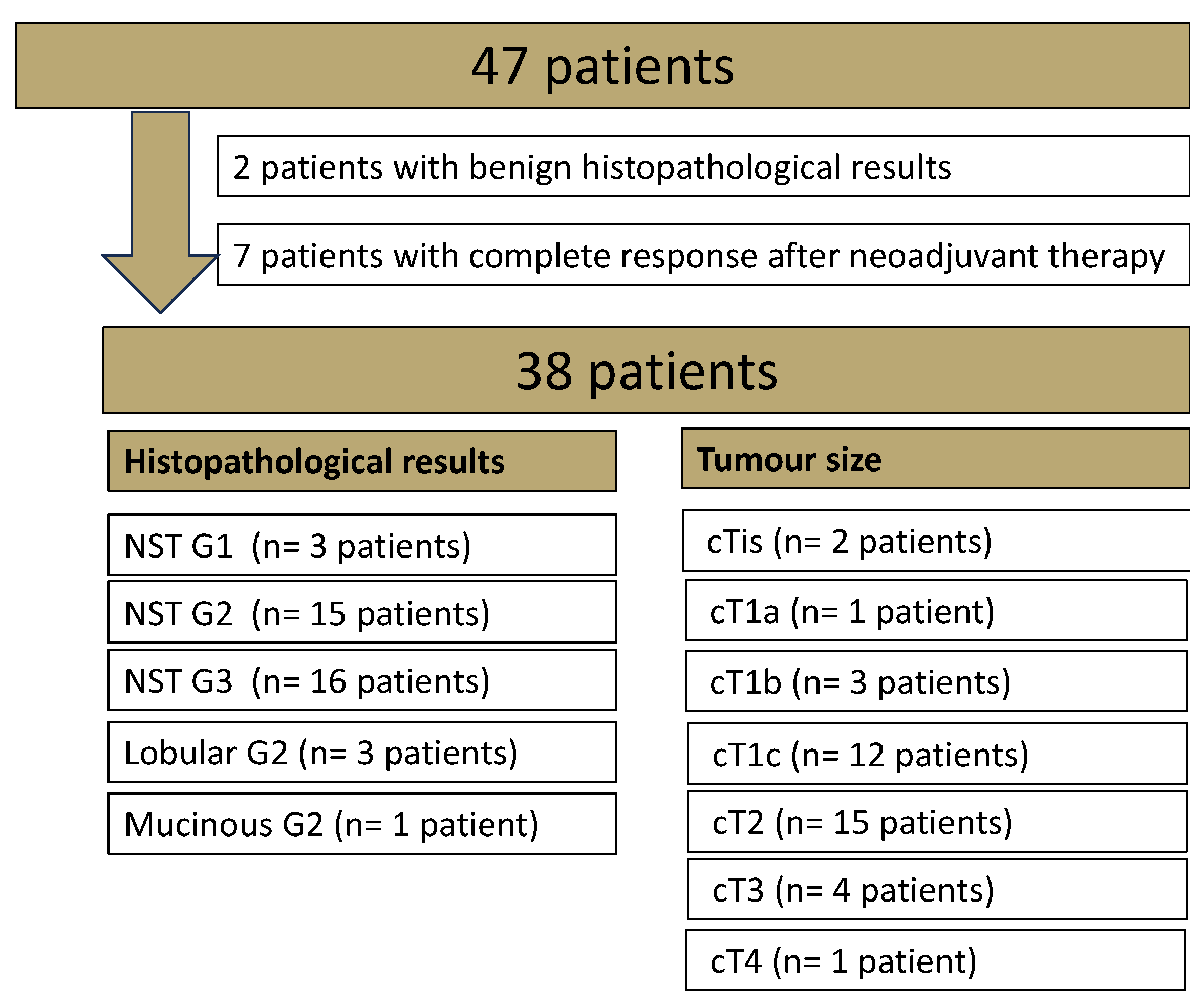
2.1. Patient Cohort

This unicenter, prospective study was approved by the Institutional Review Board of our hospital (055/201BO2). Only patients with signed informed consent were included. The inclusion criteria were histologically proven breast cancer in pre-operative patients without any prior breast cancer, who underwent a breast MRI at 1.5 T with a DWIStd and a research application package DWIDL sequence between March and April 2023 for clinical indications. For details of the histological breast cancer subtypes, see Figure 1.

2.2. Image Acquisition

All patients were scanned in a prone position using the same 1.5 T system (MAGNETOM Aera, Siemens Healthineers, Erlangen, Germany) with a dedicated 7-channel bilateral breast coil (Siemens Healthineers, Erlangen, Germany), and received a body weight-adapted dose of i.v. Gadovist (Bayer Healthcare, Berlin, Germany; 0.1 mmol Gadobutrol/kg body weight). Our standard imaging protocol encompassed a T2 fat-suppressed turbo inversion recovery magnitude sequence (T2 TIRM) and non-fat suppressed 3D T1-weighted imaging before and after contrast agent application, as well as a single-shot EPI (ssEPI) acquisition for DWIStd (TE 58 ms; TR 11,700 ms; acceleration factor 2; b values 50 s/mm2 and 800 s/mm2 with 4 and 16 averages, respectively; no partial Fourier). Additionally, a high-resolution deep-learning-based DWI acquisition (DWIDL) was performed (TE 63 ms; TR 12,900 ms; acceleration factor 2; b values 50 s/mm2 and 800 s/mm2 with 3 and 12 averages, respectively; partial Fourier factor along the phase encoding direction of 6/8). The resolution of DWIStd was 2.2 × 2.2 × 3.0 mm3, whereas the DWIDL used a higher in-plane resolution of 0.8 (i) × 0.8 (i) × 3.0 mm3. Both DWIStd and DWIDL were obtained during the same clinical scan after contrast media administration. Detailed acquisition parameters are shown in Table 1.

2.3. Image Reconstruction
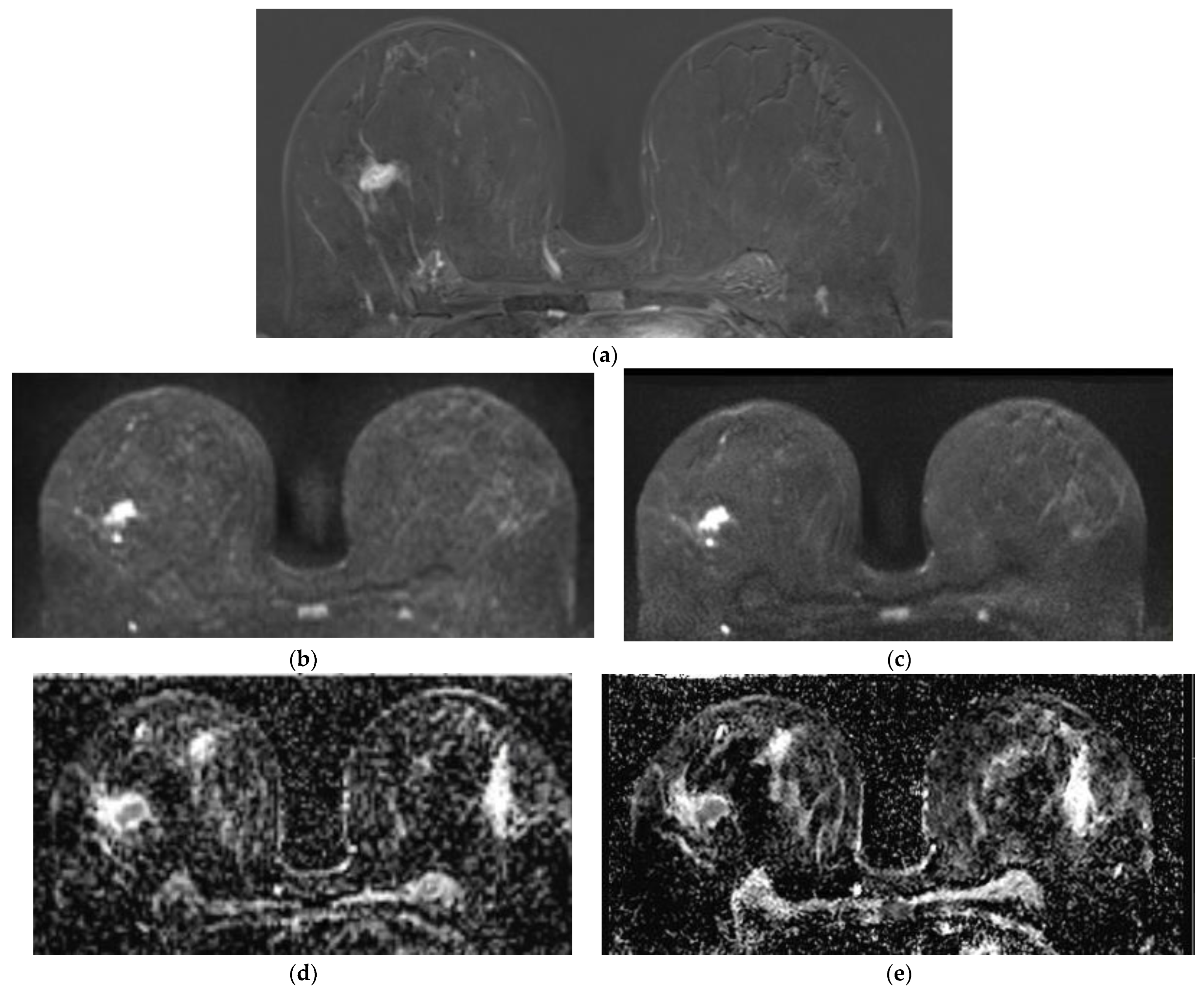
DWIStd images were reconstructed with conventional GRAPPA, while DWIDL images were reconstructed using a research application deep-learning-based reconstruction approach, which contains two different steps. The first uses raw k-space data following the scheme of a variational network [16]. Concretely, 17 unrolled iterations are performed on acquired single-shot EPI data, as well as precalculated coil sensitivity profiles. The first 6 iterations focus on parallel imaging by applying data consistency steps without additional regularization to fill in missing k-space information from PAT undersampling. The remaining 11 iterations focus on denoising by additionally using a regularization term, built via a convolutional neural network with hierarchical down–up architecture. All iterations employ trainable step sizes and Nesterov extrapolation [17]. Training was performed offline in PyTorch, using about 500,000 single-shot DWI images, acquired across different 1.5 T and 3 T clinical MR systems (MAGNETOM, Siemens, Healthineers, Erlangen, Germany) and various body regions. After k-space to image reconstruction, single-shot images were processed in a second step with an image-based super resolution network with pixel shuffle architecture [18]. Here, the goal was to increase sharpness by increasing the matrix size by a factor of two. Furthermore, blurring along the phase-encoding direction due to the applied partial Fourier factor of 6/8 in the acquisition is accounted for by extrapolating the missing 2/8 part of the k-space. By using hard data consistency, only non-measured parts of the k-space were filled in order not to modify the actual image content. To simultaneously achieve the task of super resolution and partial Fourier reconstruction, the network was trained with image pairs consisting of high-resolution images without partial Fourier (ground truth images) and retrospectively downsampled low-resolution images with simulated partial Fourier. Again, training images were acquired in volunteers from different systems and body regions. Both reconstruction steps were trained in a supervised, offline setting. Afterwards, the networks were frozen and integrated into the C++-based reconstruction pipeline at the scanner. After GRAPPA-based reconstruction for DWIStd and deep-learning based reconstruction for DWIDL, diffusion specific steps, which included averaging and ADC calculation, were performed identically with the vendor-provided conventional processing steps.

2.4. Image Analysis

Two radiologists (H.P with 12 years and S-C.O with 6 years of experience in breast MRI) evaluated first the DWIStd, followed by the DWIDL sequences, and ADCStd, followed by ADCDL, independently for all patients. Both readers were not blinded for the sequence they evaluated, as the characteristic image impression is obvious for the experienced MR reader. For lesion analysis, only histopathological proven malignant lesions were examined. All included patients were surgical treated inhouse and histopathological specimens were analysed in our local histopathology department (for details see Figure 1). Benign lesions (n = 9) were omitted in the analysis.
Each reader evaluated DWIStd, and DWIDL for b values 50 and 800, as well as ADCStd and ADCDL sequences qualitatively and quantitatively in our standard postprocessing software (syngo.via, 9.4, Siemens Healthineers, Erlangen, Germany).
Qualitative evaluation of the malignant lesion was based on a five-point Likert scale (with 1 for non-diagnostic imaging, 2 for poor, 3 for moderate, 4 for good and 5 for excellent) for image quality, sharpness, artifacts, contrast, noise and diagnostic confidence.
Quantitative analysis included the diameter of the malignant lesion in DWIStd and DWIDL at b 800 and ADCStd and ADCDL compared to our gold standard in the 2nd SUB sequence. For both DWIStd and DWIDL, SNR was analysed on both b-values, and ADC values were investigated by applying an oval-shaped two-dimensional ROI of 20 mm2 in each breast quadrant. Once a ROI was placed, it was copied to the same region in all sequences. The SNR values were obtained by the quotient of the mean and standard deviation [19].

2.5. Statistical Analysis

Statistical analysis was performed using SPSS (version 28, IBM, Chicago, IL, USA). Descriptive statistics were displayed as mean values with standard deviation. Median and IQR values were neglected, as those would not have been informative due to ratings based on a five-point Likert scale. For inferential statistics, the significance level was set to α = 0.05. Inter-reader reliability between the two readers were analysed via Pearson correlations. Paired sample t-tests were applied for the analyses of differences in image quality, lesion detection, and acquisition time between DWIStd and DWIDL. Beforehand, all outcomes were checked for the assumption of normal distribution. Whenever the assumption could not be confirmed, additional parameter-free tests (i.e., Spearman rank, and Wilcoxon tests) were calculated. These led to the same statistical test decisions and similar effect sizes as the parametric tests carried out. To be consistent in the presentation and comparison of all results, we therefore decided to report only the parametric tests in the results section.

3. Results

3.1. Subsections

3.1.1. Patients

Of the 47 patients, 2 had histologically proven benign disease (adenosis), and another 7 patients revealed a complete response after systemic therapy (Figure 1). The mean age of the included 38 female patients was 54.5 years (SD 12.35). The mean lesion diameter, measured on the T1-weighted 2nd SUB, was 25.4 mm (SD 16.7).

3.1.2. Qualitative Image Evaluation

Qualitative Image Evaluation for DWI

Regarding all analysed imaging parameters (image quality, sharpness, artifacts, image contrast, noise and diagnostic confidence), DWIDL revealed significantly superior results compared to DWIStd at b 50 and b 800 values for both readers (each p < 0.02), except for chemical shift artifacts in high-resolution DWIDL in reader 2 (p = 0.01, Table 2). Inter-reader reliability was best for image quality in DWIStd (r = 0.74), sharpness in DWIDL (r = 0.78), contrast in DWIStd (r = 0.73), artifacts in DWIStd (r = 0.70) and diagnostic confidence in DWIStd (r = 0.95) and DWIDL (r = 0.91; Table 2).

Qualitative Image Evaluation for ADC

ADCDL was significantly superior to ADCStd for all analysed parameters (image quality, sharpness, artifacts, image contrast, noise and diagnostic confidence) in both readers (each p < 0.03 Table 3, Figure 2). Inter-reader reliability was best for diagnostic confidence in ADCStd (r = 0.74) and lowest for image quality in ADCStd (r = 0.37), and artifacts in ADCStd (r = 0.47) and ADCDL (r = 0.30; Table 3).

3.1.3. Quantitative Image Evaluation

Lesion Visibility and Diameter

The primary tumour was visible in all analysed sequences (n = 38).
Compared to the gold standard of the 2nd T1w SUB after contrast media application, the lesion diameter was 6.1% lower in DWIStd and DWIDL.
Regarding the lesion diameter in ADC, 5.2% and 7.2% smaller lesion diameters were measured in ADCStd and ADCDL in comparison to the gold-standard of the 2nd SUB (Table 4, Figure 3). The signal intensities of the lesions revealed significantly higher values for ADCDL compared to ADCStd (p = 0.02; Table 5).

SNR

SNR in DWIDL was higher than in DWIStd for b 50; however, it was not statistically significant (p = 0.07 and 0.06). A comparable SNR was obtained for DWIDL and DWIStd at b 800 values (p = 0.92; Table 6).

Image Acquisition Time

The acquisition time was 3:49 min for DWIDL compared to 4:54 min for DWIStd, offering the patients a 22% shorter examination time.

4. Discussion

Comparing high-resolution DWIDL with DWIStd, all lesions were visible in both sequences, indicating that DWIDL is a clinically applicable and useful technique at 1.5 T. With a mean size of 25 mm, no lesions were missed in our DWIDL. Furthermore, the DWIDL sequence offered a mean scanning time reduction of 65 s in our study.
Breast MR examinations for cancer screening in high-risk patients are steadily increasing, according to the detection of new genetic risk profiles [20]. As these women are commonly young and have to undergo at least a yearly MRI examination, a reduction of examination time and a potential substitute for gadolinium contrast-agents would be a great step in the diagnostic work. Although this issue has been examined for several years with inferior results for DWI compared to dynamic CE breast MR [21], further development of DL sequences in DWI might have the potential to overcome this problem, as our study demonstrates with no missed lesions in high-resolution DWIDL. Furthermore, our results indicate that the exact tumor diameter can be measured with the high-resolution DWIDL b 800 and ADCDL compared to the gold-standard of the 2nd SUB, which is crucial for planning the individual therapeutic concept [22].
Regarding the choice of applied b values in breast DWI, our protocol encompassed b 800 values, which is in line with most of the published breast DWI studies for good diagnostic specificity and an acceptable SNR [23].
In general, DL-based techniques have been shown to result in higher SNR values, not only in T1 VIBE, and PD sequences, but also in DWI in musculoskeletal and abdominal imaging [11,12,13]. In our study, SNR for DWIDL compared with DWIStd was higher for b 50 and ADC, however not statistically significant (p = 0.073 and 0.069). One potential reason is that DWIDL used an increased matrix size, which in turn reduces overall SNR.
So far, deep-learning-based DWI at 1.5 T has been applied for faster image acquisition while maintaining equal image quality, contrast and diagnostic confidence [8]. With the proposed DWIDL sequence, both superior image quality and a reduction of acquisition time compared to conventional DWI is feasible as the analysed image quality, sharpness, noise, contrast and diagnostic confidence were significantly higher in high-resolution DWIDL compared to DWIStd. Thus, this new high-resolution DWI represents a relevant development for DWIDL sequences in breast MRI at 1.5 T and can strengthen the role of DWI in the clinical routine in the staging and high-risk screening population. As a consequence, abbreviated breast MRI for screening in high-risk patients might, in the future, potentially be possible without i.v. CM application in the clinical routine.
At 3 T, including benign and malignant lesions, the DL DWI sequence resulted in significantly higher contrast, while the SNR and contrast-to-noise values were comparable between DWI Std and DWI DL [10]
While high-resolution DWIDL was rated superior in almost all categories, reader 2 stated increased chemical shift artifacts compared to DWIStd; however, without affecting the diagnostic confidence in both readers (M = 4.62 vs. M = 4.36 in Likert scale, p 0.01). Given that a higher matrix size and therefore longer echo trains were used for DWIDL, this is expected and independent from the DL reconstruction. Employing segmented instead of single-shot readouts might help to reduce chemical shift, which should be investigated in further studies for breast MR at 1.5 T.
The limitations of this study include the small study cohort. However, we provided a patient collective with histologically proven breast cancers. In the future, larger patient cohorts, suffering from the same tumour histology and grading type, should be analysed to gain more information regarding the homogenous data of DL in DWI, potentially enabling cut-off values for benign vs. malignant lesions.

5. Conclusions

High resolution DL-based DWI in breast MR at 1.5 T offers superior diagnostic image quality compared to conventional DWI, while reducing the acquisition time by up to 22%. This result strengthens the role of DWI for implementation in clinical diagnostic routines, and might potentially also play a crucial role in the evaluation of gadolinium-free breast examinations. Further studies with larger patient cohorts should be performed for validation of these initial results. Additionally, homogenous cohorts should be analysed independently at 1.5 and 3 T, to gain further knowledge, potentially enabling a differentiation between benign and malignant breast lesions through the identification of a cut-off value for b 50, b 800 and ADC values.

Author Contributions

Conceptualization, S.-C.O. and D.W.; methodology, S.-C.O. and H.P.; software, E.W. and T.B.; validation, H.P., K.N., S.A. and D.W.; formal analysis, S.-C.O., H.P., D.L. and D.W.; investigation, S.-C.O., H.P. and D.L.; resources, K.N. and S.A. data curation, S.-C.O.; writing—original draft preparation, S.-C.O. and D.L.; writing—review and editing, H.P., K.N., S.A., E.W., T.B. and D.L.; visualization, S.-C.O.; supervision, H.P. and K.N.; project administration, S.-C.O. and H.P. All authors have read and agreed to the published version of the manuscript.

Funding

This research received no external funding.

Institutional Review Board Statement

The study was conducted in accordance with the Declaration of Helsinki, and approved by the Institutional Review Board of the University of Tuebingen (protocol code IRB; No 055/2017BO2 and date of approval 2022_11_16).

Informed Consent Statement

Informed consent was obtained from all subjects involved in the study.

Data Availability Statement

The data presented in this study are available on request from the corresponding author due to privacy.

Conflicts of Interest

The authors declare no conflicts of interest.

References

  1. Mann, R.M.; Balleyguier, C.; Baltzer, P.A.; Bick, U.; Colin, C.; Cornford, E.; Evans, A.; Fallenberg, E.; Forrai, G.; Fuchsjager, M.H.; et al. Breast MRI: EUSOBI recommendations for women’s information. Eur. Radiol. 2015, 25, 3669–3678. [Google Scholar] [CrossRef] [PubMed]
  2. van der Molen, A.J.; Quattrocchi, C.C.; Mallio, C.A.; Dekkers, I.A.; European Society of Magnetic Resonance in Medicine, Biology Gadolinium Research, Educational Committee (ESMRMB-GREC). Ten years of gadolinium retention and deposition: ESMRMB-GREC looks backward and forward. Eur. Radiol. 2024, 34, 600–611. [Google Scholar] [CrossRef] [PubMed]
  3. Kuhl, C.K.; Schrading, S.; Strobel, K.; Schild, H.H.; Hilgers, R.D.; Bieling, H.B. Abbreviated breast magnetic resonance imaging (MRI): First postcontrast subtracted images and maximum-intensity projection-a novel approach to breast cancer screening with MRI. J. Clin. Oncol. Off. J. Am. Soc. Clin. Oncol. 2014, 32, 2304–2310. [Google Scholar] [CrossRef] [PubMed]
  4. Sardanelli, F.; Carbonaro, L.A.; Montemezzi, S.; Cavedon, C.; Trimboli, R.M. Clinical Breast MR Using MRS or DWI: Who Is the Winner? Front. Oncol. 2016, 6, 217. [Google Scholar] [CrossRef] [PubMed]
  5. Obara, M.; Kwon, J.; Yoneyama, M.; Ueda, Y.; Cauteren, M.V. Technical Advancements in Abdominal Diffusion-weighted Imaging. Magn. Reson. Med. Sci. 2023, 22, 191–208. [Google Scholar] [CrossRef]
  6. Zhang, L.; Tang, M.; Min, Z.; Lu, J.; Lei, X.; Zhang, X. Accuracy of combined dynamic contrast-enhanced magnetic resonance imaging and diffusion-weighted imaging for breast cancer detection: A meta-analysis. Acta Radiol. 2016, 57, 651–660. [Google Scholar] [CrossRef] [PubMed]
  7. Baltzer, P.; Mann, R.M.; Iima, M.; Sigmund, E.E.; Clauser, P.; Gilbert, F.J.; Martincich, L.; Partridge, S.C.; Patterson, A.; Pinker, K.; et al. Diffusion-weighted imaging of the breast-a consensus and mission statement from the EUSOBI International Breast Diffusion-Weighted Imaging working group. Eur. Radiol. 2020, 30, 1436–1450. [Google Scholar] [CrossRef] [PubMed]
  8. Wessling, D.; Gassenmaier, S.; Olthof, S.C.; Benkert, T.; Weiland, E.; Afat, S.; Preibsch, H. Novel deep-learning-based diffusion weighted imaging sequence in 1.5 T breast MRI. Eur. J. Radiol. 2023, 166, 110948. [Google Scholar] [CrossRef]
  9. Sauer, S.T.; Christner, S.A.; Lois, A.M.; Woznicki, P.; Curtaz, C.; Kunz, A.S.; Weiland, E.; Benkert, T.; Bley, T.A.; Baessler, B.; et al. Deep Learning k-Space-to-Image Reconstruction Facilitates High Spatial Resolution and Scan Time Reduction in Diffusion-Weighted Imaging Breast MRI. J. Magn. Reson. Imaging 2023, 60, 1190–1200. [Google Scholar] [CrossRef]
  10. Wilpert, C.; Neubauer, C.; Rau, A.; Schneider, H.; Benkert, T.; Weiland, E.; Strecker, R.; Reisert, M.; Benndorf, M.; Weiss, J.; et al. Accelerated Diffusion-Weighted Imaging in 3 T Breast MRI Using a Deep Learning Reconstruction Algorithm With Superresolution Processing: A Prospective Comparative Study. Investig. Radiol. 2023, 58, 842–852. [Google Scholar] [CrossRef]
  11. Kiryu, S.; Akai, H.; Yasaka, K.; Tajima, T.; Kunimatsu, A.; Yoshioka, N.; Akahane, M.; Abe, O.; Ohtomo, K. Clinical Impact of Deep Learning Reconstruction in MRI. Radiographics 2023, 43, e220133. [Google Scholar] [CrossRef] [PubMed]
  12. Chaika, M.; Afat, S.; Wessling, D.; Afat, C.; Nickel, D.; Kannengiesser, S.; Herrmann, J.; Almansour, H.; Mannlin, S.; Othman, A.E.; et al. Deep learning-based super-resolution gradient echo imaging of the pancreas: Improvement of image quality and reduction of acquisition time. Diagn. Interv. Imaging 2022, 104, 53–59. [Google Scholar] [CrossRef] [PubMed]
  13. Herrmann, J.; Feng, Y.S.; Gassenmaier, S.; Grunz, J.P.; Koerzdoerfer, G.; Lingg, A.; Almansour, H.; Nickel, D.; Othman, A.E.; Afat, S. Fast 5-minute shoulder MRI protocol with accelerated TSE-sequences and deep learning image reconstruction for the assessment of shoulder pain at 1.5 and 3 Tesla. Eur. J. Radiol. Open 2024, 12, 100557. [Google Scholar] [CrossRef] [PubMed]
  14. Gassenmaier, S.; Warm, V.; Nickel, D.; Weiland, E.; Herrmann, J.; Almansour, H.; Wessling, D.; Afat, S. Thin-Slice Prostate MRI Enabled by Deep Learning Image Reconstruction. Cancers 2023, 15, 578. [Google Scholar] [CrossRef] [PubMed]
  15. Liu, W.; Darwish, O.; Benkert, T.; Weiland, E.; Nickel, M.D. Improved Readout-Segmented EPI Using Deep Learning Reconstruction. 2024. Available online: https://submissions.mirasmart.com/ISMRM2024/Itinerary/Login.aspx (accessed on 7 August 2024).
  16. Hammernik, K.; Klatzer, T.; Kobler, E.; Recht, M.P.; Sodickson, D.K.; Pock, T.; Knoll, F. Learning a variational network for reconstruction of accelerated MRI data. Magn. Reson. Med. 2018, 79, 3055–3071. [Google Scholar] [CrossRef]
  17. Tao, W.; Pan, Z.; Wu, G.; Tao, Q. The Strength of Nesterov’s Extrapolation in the Individual Convergence of Nonsmooth Optimization. IEEE Trans. Neural Netw. Learn. Syst. 2020, 31, 2557–2568. [Google Scholar] [CrossRef]
  18. Shi, W.; Caballero, J.; Huszár, F. Real-time single image and video super-resolution using an efficient sub-pixel convolutional neural net-work. In Proceedings of the IEEE Conference on Computer Vision and Pattern Recognition (CVPR), Las Vegas, NV, USA, 27–30 June 2016; pp. 1874–1883. [Google Scholar] [CrossRef]
  19. Song, S.E.; Woo, O.H.; Cho, K.R.; Seo, B.K.; Son, Y.H.; Grimm, R.; Liu, W.; Moon, W.K. Simultaneous Multislice Readout-Segmented Echo Planar Imaging for Diffusion-Weighted MRI in Patients With Invasive Breast Cancers. J. Magn. Reson. Imaging 2021, 53, 1108–1115. [Google Scholar] [CrossRef]
  20. Sheikh, A.; Hussain, S.A.; Ghori, Q.; Naeem, N.; Fazil, A.; Giri, S.; Sathian, B.; Mainali, P.; Al Tamimi, D.M. The spectrum of genetic mutations in breast cancer. Asian Pac. J. Cancer Prev. 2015, 16, 2177–2185. [Google Scholar] [CrossRef]
  21. Partridge, S.C.; Nissan, N.; Rahbar, H.; Kitsch, A.E.; Sigmund, E.E. Diffusion-weighted breast MRI: Clinical applications and emerging techniques. J. Magn. Reson. Imaging 2017, 45, 337–355. [Google Scholar] [CrossRef]
  22. Wekking, D.; Porcu, M.; De Silva, P.; Saba, L.; Scartozzi, M.; Solinas, C. Breast MRI: Clinical Indications, Recommendations, and Future Applications in Breast Cancer Diagnosis. Curr. Oncol. Rep. 2023, 25, 257–267. [Google Scholar] [CrossRef]
  23. Messina, C.; Bignone, R.; Bruno, A.; Bruno, A.; Bruno, F.; Calandri, M.; Caruso, D.; Coppolino, P.; Robertis, R.; Gentili, F.; et al. Diffusion-Weighted Imaging in Oncology: An Update. Cancers 2020, 12, 1493. [Google Scholar] [CrossRef] [PubMed]
Figure 1. Overview of the histopathological results of the whole study cohort examined in breast magnetic resonance imaging (MRI). Non-special type (NST).
Diagnostics 14 01742 g001
Figure 2. A 68-year-old patient with histologically proven breast carcinoma, non-special type (NST), G2 on the right side. The 2nd subtraction (SUB) is the diagnostic gold-standard (a). The lesion in diffusion-weighted imaging (DWI)Std (b) was less sharp, compared to DWIDL (c). Additionally, visibility for lesion detection was in apparent diffusion coefficient (ADC)Std inferior (d) to ADCDL (e).
Diagnostics 14 01742 g002
Figure 3. A 54-year-old patient with histologically proven breast carcinoma, NST, G2 on the right side in the 2nd subtraction (a). The exact lesion diameter could be determined more clearly in DWIDL (c) and ADCDL (e) compared to DWIStd (b) and ADCStd (d).
Diagnostics 14 01742 g003
Table 1. Details of the protocol parameters of diffusion weighted imaging (DWI)Std and DWIDL.
Protocol ParameterDWIStdDWIDL
Resolution2.2 × 2.2 × 3.0 mm0.8 (i) × 0.8 (i) × 3.0 mm
Acquisition time (TA)4:54 min3:49 min
Repitition time (TR)/
Echo time (TE)
11,700/58 ms12,900/63 ms
Fat SaturationSpectral attenuated inversion recovery (SPAIR)SPAIR
Parallel imaging factor22
b-values (averages)50 (4)/800 (16) s/mm250 (3)/800 (12) s/mm2
Diffusion mode3D Diagonal3D Diagonal
Partial FourierNone6/8
Deep Learning (DL)NoneDL reconstruction,
DL super resolution
Table 2. Visual evaluation of the image quality (IQ), sharpness, noise, contrast, artifacts and diagnostic confidence (DC) for DWIStd and DL in both readers.
Reader 1Reader 2Interreader Reliability (r)
Image Parameters DWI SequenceDWIStd
Mean (SD)
DWIDL
Mean (SD)
p-ValueDWIStd
Mean (SD)
DWIDL
Mean (SD)
p-ValueDWIStdDWIDL
Overall Image Quality
IQ 13.86
(0.58)
4.49
(0.65)
<0.0013.92
(0.54)
4.70
(0.46)
<0.0010.7460.585
Sharpness3.86
(0.67)
4.68
(0.58)
<0.0013.78
(0.41)
4.76
(0.49)
<0.0010.6840.782
Noise4.00
(0.68)
4.65
(0.63)
<0.0014.08
(0.49)
4.78
(0.47)
<0.0010.4030.567
Contrast4.49
(0.76)
4.65
(0.67)
0.0104.36
(0.68)
4.62
(0.54)
0.0100.7300.609
Artifacts4.16
(0.72)
4.43
(0.76)
0.0204.46
(0.55)
4.16
(0.60)
0.0100.7020.387
DC 24.49
(0.98)
4.59
(0.89)
0.1004.59
(0.83)
4.73
(0.60)
0.0200.9550.915
1 IQ = image quality; 2 DC = diagnostic confidence; IQR = interquartile range for values in brackets.
Table 3. Visual evaluation of the image quality (IQ), sharpness, noise, contrast, artifacts and diagnostic confidence (DC) for ADCStd and DL in both readers.
Reader 1Reader 2Interreader Reliability (r)
Image Parameters ADCADCStd
Mean (SD)
ADCIDL
Mean (SD)
p-ValueADCStd
Mean (SD)
ADCDL
Mean (SD)
p-ValueADCStdADCDL
Overall Image Quality
IQ 13.41
(0.59)
3.95
(0.91)
<0.0013.46
(0.50)
4.11
(0.51)
<0.0010.3770.486
Sharpness3.41
(0.68)
4.05
(0.91
<0.0013.41
(0.59)
4.16
(0.72)
<0.0010.6710.615
Noise3.24
(0.64)
3.62
(0.89)
<0.0013.24
(0.49)
3.73
(0.69)
<0.0010.5960.548
Contrast3.51
(0.83)
3.76
(0.89)
<0.0013.86
(0.48)
3.62
(0.54)
0.0200.5220.376
Artifacts3.41
(0.68)
3.81
(0.93)
<0.0013.78
(0.53)
3.73
(0.69)
0.6600.4740.30
DC 23.62
(1.06)
3.97
(1.04)
<0.0013.62
(1.01)
3.78
(1.03)
0.0300.7430.668
1 IQ= image quality; 2 DC = diagnostic confidence; 3 IQR = interquartile range for values in brackets.
Table 4. Lesion diameters in mm measured in DWI b 800Std and DL and ADCStd and DL. in comparison to the gold-standard of the 2nd SUB.
Lesion Size in mmStdDL
DWI b 80024.13
(−4.90%)
23.94
(−5.7%)
ADC (mm2/s)24.00
(−5.20%)
23.48
(−7.2%)
2nd SUB25.3
Table 5. Overview of quantitative image parameters obtained in DWIStd and DL at b 50 and b 800 values, as well as in ADC maps.
Quantitative Image Parameters (Lesion)DWIStd
Mean (SD 1)
DWIDL
Mean (SD 1)
p-Value
ADC (mm2/s)936.22
(262.47)
980.78
(274.00)
0.02
1 SD = standard deviation.
Table 6. Noise and SNR measured in DWIStd and DL, both at b 50 and b 800 values and in ADC maps.
NoiseDWIStd
SD 1
DWIDL
SD 1
p-ValueSNR 2DWIStd
SNR 2
DWIDL
SNR 2
p-Value
b 5096.7888.610.001b 504.174.360.073
b 80029.7227.59<0.001b 8007.277.290.925
1 SD = standard deviation, 2 SNR = signal-to-noise-ratio;.
Disclaimer/Publisher’s Note: The statements, opinions and data contained in all publications are solely those of the individual author(s) and contributor(s) and not of MDPI and/or the editor(s). MDPI and/or the editor(s) disclaim responsibility for any injury to people or property resulting from any ideas, methods, instructions or products referred to in the content.

