Article

Could SLC26A7 Be a Promising Marker for Preoperative Diagnosis of High-Grade Papillary Thyroid Carcinoma?

by
Sergei E. Titov
1,2,3,*,
Evgeniya S. Kozorezova
4,
Sergei A. Lukyanov
5,
Sergei V. Sergiyko
5,
Pavel S. Demenkov
3,6,
Yulia A. Veryaskina
1,6,
Sergey L. Vorobyev
4,
Ilya V. Sleptsov
7,
Roman A. Chernikov
7,
Natalia I. Timofeeva
7,
Svetlana V. Barashkova
4,
Elena L. Lushnikova
8,
Anna A. Uspenskaya
7,
Anna V. Zolotoukho
7,
Olga V. Romanova
9 and
Igor F. Zhimulev
1
1
Department of the Structure and Function of Chromosomes, Institute of Molecular and Cellular Biology, Siberian Branch of the Russian Academy of Sciences, Novosibirsk 630090, Russia
2
PCR Laboratory, AO Vector-Best, Novosibirsk 630117, Russia
3
Department of Natural Sciences, Novosibirsk State University, Novosibirsk 630090, Russia
4
National Center of Clinical Morphological Diagnostics, Saint Petersburg 192283, Russia
5
Department of General and Pediatric Surgery, South Ural State Medical University, Chelyabinsk 454092, Russia
6
Institute of Cytology and Genetics, Siberian Branch of the Russian Academy of Sciences, Novosibirsk 630090, Russia
7
Department of Endocrine Surgery, Saint Petersburg State University Hospital, Saint Petersburg 199034, Russia
8
Department of Molecular Pathology, Federal Research Center of Fundamental and Translational Medicine, Novosibirsk 630117, Russia
9
N.N. Blokhin National Medical Research Center of Oncology, Moscow 115478, Russia
*
Author to whom correspondence should be addressed.
Diagnostics 2024, 14(23), 2652; https://doi.org/10.3390/diagnostics14232652
Submission received: 22 October 2024 / Revised: 20 November 2024 / Accepted: 23 November 2024 / Published: 25 November 2024
(This article belongs to the Special Issue Head and Neck Surgery: Diagnosis and Management)

Abstract

:
Background/Objectives: A modern classification distinguishes between two nosological entities posing an intermediate risk between differentiated and anaplastic carcinoma: poorly differentiated thyroid carcinoma and differentiated high-grade thyroid carcinoma. There are currently few studies searching for the preoperative molecular genetic markers of high-grade papillary thyroid carcinoma (PTC HG), primarily because of a recent WHO reclassification and singling out of a separate entity: high-grade follicular cell-derived nonanaplastic thyroid carcinoma. Therefore, this work was aimed at identifying PTC HG-specific microRNAs and mRNAs that reliably distinguish them from differentiated papillary thyroid carcinoma in preoperative cytology specimens (fine-needle aspiration biopsies). Methods: A molecular genetic profile (expression levels of 14 genes and eight microRNAs) was studied in 110 cytology specimens from patients with PTC: 13 PTCs HG and 97 PTCs without features of HG. Results: Of the examined eight microRNAs and 14 genes, significant differences in the expression levels between the PTC and PTC HG groups were revealed for genes SLC26A7, TFF3, and TPO. Only one gene (SLC26A7) proved to be crucial for detecting PTC HG. It showed the largest area under the ROC curve (0.816) in differentiation between the PTC and PTC HG groups and was the key element of the decision tree by ensuring 54% sensitivity and 87.6% specificity. Conclusions: Early preoperative diagnosis of PTC HG in patients with early stages of this cancer type will allow clinicians to modify a treatment strategy toward a larger surgery volume and lymph node dissection and may provide indications for subsequent radioactive iodine therapy.

1. Introduction

The WHO Classification of Endocrine Tumors (the 5th Edition, 2022) distinguishes between two subtypes of high-grade follicular cell-derived nonanaplastic thyroid carcinoma, posing a risk that is intermediate between differentiated and anaplastic thyroid carcinoma: poorly differentiated thyroid carcinoma (PDTC) and differentiated high-grade thyroid carcinoma (DHGTC) [1]. DHGTCs are high-grade epithelium-derived carcinomas that retain their structural follicular/papillary differentiation and cytological nuclear features, typical of papillary carcinoma. DHGTC is characterized by invasive growth, a necrotic component, and an elevated mitotic index while retaining signs of differentiation, which is not typical of anaplastic carcinoma [2].
The need to introduce a new nosological entity—differentiated high-grade thyroid carcinoma—has arisen because papillary thyroid carcinoma (PTC, which represents the majority of differentiated thyroid carcinoma [DTC] cases) most often progresses indolently and does not pose a risk of death. However, researchers also see aggressive subtypes of PTC, which progress less favorably and have a worse prognosis. Moreover, in some countries, such as the United States, mortality from thyroid cancer has increased by 1.1% per year from 1994 to 2013, whereas during the same period, the annual increase in the incidence of (and mortality from) PTC is 1.7% [3].
Most DHGTCs not only have cytoarchitectonic features of papillary carcinoma but can also carry the BRAF V600E mutation [4,5] while remaining prone to regional metastasis to cervical lymph nodes [5]. The term “papillary thyroid carcinoma, high grade” (PTC HG) has been proposed for this type of tumor. These tumors usually develop in adults, are frequently metastatic (20–50%), characterized by substantial local invasion, and are often resistant to radioactive iodine therapy [6,7]. Postoperative treatment of this group of patients can involve external beam radiation therapy and/or targeted chemotherapy with tyrosine kinase inhibitors. DHGTC patients have a similar survival rate (56% 10-year survival rate) [5], although recurrence-free survival (without signs of disease) of DHGTC patients can be lower compared to that of PTC patients [7]. The unfavorable prognosis is also related to the fact that patients often receive a diagnosis of DHGTC at late stages. Therefore, an early and accurate diagnosis of this cancer is crucially important for initiating effective treatment [8].
Fine-needle aspiration biopsy (FNA) is a minimally invasive and highly effective method of PTC detection, but the specificity to HG signs is modest. PTC HG is cytologically verified regarding papillary carcinoma or a tumor suspected of papillary carcinoma (Bethesda V/VI); most often, there are no sufficient features to expect high-grade carcinoma. PTC HG can be suspected based on clinical data, such as the large size and rapid growth of the tumor, as well as the presence of regional and distant metastases [9].
DHGTC (and PTC HG in particular) typically develops from a pre-existing well-differentiated thyroid carcinoma as a result of genetic rearrangements; therefore, DHGTC (PTC HG) is caused by the known mutations in MAPK and PI3K–AKT signaling pathways [10,11]; accordingly, it is rather difficult to differentiate between papillary carcinoma and PTC HG at the molecular level. Meanwhile, some available data are indicative of an association of high-grade carcinomas with additional mutations in TP53 and TERT promoters and/or dysregulated expression of certain microRNAs (miRNAs, miRs) [12,13], although no specific genetic markers have been identified yet for this group of patients.
Various molecular genetic markers (miRNA and messenger RNA [mRNA]) are currently successfully employed for diagnosing differentiated forms of thyroid cancer in such genetic testing panels as the ThyGenX/ThyraMIR, Afirma Genomic Sequencing Classifier, and mir-THYpe [14,15,16]. These panels can detect pituitary cancer with high accuracy in patients with uncertain cytological findings (Bethesda III, IV, and V). A panel developed earlier [17,18] also allows one to improve the preoperative stratification of thyroid tumors into benign and malignant ones in FNA smears, as well as to distinguish thyroid carcinoma types such as oncocytic, papillary, and medullary with high accuracy. Nonetheless, this panel has proved to be insufficiently informative for verifying poorly differentiated thyroid carcinomas [19,20].
In comparison with anaplastic thyroid cancer, the probability of detecting a high-grade tumor of small size for the first time is much higher [21]. If the tumor size is less than 4.0 cm, then a surgeon may decide to perform a hemithyroidectomy. Because PTC HG is an aggressive tumor type, with the above treatment strategy, the patient will need a second surgical operation to the extent of removal of thyroid gland remnants and a level VI lymphadenectomy of the neck; the latter modalities are always associated with an elevated risk of complications (laryngeal nerve paresis and hypoparathyroidism) [22]. Suspicion of the high grade already at the preoperative stage will allow a surgeon to immediately choose the correct extent of the surgical intervention.
There are currently very few studies searching for the preoperative molecular genetic markers of PTC HG, primarily because of the recent WHO reclassification of endocrine tumors (2022, 5th edition) and the singling out of a separate nosological entity: high-grade follicular cell-derived nonanaplastic thyroid carcinoma. Therefore, this work was aimed at identifying PTC HG-specific miRNAs and mRNAs that reliably distinguish them from differentiated papillary thyroid carcinoma in preoperative cytology specimens collected by FNA.

2. Materials and Methods

Clinical material. Two institutions took part in this study: Saint Petersburg State University Hospital (St. Petersburg, Russia) and South Ural State Medical University (Chelyabinsk, Russia). All the cytology and histology tissue samples pertaining to this study were re-examined by two independent pathologists from the National Center for Clinical Morphological Diagnostics.
To search for patients with PTC HG who meet the criteria of the WHO classification (2022), 298 histological examinations of PTC specimens derived from patients who had been operated on in 2021–2023 were reassessed. After a comprehensive re-examination of the cytology and histology specimens, 110 specimens were selected; cases not meeting the inclusion criteria (see below) were excluded from the study. Of those, the criteria for PTC HG were met in 13 cases: features of differentiated papillary carcinoma were retained (structural and nuclear morphology); the presence of necrotic foci and an increased number of mitoses ≥ 3 × 2 mm2; and no anaplastic dedifferentiation. The comparison group contained 97 patients with PTC without features of HG.
The inclusion criteria were as follows: cytology and histology specimens being available; age > 18 years; a match between the ultrasonography findings for a thyroid nodule during preoperative diagnostics and the macroscopic characteristics of the thyroid nodule in the operative specimen; and >200 tumor cells in the cytology specimen. Cytology specimens on glass slides prepared by the conventional procedure (fixation and May–Grünwald–Giemsa staining with azure and eosin dyes) were included in the study.
Choosing a set of molecular markers. A set of mRNAs for the analysis was primarily selected, taking into account published findings. The list of mRNAs consisted of 14 genes: FN1, geminin (GMNN), CDKN2A, TPO, SLC26A7, HMGA2, CPQ, SPATA18, APOE, DIO1, NIS, TFF3, TMPRSS4, and TSHR. MiRNAs were chosen based on our own studies [23,24,25,26,27,28,29,30,31] and literature analysis. Eight miRNAs were subjected to experimental analysis: miR-146b-5p, miR-199b-5p, miR-221-3p, miR-223-3p, miR-31-5p, miR-375, miR-551b-3p, and miR-21-5p [32,33]. The ratio of mitochondrial DNA (mtDNA) to nuclear DNA (nDNA) served as a criterion for the presence of oncocytes in a cytology specimen [20]. Therefore, 23 molecular genetic markers were investigated in this work.
Total nucleic acid extraction. Nucleic acid extraction was conducted according to a published protocol [32] with modifications: each dried cytology specimen was washed in a 1.5 mL microcentrifuge tube with three 200 μL portions of guanidine lysis buffer (4 M guanidine thiocyanate; 25 mM sodium citrate, pH 7.0; 0.3% of sarcosyl; and 0.1% of 2-mercaptoethanol). The sample was vigorously mixed and incubated in a thermal shaker for 15 min at 65 °C. Next, an equal volume (600 μL) of isopropanol was added. The reaction solution was thoroughly mixed and kept at room temperature for 5 min. After centrifugation for 15 min at 14,000× g, the supernatant was discarded, and the pellet was washed with 500 μL of 70% ethanol and then with 300 μL of acetone. Finally, the RNA was dissolved in 200 μL of deionized water. If not analyzed immediately, the RNA samples were stored at –20 °C. The concentration of the isolated total RNA was measured on a NanoDrop 2000C spectrophotometer (Thermo Scientific, Waltham, MA, USA). Total RNA concentrations were in the range of 5.4–51.6 ng/μL (mean 25.2 ng/μL).
Oligonucleotide primers and probes
All oligonucleotides were synthesized at AO Vector-Best (Novosibirsk, Russia). The oligonucleotides were selected using the OligoAnalyzer™ Tool online service (https://eu.idtdna.com/calc/analyzer (accessed on 22 November 2024)). Sequences of primers and fluorescently labeled probes are presented in the Supplementary Materials: Table S1. The standard primer concentration in all the reactions was 0.5 μM, whereas the concentration of the fluorescently labeled probe was 0.25 μM.
Semiquantitative assessment of the mRNA content. This procedure was performed by real-time reverse-transcription PCR (RT-PCR) employing specific primers and fluorescently labeled probes to detect the mRNA of one of the above genes and the housekeeping gene PGK1 (phosphoglycerate kinase) used for normalization. The RT-PCR protocol was as follows: incubation at 45 °C for 30 min; heating at 95 °C for 2 min; 50 cycles: denaturation at 94 °C for 10 s; annealing and elongation at 60 °C for 20 s. Each relative expression level was calculated by the 2−ΔCq method [34].
Semiquantitative assessment of the miRNA content. Eight miRNAs were detected by real-time RT-PCR for all the types of tumors and lumps. Mature miRNAs were detected by stem-loop RT-PCR [35]. The reverse-transcription reaction and real-time PCR were carried out according to a protocol reported in ref. [32]. The reverse-transcription reaction followed by real-time PCR was carried out individually for each miRNA. Single-replicate analysis was performed on each RNA sample. The miRNA content was normalized to the geometric mean of the levels of three reference miRNAs (miR-197-3p, miR-23a-3p, and miR-29b-3p) by the 2−ΔCq method.
Analysis of the ratio of mtDNA to nDNA. MtDNA and nDNA were detected by real-time PCR using the following protocol: preheating at 95 °C for 2 min; 50 cycles: denaturation at 94 °C for 10 s; annealing and elongation at 60 °C for 20 s. The ratio was determined by the 2−ΔCq method.
Statistical analysis. The data were analyzed in the SPSS Statistics 23 (IBM, Armonk, NY, USA) and Excel (Microsoft, Redmond, WA, USA) software. For continuous variables, two independent samples were compared by the Mann–Whitney U test. The significance level was set to 0.0022 after the Bonferroni correction. Receiver operating characteristic (ROC) analysis was carried out to make it possible to use miRNA and gene expression levels to detect PTC HG. A test comparison was performed with the help of areas under the ROC curves (ROC AUCs). Tissue samples were stratified into different groups via the C4.5 decision tree algorithm [36].

3. Results

Among the 13 patients with PTC HG, there were 10 (77%) females and 3 (23%) males. The mean age at diagnosis was 64 years (range: 48–74 years). A tumor was found to be multifocal in three (23%) cases. A macroscopically visible extrathyroidal extension was observed in seven (53.8%) patients. Metastases to the regional lymph nodes were detected in eight (61.5%) patients; no distant metastases were found (Table 1). The median tumor size was 45 mm (interquartile range: 35–50 mm); the minimal and maximal tumor sizes were 17 and 60 mm, respectively. One patient had a stage T1b tumor; five patients had stage T2; and seven patients had a stage T3b tumor.
A comparative description of the relative expression levels of eight miRNAs, a relative mtDNA level, and the expression levels of 14 genes was performed. The results are summarized in Table 2.
No significant differences in miRNA expression or the relative mtDNA level were revealed between groups PTC and PTC HG. Among the protein-coding genes, significant (with an adjustment for multiple comparisons) differences were detected for the following:
  • The TPO gene (p = 0.00085) encodes thyroid peroxidase, which plays an important role in thyroid hormones’ synthesis and maintenance of stable thyroid function. According to other studies, TPO is highly expressed, mainly in normal thyroid tissue or benign lesions, whereas in thyroid cancer, its expression is reduced or absent [37].
  • The SLC26A7 gene (p = 0.000166) codes for a transmembrane Cl/HCO3 ion transporter that controls the acid–base balance in renal tubules and the gastric epithelium and appears to perform a similar function in thyroid follicular cells. This is because an optimal ionic milieu and pH are known to be critical for enzymatic activity (of TPO, for example) during thyroid hormone biosynthesis [38]. There is evidence that decreased SLC26A7 expression accompanies the development of anaplastic carcinoma and significantly correlates with a poor prognosis among the patients [26], whereas in PTC, SLC26A7 downregulation has been associated with an elevated risk of metastases outside the thyroid [39].
  • The TFF3 gene (p = 0.000614) encodes a small molecular peptide that belongs to a family of small secretory molecules involved in the protection and repair of the gastrointestinal mucosa. TFF proteins participate in the maintenance and restoration of epithelial structural integrity by activating key signaling pathways for epithelial–cell migration, proliferation, and invasiveness [40]. TFF3 expression is diminished in PTC, and patients with a lower gene expression have poorer disease-free survival but a higher immune-cell infiltration [41].
Figure 1 shows the relative expression of these genes in groups PTC and PTC HG. For comparison, Figure 1 also depicts the relative expression of the genes whose p-values lay in the range 0.0022 < p < 0.05: GMNN (p = 0.028378; its protein modulates DNA replication by binding to replication factor Cdt1, thereby altering Cdt1’s stability and activity [42]), CDKN2A (p = 0.004832; the cyclin-dependent kinase inhibitor 2A gene encodes tumor suppressor protein p16INK4a, which plays an important part in inhibition of cell cycle progression [43]), CPQ (p = 0.007873; its protein may take part in a release of hormone thyroxine; underexpression has been noted in follicular thyroid cancer [24]), SPATA18 (p = 0.012699; it codes for a protein induced by p53 and causes the accumulation of lysosomal proteins in damaged mitochondria to utilize oxidized mitochondrial proteins in order to restore damaged mitochondria [44]), and NIS (p = 0.016281; it encodes a sodium–iodine symporter; in most studies, NIS expression is lower in thyroid carcinomas than in adenomas and normal adjacent tissue [45]).
Of note, an elevated relative expression level in the PTC HG group was detected only for the CDKN2A (INK4a) gene; it codes for cyclin-dependent kinase inhibitor 2A, which acts as a negative regulator of normal cell proliferation and a tumor suppressor. Expression of other genes in the PTC HG group proved to be downregulated.
It is also worth mentioning that there is evidence that in thyroid cancer, the expression of the SPATA18 gene is low, which leads to an increase in the number of abnormal mitochondria, in the level of reactive oxygen species, and in the mtDNA/nDNA ratio and to acidification of the cytoplasm [44]. In our work, PTC HG tissue samples featured a reduced level of SPATA18 expression as compared to PTC without signs of HG and, at the same time, a slightly elevated mtDNA/nDNA ratio, albeit without statistical significance (p = 0.206242). The mtDNA/nDNA ratios in PTC and PTC HG are presented in Figure 1.
The data on the relative expression of these genes were used to calculate the ROC AUCs for assessing the ability of individual molecular markers to distinguish groups PTC and PTC HG (Figure 2).
The largest ROC AUC was shown by the relative expression of the SLC26A7 gene (AUC = 0.816, very good model performance), followed by genes TFF3 (AUC = 0.788) and TPO (AUC = 0.780) (good model performance for both genes).
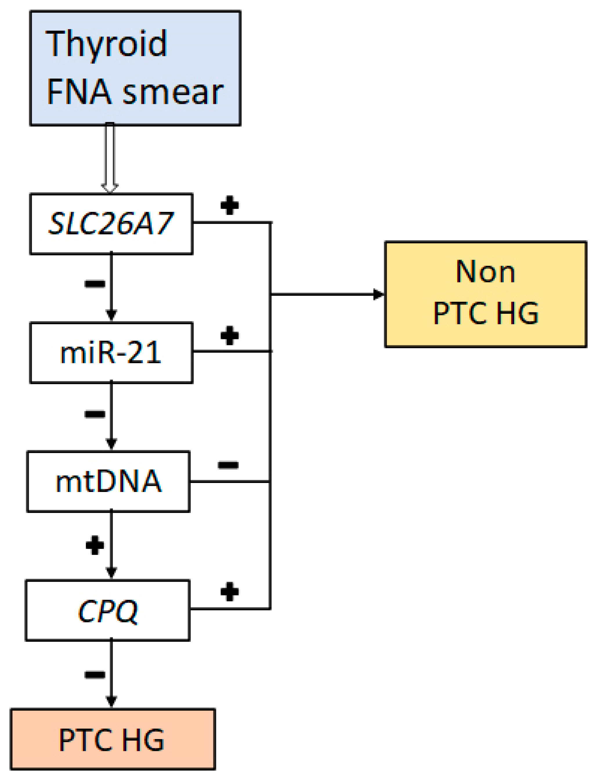
The algorithm of plotting a decision tree (C4.5) belonging to the class of logical methods was used to stratify the cytology specimens into different groups. Figure 3 illustrates the resulting decision tree.
The decision tree involved the genes SLC26A7 and CPQ as well as miR-21 and the ratio of mtDNA to nDNA. The tree has a linear structure, and basically, an FNA sample is classified as high-grade if the expression of SLC26A7 is less than the cutoff set by the algorithm (0.1), miR-21 expression is less than a cutoff of 56.96, the mtDNA/nDNA ratio is greater than 630, and the relative SLC26A7 expression is less than 0.257. If one of these conditions is not met, then the tissue sample is classified as non-high-grade. Data on diagnostic characteristics of this decision tree in terms of the identification of PTC and PTC HG are summarized in Table 3.
The resulting classifier based on the genes SLC26A7 and CPQ, as well as miR-21 and mtDNA as markers, was found to be highly specific (100%) but insufficiently sensitive (54%). In comparison, if the SLC26A7 gene was the only one used as a marker, then the sensitivity was the same (54%), but the specificity was lower (87.6%).

4. Discussion

In this study, we assessed the feasibility of preoperative detection of PTC HG (Bethesda V–VI) in cytology specimens using molecular markers. PTC is the most common malignant tumor of the thyroid gland (accounting for 85% of all thyroid cancer cases) and usually has an indolent course, with an overall 5-year survival rate of ~97.5%. Only a small percentage of well-differentiated carcinomas behave clinically aggressively and can lead to the death of the patient [46]. Some experts consider these carcinomas “true carcinomas” of the thyroid gland [47]. Until recently, however, researchers have not deemed it important to distinguish PTC HG as a separate subtype because this subdivision—according to the data available at the time—has not had any clinical or prognostic consequences [48]. According to more recent data, PTC HG should be distinguished as a separate subtype owing to its aggressive clinical course. Because molecular changes in PTC HG do not match the molecular changes in PDTC, it is appropriate to regard aggressive forms of PTC (e.g., PTC HG) as separate, not only from papillary carcinoma but also from PDTC [7].
Thus, it is crucial to search for efficient markers for the preoperative detection of PTC HG tumors. Such markers could be miRNAs, which are reported to be associated with an aggressive course of PTC [37]. We examined eight miRNAs in preoperative cytology specimens but failed to find any significant differences in miRNA expression between PTC and PTC HG.
Protein-coding genes responsible for various processes in thyrocytes are other candidates for molecular markers. Among the 14 studied genes, significant differences in expression levels between groups PTC and PTC HG were only revealed for genes SLC26A7, TFF3, and TPO. It should be noted that these three genes are related to the normal functioning of the thyroid gland rather than to malignant transformation. For instance, the TPO gene encodes thyroid peroxidase, which plays an important role in the biosynthesis of hormones; the SLC26A7 gene codes for a transmembrane ion transporter, which apparently plays an important part in the maintenance of an optimal ionic environment and pH for activities of the enzymes (including TPO) involved in the biosynthesis of thyroid hormones. The TFF3 gene is not specifically associated with the thyroid gland, although its expression is highest there (https://www.gtexportal.org/home/gene/TFF3 (accessed on 22 November 2024)); it encodes a protein belonging to the family of small secretory molecules participating in the maintenance and restoration of structural integrity of the epithelium [49]. Thus, it can be theorized that the differences between PTC HG and PTC are related to the downregulation of genes differentially expressed in the thyroid gland, i.e., with a loss of differentiation by the tumor.
Nonetheless, only one gene (SLC26A7) turned out to be crucial for detecting PTC HG. This simple classifier is characterized by the largest AUC ROC (0.816) in differentiation between the groups PTC and PTC HG and proved to be the key element of the decision tree by ensuring 54% sensitivity and 87.6% specificity. The other markers in the decision tree (CPQ, miR-21, and mtDNA) enhanced specificity (to 100%) but were possibly chosen for the algorithm randomly because of the distinctive features of the sampling procedure. On the contrary, the expression of the SLC26A7 gene is nonrandomly associated with the development of PTC HG. SLC26A7 plays an important, though not fully understood, role in thyroid hormone biosynthesis and is expressed predominantly in the thyroid gland, with dysfunction of this gene resulting in congenital goitrous hypothyroidism [50]. There is evidence that SLC26A7 is another iodine transporter because it has appreciable homology to the SLC26A4 gene (encoding an iodide exporter, also known as pendrin, which transports iodide from follicular cells to the lumen). At least when SLC26A4 is not functioning properly, SLC26A7 can perform iodine transport into the follicular lumen, thereby replacing SLC26A4 [51]. Just as in our study, SLC26A7 expression has been found to be significantly downregulated in patients with anaplastic thyroid carcinoma [26]; moreover, according to ref. [39], in PTC, SLC26A7 downregulation correlates with an elevated risk of metastases outside the thyroid.
The accumulation of data, refinement of morphological criteria, and expansion of knowledge about thyroid tumor biology will help to identify more aggressive and life-threatening subtypes of cancer at an early stage. In the present study, six out of thirteen patients with PTC HG had early disease stages T1 and T2. Early preoperative diagnosis of PTC HG in patients with early stages of this cancer will allow clinicians to adjust the treatment plan toward a broader surgical extent and lymph node dissection, and it will possibly provide indications for subsequent radioactive iodine therapy.
This study has several limitations: only 13 tissue samples of PTC HG were analyzed in the work; in the future, of course, it will be necessary to increase the sample size. Also, only a small set of markers was evaluated, which means that other potential markers could have been overlooked. Another limitation is that this study deals with a specific group of Russian patients; this means that the findings cannot be extrapolated to patient groups in other geographic locations or to other ethnic groups, owing to the effects of environmental and genetic factors. Further research will require high-throughput methods to find a sufficient number of markers for the differential diagnosis of PTC HG.

Supplementary Materials

The following supporting information can be downloaded at: https://www.mdpi.com/article/10.3390/diagnostics14232652/s1, Table S1: The oligonucleotide sequences used in the study.

Author Contributions

Conceptualization, S.E.T., E.S.K. and E.L.L.; methodology, S.E.T., E.S.K. and E.L.L.; validation, E.S.K., S.L.V., O.V.R. and S.V.B.; formal analysis, P.S.D.; investigation, Y.A.V., N.I.T., A.A.U. and A.V.Z.; resources, N.I.T., A.A.U., A.V.Z., R.A.C. and I.V.S.; data curation, S.E.T. and P.S.D.; writing—original draft preparation, S.E.T., E.S.K., S.A.L. and E.L.L.; writing—review and editing, I.F.Z., I.V.S., S.V.S. and R.A.C.; supervision, S.V.S., I.V.S. and I.F.Z.; project administration, S.A.L., S.E.T. and E.S.K. All authors have read and agreed to the published version of the manuscript.

Funding

This research was funded by the Russian Science Foundation, grant number 20-14-00074-P.

Institutional Review Board Statement

This study was conducted according to the guidelines of the Declaration of Helsinki and approved by the Ethics Committee of the South Ural State Medical University (Protocol No. 3 issued on 18 April 2019).

Informed Consent Statement

Informed consent was obtained from all patients involved in the study; all data were depersonalized.

Data Availability Statement

The raw data supporting the conclusions of this article will be made available by the authors upon request.

Acknowledgments

The cell analysis was carried out at the Multi-Access Center of Microscopic Research at the Institute of Cytology and Genetics SB RAS (supported by a government-funded project for the IC&G). The authors express their gratitude to the multi-access center Bioinformatics for the computational resources and their software, which was created within the framework of the government-funded project FWNR-2022-0020.

Conflicts of Interest

Sergei E. Titov is an employee of AO Vector-Best. The remaining authors declare that the research was conducted in the absence of any commercial or financial relationships that could be construed as a potential conflict of interest.

References

  1. Basolo, F.; Macerola, E.; Poma, A.M.; Torregrossa, L. The 5th edition of WHO classification of tumors of endocrine organs: Changes in the diagnosis of follicular-derived thyroid carcinoma. Endocrine 2023, 80, 470–476. [Google Scholar] [CrossRef] [PubMed]
  2. Baloch, Z.W.; Asa, S.L.; Barletta, J.A.; Ghossein, R.A.; Juhlin, C.C.; Jung, C.K.; LiVolsi, V.A.; Papotti, M.G.; Sobrinho-Simões, M.; Tallini, G.; et al. Overview of the 2022 WHO Classification of Thyroid Neoplasms. Endocr. Pathol. 2022, 33, 27–63. [Google Scholar] [CrossRef] [PubMed]
  3. Lim, H.; Devesa, S.S.; Sosa, J.A.; Check, D.; Kitahara, C.M. Trends in Thyroid Cancer Incidence and Mortality in the United States, 1974–2013. JAMA 2017, 317, 1338–1348. [Google Scholar] [CrossRef]
  4. Harahap, A.S.; Roren, R.S.; Imtiyaz, S. A Comprehensive Review and Insights into the New Entity of Differentiated High-Grade Thyroid Carcinoma. Curr. Oncol. 2024, 31, 3311–3328. [Google Scholar] [CrossRef] [PubMed]
  5. Xu, B.; David, J.; Dogan, S.; Landa, I.; Katabi, N.; Saliba, M.; Khimraj, A.; Sherman, E.J.; Tuttle, R.M.; Tallini, G.; et al. Primary high-grade non-anaplastic thyroid carcinoma: A retrospective study of 364 cases. Histopathology 2022, 80, 322–337. [Google Scholar] [CrossRef] [PubMed]
  6. Rivera, M.; Ghossein, R.A.; Schoder, H.; Gomez, D.; Larson, S.M.; Tuttle, R.M. Histopathologic characterization of radioactive iodine-refractory fuorodeoxyglucosepositron emission tomography-positive thyroid carcinoma. Cancer 2008, 113, 48–56. [Google Scholar] [CrossRef]
  7. Wong, K.S.; Dong, F.; Telatar, M.; Lorch, J.H.; Alexander, E.K.; Marqusee, E.; Cho, N.L.; Nehs, M.A.; Doherty, G.M.; Afkhami, M.; et al. Papillary Thyroid Carcinoma with High-Grade Features Versus Poorly Diferentiated Thyroid Carcinoma: An Analysis of Clinicopathologic and Molecular Features and Outcome. Thyroid 2021, 31, 933–940. [Google Scholar] [CrossRef]
  8. Xu, B.; Ghossein, R.A. Advances in Thyroid Pathology: High Grade Follicular Cell-derived Thyroid Carcinoma and Anaplastic Thyroid Carcinoma. Adv. Anat. Pathol. 2023, 30, 3–10. [Google Scholar] [CrossRef]
  9. Ghossein, R.; Katabi, N.; Dogan, S.; Shaha, A.R.; Tuttle, R.M.; Fagin, J.A.; Ganly, I.; Xu, B. Papillary thyroid carcinoma tall cell subtype (PTC-TC) and high-grade differentiated thyroid carcinoma tall cell phenotype (HGDTC-TC) have different clinical behaviour: A retrospective study of 1456 patients. Histopathology 2024, 84, 1130–1138. [Google Scholar] [CrossRef]
  10. Paulsson, J.O.; Backman, S.; Wang, N.; Stenman, A.; Crona, J.; Thutkawkorapin, J.; Ghaderi, M.; Tham, E.; Stalberg, P.; Zedenius, J.; et al. Whole-genome sequencing of synchronous thyroid carcinomas identifies aberrant DNA repair in thyroid cancer dediferentiation. J. Pathol. 2020, 250, 183–194. [Google Scholar] [CrossRef]
  11. Wong, K.S.; Lorch, J.H.; Alexander, E.K.; Marqusee, E.; Cho, N.L.; Nehs, M.A.; Doherty, G.M.; Barletta, J.A. Prognostic Signifcance of Extent of Invasion in Poorly Diferentiated Thyroid Carcinoma. Thyroid 2019, 29, 1255–1261. [Google Scholar] [CrossRef] [PubMed]
  12. Landa, I.; Ibrahimpasic, T.; Boucai, L.; Sinha, R.; Knauf, J.A.; Shah, R.H.; Dogan, S.; Ricarte-Filho, J.C.; Krishnamoorthy, G.P.; Xu, B.; et al. Genomic and transcriptomic hallmarks of poorly differentiated and anaplastic thyroid cancers. J. Clin. Investig. 2016, 126, 1052–1066. [Google Scholar] [CrossRef] [PubMed]
  13. Chernock, R.D.; Rivera, B.; Borrelli, N.; Hill, D.A.; Fahiminiya, S.; Shah, T.; Chong, A.S.; Aqil, B.; Mehrad, M.; Giordano, T.J.; et al. Poorly diferentiated thyroid carcinoma of childhood and adolescence: A distinct entity characterized by DICER1 mutations. Mod. Pathol. 2020, 33, 1264–1274. [Google Scholar] [CrossRef] [PubMed]
  14. Sistrunk, J.W.; Shifrin, A.; Frager, M.; Bardales, R.H.; Thomas, J.; Fishman, N.; Goldberg, P.; Guttler, R.; Grant, E. Clinical performance of multiplatform mutation panel and microRNA risk classifier in indeterminate thyroid nodules. J. Am. Soc. Cytopathol. 2020, 9, 232–241. [Google Scholar] [CrossRef]
  15. Patel, K.N.; Angell, T.E.; Babiarz, J.; Barth, N.M.; Blevins, T.; Duh, Q.Y.; Ghossein, R.A.; Harrell, R.M.; Huang, J.; Kennedy, G.C.; et al. Performance of a Genomic Sequencing Classifier for the Preoperative Diagnosis of Cytologically Indeterminate Thyroid Nodules. JAMA Surg. 2018, 153, 817–824. [Google Scholar] [CrossRef]
  16. Santos, M.; Buzolin, A.L.; Gama, R.R.; Silva, E.; Dufloth, R.M.; Figueiredo, D.; Carvalho, A.L. Molecular Classification of Thyroid Nodules with Indeterminate Cytology: Development and Validation of a Highly Sensitive and Specific New miRNA-Based Classifier Test Using Fine-Needle Aspiration Smear Slides. Thyroid 2018, 28, 1618–1626. [Google Scholar] [CrossRef]
  17. Titov, S.; Demenkov, P.S.; Lukyanov, S.A.; Sergiyko, S.V.; Katanyan, G.A.; Veryaskina, Y.A.; Ivanov, M.K. Preoperative detection of malignancy in fine-needle aspiration cytology (FNAC) smears with indeterminate cytology (Bethesda III, IV) by a combined molecular classifier. J. Clin. Pathol. 2020, 73, 722–727. [Google Scholar] [CrossRef]
  18. Lukyanov, S.A.; Sergiyko, S.V.; Titov, S.E.; Beltsevich, D.G.; Veryaskina, Y.A.; Vanushko, V.E.; Urusova, L.S.; Mikheenkov, A.A.; Kozorezova, E.S.; Vorobyov, S.L.; et al. New Opportunities for Preoperative Diagnosis of Medullary Thyroid Carcinoma. Biomedicines 2023, 11, 1473. [Google Scholar] [CrossRef]
  19. Titov, S.E.; Lukyanov, S.A.; Kozorezova, E.S.; Demenkov, P.S.; Sergiyko, S.V.; Veryaskina, Y.A.; Vorobyev, S.L.; Sleptsov, I.V.; Gostimskii, A.V. Validation of preoperative diagnosis of malignant thyroid tumors using a molecular classifier. Vopr. Onkol. 2022, 68, 741–751. [Google Scholar] [CrossRef]
  20. Titov, S.E.; Kozorezova, E.S.; Demenkov, P.S.; Veryaskina, Y.A.; Kuznetsova, I.V.; Vorobyev, S.L.; Chernikov, R.A.; Sleptsov, I.V.; Timofeeva, N.I.; Ivanov, M.K. Preoperative Typing of Thyroid and Parathyroid Tumors with a Combined Molecular Classifier. Cancers 2021, 13, 237. [Google Scholar] [CrossRef]
  21. Torous, V.F.; Jitpasutham, T.; Baloch, Z.; Cantley, R.L.; Kerr, D.A.; Liu, X.; Maleki, Z.; Merkin, R.; Nosé, V.; Pantanowitz, L.; et al. Cytologic features of differentiated high-grade thyroid carcinoma: A multi-institutional study of 40 cases. Cancer Cytopathol. 2024, 132, 525–536. [Google Scholar] [CrossRef]
  22. Haugen, B.R.; Alexander, E.K.; Bible, K.C.; Doherty, G.M.; Mandel, S.J.; Nikiforov, Y.E.; Pacini, F.; Randolph, G.W.; Sawka, A.M.; Schlumberger, M.; et al. 2015 American Thyroid Association Management Guidelines for Adult Patients with Thyroid Nodules and Differentiated Thyroid Cancer: The American Thyroid Association Guidelines Task Force on Thyroid Nodules and Differentiated Thyroid Cancer. Thyroid 2016, 26, 1–133. [Google Scholar] [CrossRef] [PubMed]
  23. Nan, B.Y.; Xiong, G.F.; Zhao, Z.R.; Gu, X.; Huang, X.S. Comprehensive Identification of Potential Crucial Genes and miRNA-mRNA Regulatory Networks in Papillary Thyroid Cancer. Biomed Res. Int. 2021, 2021, 6752141. [Google Scholar] [CrossRef] [PubMed]
  24. Wojtas, B.; Pfeifer, A.; Oczko-Wojciechowska, M.; Krajewska, J.; Czarniecka, A.; Kukulska, A. Gene Expression (mRNA) Markers for Differentiating between Malignant and Benign Follicular Thyroid Tumours. Int. J. Mol. Sci. 2017, 18, 1184. [Google Scholar] [CrossRef] [PubMed]
  25. Mussazhanova, Z.; Shimamura, M.; Kurashige, T.; Ito, M.; Nakashima, M.; Nagayama, Y. Causative role for defective expression of mitochondria-eating protein in accumulation of mitochondria in thyroid oncocytic cell tumors. Cancer Sci. 2020, 111, 2814–2823. [Google Scholar] [CrossRef] [PubMed]
  26. Ravi, N.; Yang, M.; Mylona, N.; Wennerberg, J.; Paulsson, K. Global RNA Expression and DNA Methylation Patterns in Primary Anaplastic Thyroid Cancer. Cancers 2020, 12, 680. [Google Scholar] [CrossRef]
  27. Colombo, C.; Minna, E.; Gargiuli, C.; Muzza, M.; Dugo, M.; De Cecco, L.; Pogliaghi, G.; Tosi, D.; Bulfamante, G.; Greco, A.; et al. The molecular and gene/miRNA expression profiles of radioiodine resistant papillary thyroid cancer. J. Exp. Clin. Cancer Res. 2020, 39, 245. [Google Scholar] [CrossRef]
  28. Kebebew, E.; Peng, M.; Reiff, E.; Duh, Q.Y.; Clark, O.H.; McMillan, A. ECM1 and TMPRSS4 are diagnostic markers of malignant thyroid neoplasms and improve the accuracy of fine needle aspiration biopsy. Ann. Surg. 2005, 242, 353–361. [Google Scholar] [CrossRef]
  29. Binabaj, M.M.; Soleimani, A.; Rahmani, F.; Avan, A.; Khazaei, M.; Fiuji, H.; Soleimanpour, S.; Ryzhikov, M.; Ferns, G.A.; Bahrami, A.; et al. Prognostic value of high mobility group protein A2 (HMGA2) over-expression in cancer progression. Gene 2019, 706, 131–139. [Google Scholar] [CrossRef]
  30. Chen, Z.; Guo, Y.; Zhao, D.; Zou, Q.; Yu, F.; Zhang, L.; Xu, L. Comprehensive Analysis Revealed that CDKN2A is a Biomarker for Immune Infiltrates in Multiple Cancers. Front. Cell Dev. Biol. 2021, 9, 808208. [Google Scholar] [CrossRef]
  31. Titov, S.E.; Ivanov, M.K.; Demenkov, P.S.; Katanyan, G.A.; Kozorezova, E.S.; Malek, A.V.; Veryaskina, Y.A.; Zhimulev, I.F. Combined quantitation of HMGA2 mRNA, microRNAs, and mitochondrial-DNA content enables the identification and typing of thyroid tumors in fine-needle aspiration smears. BMC Cancer 2019, 19, 1010. [Google Scholar] [CrossRef]
  32. Titov, S.E.; Ivanov, M.K.; Karpinskaya, E.V.; Tsivlikova, E.V.; Shevchenko, S.P.; Veryaskina, Y.A.; Akhmerova, L.G.; Poloz, T.L.; Klimova, O.A.; Gulyaeva, L.F.; et al. miRNA profiling, detection of BRAF V600E mutation and RET-PTC1 translocation in patients from Novosibirsk oblast (Russia) with different types of thyroid tumors. BMC Cancer 2016, 16, 201. [Google Scholar] [CrossRef] [PubMed]
  33. Maggisano, V.; Capriglione, F.; Verrienti, A.; Celano, M.; Gagliardi, A.; Bulotta, S.; Sponziello, M.; Mio, C.; Pecce, V.; Durante, C.; et al. Identification of Exosomal microRNAs and Their Targets in Papillary Thyroid Cancer Cells. Biomedicines 2022, 10, 961. [Google Scholar] [CrossRef] [PubMed]
  34. Livak, K.J.; Schmittgen, T.D. Analysis of relative gene expression data using real-time quantitative PCR and the 2(-Delta Delta C(T)) Method. Methods 2001, 25, 402–408. [Google Scholar] [CrossRef] [PubMed]
  35. Chen, C.; Ridzon, D.A.; Broomer, A.J.; Zhou, Z.; Lee, D.H.; Nguyen, J.T.; Barbisin, M.; Xu, N.L.; Mahuvakar, V.R.; Andersen, M.R.; et al. Real-time quantification of microRNAs by stem-loop RT-PCR. Nucleic Acids Res. 2005, 33, e179. [Google Scholar] [CrossRef] [PubMed]
  36. Salzberg, S.L. C4.5: Programs for Machine Learning; Quinlan, J.R., Ed.; Morgan Kaufmann Publishers, Inc.: Burlington, MA, USA, 1993; Volume 16, pp. 235–240. [Google Scholar]
  37. Li, X.; Cheng, R. TPO as an indicator of lymph node metastasis and recurrence in papillary thyroid carcinoma. Sci. Rep. 2023, 13, 10848. [Google Scholar] [CrossRef] [PubMed]
  38. Massart, C.; Hoste, C.; Virion, A.; Ruf, J.; Dumont, J.E.; Van Sande, J. Cell biology of H2O2 generation in the thyroid: Investigation of the control of dual oxidases (DUOX) activity in intact ex vivo thyroid tissue and cell lines. Mol. Cell. Endocrinol. 2011, 343, 32–44. [Google Scholar] [CrossRef]
  39. Huang, F.; Xiao, J.; Wang, L.; Xie, Y.; Jia, H. Relationships between SLC26A7 expressions and extra-thyroid metastasis of papillary thyroid carcinoma. Chin. Med. J. 2021, 135, 225–227. [Google Scholar] [CrossRef]
  40. Jahan, R.; Shah, A.; Kisling, S.G.; Macha, M.A.; Thayer, S.; Batra, S.K.; Kaur, S. Odyssey of trefoil factors in cancer: Diagnostic and therapeutic implications. Biochim. Biophys. Acta Rev. Cancer 2020, 1873, 188362. [Google Scholar] [CrossRef]
  41. Yang, L.; Zhang, X.; Zhang, J.; Liu, Y.; Ji, T.; Mou, J.; Fang, X.; Wang, S.; Chen, J. Low expression of TFF3 in papillary thyroid carcinoma may correlate with poor prognosis but high immune cell infiltration. Future Oncol. 2022, 18, 333–348. [Google Scholar] [CrossRef]
  42. Kushwaha, P.P.; Rapalli, K.C.; Kumar, S. Geminin a multi task protein involved in cancer pathophysiology and developmental process: A review. Biochimie 2016, 131, 115–127. [Google Scholar] [CrossRef] [PubMed]
  43. Liggett, W.H.; Jr Sidransky, D. Role of the p16 tumor suppressor gene in cancer. J. Clin. Oncol. 1998, 16, 1197–1206. [Google Scholar] [CrossRef] [PubMed]
  44. Dabravolski, S.A.; Nikiforov, N.G.; Zhuravlev, A.D.; Orekhov, N.A.; Mikhaleva, L.M.; Orekhov, A.N. The Role of Altered Mitochondrial Metabolism in Thyroid Cancer Development and Mitochondria-Targeted Thyroid Cancer Treatment. Int. J. Mol. Sci. 2021, 23, 460. [Google Scholar] [CrossRef] [PubMed]
  45. Tavares, C.; Coelho, M.J.; Eloy, C.; Melo, M.; da Rocha, A.G.; Pestana, A.; Batista, R.; Ferreira, L.B.; Rios, E.; Selmi-Ruby, S.; et al. NIS expression in thyroid tumors, relation with prognosis clinicopathological and molecular features. Endocr. Connect. 2018, 7, 78–90. [Google Scholar] [CrossRef]
  46. Lloyd, R.V.; Osamura, R.Y.; Kloppel, G.; Rosai, J. World Health Organization Classification of Tumours of Endocrine Organs, 4th ed.; IARC Press: Lyon, France, 2017; Volume 10. [Google Scholar]
  47. Kazaure, H.S.; Roman, S.A.; Sosa, J.A. Aggressive variants of papillary thyroid cancer: Incidence, characteristics and predictors of survival among 43,738 patients. Ann. Surg. Oncol. 2012, 19, 1874–1880. [Google Scholar] [CrossRef]
  48. Nikiforov, Y.E.; Biddinger, P.W.; Thompson, L.D. Diagnostic Pathology and Molecular Genetics of the Thyroid; Lippincott Williams & Wilkins: Philadelphia, PA, USA, 2012. [Google Scholar]
  49. GTEx Portal (dbGaP Accession phs000424.v8.p2). Available online: https://www.gtexportal.org/home/gene/TFF3 (accessed on 22 November 2024).
  50. Cangul, H.; Liao, X.H.; Schoenmakers, E.; Kero, J.; Barone, S.; Srichomkwun, P.; Iwayama, H.; Serra, E.G.; Saglam, H.; Eren, E.; et al. Homozygous loss-of-function mutations in SLC26A7 cause goitrous congenital hypothyroidism. JCI Insight 2018, 3, e99631. [Google Scholar] [CrossRef]
  51. Ishii, J.; Suzuki, A.; Kimura, T.; Tateyama, M.; Tanaka, T.; Yazawa, T.; Arimasu, Y.; Chen, I.S.; Aoyama, K.; Kubo, Y.; et al. Congenital goitrous hypothyroidism is caused by dysfunction of the iodide transporter SLC26A7. Commun. Biol. 2019, 2, 270. [Google Scholar] [CrossRef]
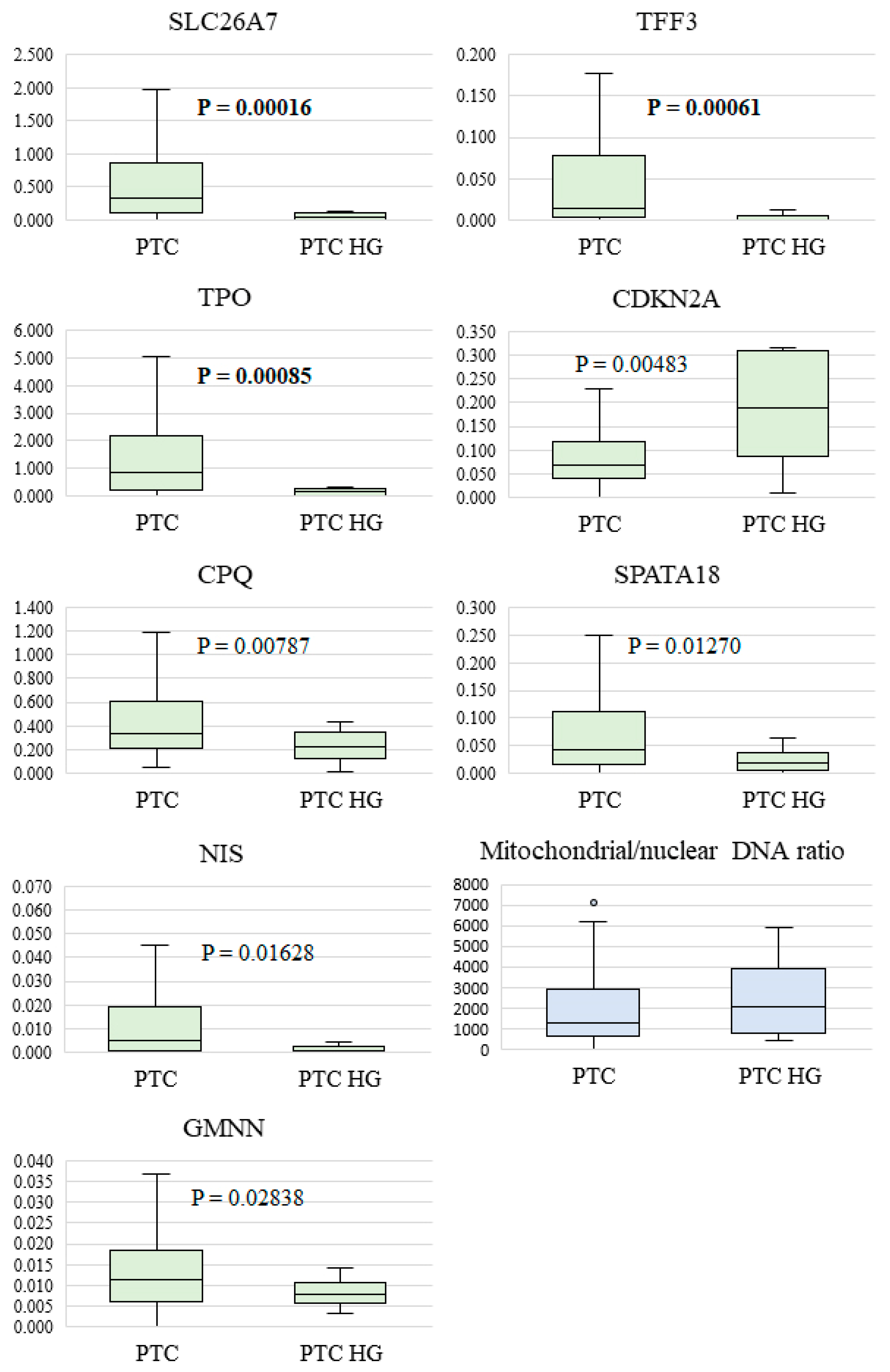
Figure 1. Relative expression levels of eight genes and the mtDNA/nDNA ratio in PTC and PTC HG specimens. The median, upper, and lower quartiles (box), a nonoutlier range (whiskers), and outliers (circles) are indicated. The p-values at the significance level chosen in this study (<0.0022) are shown in bold.
Diagnostics 14 02652 g001
Figure 2. Areas under the ROC curves (ROC AUC) for eight mRNAs for assessing the distinguishability of groups PTC and PTC HG. Whiskers denote 95% confidence intervals.
Diagnostics 14 02652 g002
Figure 3. The decision tree for classifying samples into benign or malignant ones followed by cancer typing. + or −: exceeding the chosen cutoff or not exceeding it.
Diagnostics 14 02652 g003
Table 1. Clinical characteristics of the patients.
CharacteristicValue, n (%)
PTC (n = 97)PTC HG (n = 13)
Age, median (Q1-Q3)47.5 (37–60.25)64 (48.75–74.25)
Sex ratio (M/F)21/763/10
Metastasis to central cervical lymph nodes N1a26 (26.8%)2 (15.4%)
Metastasis to lateral cervical lymph nodes N1b19 (19.6%)6 (46.2%)
Multifocality58 (59.8%)3 (23%)
Extrathyroidal extension (macroscopic invasion)18 (18.6%)7 (53.8%)
Distant metastasis00
Table 2. Statistical significance of differences in the pairwise comparison of groups PTC (n = 97) and PTC HG (n = 13).
Markerp-Value
miR-146b0.452583
miR-199b0.086416
miR-2210.744162
miR-2230.204396
miR-310.067463
miR-3750.767790
miR-551b0.629983
miR-210.359206
mtDNA0.206242
FN10.142648
GMNN0.028378
CDKN2A0.004832
TPO0.000850
SLC26A70.000166
HMGA20.558307
CPQ0.007873
SPATA180.012699
APOE0.104936
DIO10.929108
NIS0.016281
TFF30.000614
TMPRSS40.547838
TSHR0.458833
Significant differences are boldfaced (adjusted p-value < 0.0022).
Table 3. Diagnostic characteristics (including the 95% confidence interval) of PTC and PTC HG identification either by means of the decision tree or exclusively by means of the SLC26A7 gene.
Diagnostic CharacteristicsDecision TreeSLC26A7
Sensitivity53.8% (25.1–80.8%)53.8% (25.1–80.8%)
Specificity100.0% (96.3–100.0%)87.6% (79.4–93.4%)
Accuracy94.5% (88.5–97.8%)83.6% (75.5–90.0%)
Positive predictive value100.0% (59.0–100.0%)36.8% (36.8–54.8%)
Negative predictive value94.2% (90.0–96.7%)93.4% (88.7–96.2%)
Disclaimer/Publisher’s Note: The statements, opinions and data contained in all publications are solely those of the individual author(s) and contributor(s) and not of MDPI and/or the editor(s). MDPI and/or the editor(s) disclaim responsibility for any injury to people or property resulting from any ideas, methods, instructions or products referred to in the content.