

Short- and Long-Term Outcomes after Radiofrequency Ablation of Osteoid Osteomas
 , ,
, ,
Abstract
:1. Introduction
2. Materials and Methods
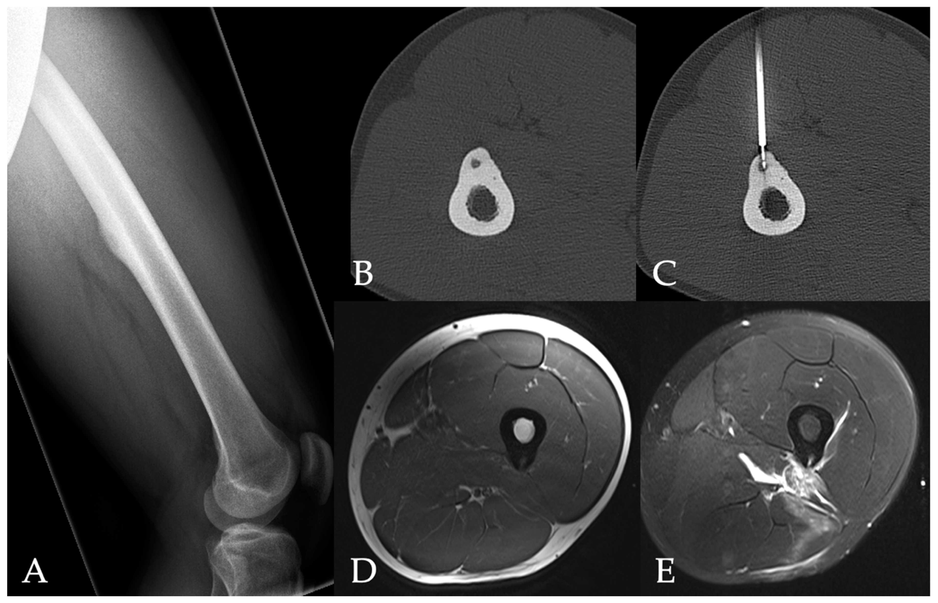
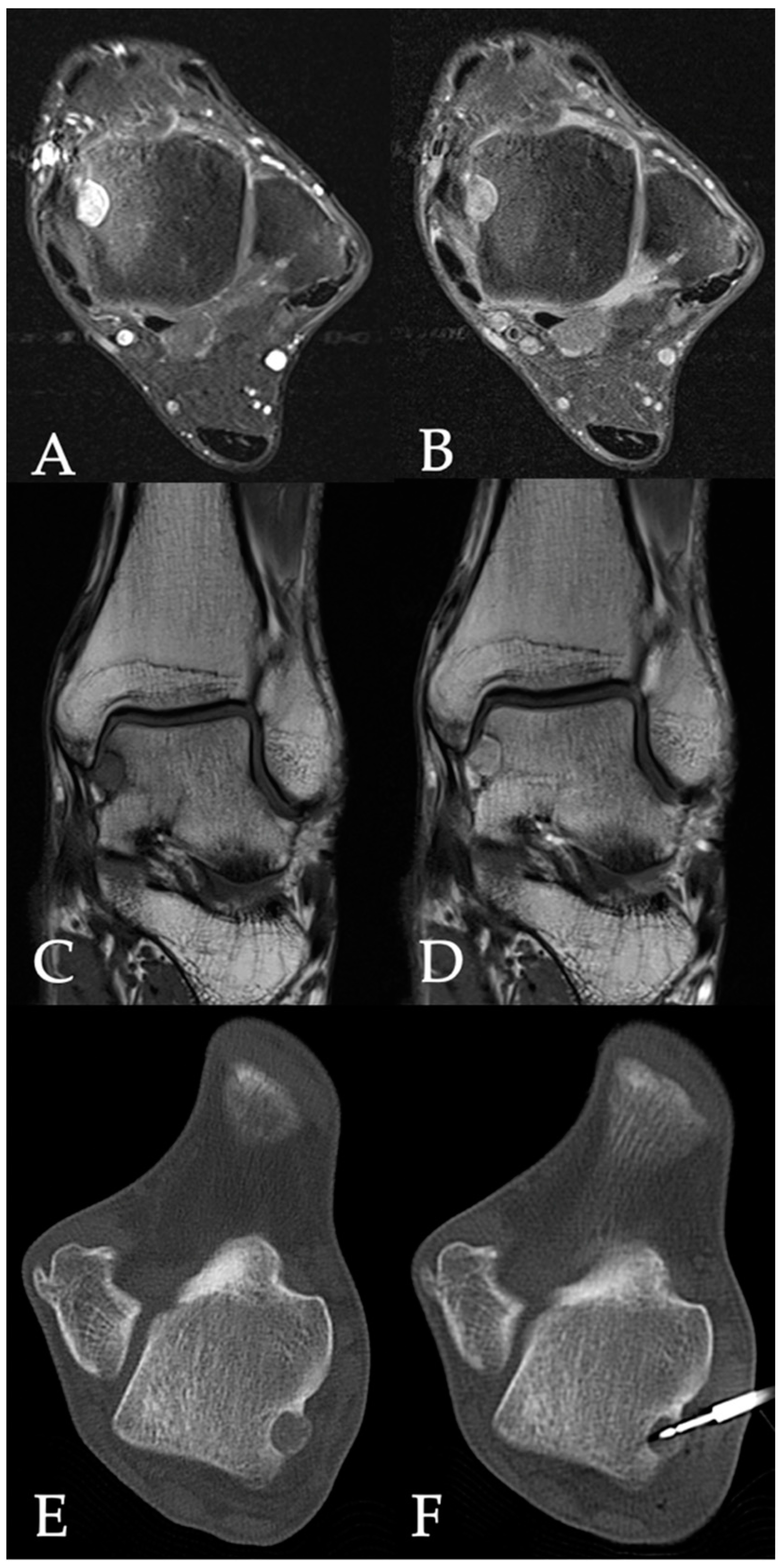
2.1. Radiofrequency Ablation
2.2. Patient Questionnaire
2.3. Data Analysis
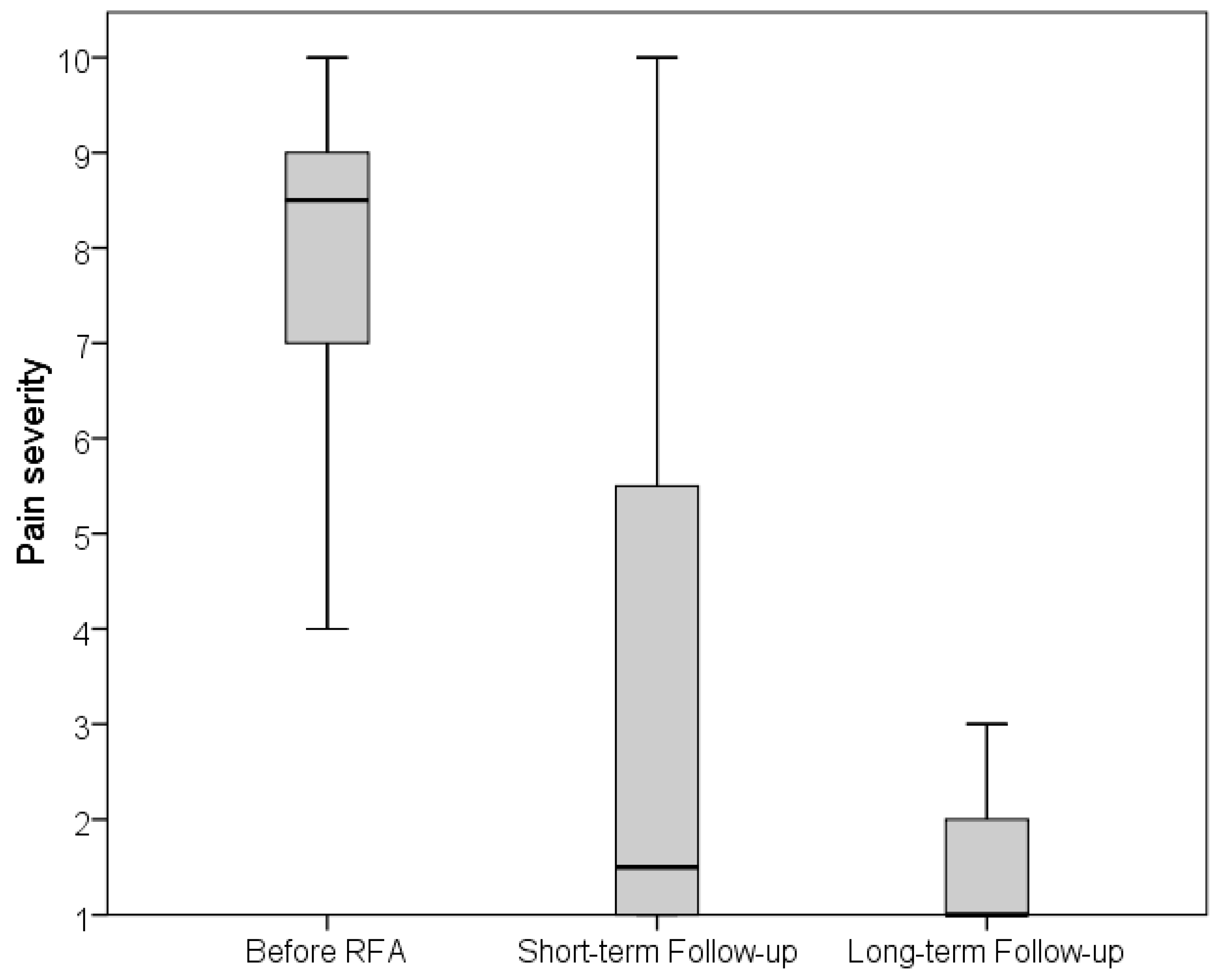
3. Results
4. Discussion
5. Limitations
6. Conclusions
Author Contributions
Funding
Institutional Review Board Statement
Informed Consent Statement
Data Availability Statement
Conflicts of Interest
References
- Lee, E.H.; Shafi, M.; Hui, J.H. Osteoid osteoma: A current review. J. Pediatr. Orthop. 2006, 26, 695–700. [Google Scholar] [CrossRef] [PubMed]
- Hakim, D.N.; Pelly, T.; Kulendran, M.; Caris, J.A. Benign tumours of the bone: A review. J. Bone Oncol. 2015, 4, 37–41. [Google Scholar] [CrossRef] [PubMed]
- Orth, P.; Kohn, D. Diagnostik und Therapie des Osteoidosteoms [Diagnostics and treatment of osteoid osteoma]. Orthopade 2017, 46, 510–521. [Google Scholar] [CrossRef] [PubMed]
- De Filippo, M.; Russo, U.; Papapietro, V.R.; Ceccarelli, F.; Pogliacomi, F.; Vaienti, E.; Piccolo, C.; Capasso, R.; Sica, A.; Cioce, F.; et al. Radiofrequency ablation of osteoid osteoma. Acta Biomed. 2018, 89, 175–185. [Google Scholar] [CrossRef] [PubMed]
- Esteban Cuesta, H.; Martel Villagran, J.; Bueno Horcajadas, A.; Kassarjian, A.; Rodriguez Caravaca, G. Percutaneous radiofrequency ablation in osteoid osteoma: Tips and tricks in special scenarios. Eur. J. Radiol. 2018, 102, 169–175. [Google Scholar] [CrossRef]
- Kayser, F.; Resnick, D.; Haghighi, P.; Pereira, E.D.; Greenway, G.; Schweitzer, M.; Kindynis, P. Evidence of the subperiosteal origin of osteoid osteomas in tubular bones: Analysis by CT and MR imaging. AJR Am. J. Roentgenol. 1998, 170, 609–614. [Google Scholar] [CrossRef] [PubMed]
- Bhure, U.; Roos, J.E.; Strobel, K. Osteoid osteoma: Multimodality imaging with focus on hybrid imaging. Eur. J. Nucl. Med. Mol. Imaging 2019, 46, 1019–1036. [Google Scholar] [CrossRef] [PubMed]
- Tepelenis, K.; Skandalakis, G.P.; Papathanakos, G.; Kefala, M.A.; Kitsouli, A.; Barbouti, A.; Tepelenis, N.; Varvarousis, D.; Vlachos, K.; Kanavaros, P.; et al. Osteoid Osteoma: An Updated Review of Epidemiology, Pathogenesis, Clinical Presentation, Radiological Features, and Treatment Option. In Vivo 2021, 35, 1929–1938. [Google Scholar] [CrossRef] [PubMed]
- Carneiro, B.C.; Da Cruz, I.A.; Ormond Filho, A.G.; Silva, I.P.; Guimarães, J.B.; Silva, F.D.; Nico, M.A.; Stump, X.M. Osteoid osteoma: The great mimicker. Insights Imaging 2021, 12, 32. [Google Scholar] [CrossRef]
- Cantwell, C.P.; Obyrne, J.; Eustace, S. Current trends in treatment of osteoid osteoma with an emphasis on radiofrequency ablation. Eur. Radiol. 2004, 14, 607–617. [Google Scholar] [CrossRef]
- Assoun, J.; Richardi, G.; Railhac, J.J.; Baunin, C.; Fajadet, P.; Giron, J.; Maquin, P.; Haddad, J.; Bonnevialle, P. Osteoid osteoma: MR imaging versus CT. Radiology 1994, 191, 217–223. [Google Scholar] [CrossRef] [PubMed]
- Hosalkar, H.S.; Garg, S.; Moroz, L.; Pollack, A.; Dormans, J.P. The diagnostic accuracy of MRI versus CT imaging for osteoid osteoma in children. Clin. Orthop. Relat. Res. 2005, 433, 171–177, Erratum in Clin. Orthop. Relat. Res. 2005, 436, 286. [Google Scholar] [CrossRef]
- Davies, M.; Cassar-Pullicino, V.N.; Davies, A.M.; McCall, I.W.; Tyrrell, P.N. The diagnostic accuracy of MR imaging in osteoid osteoma. Skelet. Radiol. 2002, 31, 559–569. [Google Scholar] [CrossRef] [PubMed]
- French, J.; Epelman, M.; Johnson, C.M.; Stinson, Z.; Meyers, A.B. MR Imaging of Osteoid Osteoma: Pearls and Pitfalls. Semin. Ultrasound CT MR 2020, 41, 488–497. [Google Scholar] [CrossRef]
- Klontzas, M.E.; Zibis, A.H.; Karantanas, A.H. Osteoid Osteoma of the Femoral Neck: Use of the Half-Moon Sign in MRI Diagnosis. AJR Am. J. Roentgenol. 2015, 205, 353–357. [Google Scholar] [CrossRef]
- Boscainos, P.J.; Cousins, G.R.; Kulshreshtha, R.; Oliver, T.B.; Papagelopoulos, P.J. Osteoid osteoma. Orthopedics 2013, 36, 792–800. [Google Scholar] [CrossRef] [PubMed]
- Alemdar, C.; Çaçan, M.A.; Dusak, A.; Özkul, E.; Atiç, R.; Kapukaya, A. A comparison of percutaneous trephine excision and open surgery in the treatment of osteoid osteoma. Int. Orthop. 2016, 40, 1481–1487. [Google Scholar] [CrossRef]
- Rosenthal, D.I.; Alexander, A.; Rosenberg, A.E.; Springfield, D. Ablation of osteoid osteomas with a percutaneously placed electrode: A new procedure. Radiology 1992, 183, 29–33. [Google Scholar] [CrossRef]
- Woertler, K.; Vestring, T.; Boettner, F.; Winkelmann, W.; Heindel, W.; Lindner, N. Osteoid osteoma: CT-guided percutaneous radiofrequency ablation and follow-up in 47 patients. J. Vasc. Interv. Radiol. 2001, 12, 717–722. [Google Scholar] [CrossRef]
- Tomasian, A.; Cazzato, R.L.; Auloge, P.; Garnon, J.; Gangi, A.; Jennings, J.W. Osteoid osteoma in older adults: Clinical success rate of percutaneous image-guided thermal ablation. Clin. Radiol. 2020, 75, 713.e11–713.e16. [Google Scholar] [CrossRef]
- Göksel, F.; Aycan, A.; Ermutlu, C.; Gölge, U.H.; Sarısözen, B. Comparison of Radiofrequency Ablation and Curettage in Osteoid Osteoma in Children. Acta Ortop. Bras. 2019, 27, 100–103. [Google Scholar] [CrossRef] [PubMed]
- Garge, S.; Keshava, S.N.; Moses, V.; Chiramel, G.K.; Ahmed, M.; Mammen, S.; Madhuri, V. Radiofrequency ablation of osteoid osteoma in common and technically challenging locations in pediatric population. Indian J. Radiol. Imaging 2017, 27, 88–91. [Google Scholar] [CrossRef] [PubMed]
- Somma, F.; Stoia, V.; D’Angelo, R.; Fiore, F. Imaging-guided radiofrequency ablation of osteoid osteoma in typical and atypical sites: Long term follow up. PLoS ONE 2021, 16, e0248589. [Google Scholar] [CrossRef] [PubMed]
- Miyazaki, M.; Arai, Y.; Myoui, A.; Gobara, H.; Sone, M.; Rosenthal, D.I.; Tsushima, Y.; Kanazawa, S.; Ehara, S.; Endo, K. Phase I/II Multi-Institutional Study of Percutaneous Radiofrequency Ablation for Painful Osteoid Osteoma (JIVROSG-0704). Cardiovasc. Interv. Radiol. 2016, 39, 1464–1470. [Google Scholar] [CrossRef] [PubMed]
- Yu, X.; Wang, B.; Yang, S.; Han, S.; Jiang, L.; Liu, X.; Wei, F.; Wu, F.; Dang, L.; Liu, Z. Percutaneous radiofrequency ablation versus open surgical resection for spinal osteoid osteoma. Spine J. 2019, 19, 509–515. [Google Scholar] [CrossRef] [PubMed]
- Sangiorgio, A.; Oldrini, L.M.; Candrian, C.; Errani, C.; Filardo, G. Radiofrequency ablation is as safe and effective as surgical excision for spinal osteoid osteoma: A systematic review and meta-analysis. Eur. Spine J. 2023, 32, 210–220. [Google Scholar] [CrossRef] [PubMed]
- Baal, J.D.; Pai, J.S.; Chen, W.C.; Joseph, G.B.; O’Donnell, R.J.; Link, T.M. Factors Associated with Osteoid Osteoma Recurrence after CT-Guided Radiofrequency Ablation. J. Vasc. Interv. Radiol. 2019, 30, 744–751. [Google Scholar] [CrossRef] [PubMed]
- Efthymiadis, A.; Tsikopoulos, K.; Uddin, F.; Kitridis, D.; Edwards, N.; Sidiropoulos, K.; Lavalette, D. Which is the optimal minimally invasive treatment for osteoid osteoma of the hip? A systematic review and proportional meta-analysis. J. Orthop. Sci. 2022, 27, 456–462. [Google Scholar] [CrossRef] [PubMed]
- Shanmugasundaram, S.; Nadkarni, S.; Kumar, A.; Shukla, P.A. Percutaneous Ablative Therapies for the Management of Osteoid Osteomas: A Systematic Review and Meta-Analysis. Cardiovasc. Interv. Radiol. 2021, 44, 739–749. [Google Scholar] [CrossRef] [PubMed]
- Lindquester, W.S.; Crowley, J.; Hawkins, C.M. Percutaneous thermal ablation for treatment of osteoid osteoma: A systematic review and analysis. Skelet. Radiol. 2020, 49, 1403–1411. [Google Scholar] [CrossRef]
- Oc, Y.; Kilinc, B.E.; Cennet, S.; Boyacioglu, M.M.; Ertugrul, R.; Varol, A. Complications of Computer Tomography Assisted Radiofrequency Ablation in the Treatment of Osteoid Osteoma. Biomed. Res. Int. 2019, 2019, 4376851. [Google Scholar] [CrossRef] [PubMed]
- Singh, D.K.; Kumar, N.; Rustagi, A.; Jalan, D.; Krishna, L.G.; Sharma, A. Percutaneous CT-guided radiofrequency ablation of osteoid osteoma: Potential Pitfalls and complications and how to avoid them. J. Clin. Orthop. Trauma 2022, 28, 101869. [Google Scholar] [CrossRef] [PubMed]
- Schmidt, D.; Clasen, S.; Schaefer, J.F.; Rempp, H.; Duda, S.; Trübenbach, J.; König, C.W.; Erdtmann, B.; Claussen, C.D.; Pereira, P.L. CT-gesteuerte Radiofrequenz (RF)-Ablation von Osteoidosteomen: Klinische Langzeitergebnisse [CT-guided radiofrequency (RF) ablation of osteoid osteoma: Clinical long-term results]. Rofo 2011, 183, 381–387. [Google Scholar] [CrossRef] [PubMed]
- Akhlaghpoor, S.; Aziz Ahari, A.; Arjmand Shabestari, A.; Alinaghizadeh, M.R. Radiofrequency ablation of osteoid osteoma in atypical locations: A case series. Clin. Orthop. Relat. Res. 2010, 468, 1963–1970. [Google Scholar] [CrossRef] [PubMed]


| Parameter | Patients n = 62 |
|---|---|
| Age, y | 26.2 ± 13.2 |
| sex | |
| Female | 19 (30.6%) |
| Male | 43 (69.4%) |
| Tumor localization | |
| Femur | 27 (42.9%) |
| Tibia | 19 (30.2%) |
| Humerus | 4 (6.3%) |
| Spine | 4 (6.3%) |
| Cervical | 1 (1.6%) |
| Thoracic | 1 (1.6%) |
| Lumbar | 2 (3.2%) |
| Talus | 3 (4.8%) |
| Ilium | 2 (3.2%) |
| Fibula | 1 (1.6%) |
| Rib | 1 (1.6%) |
| Thumb | 1 (1.6%) |
| Index finger | 1 (1.6%) |
| Nidus size, mm | 5.7 ± 2.6 |
| Parameter | Patients n = 36 |
|---|---|
| Pain quality | |
| Stinging | 15 (41.7%) |
| Oppressive | 10 (27.8%) |
| Stinging and oppressive (mixed) | 6 (16.7%) |
| Indescribable | 5 (13.8%) |
| Time of pain | |
| All-day | 16 (44.4%) |
| Nighttime | 14 (38.9%) |
| Daytime | 6 (16.7%) |
| Pain-related sleep disorders | 29 (80.6%) |
| Pain medication | |
| Yes | 30 (83.3%) |
| Pain relief | |
| Yes | 27 (90%) |
| No | 3 (10%) |
Disclaimer/Publisher’s Note: The statements, opinions and data contained in all publications are solely those of the individual author(s) and contributor(s) and not of MDPI and/or the editor(s). MDPI and/or the editor(s) disclaim responsibility for any injury to people or property resulting from any ideas, methods, instructions or products referred to in the content. |
© 2024 by the authors. Licensee MDPI, Basel, Switzerland. This article is an open access article distributed under the terms and conditions of the Creative Commons Attribution (CC BY) license (https://creativecommons.org/licenses/by/4.0/).
Share and Cite
Vogl, T.J.; Bialek, M.; Eichler, K.; Hammerstingl, R.; Bielfeldt, J.; Zangos, S.; Scholtz, J.-E.; Adwan, H. Short- and Long-Term Outcomes after Radiofrequency Ablation of Osteoid Osteomas. J. Pers. Med. 2024, 14, 401. https://doi.org/10.3390/jpm14040401
Vogl TJ, Bialek M, Eichler K, Hammerstingl R, Bielfeldt J, Zangos S, Scholtz J-E, Adwan H. Short- and Long-Term Outcomes after Radiofrequency Ablation of Osteoid Osteomas. Journal of Personalized Medicine. 2024; 14(4):401. https://doi.org/10.3390/jpm14040401
Chicago/Turabian StyleVogl, Thomas J., Michael Bialek, Katrin Eichler, Renate Hammerstingl, John Bielfeldt, Stephan Zangos, Jan-Erik Scholtz, and Hamzah Adwan. 2024. "Short- and Long-Term Outcomes after Radiofrequency Ablation of Osteoid Osteomas" Journal of Personalized Medicine 14, no. 4: 401. https://doi.org/10.3390/jpm14040401