Review

Oral Lubrication, Xerostomia, and Advanced Macromolecular Lubricants for Treatment of Dry Mouth

by
William Austin
1,2,
Maryam Hdeib
1,2,
Paige Fraser
1,2,
Maya Goldchtaub
1,2,
Elika Shams
3,
Tianyi Han
4,
Pierre-Luc Michaud
5 and
Vahid Adibnia
1,2,6,*
1
School of Biomedical Engineering, Dalhousie University, Halifax, NS B3H 4R2, Canada
2
Department of Applied Oral Sciences, Faculty of Dentistry, Dalhousie University, Halifax, NS B3H 4R2, Canada
3
Department of Biomedical Engineering, College of Engineering, University of Connecticut, Storrs, CT 06269, USA
4
State Key Laboratory of Tribology in Advanced Equipment, Tsinghua University, Beijing 100084, China
5
Department of Dental Clinical Sciences, Faculty of Dentistry, Dalhousie University, Halifax, NS B3H 4R2, Canada
6
Department of Chemistry, Dalhousie University, Halifax, NS B3H 4R2, Canada
*
Author to whom correspondence should be addressed.
Lubricants 2024, 12(4), 126; https://doi.org/10.3390/lubricants12040126
Submission received: 1 March 2024 / Revised: 25 March 2024 / Accepted: 9 April 2024 / Published: 12 April 2024
(This article belongs to the Special Issue Advanced Polymeric and Colloidal Lubricants)

Abstract

:
Dry mouth, also known as xerostomia, is a condition in which insufficient or ineffective saliva does not provide sufficient oral lubrication. The severity of this condition can vary from a mild discomfort to a debilitating condition that greatly impairs patients’ lives. Xerostomia arises as a side effect of various medications, diseases, radiation therapy, chemotherapy, or nerve damage. Various aqueous dispersions of macromolecules have been proposed to assist or replace the saliva in these patients. It is vital that these macromolecules have ample lubricity and water retention properties while showing long-lasting efficacy. The emphasis of this review is to provide a general overview on lubricating macromolecules that have been clinically used or reported in the literature as potential replacements for saliva. These include various natural or synthetic polymers, proteins, peptides, and lipids that are used in the form of solutions, gels, emulsions, and colloids. Perspectives into the future of macromolecular oral lubricants in the treatment of xerostomia are also provided.

Graphical Abstract

1. Introduction

Xerostomia is the subjective sensation of dry mouth. It affects 20% of the general population worldwide [1]. Adults over 75 years of age with co-morbid diseases and polypharmacy are more prone to this condition, resulting in the frequency rising to 46% in this age group [2]. There are multiple factors causing xerostomia, but medication side effects are among the most prominent etiologies, followed by radiation therapy and various chronic autoimmune syndromes, such as Sjögren’s syndrome [3]. If an individual does not treat xerostomia, it may cause complications such as nutritional deficiency, low mood, and depression [4]. Patients with xerostomia may face difficulty speaking, eating, and sleeping, and it could significantly impact their overall quality of life. Because saliva is important to buffer the acidity of the mouth, dry mouth could also lead to dental cavities and eventually, tooth loss [5].
Saliva is the key medium responsible for the lubrication of oral surfaces. It is a fluid mostly composed of water (99%), ions, and proteinaceous substances, including mucins, amylases, and other low molecular weight proteins [6]. As a response to biochemical inputs and environmental cues, saliva is secreted from the salivary glands through a coordinated action of the assembled acinar, ductal, and myoepithelial cells [7]. Saliva has a low surface tension (59 mN m−1 compared to 72.7 mN m−1 for distilled water [8]), which helps it wet the oral mucosa as a thin film [9]. The salivary conditioning film (SCF) contains glycoproteins like mucin, which retains water and results in hydration lubrication of the oral cavity [10]. Salivary gland dysfunction may alter the composition and the production of saliva, resulting in aberrant SCF with less mucin, which results in poor lubrication, mouth dryness, difficulty in swallowing, and may cause oral burning sensations [10]. The normal unstimulated saliva flow rate is 0.3–0.4 mL min−1, which could decrease to 0.1 mL min−1 while sleeping and increase to 0.4–0.5 mL min−1 under stimulation [11]. In patients with xerostomia, this stimulated rate can decrease to 0.1–0.2 mL min−1 [12]. Nevertheless, xerostomia could also happen with higher salivary flow, which may be caused by psychogenic reasons or as a result of changes in the saliva composition [13]. Lubrication on a surface is often quantified by coefficient of friction (CoF), which is the ratio of the friction force to normal force [14]. The lower the CoF, the more efficient the surface lubricity. For instance, the average friction coefficient of the tongue without a salivary coating μ = 0.25, while with a salivary coating μ = 0.16, as measured by a stainless steel ball that was pressed on the dorsal surface of tongue with a constant normal load of 0.1 N and sliding at a constant velocity of 0.5 mm s−1 [15].
Certain patients found to have decreased salivary production could be managed pharmaceutically with pilocarpine, a muscarinic agent, although this could also increase sweating [16]. Another option for those who still have some salivary capacity is xerostomia lozenges, which are designed to increase salivary flow. For other patients, a potentially effective approach to manage dry mouth is using lubricating agents, which alleviate the xerostomia symptoms and improve overall oral health. There are several types of lubricating agents that can be used to manage dry mouth, which can be either synthetic or natural macromolecules. Artificial saliva containing polyacrylic acid, carboxymethyl cellulose, and porcine gastric mucins, which especially mimics the viscous properties of saliva, can be considered for mouth lubrication [17]. These materials are typically used in the form of saliva substitutes, mouthwashes, and lubricating gels [18].
This review provides a general overview of the literature on the causes and symptoms of xerostomia. Natural oral lubrication by saliva will be discussed, and the clinical and non-clinical approaches for diagnosing xerostomia are reviewed. This review summarizes treatment options for dry mouth based on macromolecular biolubricants. The properties of these lubricants are discussed, and their mechanisms of action explained. The clinical performance of these artificial saliva formulations, which act as biolubricants, are also reviewed when available based on the recent literature. The focus of this review is primarily on the fundamental aspects of material design as they relate to the material performance in clinical or laboratory settings.

2. Causes of Xerostomia

Xerostomia patients can be categorized into the following two primary groups: those experiencing a reduced salivary flow or those with no saliva production whatsoever. Individuals diagnosed with Sjögren’s syndrome, or those who have lost salivary gland function due to radiation damage, may fall into the category of “no saliva production”. These patients have sustained damage to their salivary glands rendering them incapable of producing saliva, leading to the development of a dry mouth condition. On the other hand, patients with polypharmacy-induced xerostomia or with other conditions contributing to dry mouth usually retain some salivary gland function, but the saliva produced is inefficient and fails to meet the requirements for healthy saliva. Dysfunctions can range from an inadequate quantity of saliva production to improper concentrations of proteins essential for proper salivary function. Compared to patients with no saliva production, those with unproductive saliva encounter greater challenges in managing their dry mouth symptoms. This difficulty is largely attributed to the dilution of topicals intended to alleviate dry mouth symptoms by the residual salvia. Moreover, unproductive saliva often exhibits a sticky, ropey consistency, as the saliva produced has a decreased contribution from the minor salivary glands, which normally secrete mucins to lubricate the mouth [19]. This aspect further contributes to the sensation of xerostomia. The common causes of xerostomia are discussed in detail below.

2.1. Medication Side Effects

Sufferers of xerostomia are often members of the geriatric population. A prospective study conducted in Japan analyzed 600 people over the age of 70, and demonstrated that 37.3% of geriatric patients (27.8% of men and 47.3% of women) suffer from xerostomia [20]. This phenomenon is more readily explained by the extensive use of medications within this population, rather than the process of aging alone [21]. Polypharmacy is the condition that describes a patient’s simultaneous use of multiple medications (five or more) for the treatment of one or more medical conditions [22]. Polypharmacy is a primary risk factor for xerostomia, regardless of the medication ingested [22].
There is an extensive list of medications that have been shown to induce xerostomia, including antimuscarinic agents [23], bronchodilators [24], antipsychotics [25], antidepressants [26], and hypertensive medications [27]. Drugs that act by inhibiting the release of acetylcholine at the parasympathetic effector junction are the primary contributors to oral dryness [23]. Salivary secretory cells possess the muscarinic M1 and M3 receptors and adrenergic α1 and α2 receptors, all of which are involved in the salivary secretion mechanism (see Figure 1a) [28].
Antimuscarinic medications cause oral dryness due to the inhibition of M3 receptors, thus preventing parasympathetic innervation from establishing salivary secretion [23]. Similarly, antipsychotics exhibit anticholinergic properties, which induce xerostomia via a lack of salivary secretions [30]. Bronchodilators have also been shown to inhibit the muscarinic receptors required to initiate the secretion of saliva via blockage of the muscarinic receptors [24]. Antidepressants, including tricyclic antidepressants (TCAs), cause xerostomia via the inhibition of cholinergic, histaminic, and α1 adrenergic receptors located on the salivary glands [31]. TCAs have been found to not only decrease the salivary flow rate, but also alter the composition of saliva. Muscarinic receptor inhibition by TCAs results in a marked decrease in salivary sodium concentration, and an increase in salivary potassium concentration [31,32]. This unhealthy salivary composition is not congruent with physiological salivary function and causes sensations of dry mouth. Hypertensive medications, including beta-blockers, cause the stimulation of the α2 adrenergic receptors present on the salivary glands. This stimulation affects the composition of saliva, notably a decrease in the salivary protein, amylase, potassium, calcium, and phosphate concentration, causing xerostomia [33]. Notably, the effects of dose influences, duration of drug use, drug metabolism, and drug–drug interactions can induce xerostomia, regardless of the drugs direct metabolic effect on the salivary glands [34].

2.2. Cancer

Surgical and non-surgical cancer treatments have also been proven to cause xerostomia [35]. Cytotoxic agents cause damage to the oral mucosa, which in turn causes the sensation of dry mouth. Cytotoxic agents induce changes to the composition of saliva, which further contributes to the feeling of dry mouth. Following chemotherapy, the salivary flow rate reduces, and the concentrations of immunoglobulin A and amylase in the saliva decrease [36].
Xerostomia is the most prominent in patients suffering from head and neck cancer, those of whom are undergoing external-beam radiation therapies (RT). A cross-sectional study which surveyed 906 oropharyngeal cancer survivors concluded that 39% of this population suffered from dry mouth [37]. These therapies cause severe and potentially life-long hyposalivation, thus inducing dry mouth and negative effects on the patients’ overall oral health [38]. The salivary glands are largely affected by radiation treatments, which often proceed the onset of xerostomia. Exposure to radiation causes damage to the acinar cells (saliva-producing cells) and impairment to duct function [39]. This impairment is thought to be caused by radiation directly disrupting the plasma membrane of the secretory cells, thus damaging muscarinic receptor-stimulated secretion [40]. When salivary glands are located within the region that is being treated by radiation, a significant decrease in glandular function results within the first week of treatment [41]. This decrease in function progresses over the course of treatment, until the salivary flow rate becomes insignificant. Following radiation therapy, secretary function in the salivary glands continues to decline, and there is little recovery of glandular function over time [42]. One month post RT, stimulated salivary flow rate drops to 17% of the baseline function (BF), and unstimulated salivary flow rate drops to 12.5% of the BF. One year post RT, stimulated salivary flow rate increases slightly to 29% of the BF, and unstimulated salivary flow rate increases slightly to 25% of the BF. Two years post RT, stimulated salivary flow rate increases to 50% of the BF, and unstimulated salivary flow rate increases slightly to 37.5% of the BF (see Figure 1b) [29].

2.3. Other Conditions Leading to Xerostomia

The pathological mechanisms of many medical conditions, including autoimmune diseases, endocrine diseases, viral infections, and storage diseases, can directly induce xerostomia. Sjögren’s syndrome is an autoimmune disease caused by inflammation of the exocrine glands, coupled with the production of autoantibodies against the ribonucleoprotein particles SSA/Ro and SSB/La. This mechanism causes severe destruction of the salivary glands, and the affected person produces very little to no saliva [43]. Additional autoimmune diseases, including rheumatoid arthritis, give rise to xerostomia due to the lymphocytic infiltration of the submandibular glands, and the affected protein secretory mechanisms further disable the submandibular glands. The saliva composition of patients with rheumatoid arthritis has decreased concentration of total salivary proteins and peroxidase activity, which induces the subjective feeling of xerostomia [44].
Diabetes mellitus is a disease of the endocrine system that often gives rise to xerostomia. High levels of glucose in the blood, caused by disordered glycemic control, results in damage to the salivary gland parenchyma and to the circulation supplying the salivary glands. These physiological changes modify the composition of salivary proteins, decrease salivary flow rate, and decrease salivary pH, resulting in xerostomia [45]. Viral infections, such as human immunodeficiency virus (HIV), manifests oral complications including xerostomia. The decreased salivary flow rate in HIV patients is thought to be caused by the infiltration of cytotoxic CD8+ T-cells in the lymph nodes, causing significant hyperplasia in the parotid gland, thus producing less salivary secretions [46]. Amyloidosis is a storage disease that causes xerostomia due to amyloid infiltration and the destruction of salivary glands. The damage made to the salivary glands negatively impacts saliva production [47].
One of the most prevalent diseases with symptoms of xerostomia is diabetes mellitus [12,48], commonly known as type 1 and type 2 diabetes. This could be a result of damage to the salivary gland’s functional tissue, variability in the patient’s glycemic control, dehydration, or alterations to the microcirculation to the salivary glands [48,49]. Further studies have noted that xerostomia is more prevalent and severe in patients with type 1 diabetes compared to those with type 2 diabetes [12]. In addition to diabetes mellitus, xerostomia has also been associated with thyroid dysfunction [48,49], and is more prevalent in hypothyroidism as opposed to hyperthyroidism [49]. The cause behind this association is the atrophy of the parotid saliva gland [49].
Alongside endocrine disorders, xerostomia has been noted as a symptom of chronic inflammatory disorders, metabolic disorders, neurological disorders, genetic disorders, and infectious disorders [48,49]. The most common rheumatological chronic inflammatory disorder associated with xerostomia is Sjögren’s syndrome, which involves an autoimmune attack on the exocrine glands, particularly the salivary and lacrimal glands [49]. Various metabolic disorders, ranging from kidney diseases to chronic alcoholism, can result in xerostomia [48,49]. This is primarily due to dehydration in the body and excess water secretions in the urine [49]. Certain neurological conditions have also been associated with xerostomia, including Parkinson’s disease and depression [48,49]. Parkinson’s disease likely induces hyposalivation due to the autonomic dysfunction associated with the disease [49]. Depression-associated xerostomia is often a result of the medication that a patient is taking; however, some studies have shown psychological associations between depression and xerostomia [49]. Genetic disorders that result in xerostomia can range from issues with hyposalivation to issues with saliva composition [49]. Patients with Prader–Willi syndrome have very viscous saliva [49], likely due to excess proline-rich proteins (PRPs) or mucins in the saliva. Additional genetic disorders include Down syndrome and Gaucher disease, both of which result in low salivary excretions [49]. Certain infectious viral diseases can induce xerostomia, including HIV and Hepatitis C [48,49]. The reduced salivary output for these diseases may be associated with their treatments; however, some theories suggest that xerostomia in Hepatitis C patients may have resulted from the virus infiltrating the salivary glands, or the immune responses damaging the salivary glands to contain the infection [49].

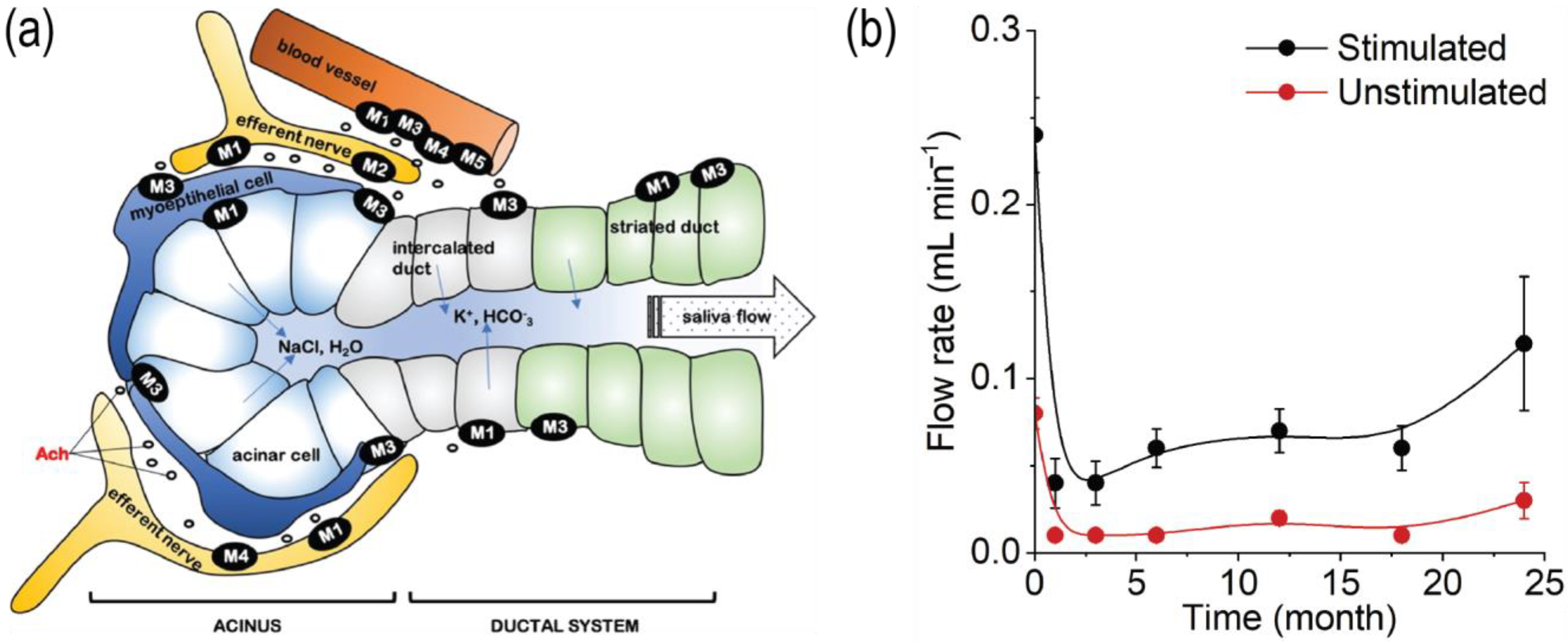
3. Mechanism of Lubrication in the Mouth

Saliva plays several important roles in the mouth, such as lubrication, food processing and perception, and microbial defense. It is composed of 99% water and 1% proteins and enzymes [50]. By minimizing contact between the soft and hard tissues of the mouth, saliva decreases any dynamic friction and prevents adherence or static friction between the soft and hard tissues [51]. Despite its high water concentration, saliva is best described as a shear-thinning non-Newtonian fluid because its viscosity decreases as shearing force on it increases [50]. The viscoelasticity of saliva can be attributed to the presence of mucins—a high molecular weight glycoprotein [50]. Mucin, which is a natural glycoprotein in tears, saliva, mucus, and synovial fluid, can attach to the surface of tissue and organs and act as a lubricant [52,53].
The ability of macromolecules to form a film on and between the hard and soft oral surfaces depends upon the strength of the attractive intermolecular forces between the macromolecule and the oral surfaces. Macromolecules must associate with the enamel surface of the teeth and with the soft tissue [54,55]. The surface enamel of teeth is largely composed of hydroxyapatite and fluorapatite, which are minerals composed of a mixture of Ca10(PO4)6(OH)2 and Ca10(PO4)6(F)2 [56,57]. The ionic nature of calcium apatites suggests that polar substances will associate with it. The surface of most of the oral soft tissue is nonkeratinized stratified squamous epithelium, but the gingiva, the dorsal surface of the tongue, the hard palate, and the retromolar pads, are keratinized stratified squamous epithelium [55]. Keratinized tissue has a layer of cornified cells facing outward [55]. This layer is rich in the protein keratin and is water repellant [58].
Mucins are composed of ~50% carbohydrates in the form of oligosaccharides—three to ten sugar molecules bound together—bonded to serine or threonine, forming the glycoprotein structure [59]. The high serine content, in part, allows the protein to form an extended structure [50]. The two types of mucins typically found in saliva are MUC1 and MUC2 [59], with MUC2 being more prominent and a gel-forming extracellular mucin [60]. MUC2 is larger and has more o-linked glycosylation domains that allow it to form branched structures (Figure 2a). The lubricating properties of mucins can be attributed to their ability to bind ions and, consequently, water. Mucins have charged residues in their structure which, upon binding certain ions like sulfates, can lead to a swollen tertiary structure due to the repulsion of these negatively charged residues and, consequently, the uptake of water [59]. This swollen tertiary structure generates a protective barrier between the mucin molecules which allows for further lubrication. Unstimulated saliva, which is mostly submandibular and sublingual saliva that are rich in mucins (Table 1), generate a coefficient of friction of 0.25 between a pig’s esophagus and tongue soft tissues [61]. Stimulated saliva, which is primarily parotid saliva that does not contain mucins (Table 1), generates a friction coefficient of 0.33 between the soft tissues [61]. While parotid saliva only makes up 20% of the whole-mouth saliva [62], it contributes to >50% of the oral saliva during periods of high saliva flow, leading to a lower mucin content during mastication and swallowing [62]. During periods of low salivary flow rate, the submandibular glands produce ~65% of the oral saliva, allowing the mucins to form protective layers on the oral tissues [59,62]. The protective layer that mucins provide is so important that the loss of mucin production is a major cause of xerostomia [59]. Without the protection of mucins during low salivary flow, patients can develop oral ulcers, caries, and infections that are not native to the oral cavity [59].
Table 1. Composition of healthy parotid, submandibular, sublingual, and minor gland saliva measured using SDS-PAGE and ELISA for qualitative and quantitative concentrations, respectively. ↑ = low, ↑↑ = high, ND = no data.
ParotidSubmandibularSublingualMinor Glands
Mucins MG1/MG20 [61,62,63]↑/↑ [61,63]↑↑/↑↑ [63]↑/0 [63]
Statherin (μM)12.8 [64]NDNDND
Amylases (U/mL)161.8 [63]15.9 [63]15.9 [63]101.4 [63]
Water99% [50,62,63]99% [50,62,63]99% [50,62,63]99% [50,62,63]
Proline-rich Proteins (mg/mL)1.7 [63]1.3 [63]1.8 [63]2.1 [63]
Cystatin S (μg/mL)0.5 [63]177 [63]28 [63]56 [63]
Lysozymes~0 [63]↑ [63]↑↑ [63]~0 [63]
Figure 2. (a) Schematic representation of the structure of mucins 1 and 2, showing the peptide backbone with a heavy oligosaccharide binding alongside various other domains in the mucin structure. MUC2 is a gel-forming mucin, whereas MUC1 is typically a membrane-bound mucin. Reproduced from reference [60] with permission. (b) A proposed structure for how statherin molecules aggregate on hydrophilic (HP) and hydrophobic (HB) surfaces. Reprint from reference [65] with permission. Note that the second layer of statherin is less dense than the first. (c) The primary structure and a proposed secondary structure of the statherin protein, giving it amphipathic properties. Reprinted from reference [65] with permission.
Lubricants 12 00126 g002
Like mucins, PRPs are found throughout the oral mucosa with the function of providing lubricity to the oral cavity [50]. Unlike mucins, these proteins, which are mostly produced in the minor glands, appear in all types of saliva in relatively similar quantities [63] (Table 1). There are over 50 different types of PRPs [50] with slightly differing structures between them, giving slightly different properties to each type. Parotid saliva appears to contain basic and acidic PRPs, whereas the submandibular and sublingual saliva appear to exclusively contain acidic PRPs [50]. Additionally, the high proline content in PRPs–similar to the serine content in mucins–allows the structure of these proteins to extend [50], providing a cushioned surface to lower the coefficient of friction between the different tissues of the mouth and provide lubrication [66]. PRPs also become part of the enamel pellicle by binding to hydroxyapatite on the tooth surface and controlling the crystallization and demineralization of calcium on the surface [50,62]. This pellicle, also composed of cystatins and statherin, helps secure and stabilize the bonding of hydroxyapatite to the tooth surface [54], maintaining the healthy enamel.
Despite being the main oral lubricant, saliva also contains various proteins for antimicrobial purposes. Cystatins are cysteine protease inhibitors which block the degrading activity of proteases from various origins including bacteria [67], protozoans [68], and endogenous [68]. Cystatin S is also known to bind lipopolysaccharide (LPS) [69], which is a surface protein and toxin found in Gram negative bacteria and is responsible for causing inflammatory responses that can destroy healthy tissues. Cystatin S is particularly found in the human saliva, with the highest concentration being found in submandibular saliva [63] (Table 1). Very little cystatin S resides in the parotid saliva [63] (Table 1). The likely reason for this is that parotid saliva is stimulated due to mastication and swallowing [63], whereas submandibular saliva is used to protect and coat the oral cavity during unstimulated resting periods [63]. There is very little reason for parotid saliva to have antimicrobial defenses because the primary function of stimulated saliva is to wet food particles to create a bolus that could be swallowed easier. It does not stay in the oral cavity for an extended period. In opposition, unstimulated saliva from the sublingual and submandibular glands is mucous saliva which coats and protects the oral tissues and must be saturated with antimicrobial proteins to protect the oral cavity during periods of inactivity. Similar to cystatin S, lysozymes are immunological proteins present in the mucosa which provide antimicrobial activity against Gram positive and Gram negative bacteria as well as some viruses and fungi [70]. A similar trend is observed with the presence of lysozymes in different types of saliva as is seen with cystatin S (Table 1), where there is little to no presence of lysozymes in the parotid saliva, yet there is a large amount in the submandibular and in particular, sublingual saliva.
Saliva contains many enzymes and proteins that aid in digestion and microbial defense; however, some proteins play an active role in oral lubrication [71]. Statherin is a calcium-binding salivary protein that sticks to the tooth enamel to prevent the teeth from damaging and chipping during mastication by lowering the friction coefficient between the teeth [65]. Statherin can do this because of its amphipathic structure, allowing it to bind to both hydrophilic and hydrophobic surfaces [65] (Figure 2b). It can also bind to itself in aggregates and form layers of statherin [65]. Each layer is less dense than the previous, allowing for compression and easy removal of layers due to shear stress [65]. The friction coefficient is reduced more on hydrophilic surfaces than hydrophobic because the hydrophobic interactions of statherin’s non-polar region—between the different layers of statherin—in the presence of a hydrophilic surface are weaker than the ionic interactions of its polar region in the presence of a hydrophobic surface, allowing for easier removal of layers [65]. Statherin’s charged regions (Figure 2c) bind to the hydroxyapatite of the enamel, providing a low coefficient of friction between the teeth themselves, and the tongue and the teeth [72]. Additionally, statherin appears to be one of the first proteins to form the enamel pellicle, leading to the further protection of the clean tooth surface [73]. Further research shows that statherin is the primary protein that can be found at the saliva–air interface, at a concentration of 7 mM [74]. Statherin can also bind most of the calcium that is found in saliva, preventing excessive buildup of calcium onto the teeth, leading to calculus [75]. An abundance of statherin allows for easy calcium exchange with the hydroxyapatite of the tooth while moderating the mineralization to the tooth surface [50,74], maintaining healthy enamel in the mouth.
The enamel pellicle—composed of many of the proteins listed in Table 1—forms within seconds of the tooth being cleaned [76]. This formation is vital to tooth health as it protects the tooth from demineralization and crystallization, while also stabilizing the binding between the enamel and the hydroxyapatite on the surface of the tooth [50,62]. While this is beneficial in the short term, the enamel pellicle can become dental plaque—a biofilm of bacteria feasting on the sugars of the pellicle—within a matter of days unless it is removed through brushing [76]. Dental plaque begins to degrade the hydroxyapatite of the tooth and damages the enamel, leading to dental caries and oral infections [76]. Once dental plaque becomes petrified due to the crystallization of calcium and mineral deposits in the old biofilm [77], it becomes hard dental calculus [78]. While dental calculus is hard, it has a value of 30–60 on the Vicker’s Hardness Scale compared to a value of 350 for solid tooth enamel [78]. Despite the generation of this hard tissue on the tooth enamel, there is no reference in the literature regarding the effect of calculus on the tooth-on-tooth coefficient of friction and the damage that this can cause.
Despite different saliva having different effects on friction in the oral cavity, most tribological experiments focus on whole-mouth saliva, a mix of all saliva in the mouth in an unstimulated condition, which is ~65% submandibular/sublingual, to evaluate the frictional forces that take place in the mouth at rest. Berg et al. used whole-mouth saliva, which has been shown to reduce the friction coefficient between the hard silica from 0.66 to 0.03 under physiologically relevant pressures [79]. Studies also show that whole-mouth saliva loses most of its elasticity and viscosity when it has been aged or centrifuged instead of being used fresh [80]. This is likely due to the separation of the heavy mucins and PRPs from the saliva mixture [80].

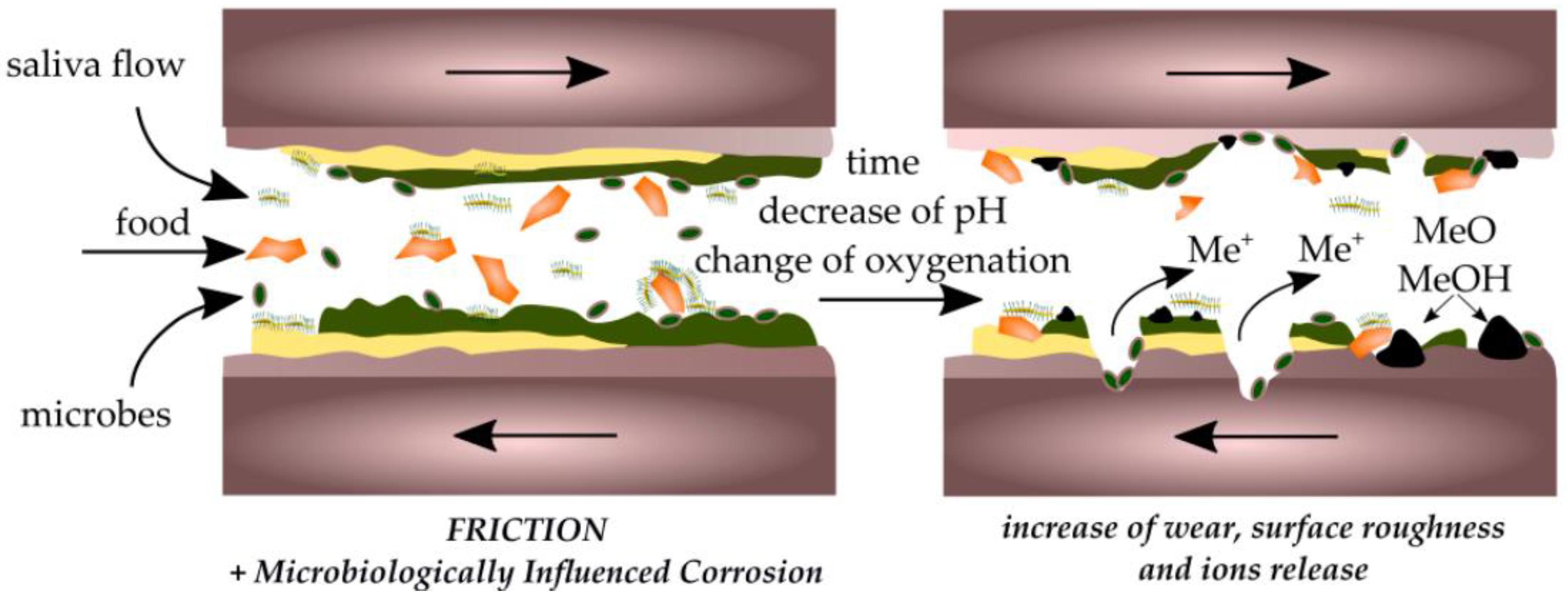
4. Effect of Biofilm and Implants on Oral Friction and Wear

Biofilms are vital for reducing the oral friction between the teeth, and between enamel and dental implants, due to their viscoelastic properties [81]. Oral biofilms are formed when foreign bacteria enter the oral cavity, adhere to, and proliferate on either teeth and/or implants [82]. The salivary pellicle formed on the tooth enamel is particularly rich in nutrients and provides a safer environment for the bacteria to grow and proliferate [82]. The bacteria can then secrete adhesive proteins like glucans [83] to become adherent to the tooth/implant surface and multiply into a biofilm. More than 700 bacteria have been identified as living inside the oral cavity [84]. In the salivary pellicle, some bacteria include the following: Streptococcus, Campylobacter, and Actinomyces [84]. Over the short term, biofilms can create a hydrated interface and lubricate biomaterials in vivo, which lead to less wear on the implant and the surrounding tissues [81]. While the biofilm can lower the coefficient of friction between biomaterials and implants, studies have shown that over the long term, biofilms can decrease an implant restoration’s resistance to corrosion or degradation, thus making the implant restoration’s surface rougher [81,85]. This leads to increased wear on the oral tissues in the presence of an implant and will increase the friction coefficient in the oral cavity [81,85]. This degradation comes from the interaction of implants with the lipopolysaccharides in Gram negative bacteria [85] and hydrogen peroxide, which is released from bacteria and leukocytes during inflammatory responses [86]. Additionally, some cariogenic biofilms can degrade resin-modified glass ionomers, and oral bacteria can release esterases, which can degrade other resin-based restorations and cements [82]. Biofilms will degrade implants and increase the coefficient of friction up to the point where the increased frictional forces tear apart the biofilm and lead to a significant increase in oral frictional forces [81] (see Figure 3). Ongoing research on dental implants studies the design of biofilm-resistant implants to ensure that implants are not degraded and that oral lubricity is maintained following installment of the implant [82]. Similar to the consequences of biofilms on the teeth surface lubricity, the salivary pellicle that these biofilms form also affect the lubricity of the oral cavity [76]. The salivary pellicle is rich in mucins, statherin, and acidic PRPs, providing plenty of lubricity for the oral cavity, yet it also contains plenty of nutrients for cariogenic biofilms to form, which eventually leads to the surface wear and diminished lubricity [76].

5. Methods of Studying Oral Friction

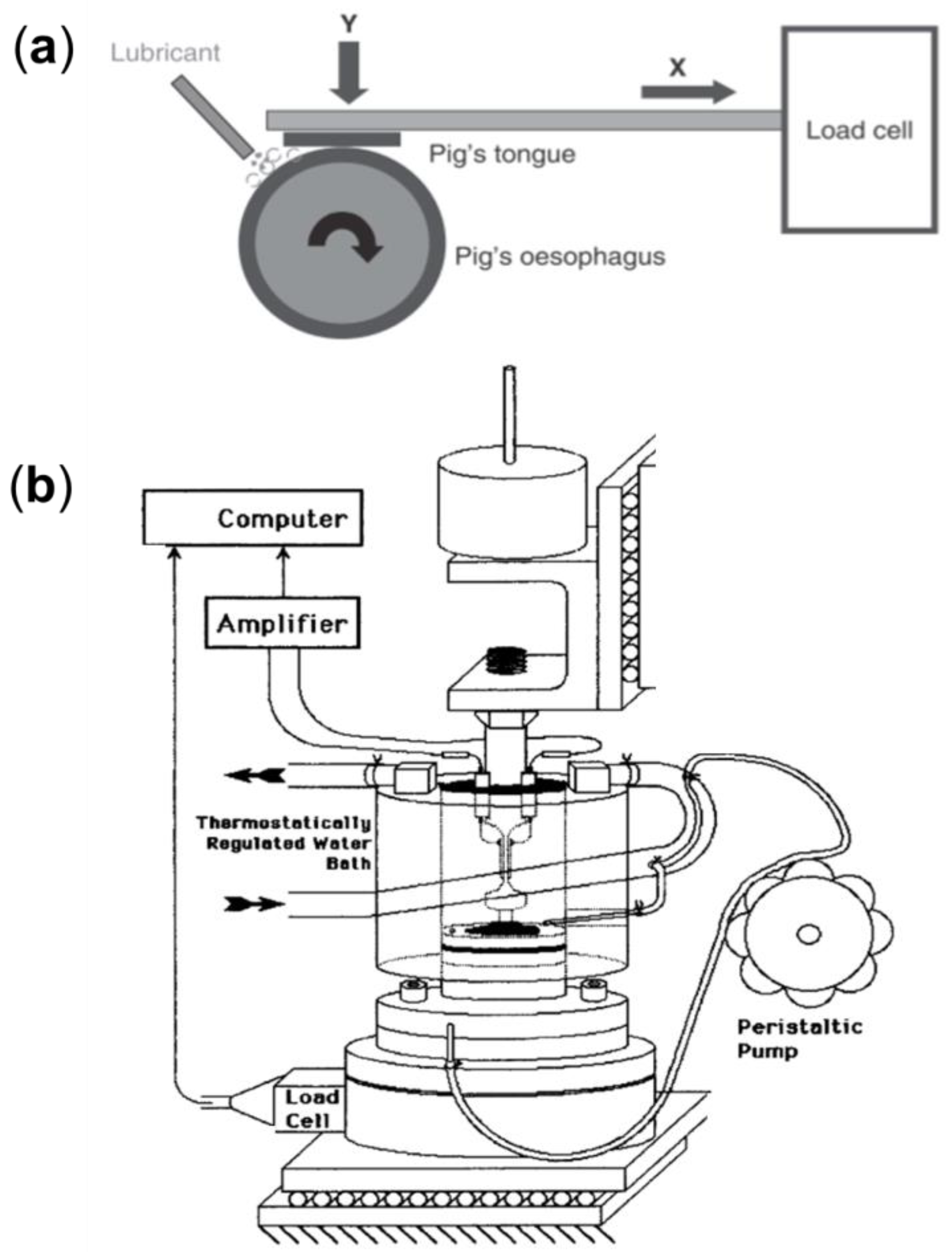
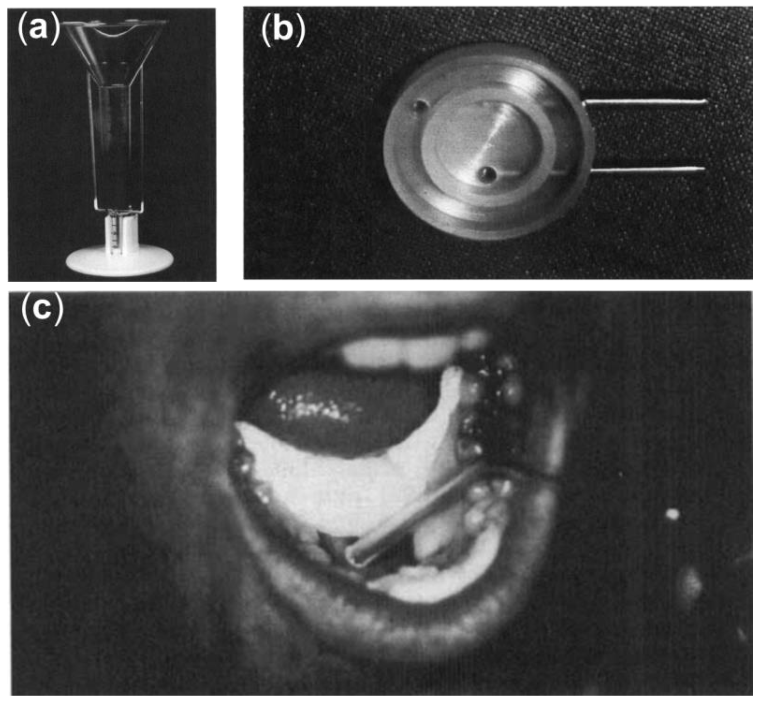
5.1. Ex Vivo Models for Studying Oral Friction

Lack of salivary lubrication leads to an increased coefficient of friction between the tongue and tooth enamel of 1.87–3.21 [87,88], compared to a value of 0.3–0.5 for tissues that are lubricated, as measured ex vivo using a tongue–enamel friction system [87,89]. This increase can induce significant discomfort and pain for patients. The role of macromolecular lubricants is to reduce the friction coefficient through various mechanisms of lubrication. To test the efficacy of these lubricants, ex vivo tribological tests and models that can replicate oral friction are necessary. Any model of oral friction must have an accurate representation of the various oral tissues and how they function, in addition to replicating the biomechanics of the entire oral cavity. Very few studies use oral biological tissues for tribological studies and instead focus on the ability of whole-mouth saliva and various saliva substitutes to reduce the friction coefficient between various hard enamel-like surfaces, such as silica discs [73,79,88], spheres [80,90], and smooth glass [90], yet the few that use biological tissues focus on silicone balls or enamel [87,88] friction models on porcine tongues (see Figure 4). The topography of the tongue has a significant influence on the lubrication of the mouth [90]. Pig tongues are often used for tribological tests because of their inherent similarities to the human tongue [90]. Despite their usefulness, these animal tissue models have some significant flaws as they decompose quickly [90], they have complex surface chemistry [91], and there is a significant variability between the surface chemistry and topography of each tongue that is used as a model [91]. Because of these issues, studies are pointing towards using synthetic materials as alternatives to biological tongue models [90,91]. For example, PDMS tongue models can easily be synthesized in the lab space, allowing researchers to have control over the surface chemistry and topography of their tongue model [91]. Furthermore, PDMS can be modified to mimic the deformability and wettability of biological tongues [91].
Due to the difficulty of measuring oral friction in vivo, most in vivo studies revolve around mouthfeel, in which patients are asked to describe the texture of a material or food in their mouth using specific adjectives that can be correlated to different levels of friction. Some standard questions regarding mouthfeel ask whether the patient sips liquids to swallow food, gets up at night for a drink, has difficulty swallowing dry food, sucks on candies to relieve dry mouth, has too much or too little saliva, and has difficulty swallowing certain foods [93].

5.2. Clinical Approaches for Studying Dry Mouth

In clinical settings, dry mouth is determined by collecting a patient’s history with hyposalivation, the medications they take, severity of symptoms, and the patient’s assessment of their salivary flow [94]. Clinicians can also perform visual and physical tests including having the patient swallow a dry biscuit without water or testing the adhesion of a dental mirror on the buccal mucosal surface [94]. The most common method for clinicians to test for xerostomia is to measure the unstimulated whole-mouth salivary flow rate [94]. Visual signs of severe xerostomia include caries around the gum line—that is the cervical line of the tooth—as well as depapillation or erythema of the dorsal surface of the tongue [94]. Clinicians may also palpate the salivary glands to assess firmness, enlargement, and tenderness to aid with diagnosis. Occasionally, the minor salivary glands may be biopsied from the inside of the lip to look for diagnostic indications of Sjögren’s syndrome [94].
There are a variety of ways to measure stimulated and unstimulated salivary flow rate to test for xerostomia [95] (see Figure 5). Measuring unstimulated salivary flow rate can involve patients consistently pouring saliva from the lip into a graduated cylinder for 15 min, spitting saliva that has accumulated every 60 s into a graduated cylinder, or aspirating saliva from the floor of the mouth continuously [96]. Stimulated salivary flow rate is measured similarly, except it is measured after having the patient chew unflavored gum for 1 min [95]. The saliva is collected for a period of five minutes [95].
A standardized test has been developed to provide clinicians with a consistent means of testing for xerostomia, called the Clinical Oral Dryness Score (CODS) [94]. This test uses a variety of qualitative tests to determine whether the patient has dry mouth and how severe it is, ranked on a scale from 1 to 10 [94]. The tests involved in the CODS include the following: dental mirror sticking to tongue and buccal mucosa, frothy saliva, no pooling saliva under the tongue, loss of papillae on tongue, altered or smooth gingival texture, “glassy” appearance to the oral mucosa as a whole, fissured tongue, active cervical caries or cervical carries restored in the past six months, and debris or food adhered to the palate [94].

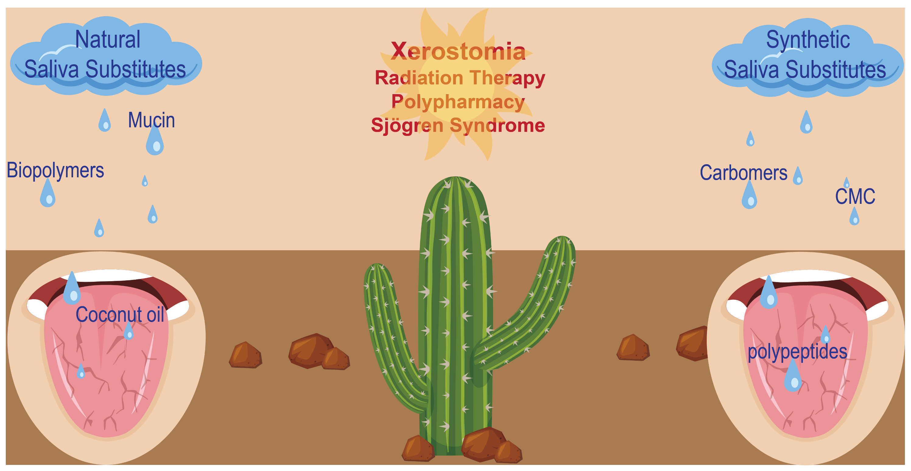
6. Lubricating Saliva Substitutes

Lubricating saliva substitutes aim to supplement patients that may be deficient in native saliva or when their saliva composition has been negatively affected. Saliva substitutes provide moisture as well as lubrication to oral surfaces and prevent damage resulting from direct solid–solid contact. This lubrication is essential for addressing the symptoms of xerostomia, such as difficulty in swallowing and speaking [10]. The ingredients of saliva substitutes have different functions. These ingredients include hydrophilic macromolecules as lubricating agents, active molecules like malic acid and tartaric acid as saliva stimulants, buffering agents like phosphate buffers for controlling pH and ionic content, as well as artificial flavoring agents [2]. Here, we describe effective lubricating macromolecules that have been used in the literature as artificial saliva, and discuss their structure, mechanism of lubrication, and potential shortcomings for long-lasting oral lubrication.

6.1. Natural Macromolecular Lubricants for the Treatment of Xerostomia

This section discusses macromolecules that are extracted from biological environments, including natural proteins, glycoproteins, lipids, and polysaccharides, and have been used in artificial saliva formulations.

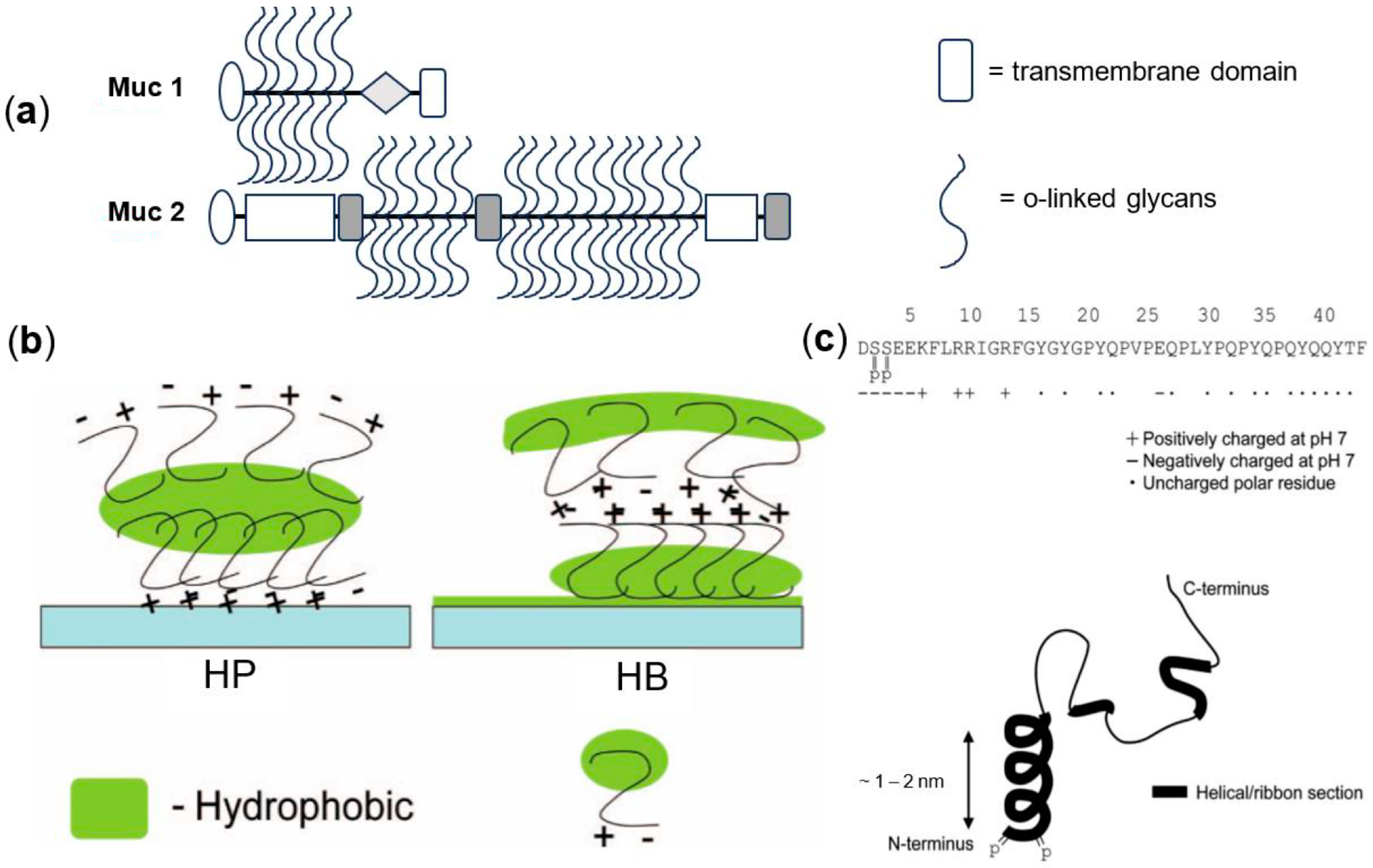
6.1.1. Lubricating Proteins and Glycoproteins

Within native saliva, mucins are the major source of lubrication [97]. Patients can be supplemented with animal-derived mucins as a saliva substitute [98]. Porcine gastric mucins or bovine submaxillary mucins are commonly used for this purpose [97,98]. These animal-derived mucins are similar in structure and function to human oral mucins and function similarly to the native saliva [98].
Vissink et al. compared artificial saliva supplements containing mucins to those containing carboxymethyl cellulose in a controlled trial over 3 years following three groups: two groups were given either carboxymethyl cellulose- (group I) or mucin- (group III) based artificial saliva, and a third group was given both types of artificial saliva for a month each and then asked to choose one to continue with (group II) [99]. If patients in group I or III complained, they were given the other solution. Out of the 61 patients in group II, 57 preferred the mucin saliva substitution after trying both the mucin and carboxymethyl cellulose solutions. Patients commented that the mucin provided longer post-application effectivity (30 min for mucins compared to 10 min for carboxymethyl cellulose) and greater comfort. Patients using the carboxymethyl cellulose complained of stickiness and caking.

6.1.2. Lubricating Lipids

Native saliva has lipid concentrations of 0.2, 0.9, and 1.3 mg/dL in the parotid, submandibular, and whole stimulated saliva, respectively [100]. Most are not membrane lipids because 96–99% of these lipids are cholesteryl esters, cholesterol, triglycerides, diglycerides, monoglycerides, and free fatty acids, rather than polar lipids (which include phospholipids) [100]. Little is known about the function of lipids within saliva or how changes to lipid production affects the oral cavity [101].
The nature-derived lipids discussed in this section are nonpolar hydrocarbon chains that interact with oral surfaces primarily through relatively weak London dispersion intermolecular forces. The ability of lipids to provide oral lubrication depends on their ability to coat oral surfaces and reduce the coefficient of friction through viscous dissipation of friction energy. This mechanism of action differentiates lipids from the other macromolecules discussed in this paper, which primarily lubricate surfaces through water retention and hydration lubrication. Longer chain lipids tend to be more viscous, allowing them to potentially stay on oral surfaces longer than short-chain lipids. In this section, we discuss rapeseed oil, coconut oil, and olive oil in the context of oral lubrication.
Rapeseed oil is a vegetable oil used as an artificial saliva substitute [102], although literature on the tribology of rapeseed oil on oral hard and soft tissues is not available. Canola oil is rapeseed oil that is low in erucic acid and glucosinolate [103]. Rapeseed oil is extracted from the seed of the Brassica genus from the Cruciferae family [103]. The low erucic acid and glucosinolate content of the Brassica plant makes them more attractive candidates because of the linkage of these substances to poor health outcomes and diseases in animals, including fat deposits in the heart, skeletal muscles, and adrenal glands, and inhibited uptake of iodine by the thyroid [103]. Like other vegetable oils, rapeseed oil contains mainly triacylglycerides (91.8–99.0%) [103]. Rapeseed oil was shown to improve xerostomia in a prospective crossover study of 120 head and neck radiotherapy patients [104]. Xerostomia was assessed using questionnaires before and during each treatment. There was no statistically significant difference between the average summative effectivity of rapeseed oil and the three other treatments tested (carboxymethylcellulose, animal mucins, and Aloe vera). Rapeseed oil performed better than carboxymethyl cellulose in the frequency of application (p-value = 0.028) and sleep quality (p-value = 0.015). It did not perform worse than carboxymethylcellulose, animal mucins, or Aloe vera in any category. However, when asked to pick their favorite macromolecule, 17.5% chose rapeseed oil, while approximately 27% chose each of the other three. This may be due to the poor taste of rapeseed oil (4.3 ± 0.15 compared to approximately 3.2 ± 0.14, on a scale of 1 as very good and 6 as poor).
Coconut oil is a lauric vegetable oil largely composed of low molecular weight saturated triacylglyceride fats [103,105]; however, coconut oil is calorie dense. It is digested easily by most people and is resistant to becoming rancid when stored [105]. Virgin coconut oil (and also coconut milk and coconut water) might be capable of demineralizing the enamel of teeth [106]. Commercial grade coconut oil is extracted from dried coconut kernels and purified [105]. Virgin coconut oil is extracted from coconut milk [105]. The lipid composition of these two forms is different [105]. Commercial grade coconut oil is more prone to experiencing microbial contamination during processing [105]. Tribological studies of coconut oil have been conducted in relation to using it as a biolubricant for machinery [107]. Although literature on the tribology of coconut oil on oral hard and soft tissues is not available, in vivo studies of coconut oil as a saliva substitute have also been conducted. In a study of 30 radiotherapy head and neck xerostomia patients, participants were asked to use coconut oil for two weeks [108]. Participants chose to administer an average of 5 mL of oil 3 times per day. The results indicated that 41.4% continued to use coconut oil after the study concluded.
Olive oil, with 99% triacylglycerides [109], is considered a home remedy for xerostomia but is also found as an ingredient within some artificial saliva substitutes [110,111]. In a crossover study on 39 patients, an artificial saliva formulation containing olive oil showed increased unstimulated whole salivary flow rates, reduced complaints of xerostomia, and improved xerostomia-associated quality of life, although no specific information was provided on the role of olive oil in this performance [110]. In addition to potentially providing lubrication, olive oil has antimicrobial and anti-inflammatory properties [111].

6.1.3. Lubricating Polysaccharides

Polysaccharides are chains of monosaccharides covalently bonded with glycosidic bonds. Polysaccharides have carbon backbones and are hydrophilic due to the presence of multiple hydroxyl groups on each monosaccharide. The hydroxyl groups hydrogen bond with each other and with water. The excellent water retention properties of polysaccharides make them attractive candidates for biolubrication [112].
Xanthan gum is an anionic [113] (at pH > 7) exopolysaccharide [114] formed from the monosaccharides glucose, mannose, and glucuronic acid [115]. It is produced commercially from the Xanthomonas campestris bacteria [116]. Xanthan gum is hydrophilic and creates viscous solutions at low concentrations [113]. The viscoelastic properties of xanthan gum dispersions resemble native saliva, which may help it approximate the feel of native saliva in speech and eating [117]. A randomized, double-blinded study compared Xialine® to a placebo (Xialine® without xanthan gum) in 30 head and neck radiotherapy xerostomia patients [117]. Xanthan gum did not reduce the symptoms of xerostomia better than the placebo except for the quality of speech, where presence of the gum made the formulation twice as effective.
Sodium hyaluronate is the sodium salt of hyaluronic acid (HA), which is a glycosaminoglycan composed of d-glucuronic acid and N-acetylglucosamine [118]. HA is a naturally occurring mucoadhesive polysaccharide found in the extracellular matrix [119], connective tissues such as cartilage [120], and human saliva [121]. Sodium hyaluronate is highly anionic, allowing it to absorb large amounts of water and provide lubricating properties [122]. HA is biocompatible, non-immunogenic, and can be degraded by the hyaluronidase enzyme found in the human body [119]. HA aqueous dispersion displays non-Newtonian fluid mechanics and similar viscosity values to stimulated and unstimulated whole saliva when tested with the shearing forces representative of those in the oral cavity [122]. In a study by Takemura et al. (2022), commercial artificial saliva in the form of a rinse, containing sodium hyaluronate as the main active ingredient, significantly increased the unstimulated salivary flow rate compared to a placebo and an artificial saliva that did not contain sodium hyaluronate. Interestingly, all the treatments tested had similar scores rating the subjective sensation of lasting intraoral moisture after 1 h of use [119]. This could be because despite HA having a similar viscoelastic profile to human saliva, it showed inferior wettability (i.e., film-forming ability) to oral surfaces compared to the whole human saliva. Thus, chemical modification of HA to increase wettability may be required to provide more effective and long-lasting xerostomia relief [122]. The molecular weight of hyaluronic acid is an important consideration in designing HA-based artificial saliva: high molecular weight hyaluronic acid has been shown to inhibit lysozyme and peroxidase antimicrobial—particularly candidacidal—activities. This inhibition of lysozyme and peroxidase activity was more pronounced when the high molecular weight hyaluronic acid was adsorbed onto hydroxyapatite ceramic beads mimicking teeth than when simply dissolved [123]. Considering that artificial saliva will likely adsorb onto the salivary conditioning film and/or intraoral structures, low molecular weight hyaluronic acid should be used in HA artificial saliva formulations. High molecular weight HA (up to 20,000 kDa) can be extracted from animal sources such as bovine cartilage, while lower molecular weight HA (1000–4000 kDa) can be extracted from bacteria or yeast [124].

6.1.4. Complex Mixtures

Naturally derived complex materials with good water retention properties are attractive candidates for oral lubrication. There are a few examples of these materials discussed in the literature for treatment of dry mouth as indicated in Table 2.
Yam tuber extract is an attractive candidate for saliva replacement because it contains mannan glycoproteins called mucilages [126]. Raw Korean yam (Dioscorea batatas) extract has been studied as a saliva replacement material. Solutions with a similar viscosity to unstimulated and stimulated saliva can be achieved through a mixture of yam extract and simulated salivary buffer solutions [131]. The yam solutions had greater wettability on resins than human saliva, supporting their potential to relieve the sensation of oral dryness. Further testing is required to assess how the tribology of yam tuber artificial saliva solutions compares to that of native saliva, and to further assess the effect of yam tubers on the biological environment of the oral cavity.
Linseed extract contains polysaccharides, glycoproteins, and proteins that mimic mucins, creating an aqueous solution with properties similar to those of native saliva [127]. This makes linseed extract a good prospect as an artificial saliva substitute. Salinum®, a market saliva substitute containing linseed extract as the active ingredient, was compared to MAS-84, a carboxymethylcellulose formulation, in a single-blind crossover study of 21 head and neck radiotherapy patients [132]. The patients used each formulation for three weeks (with a one-week break in between) and were randomly assigned to use either one first. Linseed extract required fewer applications and lasted longer than carboxymethylcellulose (60 min compared to 30 min), with statistical significance. Linseed extract also reduced the plaque index and gingival bleeding, while carboxymethylcellulose did not. Subjective measures of taste, speech, overall relief, and chewing and swallowing also showed increased improvement with linseed extract. Although Salinum® and MAS-84 differ in composition and mechanical properties, the results of this study may suggest that linseed extract is superior to carboxymethylcellulose as a saliva substitute for head and neck radiotherapy xerostomia patients.
Whole bovine milk is an aqueous colloid that comprises 3.5–5% fats (98% of which are triacylglycerol) that are emulsified by the protein surfactant casein [133,134]. If not for its sugar content and the effect of sugar at increasing dental caries, bovine milk has characteristics that are attractive for saliva substitutes [128]. Milk can buffer acids within the mouth and protect enamel by decreasing the solubility of enamel and possibly helping to remineralize the enamel [128]. A unique macromolecule within milk is the protein casein, which constitutes 20% of the solids within milk [134]. It is a molecule that can inhibit the dissolution of hydroxyapatite [135]. Caseins exist as unfolded non-globular structures in milk and form micelles [129,130]. There are no clinical studies on milk as a saliva substitute in the literature. In an oral tribology study using PDMS to represent the oral surfaces and milk as the saliva substitute, decreased coefficient of friction values were achieved by increasing the fat content of the milk and increasing thickness and viscosity by addition of xanthan gum [136]. This study also found that a larger fat droplet size is associated with a lower coefficient of friction. They hypothesized that this occurred because larger droplets can be compressed, dragged, and trailed along surfaces more easily.
Aloe vera gel is an extract from the Aloe barbadensis miller succulent plant known for its antibacterial, anti-inflammatory, and healing properties [137]. Aloe vera gel is produced by the parenchyma cells of the plant [138]. Aloe vera gel is highly hydrated (>98% water) and its gel consistency is attributed to polysaccharides [137]. More than 60% of the solid within the gel is polysaccharides [139]. Acemannan, a mannose-containing polysaccharide, has been reported as the main active substance present in an Aloe vera filet [139]. The tribology of Aloe vera has been studied in the context of applying it as a biolubricant in machinery [140]. Literature on the tribology of Aloe vera on oral hard and soft tissues is not available. Aloe vera is used commercially in artificial saliva substitutes. Aldiamed oral gel, an artificial saliva substitute that uses Aloe vera gel as its major active ingredient, was compared to carboxymethylcellulose, rapeseed oil, and animal mucins in a prospective crossover study of 120 radiotherapy (head and neck) patients [104]. Xerostomia was assessed with a questionnaire before and during each treatment. The average additive scores indicated, with statistical significance, that the four treatments improved xerostomia. However, there was no significant statistical difference between the average additive scores of the four treatments. Instead of one solution working best, each had unique properties most helpful to patients in different contexts. Aloe vera required the lowest frequency of use. This result had statistical significance against baseline xerostomia (p < 0.0001), carboxymethylcellulose (p < 0.0001), and mucins (p = 0.014). Aloe vera gel also improved sleep quality the most. This result had statistical significance against baseline xerostomia (p < 0.0001) and carboxymethylcellulose (p = 0.006).

6.2. Synthetic Macromolecular Mouth Lubricants

This section highlights the application of synthetic macromolecules or synthetically modified natural materials, including synthetic polymers, chemically modified biomacromolecules and synthetic polypeptides, as mouth lubricants.

6.2.1. Chemically Modified Biomacromolecules

Carboxymethyl cellulose is a polymer mainly used in artificial saliva as an agent with thickening, lubricating, mucoadhesive, and film-forming properties. Carboxymethyl cellulose has hydrophilic groups which can absorb a significant amount of water and form hydrogels. Also, this polyanionic polymer has mucoadhesive properties that can be used for transmucosal applications [141]. Sarideechaigul et al. investigated the effect of two combinations of artificial saliva containing 0.1% pilocarpine, one with sodium carboxymethylcellulose (SCMC), the other with sodium polyacrylate (SPA). They conducted a double-blinded clinical assessment for six weeks of xerostomia treatment on 31 patients, using the Clinical Oral Dryness Score (CODS) and Xerostomia Inventory (XI). Overall, both the SCMC and SPA formulas improved hyposalivation to normal saliva flow rates, which is 0.3–0.4 mL min−1. However, in the SCMC-treated group, the unstimulated and stimulated whole salivary flow rates were greater, and CODS were considerably lower, which indicates better performance of SCMC-based artificial saliva over its SPA-based counterpart [142].
Chitosan is a cationic mucoadhesive polymer capable of forming strong electrostatic interactions and hydrogen bonding with many other macromolecules [143]. It can be modified with catechol to boost its water solubility and its affinity to glycoprotein [144]. Mucoadhesion takes place in two steps where at the contact step, the polymer contacts the mucosa, then at consolidation step, the polymer has to react with the mucosa by forming mucoadhesive bonds [145]. Wan et al. [87] obtained various degrees of catechol conjugation on chitosan (with 7.6%, 14.5%, and 22.4% catechol conjugation to the backbone of the polymer) as a mucoadhesive with a layered structure, including a rigid bottom and a soft secondary salivary conditioning film (S-SCF). They collected healthy and diseased simulated whole saliva (SWS) from Sjögren’s syndrome patients and flowed them over a Quartz Crystal Microbalance with Dissipation (QCM-D) sensor followed by the flow of Chi-C and reflow of saliva to analyze the formation kinetics of SCFs. A larger frequency shift showed that after the reflow of saliva, more salivary proteins adsorbed on the sensor in the chitosan–catechol (Chi-C) samples with higher catechol conjugation. Also, the ratio of dissipation to frequency shifts demonstrated that the structural softness was higher with higher catechol conjugation.
Adamczak et al. investigated the water adsorption capabilities of polymer-coated liposomes, made of low-methoxylated pectin (LM-pectin), high-methoxylated pectin (HM-pectin), alginate, chitosan, and hydrophobically modified ethyl hydroxyethyl cellulose (HM-EHEC), using dynamic water sorption measurements (DVS). The findings revealed that alginate-coated liposomes absorbed the most water, which prolonged the moisture protection of oral surface. Chitosan-coated liposomes had the highest water sorption capacity with a high mucoadhesive property [146]. Hiorth et al. studied the oral lubricating properties of positively, negatively, and neutrally charged polymer-coated liposomes using a ball-on-disc tribometer at 2 N load at 37 °C. This study revealed that the positively charged formulation of the chitosan-coated liposomes gave rise to better lubrication properties (lower friction coefficient, μ < 0.1) at orally relevant shearing speeds (~50 mm/s) than the negatively and neutrally charged polymer-coated liposomes [147].
Phosphatidylcholine (PC) is the zwitterionic head group in some phospholipids such as lecithin [148], which is a class of essential phospholipids that make up a significant portion of the phospholipid mass in eukaryotic cell membranes. Lecithin is an amphipathic phospholipid, comprising a hydrophilic zwitterionic group headgroup and two hydrophobic fatty acid tails [149]. PC is found in all human cells [149] and many other natural sources, such as eggs [148], soybeans, and sunflowers [150]. PC is also abundant in lipoproteins, biliary lipid aggregates, and lung surfactants [151]. Phospholipids usually self-assemble into bilayers due to a hydrophobic effect, when the aggregated phase is more energetically favorable [152]. Macromolecules containing PCs have excellent hydration capabilities and exhibit hydration lubrication [14,153,154]. The zwitterionic headgroup forms an abundance of strong hydrogen bonds with water molecules while retaining the ability to rapidly relax when exposed to shearing forces. Thus, the hydration of the zwitterionic groups is exceptionally stable while subjected to large stress [155].
An important consideration when selecting phospholipids for lubrication applications is the lipid bilayer phase behavior. A lipid bilayer’s solid/gel to liquid phase transition is dependent on the temperature and on the diacyl chain length of the phospholipid. Phospholipids with longer acyl chains are much more resistant to increases in temperature before transitioning from the solid/gel phase to liquid phase. A study investigating the effect of the bilayer phase on lubrication properties of the PC vesicles explored the tribological and mechanical properties of dipalmitoyl phosphatidylcholine (DPPC) and 1,2-dioleoyl-sn-glycero-3-phosphatidylcholine (DOPC). DPPC is a solid/gel phase phospholipid at the physiological temperature with a phase transition temperature of 42 °C, while DOPC is a liquid phase phospholipid with a phase transition temperature of −19 °C. The tribological properties of DPPC and DOPC were measured using a tribometer. DPPC and DOPC bilayers were subject to approximately 180 back-and-forth cycles of 6 mm displacement over the course of 1 h. The coefficient of friction of DPPC bilayers was 0.002 ± 0.0008 and did not change over time. The DOPC coefficient of friction was initially 0.01 ± 0.005 and after 1 h of friction, the friction DOPC friction coefficient increased to 0.05 ± 0.02. The DOPC bilayers were always deformed after being subjected to 1 h of frictional force, as visualized using fluorescence microscopy. The mechanical resistance to puncture and deformation was tested using AFM force spectroscopy. Force versus Z-piezo displacement distance curves were recorded in response to hundreds of approach–retraction cycles. The DPPC bilayers were not punctured by the AFM and remained mechanically stable in the range of normal forces tested (0–20 nN). The DOPC bilayers, however, were always deformed during friction tests. Additionally, DPPC showed high packing density compared to DOPC, which is also associated with increased resistance [156] to indentation and lower coefficients of friction [155]. Thus, solid phase phospholipids were much more resistant to indentation and deformation than liquid phase bilayers. Even though both DPPC and DOPC bilayers have significantly lower coefficients of friction than saliva, DPPC liposomes would likely be more favorable in artificial saliva formulations because they are more densely packed and because their lubricity does not diminish over time due to their enhanced mechanical stability.
A challenge with using DPPC and other phospholipids is that they are not mucoadhesive on their own [157] due to the phospholipids having a weak negative charge at physiological pH, thus it is unlikely to be involved in strong electrostatic attraction with mucins in the oral mucosa [158]. A possible solution to increase mucoadhesion and adsorption on the oral mucosa would be to add positively charged functional groups to some of the liposome PC heads. Because solid/gel phase PCs have a much lower coefficient of static friction than human saliva, some addition of positively charged functional groups would likely not compromise the intraliposomal water layer crucial to effective lubrication. For example, liposomes made of DPPC and dicetyl phosphate (DCP) in a molar ratio of 8:2 were coated with either chitosan, polyvinyl alcohol with a long alkyl chain, or polyacrylic acid (i.e., carbomer) bearing cholesterols, and all showed enhanced mucoadhesion to rat intestinal cells. Liposomes coated with chitosan showed the highest mucoadhesive percentage confirmed through fluorescence microscopy [157].

6.2.2. Synthetic Polymers

For lubricating and protecting the oral cavity, a saliva substitute must be maintained for a long period of time. This could be achieved using a bioadhesive polymer in the saliva substitute. Some highly hydrophilic mucoadhesive polymers are anionic such as carboxymethyl cellulose (CMC) and polyacrylic acid (PAA), which are rich in carboxylic moiety (-COOH) and act as moisturizing agent because of hydrogen bonding with mucosal surfaces. Cationic mucoadhesive polymers, such as chitosan and cationic hydroxyethyl cellulose, however, interact electrostatically with residual anionic mucin in the mucus layer. Other non-ionic polymers, such as polyethylene glycol (PEG) and methyl cellulose (MC), could be used as mucoadhesive agents mainly through hydrogen bonding. Novel mucoadhesive polymers such as thiolated polymers react with mucus by thiol–disulfide exchange, resulting in strong mucoadhesion with covalent disulfide bridges with the mucus layer [159]. Incorporation of these highly hydrophilic polymers in the oral cavity may result in long-lasting hydration and lubrication of oral surfaces.
By immobilization of polyethylene glycol (PEG) to the oral epithelial cells, a salivary film could be mimicked to provide lasting relief from the symptoms of mucosal dryness for patients suffering from xerostomia [160]. In a study conducted by Blakeley et al., a sugar-binding lectin, wheat germ agglutinin, was used to enhance PEG adhesion to cells, and the ability of wheat germ agglutinin-coated PEG (WGA-PEG) to reduce oral friction and improve dry mouth was studied by an ex vivo oral tissue tribology rig where the bovine enamel section rubbed against a porcine tongue. Both forms of functionalized PEG and free PEG provided lasting hydration to the tissue when trapped between the enamel and tongue, resulting in sustainable lubrication. However, in cases where there are more complex oral states, such as swallowing or drinking, the free PEG may be removed easily. Therefore, it was anticipated that the WGA functionalized PEG would provide longer relief than PEG due to its improved bioadhesion [160]. Moreover, Xiaoyan et al. investigated the water adsorption and structural properties of the mixture of mucin and thiolated PEG-SH with QCM-D. Their results suggested that thiolated PEG was more efficient for lubrication behavior compared to mucin [161].
Carbomers are synthetic mucoadhesive PAA polymers. Carbomers are available in different grades, with the grade depending on the conjugation with different functional groups, such as allyl sucrose and allyl pentaerythritol. These functional groups account for 0.75–2% of the molecular weight. Carbomers also contain different amounts of carboxylic acid groups, usually making up between 56% and 68% of the molecular weight [162]. Carbomers exhibit excellent swelling due to the highly hydrophilic carboxylic acid groups hydrogen bonding with water. Hydrated carbomers exhibit different water absorption capacities and different viscosities under different pH conditions. Initially, hydrated carbomers are acidic, but at the physiological pH of the mouth (6.2–7.6) [163], the carbomers become partially deprotonated and highly hydrophilic due to the COO- groups, thus absorbing large amounts of water [164]. Furthermore, during this neutralization process, carbomer chains become ionized and the resulting internal repulsion causes the carbomer chains to uncoil [165] and form a bulk network, which increases the viscosity of the gel [166]. Moreover, carbomers are highly mucoadhesive because the carboxyl groups form hydrogen bonds with the oligosaccharide chains on mucins [167]. In a 2009 study by Mehravaran et al. investigating the rheological behavior and mucoadhesiveness of artificial saliva pump spray formulations, their carbomer-containing artificial saliva formulations exhibited enhanced viscosity compared to other artificial cellulose-based saliva formulations. Moreover, when carbomers 971 and 940 were tested against shearing rates similar to those in the oral cavity, the carbomer-based formulations exhibited pseudoplastic viscosity behavior similar to natural saliva and a lower viscosity reduction compared to natural saliva, indicating a longer duration of action due to its higher resistance against being dislodged from the oral cavity. All artificial saliva formulations containing carbomers tested in the Mehravran et al. study showed higher mucoadhesive strength when compared to natural saliva, again indicating their potential for longer durations of action [168]. The findings of Mehravran et al. (2009) are consistent with that of Vinke et al. (2020), which showed that saliva substitutes containing carbomers adsorbed to the SCF and increased the relief period [168,169]. Interestingly, despite showing considerable mucoadhesion and adsorption to the SCF, carbomers on their own did not increase structural softness of the S-SCF; however, carbomers and the cellulosic polymer hydroxyethyl cellulose (HEC) together displayed synergy and were highly mucoadhesive and lubricating [169]. Thus, carbomers may be especially effective in artificial saliva formulations when combined with other highly hydrophilic materials. Some commercial saliva substitutes that contain carbomers—listed as polyacrylic acid in the ingredients—are Biotene and BioXtra [169], which have been shown to alleviate xerostomia symptoms during the day and improve speech in a clinical study. However, swallowing was not improved in these studies by using these products [170]. Furthermore, carbomer-based pump spray formulations were more easily and uniformly applicable to a glass tile meant to simulate the buccal mucosa compared to other formulations [168].

6.2.3. Synthetic Polypeptides

Recently, some hydrogels have been proposed as novel candidates for artificial saliva. However, they need to mimic saliva more closely from chemical and structural perspectives, which has led to the use of peptides and other bioactive ingredients in their structure [94]. Recombinant supercharged, unfolded polypeptides (SUPs) with elastin-like motif are a promising class of proteinaceous materials that could restore biolubrication in polyelectrolytes. Veeregowda et al. investigated the effect of positively charged SUPs on the lubrication and structure of the salivary conditioning film after adsorption of the polypeptides and exposure to saliva [17]. They demonstrated that if the number of positive charges is sufficiently high on cationic SUPs, they can adsorb on the SCFs to recruit glycosylated mucins from the saliva. These hydrated and rigid films improve lubrication and maintain the film’s structural integrity upon high contact pressures.
Wan et al. conducted biophysical and in vitro friction measurements to investigate the effect of SUPs with the repetitive motif K (GVGKP) on the improvement of oral lubrication with saliva from patients with xerostomia, by collecting saliva from patients with Sjögren’s syndrome. Five different SUPs, including K72, K108, and K144, and two cysteine-modified K108cys and K144cys were investigated in this research. The QCM-D was used to analyze the structural softness and formation kinetics of SCF for each sample. Through this QCM-D analysis of these five variants of SUP and a buffer, they realized that after adsorption to the surface, both K108cys and K144cys had higher structural softness due to the adsorption of more salivary glycoproteins. Using samples from four healthy volunteers and four dry mouth patients with Sjogren’s disease, the ex vivo assessment of K108cys in terms of salivary lubrication was carried out using a customized tongue–enamel friction device. The study involved sliding enamel against the tongue for 2.5 s and calculating the COF. Saliva was introduced to create the initial SCF, followed by K108cys or buffer solution for four cycles. The relief period was monitored until the COF increased, marking the relief period. The relief period for patients with S-SCF increased from 15 ± 2.5 (lowest) to 30 ± 3.6 (highest) min with intermediate exposure to K108cys. In fact, the S-SCF treated with K108cys assisted in the retention of water on the surface and provided high lubricity for a longer length of time by producing a soft layer on top of a slightly stiff charge-stabilized layer (Figure 6). The findings showed that an intermediate layer of K108cys induced electrostatic stabilization of SCFs, which is associated with strong water retention, resulting in a prolonged relief time for both healthy and patient saliva [171]. Table 3 summarizes all the saliva substitute macromolecules discussed in this review.

7. Summary and Outlook

This review provides an overview of the literature on xerostomia. Xerostomia is a medical condition that results from lack of or ineffective oral lubrication. Various medical conditions that result in xerostomia were reviewed here in detail, with polypharmacy, radiation therapy, and Sjögren’s syndrome being among the most common causes of dry mouth. Native saliva was reviewed as the primary lubricating medium in the oral cavity. The composition of saliva and its active macromolecules in biolubrication were discussed in detail under stimulated, unstimulated, and pathogenic conditions. Methods of evaluation of oral lubrication and xerostomia were examined both in clinical settings and ex vivo simulated conditions, where a significant shortcoming in the literature can be recognized in correlating the ex vivo and clinical studies. This review also provided an overview of the various natural and synthetic macromolecules that have been identified as potential ingredients for the preparation of artificial saliva formulations for xerostomia treatment. These macromolecules, including proteins, glycoproteins, biopolymers, synthetic polymers, lipids, and phospholipids, play a primary role in oral lubrication, with most of them functioning by improving the retention of water at the oral surface. The performance of these macromolecules was typically assessed in clinical studies where qualitative parameters, such as mouth feel and comfort, determine the outcome, although a limited number of mechanistic studies also investigated their mechanisms of actions in precise fundamental ex vivo experiments.
Developing saliva substitutes for patients suffering from xerostomia remains a significant challenge as performance metrics, such as the duration of action and effective lubrication, must be in concert with subjective qualities, such as mouth feel and taste. Identifying an ex vivo model that can assess artificial saliva performance and provide outcomes that correlate with patient reports will be valuable for effectively testing the performance of new formulations without the need for early clinical studies. Although tongue–enamel tribological systems provide such a platform to some extent, identifying the appropriate conditions for humidity, duration of friction, evaporation, tribopair geometries, and force in a way that replicate the oral conditions of xerostomia patients is important for developing an effective ex vivo model. Validating the model with clinical studies is imperative in achieving such effective ex vivo models. The field of biolubrication has advanced significantly over the past two decades as our understanding about the role of water retention for boundary lubrication has improved, and novel natural and synthetic materials have been introduced for biolubrication based on this improved knowledge [14,112,172,173]. The use of such materials for oral lubrication, however, has not been evaluated, perhaps because of a low number of research groups that are active in studying the fundamental aspects of oral lubrication. With the current pattern of population aging, increased medication use, and cancer incidents, xerostomia is a medical problem that will only get aggravated over the next few decades, and thus demands more attention from scientists to discover new materials that can alleviate the symptoms of this condition and help improve patients’ quality of life.

Author Contributions

All authors contributed to writing, reviewing and editing the manuscript. All authors have read and agreed to the published version of the manuscript.

Funding

This research received no external funding.

Data Availability Statement

No new data were created or analyzed in this study. Data sharing is not applicable to this article.

Conflicts of Interest

The authors declare no conflict of interest.

References

  1. Kim, Y.J. Xerostomia and Its Cellular Targets. Int. J. Mol. Sci. 2023, 24, 5358. [Google Scholar] [CrossRef] [PubMed]
  2. Hu, J.; Andablo-Reyes, E.; Mighell, A.; Pavitt, S.; Sarkar, A. Dry Mouth Diagnosis and Saliva Substitutes—A Review from a Textural Perspective. J. Texture Stud. 2021, 52, 141–156. [Google Scholar] [CrossRef]
  3. Bhayani, M.K.; Lai, S.Y. Xerostomia. In Gland-Preserving Salivary Surgery; Springer: Berlin/Heidelberg, Germany, 2017; pp. 175–183. [Google Scholar] [CrossRef]
  4. Tanasiewicz, M.; Hildebrandt, T.; Obersztyn, I. Xerostomia of Various Etiologies: A Review of the Literature. Adv. Clin. Exp. Med. 2016, 25, 199–206. [Google Scholar] [CrossRef] [PubMed]
  5. Porter, S.R.; Scully, C.; Hegarty, A.M. An Update of the Etiology and Management of Xerostomia. Oral Surg. Oral Med. Oral Pathol. Oral Radiol. Endod. 2004, 97, 28–46. [Google Scholar]
  6. Sarkar, A.; Xu, F.; Lee, S. Human Saliva and Model Saliva at Bulk to Adsorbed Phases—Similarities and Differences. Adv. Colloid Interface Sci. 2019, 273, 102034. [Google Scholar]
  7. Ozdemir, T.; Fowler, E.W.; Hao, Y.; Ravikrishnan, A.; Harrington, D.A.; Witt, R.L.; Farach-Carson, M.C.; Pradhan-Bhatt, S.; Jia, X. Biomaterials-Based Strategies for Salivary Gland Tissue Regeneration. Biomater. Sci. 2016, 4, 592–604. [Google Scholar] [CrossRef]
  8. Gittings, S.; Turnbull, N.; Henry, B.; Roberts, C.J.; Gershkovich, P. Characterisation of Human Saliva as a Platform for Oral Dissolution Medium Development. Eur. J. Pharm. Biopharm. 2015, 91, 16–24. [Google Scholar] [CrossRef]
  9. Carpenter, G. Artificial Salivas. Clin. Dent. Rev. 2018, 2, 24. [Google Scholar] [CrossRef]
  10. Guggenheimer, J.; Moore, P.A. Xerostomia: Etiology, Recognition, and Treatment. J. Am. Dent. Assoc. 2003, 134, 61–69. [Google Scholar] [CrossRef]
  11. Iorgulescu, G. Saliva between Normal and Pathological. Important Factors in Determining Systemic and Oral Health. J. Med. Life 2009, 2, 303–307. [Google Scholar]
  12. Malicka, B.; A-F, U.K.; Skośkiewicz-Malinowska, K. Prevalence of Xerostomia and the Salivary Flow Rate in Diabetic Patients. Adv. Clin. Exp. Med. 2014, 23, 225–233. [Google Scholar] [CrossRef] [PubMed]
  13. Edgar, W.M. Saliva: Its Secretion, Composition and Functions. Br. Dent. J. 1992, 172, 305–312. [Google Scholar] [CrossRef] [PubMed]
  14. Adibnia, V.; Mirbagheri, M.; Faivre, J.; Robert, J.; Lee, J.; Matyjaszewski, K.; Lee, D.W.; Banquy, X. Bioinspired Polymers for Lubrication and Wear Resistance. Prog. Polym. Sci. 2020, 110, 101298. [Google Scholar] [CrossRef]
  15. Ranc, H.; Elkhyat, A.; Servais, C.; Mac-Mary, S.; Launay, B.; Humbert, P. Friction Coefficient and Wettability of Oral Mucosal Tissue: Changes Induced by a Salivary Layer. Colloids Surfaces A Physicochem. Eng. Asp. 2006, 276, 155–161. [Google Scholar] [CrossRef]
  16. Kagami, H.; Wang, S.; Hai, B. Restoring the Function of Salivary Glands. Oral Dis. 2008, 14, 15–24. [Google Scholar] [CrossRef] [PubMed]
  17. Veeregowda, D.H.; Kolbe, A.; Van Der Mei, H.C.; Busscher, H.J.; Herrmann, A.; Sharma, P.K. Recombinant Supercharged Polypeptides Restore and Improve Biolubrication. Adv. Mater. 2013, 25, 3426–3431. [Google Scholar] [CrossRef] [PubMed]
  18. Furness, S.; Worthington, H.V.; Bryan, G.; Birchenough, S.; McMillan, R. Interventions for the Management of Dry Mouth: Topical Therapies. Cochrane Database Syst. Rev. 2011, CD008934. [Google Scholar] [CrossRef] [PubMed]
  19. Satoh-Kuriwada, S.; Iikubo, M.; Shoji, N.; Sakamoto, M.; Sasano, T. Diagnostic Performance of Labial Minor Salivary Gland Flow Measurement for Assessment of Xerostomia. Arch. Oral Biol. 2012, 57, 1121. [Google Scholar] [CrossRef] [PubMed]
  20. Iwasaki, M.; Borgnakke, W.S.; Yoshihara, A.; Ito, K.; Ogawa, H.; Nohno, K.; Sato, M.; Minagawa, K.; Ansai, T. Hyposalivation and 10-Year All-Cause Mortality in an Elderly Japanese Population. Gerodontology 2018, 35, 87–94. [Google Scholar] [CrossRef]
  21. Åstrøm, A.N.; Lie, S.A.; Ekback, G.; Gülcan, F.; Ordell, S. Self-Reported Dry Mouth among Ageing People: A Longitudinal, Cross-National Study. Eur. J. Oral Sci. 2019, 127, 130–138. [Google Scholar] [CrossRef]
  22. Marcott, S.; Dewan, K.; Kwan, M.; Baik, F.; Lee, Y.; Sirjani, D. Where Dysphagia Begins: Polypharmacy and Xerostomia. Fed. Pract. 2020, 37, 234–241. [Google Scholar] [PubMed]
  23. Arany, S.; Kopycka-Kedzierawski, D.T.; Caprio, T.V.; Watson, G.E. Anticholinergic Medication: Related Dry Mouth and Effects on the Salivary Glands. Oral Surg. Oral Med. Oral Pathol. Oral Radiol. 2021, 132, 662–670. [Google Scholar] [CrossRef] [PubMed]
  24. Khan, A.; Gilani, A.H. Antispasmodic and Bronchodilator Activities of Artemisia Vulgaris Are Mediated through Dual Blockade of Muscarinic Receptors and Calcium Influx. J. Ethnopharmacol. 2009, 126, 480–486. [Google Scholar] [CrossRef] [PubMed]
  25. Tardy, M.; Dold, M.; Engel, R.; Leucht, S. Flupenthixol versus Low-Potency First-Generation Antipsychotic Drugs for Schizophrenia. Cochrane Database Syst. Rev. 2014, CD009227. [Google Scholar] [CrossRef] [PubMed]
  26. Kumar, N.N.; Panchaksharappa, M.G.; Annigeri, R.G. Modified Schirmer Test-a Screening Tool for Xerostomia among Subjects on Antidepressants. Arch. Oral Biol. 2014, 59, 829–834. [Google Scholar] [CrossRef] [PubMed]
  27. Habbab, K.M.; Moles, D.R.; Porter, S.R. Potential Oral Manifestations of Cardiovascular Drugs. Oral Dis. 2010, 16, 769–773. [Google Scholar] [CrossRef] [PubMed]
  28. Mohandoss, A.A.; Thavarajah, R. Salivary Flow Alteration in Patients Undergoing Treatment for Schizophrenia: Disease-Drug-Target Gene/Protein Association Study for Side-Effects. J. Oral Biol. Craniofacial Res. 2019, 9, 286–293. [Google Scholar] [CrossRef] [PubMed]
  29. Murdoch-Kinch, C.A.; Kim, H.M.; Vineberg, K.A.; Ship, J.A.; Eisbruch, A. Dose-Effect Relationships for the Submandibular Salivary Glands and Implications for Their Sparing by Intensity Modulated Radiotherapy. Int. J. Radiat. Oncol. Biol. Phys. 2008, 72, 373–382. [Google Scholar] [CrossRef]
  30. Stroup, T.S.; Gray, N. Management of Common Adverse Effects of Antipsychotic Medications. World Psychiatry 2018, 17, 341–356. [Google Scholar] [CrossRef]
  31. Fortuna, G.; Whitmire, S.; Sullivan, K.; Alajbeg, I.; Andabak-Rogulj, A.; Pedersen, A.M.; Vissink, A.; di Fede, O.; Aria, M.; Jager, D.J.; et al. Impact of Medications on Salivary Flow Rate in Patients with Xerostomia: A Retrospective Study by the Xeromeds Consortium. Clin. Oral Investig. 2023, 27, 235–248. [Google Scholar] [CrossRef]
  32. Hunter, K.D.; Wilson, W.S. The Effects of Antidepressant Drugs on Salivary Flow and Content of Sodium and Potassium Ions in Human Parotid Saliva. Arch. Oral Biol. 1995, 40, 983–989. [Google Scholar] [CrossRef] [PubMed]
  33. Nederfors, T.; Dahlöf, C. Effects on Salivary Flow Rate and Composition of Withdrawal of and Re-Exposure to the Β1 -Selective Antagonist Metoprolol in a Hypertensive Patient Population. Eur. J. Oral Sci. 1996, 104, 262–268. [Google Scholar] [CrossRef] [PubMed]
  34. Villa, A.; Wolff, A.; Narayana, N.; Aframian, D.; Vissink, A.; Ekström, J.; Proctor, G.; McGowan, R.; Narayana, N.; Aliko, A. World Workshop on Oral Medicine VI: A Systematic Review of Medication-Induced Salivary Gland Dysfunction: Prevalence, Diagnosis, and Treatment. Clin. Oral Investig. 2016, 19, 365–382. [Google Scholar] [CrossRef] [PubMed]
  35. Wilberg, P.; Hjermstad, M.J.; Ottesen, S.; Herlofson, B.B. Chemotherapy-Associated Oral Sequelae in Patients With Cancers Outside the Head and Neck Region. J. Pain Symptom Manag. 2014, 48, 1060–1069. [Google Scholar] [CrossRef] [PubMed]
  36. Main, B.E.; Calman, K.C.; Ferguson, M.M.; Kaye, S.B.; MacFarlane, T.W.; Mairs, R.J.; Samaranayake, L.P.; Willos, J.; Welsh, J. The Effect of Cytotoxic Therapy on Saliva and Oral Flora. Oral Surg. Oral Med. Oral Pathol. Oral Radiol. 1984, 58, 545–548. [Google Scholar] [CrossRef] [PubMed]
  37. Cardoso, R.C.; Qazali, A.; Zaveri, J.; Chambers, M.S.; Gunn, G.B.; Fuller, C.D.; Lai, S.Y.; Mott, F.E.; Hutcheson, K.A. Self-Reported Oral Morbidities in Long-Term Oropharyngeal Cancer Survivors: A Cross-Sectional Survey of 906 Survivors. Oral Oncolology 2018, 84, 88–94. [Google Scholar] [CrossRef] [PubMed]
  38. Jensen, S.B.; Vissink, A.; Limesand, K.H.; Reyland, M.E. Salivary Gland Hypofunction and Xerostomia in Head and Neck Radiation Patients. J. Natl. Cancer Inst. Monogr. 2019, 2019, 95–107. [Google Scholar] [CrossRef] [PubMed]
  39. Jasmer, K.J.; Gilman, K.E.; Muñoz Forti, K.; Weisman, G.A.; Limesand, K.H. Radiation-Induced Salivary Gland Dysfunction: Mechanisms, Therapeutics and Future Directions. J. Clin. Med. 2020, 9, 4095. [Google Scholar] [CrossRef] [PubMed]
  40. Vissink, A.; Mitchell, J.B.; Baum, B.J.; Limesand, K.H.; Jensen, S.B.; Fox, P.C.; Elting, L.S.; Langendijk, J.A.; Coppes, R.P.; Reyland, M.E. Clinical Management of Salivary Gland Hypofunction and Xerostomia in Head and Neck Cancer Patients: Successes and Barriers. Int. J. Radiat. Oncol. Biol. Phys. 2010, 78, 983–991. [Google Scholar] [CrossRef]
  41. Beetz, I.; Schilstra, C.; Visink, A.; van der Schaaf, A.; Bijl, H.P.; van der Laan, B.F.; Steenbakkers, R.J.H.M.; Langendijk, J.A. Role of Minor Salivary Glands in Developing Patient-Rated Xerostomia and Sticky Saliva during Day and Night. Radiother. Oncol. 2013, 1092, 311–316. [Google Scholar] [CrossRef]
  42. Winter, S.C.; Cassell, O.; Corbridge, R.J.; Goodacre, T.; Cox, G.J. Quality of Life Following Resection, Free Flap Reconstruction and Postoperative External Beam Radiotherapy for Squamous Cell Carcinoma of the Base of Tongue. Clin. Otolaryngol. Allied Sci. 2004, 29, 274–278. [Google Scholar] [CrossRef] [PubMed]
  43. Shiboski, C.H.; Shiboski, S.C.; Seror, R.; Criswell, L.A.; Labetoulle, M.; Lietman, T.M.; Rasmussen, A.; Scofield, H.; Vitali, C.; Bowman, S.J.; et al. 2016 American College of Rheumatology/European League Against Rheumatism Classification Criteria for Primary Sjögren’s Syndrome: A Consensus and Data-Driven Methodology Involving Three International Patient Cohorts. Arthritis Rheumatol. 2017, 69, 35–45. [Google Scholar] [CrossRef] [PubMed]
  44. Zalewska, A.; Knaś, M.; Waszkiewicz, N.; Waszkiel, D.; Sierakowski, S.; Zwierz, K. Rheumatoid Arthritis Patients with Xerostomia Have Reduced Production of Key Salivary Constituents. Oral Surg. Oral Med. Oral Pathol. Oral Radiol. 2013, 115, 483–490. [Google Scholar] [CrossRef] [PubMed]
  45. Aitken-Saavedra, J.; Rojas-Alcayaga, G.; Maturana-Ramírez, A.; Escobar-Álvarez, A.; Cortes-Coloma, A.; Reyes-Rojas, M.; Viera-Sapiain, V.; Villablanca-Martínez, C.; Morales-Bozo, I. Salivary Gland Dysfunction Markers in Type 2 Diabetes Mellitus Patients. J. Clin. Exp. Dent. 2015, 7, 501–505. [Google Scholar] [CrossRef] [PubMed]
  46. López-Verdín, S.; Andrade-Villanueva, J.; Zamora-Perez, A.L.; Bologna-Molina, R.; Cervantes-Cabrera, J.J.; Molina-Frechero, N. Differences in Salivary Flow Level, Xerostomia, and Flavor Alteration in Mexican HIV Patients Who Did or Did Not Receive Antiretroviral Therapy. AIDS Res. Treat. 2013, 2013, 613278. [Google Scholar] [CrossRef] [PubMed]
  47. Wey, S.J.; Chen, Y.M.; Lai, P.J.; Chen, D.Y. Primary Sjögren Syndrome Manifested as Localized Cutaneous Nodular Amyloidosis. J. Clin. Rheumatol. 2011, 17, 368–370. [Google Scholar] [CrossRef] [PubMed]
  48. López-Pintor, R.M.; Casañas, E.; González-Serrano, J.; Serrano, J.; Ramírez, L.; De Arriba, L.; Hernández, G. Xerostomia, Hyposalivation, and Salivary Flow in Diabetes Patients. J. Diabetes Res. 2016, 2016, 4372852. [Google Scholar] [CrossRef]
  49. Saleh, J.; Figueiredo, M.A.Z.; Cherubini, K.; Salum, F.G. Salivary Hypofunction: An Update on Aetiology, Diagnosis and Therapeutics. Arch. Oral Biol. 2015, 60, 242–255. [Google Scholar] [CrossRef] [PubMed]
  50. Carpenter, G.H. The Secretion, Components, and Properties of Saliva. Annu. Rev. Food Sci. Technol. 2013, 4, 267–276. [Google Scholar] [CrossRef]
  51. Yakubov, G.E. Lubrication. Monogr. Oral Sci. 2014, 24, 71–87. [Google Scholar]
  52. Käsdorf, B.T.; Weber, F.; Petrou, G.; Srivastava, V.; Crouzier, T.; Lieleg, O. Mucin-Inspired Lubrication on Hydrophobic Surfaces. Biomacromolecules 2017, 18, 2454–2462. [Google Scholar] [CrossRef] [PubMed]
  53. Adibnia, V.; Mirbagheri, M.; Salimi, S.; De Crescenzo, G.; Banquy, X. Nonspecific Interactions in Biomedical Applications. Curr. Opin. Colloid Interface Sci. 2020, 47, 70–83. [Google Scholar] [CrossRef]
  54. Elkayar, A.; Elshazly, Y.; Assaad, M. Properties of Hydroxyapatite from Bovine Teeth. Bone Tissue Regen. Insights 2009, 2, 31–36. [Google Scholar] [CrossRef]
  55. Lindberg, K.; Rheinwald’, J.G. Three Distinct Keratinocyte Subtypes Identified in Human Oral Epithelium by Their Patterns of Keratin Expression in Culture and in Xenografts. Differentiation 1990, 45, 230–241. [Google Scholar] [CrossRef] [PubMed]
  56. Kono, T.; Sakae, T.; Nakada, H.; Kaneda, T.; Okada, H. Confusion between Carbonate Apatite and Biological Apatite (Carbonated Hydroxyapatite) in Bone and Teeth. Minerals 2022, 12, 170. [Google Scholar] [CrossRef]
  57. Kay, D.M.I.; Young, P.R.A.; Posner, D.A.S. Crystal Structure of Hydroxyapatite. Nature 1964, 204, 1050–1052. [Google Scholar] [CrossRef] [PubMed]
  58. Eckhart, L.; Lippens, S.; Tschachler, E.; Declercq, W. Cell Death by Cornification. Biochim. Biophys. Acta—Mol. Cell Res. 2013, 1833, 3471–3480. [Google Scholar] [CrossRef] [PubMed]
  59. Tabak, L.A.; Levine, M.J.; Mandel, I.D.; Ellison, S.A. Role of Salivary Mucins in the Protection of the Oral Cavity. J. Oral Pathol. Med. 1982, 11, 1–17. [Google Scholar] [CrossRef] [PubMed]
  60. Moran, A.P.; Gupta, A.; Joshi, L. Sweet-Talk: Role of Host Glycosylation in Bacterial Pathogenesis of the Gastrointestinal Tract. Gut 2011, 60, 1412–1425. [Google Scholar] [CrossRef]
  61. Prinz, J.F.; de Wijk, R.A.; Huntjens, L. Load Dependency of the Coefficient of Friction of Oral Mucosa. Food Hydrocoll. 2007, 21, 402–408. [Google Scholar] [CrossRef]
  62. Humphrey, S.P.; Williamson, R.T. A Review of Saliva: Normal Composition, Flow and Function. J. Prosthet. Dent. 2001, 85, 162–169. [Google Scholar] [CrossRef] [PubMed]
  63. Veerman, E.C.I.; Van Den Keybus, P.A.M.; Vissink, A.; Amerongen, A.V.N. Human Glandular Salivas: Their Separate Collection and Analysis. Eur. J. Oral Sci. 1996, 104, 346–352. [Google Scholar] [CrossRef]
  64. Hay, D.I.; Smith, D.J.; Schluckebier, S.K.; Moreno, E.C. Basic Biological Sciences Relationship between Concentration of Human Salivary Statherin and Inhibition of Calcium Phosphate Precipitation in Stimulated Human Parotid Saliva. J. Dent. Res. 1984, 63, 857–863. [Google Scholar] [CrossRef]
  65. Harvey, N.M.; Carpenter, G.H.; Proctor, G.B.; Klein, J. Normal and Frictional Interactions of Purified Human Statherin Adsorbed on Molecularly-Smooth Solid Substrata. Biofouling 2011, 27, 823–835. [Google Scholar] [CrossRef] [PubMed]
  66. Boze, H.; Marlin, T.; Durand, D.; Pérez, J.; Vemhet, A.; Canon, F.; Sami-Manchado, P.; Cheynier, V.; Cabane, B. Proline-Rich Salivary Proteins Have Extended Conformations. Biophys. J. 2010, 99, 656–665. [Google Scholar] [CrossRef] [PubMed]
  67. Gorr, S.U. Antimicrobial Peptides of the Oral Cavity. Periodontology 2000 2009, 51, 152–180. [Google Scholar] [CrossRef]
  68. Dickinson, P.D. Salivary (SD-Type) Cystatins: Over One Billion Yearsin the Making—But to What Purpose? Crit. Rev. Oral Biol. Med. 2002, 13, 485–508. [Google Scholar] [CrossRef]
  69. Choi, S.; Baik, J.E.; Jeon, J.H.; Cho, K.; Seo, D.G.; Kum, K.Y.; Yun, C.H.; Han, S.H. Identification of Porphyromonas Gingivalis Lipopolysaccharide-Binding Proteins in Human Saliva. Mol. Immunol. 2011, 48, 2207–2213. [Google Scholar] [CrossRef]
  70. Ferraboschi, P.; Ciceri, S.; Grisenti, P. Applications of Lysozyme, an Innate Immune Defense Factor, as an Alternative Antibiotic. Antibiotics 2021, 10, 1534. [Google Scholar] [CrossRef]
  71. Douglas, W.H.; Reeh, E.S.; Ramasubbu, N.; Raj, P.A.; Bhandary, K.K.; Levine, M.J. Statherin: A Major Boundary Lubricant of Human Saliva. Biochem. Biophys. Res. Commun. 1991, 180, 91–97. [Google Scholar] [CrossRef]
  72. Makrodimitris, K.; Masica, D.L.; Kim, E.T.; Gray, J.J. Structure Prediction of Protein-Solid Surface Interactions Reveals a Molecular Recognition Motif of Statherin for Hydroxyapatite. J. Am. Chem. Soc. 2007, 129, 13713–13722. [Google Scholar] [CrossRef] [PubMed]
  73. Svendsen, I.E.; Arnebrant, T.; Lindh, L. Human Palatal Saliva: Adsorption Behaviour and the Role of Low-Molecular Weight Proteins. Biofouling 2004, 20, 269–277. [Google Scholar] [CrossRef] [PubMed]
  74. Proctor, G.B.; Hamdan, S.; Carpenter, G.H.; Wilde, P. A Statherin and Calcium Enriched Layer at the Air Interface of Human Parotid Saliva. Biochemistry 2005, 389, 111–116. [Google Scholar] [CrossRef] [PubMed]
  75. Hay, D.I.; Schluckebier, K.; Moreno, E.C. Calcified Tissue International Saturation of Human Salivary Secretions with Respect to Calcite and Inhibition of Calcium Carbonate Precipitation by Salivary Constituents. Calcif. Tissue Int. 1986, 39, 151–160. [Google Scholar] [CrossRef]
  76. Chawhuaveang, D.D.; Yu, O.Y.; Yin, I.X.; Lam, W.Y.H.; Mei, M.L.; Chu, C.H. Acquired Salivary Pellicle and Oral Diseases: A Literature Review. J. Dent. Sci. 2021, 16, 523–529. [Google Scholar] [CrossRef]
  77. White, D.J. Dental Calculus: Recent Insights into Occurrence, Formation, Prevention, Removal and Oral Health Effects of Supragingival and Subgingival Deposits. Eur. J. Oral Sci. 1997, 105, 508–522. [Google Scholar] [CrossRef]
  78. White, D.J. Processes Contributing to the Formation of Dental Calculus. Biofouling 1991, 4, 209–218. [Google Scholar] [CrossRef]
  79. Berg, I.C.H.; Rutland, M.W.; Arnebrant, T. Lubricating Properties of the Initial Salivary Pellicle—An AFM Study. Biofouling 2003, 19, 365–369. [Google Scholar] [CrossRef]
  80. Bongaerts, J.H.H.; Rossetti, D.; Stokes, J.R. The Lubricating Properties of Human Whole Saliva. Tribol. Lett. 2007, 27, 277–287. [Google Scholar] [CrossRef]
  81. Mystkowska, J.; Niemirowicz-Laskowska, K.; Łysik, D.; Tokajuk, G.; Dąbrowski, J.R.; Bucki, R. The Role of Oral Cavity Biofilm on Metallic Biomaterial Surface Destruction–Corrosion and Friction Aspects. Int. J. Mol. Sci. 2018, 19, 743. [Google Scholar] [CrossRef]
  82. Lin, N.J. Biofilm over Teeth and Restorations: What Do We Need to Know? Dent. Mater. 2017, 33, 667–680. [Google Scholar] [CrossRef] [PubMed]
  83. Kozmos, M.; Virant, P.; Rojko, F.; Abram, A.; Rudolf, R.; Raspor, P.; Zore, A.; Bohinc, K. Bacterial Adhesion of Streptococcus Mutans to Dental Material Surfaces. Molecules 2021, 26, 1152. [Google Scholar] [CrossRef] [PubMed]
  84. Maddi, A.; Scannapieco, F.A. Oral Biofilms, Oral and Periodontal Infections, and Systemic Disease. Am. J. Dent. 2013, 26, 249–254. [Google Scholar] [PubMed]
  85. Mathew, M.T.; Barão, V.A.; Yuan, J.C.C.; Assunção, W.G.; Sukotjo, C.; Wimmer, M.A. What Is the Role of Lipopolysaccharide on the Tribocorrosive Behavior of Titanium? J. Mech. Behav. Biomed. Mater. 2012, 8, 71–85. [Google Scholar] [CrossRef]
  86. Messer, R.L.W.; Tackas, G.; Mickalonis, J.; Brown, Y.; Lewis, J.B.; Wataha, J.C. Corrosion of Machined Titanium Dental Implants under Inflammatory Conditions. J. Biomed. Mater. Res. 2009, 88, 474–481. [Google Scholar] [CrossRef] [PubMed]
  87. Wan, H.; Vissink, A.; Sharma, P.K. Enhancement in Xerostomia Patient Salivary Lubrication Using a Mucoadhesive. J. Dent. Res. 2020, 99, 914–921. [Google Scholar] [CrossRef] [PubMed]
  88. Vinke, J.; Kaper, H.J.; Vissink, A.; Sharma, P.K. An Ex Vivo Salivary Lubrication System to Mimic Xerostomic Conditions and to Predict the Lubricating Properties of Xerostomia Relieving Agents. Sci. Rep. 2018, 8, 9087. [Google Scholar] [CrossRef] [PubMed]
  89. Pailler-Mattei, C.; Vargiolu, R.; Tupin, S.; Zahouani, H. Ex Vivo Approach to Studying Bio-Adhesive and Tribological Properties of Artificial Salivas for Oral Dryness (Xerostomia). Wear 2015, 332–333, 710–714. [Google Scholar] [CrossRef]
  90. Dresselhuis, D.M.; de Hoog, E.H.A.; Stuart, M.A.C.; van Aken, G.A. Application of Oral Tissue in Tribological Measurements in an Emulsion Perception Context. Food Hydrocoll. 2008, 22, 323–335. [Google Scholar] [CrossRef]
  91. Stokes, J.R.; Boehm, M.W.; Baier, S.K. Oral Processing, Texture and Mouthfeel: From Rheology to Tribology and Beyond. Curr. Opin. Colloid Interface Sci. 2013, 18, 349–359. [Google Scholar] [CrossRef]
  92. Reeh, E.S.; Douglas, W.H.; Levine, M.J. Pergamon Lubrication of Human and Bovine Enamel Compared in an Artificial Mouth. Arch. Oral Biol. 1995, 40, 1063–1072. [Google Scholar] [CrossRef] [PubMed]
  93. Thomson, W.M.; Spencer, A.J.; Williams, S. The Xerostomia Inventory: A Multi-Item Approach to Measuring Dry Mouth Prevalence of Periodontal Diseases in Oman View Project The Xerostomia Inventorv: A Multi-Item Approach to Measuring Dry Mouth. Community Dent. Health 1999, 16, 12–17. [Google Scholar] [PubMed]
  94. Challacombe, S.J.; Osailan, S.M.; Proctor, G.B.; Carpenter, G. Dry Mouth A Clinical Guide on Causes, Effects and Treatments; Carpenter, G., Ed.; Springer: Berlin/Heidelberg, Germany, 2015. [Google Scholar]
  95. Villa, A.; Connell, C.L.; Abati, S. Diagnosis and Management of Xerostomia and Hyposalivation. Ther. Clin. Risk Manag. 2014, 11, 45–51. [Google Scholar] [CrossRef] [PubMed]
  96. Navazesh, M. Methods for Collecting Saliva. Ann. N. Y. Acad. Sci. 1993, 694, 72–77. [Google Scholar] [CrossRef] [PubMed]
  97. Park, M.S.; Chung, J.W.; Kim, Y.K.; Chung, S.C.; Kho, H.S. Viscosity and Wettability of Animal Mucin Solutions and Human Saliva. Oral Dis. 2007, 13, 181–186. [Google Scholar] [CrossRef] [PubMed]
  98. Park, W.K.; Chung, J.W.; Kim, Y.K.; Chung, S.C.; Kho, H.S. Influences of Animal Mucins on Lysozyme Activity in Solution and on Hydroxyapatite Surfaces. Arch. Oral Biol. 2006, 51, 861–869. [Google Scholar] [CrossRef] [PubMed]
  99. Vissink, A.; ’s-Gravenmade, E.J.; Panders, A.K.; Vermey, A.; Petersen, J.K.; Visch, L.L.; Schaub, R.M.H. A Clinical Comparison between Commercially Available Mucin- and CMC-Containing Saliva Substitutes. Int. J. Oral Surg. 1983, 12, 232–238. [Google Scholar] [CrossRef]
  100. Larsson, B.; Olivecrona, G.; Ericson, T. Lipids in Human Saliva. Arch. Oral Biol. 1996, 41, 105–110. [Google Scholar] [CrossRef]
  101. Matczuk, J.; Zendzian-Piotrowska, M.; Maciejczyk, M.; Kurek, K. Salivary Lipids: A Review. Adv. Clin. Exp. Med. 2017, 26, 1023–1031. [Google Scholar] [CrossRef]
  102. Piazza, G.; Foglia, T. Rapeseed Oil for Oleochemical Usage. Eur. J. Lipid Sci. Technol. 2001, 103, 450–454. [Google Scholar] [CrossRef]
  103. Shahidi, F. Bailey’s Industrial Oil and Fat Products, 6th ed.; John Wiley & Sons: Hoboken, NJ, USA, 2005. [Google Scholar]
  104. Momm, F.; Volegova-Neher, N.J.; Schulte-Mönting, J.; Guttenberger, R. Different Saliva Substitutes for Treatment of Xerostomia Following Radiotherapy a Prospective Crossover Study. Strahlenther. Onkol. 2005, 181, 231–236. [Google Scholar] [CrossRef] [PubMed]
  105. Marina, A.M.; Man, Y.B.C.; Nazimah, S.A.H.; Amin, I. Chemical Properties of Virgin Coconut Oil. J. Am. Oil Chem. Soc. 2009, 86, 301–307. [Google Scholar] [CrossRef]
  106. Rahamat, S.F.; Hayati, W.N.; Manan, W.A.; Jalaludin, A.A.; Abllah, Z. Enamel Subsurface Remineralization Potential of Virgin Coconut Oil, Coconut Milk and Coconut Water. Mater. Today Proc. 2019, 16, 2238–2244. [Google Scholar] [CrossRef]
  107. Jayadas, N.H.; Nair, K.P.; Ajithkumar, G. Tribological Evaluation of Coconut Oil as an Environment-Friendly Lubricant. Tribol. Int. 2007, 40, 350–354. [Google Scholar] [CrossRef]
  108. Quimby, A.E.; Hogan, D.; Khalil, D.; Hearn, M.; Nault, C.; Johnson-Obaseki, S. Coconut Oil as a Novel Approach to Managing Radiation-Induced Xerostomia: A Primary Feasibility Study. Int. J. Otolaryngol. 2020, 2020, 8537643. [Google Scholar] [CrossRef] [PubMed]
  109. Boskou, D.; Blekas, G.; Tsimidou, M. Fatty Acids, Triacylglycerols, and Partial Glycerides. In Olive Oil—Chemistry and Technology; AOCS Press: Champaign, IL, USA, 2006; pp. 41–42. [Google Scholar]
  110. Ship, J.A.; McCutcheon, J.A.; Spivakovsky, S.; Kerr, A.R. Safety and Effectiveness of Topical Dry Mouth Products Containing Olive Oil, Betaine, and Xylitol in Reducing Xerostomia for Polypharmacy-Induced Dry Mouth. J. Oral Rehabil. 2007, 34, 724–732. [Google Scholar] [CrossRef] [PubMed]
  111. Dost, F.; Farah, C.S. Stimulating the Discussion on Saliva Substitutes: A Clinical Perspective. Aust. Dent. J. 2013, 58, 11–17. [Google Scholar] [CrossRef] [PubMed]
  112. Adibnia, V.; Ma, Y.; Halimi, I.; Walker, G.C.; Banquy, X.; Kumacheva, E. Phytoglycogen Nanoparticles: Nature-Derived Superlubricants. ACS Nano 2021, 15, 8953–8964. [Google Scholar] [CrossRef] [PubMed]
  113. Petri, D.F.S. Xanthan Gum: A Versatile Biopolymer for Biomedical and Technological Applications. J. Appl. Polym. Sci. 2015, 132, 42035. [Google Scholar] [CrossRef]
  114. Bhat, I.M.; Wani, S.M.; Mir, S.A.; Masoodi, F.A. Advances in Xanthan Gum Production, Modifications and Its Applications. Biocatal. Agric. Biotechnol. 2022, 42, 102328. [Google Scholar] [CrossRef]
  115. Jansson, P.-E.; Kenne, L.; Lindberg, B. Structure of the Extracellular Polysaccharide from Xanthomonas Campestris. Carbohydr. Res. 1975, 45, 275–282. [Google Scholar] [CrossRef]
  116. Rosalam, S.; England, R. Review of Xanthan Gum Production from Unmodified Starches by Xanthomonas comprestris sp. Enzyme Microb. Technol. 2006, 39, 197–207. [Google Scholar] [CrossRef]
  117. Jellema, A.P.; Langendijk, H.; Bergenhenegouwen, L.; Reijden, W.V.D.; Leemans, R.; Smeele, L.; Slotman, B.J. The Efficacy of Xialine in Patients with Xerostomia Resulting from Radiotherapy for Head and Neck Cancer: A Pilot-Study. Radiother. Oncol. 2001, 59, 157–160. [Google Scholar] [CrossRef] [PubMed]
  118. Chen, Q.; Shao, X.; Ling, P.; Liu, F.; Han, G.; Wang, F. Recent Advances in Polysaccharides for Osteoarthritis Therapy. Eur. J. Med. Chem. 2017, 139, 926–935. [Google Scholar] [CrossRef]
  119. Takemura, A.; Hashimoto, K.; Ho, A.; Bessinger, M.; Law, S.; Schifferle, R.E.; Ciancio, S.G. Efficacy of New Oral Rinse Containing Sodium Hyaluronate in Xerostomia: A Randomized Crossover Study. Oral Dis. 2022, 29, 2747–2755. [Google Scholar] [CrossRef] [PubMed]
  120. Gupta, R.C.; Lall, R.; Srivastava, A.; Sinha, A. Hyaluronic Acid: Molecular Mechanisms and Therapeutic Trajectory. Front. Vet. Sci. 2019, 6, 192–216. [Google Scholar] [CrossRef] [PubMed]
  121. Pogrel, M.A.; Lowe, M.A.; Stern, R. Hyaluronan (Hyaluronic Acid) in Human Saliva. Arch. Oral Biol. 1996, 41, 667–671. [Google Scholar] [CrossRef]
  122. Park, M.S.; Chang, J.Y.; Kang, J.H.; Park, K.P.; Kho, H.S. Rheological Properties of Hyaluronic Acid and Its Effects on Salivary Enzymes and Candida. Oral Dis. 2010, 16, 382–387. [Google Scholar] [CrossRef] [PubMed]
  123. Kim, J.; Chang, J.Y.; Kim, Y.Y.; Kim, M.J.; Kho, H.S. Effects of Molecular Weight of Hyaluronic Acid on Its Viscosity and Enzymatic Activities of Lysozyme and Peroxidase. Arch. Oral Biol. 2018, 89, 55–64. [Google Scholar] [CrossRef] [PubMed]
  124. Rodriguez-marquez, C.D.; Arteaga-marin, S.; Rivas-sánchez, A.; Autrique-hernández, R.; Castro-muñoz, R. A Review on Current Strategies for Extraction and Purification of Hyaluronic Acid. Int. J. Mol. Sci. 2022, 23, 6038. [Google Scholar] [CrossRef]
  125. Kho, H.S.; Park, M.S.; Chang, J.Y.; Kim, Y.Y. Yam Tuber Mucilage as a Candidate Substance for Saliva Substitute: In Vitro Study of Its Viscosity and Influences on Lysozyme and Peroxidase Activities. Gerodontology 2014, 31, 34–41. [Google Scholar] [CrossRef] [PubMed]
  126. Misaki, A.; Ito, T.; Harada, T. Constitutional Studies on the Mucilage of “Yamanoimo,” Dioscorea Batatas Decne, Forma Tsukune. Agric. Biol. Chem. 1972, 36, 761–771. [Google Scholar] [CrossRef]
  127. Johansson, G.; Andersson, G.; Attström, R.; Glantz, P.O.; Larsson, K. The Effect of Salinum on the Symptoms of Dry Mouth: A Pilot Study. Gerodontology 1994, 11, 46–49. [Google Scholar] [CrossRef] [PubMed]
  128. Herod, E.L. The Use of Milk as a Saliva Substitute. J. Public Health Dent. 1994, 54, 184–189. [Google Scholar] [CrossRef] [PubMed]
  129. Horne, D.S. Casein Structure, Self-Assembly and Gelation. Curr. Opin. Colloid Interface Sci. 2002, 7, 456–461. [Google Scholar] [CrossRef]
  130. Huppertz, T. Chemistry of the Caseins. Adv. Dairy Chem. 2013, 1A, 135–160. [Google Scholar]
  131. Park, M.S.; Chang, J.Y.; Kim, Y.Y.; Kang, J.H.; Kho, H.S. Physical and Biological Properties of Yam as a Saliva Substitute. Arch. Oral Biol. 2010, 55, 177–183. [Google Scholar] [CrossRef]
  132. Andersson, G.; Johansson, G.; Attström, R.; Edwardsson, S.; Glantz, P.; Larsson, K. Comparison of the Effect of the Linseed Extract Salinum® and a Methyl Cellulose Preparation on the Symptoms of Dry Mouth. Gerodontology 1995, 12, 12–17. [Google Scholar] [CrossRef]
  133. Hirst, L.S. Fundamentals of Soft Matter Science, 2nd ed.; CRC Press: Boca Raton, FL, USA, 2020. [Google Scholar]
  134. Jensen, R.G.; Ferris, A.M.; Lammi-Keefe, C.J. The Composition of Milk Fat. J. Dairy Sci. 1991, 74, 3228–3243. [Google Scholar] [CrossRef]
  135. Barbour, M.E.; Shellis, R.P.; Parker, D.M.; Allen, G.C.; Addy, M. Inhibition of Hydroxyapatite Dissolution by Whole Casein: The Effects of PH, Protein Concentration, Calcium, and Ionic Strength. Eur. J. Oral Sci. 2008, 116, 473–478. [Google Scholar] [CrossRef]
  136. Wang, Q.; Zhu, Y.; Ji, Z.; Chen, J. Lubrication and Sensory Properties of Emulsion Systems and Effects of Droplet Size Distribution. Foods 2021, 10, 3024. [Google Scholar] [CrossRef]
  137. Quispe, C.; Villalobos, M.; Bórquez, J.; Simirgiotis, M. Chemical Composition and Antioxidant Activity of Aloe vera from the Pica Oasis (Tarapacá, Chile) by UHPLC-Q/Orbitrap/MS/MS. J. Chem. 2018, 2018, 6123850. [Google Scholar] [CrossRef]
  138. Femenia, A.; Sánchez, E.S.; Simal, S.; Rosselló, C. Compositional Features of Polysaccharides from Aloe vera (Aloe barbadensis Miller) Plant Tissues. Carbohydr. Polym. 1999, 39, 109–117. [Google Scholar] [CrossRef]
  139. McAnalley, B.H. Process for Preparation of Aloe Products. Patent ZA889733B, 29 August 1990. [Google Scholar]
  140. Xu, J.; Luo, J.B.; Liu, S.H.; Xie, G.X.; Ma, L. Tribological Characteristics of Aloe Mucilage. Tribol.—Mater. Surf. Interfaces 2008, 2, 72–76. [Google Scholar] [CrossRef]
  141. Javanbakht, S.; Shaabani, A. Carboxymethyl Cellulose-Based Oral Delivery Systems. Int. J. Biol. Macromol. 2019, 133, 21–29. [Google Scholar] [CrossRef]
  142. Sarideechaigul, W.; Priprem, A.; Limsitthichaikoon, S.; Phothipakdee, P.; Chaijit, R.; Jorns, T.P.; Lungruammit, N.; Chaiya, K. Efficacy and Safety of Two Artificial Saliva-Based Polymers Containing 0.1% Pilocarpine for Treatment of Xerostomia: A Randomized Clinical Pilot Trial. J. Clin. Exp. Dent. 2021, 13, 994–1000. [Google Scholar] [CrossRef]
  143. Adibnia, V.; Mirbagheri, M.; Latreille, P.L.; Faivre, J.; Cécyre, B.; Robert, J.; Bouchard, J.F.; Martinez, V.A.; Delair, T.; David, L.; et al. Chitosan Hydrogel Micro-Bio-Devices with Complex Capillary Patterns via Reactive-Diffusive Self-Assembly. Acta Biomater. 2019, 99, 211–219. [Google Scholar] [CrossRef]
  144. Neto, A.I.; Cibrão, A.C.; Correia, C.R.; Carvalho, R.R.; Luz, G.M.; Ferrer, G.G.; Botelho, G.; Picart, C.; Alves, N.M.; Mano, J.F. Nanostructured Polymeric Coatings Based on Chitosan and Dopamine-Modified Hyaluronic Acid for Biomedical Applications. Small 2014, 10, 2459–2469. [Google Scholar] [CrossRef]
  145. Boddupalli, B.M.; Mohammed, Z.N.K.; Nath, R.A.; Banji, D. Mucoadhesive Drug Delivery System: An Overview. J. Adv. Pharm. Technol. Res. 2010, 1, 381–387. [Google Scholar] [CrossRef]
  146. Adamczak, M.I.; Martinsen, Ø.G.; Smistad, G.; Hiorth, M. Polymer Coated Mucoadhesive Liposomes Intended for the Management of Xerostomia. Int. J. Pharm. 2017, 527, 72–78. [Google Scholar] [CrossRef]
  147. Hiorth, M.; Mihailovic, L.; Adamczak, M.; Goycoolea, F.M.; Sarkar, A. Lubricating Performance of Polymer-Coated Liposomes. Biotribology 2023, 35–36, 100239. [Google Scholar] [CrossRef]
  148. Vance, D.E.; Vance, J.E. Phospholipid Biosynthesis in Eukaryotes. Biochem. Lipids Lipoproteins Membr. 2008, 2008, 213–244. [Google Scholar]
  149. Ridgway, N.D. Phospholipid Synthesis in Mammalian Cells. In Biochemistry of Lipids, Lipoproteins and Membranes; Elsevier: Amsterdam, The Netherlands, 2021; pp. 227–258. [Google Scholar]
  150. Abdelkafi, S.; Abousalham, A. The Substrate Specificities of Sunflower and Soybean Phospholipases D Using Transphosphatidylation Reaction. Lipids Health Dis. 2011, 10, 196. [Google Scholar] [CrossRef]
  151. Kanno, K.; Wu, M.K.; Scapa, E.F.; Roderick, S.L.; Cohen, D.E. Structure and Function of Phosphatidylcholine Transfer Protein (PC-TP)/StarD2. Biochim. Biophys. Acta—Mol. Cell Res. 2007, 1771, 654–662. [Google Scholar] [CrossRef]
  152. Israelachvili, J.N.; Marcelja, S.; Horn, R.G.; Israelachvili, J.N. Physical Principles of Membrane Organization. Q. Rev. Biophys. 1980, 13, 121–200. [Google Scholar] [CrossRef]
  153. Adibnia, V.; Olszewski, M.; De Crescenzo, G.; Matyjaszewski, K.; Banquy, X. Superlubricity of Zwitterionic Bottlebrush Polymers in the Presence of Multivalent Ions. J. Am. Chem. Soc. 2020, 142, 14843–14847. [Google Scholar] [CrossRef]
  154. Faivre, J.; Shrestha, B.; Xie, G.; Olszewski, M.; Adibnia, V.; Moldovan, F.; Montembault, A.; Sudre, G.; Delair, T.; David, L.; et al. Intermolecular Interactions between Bottlebrush Polymers Boost the Protection of Surfaces against Frictional Wear. Chem. Mater. 2018, 30, 4140–4149. [Google Scholar] [CrossRef]
  155. Goldberg, R.; Schroeder, A.; Silbert, G.; Turjeman, K.; Barenholz, Y.; Klein, J. Boundary Lubricants with Exceptionally Low Friction Coefficients Based on 2D Close-Packed Phosphatidylcholine Liposomes. Adv. Mater. 2011, 23, 3517–3521. [Google Scholar] [CrossRef]
  156. Trunfio-Sfarghiu, A.M.; Berthier, Y.; Meurisse, M.H.; Rieu, J.P. Role of Nanomechanical Properties in the Tribological Performance of Phospholipid Biomimetic Surfaces. Langmuir 2008, 24, 8765–8771. [Google Scholar] [CrossRef]
  157. Takeuchi, H.; Yamamoto, H.; Niwa, T.; Hino, T.; Kawashima, Y. Mucoadhesion of Polymer-Coated Liposomes to Rat Intestine in Vitro. Chem. Pharm. Bull. 1994, 42, 1954–1956. [Google Scholar] [CrossRef]
  158. Hanning, S.M.; Yu, T.; Jones, D.S.; Andrews, G.P.; Kieser, J.A.; Medlicott, N.J. Lecithin-Based Emulsions for Potential Use as Saliva Substitutes in Patients with Xerostomia—Viscoelastic Properties. Int. J. Pharm. 2013, 456, 560–568. [Google Scholar] [CrossRef]
  159. Hu, J. Aqueous Lubricants for Dry Mouth Applications; University of Leeds: Leeds, UK, 2020. [Google Scholar]
  160. Blakeley, M.; Sharma, P.K.; Kaper, H.J.; Bostanci, N.; Crouzier, T. Lectin-Functionalized Polyethylene Glycol for Relief of Mucosal Dryness. Adv. Healthc. Mater. 2022, 11, 2101719. [Google Scholar] [CrossRef]
  161. He, X.; Smart, P.; Taufiqurrakhman, M.; Wang, C.; Bryant, M. Stable Oral Lubrication Enhancer Obtained from Thiolated Polyethylene Glycol and Mucin. Friction 2023, 11, 617–634. [Google Scholar] [CrossRef]
  162. Kaur, G.; Grewal, J.; Jyoti, K.; Jain, U.K.; Chandra, R.; Madan, J. Oral Controlled and Sustained Drug Delivery Systems: Concepts, Advances, Preclinical, and Clinical Status. Drug Target. Stimuli Sensitive Drug Deliv. Syst. 2018, 2018, 567–626. [Google Scholar]
  163. Baliga, S.; Muglikar, S.; Kale, R. Salivary PH: A Diagnostic Biomarker. J. Indian Soc. Periodontol. 2013, 17, 461–465. [Google Scholar] [CrossRef]
  164. Suhail, M.; Wu, P.C.; Minhas, M.U. Using Carbomer-Based Hydrogels for Control the Release Rate of Diclofenac Sodium: Preparation and in Vitro Evaluation. Pharmaceuticals 2020, 13, 399. [Google Scholar] [CrossRef]
  165. Mastropietro, D.; Park, K.; Omidian, H. 4.23 Polymers in Oral Drug Delivery. Compr. Biomater. II 2017, 4, 430–444. [Google Scholar]
  166. Maslii, Y.; Ruban, O.; Kasparaviciene, G.; Kalveniene, Z.; Materiienko, A.; Ivanauskas, L.; Mazurkeviciute, A.; Kopustinskiene, D.M.; Bernatoniene, J. The Influence of PH Values on the Rheological, Textural and Release Properties of Carbomer Polacril® 40P-Based Dental Gel Formulation with Plant-Derived and Synthetic Active Components. Molecules 2020, 25, 5018. [Google Scholar] [CrossRef]
  167. Subramanian, D.A.; Langer, R.; Traverso, G. Mucus Interaction to Improve Gastrointestinal Retention and Pharmacokinetics of Orally Administered Nano-Drug Delivery Systems. J. Nanobiotechnol. 2022, 20, 362. [Google Scholar] [CrossRef]
  168. Mehravaran, N.; Moghimi, H.; Mortazavi, S.A. The Influence of Various Mucoadhesive Polymers on In Vitro Performance of the Resulting Artificial Saliva Pump Spray Formulations. Iran. J. Pharm. Res. 2009, 8, 3–13. [Google Scholar]
  169. Vinke, J.; Kaper, H.J.; Vissink, A.; Sharma, P.K.; Nl, K.S. Dry Mouth: Saliva Substitutes Which Adsorb and Modify Existing Salivary Condition Films Improve Oral Lubrication Salivary Conditioning Film SCF AT Salivary Conditioning Film after Treatment SN Saliva Natura. Clin. Oral Investig. 2020, 24, 4019–4030. [Google Scholar] [CrossRef]
  170. Gookizadeh, A.; Emami, H.; Najafizadeh, N.; Roayaei, M. Clinical Evaluation of BIOXTRA in Relieving Signs and Symptoms of Dry Mouth after Head and Neck Radiotherapy of Cancer Patients at Seyed-Al-Shohada Hospital, Isfahan, Iran. Adv. Biomed. Res. 2012, 1, 72. [Google Scholar]
  171. Wan, H.; Ma, C.; Vinke, J.; Vissink, A.; Herrmann, A.; Sharma, P.K. Next Generation Salivary Lubrication Enhancer Derived from Recombinant Supercharged Polypeptides for Xerostomia. ACS Appl. Mater. Interfaces 2020, 12, 34524–34535. [Google Scholar] [CrossRef]
  172. Banquy, X.; Burdyńska, J.; Lee, D.W.; Matyjaszewski, K.; Israelachvili, J. Bioinspired Bottle-Brush Polymer Exhibits Low Friction and Amontons-like Behavior. J. Am. Chem. Soc. 2014, 136, 6199–6202. [Google Scholar] [CrossRef]
  173. Kluzek, M.; Oppenheimer-Shaanan, Y.; Dadosh, T.; Morandi, M.I.; Avinoam, O.; Raanan, C.; Goldsmith, M.; Goldberg, R.; Klein, J. Designer Liposomic Nanocarriers Are Effective Biofilm Eradicators. ACS Nano 2022, 16, 15792–15804. [Google Scholar] [CrossRef]
Figure 1. (a) Schematic of the muscarinic receptors in salivary glands. The diagram depicts the secretory unit of the salivary glands. Acetylcholine is released into the synaptic cleft and signals for the activation of the muscarinic receptors. Acetylcholine bonds to the acinar M1 and M3 receptors, which leads to the activation of the intracellular production of salivary fluid and proteins. Reprinted from reference [23] with permission. (b) Salivary gland function following radiation treatments. The graph plots the salivary flow rate of the submandibular glands’ saliva secretion following conclusion of radiation therapy (RT) as a function of time. Standard error is shown by error bars. Reproduced from reference [29] with permission.
Lubricants 12 00126 g001
Figure 3. The effects of biofilms on oral implants’ surface roughness, demonstrating their effects on the friction and wear of the oral cavity after the corrosion. Reprinted from reference [81] with permission.
Lubricants 12 00126 g003
Figure 4. Various models of measuring the friction coefficient of friction between biological and synthetic surfaces. (a) Measuring the friction between two soft biological surfaces, while testing the saliva and saliva substitutes. Reprinted from reference [61] with permission. (b) An artificial mouth simulator maintaining the frictional area at 37 °C and using enamel as the hard surface. Reprinted from reference [92] with permission.
Lubricants 12 00126 g004
Figure 5. Methods for collecting saliva and measuring salivary flow in the mouth. (a) A SialometerTM graduated cylinder for collecting stimulated or unstimulated saliva. (b) A Carlson–Crittenden collector for collecting parotid saliva from the mouth. (c) The suction technique where an aspirator is placed on the floor of the mouth to collect saliva and measure its flow rate. Reprinted from reference [96] with permission.
Lubricants 12 00126 g005
Figure 6. The introduction of SUPs results in strong water immobilization at the layered S-SCF. Extracted from [171] with permission.
Lubricants 12 00126 g006
Table 2. Lubricating components of natural complex mixtures used in artificial saliva substitutes.
SubstanceComponents that Provide Lubrication
Yam tuberMucilages (mannan glycoproteins) [124,125,126]
Linseed extractpolysaccharides, glycoproteins, and proteins that mimic mucins [127]
MilkFats and protein [128,129,130]
Aloe vera gelAcemannan polysaccharides [106]
Table 3. Summary of natural and synthetic saliva substitutes with their respective advantages and disadvantages.
Saliva SubstituteAdvantagesDisadvantages
Natural
Porcine/Bovine MucinsSimilar in structure and function to mucins in human saliva [98], loses effectiveness within 30 min of applying [99]No noted disadvantages
Rapeseed OilLow erucic acid and glucosinolate indicates it is healthier than other oils [103]; is on par with mucins, carboxymethyl cellulose, and Aloe vera with regards to effectivity [104]Poor taste [104]
Coconut OilEasily digested and preserves well without becoming rancid [105], 41% of study participants chose to continue using coconut oil after the study [108]Calorie dense [105], may demineralize tooth enamel [106]
Olive OilArtificial saliva containing olive oil increased unstimulated whole saliva flow rate and improved patient’s xerostomia [110], antimicrobial and anti-inflammatory properties [111]No clear correlation between olive oil itself and improved xerostomia
Xanthan GumMimics native saliva’s mouthfeel during eating and speech [117]No distinct advantages of xanthan gum over placebos (Xialine® without Xanthan Gum) aside from speech production [117]
Sodium HyaluronateHigh water content providing lubrication [122]; biocompatible, non-immunogenic, and degraded by the body’s hyaluronidase [119]; similar rheometric and non-Newtonian qualities as saliva under the same shearing forces [122]; increases unstimulated salivary flow rates compared to placebos [122]Inferior wettability and film-forming ability to whole human saliva [122]; high molecular weight HA (up to 20,000 kDa) reduces lysozyme and peroxidase, leading to infection [123], so low molecular weight HA from bacteria and yeast will have to be used [123,124]
Yam tuber extractCan achieve a similar viscosity to saliva by mixing with simulated salivary buffer solutions [131], greater wettability on resins compared to whole human saliva [131]No noted disadvantages
Linseed extractSimilar properties to native saliva [127], lasts longer (60 min compared to 30 min) than commercial carboxymethyl cellulose saliva substitutes [132], reduces gingival bleeding and plaque index [132], improved subjective taste, speech, chewing, swallowing, and overall relief compared to commercial carboxymethyl cellulose saliva substitutes [132]No noted disadvantages
Whole bovine milkProtect enamel by buffering acids, decreasing enamel’s solubility, and helping remineralize enamel [128]; contains casein which can inhibit hydroxyapatite dissolution [135]; combining whole bovine milk with xanthan gum decreases the coefficient of friction on PDMS [136]High sugar content leading to dental caries [128], no studies on whole bovine milk as a saliva substitute to date
Aloe veraLowest frequency of use compared to carboxymethyl cellulose, animal mucins, and rapeseed oil [104]; statistically significant improvement in xerostomia symptoms [104]; improved sleep quality more than carboxymethyl cellulose, rapeseed oil, and animal mucins [104]No noted disadvantages
Synthetic
Carboxymethyl celluloseStatistically significant improvement in xerostomia symptoms [104], mucoadhesive and high water retention properties [141], improves stimulated and unstimulated whole saliva flow rates [142] Stickiness, caking, loses effectiveness within 10 min of applying [99]
ChitosanMucoadhesive properties [145], can be modified with catechol to become softer and adsorb more salivary proteins [145], chitosan-coated liposomes have the highest water sorption properties compared to other polymer-coated liposomes [146]No noted disadvantages
Alginate-coated liposomesOutperform chitosan, methoxylated pectin, and hydrophobically modified ethyl hydroxyethyl cellulose coated liposomes for water retention [146]No noted disadvantages
Phosphatidylcholine-modified macromoleculesPhosphatidylcholine (PC) is abundant in all organisms and easy to obtain [150]; imbues macromolecules with excellent hydration, hydration lubrication, and allows them to rapidly relax when under shearing forces [153,154,155]No noted disadvantages
DPPCDensely packed phospholipids indicate a low coefficient of friction and resistance to deformation in the presence of shear forces [155], highly mechanically stable [155], very low coefficient of friction of 0.002 ± 0.0008 [155]Needs extensive chemical modification to become mucoadhesive [157]
DOPCVery low coefficient of friction of 0.01 ± 0.005 [155] Less resistant to deformation than DPPC due to its liquid bilayer state and the lower density of its phospholipids [155], needs extensive modification to become mucoadhesive [157]
Polyethylene glycol (PEG)Sustainable lubrication while providing lasting hydration [160], thiolated PEG is more efficient at lubricating than mucin [161]PEG must be coated with wheat-germ agglutinin to become bioadhesive to avoid being removed from the mouth during swallowing [160]
CarbomersExcellent water uptake and swelling due to the high concentration of carboxylic acid groups that become negatively charged in the mouth [164], more mucoadhesive than natural human saliva due to the high number of carboxyl groups [167], highly resistant to being dislodged from the oral cavity [168], exhibit similar changes in viscosity in response to force as natural human saliva [168]Must be combined with other hydrophilic materials to become effective in artificial saliva formulations [169], artificial saliva using carbomers and polyacrylic acid do not improve swallowing despite improving other xerostomia symptoms and speech [170]
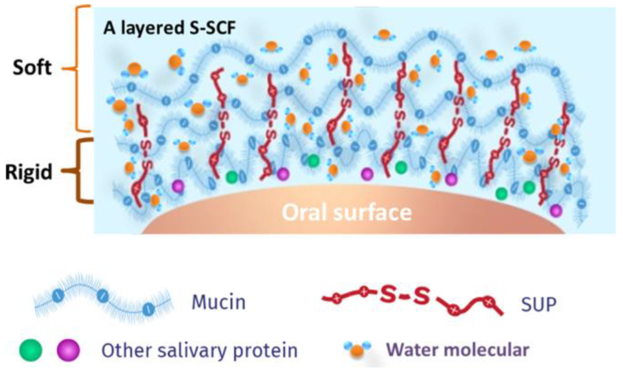
SUPsImprove oral lubrication and maintains its structural integrity during high contact pressures [17], SUPs with a sufficient number of positive charges can adsorb onto the SCF and retrieve mucins from the saliva [17], K108cys modified SUPs resulted in higher salivary glycoprotein adsorption and softness while also doubling the period of effectiveness of natural human saliva [17]No noted disadvantages
Disclaimer/Publisher’s Note: The statements, opinions and data contained in all publications are solely those of the individual author(s) and contributor(s) and not of MDPI and/or the editor(s). MDPI and/or the editor(s) disclaim responsibility for any injury to people or property resulting from any ideas, methods, instructions or products referred to in the content.

