Article

Optimizing Hyaluronan-Based Lubricants for Treating Thoracolumbar Fascia Pathologies: Insights from Tribological and Pharmacokinetic Studies

1
Biotribology Research Group, Faculty of Mechanical Engineering, Brno University of Technology, Technická 2896/2, 616 69 Brno, Czech Republic
2
Contipro a.s., Dolní Dobrouč 401, 561 02 Dolní Dobrouč, Czech Republic
3
Faculty of Medicine in Hradec Králové, Charles University, Šimkova 870, 500 03 Hradec Králové, Czech Republic
4
CEITEC—Central European Institute of Technology, Brno University of Technology, Purkyňova 656/123, 612 00 Brno, Czech Republic
*
Author to whom correspondence should be addressed.
Lubricants 2025, 13(4), 184; https://doi.org/10.3390/lubricants13040184
Submission received: 6 March 2025 / Revised: 14 April 2025 / Accepted: 15 April 2025 / Published: 16 April 2025

Abstract

:
In a world where the incidence of non-specific lower back pain (LBP) is steadily increasing, researchers are still searching for effective solutions for patients. Hyaluronic acid (HA) viscosupplementation is commonly used to restore lubrication in osteoarthritis (OA) and other medical applications, but its rapid metabolism limits efficacy. This study evaluates whether an HA derivative can replace native HA for the treatment of non-specific LBP while maintaining or enhancing its frictional properties and improving in vivo stability. Six HA-based lubricants, both native and derivatized, were tested in a tribological rabbit fascia model and a new synthetic model. Reduced HA derivative showed better tribological properties and longer in vivo residence time compared to native HA, as demonstrated in pharmacokinetic studies in rabbits. The 316 kDa HA and reduced HA exhibited the most stable tribological properties, which were influenced by their molecular weight and concentration. These findings suggest that both native and reduced HA are promising viscosupplements for intrafascial injection in the treatment of LBP, with reduced HA potentially enhancing effectiveness through a prolonged effect.

1. Introduction

In recent years, there has been a significant increase in cases of lower back pain (LBP) among individuals [1,2]. The treatment for LBP can be financially burdensome and prolonged, often without accurately diagnosing the underlying cause. As a result, many individuals may experience work incapacity [1]. Research suggests that one possible cause of LBP could be the thoracolumbar fascia (TLF), which undergoes pathological changes [3,4,5]. Healthy fascia typically consists of two or three layers of connective tissue that glide smoothly and are lubricated by hyaluronic acid (HA) [6]. However, an unhealthy lifestyle and overuse syndromes can lead to structural changes in the fascia.
Such changes in the fascia can include densification and fibrosis, which are distinct pathological alterations. The two alterations often occur simultaneously. Fibrosis refers to a pathological change in the fibrous component of the fascia, characterized by the random accumulation of collagen fibers. This type of change is challenging to modify manually during rehabilitation. On the other hand, densification is a change in the viscosity of the loose connective tissue within the fascia, resulting from the more viscous HA [7,8,9]. This causes patients with chronic LBP to exhibit reduced gliding of the TLF [5]. Furthermore, it has been proposed that heightened muscle exertion elevates the molecular weight (MW) of HA, consequently increasing its viscosity [10,11], while the increase in viscosity of HA is also associated with immobilization [12]. This elevation in viscosity may impede normal muscle movement within loose connective tissue. Analogous to its function in joint and synovial fluid, heightened HA production represents an initial effort by the fascia to enhance its mobility. At elevated concentrations, HA behaves similarly to a non-Newtonian fluid, exhibiting increased viscosity as the HA chains entangle, thereby contributing to the solution’s hydrodynamic characteristics [6]. The heightened viscosity of HA, coupled with the sparsity of connective tissue due to pathological alterations within and upon the fascia, reduces the sliding between the collagen fiber layers of the deep fascia, consequently resulting, again, in a sensation of stiffness in the patient’s back.
Viscosupplementation with HA is widely used to restore lubrication and improve the gliding of connective tissues in osteoarthritis (OA), as well as to reduce friction and enhance comfort between contact lenses and the eye [13]. Intrafascial injections of HA, a promising therapeutic approach recently introduced by our group, could similarly restore the reduced gliding ability of the fascia by reducing friction between its layers [14]. However, a key limitation of using HA in medicine is its relatively rapid degradation in the human body, which restricts its duration of action. To extend its residence time at the application site, HA can be chemically modified [15] while preserving its essential mucoadhesiveness and biocompatibility [16]. For example, a form of HA with partially reduced carboxyl groups offers increased thermal stability and enzymatic resistance [16]. While reduced HA has not yet been studied for LBP treatment, it has been widely researched for restoring joint lubrication in OA [17]. The amphiphilic form of HA is known for its resistance to enzymatic degradation and its ability to interact through both ionic and hydrophobic interactions [18]. Compared to native HA, this derivative exhibits significantly enhanced shear-thinning behavior [19]. It has been previously studied for use in eye drops to treat dry eyes [20], where this form demonstrated extremely low friction and effectively prevented surface eye cells from drying out, outperforming regular HA.
Therefore, this study aims to determine if native HA used in viscosupplementation for treating non-specific LBP can be effectively replaced with a derivative that maintains or even enhances its frictional properties while offering improved in vivo stability. We rigorously tested six HA-based lubricants, in both native and derivatized forms, on a tribological model of rabbit fascia introduced in [21]. Simultaneously, a new synthetic model was introduced to verify the lubricants’ influence on fascia friction. The most promising derivative based on tribological testing, reduced HA [22], exhibits nontoxic properties and biodegradability comparable to native HA, as demonstrated in a pharmacokinetic study involving rabbits.

2. Materials and Methods

2.1. Lubricants

Native HA (101, 316 and 610 kDa) and HA derivatives (reduced HA (HA RED and lauroyl-modified HA (HA-C12)), provided and biotechnologically manufactured by Contipro a.s. (Dolní Dobrouč, Czech Republic), were dissolved in PBS and sterilized by filtration. Concentrations and viscosity of tested solutions are summarized in Table 1. Structures of HA derivatives are displayed in Figure S1.
For the pharmacokinetics study, a solution of 13C-labelled 316 kDa HA and a solution of HA-RED were prepared in normal saline (0.9% NaCl) at a concentration of 10 mg/mL. Labelling of HA with the nonradioactive isotope 13C was used to distinguish the administered HA from endogenous HA [23]. After sterilization by filtration, they were additionally autoclaved. Apyrogenicity of the solutions was then verified using a portable endotoxin testing system (Endosafe® nexgenPTS™ testing system, Charles River Laboratories, Wilmington, MA, USA) and only apyrogenic samples (endotoxin levels < 0.1 IU/mL) were used for study.

2.2. Tribological Setup

Bruker UMT TriboLab (Bruker, Billerica, MA, USA) was used to measure friction and lubrication properties of test samples in a pin-on-plate configuration. The tribometer sensor recorded the normal and friction force, from which the value of the friction coefficient was calculated as a function of time. The static pin was loaded with 2 N normal force. The plate moved with a 1 Hz frequency of reciprocal linear motion and 12 mm track length. The same amount of lubricant and test temperature were kept for each test (2 mL of lubricant, 37 °C). Two types of experiments were conducted based on changes in the test time. The short-term 300 s tests and the preload effect tests were repeated five times. The long-term 3000 s tests were tested three times. Experimental conditions were chosen based on physiological conditions, therapy, and the limitations of the test facility. For more information, refer to the Materials and Methods Section in the previous publication [21].
The experimental apparatus, in pin-on-plate configuration, consisted of a pot and pin part. The pin has a convex surface with a 50 mm radius on which the polydimethylsiloxane (PDMS) with a 1 mm thickness is embedded. The fascia is stretched over the surface of the pin and PDMS, which is attached with jaws screwed into the pin. The pot is divided into two main parts. The lower part provides attachment to the tribometer, heating cartridges, and an assembly of jaws to hold the sample. In the bottom part, PDMS is inserted and, over it, fascia is prestressed by the jaws. This study examines the effect of different levels of prestressing when the fascia is stretched to three degrees: strong, medium, and weak. These levels are defined by the width of the fascia when stretched at the center and edges of the specimen. The lubricants examined in this study were laid on elastomer PDMS of 10 ShA stiffness simulating underlying muscle. Sliding friction tests were carried out for prepared thoracolumbar rabbit fascia and SynDaverTM (Tampa, FL, USA) synthetic fibrous fascia tissue.

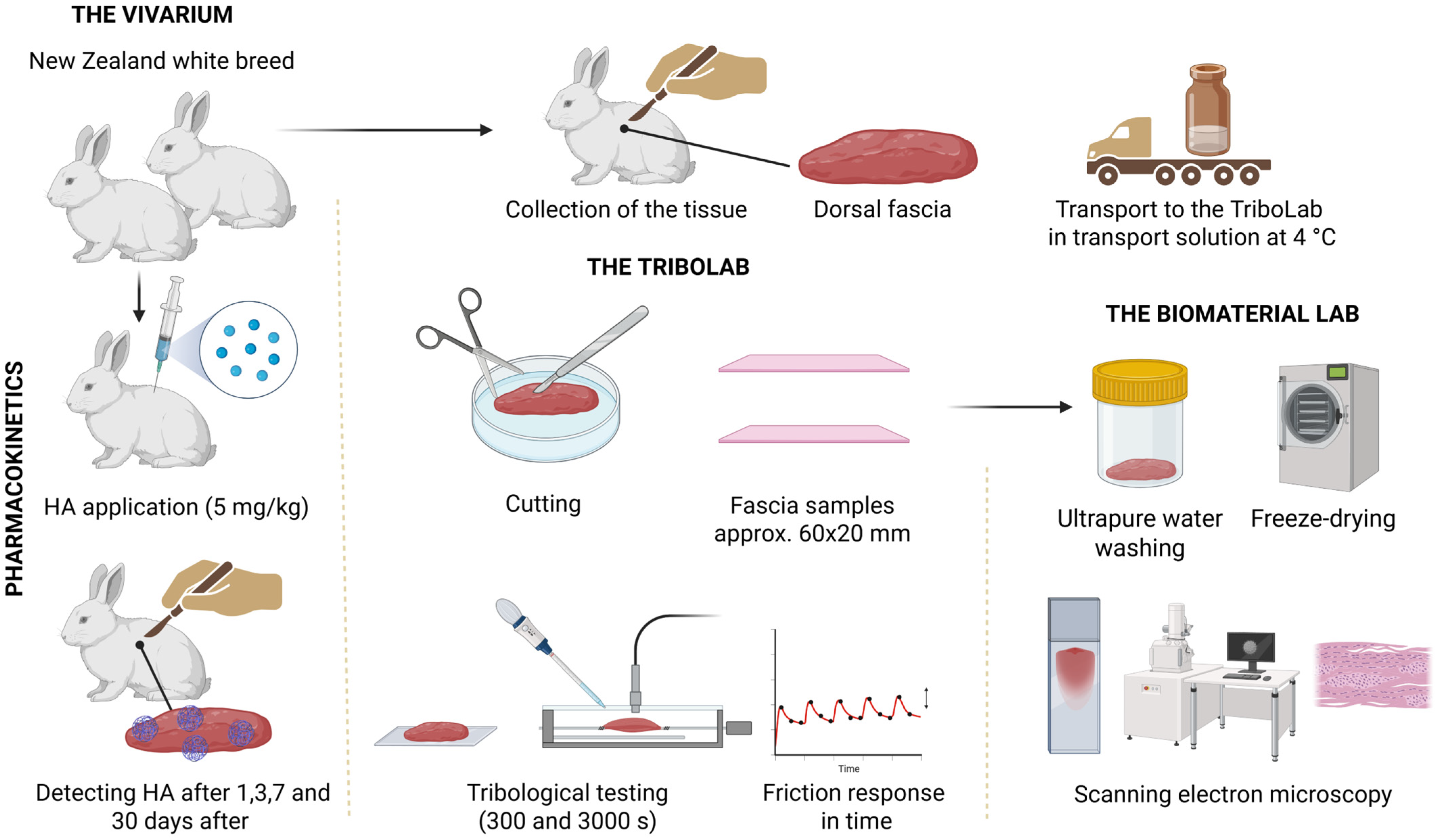
2.3. Preparation of Samples of Rabbit TLF

Sexually mature New Zealand white rabbits, with body weights of at least 3 kg, were used for preparation of TLF for tribological testing. The rabbits were first sedated by subcutaneous administration of medetomidine (0.3 mg/kg; Domitor inj., Orion Corporation, Espoo, Finland) and then induced into general anesthesia by intramuscular administration of ketamine (25 mg/kg; Narkamon inj., Bioveta, Ivanovice na Hane, Czech Republic). Subsequently, the animals were stunned with a captive bolt gun and then exsanguinated by transecting the carotid artery (arteria carotis communis). After removing the skin from the cadaver, a paravertebral incision was made, thereby releasing the TLF from the spine. Then, the fascia was released by blunt dissection from the back muscle up to the latissimus dorsi muscle, tensor fascia late muscle and external oblique abdominis muscle. The collected TLF was immersed in a transport solution (Hanks balanced salt solution) and transported to the laboratory at 4 °C. The chilled fascia was removed from the test tube after 16 h at 4 °C and moved, along with the transport solution, to a sterilized Petri dish to prevent the tissue from drying out. Subsequently, the right or left part of the fascia was taken out and spread on a glass plate, see Figure 1. After measuring, a sample of approximately 60 × 20 mm of the test sample was cut out with dissecting scissors. The prepared sample was attached to the tribometer.
Fascia for electron microscopy scanning was washed with ultrapure water (Type II according to ISO 3696 [24], prepared on an Elix 5 UV Water Purification System (Merck, Darmstadt, Germany)). Then, the specimen was freeze-dried on an Epsilon 2-10D machine (Martin Christ, Osterode am Hartz, Germany). A scanning electron microscope, MIRA3 (TESCAN, Brno, Czech Republic), was used to study the morphology of the dried fascia sample. Images were taken in secondary electron emission mode; the scan mode was DEPTH, the beam density was 10, and the high voltage was 10 kV. The working distance was set to 15 mm. The surface of the sample was coated with a 15 nm thin layer of Au using an EM ACE 600 (Leica Microsystems, Wetzlar, Germany).

2.4. Pharmacokinetic Study

This study was approved by the Ethics Committee of the Ministry of Education, Youth and Sports (approval No. MSMT-2036/2022-3). For this study, 48 healthy, sexually mature New Zealand white male rabbits (weighing 2.7–3.5 kg) obtained from a certified laboratory animal breeder and supplier, VELAZ, Ltd. (Prague, Czech Republic), were used. The rabbits were acclimated for a period of two weeks before the experiment began. The animals were housed individually in clean stainless-steel cages under standardized environmental conditions (12-h light/dark cycle, temperature 17–21 °C, air humidity 30–70%). The rabbits had access ad libitum to the standard diet for rabbits (VELAZ Ltd., Prague, Czech Republic) and drinking water. The rabbits were randomly allocated into eight groups, with six rabbits per group. The groups differed in the amount of intrafascial injected solution (HA-RED/13C-labelled 316 kDa HA) and in the euthanasia time after the solution administration (1/3/7/30 days). The rabbits were first sedated by subcutaneous administration of medetomidine (0.3 mg/kg; Domitor inj., Orion Corporation, Espoo, Finland) and then induced into general anesthesia by intramuscular administration of ketamine (25 mg/kg; Narkamon inj., Bioveta, Ivanovice na Hane, Czech Republic). Subsequently, the rabbits were shaved on their backs (approximately 4 × 4 cm in the application sites of the thoracolumbar spine area), and, using a 25G needle under ultrasound guidance (Lumify, Philips, Amsterdam, The Netherlands), they were injected with a 1% solution of 13C-labelled 316 kDa HA or a 1% solution of HA-RED (according to their experimental group) in the amount of 0.7 mL on each side between the superficial and deep layers of the thoracolumbar fascia at the level of the L2–L3 intervertebral junction, 50 mm lateral to the spine (left and right). The application sites were then gently massaged manually to evenly spread the injected solution between the fascial layers throughout the entire extent of the fascia. At 1, 3, 7, or 30 days after the intrafascial administration of HA-RED or 13C-labelled 316 kDa HA solution, depending on the experimental group, the animals were sedated by subcutaneous administration of medetomidine (0.3 mg/kg; Domitor inj., Orion Corporation, Espoo, Finland) and then induced into general anesthesia by intramuscular administration of ketamine (25 mg/kg; Narkamon inj., Bioveta, Ivanovice na Hane, Czech Republic). A non-coagulated blood sample (1 mL) was then collected from the central ear artery (arteria auricularis centralis), centrifuged, and the plasma was separated. Subsequently, the animals were stunned with a captive bolt gun and then exsanguinated by transecting the carotid artery (arteria carotis communis). Then, after removing the skin from the rabbit cadavers, the TLF tissue samples were obtained as two separate samples: a partial-thickness fascia (without the epimysial layer) by separation from the spine through a paravertebral incision and releasing by blunt dissection from the back muscles, and the corresponding epimysial layer with muscle tissue underneath. Both types of samples were weighed separately, frozen individually at −20 °C, and transported to the laboratory for quantification of administered 13C-labelled 316 kDa HA and HA-RED in the fascia and muscle by LC-MS.

2.5. Determination of Isotopically Labelled HA and HA-RED in Rabbit Fascia and Muscle

For the quantification of HA derivatives, a modified LC-MS method based on quantification of HA disaccharides after enzymatic hydrolysis was employed [25] (Figure S2). Briefly, weighted samples of fascia and muscle were diluted with buffer (100 mM PBS with 10 mM EDTA, pH 7.4) and homogenized with Precellys Evolution homogenizer (Bertin Technologies, France). To 100 µL aliquot, internal standard of fully isotopically labelled 8×13C-HA (lower conc.) was added, and samples were incubated for 3 h at 45 °C with 40 µL of 1% Actinase E solution (Merck, Germany) in HEPES buffer (pH 8). Afterwards, 1 µL of solution of hyaluronan lyase from Streptococcus pneumoniae (Contipro a.s., Czech Republic) was added and samples were incubated for 30 min at 37 °C. Finally, 400 µL of acetonitrile (LC-MS grade, VWR, Radnor, PA, USA) was added, the samples were centrifuged (30 s, 10,000 rpm) and supernatants were analyzed by LC-MS. A Vanquish Horizon chromatography system and a TQS Altis plus triple quadrupole mass spectrometer (both Thermo Fisher Scientific, Waltham, MA, USA) were used for analysis. A total of 2 µL of sample were injected onto an Acquity BEH HILIC Amide (2.1 × 100 mm) analytical column (Waters, Wilmslow, UK) tempered at 60 °C. The mobile phases consisted of 0.1% formic acid (v/v) (LC-MS grade, Merck, Germany) and acetonitrile (LC-MS grade, VWR, USA). The gradient of acetonitrile was as follows: 0–0.5 min 90%, 0.5–5 min 90–74%, 5–7 min 74–40%, 7–7.5 min 40–90%, 7.5–9 min 90%. The electrospray settings for the analysis were as follows: capillary voltage −4000 V, Sheath gas 50, Aux gas 10, Sweep gas 0.5, ion transfer tube temp. 275 °C, vaporizer temp. 350 °C. MRM transitions of unsaturated disaccharides for 1×13C-316 kDa HA (lower conc.) and fully labelled HA together with unsaturated tetrasaccharide of HA-RED were detected during the analysis.

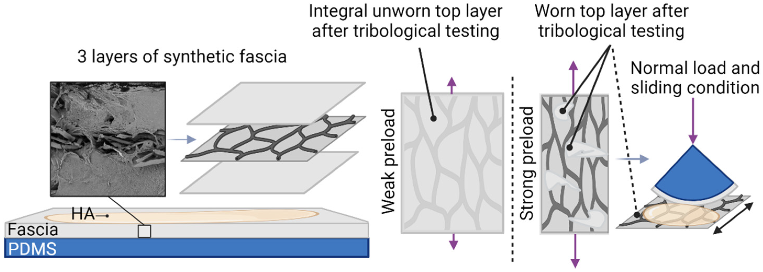
2.6. Preparation of Tribological Samples from Synthetic Fascia

The 1 mm synthetic substitute fascia, consisting of multiple fiber planes, was purchased from SynDaverTM (Fibrous fascia, SKU: 141620), and is designed primarily for medical device design testing and for medical professionals to practice surgery. Pre-preparation and storage of synthetic fascia were performed according to manufacturer’s instructions. The procedure for cutting, sizing, and mounting the specimens further coincides with the preparation of tribological samples from rabbit fascia.

2.7. Statistical Methods

Results were presented as a mean from three measurements ± SD (standard deviation) unless otherwise stated. Statistical criteria were verified by one-way or two-way ANOVA followed by Tukey’s or Bonferroni’s multiple comparisons tests, with individual variances computed for each comparison. The Tukey test was applied for short-term friction tests involving multiple group comparisons to detect significant differences while controlling the overall error rate, while the Bonferroni correction was used for long-term tests with a limited number of planned comparisons to minimize the risk of false positives. For all tests, * p < 0.05, ** p < 0.01, *** p < 0.001, and **** p < 0.0001 were considered to be significant.

3. Results

3.1. Testing of Preload Effect of Fascia on Friction

In tribological testing, the material in the experimental setup is preloaded to simulate the conditions experienced during muscle movement. Since stress levels can significantly impact friction results, this study begins by parametrically analyzing the effect of material preload on friction, utilizing jaw prestressing. Subsequent friction experiments were conducted five times using the 316 kDa HA lubricant, chosen due to its medium MW and widespread use among the lubricants in this study. Figure 2 illustrates that the bias level’s effect on friction had almost no effect on rabbit fascia when the fascia is prestressed. Statistical analysis revealed the following significance levels: p = 0.0427 for the difference between weak and middle bias levels, p = 0.0542 for the difference between weak and strong, and p = 0.9752 for the difference between middle and strong for synthetic fascia. However, the influence of fascia prestressing emerges as a slightly more significant factor in synthetic fascia. In instances where the fascia is moderately or not prestressed, friction levels are lower compared to instances where the fascia is strongly prestressed. The coefficient of friction (COF) increased from 0.0517 (SD = 0.0076) or 0.04931 (SD = 0.0016) to 0.6275 (SD = 0.0023). Importantly, the bias level ceases to affect friction in cases of strong prestress levels. For rabbit fascia, the significance levels were p = 0.8523 between weak and middle, p = 0.2301 between weak and strong, and p = 0.0002 between middle and strong bias levels. Considering the results from the parametric study, the tribological tests in the following parts of the study were performed on a middle-prestressed fascia.

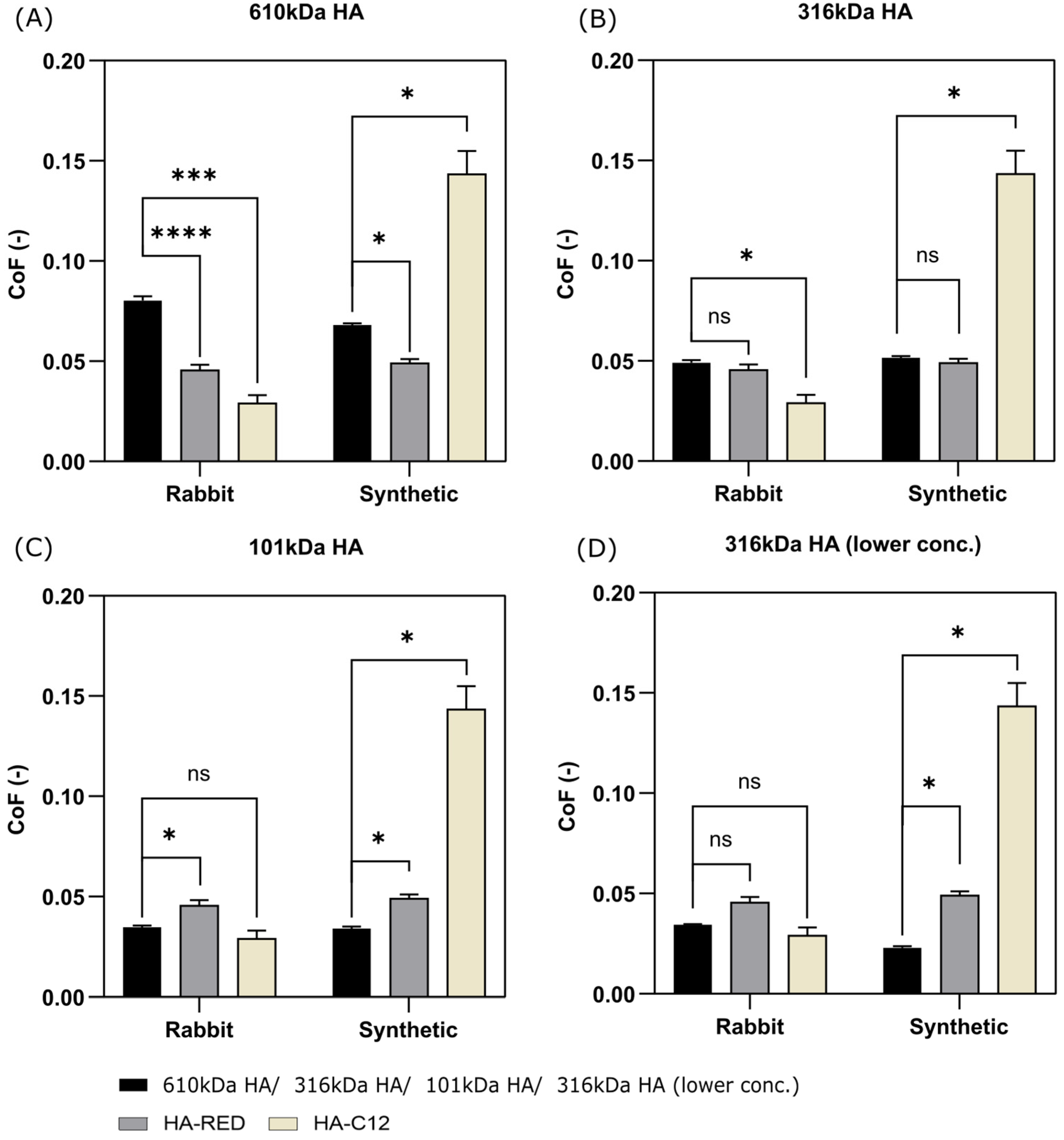
3.2. Short-Term and Long-Term Testing of Fascia Friction

The friction experiments were conducted in two distinct stages. Initially, short-term tribological tests were conducted for 300 s. The results in Figure 3 were analyzed to determine which lubricant most effectively reduces the friction of the fascia and its layers. This information is valuable in identifying suitable properties for Visco supplements for treating non-specific LBP. Statistical performance between the different lubricants is listed in Table S1. 101 kDa HA and 316 kDa HA (lower conc.) exhibited the lowest COF for both materials. However, 316 kDa HA and HA-RED also demonstrated low friction with no significant difference observed for both ex vivo rabbit and synthetic fascia. Conversely, HA-C12 displayed the highest friction and standard deviation (SD) for synthetic fascia but the lowest for rabbit fascia. Lastly, 610 kDa HA exhibited the highest friction, nearly double that of 101 kDa HA and 316 kDa HA (lower conc.), for both materials. The results of the short-term tests indicate that HA-RED has the same efficacy on fascia friction as native HA with the same concentration and molecular weight. However, the interchangeability of HA-C12 with native HA remains debatable. Among the native HAs, 101 kDa HA and 316 kDa HA (lower conc.) are the most effective candidates for successfully reducing friction in fascia and its layers.
The second phase aimed to confirm the suitability of the lubricant for fascia lubrication based on long-term effects. Long-term tribological tests were conducted for 3000 s with other experimental conditions maintained. The results were presented for each material in Figure 4, illustrating the evolution of friction over time for rabbit fascia and synthetic fascia, respectively. These graphs are depicted as curves because the behavior of the lubricant in contact over time is deemed more important than the final or average friction value. An overview of the graph indicates that synthetic fascia exhibited more consistent behavior throughout the long-term test compared to other specimens, with fewer abrupt transitions and instabilities in the friction curves. However, HA-C12 lubrication notably deteriorated the performance of synthetic fascia, rendering it unsuitable for effective tribological testing with this lubricant. Conversely, the performance of rabbit fascia remained relatively steady across most scenarios, showing no significant deterioration in either case. Nonetheless, when paired with 610 kDa HA lubricant, slight fluctuations in performance were observed, with friction levels gradually increasing over time. A similar but more nuanced trend was observed with 316 kDa HA (lower conc.) lubrication of rabbit fascia. Regarding HA-RED lubrication, one friction curve of rabbit fascia exhibited instability and a slight upward trend over time. The results of the long-term assays are consistent with the results of the short-term assays, confirming that HA-RED can replace native HA with the same properties if it demonstrates longer stability in vivo. While HA-C12 exhibits low friction on biological fascia, its performance on synthetic fascia raises questions about its long-term lubricating effects. Therefore, the benefits of HA-C12 compared with HA-RED need to be carefully evaluated. Native HAs generally demonstrated good stability over time. However, lower MW is associated with greater temporal stability, and lower concentrations result in reduced friction. Therefore, the best native candidate for reducing friction in the fascia and its layers is 316 kDa HA (lower conc.).

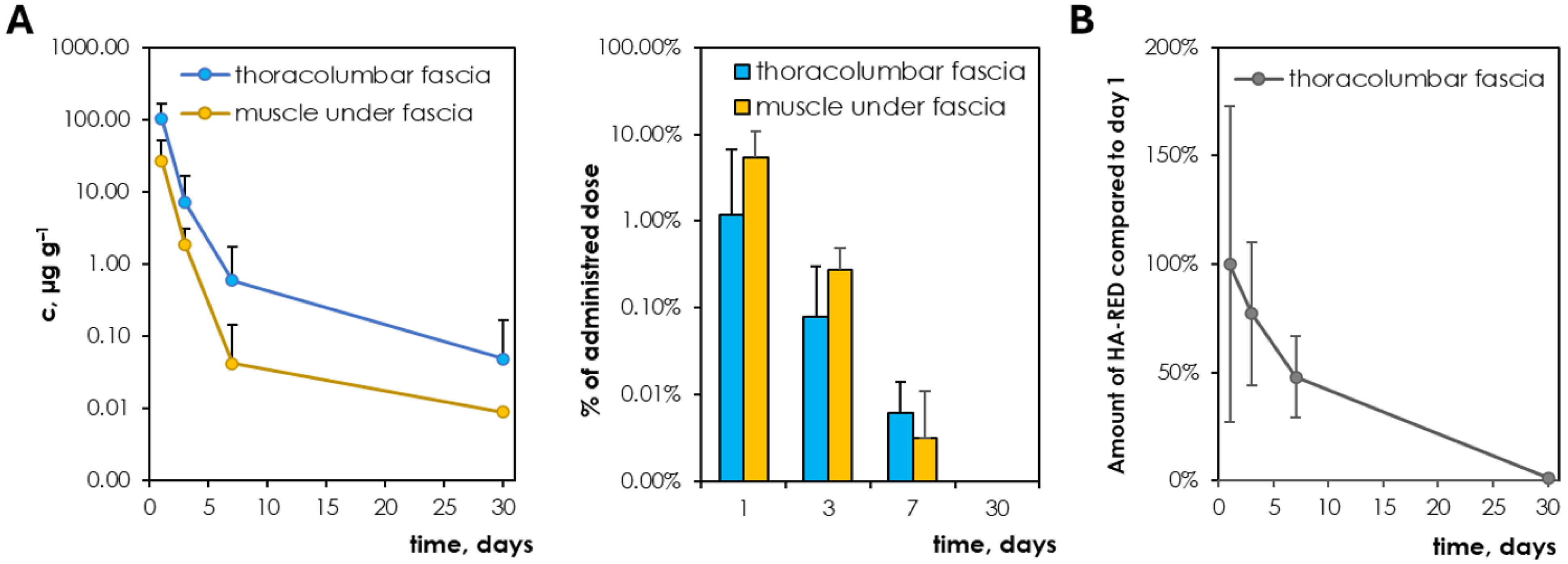
3.3. Pharmacokinetics of 316 kDa HA (Lower Conc.) and HA-RED After Application to the Fascia

Based on the tribological results, 316 kDa HA (lower conc.) and HA-RED were singled out as the candidates for the development of viscosupplementation for the treatment of non-specific LBP. To determine the pharmacokinetics of both materials and biodegradability of HA-RED, 13C-labelled HA and HA-RED were administered to live rabbits via intrafascial injection and the number of administered materials were quantified in fascia and muscle by LC-MS.
Although a relatively large amount of 316 kDa HA (lower conc.) is injected into the fascia (14 mg corresponds to a dose of 5 mg/kg), after 3 days the amount of exogenous HA present in the fascia is very small (<2%) relative to the amount of endogenous HA in the tissue (see Figure 5). The amount of endogenous HA in fascia was determined to be 412 ± 73 µg/g and in muscle 12.8 ± 1.9 µg/g. Thus, the ratio of the concentration of 13C-HA in the fascia to that of endogenous HA decreased during 1, 3, 7 and 30 days as follows: 25.3%, 1.7%, 0.1%, 0.0%, respectively. In muscle, the ratio is then 14.4%, 0.3%, and 0.1% at 3 days, 7 days, and 30 days, respectively. The rate of elimination of applied 13C-HA from the fascia is thus relatively rapid. The elimination half-life in fascia and muscle was determined to be 20.1 h and 15.7 h, respectively. The concentration of 13C-HA was also determined in plasma and was determined to be below the detection limit of <100 ng/mL.
Quantification of HA-RED in fascial and muscle tissue by LC-MS was problematic since the main unique product of enzymatic degradation, unsaturated disaccharide of HA-RED, lacks an ionizable carboxyl group and was undetectable by LC-MS (Figure S2). Therefore, unsaturated tetrasaccharide, the site product of enzymatic depolymerization, was used for detection. However, the low abundance of tetrasaccharide and interferents present in the fascia and muscle specimens prevented accurate quantification of HA-RED, and, thus, its abundance in fascia and muscle was evaluated at least semiquantitatively. The HA-RED signal was detected at 1, 3 and 7 days after administration. As in the case of 13C-HA, only trace amounts were present in the samples 30 days after administration. The elimination half-life in fascia was around 108 h. The rate of elimination could not be accurately evaluated, but the signal at 7 days was still significant, and the elimination half-life in fascia was around 108 h, suggesting that the HA-RED is a biodegradable material with a slightly slower elimination rate than native HA.

4. Discussion

In this study, we examined six HA-based lubricants using ex vivo and synthetic fascia to identify optimal HA properties that minimize friction within the fascia and its layers. HA plays a crucial role in facilitating smooth movement within the fascia. Pathological conditions cause HA to thicken, leading to tissue adhesion. In a previous study [14], we proposed a promising treatment involving native HA intrafascial injections to reduce tissue friction and alleviate pain. Effective agents must possess specific properties for optimal functionality. Therefore, our experiments focused on understanding how the MW, chemical modification and concentration of HA influence friction in fascia and its layers. The most promising HA derivative from tribological experiments, HA-RED, was also tested for biodegradability using rabbits to ensure its safety use for intrafascial injections.

4.1. Influence of Preload of Fascia on Friction

This study begins by examining how stretching materials affect fascia friction. This investigation is motivated by several factors. Firstly, during testing, the material is held in place and slightly prestressed to mimic the natural tension fascia experiences. Fascia, which separates muscles or fat, naturally undergoes stress with every movement due to muscle contractions or external forces [26]. Structurally, fascia consists of three sublayers of connective tissue with different densities and orientations [27,28]. Studies have shown that within each sublayer collagen fibers run parallel, but the fibers in adjacent layers are oriented at angles of about 70–80 degrees to each other [27]. Research on rabbit dorsal fascia demonstrated a similar structure to human TLF, with collagen fibers and loose connective tissue [14]. Notably, only the superficial sublayer is richly innervated and contains few elastic fibers [28]. Collagen fibers, visible in the study’s images (see Figure 6), provide resistance to tension and stretching, common in various fascial tissues like ligaments, tendons, sheaths, muscular fascia, and deeper fascial sublayers [29]. The collagen and three-layer structure of the fascia, designed to allow fiber sliding within each layer [30,31], likely explains why preloading did not affect the friction of HA within the rabbit fascia. In contrast, synthetic fascia showed higher friction under heavier preload. The synthetic material, consisting of three sublayers with hydrophilic polymer on the top and bottom and fibers in the middle, might have experienced top-layer wear under increased stress.
These thinner layers are more prone to damage from stretching and tangential forces during friction tests, leading to rougher fiber surfaces (Figure 7). When the top fiber surface contacts the material, additional contact area is observed with more compliant elastomers, indicating hysteretic friction [32]. Hysteretic friction likely increases with higher surface roughness, leading to a higher COF [33]. However, this hypothesis requires further analysis. Future viscosupplements should maintain low friction levels without being compromised by stress and wear in pathological conditions. Additionally, future experiments should note that there is no significant difference in friction between lightly and moderately prestressed material, so materials will not be overstressed in further tests.

4.2. Selection of Appropriate Properties of HA-Based Solution as a Viscosupplement of TLF Fascia Based on Friction Results

As mentioned in the Introduction, one of the main limitations of HA in medical applications is its quick degradation in the human body, which reduces its effectiveness over time. To extend its presence at the application site, HA can undergo chemical modifications. In this study, both native HA (610 kDa, 316 kDa and 101 kDa HA) and two HA derivatives (HA-RED and HA-C12, see Figure S1) were tested. HA-RED is HA with carboxyl groups partly reduced to primary alcohols [22]. Comparing the results of native HA and HA-RED with similar MW (316 vs. 275 kDa), we observe that the reduction of carboxyl groups had no significant impact on fascia friction. Despite a minor decrease in viscosity, the influential factors remained MW and concentration parameters. HA-C12 is an amphiphilic form of HA [18]. In studies [19,20], HA-C12 showed extremely low friction and effectively prevented surface eye cells from drying out, surpassing the effectiveness of regular HA. However, the effect of HA-C12 derivation on fascia friction alone cannot be assessed as the lubricant has a different concentration than the others used.
In this study, the viscosity of HA solutions increased with increasing MW and concentration, as shown in Table 1. The reason for this increase is straightforward: the viscosity of a polymer solution depends on the proportion of the solution’s volume taken up by the polymer chains. This proportion can be described by the concentration of the polymer multiplied by its specific volume [6]. In simpler terms, viscosity is influenced by the length and number of polymer chains in the solution, as well as the size of the molecules themselves. As MW and concentration rise, the polymer chains become more tightly packed, making it harder for them to move freely within the solution. Consequently, when minimal sliding and pressure are applied, friction between the molecules increases. This conclusion is supported by the findings of the short-term friction experiments, as seen in Figure 3.
The tribological experiments also supported the statement that a high-viscosity solution may not be ideal for promoting fascial glide [10,11,12]. These solutions (610 kDa HA, 316 kDa HA, and HA-RED) exhibited higher friction coefficients. In contrast, Figure 3 shows a noticeable decrease in friction of native HA (101 kDa HA and 316 kDa HA (lower conc.)) with decreasing MW and concentration for both fascial models. Surprisingly, this is the opposite of what happens in an osteoarthritic joint [34,35] or the eye and eyelid [13]. The most interesting comparison is with HA-C12, which may explain how HA behaves toward fascia. For rabbit fascia, a solution with a concentration of 3 mg/mL and a low MW of 318 kDa achieved super-low friction. However, the friction of the synthetic fascia was almost five times higher. This demonstrates that HA has limited capacity to adhere to both hydrophilic and hydrophobic artificial surfaces [36]. In contrast, for tissue, collagen fibers on cartilage deform under boundary lubrication [37], reducing gaps laterally while maintaining size normally. This deformation allows collagen fibers to mechanically entrap HA molecules. While the forces binding the molecules to the fibers are not strong enough to create a complete lubricating layer [38], unlike phospholipids and HA [39,40,41], the HA trapped in this manner can still serve as a boundary layer, preventing cartilage damage. HA-C12 has a low concentration and viscosity, forming a weaker lubricating film (Table 1). Therefore, the synergy between collagen fibers and HA helps protect fascia against wear and damage. Synthetic fascia, lacking collagen, does not form this layer, leading to rapid wear. Even a concentration as small as 3 mg/mL is a suitable option for viscosupplementation of fascia.
The HA-collagen synergy is likely manifested in other cases as well. For 610 kDa HA, the longitudinal test curves for synthetic fascia show a smooth progression throughout the experiment, as seen in Figure 4. The MW and concentration of 610 kDa HA and the likely low roughness of the synthetic fascia contributed to forming a minimally weak lubricating film, protecting the fascia from damage. For rabbit fascia, regular waves can be seen in the progress. This may be explained by the viscoelastic properties of HA at higher MWs, as shown by [42], combined with how collagen entraps HA between its fibers [37]. The friction gradually increases until it reaches its peak and then decreases gradually. This suggests that 610 kDa HA, due to its large macromolecular clusters [6], does not reach deep between the collagen fibers but attaches mechanically to their surface, causing an increase in friction. Due to the viscoelastic properties of HA and the shear frictional bias, the chains tear into smaller ones, as described in [6,43,44], again protecting the fascia from damage. The interaction between lubricin and HA also plays a crucial role in the lubrication of connective tissues, reducing friction and preventing tissue adhesion [45]. Lubricin penetrates chemically bound HA, forming a viscoelastic gel that lowers the COF and enhances surface resistance to wear. Increasing lubricin concentration further improves lubrication by elevating the threshold load before wear occurs and reducing friction between HA-coated surfaces. Additionally, the HA-lubricin gel forms a stable lubricating film, maintaining its thickness under load [46]. Its shear viscosity increases significantly under confinement, and lubricin’s interaction with HA prevents further adsorption onto negatively charged surfaces, potentially minimizing tissue adhesion.

4.3. HA Degradation in Living Organisms and Viscosupplement Confrontation

The suitability of native versus reduced HA for treating non-specific LBP was further assessed through pharmacokinetics (Figure 5). The viscosupplement is designed to dilute endogenous HA, which becomes highly viscous due to pathological changes in fascial tissue [10,11]. Based on friction results, 316 kDa HA (lower conc.) and HA-RED (275 kDa) were selected for comparison. However, previous research has shown that HA solutions degrade more slowly in vivo with higher MW compared to lower MW [23]. Consequently, 101 kDa HA, despite having the same COF but a lower MW, was not chosen for in vivo experiments. The turnover rate of endogenous HA in the body is relatively rapid, with a half-life of 2–5 min in the bloodstream [47] and a few days in tissues [48]. If low doses of exogenous HA are used (~µg kg−1), its elimination is fast and similar to elimination of endogenous HA; however, if HA is dosed at relatively high doses in the range of mg kg−1 HA elimination follows saturable kinetics, where the elimination half-life can reach several hours for HA. HA is eliminated from the body through local cleavage reactions and systemic elimination via the liver, lymphatics, and kidneys. At the cellular level, HA undergoes non-specific degradation by free radicals or specific depolymerization by various enzymes [23].
HA administered via intrafascial injection at a dose of 5 mg/kg exhibited an elimination half-life which is expected for this MW [23]. It is appealing to use HA with higher MW to prolong its stay in fascia and enhance the effectiveness of the viscosupplement. However, formulations with excessively high MW have been reported to cause local harm [49] and its application is problematic due to the high viscosity of solutions of high-MW HA [14]. The chemical derivatization of HA seems a more promising approach to prolong the stay of viscosupplementation at application sites, as the viscosity is unaltered and resistance to enzymatic degradation is enhanced [50]. The pharmacokinetics of HA-RED showed excellent biocompatibility and biodegradability, since after 30 days all HA-RED was eliminated similarly to native HA of similar MW. HA-RED exhibited a longer elimination half-life (108 h) than native HA (20.1 h), as seen in Figure 5. Although chemical modification of HA ensures higher in vivo stability, the elimination of viscosupplementation remains relatively fast. Despite this, viscosupplementation based on HA can be effective, as evidenced in the case of viscosupplementation for OA. Although the on-site quantification of HA-RED was challenging and only allowed for semi-quantitative determination, the prolonged retention of HA-RED was inferred from its elimination kinetics, where the limitations of semi-quantitative analysis do not affect the validity of the conclusion.
Viscosupplementation with HA is a well-established treatment for OA of the joints, where the administered HA is absorbed within days [51]. However, the clinical efficacy of intra-articular HA injections for managing knee OA pain reaches its maximum around 6–8 weeks after administration, with uncertain benefits at 6 months [52]. This extended relief may be attributed to HA’s ability to initiate secondary processes despite its rapid degradation. HA can reduce nerve impulses and sensitivity linked to OA pain [53]. In experimental OA models, HA has shown protective effects on cartilage, likely mediated by its molecular and cellular interactions observed in vitro. Exogenous HA stimulates HA and proteoglycan synthesis in chondrocytes, reduces the production and activity of proinflammatory mediators and matrix metalloproteinases, and modulates immune cell behavior. Furthermore, dermal HA fillers are known to enhance fibroblast production and alleviate inflammatory skin conditions [54]. Fasciacytes [55], which produce HA in the fascia, are fibroblast-like cells contributing to these effects. HA has documented wound-healing properties [56] and it is crucial in regulating cell behavior and promoting cell proliferation within an HA-rich extracellular matrix [57]. These physiological effects collectively contribute to the mechanisms through which HA exerts its clinical benefits in the viscosupplement treatment, even for fascia tissue and non-specific LBP.

5. Conclusions

In this study, HA-based lubricants were tribologically tested to optimize the solution’s properties, leading to low friction of fascia tissue. The aim of this study was to assess whether the HA derivative could substitute the native HA form, preserving its superior frictional properties with an assumed slower degradation in vivo. Tribological tests were supplemented by pharmacokinetics of the two most promising lubricants using a living rabbit model, contributing to the development of advanced therapeutic solutions. To summarize the main conclusions and contributions of the study:
  • Tribological model—Optimization of fascia prestressing: The medium rabbit and synthetic fascia prestress ensures that the final friction results will be influenced by the effect of the lubricant without being affected by the degree of fascia tension;
  • Lubricants: 316 kDa HA and HA-RED were identified as having the best and most stable tribological properties for fascia lubricants, driven by their MW and concentration;
  • Pharmacokinetics of intrafascial HA-based viscosupplementation: Chemically modified HA (HA-RED) is eliminated within 30 days as well as native HA; however, it exhibits a longer residence time compared to native HA.

Supplementary Materials

The following supporting information can be downloaded at: https://www.mdpi.com/article/10.3390/lubricants13040184/s1, Figure S1: Structures of native HA and its derivatives: reduced HA (HA-RED) and lauroyl-modified HA (HA-C12). Figure S2: Enzymatic depolymerization of fully labelled native HA (14×13C-HA), reduced-HA (HA) and 2×13C-labelled native HA (2×13C-HA) into unsaturated oligosaccharides. Figure S3: COF comparison of HA-based lubricants on rabbit and synthetic models. Table S1: Significance of friction results between individual lubricants.

Author Contributions

A.S.—investigation, conceptualization, writing—original draft preparation, data curation, methodology; M.Š.—visualization, investigation, writing—original draft preparation, data curation, methodology; J.M.—writing-original draft preparation, formal analysis; D.N.—writing-review and editing, supervision; M.V.—visualization, supervision, project administration; J.S.—resources, methodology; V.P.—methodology, writing—review and editing; L.V.—resources, funding acquisition; M.H.—supervision; I.K.—funding acquisition; K.N.—supervision, resources, project administration. All authors have read and agreed to the published version of the manuscript.

Funding

This publication was supported by the project “Mechanical Engineering of Biological and Bio-inspired Systems”, funded as project No. CZ.02.01.01/00/22_008/0004634 by Programme Johannes Amos Comenius, call Excellent Research. This work was partly supported by the Ministry of Health of the Czech Republic under project No. NU22-08–00454. CzechNanoLab project LM2023051 funded by MEYS CR is gratefully acknowledged for the financial support of the measurements/sample fabrication at CEITEC Nano Research Infrastructure.

Institutional Review Board Statement

The study was approved by the Ethics Committee of the Ministry of Education, Youth and Sports (approval No. MSMT-2036/2022-3).

Data Availability Statement

Experimental data for Hyaluronic Acid-Based Lubricants Tribology for Alleviating Non-Specific Lower Back Pain available at resource https://doi.org/10.5281/zenodo.11239441 (accessed on 22 February 2024).

Conflicts of Interest

Authors Matěj Šimek, Jana Matonohová, Kristina Nešporová were employed by Contipro a.s. The remaining authors declare that the research was conducted in the absence of any commercial or financial relationships that could be construed as a potential conflict of interest.

Abbreviations

The following abbreviations are used in this manuscript:
LBPlower back pain
HAhyaluronic acid
OAosteoarthritis
SDstandard deviation
COFcoefficient of friction
PDMSpolydimethylsiloxane

References

  1. Wu, A.; March, L.; Zheng, X.; Huang, J.; Wang, X.; Zhao, J.; Blyth, F.M.; Smith, E.; Buchbinder, R.; Hoy, D. Global low back pain prevalence and years lived with disability from 1990 to 2017: Estimates from the Global Burden of Disease Study 2017. Ann. Transl. Med. 2020, 8, 299. [Google Scholar] [CrossRef] [PubMed]
  2. Hoy, D.; Bain, C.; Williams, G.; March, L.; Brooks, P.; Blyth, F.; Woolf, T.V.; Buchbinder, R. A systematic review of the global prevalence of low back pain. Arthritis Rheumatol. 2012, 64, 2028–2037. [Google Scholar] [CrossRef] [PubMed]
  3. Casato, G.; Stecco, C.; Busin, R. Role of fasciae in nonspecific low back pain. Eur. J. Transl. Myol. 2019, 29, 8330. [Google Scholar] [CrossRef] [PubMed]
  4. Wilke, J.; Schleip, R.; Klingler, W.; Stecco, C. The Lumbodorsal Fascia as a Potential Source of Low Back Pain: A Narrative Review. Biomed Res. Int. 2017, 2017, 5349620. [Google Scholar] [CrossRef]
  5. Langevin, H.M.; Fox, J.R.; Koptiuch, C.; Badger, G.J.; Greenan-Naumann, A.C.; Bouffard, N.A.; Konofagou, E.A.; Lee, W.-N.; Triano, J.J.; Henry, S.M. Reduced thoracolumbar fascia shear strain in human chronic low back pain. BMC Musculoskelet. Disord. 2011, 12, 203. [Google Scholar] [CrossRef]
  6. Cowman, M.K.; Schmidt, T.A.; Raghavan, P.; Stecco, A. Viscoelastic Properties of Hyaluronan in Physiological Conditions. F1000Research 2015, 4, 622. [Google Scholar] [CrossRef]
  7. Matteini, P.; Dei, L.; Carretti, E.; Volpi, N.; Goti, A.; Pini, R. Structural Behavior of Highly Concentrated Hyaluronan. Biomacromolecules 2009, 10, 1516–1522. [Google Scholar] [CrossRef]
  8. Fede, C.; Angelini, A.; Stern, R.; Macchi, V.; Porzionato, A.; Ruggieri, P.; De Caro, R.; Stecco, C. Quantification of hyaluronan in human fasciae: Variations with function and anatomical site. J. Anat. 2018, 233, 552–556. [Google Scholar] [CrossRef]
  9. Stecco, A.; Meneghini, A.; Stern, R.; Stecco, C.; Imamura, M. Ultrasonography in myofascial neck pain: Randomized clinical trial for diagnosis and follow-up. Surg. Radiol. Anat. 2014, 36, 243–253. [Google Scholar] [CrossRef]
  10. Stecco, A.; Gesi, M.; Stecco, C.; Stern, R. Fascial Components of the Myofascial Pain Syndrome. Curr. Pain Headache Rep. 2013, 17, 352. [Google Scholar] [CrossRef]
  11. Stecco, A.; Stern, R.; Fantoni, I.; De Caro, R.; Stecco, C. Fascial Disorders: Implications for Treatment. Phys. Med. Rehabil. 2016, 8, 161–168. [Google Scholar] [CrossRef] [PubMed]
  12. Wilke, J.; Schleip, R.; Yucesoy, C.A.; Banzer, W. Not merely a protective packing organ? A review of fascia and its force transmission capacity. J. Appl. Physiol. 2018, 124, 234–244. [Google Scholar] [CrossRef] [PubMed]
  13. Iwashita, H.; Mabuchi, K.; Itokawa, T.; Okajima, Y.; Suzuki, T.; Hori, Y. Evaluation of the Lubricating Effect of Hyaluronic Acid on Contact Lenses Using a Pendulum-Type Friction Tester Under Mimicking Physiological Conditions. Eye Contact Lens 2022, 48, 83–87. [Google Scholar] [CrossRef] [PubMed]
  14. Nešporová, K.; Matonohová, J.; Husby, J.; Toropitsyn, E.; Stupecká, L.D.; Husby, A.; Kleplová, T.S.; Streďanská, A.; Šimek, M.; Nečas, D.; et al. Injecting hyaluronan in the thoracolumbar fascia: A model study. Int. J. Biol. Macromol. 2023, 253, 126879. [Google Scholar] [CrossRef]
  15. Hintze, V.; Schnabelrauch, M.; Rother, S. Chemical Modification of Hyaluronan and Their Biomedical Applications. Front. Chem. 2022, 10, 830671. [Google Scholar] [CrossRef]
  16. Griesser, J.; Hetényi, G.; Bernkop-Schnürch, A. Thiolated Hyaluronic Acid as Versatile Mucoadhesive Polymer: From the Chemistry Behind to Product Developments—What Are the Capabilities? Polymers 2018, 10, 243. [Google Scholar] [CrossRef]
  17. Ikeda, J.; Zhao, C.; Sun, Y.-L.; An, K.-N.; Amadio, P.C. Carbodiimide-Derivatized Hyaluronic Acid Surface Modification of Lyophilized Flexor Tendon: A biomechanical study in a canine in vitro model. JBJS 2010, 92, 388–395. [Google Scholar] [CrossRef]
  18. Huerta-Ángeles, G.; Brandejsová, M.; Kopecká, K.; Ondreáš, F.; Medek, T.; Židek, O.; Kulhánek, J.; Vagnerová, H.; Velebný, V. Synthesis and Physicochemical Characterization of Undecylenic Acid Grafted to Hyaluronan for Encapsulation of Antioxidants and Chemical Crosslinking. Polymers 2020, 12, 35. [Google Scholar] [CrossRef]
  19. Huin-Amargier, C.; Marchal, P.; Payan, E.; Netter, P.; Dellacherie, E. New physically and chemically crosslinked hyaluronate (HA)-based hydrogels for cartilage repair. J. Biomed. Mater. Res. Part A 2006, 76A, 416–424. [Google Scholar] [CrossRef]
  20. Černohlávek, M.; Brandejsová, M.; Štěpán, P.; Vagnerová, H.; Hermannová, M.; Kopecká, K.; Kulhánek, J.; Nečas, D.; Vrbka, M.; Velebný, V.; et al. Insight into the Lubrication and Adhesion Properties of Hyaluronan for Ocular Drug Delivery. Biomolecules 2021, 11, 1431. [Google Scholar] [CrossRef]
  21. Streďanská, A.; Nečas, D.; Vrbka, M.; Suchánek, J.; Matonohová, J.; Toropitsyn, E.; Hartl, M.; Křupka, I.; Nešporová, K. Understanding frictional behavior in fascia tissues through tribological modeling and material substitution. J. Mech. Behav. Biomed. Mater. 2024, 155, 106566. [Google Scholar] [CrossRef] [PubMed]
  22. Buffa, R.; Klejch, T.; Hermannová, M.; Hejlová, L.; Svozil, V.; Vágnerová, H.; Škubalová, H.; Nešporová, K.; Velebný, V. Modified hyaluronic acid with enhanced resistance to degradation. Carbohydr. Polym. 2023, 320, 121241. [Google Scholar] [CrossRef] [PubMed]
  23. Šimek, M.; Nešporová, K.; Kocurková, A.; Foglová, T.; Ambrožová, G.; Velebný, V.; Kubala, L.; Hermannová, M. How the molecular weight affects the in vivo fate of exogenous hyaluronan delivered intravenously: A stable-isotope labelling strategy. Carbohydr. Polym. 2021, 263, 117927. [Google Scholar] [CrossRef] [PubMed]
  24. ISO 3696:1987; Water for Analytical Laboratory Use—Specification and Test Methods. International Organization for Standardization: Geneva, Switzerland, 1987.
  25. Šimek, M.; Turková, K.; Schwarzer, M.; Nešporová, K.; Kubala, L.; Hermannová, M.; Foglová, T.; Šafránková, B.; Šindelář, M.; Šrůtková, D.; et al. Molecular weight and gut microbiota determine the bioavailability of orally administered hyaluronic acid. Carbohydr. Polym. 2023, 313, 120880. [Google Scholar] [CrossRef]
  26. Schleip, R.; Klingler, W.; Lehmann-Horn, F. Active fascial contractility: Fascia may be able to contract in a smooth muscle-like manner and thereby influence musculoskeletal dynamics. Med. Hypotheses 2005, 65, 273–277. [Google Scholar] [CrossRef]
  27. Stecco, C.; Pavan, P.G.; Porzionato, A.; Macchi, V.; Lancerotto, L.; Carniel, E.L.; Natail, A.N.; De Caro, R. Mechanics of crural fascia: From anatomy to constitutive modelling. Surg. Radiol. Anat. 2009, 31, 523–529. [Google Scholar] [CrossRef]
  28. Benetazzo, L.; Bizzego, A.; De Caro, R.; Frigo, G.; Guidolin, D.; Stecco, C. 3D reconstruction of the crural and thoracolumbar fasciae. Surg. Radiol. Anat. 2011, 33, 855–862. [Google Scholar] [CrossRef]
  29. Kumka, M.; Bonar, J. Fascia: A morphological description and classification system based on a literature review. J. Can. Chiropr. Assoc. 2012, 56, 179–191. [Google Scholar]
  30. Yahia, L.H.; Rhalmi, S.; Newman, N.; Isler, M. Sensory innervation of human thoracolumbar fascia. Acta Orthop. Scand. 2009, 63, 195–197. [Google Scholar] [CrossRef]
  31. Stecco, C.; Porzionato, A.; Lancerotto, L.; Stecco, A.; Macchi, V.; Ann Day, J.; De Caro, R. Histological study of the deep fasciae of the limbs. J. Bodyw. Mov. Ther. 2008, 12, 225–230. [Google Scholar] [CrossRef]
  32. Tiwari, A.; Dorogin, L.; Bennett, A.I.; Schulze, K.D.; Sawyer, W.G.; Tahir, M.; Heinrich, G.; Persson, B.N.J. The effect of surface roughness and viscoelasticity on rubber adhesion. Soft Matter 2017, 13, 3602–3621. [Google Scholar] [CrossRef] [PubMed]
  33. Ido, T.; Yamaguchi, T.; Shibata, K.; Matsuki, K.; Yumii, K.; Hokkirigawa, K. Sliding friction characteristics of styrene butadiene rubbers with varied surface roughness under water lubrication. Tribol. Int. 2019, 133, 230–235. [Google Scholar] [CrossRef]
  34. Anadere, I.; Chmiel, H.; Laschner, W. Viscoelasticity of “normal” and pathological synovial fluid. Biorheology 1979, 16, 179–184. [Google Scholar] [PubMed]
  35. Balazs, E.A. Viscosupplementation for treatment of osteoarthritis: From initial discovery to current status and results. Surg. Technol. 2004, 12, 278–289. [Google Scholar]
  36. Chang, D.P.; Abu-Lail, N.I.; Coles, J.M.; Guilak, F.; Jay, G.D.; Zauscher, S. Friction force microscopy of lubricin and hyaluronic acid between hydrophobic and hydrophilic surfaces. Soft Matter 2009, 5, 3438–3445. [Google Scholar] [CrossRef]
  37. Greene, G.W.; Zappone, B.; Banquy, X.; Lee, D.W.; Söderman, O.; Topgaard, D.; Israelachvili, J.N. Hyaluronic acid–collagen network interactions during the dynamic compression and recovery of cartilage. Soft Matter 2012, 8, 9906–9914. [Google Scholar] [CrossRef]
  38. Jay, G.D.; Torres, J.R.; Warman, M.L.; Laderer, M.C.; Breuer, K.S. The role of lubricin in the mechanical behavior of synovial fluid. Proc. Natl. Acad. Sci. USA 2007, 104, 6194–6199. [Google Scholar] [CrossRef]
  39. Hilšer, P.; Suchánková, A.; Mendová, K.; Filipič, K.E.; Daniel, M.; Vrbka, M. A new insight into more effective viscosupplementation based on the synergy of hyaluronic acid and phospholipids for cartilage friction reduction. Biotribology 2021, 25, 100166. [Google Scholar] [CrossRef]
  40. Herzog, M.; Li, L.; Galla, H.-J.; Winter, R. Effect of hyaluronic acid on phospholipid model membranes. Colloids Surf. B Biointerfaces 2019, 173, 327–334. [Google Scholar] [CrossRef]
  41. Dėdinaitė, A.; Wieland, D.C.F.; Bełdowski, P.; Claesson, P.M. Biolubrication synergy: Hyaluronan—Phospholipid interactions at interfaces. Adv. Colloid Interface Sci. 2019, 274, 102050. [Google Scholar] [CrossRef]
  42. Rebenda, D.; Vrbka, M.; Čípek, P.; Toropitsyn, E.; Nečas, D.; Pravda, M.; Hartl, M. On the Dependence of Rheology of Hyaluronic Acid Solutions and Frictional Behavior of Articular Cartilage. Materials 2020, 13, 2659. [Google Scholar] [CrossRef] [PubMed]
  43. Balazs, E.A. Viscoelastic properties of hyaluronic acid and biological lubrication. Univ. Mich. Med. Cent. J. 1968, 255–259. [Google Scholar]
  44. Tamer, T.M. Hyaluronan and synovial joint: Function, distribution and healing. Interdiscip. Toxicol. 2013, 6, 111–125. [Google Scholar] [CrossRef]
  45. Han, M.; Russo, M.J.; Desroches, P.E.; Silva, S.M.; Quigley, A.F.; Kapsa, R.M.; Moulton, S.E.; Greene, G.W. Calcium ions have a detrimental impact on the boundary lubrication property of hyaluronic acid and lubricin (PRG-4) both alone and in combination. Colloids Surf. B Biointerfaces 2024, 234, 113741. [Google Scholar] [CrossRef]
  46. Das, S.; Banquy, X.; Zappone, B.; Greene, G.W.; Jay, G.D.; Israelachvili, J.N. Synergistic Interactions between Grafted Hyaluronic Acid and Lubricin Provide Enhanced Wear Protection and Lubrication. Biomacromolecules 2013, 14, 1669–1677. [Google Scholar] [CrossRef]
  47. Fraser, J.R.F.; Laurent, T.C.; Pertoft, H.; Baxter, E. Plasma clearance, tissue distribution and metabolism of hyaluronic acid injected intravenously in the rabbit. Biochem. J. 1981, 200, 415–424. [Google Scholar] [CrossRef]
  48. Kogan, G.; Šoltés, L.; Stern, R.; Schiller, J.; Mendichi, R. Hyaluronic Acid: Its Function and Degradation in in vivo Systems. Bioact. Nat. Prod. (Part N) 2008, 34, 789–882. [Google Scholar] [CrossRef]
  49. Reichenbach, S.; Blank, S.; Rutjes, A.W.; Shang, A.; King, E.A.; Dieppe, P.A.; Jüni, P.; Trelle, S. Hylan versus hyaluronic acid for osteoarthritis of the knee: A systematic review and meta-analysis. Arthritis Care Res. 2007, 57, 1410–1418. [Google Scholar] [CrossRef]
  50. Klejch, T.; Buffa, R.; Šimek, M.; Nešporová, K.; Exnerová, A.; Bednařík, J.; Brandejsová, M.; Vágnerová, H.; Fiala, F.; Velebný, V. Enzymatically stable unsaturated hyaluronan-derived oligosaccharides with selective cytostatic properties. Carbohydr. Polym. 2024, 336, 122129. [Google Scholar] [CrossRef]
  51. Larsen, N.E.; Dursema, H.D.; Pollak, C.T.; Skrabut, E.M. Clearance kinetics of a hylan-based viscosupplement after intra-articular and intravenous administration in animal models. J. Biomed. Mater. Res. Part B Appl. Biomater. 2012, 100B, 457–462. [Google Scholar] [CrossRef]
  52. Trigkilidas, D.; Anand, A. The effectiveness of hyaluronic acid intra-articular injections in managing osteoarthritic knee pain. Ann. R. Coll. Surg. Engl. 2013, 95, 545–551. [Google Scholar] [CrossRef] [PubMed]
  53. Moreland, L.W. Intra-articular hyaluronan (hyaluronic acid) and hylans for the treatment of osteoarthritis: Mechanisms of action. Arthritis Res. Ther. 2003, 5, 54–67. [Google Scholar] [CrossRef] [PubMed]
  54. Walker, K.; Basehore, B.M.; Goyal, A.; Zito, P.M. Hyaluronic Acid. Statpearls [Internet]. Available online: https://www.ncbi.nlm.nih.gov/books/NBK482440/ (accessed on 19 June 2024).
  55. Stecco, C.; Fede, C.; Macchi, V.; Porzionato, A.; Petrelli, L.; Biz, C.; Stern, R.; De Caro, R. The fasciacytes: A new cell devoted to fascial gliding regulation. Clin. Anat. 2018, 31, 667–676. [Google Scholar] [CrossRef] [PubMed]
  56. Fallacara, A.; Vertuani, S.; Panozzo, G.; Pecorelli, A.; Valacchi, G.; Manfredini, S. Novel Artificial Tears Containing Cross-Linked Hyaluronic Acid: An In Vitro Re-Epithelialization Study. Molecules 2017, 22, 2104. [Google Scholar] [CrossRef]
  57. Owen, S.C.; Kuo, J.-W.; Prestwich, G.D. 2.14 Hyaluronic Acid. Compr. Biomater. II 2017, 2, 306–331. [Google Scholar] [CrossRef]
Figure 1. A summary diagram of the methods used for rabbit fascia in the present study. Created with www.Biorender.com (accessed on 21 May 2024).
Lubricants 13 00184 g001
Figure 2. Level and friction influence of material prestressing of rabbit and synthetic fascia in experimental apparatus. (A) Illustration of specimen stretching and the locations of verifying specimen width measurements to categorize them as weak, medium, and strong. (B) Friction results of tribological testing. One-way ANOVA was used for statistical analysis.
Lubricants 13 00184 g002
Figure 3. COF comparison of HA-based lubricants on rabbit and synthetic models. Two-way ANOVA was used for statistical analysis. Differences between native HA lubricants and derivatives tested on synthetic fascia were * p < 0.05, shown in Table S1. The graph presents a comparison of native HA forms against derivatives, therefore the results for HA-RED and HA-C12 are the same for parts (AD). Black bar is labelled in terms of the plot name.
Lubricants 13 00184 g003
Figure 4. Development of friction in time of 3000 (s) for rabbit and synthetic fascia. One-way ANOVA was used for statistical analysis.
Lubricants 13 00184 g004
Figure 5. Pharmacokinetics of 13C-316 kDa HA (lower conc.) after injection into the fascia expressed as (A) the concentration of 13C-316 kDa HA (lower conc.) per 1 g of tissue and (B) the amount of 13C-316 kDa HA (lower conc.) in the tissue expressed as a proportion of the total amount injected. One-way ANOVA was used for statistical analysis.
Lubricants 13 00184 g005
Figure 6. Close-ups of the fascia specimens used, captured using the SEM method.
Lubricants 13 00184 g006
Figure 7. Synthetic fascia layers’ behavior under the preloading, normal load and sliding conditions. The direction of the purple arrows shows the direction of stress on the material.
Lubricants 13 00184 g007
Table 1. Lubricants used for tribological tests.
DesignationMolecular Weight (kDa)Degree of SubstitutionConcentration (mg/mL)Viscosity
(mPa·s, 37 °C)
610 kDa HA610-201191
316 kDa HA316-20202
101 kDa HA101-2027
316 kDa HA (lower conc.)316-1059
HA-RED27518%20122
HA-C123189.1%337
Disclaimer/Publisher’s Note: The statements, opinions and data contained in all publications are solely those of the individual author(s) and contributor(s) and not of MDPI and/or the editor(s). MDPI and/or the editor(s) disclaim responsibility for any injury to people or property resulting from any ideas, methods, instructions or products referred to in the content.

