Article

Detection of the Lychee Erinose Mite, Aceria litchii (Keifer) (Acari: Eriophyidae) in Florida, USA: A Comparison with Other Alien Populations

1
Tropical Research and Education Center, University of Florida, Homestead, FL 33031, USA
2
Electron and Confocal Microscopy Unit, United States Department of Agriculture, Agricultural Research Service, Beltsville, MD 20705, USA
3
Systematic Entomology Laboratory, United States Department of Agriculture, Agricultural Research Service, Beltsville, MD 20705, USA
4
Subtropical Horticulture Research Station, Miami, United States Department of Agriculture, Agricultural Research Service, Miami, FL 33158, USA
5
Division of Plant Industry, Florida Department of Agriculture and Consumer Services, Gainesville, FL 32614, USA
*
Author to whom correspondence should be addressed.
Insects 2020, 11(4), 235; https://doi.org/10.3390/insects11040235
Submission received: 20 March 2020 / Revised: 6 April 2020 / Accepted: 8 April 2020 / Published: 9 April 2020
(This article belongs to the Special Issue Invasive Arthropod Pests)

Abstract

:
The lychee erinose mite (LEM), Aceria litchii (Keifer) is a serious pest of lychee (Litchi chinensis Sonn.). LEM causes a type of gall called ‘erineum’ (abnormal felty growth of trichomes from the epidermis), where it feeds, reproduces and protects itself from biotic and abiotic adversities. In February of 2018, LEM was found in a commercial lychee orchard on Pine Island, Florida. Infestations were recorded on young leaves, stems, and inflorescences of approximately 30 young trees (1.5–3.0 yrs.) of three lychee varieties presenting abundant new growth. Although LEM is present in Hawaii, this mite is a prioritized quarantine pest in the continental USA and its territories. Florida LEM specimens showed small morphological differences from the original taxonomic descriptions of Keifer (1943) and Huang (2008). The observed differences are probably an artifact of the drawings in the original descriptions. Molecular comparisons were conducted on the DNA of LEM specimens from India, Hawaii, Brazil, Taiwan, Australia and Florida. The amplified COI fragment showed very low nucleotide variation among the locations and thus, could be used for accurate LEM identification. The ITS1 sequences and partial 5.8S fragments displayed no nucleotide differences for specimens from any of the locations except Australia. Consistent differences were observed in the ITS2 and 28S fragments. The ITS1-ITS2 concatenated phylogeny yielded two lineages, with Australia in one group and Hawaii, India, Brazil, Florida and Taiwan in another. Specimens from Taiwan and Florida present identical ITS and rDNA segments, suggesting a common origin; however, analysis of additional sequences is needed to confirm the origin of the Florida population.

1. Introduction

The Lychee Erinose Mite (LEM), Aceria litchii (Keifer) (Acari: Eriophyidae) is one of the most important pests of lychee trees (Litchi chinensis Sonn., Sapindaceae) in Asia. It occurs throughout India [1], Pakistan [2], Bangladesh [3], Thailand [4], China and Taiwan [5]. The mite has also been reported from Hawaii [6] and Australia [7]. More recently, LEM was found in Brazil [8], where it has spread to all major lychee producing areas and has caused an estimated 70%–80% yield reduction and a 20% increase in production costs [9]. An 80% yield reduction caused by LEM was also reported in India [10].
Although LEM is present in Hawaii [6], this mite is a prioritized quarantine pest in the continental United States of America and its territories [11]. Florida is the leading producer of lychee in the United States, followed by Hawaii and California [12], making LEM a pest of significant concern when it was detected in a 1.2 ha. commercial lychee orchard on Pine Island, Lee County, Florida in February 2018. Infestations were recorded on young leaves, stems, and inflorescences of approximately 30 young trees (1.5–3.0 yrs.) of three varieties (‘Mauritius’, ‘Hak Ip’, and ‘Sweet Heart’), presenting abundant new flush. Pine Island houses several nurseries that produce lychee tree propagative material and LEM-infested plants were transported to other Florida counties. Subsequent surveys conducted by the Florida Department of Agriculture and Consumer Services (FDACS) confirmed that Lee County was the epicenter of the LEM infestation, but small isolated LEM infestations were detected in 13 additional Florida counties (Brevard, Charlotte, Collier, Hendry, Highlands, Martin, Manatee, Palm Beach, Pinellas, Broward, Sarasota, Saint Lucie and Miami-Dade) (Figure 1). To prevent further spread of this major pest, FDACS imposed a quarantine on Lee County, prohibiting the movement of lychee fruit or plant parts (trees, leaves or stems) out of the county, and is currently implementing mitigation strategies in the other counties [13]. So far, isolated LEM infestations have been eradicated from six counties (Manatee, Broward, Highlands, Pinellas, Sarasota and Saint Lucie).
As with other eriophyids, LEM uses a series of stylets to pierce and feed on leaf epidermal cells. Punctured cells often die, but surrounding epidermal cells undergo morphological alterations resulting in the hyperplasia of leaf trichomes, referred to as “erinea” [14]. How LEM induces the formation of erinea is unknown. The enlargement and excessive branching of trichomes provide mites with a favorable habitat along with protection from natural enemies and abiotic adversities. The relative degree of erineum maturity can be gauged from the color, thickness and density of the trichomes (Figure 2). Erinea can also develop on stems, flowers and fruit.
LEM prefers to feed on new flush, which they infest by dispersing ambulatorily on the plant [2,15]. For long-range dispersal, LEM is phoretic on honeybees during the blooming season and can also drift on air currents [16,17]. LEM lays its eggs in the erinea and development from egg to adult takes approximately 14 days [18]. Multiple, overlapping generations can occur over the course of a year and population growth seems to be favored by new growth on trees during moderately hot and dry periods. LEM is highly host-specific and is only known to attack lychee [19], with young trees being more susceptible. The main lychee varieties grown in Florida are ‘Mauritius’, ‘Brewster’ and ‘Sweetheart’, while several other varieties (19), including ‘Hak Ip’, ‘Bengal’, ‘Ohia’ and ‘Kaimana’, are cultivated on a smaller scale [20]. LEM infestations in Florida have been found in all major cultivars as well as on ‘Hak Ip’ and ‘Ohia’ (Carrillo and Revynthi, personal observations). There have been reports of longan (Dinocarpus longan Lour, Sapindaceae) serving as a host for LEM [5,21]. However, lychee and longan are often planted together in areas with high LEM pressure in Brazil and Florida and, so far, there is no other record of LEM infesting longan. Huang’s finding may be a case of dispersing LEM landing on a longan tree, a misidentification of the host plant (there are lychee–longan hybrids that could be mistaken for pure longan), or taxonomic confusion with the close mite relative Aceria longana Boczek and Knihinicki, which is specific to longan.
Proper identification is crucial for regulated pests. The identification of eriophyoids based exclusively on morphological characters requires very specific skills and relies on the availability of voucher specimens and accurate descriptions. The specimens detected in Florida resembled the descriptions of LEM by Keifer [6] and Huang [21]. However, there were noticeable differences between those descriptions and the Florida specimens. Unfortunately, Keifer’s original type specimens, housed at the Beltsville Agricultural Research Center (USDA), were unavailable for study due to their poor condition. Therefore, fresh specimens were collected from Hawaii, the type locality of LEM [6], for morphological comparison with the Florida specimens. In addition, DNA sequences widely used as molecular markers for other mite species were analyzed. These sequences were used to compare LEM populations from different parts of the world.

2. Materials and Methods
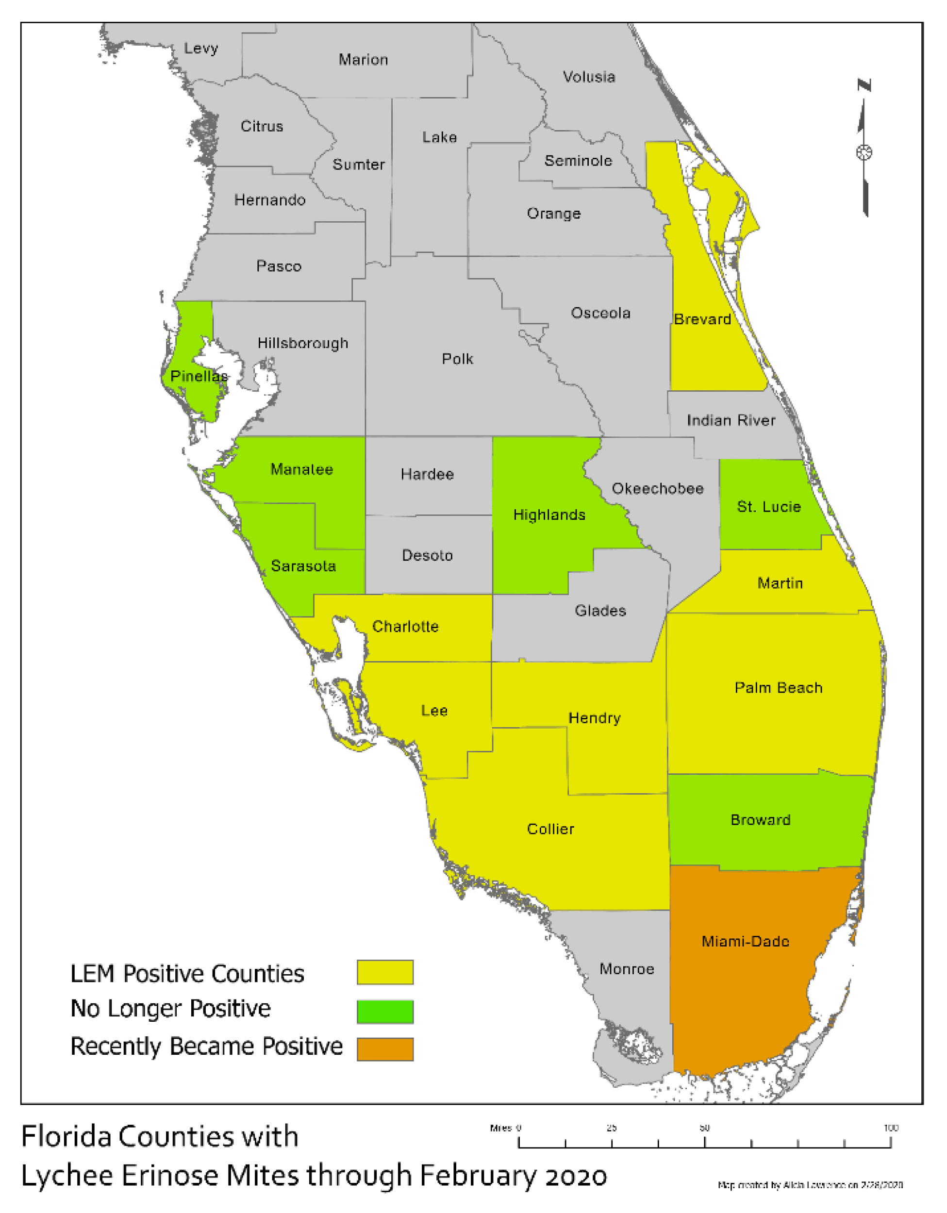
For morphological comparison, lychee erinose mites were freshly collected from Hawaii and Florida, preserved in 70% ethanol and then examined under Low-Temperature SEM (LTSEM), following the technique delineated by Bolton et al. [22]. Due to a poor state of preservation, the slide mounted type specimens of Keifer [6] were unavailable for comparison with fresh material.
DNA was extracted from ten individual mites from Hawaii, Florida, India, Brazil, Australia, and Taiwan, using the protocol reported by Carew et al. [23]. Amplification of the cytochrome oxidase subunit I (COI) was carried out with the primer and conditions reported by Navajas et al. [24].The internal transcribed spacer 1 (ITS1) along with the 5′ terminal region of 5.8S were amplified with the primer sets and conditions reported by Fenton et al. [25]. The internal transcribed spacer 2 (ITS2) along with the 5′ terminal region of the 28S nuclear ribosomal rDNA (28S) were amplified with the primer sets and conditions reported by Sonnenberg et al. [26] and Chetverikov et al. [27]. DNA was amplified on a nexus G×2 thermocycler (Eppendorf, Hamburg, AG) in reaction mixtures (25 µL) containing the following components: 12.5 µL Platinum SuperFi Green PCR master Mix (Invitrogen, Carlsbad, CA, USA) 0.4 µM each primer and 3 µL DNA template. PCR products were verified by gel electrophoresis. The amplicon size was 460 bp for COI, 500 bp for ITS1a and 1.6kb for ITS2. PCR products were purified using ExoSAP-IT (Affymetrix, Santa Clara, CA, USA) following the manufacturer’s protocols. Sanger sequencing was performed by Eurofins genomics (Louisville, KY, USA), amplification primers were also used as sequencing primers.
Geneious software, version 9.1.5 [28] and the Staden Package [29], were used to trim low-quality sequence end reads, create consensus gene sequences, and align sequences of the individual molecular markers of A. litchii from the different locations. The length of the resulting trimmed sequences used for the analyses was 492 bp for ITS1, 936 for ITS2 and 363 for COI. Sequences were deposited in GenBank (Table 1).
Phylogenies were generated for the individual COI and ITS1 sequence alignments and for the concatenated ITS1 and ITS2 segments by retrieving GenBank accessions of related Aceria species (Table S1). Abacarus sacchari Channabasavanna, 1966 was chosen as the outgroup.
The datasets of individual markers and the concatenated ITS1- ITS2 were aligned and trimmed to a similar length in Mega 7 software [30]. Phylogenies were generated under the maximum-likelihood method using 1000 bootstraps with Mega 7 software [30]. COI phylogeny was based on the Hasegawa–Kishino–Yano model [31] with discrete Gamma distribution to model evolutionary rate differences among sites, a total of 439 positions were used for this dataset. ITS1 phylogeny was based on Tamura 3-parameter model [32] with discrete Gamma distribution, a total of 509 positions were used in the final dataset. The concatenated ITS1-ITS2 was based on the Kimura 2-parameter model [33]; this dataset contained a total of 1170 positions.
Genetic diversity of the three molecular markers among the collection sites was described by estimating the number of haplotypes (COI) or alleles (ITS1 and ITS2), nucleotide diversity (π) and number of polymorphic segregating sites, these parameters were calculated in DnaSP version 6 software [34]. Estimations of pairwise divergency were computed using Kimura’s 2-parameter (K2P) distance model [33]. For COI, the first codon position was included and transition + transversion was chosen as the substitution type.

3. Results

The specimens from Florida resemble the descriptions of LEM by Keifer [6] and Huang [21]. However, with respect to the prodorsum, there are three noticeable differences between the descriptions and the Florida specimens: (1) the median line of the Florida specimens is almost always broken, whereas it is unbroken in the drawn figures from the two descriptions by Keifer [6] and Huang [21]; (2) in the Florida specimens, the arrangement of ridges in the posteromedian region is typically asymmetrical and highly variable among specimens (Figure 3A–E), whereas it is symmetrical and varies little between the figures of Keifer [6] and Huang [21]; (3) the Florida specimens have three lateral cells on each side of the prodorsum (Figure 3B), whereas the Keifer description only appears to show one of those cells. LEM obtained from Hawaii were similar to the Florida specimens with respect to all three characters (Figure 3F).
All the individuals from each location had identical sequences for all the molecular markers. Analysis of the LEM-COI segments showed low sequence variability, as indicated by the lowest nucleotide diversity, number polymorphic segregating sites (Table 2), as well as the smallest genetic distances between the locations (Table 3). COI segments from Australia, Brazil and Hawaii shared 100% of identity, whereas Florida, Taiwan and India showed unique alleles. The ITS1 sequence and partial 5.8S fragment of the Florida LEM were identical to those of Hawaii, India, Brazil, and Taiwan. By contrast, Australia, with 62 polymorphic sites, shared 79.9% of identity with the rest of the locations (Table 2 and Table 3). The ITS2 and 28S partial sequences from Australia and Hawaii, as well as Taiwan and Florida, shared 100% sequence identity. This dataset showed the largest number of segregation sites distributed across the locations and the largest genetic distances among the molecular markers (Table 2 and Table 3). The phylogeny generated from the COI analysis resulted in a tree with two major clades. One clustered the LEM sequences from the six locations and the second clade contained all other Aceria spp. Due to the low intraspecific variation of the COI segment, no resolution was observed within the LEM clade (Figure S1). The analysis of the concatenated ITS1 and ITS2 provided resolution within the LEM clade (Figure 4), with Australia in the most basal position, followed by Hawaii, India and then Brazil, so that each of these locations is sister to a clade comprising the remaining locations, while Florida and Taiwan form the two most recently diverging locations.

4. Discussion

Differences between the Florida specimens and the descriptions of Keifer [6] and Huang [21] are probably an artifact of the drawings, which appear to simplify the detail and variation revealed in the LTSEM images.
The molecular analysis of the LEM specimens from different geographic areas suggests that the mitochondrial CO1 gene region, a standard genetic marker for a broad range of animals [35], is potentially appropriate for accurate identification of LEM. The COI sequence could be an important tool to identify LEM immature stages. Our results also suggest that the ITS1 and ITS2 regions are useful for studies on intraspecific variability. The ITS1 and ITS2 regions may be useful to trace specific LEM populations and identify introduction routes. This information may be useful to prevent reintroduction or further spread of this pest. The ITS2 and 28S sequences vary by geographic location. Divergence of the Australian population may have occurred earliest, resulting in the observed tree topology. Interestingly, LEMs from Taiwan and Florida seem to present identical genotypes, perhaps indicating the same origin. Further analysis is required to ascertain the origin of the Florida and Taiwan populations. We were unable to obtain specimens from Southern China, where lychee originates [36]. Future efforts to study the biogeography of LEM should include specimens from Southern China.
Lychee is a difficult crop for farmers in Florida because yield can vary greatly due to irregular flowering and poor fruiting retention [12]. The establishment of LEM in commercial orchards could become a major pest problem that could further reduce yields. It is unknown how LEM was introduced into Lee County, Florida. Considering the geographic distance between the known geographic range of LEM and Florida, the founders of the introduced populations must have been transported by human activities. The detection reported here is the third introduction of LEM in Florida. The first detection was in 1955 in a lychee grove located at Nokomis, Sarasota County. This introduction failed to establish and was likely eradicated through a combination of extensive pruning, acaricide treatments and extremely cold temperatures, which were reported in 1960 [37]. The second interception was in Coral Gables, Miami-Dade County in 1993; LEM infested containerized trees imported from China, which were destroyed to eradicate the pest [38]. The population recently detected on Pine Island has already spread to multiple Florida counties. Notably, the principal lychee commercial growing area in South Florida is, so far, free of LEM. Suppression or eradication of this current LEM invasion in Florida will depend largely on the strategic actions of regulatory agencies and industry to manage this pest.
Known management tools targeting LEM are restricted to cultural and chemical practices. Postharvest pruning, which includes eliminating and burning infested branches, is the main cultural control strategy against LEM [39,40]. Chemical control has been studied in Australia, China, and Brazil [7,14,39]. Several active ingredients have shown some efficacy on LEM [15,41]. The main management strategy focuses on protection of new flush after the postharvest sanitation through carefully timed acaricide applications [39,42]. Sprays of dimethoate or wettable sulfur at 2–3 week intervals, starting at bud emergence until leaves have hardened, provided protection of new flush against LEM in Australia [39]. In Hawaii, sulfur applications have been used to protect new flush against LEM [43]. A recent study indicates that post-harvest paraffinic oil dips have potential to disinfest lychee fruit from LEM, thereby minimizing risk of further pest spread in Florida [44].
Biological control might provide a long-term solution to managing this invasive mite. Several natural enemies have been reported in association with LEM in India [45], Australia [46,47], Brazil [42,48] and China [49]. However, predation on LEM has only been confirmed for a few species of phytoseiid mites, including Amblyseius largoensis (Muma) in China [50], and Phytoseius intermedius Evans and MacFarlane in Brazil [48]. In Florida, A. largoensis, Euseius membrasicus, Hemycheyletia bakeri and Cheylotogenes ornatus have been found to be associated with LEM. The mite pathogen Hirsutella thompsonii (Fischer) has also been reported to infect LEM in Brazil [51].
The State of Florida is conducting an eradication program for LEM [13]. If the eradication program is not successful, a comprehensive program for managing LEM will require: (1) detailed investigations on the biology and ecology of LEM, (2) implementation of effective regulatory actions to prevent movement of LEM infested material, (3) development of phytosanitary postharvest treatments, (4) determination of the susceptibility of lychee cultivars grown in neighboring areas, and (5) development of biological and chemical control programs against LEM.

5. Conclusions

The invasive LEM was recently found on Pine Island, Florida. Infestations have spread to several FL counties but have not reached the main commercial production area in South Florida. Initial taxonomic uncertainties were resolved through comparison of the Florida specimens with fresh specimens collected from Hawaii, the type locality. A molecular analysis of specimens from India, Hawaii, Brazil, Taiwan, Australia and Florida revealed that LEM from Australia had diverged earlier and that LEM from Taiwan and Florida may have a common origin. The methodologies used here can be used to accurately identify LEM and trace further movement of LEM populations. The impact of LEM on Florida’s lychee industry will depend on the success of an eradication program currently conducted by the state’s regulatory agency. Management programs specific for LEM are lacking in Florida.

Supplementary Materials

The following are available online at https://www.mdpi.com/2075-4450/11/4/235/s1, Figure S1: Phylogeny of Aceria species Cytochrome oxidase 1 by the Maximum Likelihood method, Figure S2: Phylogeny of Aceria species ITS1 region by the Maximum Likelihood method, Table S1: GenBank accession of sequences used in the phylogenetic analyses.

Author Contributions

Conceptualization, investigation, funding acquisition, project administration, writing—original draft preparation D.C.; formal analysis L.F.C.; investigation, writing—original draft preparation A.M.R.; investigation, R.E.D.; methodology, writing—review and editing, G.R.B.; investigation, writing—review and editing R.O.; writing—review and editing, funding acquisition, P.E.K.; formal analysis, writing—review and editing, S.J.B. All authors have read and agreed to the published version of the manuscript.

Funding

This research was funded by USDA ARS-UF Non-Assistance Cooperative Agreement No. 5860388004. The findings and conclusions in this preliminary publication have not been formally disseminated by the U.S. Department of Agriculture and should not be construed to represent any Agency determination or policy. Mention of trade names or commercial products in this publication is solely for the purpose of providing specific information and does not imply recommendation or endorsement by the USDA; USDA is an equal opportunity provider and employer.

Acknowledgments

We thank Janis Matsunaga, Krishna Karmakar, Hsio-Hua Hao, Gilberto de Moraes, Jenny Beard for providing Aceria litchii specimens from Hawaii, India and Taiwan, Brazil and Australia, respectively. We thank Cheng-Fang Hong for translations of literature in Mandarin and facilitating contacts to obtain A. litchii specimens from Taiwan. We thank Armando Monterroso from Brooks Tropicals for allowing access to the site of the original detection. We thank Araceli Lamelas for guidance in the phylogenetic analysis. We thank the Florida Department of Agriculture and Consumer Services, Division of Plant Industry, for providing information on the distribution of Aceria litchii in Florida.

Conflicts of Interest

The authors declare no conflict of interest.

References

  1. Sharma, D.D. Major pests of litchi in Bihar. Indian Farming 1985, 35, 25–26. [Google Scholar]
  2. Alam, Z.M.; Wadud, M.A. On the biology of Litchi mite, Aceria litchii Keifer (Eriophyidae: Acarina) in East Pakistan. Pak. J. Sci. 1963, 15, 232–240. [Google Scholar]
  3. Haque, M.M.; Das, B.C.; Khalequzzaman, M.; Chakrabarti, S. Eriophyoid mites (Acari: Eriophyoidea) from Bangladesh. Orient. Insects 1998, 32, 35–40. [Google Scholar] [CrossRef]
  4. Keifer, H.H.; Knorr, L.C. Eriophyid Mites of Thailand. Plant Prot. Serv. Tech. Bull. (Bangkok) 1978, 38, 1–36. [Google Scholar]
  5. Huang, T.A. Study on Morphological Features of Erinose Mite of Litchi (Eriophyes litchii Keifer) and an observation on the conditions of its damage. Plant Prot. Bull. 1967, 9, 35–46. [Google Scholar]
  6. Keifer, H.H. Eriophyid studies XIII. State Calif. Dep. Agric. Bull. 1943, 32, 212–222. [Google Scholar] [CrossRef]
  7. Pinese, B. Erinose mite—A serious litchi pest. Queensl. Agric. J. 1981, 107, 79–81. [Google Scholar]
  8. Raga, A.; Mineiro, J.L.D.C.; Sato, M.E.; De Moraes, G.J.; Flechtmann, C.H.W. Primeiro relato de Aceria litchii (Keifer) (prostigmata: Eriophyidae) em plantas de lichia no Brasil. Rev. Bras. Frutic. 2010, 32, 628–629. [Google Scholar] [CrossRef] [Green Version]
  9. Navia, D.; Júnior, A.L.M.; Gondim, M.G.C., Jr.; de Mendonza, R.s.; da Silva Pereira, P.R.V. Recent mite invasions in South America. In Potential Invasive Pests Agric Crop, 1st ed.; Peña, J.E., Ed.; CABI: Oxfordshire, UK, 2013; pp. 251–287. [Google Scholar]
  10. Prasad, V.G.; Singh, R.K. Prevalence and control of litchi mite, Aceria litchii (Keifer) in Bihar. Indian J. Entomol. 1981, 43, 67–75. [Google Scholar]
  11. The USDA 2012 Prioritized Offshore Pest List. Available online: https://www.aphis.usda.gov/plant_health/plant_pest_info/pest_detection/downloads/farmbill/PrioritizedOffshorePestList.pdf (accessed on 18 June 2019).
  12. Evans, E.; Degner, R.; Morgan, K. Six Ways to Improve the Profitability of Lychee in South Florida. 2004. EDIS 18. Available online: https://journals.flvc.org/edis/article/view/114112 (accessed on 8 April 2020).
  13. FDACS (2020) Lychee Erinose Mite Quarantine Information. Available online: https://www.fdacs.gov/Agriculture-Industry/Pests-and-Diseases/Plant-Pests-and-Diseases/Lychee-Erinose-Mite (accessed on 18 March 2020).
  14. Karioti, A.; Tooulakou, G.; Bilia, A.R.; Psaras, G.K.; Karabourniotis, G.; Skaltsa, H. Erinea formation on Quercus ilex leaves: Anatomical, physiological and chemical responses of leaf trichomes against mite attack. Phytochemistry 2011, 72, 230–237. [Google Scholar] [CrossRef]
  15. Azevedo, L.H.; Moraes, G.J.; Yamamoto, P.T.; Zanardi, O.Z. Development of a Methodology and Evaluation of Pesticides Against Aceria litchii and Its Predator Phytoseius intermedius (Acari: Eriphyidae, Phytoseiidae). J. Econ. Entomol. 2013, 106, 2183–2189. [Google Scholar] [CrossRef] [PubMed]
  16. Waite, G.K.; McAlpine, J.D. Honey bees as carriers of lychee erinose mite Eriophyes litchii (Acari: Eriophyiidae). Exp. Appl. Acarol. 1992, 15, 299–302. [Google Scholar] [CrossRef]
  17. Waite, G.K. New evidence further incriminates honey bees as vectors of lychee erinose mite Aceria litchii (Acari: Eriophyiidae). Exp. Appl. Acarol. 1999, 23, 145–147. [Google Scholar] [CrossRef]
  18. Jeppson, L.R.; Keifer, H.H.; Baker, E.W. Mites Injurious to Economic Plants; University of California Press: Berkeley, CA, USA, 1975; p. 360. [Google Scholar]
  19. Oldfield, G.N. Diversity and host plant specificity. In Eriophyoid Mites—Their Biology, Natural Enemies and Control; Lindquist, E.E., Sabelis, M.W., Bruin, J., Eds.; World Crop Pests vol 6; Elsevier Science Publishing: Amsterdam, The Netherlands, 1996; pp. 199–216. [Google Scholar]
  20. Crane, J.H.; Balerdi, C.F.; Maguire, I. Lychee Growing in the Florida Home Landscape. EDIS, 2016(HS6). Available online: https://edis.ifas.ufl.edu/pdffiles/MG/MG05100.pdf (accessed on 8 April 2020).
  21. Huang, K. Aceria (Acarina: Eriophyoidea) in Taiwan: Five new species and plant abnormalities caused by sixteen species. Zootaxa 2008, 1829, 1–30. [Google Scholar] [CrossRef]
  22. Bolton, S.J.; Klompen, H.; Bauchan, G.R.; Ochoa, R. A new genus and species of Nematalycidae (Acari: Endeostigmata). J. Nat. Hist. 2014, 48, 1359–1373. [Google Scholar] [CrossRef]
  23. Carew, M.E.; Goodisman, M.A.D.; Hoffmann, A.A. Species status and population genetic structure of grapevine eriophyoid mites. Entomol. Exp. Appl. 2004, 111, 87–96. [Google Scholar] [CrossRef]
  24. Navajas, M.; Gutierrez, M.; Lagnel, J.; Boursot, P. Mitochondrial cytochrome oxidase 1 in tetranychid mites: A comparison between molecular phylogeny and changes of morphological and life history traits. Bull. Ent. Res. 1996, 86, 407–417. [Google Scholar] [CrossRef] [Green Version]
  25. Fenton, B.; Malloch, G.; Moxey, E. Analysis of eriophyid mite rDNA internal transcribed spacer sequences reveals multiple simple sequence repeats. Insect. Mol. Biol. 1997, 6, 23–32. [Google Scholar] [CrossRef]
  26. Sonnenberg, R.; Nolte, A.W.; Tautz, D. An evaluation of LSU rDNA D1-D2 sequences for their use in species identification. Front. Zool. 2007, 4, 6. [Google Scholar] [CrossRef] [Green Version]
  27. Chetverikov, P.E.; Cvrković, T.; Makunin, A.; Sukhareva, S.; Vidović, B.; Petanović, R. Basal divergence of Eriophyoidea (Acariformes, Eupodina) inferred from combined partial COI and 28S gene sequences and CLSM genital anatomy. Exp. Appl. Acarol. 2015, 67, 219–245. [Google Scholar] [CrossRef]
  28. Kearse, M.; Moir, R.; Wilson, A.; Stones-Havas, S.; Cheung, M.; Sturrock, S.; Buxton, S.; Cooper, A.; Markowitz, S.; Duran, C.; et al. Geneious Basic: An integrated and extendable desktop software platform for the organization and analysis of sequence data. Bioinformatics 2012, 28, 1647–1649. [Google Scholar] [CrossRef]
  29. Bonfield, J.K.; Smith, K.F.; Staden, R. A new DNA sequence assembly program. Nucleic Acids Res. 1995, 23, 4992–4999. [Google Scholar] [CrossRef] [PubMed] [Green Version]
  30. Kumar, S.; Stecher, G.; Tamura, K. Molecular Evolutionary Genetics Analysis version 7.0 for bigger datasets. Mol. Biol. Evol. 2017, 33, 1870–1874. [Google Scholar] [CrossRef] [PubMed] [Green Version]
  31. Hasegawa, M.; Kishino, H.; Yano, T. Dating the human-ape split by a molecular clock of mitochondrial DNA. J. Mol. Evol. 1985, 22, 160–174. [Google Scholar] [CrossRef] [PubMed]
  32. Tamura, K. Estimation of the number of nucleotide substitutions when there are strong transition-transversion and G + C-content biases. Mol. Biol. Evol. 1992, 9, 678–687. [Google Scholar]
  33. Kimura, M. A simple method for estimating evolutionary rate of base substitutions through comparative studies of nucleotide sequences. J. Mol. Evol. 1980, 16, 111–120. [Google Scholar] [CrossRef]
  34. Rozas, J.; Ferrer-Mata, A.; Sanchez-DelBarrio, J.C.; Guirao-Rico, S.; Librado, P.; Ramos-Onsins, S.E. DnaSP 6: DNA Sequence Polymorphism Analysis of Large Data Sets. Mol. Biol. Evol. 2017, 34, 3299–3302. [Google Scholar] [CrossRef]
  35. Rach, J.; Bergmann, T.; Paknia, O.; DeSalle, R.; Schierwater, B.; Hadrys, H. The marker choice: Unexpected resolving power of an unexplored CO1 region for layered DNA barcoding approaches. PLoS ONE 2017, 12, e017484. [Google Scholar] [CrossRef] [Green Version]
  36. Huang, X.; Subhadrabandhu, S.; Mitra, S.K.; Ben-Aire, R.; Stern, R.A. Origin, History, Production and Processing. In Litchi and Longan: Botany, Production and Uses; Menzel, C.M., Waite, G.K., Eds.; CABI Publishing: Oxfordshire, UK, 2005; pp. 1–25. [Google Scholar]
  37. Dekle, G.W. Twenty-third biennial Report for the period July 1, 1958-June 30, 1960. State Plant Board Fla. Bull. 1960, 14, 65–70. [Google Scholar]
  38. Mead, F.A. Entomology Section. Triy-ology 1993, 32, 3. [Google Scholar]
  39. Waite, G.K. Pests. In Litchi and Longan: Botany, Production and Uses; Menzel, C.M., Waite, G.K., Eds.; CABI Publishing: Oxfordshire, UK, 2005; pp. 237–259. [Google Scholar]
  40. Castro, B.M.C.; Plata-Rueda, A.; Meloni-Silva, W.; de Menezes, C.W.G.; Wilcke, C.F.; Zanuncio, J.C. Manejo del ácaro Aceria litchii (Acari: Eriophyidae) en Litchi chinensis. Rev. Colomb. Entomol. 2018, 44, 2. [Google Scholar] [CrossRef]
  41. Schulte, M.J.; Martin, K.; Sauerborn, J. Efficacy of spiromesifen on Aceria litchii (Keifer) in relation to Cephateuros virescens Kunze colonization on leaves of litchi (Litchi chinensis Sonn.). J. Plant. Dis. Prot. 2007, 114, 133–137. [Google Scholar] [CrossRef]
  42. Picoli, P.R.F.; Vieira, M.R.; da Silva, E.A.; da Mota, M.S.d.O. Ácaros Predadores Associados Ao Ácaro-Da-Erinose Da Lichia. Pesqui Agropecu Bras 2010, 45, 1246–1252. [Google Scholar] [CrossRef] [Green Version]
  43. Nishida, T.; Holdaway, F.G. The Erinose Mite of Lychee. Circular No. 48; Hawaii Agriculture Experiment Station: Honolulu, HI, USA, 1955; p. 10. [Google Scholar]
  44. Revynthi, A.M.; Duncan, R.E.; Mannion, C.; Kendra, P.E.; Carrillo, D. Post-harvest paraffinic oil dips to disinfest lychee fruit from lychee erinose mite. Fla. Entomol. 2020, (in press). [Google Scholar]
  45. Lall, B.S.; Rahman, M.F. Studies on the bionomics and control of the erinose mite Eriophyes litchii Keifer (Acarina: Eriophyidae). Pesticides 1975, 9, 49–54. [Google Scholar]
  46. Schicha, E. Phytoseiidae of Australia and Neighbouring Areas; Indira Publishing House: West Bloomfield, MI, USA, 1987; 187p. [Google Scholar]
  47. Waite, G.K.; Gerson, U. The predator guild associated with Aceria litchii (Acari: Eriophyidae) in Australia and China. Entomophaga 1994, 39, 275–280. [Google Scholar] [CrossRef]
  48. Azevedo, L.H.; Maeda, E.Y.; Inomoto, M.M.; De Moraes, G.J. A Method to Estimate the Population Level of Aceria litchii (Prostigmata: Eriophyidae) and a Study of the Population Dynamics of This Species and Its Predators on Litchi Trees in Southern Brazil. J. Econ. Entomol. 2014, 107, 361–367. [Google Scholar] [CrossRef] [PubMed]
  49. Waite, G.K.; Hwang, J.S. Pests of litchi and longan. In Tropical Fruit Pests and Pollinators: Biology Economic Importance, Natural Enemies and Control; Peña, J.E., Sharp, J.L., Wysoki, M., Eds.; CAB International: Wallingford, UK, 2002; pp. 331–359. [Google Scholar]
  50. Cheng, L.; Zhang, X.; Sha, L.; Lu, A.; Chen, P. Functional and numerical response of Amblyseius largoensis to Aceria litchii. J. Trop. Crop. 2005, 26, 53. [Google Scholar]
  51. Picoli, P.R.F.; Vieira, M.R. Primeiro relato de atividade patogênica de Hirsutella thompsonii (Fischer) sobre o ácaro-da-erinose-da-lichia Aceria litchii (Keifer). Semin. Agrar. 2013, 34, 187–190. [Google Scholar] [CrossRef] [Green Version]
Figure 1. Current distribution of lychee erinose mite (LEM) (Aceria litchii) in Florida.
Insects 11 00235 g001
Figure 2. Erinea types associated with different levels of infestation of LEM-topside on left, underside on right (note that most of the topside of (D) is obscured because the leaf has folded). (A) Low level of infestation. (B) Moderate level of infestation. (C) High level of infestation. (D) Extreme level of infestation (topside of leaf with some erineum hairs).
Insects 11 00235 g002
Figure 3. Variation of the prodorsum of LEM from Florida and Hawaii examined under Low-Temperature SEM (LTSEM), following the technique delineated by Bolton et al. [22] (A)–(E) Dorsal view of Florida specimens (unknown instars) (only (B) is labeled). Note that the median line is sometimes broken anteriorly and/or posteriorly. The arrangement of the ridges in the posteromedian region also varies dramatically so that the posterior part of the median line can run centrally, or curve left or right. (F) Lateral view of Hawaii specimen (unknown instar), showing the broken median line and the two additional lateral cells also observed in the Florida specimens. M = Median line; C = Cells missing from figure of Keifer [6]. Adults 110-135 μm long.
Insects 11 00235 g003
Figure 4. Phylogeny of Aceria species as inferred by Maximum Likelihood of the ITS1 and ITS2 concatenated sequences. Numbers presented above and below each node indicate maximum likelihood bootstrap values and Bayesian a posteriori probability for the corresponding inner branch.
Insects 11 00235 g004
Table 1. Lychee erinose mite (Aceria litchii) specimens collected from different countries and used for DNA fragment sequence analysis. GenBank accession numbers for all analyzed sequences are provided.
Geographic OriginCoordinatesCollectorCollection DateGenBank Sequences
COIITS1-5.8ITS2-28S 5’
Australia24.87 S 56.78 EE. Dunn & J.J. Beard3-Apr-19MT027817MN594262MN594268
Brazil22.71 S 43.63 WG.J de Moraes25-Mar-19MT027818MN594263MN594269
Florida26.6 N 82.1 WD. Carrillo & A.M Revynthi20-Feb-18MT027819MN594264MN594270
Hawaii21.30 N 157.82 WB. Azama & M. Ramadan15-Mar-18MT027820MN594265MN594271
India22.94 N 88.53 EK. Karmakar25-Mar-19MT027821MN594266MN594272
Taiwan22.64 N 120.35 EC.F. Hong22-Apr-19MT027822MN594267MN594273
Table 2. Sequence diversity estimates for A. litchii.
Molecular Marker Number of SitesNo. of Alleles/HaplotypesNo. of Segregating Sites Nucleotide Diversity Per Site (π)
ITS1 4922620.04665
ITS29364640.03874
COI363460.00724
Table 3. Pairwise K2P distances between the A. litchii from six locations.
ITS1ITS2COI
FLAuBrHWIndFLAuBrHWIndFLAuBrHWInd
Au0.140 0.066 0.008
Br0.0000.140 0.0040.069 0.0080.000
HW0.0000.1400.000 0.0660.0000.069 0.0080.0000.000
Ind0.0000.1400.0000.000 0.0090.0670.0090.067 0.0170.0080.0080.008
Tw0.0000.1400.0000.0000.0000.0000.0660.0040.0660.0090.0170.0080.0080.0080.000

