Article

Bombyx mori Nucleopolyhedrovirus p26 Is Associated with Viral Late Stage Replication

1
Institute of Biotechnology, Fujian Academy of Agricultural Sciences, Wusi Road 247, Fuzhou 350003, China
2
Department of Entomology, College of Plant Protection, Fujian Agriculture and Forestry University, Shangxiadian Road 15, Fuzhou 350002, China
*
Author to whom correspondence should be addressed.
Insects 2021, 12(8), 707; https://doi.org/10.3390/insects12080707
Submission received: 28 June 2021 / Revised: 3 August 2021 / Accepted: 4 August 2021 / Published: 6 August 2021
(This article belongs to the Special Issue Advances in the Use of Insect Cell Culture and Biotechnology)

Abstract

:

Simple Summary

p26 is conserved among all completely sequenced Lepidoptera baculoviruses, and some baculoviruses even have two copies of p26 (p26a and p26b), which suggested that p26 may have a basic role in the baculovirus infection cycle. p26 may be transcribed by the host RNA polymerase II in both early and late infection. Here, protein analyses showed that Bombyx mori nucleopolyhedrovirus (BmNPV) p26 levels were very low amounts during the early phases of infection, which then increased and then declined during the late infection phase. Thus, BmNPV p26 might have functions in both the cytoplasm and nucleus. Previous p26 knockout study indicated that p26 may be an auxiliary gene that does not influence key aspects of viral replication or transmission, and RNAi response to p26 may somewhat regulate viral replication. Therefore, in order to maintain low p26 expression and measure BmNPV p26 function, a RNAi-based knockdown method was chosen. The results indicated that high p26 expression during the middle interval is necessary for late-stage viral replication. Since p26 is not essential for baculovirus replication and transmission, it would be interesting to investigate whether p26 is involved in regulating host innate immune response.

Abstract

Bombyx mori nucleopolyhedrovirus (BmNPV) p26 is conserved among all Lepidoptera baculoviruses that have been completely sequenced thus far, and some baculoviruses even have two copies of p26, which suggested that p26 may play an important role in the virus infection cycle. This study aimed to characterize BmNPV p26. We found that BmNPV p26 transcripts were detectable as early as 3 h post-infection (hpi), and the transcript levels rapidly increased starting from 12 hpi. Western blot analysis using an anti-p26 polyclonal antibody demonstrated that the corresponding protein was also detectable from 6 hpi in BmNPV-infected cell lysates. Immunofluorescence analysis demonstrated that p26 was mainly dispersed in the infected cell cytoplasm, whereas the over-expressed fusion protein EGFP-p26 also accumulated in the nucleus. These results indicated that p26 is an early BmNPV gene and has functions both in the cytoplasm and the nucleus. RNAi-based knockdown of p26 could produce infectious virus and normal-appearing virions but decreased budded virus (BV) production in BmNPV-infected cells at 72 hpi. Moreover, the results of further quantitative PCR (Q-PCR) analysis indicated that the gp64 and p74 transcripts levels decreased significantly. These results indicated that BmNPV p26 may be associated with BmNPV replication during the late infection stage.

1. Introduction

Baculovirus genes are expressed via a highly regulated cascade [1]. Genes expressed in the early phase are transcribed by the host RNA polymerase II in the cell nucleus at 0–3 h post-infection (hpi) [2], and the resulting products are required for DNA replication and late gene expression [3,4]. Viral DNA replication marks the transition from early to late gene expression. Late-stage expression genes are transcribed by virus-encoded RNA polymerase [5], and most of them are involved in viral genome replication and budded virus (BV) production [6]. At very late stages, the major matrix protein for mature occluded viruses, polyhedrin (ph), is hyperexpressed.
The Bombyx mori nucleopolyhedrovirus (BmNPV) genome has been completely sequenced; it contains 136 open reading frames (ORFs) encoding predicted proteins that are > 60 amino acids in length [7]. BmNPV p26 is encoded by ORF113; its homolog in Autographa californica nucleopolyhedrovirus (AcMNPV) is encoded by ORF136 and shares a 97% amino acid identity with BmNPV p26. p26 is usually adjacent to an enhancer sequence (hr5) in baculovirus genomic loci and possesses an early promoter motif TATAA [8]. AcMNPV p26 is transcribed by the host RNA polymerase II during both early and late infection stages [9], and its transcript can be detected at 6 hpi via deep sequencing [10], which indicated that AcMNPV p26 is an early gene [11]. AcMNPV p26 knockout analysis indicated that p26 was nonessential for viral replication [12], had no apparent effect on infectious BV and occlusion-derived virion (ODV) timing or production [13], or required proper virion occlusion in the AcMNPV polyhedra [14]. Moreover, BmNPV p26 knockout virus could also replicate and produce infectious BVs in B. mori cell line BmN [15].
In this study, we examined the expression and subcellular localization of BmNPV p26 as well as its involvement in BV production in virus-infected BmN cells. The results indicated that BmNPV p26 transcripts could be detected from 3 hpi, whereas p26 protein was detectable as early as 6 hpi. Immunofluorescence microscopy showed that p26 was mainly localized in the cytoplasm, whereas over-expressed EGFP-fusion p26 was localized in both the cytoplasm and nucleus, and was most accumulated in the nucleus. RNAi-based BmNPV p26 knockdown could produce infectious virus and normal-appearing virions, but resulted in decreased BV production in BmNPV-infected BmN cells at 72 hpi. Taken together, these results indicated that BmNPV p26 is a baculovirus early gene but associated with viral late infection events.

2. Materials and Methods

2.1. Cells and Viruses

BmN cells were maintained at 27 °C in TC-100 insect medium supplemented with 10% (v/v) fetal bovine serum (Gibco). The BmNPV (T3 strain) virus was used as the wild-type virus and propagated in BmN cell line.

2.2. Computer-Assisted Sequence Analysis

The ExPASy server software (http://www.expasy.org/tools (accessed on 7 February 2017)) was used for predicting p26 domains, motifs, signal sequences, and post-translational modifications. Protein homologs were compared using BLASTP with updated GenBank/EMBL databases. Multiple sequence alignments were performed on ClustalW software (http://www.ebi.ac.uk/clustalw (accessed on 7 February 2017)) and edited by using GeneDoc software (version 2.04).

2.3. Expression of p26 in E. coli and Generation of Anti-p26 Serum

The p26 ORF was amplified from the BmNPV genome using the primers p26FW and p26RW (with BamHI and HindIII sites, respectively). The PCR products were cloned into the expression vector pET-28a (Novagen, Darmstadt, Germany) to generate the plasmid pET-28a-p26. It was then transformed into E. coli BL21 cells, which were induced to express the fusion protein His-p26. The His-p26 fusion protein was purified, extracted, and used to produce anti-p26 serum in rabbits.

2.4. RT-PCR

BmNPV-infected BmN cells were collected at 1, 3, 6, 12, 24, 48, and 72 hpi to isolate total RNA using TRIzol reagent (Invitrogen) according to the manufacturer’s instructions. The first cDNA strand was synthesized by using an oligo-p(dT)18 primer and AMV Reverse Transcriptase (TaKaRa, Dalian, China). Subsequently, p26, p74, and ie1 were partially amplified via PCR by using the primer pairs Qp26FW/Qp26RW, Qp74FW/Qp74RW, and Qie1FW/Qie1RW, respectively. The PCR products were analyzed on a 2% agarose gel. gapdh was also partially amplified by using the primer pair QBmGapdhFW/QBmGapdhRW and used as the RT-PCR control. The sequences of the primers are included in Table 1.

2.5. Immunodetection of p26

Protein samples were prepared from BmNPV-infected BmN cells harvested at 0, 3, 6, 12, 24, 36, 48, and 72 hpi and same number of lysate cell-equivalents were loaded for SDS-PAGE separation. The protein samples were then transferred onto PVDF membrane (Millipore) using a semi-dry Trans-Blot Cell apparatus (Bio-Rad). The rabbit-derived anti-p26 polyclonal antibody and horseradish peroxidase (HRP)-conjugated goat anti-rabbit IgG were used as the primary and secondary antibodies, respectively. The signal was developed with H2O2 and diaminobenzidine (DAB) as a chromogenic substrate. The BmNPV ORF122 protein (Bm122) [16], a BmNPV early gene product, was also detected from the collected protein sample series and used as reference control.

2.6. Immunofluorescence Microscopy

In order to detect p26 subcellular localization, BmNPV-infected BmN cells were subjected to confocal microscopy [17]. At 48 hpi, the cells were collected, fixed, and incubated with the anti-p26 polyclonal antibody or the pre-immune antiserum in 1× PBS for 2 h. The primary antibody was removed and incubated with protein G-fused enhanced green fluorescence protein (EGFP) for 2 h and with the nucleus (DNA)-specific stain DAPI (Sigma-Aldrich, Shanghai, China) for 1 h. Subsequently, the cells were directly visualized and photographed on a Zeiss LSM 510 confocal laser scanning microscope.

2.7. EGFP-p26 Over-Expression in BmN Cells

In order to observe potential nuclear localization of p26, EGFP-p26 was expressed in BmN cells using the Bac-to-Bac expression system. The recombinant donor vector pFastBacHTb (Invitrogen, Shanghai, China) was reconstructed to contain egfp and p26, designated as pBacHT-EGFP-p26. The plasmid was transformed into BmDH10Bac E. coli (Invitrogen) to generate the recombinant bacmid, designated as EGFP-p26/rBmBac. The extracted EGFP-p26/rBmBac DNA was transfected into BmN cells by using Cellfectin II (Invitrogen). The recombinant virus was obtained and used to infect BmN cells, and fluorescence was directly viewed under a confocal laser scanning microscope at 24 and 48 hpi. EGFP/rBmBac-infected cells were used as the control.

2.8. RNAi-Based Knockdown

The primer pair T7p26iFW/T7p26iRW with T7 RNA polymerase promoter sequences at both ends were designed. Partial BmNPV p26 sequences were then amplified from the BmNPV bacmid. Moreover, gfp was amplified by the primer pair T7GFPiFW/T7GFPiRW. The primers used here are included in Table 1. The PCR products were purified and used as templates in order to generate dsRNA by using the MEGAscript dsRNA Kit (Ambion, Shanghai, China), according to the manufacturer’s instructions. The synthesized p26 dsRNA (dsP26) and gfp dsRNA (dsGFP) were purified by using the MEGAclear Transcription Clean-Up Kit (Ambion).
BmN cells were cultured in six-well plates and infected with BVs of BmNPV (T3) (MOI = 1.0). At 3 hpi, the cells were washed and transfected with 5 μg each of dsP26 and dsGFP by using Lipofectamine 3000 (Invitrogen), and non-dsRNA-treated BmN cells were used as the controls. At 24, 48, and 72 h after dsRNA treatment, the cellular supernatants were collected for TCID50 titration analysis.

2.9. Verification of p26 Knockdown

Total RNA extraction and reverse transcription were conducted as described above, and cDNA synthesized from BmNPV-infected BmN cells treated with dsRNA was used as templates to analyze the p26 transcript levels via quantitative PCR (Q-PCR) using the primer pair Qp26FW/Qp26RW. Q-PCR was performed on the ABI 7500 real-time PCR system by using SYBR premix Ex Taq (TaKaRa), according to the manufacturer’s protocol. Moreover, ie1 transcripts amplified by using the primer pair Qie1FW/Qie1RW were included as an internal control. The derived relative quantity (RQ) values were normalized to those of non-dsRNA-treated controls using the △△Ct method. Sequences of the primers are included in Table 1.

2.10. Infectious Virus Titration

BmN cells were seeded onto 96-well plates 1 day before titration. BVs from the supernatants of BmNPV-infected BmN cells exposed to dsRNAs were collected, serially diluted, and then added to plates (with 8 wells/sample). After 5 days, the plates were scored for infection by observing cellular cytopathic effects, and TCID50/mL was calculated by using the Reed–Muench method.

2.11. Electron Microscopy

BmN cells were cultured in six-well plates and infected with BV of BmNPV (T3) (MOI = 1.0). At 3 hpi, the cells were transfected with dsP26 or dsGFP. At 72 h post dsRNA treatment, the cells were harvested for transmission electron microscopy as described previously [18]. Briefly, the cells were fixed with 2.5% glutaraldehyde and then post-fixed in osmium tetroxide, dehydrated in a standard ethanol-acetone series, infiltrated, and embedded in the Spurr medium. Finally, the embedded cell blocks were cut into superthin sections, stained, and viewed under a Hitachi transmission electron microscope.

2.12. Transcription of gp64 and p74

In order to further analyze whether p26 knockdown influences BV or ODV assembly, transcripts of the BV-specific structural protein gp64 [19] and the ODV-specific structural protein p74 [20] were analyzed via Q-PCR using the △△Ct method as described above. gapdh transcripts were used as the endogenous control [21]. The sequences of the primer pair Qgp64FW/Qgp64RW for gp64 and Qp74FW/Qp74RW for p74 are included in Table 1.

3. Results

3.1. Sequence Analysis of p26

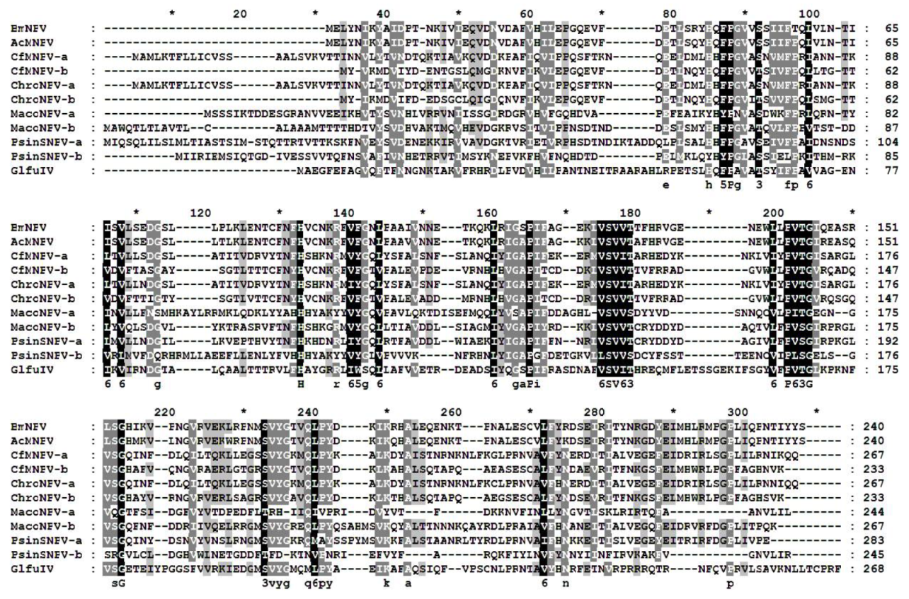
p26 is 723 nt in length and is located at 107,702–108,422 nt in the BmNPV (T3 strain) genome, which was adjacent to the enhancer sequence (hr5) and upstream of p10. Some predicted post-translational modification sites and functional motifs of p26 were explored by using EXPASY tools. However, p26 does not contain any directed domains or signal peptides that may indicate its function. Here, homologs from other baculoviruses were noted to share 27–98% identity with BmNPV p26; interestingly, a structural protein p26 (GenBank AKD28026) was found in Glypta fumiferanae ichnovirus (GlfuIV) with 35% identity (Figure A1). Furthermore, some group I and II alphabaculoviruses were found to have two copies of p26, such as Choristoneura fumiferana multiple nucleopolyhedrovirus (CfMNPV) [22], Choristoneura rosaceana nucleopolyhedrovirus (ChroNPV) [23], Mamestra configurata nucleopolyhedrovirus A (MacoNPV) [24], and Pseudoplusia includens single nucleopolyhedrovirus (PsinSNPV) [25], suggesting that p26 may play an important role in the virus infection cycle (Figure A1).

3.2. Transcription of p26

The temporal transcription of p26 was examined via RT-PCR by using total RNA isolated from BmNPV-infected BmN cells at different time points. The BmNPV p26 transcript was detectable as early as 3 hpi, much more abundant from 12 hpi, and continued to be detectable until 72 hpi (Figure 1). Meanwhile, the BmNPV late gene, p74, was detectable from 12 hpi, and the early gene, ie1, was detectable from 1 hpi. This result indicated that BmNPV p26 is an early expression gene, which corresponds to the presence of the early transcription start motif [10].

3.3. Immunodetection of p26

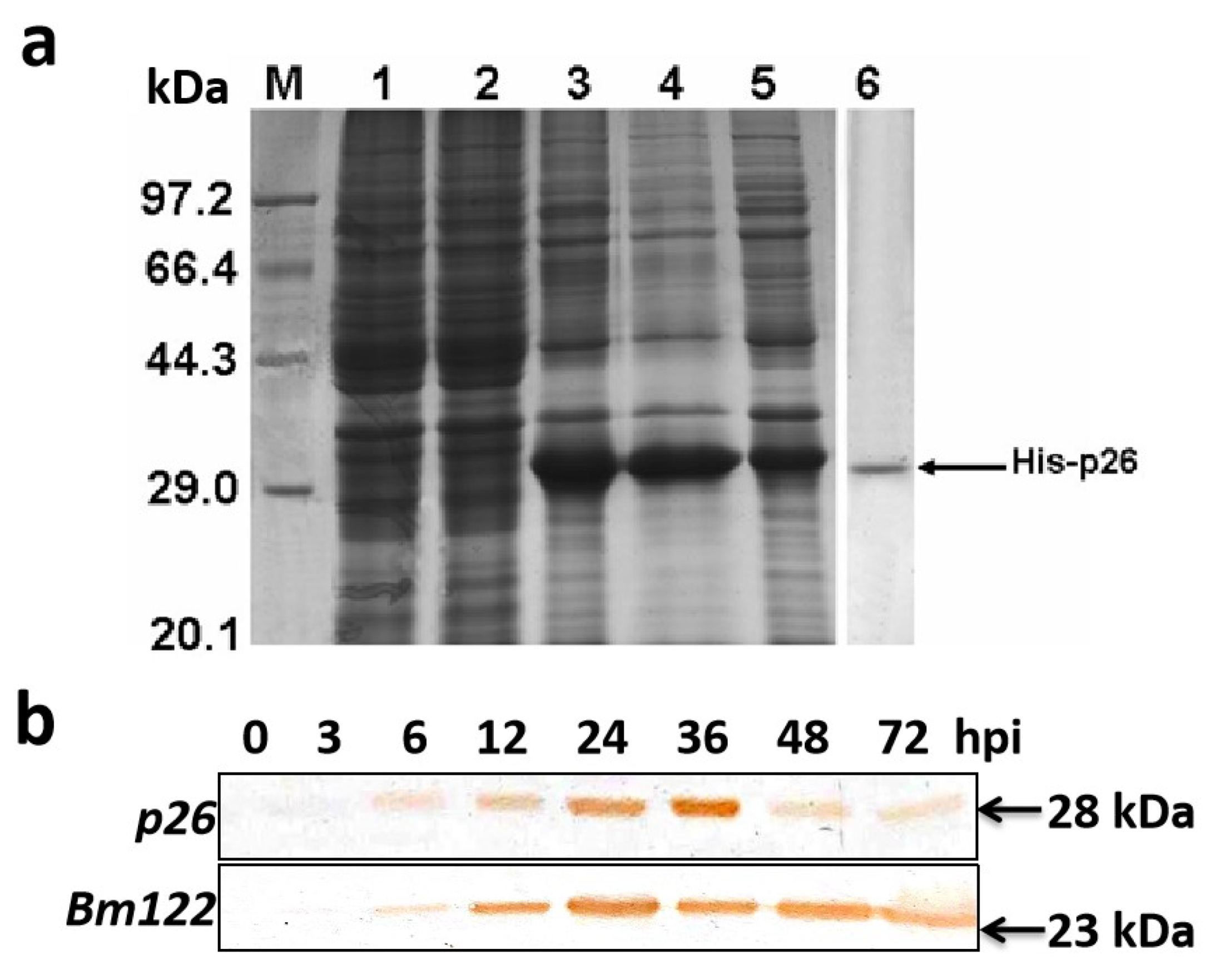
In order to obtain anti-p26 serum, the p26 coding region was expressed in E. coli (Figure 2a). The expressed fusion protein His-p26 was purified and used to raise polyclonal anti-p26 serum. Western blot analysis of protein samples extracted from BmNPV-infected BmN cells detected a specific protein of approximately 28 kDa (Figure 2b). The protein was first detected at 6 hpi, and became much more abundant at 36 hpi. However, by 48 and 72 hpi, its levels declined and it became almost undetectable. We had tried to utilize a mouse monoclonal antibody to detect beta-actin as a loading control but failed to detect any positive band. However, another BmNPV early gene, Bm122, could maintain high expression levels at 48 and 72 hpi, confirming that BmNPV p26 had a low expression level in the late stage.

3.4. Subcellular Localization of p26

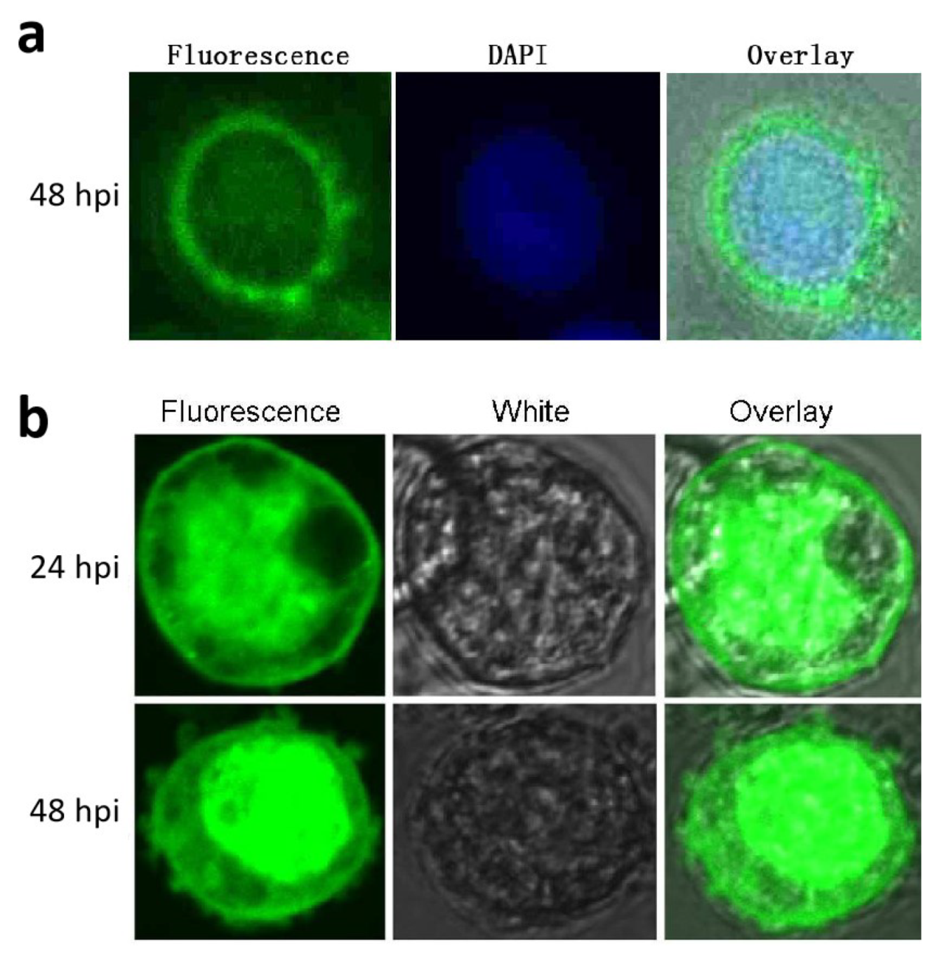
Subcellular localization of p26 was performed via immunofluorescence analysis. At 48 hpi, BmNPV-infected cells were examined for fluorescence. It was mainly detected in the cytoplasm, and some staining of the nucleus was also observed (Figure 3a). In the controls, no fluorescence was detected when pre-immune serum was used (data not shown), suggesting that p26 might play a role in both the cytoplasm and the nucleus.
As p26 were detected in the nucleus via immunofluorescence analysis, in order to more clearly confirm potential nuclear localization of p26, EGFP-p26 was over-expressed under the AcMNPV ph promoter in BmN cells using the Bac-to-Bac expression system. A recombinant bacmid, EGFP-p26/rBmBac, was extracted and transfected into BmN cells. At 24 and 48 hpi, the cells were directly observed under a confocal laser scanning microscope. The results indicated that the fluorescence still could be observed in the cytoplasm, but it became accumulated in the nucleus (Figure 3b). By contrast, when only EGFP was expressed, uniform fluorescence was observed in both the cytoplasm and the nucleus (data not shown).

3.5. Knockdown of p26

Considering that p26 transcripts were first detectable at 3 hpi, we used this time point for RNAi treatment. Q-PCR analysis indicated that the p26 transcript level was downregulated by about 90% from 24 to 72 h post-dsRNA treatment (Figure 4a), demonstrating that the synthesized dsP26 could be used to effectively knockdown p26 transcription
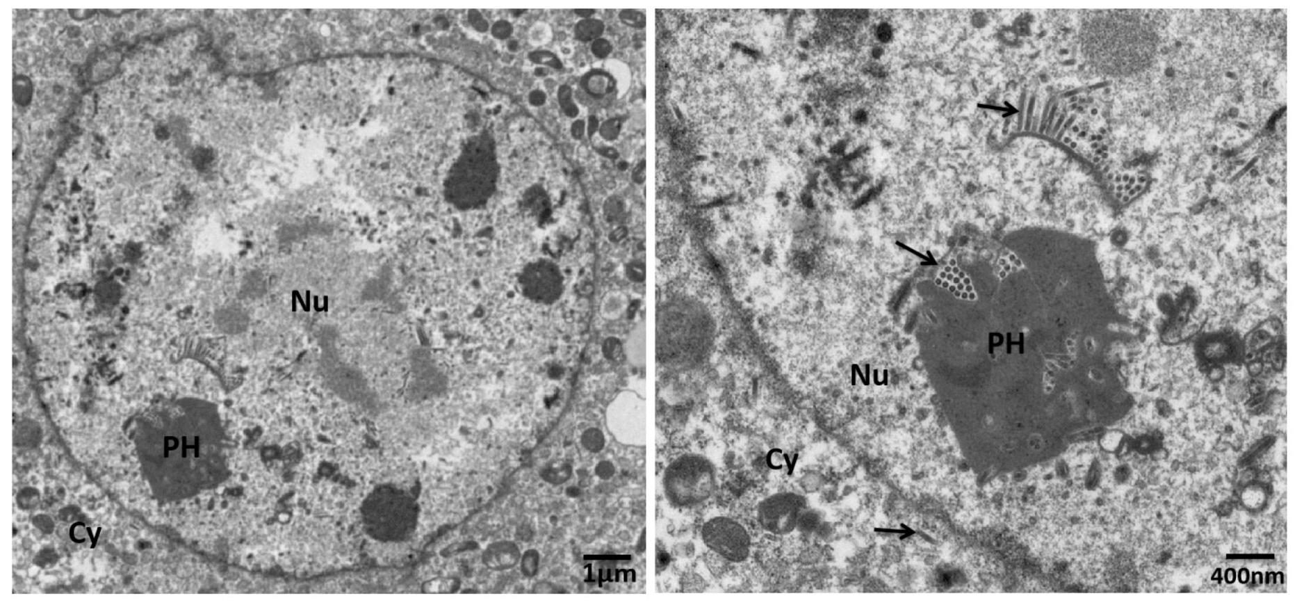
In order to determine the effects of p26 knockdown on BVs production, cell culture supernatants of BmNPV-infected cells were harvested for BV titration at the selected time points. BV production from p26 knockdown cells demonstrated no differences compared with the controls at 24 and 48 h post-dsRNA treatment, but it was significantly reduced at 72 h post-dsRNA treatment (Figure 4b). Furthermore, in order to clarify whether p26 knockdown influences of BV or ODV assembly, transcripts of BV specific gene gp64 and ODV specific gene p74 were detected by Q-PCR. The results showed that both gp64 and p74 had expression patterns consistent with above titration results, with no difference at 24 and 48 h post-dsRNA treatment but with a reduction at 72 h post-dsRNA treatment (Figure 4c). However, electron microscopy revealed that p26 knockdown virus could produce nucleocapsids with normal appearance; enveloped nucleocapsids were noted in the nucleus and cytoplasm, and assembled in the polyhedra (Figure 5).

4. Discussion

We used insect cell cultures and RNAi knockdown of p26 to explore the function of baculovirus genes. Results from this study show that BmNPV p26 knockdown did not influence BV production at 24 or 48 h post-dsRNA treatment. However, at 72 h post-dsRNA treatment, the BV production titer was significantly reduced. This result indicates that increased p26 expression during the middle interval of infection is necessary for late-stage viral replication. Even though p26 is conserved among all sequenced Leipdoptera baculovirus genomes, some baculoviruses are reported to have two copies of p26 (p26a and p26b) [25]. The RNAi pathway plays an important role in antiviral responses in insects [26], and the hotspots of siRNA in the HaSNPV genome were detected within p26, suggesting that an RNAi response to p26 may in some way regulate viral replication [10,27]. The two p26 copies likely have distinct functions [25] and are acquired independently from different sources [22]. Phylogenetic analysis suggests that p26 was obtained from three independent acquisition events within the baculoviridae family [25]. p26a is usually adjacent to p10, and p26b is usually adjacent to iap-2 [25]. BmNPV p26 was noted to be adjacent to p10, indicating that it might have the same origin as p26a.
In AcMNPV, p26 has an early promoter with a canonical TATA box, but it lacks a late promoter with a TAAG motif. Nevertheless, it is transcribed by the host RNA polymerase II both early and late in infection [10,28]. MacoNPV p26a contains a consensus early promoter, whereas MacoNPV p26b has a late promoter; notably, p26 functions are required during both early and late infection [24]. Sequence analysis results showed that an early transcription TATA box motif was found 24 nt upstream of the putative p26 initiation codon (ATG). Moreover, p26 transcripts could be detected at 3 hpi, confirming that p26 is an early BmNPV gene. Furthermore, its transcription level was investigated via Q-PCR and compared with the early gene ie1 and late gene p74. The results indicated that p26 had a high transcription level from 12 hpi [10], but no obvious differences were found among p26, ie1, and p74 (data not shown).
The BmNPV p26-specific antibody detected an approximately 28 kDa protein in BmNPV infected BmN cells, which is an approximation relative to the predicted molecular mass of 27.0 kDa. Protein analyses showed that BmNPV p26 was detected in very low amounts during the early phase of infection; it later accumulated and then declined in levels during the late infection phase. In the proteomic analyses of AcMNPV ODV proteins, p26 has not been detected [29], suggesting that p26 might not be a structural virion protein. In order to investigate whether p26 is a structural component of BmNPV virions, Western blot analysis of BVs and ODVs was performed by using the p26-specific antibody, however, no positive bands were detected (data not shown). Immunofluorescence analysis indicated that BmNPV p26 was mainly localized in the cytoplasm of infected cells. Nevertheless, weak signals were detected in the nucleus, which is similar to the result noted for AcMPNV p26 [13,30]. However, the over-expressed fusion protein EGFP-p26 accumulated in the nucleus. Although over-expressed p26 might have altered its localization, it provided further evidence that p26 might play a role in both the cytoplasm and the nucleus. Although p26 does not have a recognizable nuclear localization signal, it was observed to form dimers under physiological conditions [30]; thus, its transport to the nucleus late in infection probably depends on another protein.
AcMNPV p26 knockout studies have been performed before and indicated that p26 may be an auxiliary gene that does not influence key aspects of viral replication or transmission [12,13]. For example, the p26 of a polydnavirus (PDV), Glypta fumiferanae ichnovirus (GlfuIV) [31], is transmitted by endoparasitic wasps during egg laying into caterpillar hosts. The main function of PDV is the manipulation of host immunity and improvement of host suitability for the parasitoid [32]. Since p26 is not essential for baculovirus replication and transmission [13,14], it may provide acceptance for infection by suppressing the insect host innate immune response.

Author Contributions

J.-Q.G., Z.-H.W., X.C., and H.C. performed the experiments. J.-Q.G. conceived and designed the experiments. J.-Q.G. and J.H. analyzed the data and wrote the manuscript. All authors have read and agreed to the published version of the manuscript.

Funding

This research was funded by the Fujian Academy of Agricultural Sciences, grant number A2016-3 and National Natural Science Foundation of China, grant number 31101933.

Institutional Review Board Statement

Not applicable.

Conflicts of Interest

The authors declare no conflict of interest.

Appendix A

Figure A1. Amino acid sequence alignment of p26 homologs in Bombyx mori nucleopolyhedrovirus (BmNPV, GenBank NP_047534), Autographa californica nucleopolyhedrovirus (AcMNPV, GenBank NP_054166), Choristoneura fumiferana nucleopolyhedrovirus (CfMNPV-a, GenBank NP_848319; and CfMNPV-b, GenBank NP_848439), Choristoneura rosaceana nucleopolyhedrovirus (ChroNPV-a, GenBank YP_008378498; and ChroNPV-b, GenBank YP_008378375), Mamestra configurata nucleopolyhedrovirus A (MacoNPV-a, GenBank NP_613192; and MacoNPV-b, GenBank NP_613241), Pseudoplusia includens single nucleopolyhedrovirus (PsinSNPV-a, GenBank YP_009116933; and PsinSNPV-b, GenBank YP_009116975), and Glypta fumiferanae ichnovirus (GlfuIV, GenBank AKD28026). GeneDoc software was used for homolog shading: black, dark gray, and light gray shading denotes 100%, 80%, and 60% similarity, respectively.
Insects 12 00707 g0a1

References

  1. Blissard, G.W.; Rohrmann, G.F. Baculovirus diversity and molecular biology. Annu. Rev. Entomol. 1990, 35, 127–155. [Google Scholar] [CrossRef]
  2. Hoopes, R.R.; Rohrmann, G.F. In vitro transcription of baculovirus immediate early genes: Accurate mRNA initiation by nuclear extracts from both insect and human cells. Proc. Natl. Acad. Sci. USA 1991, 88, 4513–4517. [Google Scholar] [CrossRef] [PubMed] [Green Version]
  3. Huijskens, I.; Li, L.; Willis, L.G. Theilmann DA. Role of AcMNPV IE0 in baculovirus very late gene activation. Virology 2014, 323, 120–130. [Google Scholar] [CrossRef] [PubMed]
  4. Yamada, Y.; Matsuyama, T.; Quan, G.X.; Kanda, T.; Tamura, T.; Sahara, K.; Asano, S.; Bando, H. Use of an N-terminal half truncated IE1 as an antagonist of IE1, an essential regulatory protein in baculovirus. Virus Res. 2002, 90, 253–261. [Google Scholar] [CrossRef]
  5. Guarino, L.A.; Xu, B.; Jin, J.; Dong, W.A. virus-encoded RNA polymerase purified from baculovirus-infected cells. J. Virol. 1998, 72, 7985–7991. [Google Scholar] [CrossRef] [Green Version]
  6. Lu, A.; Miller, L.K. The roles of eighteen baculovirus late expression factor genes in transcription DNA, replication. J. Virol. 1995, 69, 975–982. [Google Scholar] [CrossRef] [Green Version]
  7. Gomi, S.; Majima, K.; Maeda, S. Sequence analysis of the genome of Bombyx mori nucleopolyhedrovirus. J. Gen. Virol. 1999, 80 Pt 5, 1323–1337. [Google Scholar] [CrossRef]
  8. Liu, A.; Qin, J.C.; Rankin, C.; Hardin, S.E.; Weaver, R.F. Nucleotide sequence of a portion of the Autographa californica nuclear polyhedrosis virus genome containing the EcoRI site-rich region (hr5) and an open reading frame just 5′ of the p10 gene. J. Gen. Virol. 1986, 67 Pt 11, 2565–2570. [Google Scholar] [CrossRef]
  9. Huht, N.E.; Weaver, R.F. Categorizing some early and late transcripts directed by the Autographa californica nuclear polyhedrosis virus. J. Gen. Virol. 1990, 71, 2195–2200. [Google Scholar]
  10. Chen, Y.R.; Zhong, S.; Fei, Z.; Hashimoto, Y.; Xiang, J.Z.; Zhang, S.; Blissard, G.W. The transcriptome of the baculovirus Autographa californica multiple nucleopolyhedrovirus in Trichoplusia ni cells. J. Virol. 2013, 87, 6391–6405. [Google Scholar] [CrossRef] [PubMed] [Green Version]
  11. Glocker, B.; Hoopes, R.R., Jr.; Rohrmann, G.F. In vitro transactivation of baculovirus early genes by nuclear extracts from Autographa californica nuclear polyhedrosis virus-infected Spodoptera frugiperda cells. J. Virol. 1992, 66, 3476–3484. [Google Scholar] [CrossRef] [PubMed] [Green Version]
  12. Rodems, S.M.; Friesen, P.D. The hr5 transcriptional enhancer stimulates early expression from the Autographa californica nuclear polyhedrosis virus genome but is not required for virus replication. J. Virol. 1993, 67, 5776–5785. [Google Scholar] [CrossRef] [PubMed] [Green Version]
  13. Simon, O.; Williams, T.; Caballero, P.; Possee, R.D. Effects of Acp26 on in vitro and in vivo productivity, pathogenesis and virulence of Autographa californica multiple nucleopolyhedrovirus. Virus Res. 2008, 136, 202–205. [Google Scholar] [CrossRef]
  14. Wang, L.; Salem, T.Z.; Campbell, D.J.; Turney, C.M.; Kumar, C.M.; Cheng, X.W. Characterization of a virion occlusion-defective Autographa californica multiple nucleopolyhedrovirus mutant lacking the p26, p10 and p74 genes. J. Gen. Virol. 2009, 90 Pt 7, 1641–1648. [Google Scholar] [CrossRef]
  15. Ono, C.; Kamagata, T.; Taka, H.; Sahara, K.; Asano, S.; Bando, H. Phenotypic grouping of 141 BmNPVs lacking viral gene sequences. Virus Res. 2012, 165, 197–206. [Google Scholar] [CrossRef] [PubMed] [Green Version]
  16. Ge, J.Q.; Zhao, J.F.; Shao, Y.M.; Tian, C.H.; Zhang, C.X. Characterization of an early gene orf122 from Bombyx mori nucleopolyhedrovirus. Mol. Biol. Rep. 2009, 36, 543–548. [Google Scholar] [CrossRef]
  17. Wang, D.; Zhang, C.X. HearSNPV orf83 encodes a late nonstructural protein with an active chitin-binding domain. Virus Res. 2006, 117, 237–243. [Google Scholar] [CrossRef]
  18. Ge, J.Q.; Yang, Z.N.; Tang, X.D.; Xu, H.J.; Hong, J.; Chen, J.G.; Zhang, C.X. Characterization of a nucleopolyhedrovirus with a deletion of the baculovirus core gene Bm67. J. Gen. Virol. 2008, 89 Pt 3, 766–774. [Google Scholar] [CrossRef]
  19. Monsma, S.A.; Oomens, A.G.; Blissard, G.W. The GP64 envelope fusion protein is an essential baculovirus protein required for cell-to-cell transmission of infection. J. Virol. 1996, 70, 4607–4616. [Google Scholar] [CrossRef] [Green Version]
  20. Faulkner, P.; Kuzio, J.; Williams, G.V.; Wilson, J.A. Analysis of p74, a PDV envelope protein of Autographa californica nucleopolyhedrovirus required for occlusion body infectivity in vivo. J. Gen. Virol. 1997, 78 Pt 12, 3091–3100. [Google Scholar] [CrossRef] [Green Version]
  21. Guo, H.; Jiang, L.; Xia, Q. Selection of reference genes for analysis of stress-responsive genes after challenge with viruses and temperature changes in the silkworm Bombyx mori. Mol. Genet. Genomics 2016, 291, 999–1004. [Google Scholar] [CrossRef]
  22. De Jong, J.G.; Lauzon, H.A.; Dominy, C.; Poloumienko, A.; Carstens, E.B.; Arif, B.M.; Krell, P.J. Analysis of the Choristoneura fumiferana nucleopolyhedrovirus genome. J. Gen. Virol. 2005, 86 Pt 4, 929–943. [Google Scholar] [CrossRef]
  23. Thumbi, D.K.; Beliveau, C.; Cusson, M.; Lapointe, R.; Lucarotti, C.J. Comparative genome sequence analysis of Choristoneura occidentalis Freeman and C. rosaceana Harris (Lepidoptera: Tortricidae) alphabaculoviruses. PLoS ONE 2013, 8, e68968. [Google Scholar] [CrossRef] [PubMed] [Green Version]
  24. Li, Q.; Donly, C.; Li, L.; Willis, L.G.; Theilmann, D.A.; Erlandson, M. Sequence and organization of the Mamestra configurata nucleopolyhedrovirus genome. Virology 2002, 294, 106–121. [Google Scholar] [CrossRef] [Green Version]
  25. Craveiro, S.R.; Inglis, P.W.; Togawa, R.C.; Grynberg, P.; Melo, F.L.; Ribeiro, Z.M.; Ribeiro, B.M.; Bao, S.N.; Castro, M.E. The genome sequence of Pseudoplusia includens single nucleopolyhedrovirus and an analysis of p26 gene evolution in the baculoviruses. BMC Genom. 2015, 16, 127. [Google Scholar] [CrossRef] [PubMed] [Green Version]
  26. Van Mierlo, J.T.; van Cleef, K.W.; van Rij, R.P. Defense and counterdefense in the RNAi-based antiviral immune system in insects. Methods Mol. Biol. 2011, 721, 3–22. [Google Scholar] [PubMed]
  27. Jayachandran, B.; Hussain, M.; Asgari, S. RNA interference as a cellular defense mechanism against the DNA virus baculovirus. J. Virol. 2012, 86, 13729–13734. [Google Scholar] [CrossRef] [PubMed] [Green Version]
  28. Huh, N.E.; Weaver, R.F. Identifying the RNA polymerases that synthesize specific transcripts of the Autographa californica nuclear polyhedrosis virus. J. Gen. Virol. 1990, 71 Pt 1, 195–201. [Google Scholar] [CrossRef]
  29. Braunagel, S.C.; Russell, W.K.; Rosas-Acosta, G.; Russell, D.H.; Summers, M.D. Determination of the protein composition of the occlusion-derived virus of Autographa californica nucleopolyhedrovirus. Proc. Natl. Acad. Sci. USA 2003, 100, 9797–9802. [Google Scholar] [CrossRef] [Green Version]
  30. Goenka, S.; Weaver, R.F. The p26 gene of the Autographa californica nucleopolyhedrovirus: Timing of transcription, and cellular localization and dimerization of product. Virus Res. 2008, 131, 136–144. [Google Scholar] [CrossRef]
  31. Beliveau, C.; Cohen, A.; Stewart, D.; Periquet, G.; Djoumad, A.; Kuhn, L.; Stoltz, D.; Boyle, B.; Volkoff, A.N.; Herniou, E.A.; et al. Genomic and Proteomic Analyses Indicate that Banchine and Campoplegine Polydnaviruses Have Similar, if Not Identical, Viral Ancestors. J. Virol. 2015, 89, 8909–8921. [Google Scholar] [CrossRef] [PubMed] [Green Version]
  32. Tan, C.W.; Peiffer, M.; Hoover, K.; Rosa, C.; Acevedo, F.E.; Felton, G.W. Symbiotic polydnavirus of a parasite manipulates caterpillar and plant immunity. Proc. Natl. Acad. Sci. USA 2018, 115, 5199–5204. [Google Scholar] [CrossRef] [PubMed] [Green Version]
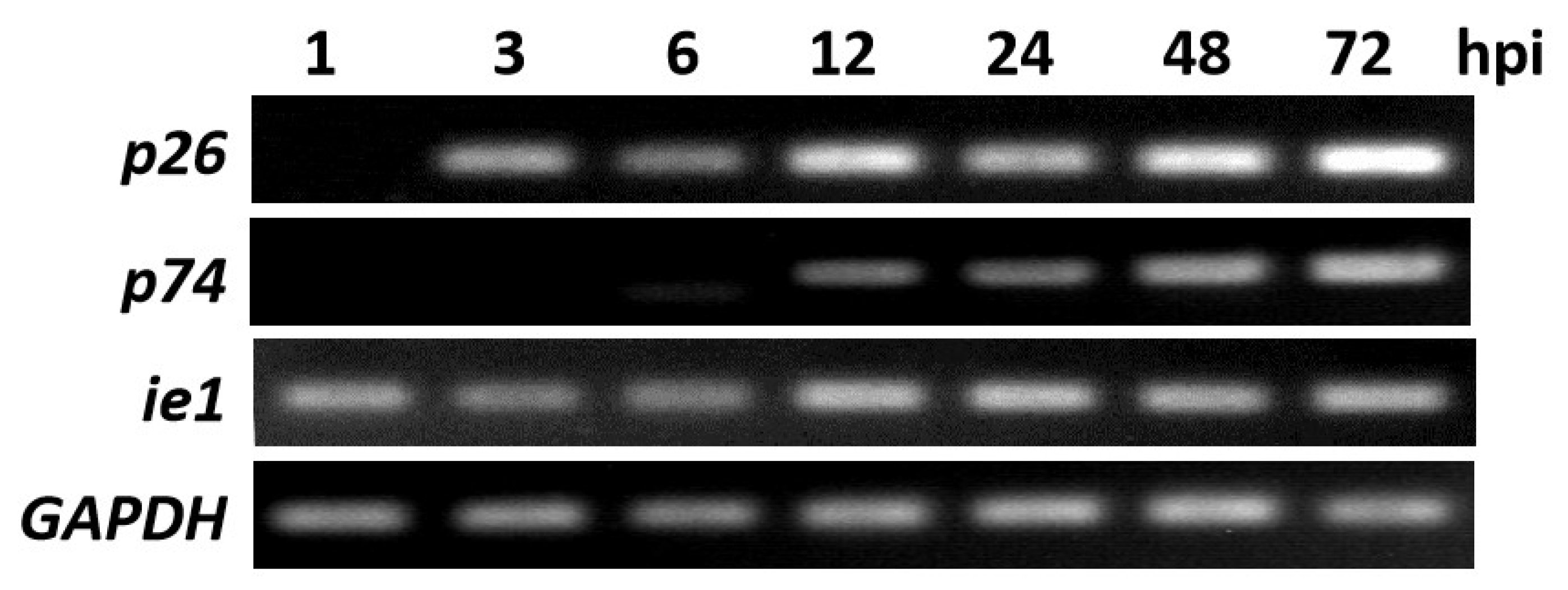
Figure 1. RT-PCR analysis of p26 transcripts in BmNPV-infected BmN cells. RT-PCR was performed by using purified RNA isolated from virus-infected cells at the indicated time points (1, 3, 6, 12, 24, 48, and 72 hpi). The first cDNA strand was synthesized with AMV Reverse Transcriptase and the oligo-p(dT)18 primer, and nested PCR was then performed with the p26, p74, and ie1-specific primers, respectively. gapdh was used as the control.
Insects 12 00707 g001
Figure 2. Overexpression of p26 in E. coli and Western blot analysis of p26 in BmNPV-infected BmN cells. (a) SDS-PAGE and Western blot analysis of the fusion protein His-p26 expressed in E. coli BL21 cells. M, protein molecular weight markers; Lane 1, BL21 cell proteins; Lane 2, proteins from BL21 cells transformed with pET-28a plasmid; Lane 3, proteins from BL21 cells transformed with pET-28a-p26; Lane 4, proteins in the sediment of supersonically broken BL21 cells transformed with pET-28a-p26; Lane 5, proteins in the supernatant of supersonically broken BL21 cells transformed with pET-28a-p26; Lane 6, Western blot analysis of the expressed His-p26 protein using an anti-His monoclonal antibody. (b) BmN cells were infected with wild-type BmNPV (MOI = 10.0), and protein samples were then harvested at 0, 3, 6, 12, 24, 36, 48, and 72 hpi, separated via 10% SDS-PAGE, transferred onto PVDF membranes, reacted with an anti-p26 polyclonal antibody or an anti-Bm122 polyclonal antibody, and detected with a DAB substrate. The immunoreactive bands are indicated by arrows, and the size is indicated on the right.
Insects 12 00707 g002
Figure 3. Subcellular localization of BmNPV p26. (a) Localization of p26 in BmNPV-infected BmN cells. The cells were collected at 48 hpi, reacted with anti-p26 serum, treated with EGFP-conjugated goat anti-rabbit IgG, and viewed under a confocal laser fluorescence microscope. In order to visualize the nucleus, the cells were also stained with DAPI (blue). (b) Localization of the over-expressed fusion protein EGFP-p26 in recombinant BmNPV infected BmN cells. EGFP-fused p26 was driven by the AcMNPV ph promoter and over-expressed in BmN cells. At 24 and 48 hpi, the cells were directly collected for fluorescence microscopy analysis.
Insects 12 00707 g003
Figure 4. p26 knockdown by RNAi and its influence on viral replication. BmN cells were infected with BmNPV. At 3 hpi, the cells were transfected with dsP26 and dsGFP, and non-dsRNA-treated BmN cells (CK) were used as the control. (a) Q-PCR analysis of p26 knockdown in BmNPV-infected BmN cells. Total RNA was extracted from BmNPV-infected BmN cells at 24, 48, and 72 h post-dsRNA treatment, reverse transcription and Q-PCR were performed, and the relative p26 transcript levels were analyzed using the △△Ct method. The derived relative quantity (RQ) values were normalized to those of non-dsRNA-treated controls, and ie1 transcript detection was included as the internal control. Data are presented as the mean RQs ± SEMs for three replicates. (b) Titration of BV from BmNPV-infected BmN cells in which p26 was knocked down by using dsRNA. At 24, 48, and 72 h post-dsRNA treatment, the supernatants of BmNPV-infected BmN cells were collected for titration by using the TCID50/mL method. Data are presented as the mean RQs ± SEMs for three replicates. (c) Q-PCR analysis of gp64 and p74 transcripts in BmNPV-infected cells with p26 knockdown. gapdh detection was included as internal controls. Data are presented as the mean RQs ± SEMs for three replicates.
Insects 12 00707 g004
Figure 5. Electron microscope analysis of BmNPV infected BmN cells after p26 knockdown. At 72 h post-dsRNA treatment, the cells were harvested for transmission electron microscope analysis. Cy, Nu, and PH denote cytoplasm, nucleus, and polyhedra, respectively. The black arrow refers to nucleocapsids and virions.
Insects 12 00707 g005
Table 1. Primers used in this study.
NameSequenceTarget
p26FW5’-CGGATCCATGGAATTGTATAATATTAAAT-3’p26
p26RW5’-CAAGCTTTTAGCTGTAATATATTGTGTTG-3’
T7p26iFW5’-TAATACGACTCACTATAGGGTTTCCTGGCGTCGTTAGTTC-3’p26
T7p26iRW5’-TAATACGACTCACTATAGGGTTGCACAGTCCCGTAAACAG-3’
T7GFPiFW5’-TAATACGACTCACTATAGGGTGGTAAAAGGACAGGGCCATC-3’gfp
T7GFPiRW5’-TAATACGACTCACTATAGGGCCATGGCCAACACTTGTCAC-3’
Qp26FW5’-TGTAATAGAGCAAGTCGACAATGTG-3’p26
Qp26RW5’-TGGTACCGGCTTAGCGTTTC-3’
Qie1FW5’-AACATTTGCACGGTCGCTTC-3’ie1
Qie1RW5’- GGTCGGAGAACCTGTTGGAA-3’
Qgp64FW5’-ACGGCATCAGCAAAAACGTG-3’gp64
Qgp64RW5’-AAGGTGGACGAGCGTTTGAT-3’
Qp74FW5’-TCTGTAGTGGTATCGCGCAC-3’p74
Qp74RW5’-AGCGCCTTCCAGCATACTAC-3’
QBmGapdhFW5’-AGGGCAGTGTTGAGGTTCAG-3’gapdh
QBmGapdhRW5’-GGCCTTAGGGTCCCTTTCTG-3’
Publisher’s Note: MDPI stays neutral with regard to jurisdictional claims in published maps and institutional affiliations.