Article

Characteristics and Comparative Analysis of Mitochondrial Genomes of the Aphid Genus Hyalopterus Koch (Hemiptera: Aphididae: Aphidinae)

1
Key Laboratory of Zoological Systematics and Evolution, Institute of Zoology, Chinese Academy of Sciences, Beijing 100101, China
2
College of Life Sciences, University of Chinese Academy of Sciences, Beijing 100049, China
*
Authors to whom correspondence should be addressed.
Insects 2024, 15(6), 389; https://doi.org/10.3390/insects15060389
Submission received: 22 April 2024 / Revised: 18 May 2024 / Accepted: 24 May 2024 / Published: 27 May 2024
(This article belongs to the Section Insect Molecular Biology and Genomics)

Abstract

:

Simple Summary

Hyalopterus aphids pose a significant threat to stone fruit trees in the genus Prunus and are distributed worldwide. In this study, we generated and compared the mitochondrial genomes of three constituent species of Hyalopterus. The repeat region located between trnE and trnF exists but differs among the three species, and the repeat unit shows variation among different geographic populations within the widely distributed Hyalopterus arundiniformis. The phylogenetic trees based on complete mitochondrial genomes supported the monophyly of Hyalopterus, with H. arundiniformis placed at the basal position. Our study enriches the mitochondrial genomic library of Aphidinae and suggests the potential utility of repeat regions in intraspecific aphid diversification studies.

Abstract

Using Illumina sequencing technology, we generated complete mitochondrial genomes (mitogenomes) of three constituent species of the aphid genus Hyalopterus Koch, Hyalopterus amygdali (Blanchard), Hyalopterus arundiniformis Ghulamullah, and Hyalopterus pruni (Geoffroy). The sizes of the Hyalopterus mitogenomes range from 15,306 to 15,410 bp, primarily due to variations in the length of non-coding regions. The Hyalopterus mitogenomes consist of 37 coding genes arranged in the order of the ancestral insect mitogenome, a control region, and a repeat region between trnE and trnF. According to the COI-based analysis, one previously reported mitogenome of H. pruni should be assigned to H. arundiniformis. The gene order, nucleotide composition, and codon usage in the Hyalopterus mitogenomes are highly conserved and similar to those of other species of Aphidinae. The tandem repeat units differ in nucleotide composition, length, and copy number across three Hyalopterus species. Within the widespread Eurasian species H. arundiniformis, variation in repeat units among different geographic populations is observed, indicating that the repeat region may provide valuable insights for studying the intraspecific diversification of aphids. Phylogenetic analyses based on 28 complete mitogenomes of Aphidinae supported the monophyly of Aphidinae, Aphidini, Macrosiphini, and two subtribes of Aphidini. Hyalopterus was monophyletic. H. amygdali and H. pruni formed a sister group, while H. arundiniformis was placed basally. Characterization of the mitogenomes of Hyalopterus provides valuable resources for further comparative studies and for advancing our understanding of the aphid mitogenome architecture.

1. Introduction

Aphids of the genus Hyalopterus Koch (Aphidinae: Aphidini) are widespread pests that cause severe damage to stone fruit crops of Prunus (Rosaceae) [1]. These species are characterized by elongated bodies, small siphunculi, and white powdery wax covering [1]. Recent studies, employing morphological and molecular techniques, have confirmed the existence of three species that alternate between different Prunus species and Phragmites within Hyalopterus [2,3,4,5,6,7]. Hyalopterus amygdali (Blanchard) feeds on the undersides of Prunus dulcis leaves and, occasionally, on Prunus armeniaca leaves. It migrates to reeds and possibly grasses as its secondary host plants. This species is mainly distributed in the Mediterranean area, central Asia, and possibly North America [1]. Hyalopterus arundiniformis Ghulamullah primarily feeds on Prunus persica and sometimes on P. armeniaca, Prunus salicina, and Prunus cerasifera. It is distributed throughout Eurasia [1]. Hyalopterus pruni (Geoffroy) inhabits the undersides of the leaves of Prunus domestica and sometimes is found on other Prunus species, except for P. dulcis. H. pruni is widely distributed in Europe and Asia and has been introduced to North America [1,8].
Due to its small size, maternal inheritance, fast evolutionary rate, and limited recombination, the insect mitogenome has been extensively employed in phylogenetic analysis across various taxonomic levels [9,10,11,12,13,14]. Compared to a single gene marker, the mitogenome provides more comprehensive information about the composition and arrangement of genes, which allows for a deeper understanding of the characteristics of species at the genetic level [10,15]. To date, complete mitogenomes of 27 aphid species of the subfamily Aphidinae have been sequenced and released on NCBI. The mitogenome of Aphidinae is a closed naked circular DNA, ranging from 15,111 to 17,954 bp in length, with a high A + T content ranging from 83.5% to 84.9%. The genus Hyalopterus belongs to the subtribe Rhopalosiphina of Aphidini. To date, complete mitogenomes are available for only five species of Rhopalosiphina [16,17,18].
In this study, we obtained the complete mitogenomes of H. amygdali, H. arundiniformis, and H. pruni via high-throughput sequencing to provide valuable mitogenomic information for Rhopalosiphina aphids. We compared the size, organization, nucleotide composition, codon usage, and non-coding regions of the Hyalopterus mitogenomes and constructed phylogenetic relationships of Aphidinae using maximum-likelihood and Bayesian methods based on the complete mitogenome sequences.

2. Materials and Methods

2.1. Sample Collection and DNA Extraction

Aphid specimens of the genus Hyalopterus were deposited in the National Animal Collection Resource Center, Institute of Zoology, Chinese Academy of Sciences, Beijing, China, and stored in 95% ethanol at −30 °C. Samples of H. amygdali were collected on Phragmites australis from Osh, Kyrgyzstan (voucher no. 40542); H. arundiniformis on P. persica from Fangchenggang, Guangxi, China (voucher no. AA197); and H. pruni on P. australis from Jambyl, Kazakhstan (voucher no. 47219). The slide-mounted voucher specimens were identified based on morphology using the taxonomic keys in Lozier et al. [5] and Rakauskas et al. [6]. The DNA was extracted from whole-body tissues using TRIzol® Reagent (Invitrogen, Carlsbad, CA, USA).

2.2. Mitogenome Sequencing, Assembly, and Annotation

Mitogenome sequencing was carried out on an Illumina HiSeq 4000 platform (BIOZERON Co., Ltd., Shanghai, China), and quality control was performed with Trimmomatic-0.39 [19]. The scaffolds generated by de novo assembly with SPAdes v3.10.1 [20] were gap-filled and optimized using GapCloser v1.12 [21]. The preliminary annotation results were obtained by the MITOS2 [22] with the invertebrate mitochondrial genetic code and RefSeq 89 Metazoa as a reference. The annotation and secondary structures of transfer RNA genes (tRNAs) were predicted by MITOS. The ribosomal RNA genes (rRNAs) and protein-coding genes (PCGs) were annotated by aligning with the mitogenomes of related aphid species, and the PCGs were further validated using ORF Finder (https://www.ncbi.nlm.nih.gov/orffinder/, accessed on 5 January 2024). The circular maps of mitogenomes were visualized with GCView Server (https://proksee.ca/, accessed on 11 January 2024) [23].

2.3. Sequence Analyses

The nucleotide composition was determined by MEGA-X [24]. Nucleotide composition bias was measured by calculating AT skew = (A − T)/(A + T) and GC skew = (G − C)/(G + C) [25]. The relative synonymous codon usage (RSCU) of the PCGs was calculated and drawn using PhyloSuite v1.2.3pre3 [26]. The ratio of nonsynonymous substitution rate (Ka) to synonymous substitution rate (Ks) was calculated for each PCG using DnaSP 6 [27], with the mitogenome of Pachypsylla venusta (GenBank accession no. NC_006157) used as a reference [16,28]. The repeat region was detected through the Tandem Repeats Finder web server (https://tandem.bu.edu/trf/home, accessed on 19 January 2024) [29]. Secondary structures of the repeat unit and control region were folded using TBtools v1.064 [30] and drawn by VARNA v3-93 [31]. We calculated the sequence similarity of the repeat units in different mitogenomes using Sequence Manipulation Suite (Version 2) (https://www.detaibio.com/sms2/, accessed on 23 January 2024) [32].

2.4. Phylogenetic Analyses

The phylogenetic relationships of Aphidinae were inferred based on whole mitogenomes of 3 species from Hyalopterus and 24 other aphid species (Table S1) by maximum-likelihood (ML) approach and Bayesian inference (BI). Four species from Calaphidinae and Chaitophorinae were taken as outgroups based on previous phylogenetic studies [12,33,34,35]. The PCGs were aligned by MAFFT on the TranslatorX online server (http://translatorx.co.uk, accessed on 2 February 2024) [36]. We aligned the tRNA and rRNA genes with the MAFFT (version 7) online service (https://mafft.cbrc.jp/alignment/server/, accessed on 2 February 2024) [37,38] and removed the unreliable alignment regions using Gblocks 0.91b [39,40]. PartitionFinder2 [41] was used to evaluate the optimal partitioning scheme and substitution models on PhyloSuite v1.2.3pre3 [26] with linked branch lengths, BIC, and searching by the greedy algorithm. All 37 genes with a total of 14,352 bp were divided into six partitions. The ML analysis was inferred using RAxML v8.2.10 [42] with 1000 bootstrap replications and GTRGAMMAI model for each partition. The BI analysis was carried out by MrBayes 3.2.7 [43] with four chains, running for 2,000,000 generations. The sampling frequency was set to every 100 generations to obtain the average deviation of the split frequencies falling below 0.01. The first 25% trees were discarded as burn-in. The resulting phylogenetic trees were annotated by Interactive Tree Of Life (iTOL) v5 (https://itol.embl.de/, accessed on 16 February 2024) [44].

3. Results and Discussion

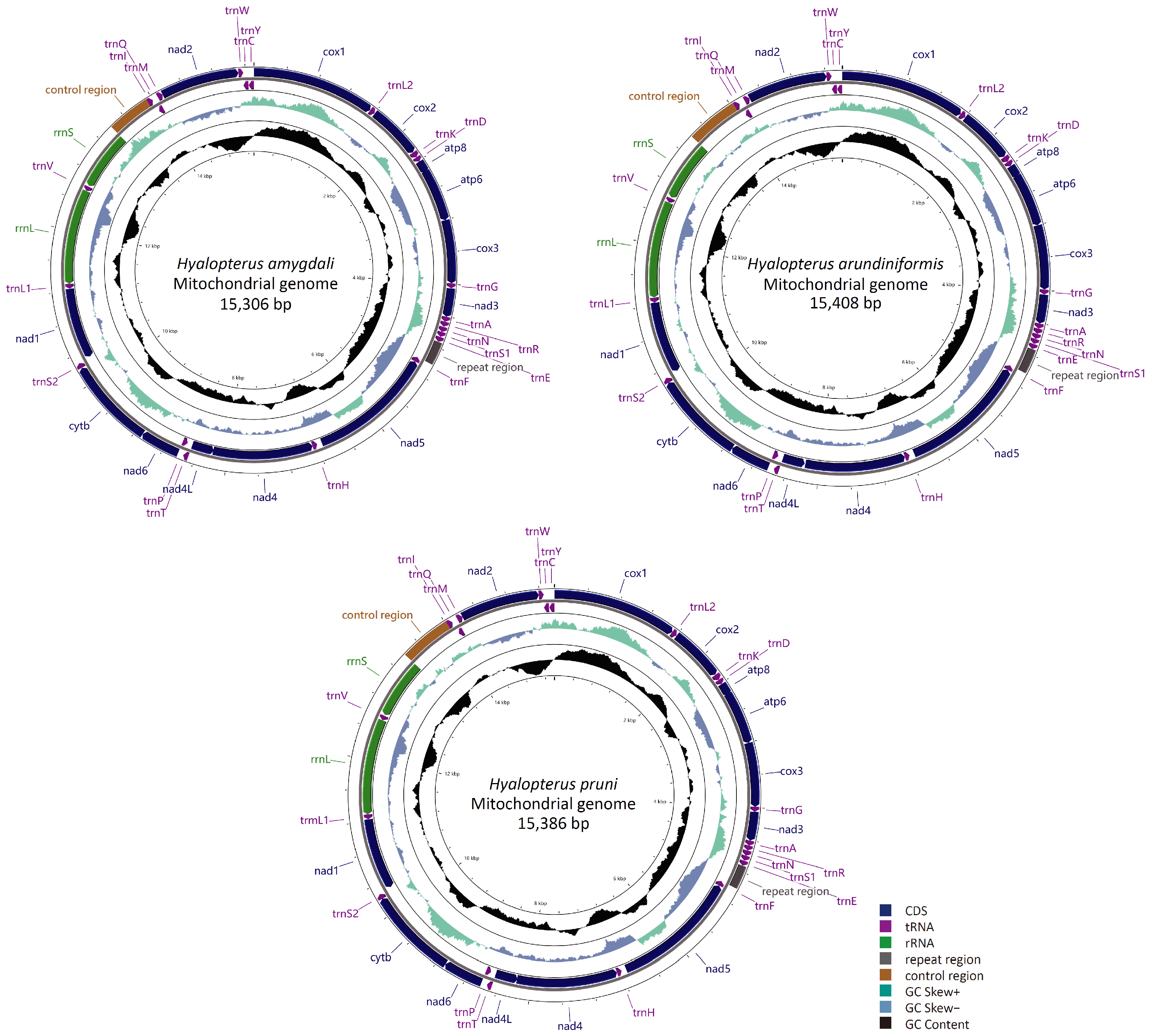
3.1. General Features of the Mitogenomes of Hyalopterus

The complete mitogenome sequences of H. amygdali, H. arundiniformis, and H. pruni are closed circular molecules of 15,306 bp, 15,408 bp, and 15,386 bp in length, respectively (Figure 1). All sequences have been submitted to GenBank under accession numbers OK641613 (H. amygdali), OK274075 (H. arundiniformis), and OK641614 (H. pruni). The gene order of the Hyalopterus mitogenomes is conserved, following that of the putative ancestral insect mitogenome [45]. Each of the three mitogenomes contain 37 coding genes, including 13 PCGs, 22 tRNAs, and 2 rRNAs. Four PCGs, eight tRNAs, and two rRNAs are encoded by the minority strand (N strand), while the remaining twenty-three coding genes (nine PCGs and fourteen tRNAs) are located on the majority strand (J strand) (Figure 1, Table S2). The longest non-coding region in the mitogenome of Hyalopterus, known as the control region, is located between rrnS and trnI. Another long non-coding region is found between trnE and trnF, often referred to as the repeat region, which has been reported to be a unique feature of aphid mitogenomes [46,47,48].
There are ten overlaps and fourteen intergenic spacers in H. amygdali, ten overlaps and thirteen intergenic spacers in H. arundiniformis, and eleven overlaps and twelve intergenic spacers in H. pruni (Table S2). The longest gene overlap within the Aphidinae mitogenomes appears between atp8 and atp6, ranging in size from 14 to 20 bp, except for Aphis glycines (NC_045236), which has a 1 bp intergenic spacer between atp8 and atp6 [47]. In the Hyalopterus mitogenomes, the longest overlap is 20 bp long and it is also located between atp6 and atp8, similarly to most Aphidinae aphids. The largest intergenic spacer (63 bp) is located between nad5 and trnH. Additionally, there is a 3 bp intergenic spacer between trnR and trnN in H. amygdali, but not in the other two species. In H. arundiniformis and H. pruni, a 2 bp overlap exists between cob and trnS (UCN), while H. amygdali has a 2 bp spacer between these two genes. There is a 7 bp overlap between nad4 and nad4L in H. amygdali and H. pruni but an 8 bp intergenic spacer in H. arundiniformis.
The lengths of the mitogenomes reported in Aphididae range from 14 to 20 kb. The largest aphid mitogenome is 19,200 bp long in Therioaphis tenera, containing the largest control region of 1452 bp and a long repeat region [49,50]. The shortest mitogenome is 14,990 bp long in Schizoneuraphis gallarum and it has a compact gene organization [51]. The Hyalopterus mitogenomes all fall within the normal size range. The variable size of aphid mitogenomes is not closely related to the coding genes but is mainly due to differences among the non-coding regions, especially the control regions and repeat regions [28,48,50,52].
Liang et al. [17] reported the complete mitogenome of H. pruni under GenBank accession no. NC_050904. This aphid sample was collected from Ningxia, China. However, according to Liu et al. [7], H. pruni in China is only found in the northern Tianshan Mountains, and most common Chinese records of H. pruni are actually referring to H. arundiniformis. To confirm this, we constructed a neighbor-joining (NJ) tree with MEGA-X [24] based on the COI sequences obtained from Liu et al. [7] and the extracted COI sequences from NC_050904 and the mitogenome of H. arundiniformis sequenced in this study (voucher no. AA197). The NC_050904 and AA197 samples were both placed within the H. arundiniformis clade (Figure S4). Therefore, the mitogenome of H. pruni (NC_050904) reported by Liang et al. [17] should be assigned to H. arundiniformis.

3.2. Nucleotide Composition

The nucleotide composition of insect mitogenomes is generally unbalanced [16,53]. In the reported Aphidinae mitogenomes, the nucleotide compositions all display a strong bias toward A + T, with the A + T content ranging from 83.50% (Aphis aurantii, GenBank accession no. NC_052865) to 84.90% (Uroleucon sonchi, GenBank accession no. MT533446). The whole mitogenomes of H. amygdali, H. arundiniformis, and H. pruni exhibit A + T contents of 83.90%, 83.80% and 83.88%, respectively (Table 1). The A + T content of the repeat regions is 89.90%, 89.30%, and 88.38% in H. amygdali, H. arundiniformis, and H. pruni, respectively. The third codon position of the PCGs exhibits the highest A + T content (93.20–93.70%), while that of the other codon positions varies from 75.66% to 80.14% (Table 1). The whole mitogenome of Hyalopterus displays a slight bias toward A, with an AT-skew value of 0.07–0.08, and a moderate bias toward C, with GC-skew values ranging from −0.28 to −0.25 (Table 1). The PCGs encoded on the J strand are C-skewed, with GC-skew values ranging from −0.24 to −0.22, while the PCGs encoded on the N strand are G-skewed, with a GC-skew value of 0.30–0.35. These results indicate the presence of nucleotide composition heterogeneity between two strands in the aphid mitogenomes. The strand asymmetry may be attributed to the spontaneous deamination of the A and C nucleotides in the N strand during replication [53].

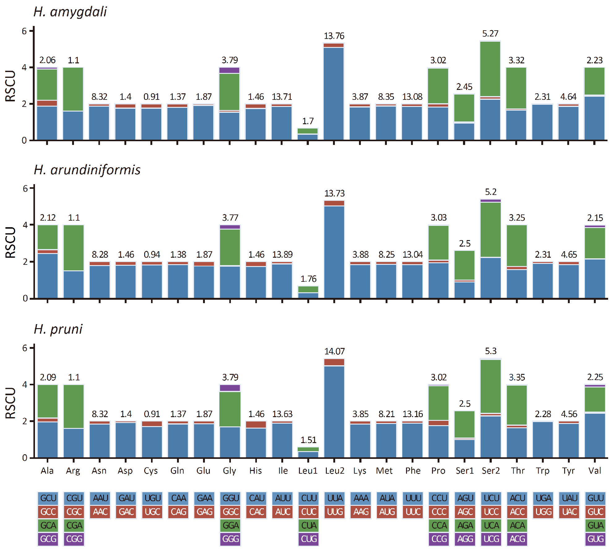
3.3. Protein-Coding Genes

The total lengths of thirteen PCGs in the mitogenomes of H. amygdali, H. arundiniformis, and H. pruni are 10,920 bp, 10,905 bp, and 10,920 bp, respectively. Nine of them (cox1, cox2, atp8, atp6, cox3, nad3, nad6, cob, and nad2) are located on the J strand, while the remaining four PCGs (nad5, nad4, nad4L, and nad1) are encoded by the N strand (Table S2). In aphid mitogenomes, most start codons are ATN and stop codons are TAA or TAG [28,48,50]. The start codons of the Hyalopterus mitogenomes are the same as those of the other aphids, which are ATA, ATT, and ATG, except for nad3 in H. arundiniformis, which starts with ATC. In terms of stop codons, cob ends with TAG, while cox1 and nad4 end with a single T, which is common in insects and can be completed by post-transcriptional polyadenylation [54]. The remaining PCGs in the Hyalopterus mitogenomes all end with TAA (Table S2).
The codon usage of PCGs in the mitogenomes of Hyalopterus is shown in Figure 2 by RSCU. As in most aphids, the five most abundant codon families in the Hyalopterus mitogenomes are Phe, Ile, Leu (UUR), Met, and Asn, while Cys is the least abundant (Figure 2) [28]. Among the 51 amino acid-coding codons in the mitogenomes of Hyalopterus, the most frequently used codon is UUA(L), followed by AUU(I), UUU(F), AUA(M), and AAU(N). These frequently used codons are composed mainly of A or U nucleotides, which is consistent with the strong A + T-content bias of the whole mitogenomes in Hyalopterus.
The Ka/Ks values of thirteen PCGs in the Hyalopterus mitogenomes are displayed in Figure 3a. All Ka/Ks values were below one, indicating that the primary evolutionary force shaping the Hyalopterus mitogenome sequences was purifying selection. The atp8 gene showed the highest evolutionary rate, particularly in H. pruni with a value of 0.80, while in H. amygdali and H. arundiniformis, the values were 0.52 and 0.60, respectively. The second highest Ka/Ks values were observed for nad6 (H. amygdali: 0.48; H. arundiniformis: 0.42; H. pruni: 0.48). The cox1 gene exhibited the strongest purifying pressure with Ka/Ks values of 0.04, 0.03, and 0.04 in H. amygdali, H. arundiniformis, and H. pruni, respectively (Figure 3a).

3.4. Transfer and Ribosomal RNA Genes
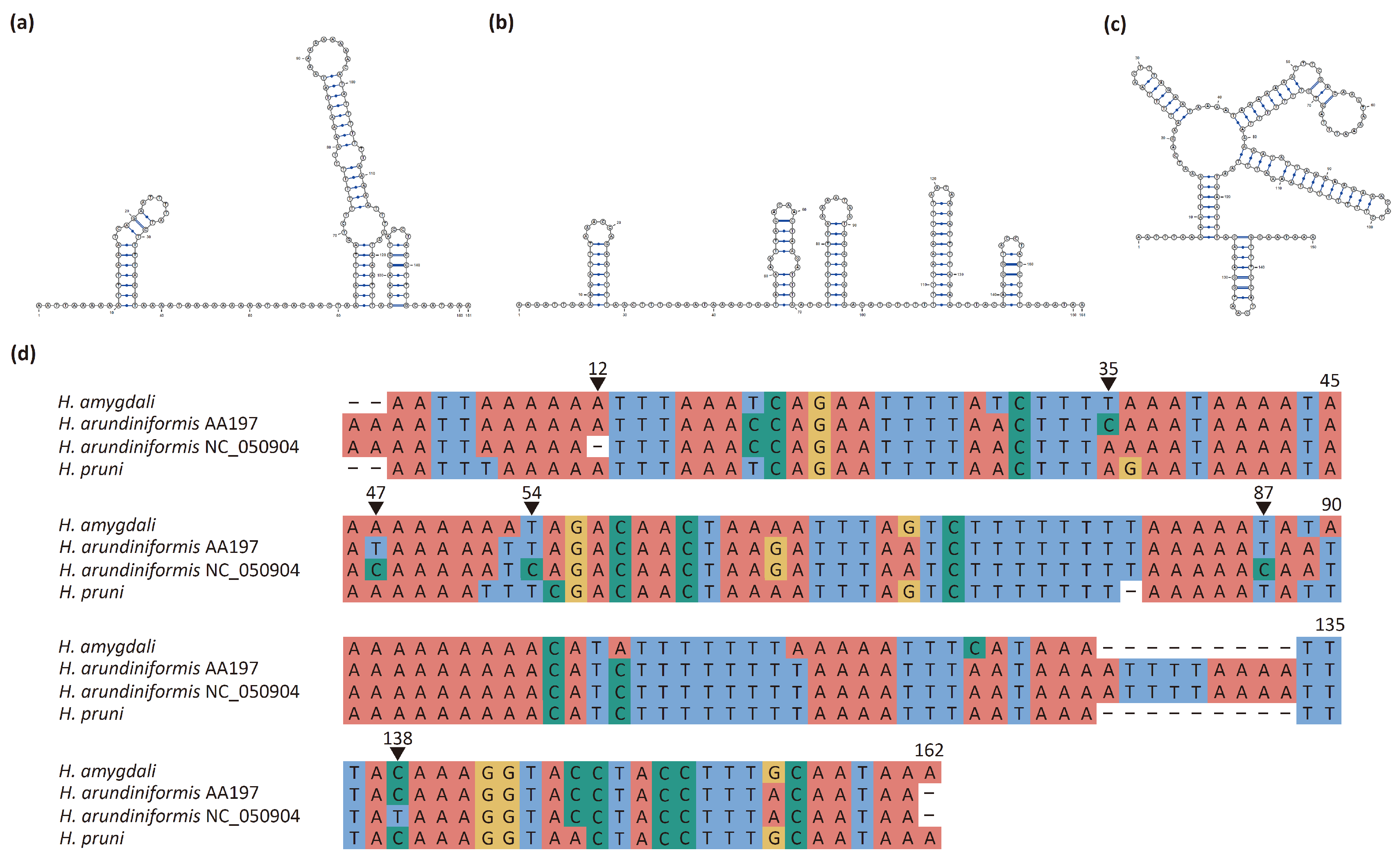
Each mitogenome of the three Hyalopterus species contains 22 tRNAs, ranging from 63 to 73 bp in size (Table S2). All tRNAs can form the typical clover-leaf secondary structures except for trnS (AGN), which lost the dihydrouridine (DHU) arm (Figures S1–S3). This feature is commonly observed in aphid mitogenomes.
The mitogenomes of Hyalopterus species encode two rRNAs on the N strand, both of which are in typical locations and have typical sizes for aphid mitogenomes (Table S2). The rrnL gene is located between trnL (CUN) and trnV without any overlapping or intergenic spacers. In H. amygdali and H. pruni, rrnL is 1257 bp long, while in H. arundiniformis, it is 1260 bp long. The lengths of rrnS are 770 bp, 768 bp, and 767 bp in H. arundiniformis, H. amygdali, and H. pruni, respectively. There is an intergenic spacer of 12 or 13 bp between trnV and the control region (12 bp in H. amygdali and H. arundiniformis, and 13 bp in H. pruni).

3.5. Control Region

The control region is a long non-coding region characterized by an extremely high A + T content in aphid mitogenomes. In Hemiptera insects, the A + T content of the control region ranges from 63.87% to 93.02% [50]. In H. amygdali, H. arundiniformis, and H. pruni, the A + T contents of the control region are 84.10%, 85.53%, and 86.31%, respectively (Table 1). The control region is involved in the initiation of DNA replication and contains promoters for the transcription of both mitochondrial DNA strands [46,55,56]. It is located between trnI and rrnS in the Hyalopterus mitogenome, and consists of an AT-rich zone, a poly-thymidine stretch, and a stem-loop region, which can be folded into a stable secondary structure (Figure 3b).
The control region exhibits poor conservation among Hyalopterus species due to high rates of nucleotide substitution and indels [52,55]. The lengths of the control regions are 535 bp, 629 bp, and 628 bp in H. amygdali, H. arundiniformis, and H. pruni, respectively (Table S2). The control region of H. amygdali is notably shorter than those of the other two species, possibly due to its shorter AT-rich zone of 413 bp. In comparison, the AT-rich zones of H. arundiniformis and H. pruni are 508 bp and 507 bp long, respectively.

3.6. Repeat Region

The repeat region in aphid mitogenomes is a tandem repeat sequence that is commonly found between trnE and trnF and sometimes within the control region [28,46,48,50]. The repeat region located between trnE and trnF is surmised to serve as another origin for replication [46,47]. It is hypothesized to have originated in the common ancestor of Aphididae and subsequently experienced numerous losses during species diversification [47,48,57]. In the Hyalopterus mitogenomes, the repeat region is only present between trnE and trnF and it exhibits the highest A + T content compared to other regions (H. amygdali: 89.90%; H. arundiniformis: 89.30%; H. pruni: 88.38%) (Table 1). H. amygdali has a repeat region of 286 bp in length, containing 1.834 151 bp-long repeat units; in H. arundiniformis, the repeat region is 299 bp in length and contains 1.832 161 bp-long repeat units; and the repeat region in H. pruni is 284 bp long, containing 1.860 150 bp-long repeat units (Figure 4d, Table 2). Each repeat unit can fold into a stem-loop structure (Figure 4a–c). The sequence similarity of the repeat units among the three species of Hyalopterus ranged from 82.10% to 91.39% (Table 2), which aligns with previous findings that tandem repeats exhibit high sequence similarity among closely related species [48].
The repeat unit of H. arundiniformis sequenced in the present study (voucher no. AA197) has a sequence similarity of 96.27% with the reported one (NC_050904) (Table 2). The repeat region is 299 bp long in AA197 and 298 bp long in NC_050904, containing 1.832 repeat units of 161 bp and 1.838 repeat units of 160 bp, respectively. In the repeat unit of AA197, the bases at positions 35, 47, 54, 87, and 138 are C, T, T, T, and C, respectively, while in the same position of NC_050904, the bases are A, C, C, C, and T, respectively (Figure 4d). Furthermore, compared with AA197, NC_050904 has a single nucleotide deletion at position 12 (Figure 4d). It has been reported that high nucleotide substitution rates, insertions or deletions, and variations in copy number are the factors that contribute to the intraspecific differences in non-coding regions of insect mitogenomes [56,58].
H. arundiniformis is a widespread Eurasian species [1]. To determine whether the variation in the repeat region of the two samples of H. arundiniformis was caused by geographical isolation, we constructed an ML tree using COI sequences of H. arundiniformis from AA197, NC_050904, and Liu et al. [59] (Figure S5). The AA197 sample collected from Guangxi, China was clustered within clade AL4, which was mainly composed of populations from southeastern and southwestern China [59]. The NC_050904 sample collected from Ningxia, China was nested within clade AL6, which spanned the Eurasia continent [59]. Therefore, the two samples of H. arundiniformis belong to distinct genetic lineages, and the intraspecific variation in the repeat region reported here may be applicable to aphid population genetic studies.

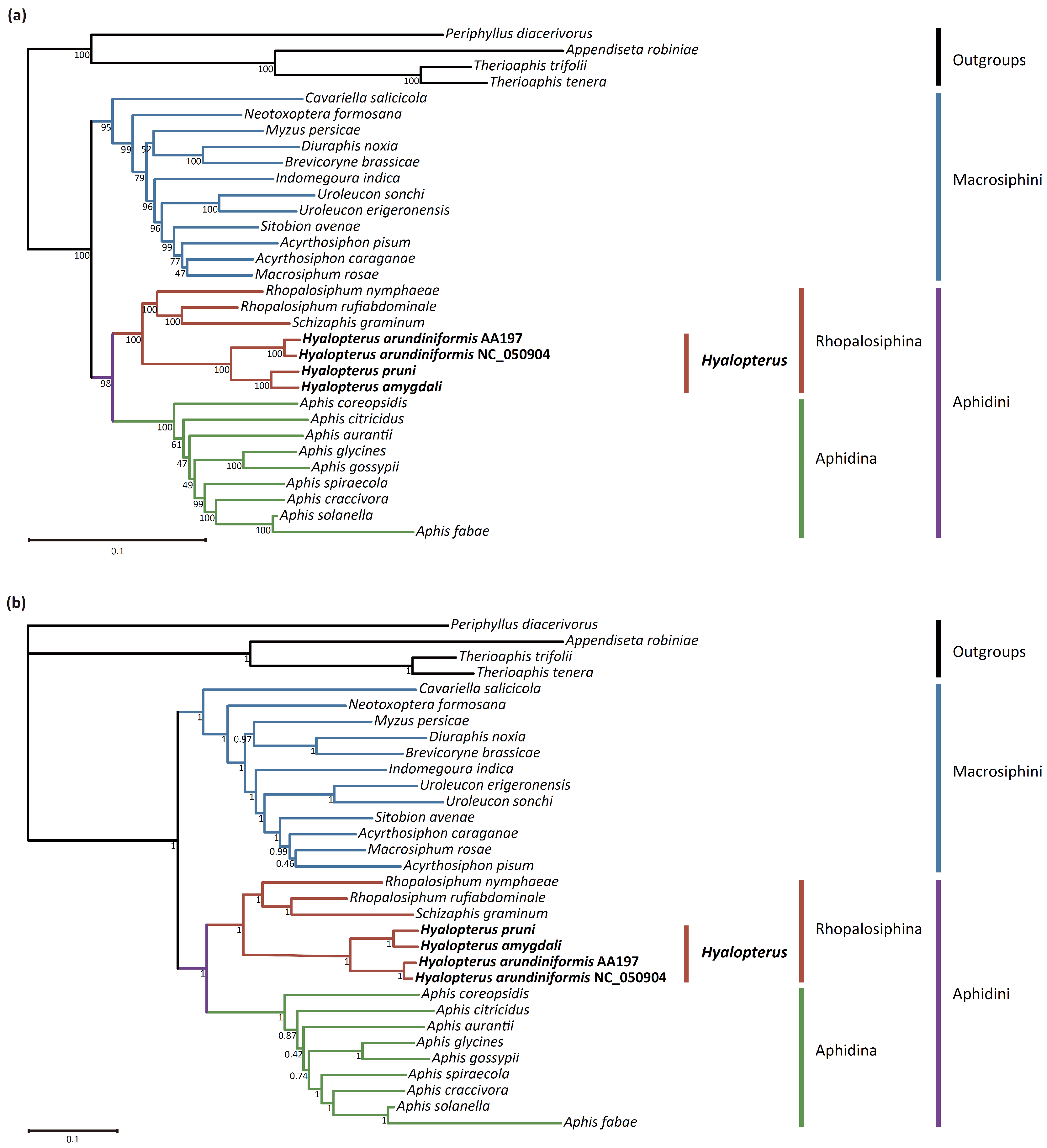
3.7. Phylogenetic Analyses

To determine the phylogenetic relationships among aphid species of Aphidinae, we performed ML and BI phylogenetic analyses with complete mitogenome sequences. The ML and BI trees were almost congruent in topology (Figure 5). Both trees supported the monophyly of Aphidinae, two tribes (Aphidini and Macrosiphini), and two subtribes within Aphidini (Aphidina and Rhopalosiphina). The generic relationships within Macrosiphini were in line with those reported by Choi et al. [60]. Cavariella and Neotoxoptera were the earliest and second earliest diverging branches, respectively. Myzus, Brevicoryne, and Diuraphis were clustered together, and Uroleucon, Sitobion, Acyrthosiphon, and Macrosiphum formed a well-supported clade.
Within the Rhopalosiphina clade, the genus Hyalopterus was monophyletic with strong support (bootstrap, BS = 100%; posterior probability, PP = 1) and was placed as a sister to the clade of Rhopalosiphum and Schizaphis, which was consistent with previous research [61]. The reported mitogenome NC_050904 strongly grouped with H. arundiniformi (AA197) (BS = 100%, PP = 1), which was in line with the result of the COI NJ tree (Figure S4) and supported the conclusion that NC_050904 should be assigned to H. arundiniformi. H. amygdali was clustered with H. pruni (BS = 100%; PP = 1), while H. arundiniformis was placed basally. The interspecific phylogenetic relationship of Hyalopterus based on whole mitogenome sequences was consistent with previous studies using multiple genes [4,7].

4. Conclusions

Complete mitogenomes of the aphid genus Hyalopterus, including three species, H. amygdali, H. arundiniformis, and H. pruni, were obtained using next-generation sequencing technology. The Hyalopterus mitogenomes show high conservation in terms of gene order, nucleotide composition, and codon usage, as observed in other Aphidinae mitogenomes. The repeat region is variable in length and nucleotide sequence among the three species, and intraspecific variation in the repeat unit is found in H. arundiniformis. Phylogenetic analysis based on the Aphidinae mitogenomes demonstrated the usefulness of the mitogenomes in resolving the phylogenetic relationships of tribes and genera. These newly produced mitogenome sequences of Hyalopterus are valuable data resources for the study of Aphidinae aphids. The acquisition of more aphid mitogenomes is necessary to provide additional mitogenome characteristics and phylogenetic information to enhance our understanding of the phylogeny and evolution of aphids.

Supplementary Materials

The following supporting information can be downloaded at: https://www.mdpi.com/article/10.3390/insects15060389/s1, Figure S1: Secondary structures of 22 transfer RNAs in H. amygdali, Figure S2: Secondary structures of 22 transfer RNAs in H. arundiniformis, Figure S3: Secondary structures of 22 transfer RNAs in H. pruni, Figure S4: Neighbor-joining tree based on the COI sequences obtained from Liu et al. [7] and the extracted COI sequences from NC_050904 and the mitogenome of H. arundiniformis sequenced in this study (voucher no. AA197), Figure S5: Maximum-likelihood tree constructed based on COI sequences of H. arundiniformis from Liu et al. [59], AA197, and NC_050904, using IQ-TREE v2.2.0 with the “TEST” model and 5000 bootstrap replications. Clades were numbered following Liu et al. [59], Table S1: Complete mitogenomes of aphids used for phylogenetic analysis, Table S2: Mitogenome organizations of three Hyalopterus species.

Author Contributions

Conceptualization, J.C., G.Q. and L.J.; methodology, data curation, and formal analysis, X.Z.; investigation, X.Z. and C.L.; writing—original draft preparation, X.Z.; writing—review and editing, J.C., L.J. and G.Q.; visualization, X.Z. and C.L.; supervision, J.C.; funding acquisition, G.Q. and J.C. All authors have read and agreed to the published version of the manuscript.

Funding

This work was supported by the Key Collaborative Research Program of the Alliance of International Science Organizations (Grant No. ANSO-CR-KP-2020-04), the National Natural Science Foundation of China (Grant No. 32030014), and the Youth Innovation Promotion Association of Chinese Academy of Sciences (Grant No. 2020087).

Data Availability Statement

The data that support the findings of this study are openly available in GenBank with accession numbers OK641613, OK274075, and OK641614.

Acknowledgments

Thanks to Fendi Yang for making voucher slides.

Conflicts of Interest

The authors declare no conflicts of interest.

References

  1. Blackman, R.L.; Eastop, V.F. Aphids on the world’s Plants: An Online Identification and Information Guide. Available online: https://aphidsonworldsplants.info/ (accessed on 6 April 2024).
  2. Mosco, M.C.; Arduino, P.; Bullini, L.; Barbagallo, S. Genetic heterogeneity, reproductive isolation and host preferences in mealy aphids of the Hyalopterus pruni complex (Homoptera, Aphidoidea). Mol. Ecol. 1997, 6, 667–670. [Google Scholar] [CrossRef]
  3. Lozier, J.D.; Roderick, G.K.; Mills, N.J. Genetic evidence from mitochondrial, nuclear, and endosymbiont markers for the evolution of host plant associated species in the aphid genus Hyalopterus (Hemiptera: Aphididae). Evolution 2007, 61, 1353–1367. [Google Scholar] [CrossRef]
  4. Poulios, K.D.; Margaritopoulos, J.T.; Tsitsipis, J.A. Morphological separation of host adapted taxa within the Hyalopterus pruni complex (Hemiptera: Aphididae). Eur. J. Entomol. 2007, 104, 235–242. [Google Scholar] [CrossRef]
  5. Lozier, J.D.; Foottit, R.G.; Miller, G.L.; Mills, N.J.; Roderick, G.K. Molecular and morphological evaluation of the aphid genus Hyalopterus Koch (Insecta: Hemiptera: Aphididae), with a description of a new species. Zootaxa 2008, 1688, 1–19. [Google Scholar] [CrossRef]
  6. Rakauskas, R.; Havelka, J.; Zaremba, A. Mitochondrial COI and morphological specificity of the mealy aphids (Hyalopterus ssp.) collected from different hosts in Europe (Hemiptera, Aphididae). ZooKeys 2013, 319, 255–267. [Google Scholar] [CrossRef]
  7. Liu, T.Y.; Chen, J.; Jiang, L.Y.; Qiao, G.X. Phylogeny and species reassessment of Hyalopterus (Aphididae, Aphidinae). Zool. Scr. 2020, 49, 755–767. [Google Scholar] [CrossRef]
  8. Lozier, J.D.; Roderick, G.K.; Mills, N.J. Tracing the invasion history of mealy plum aphid, Hyalopterus pruni (Hemiptera: Aphididae), in North America: A population genetics approach. Biol. Invasions 2009, 11, 299–314. [Google Scholar] [CrossRef]
  9. Boore, J.L. Animal mitochondrial genomes. Nucleic Acids Res. 1999, 27, 1767–1780. [Google Scholar] [CrossRef]
  10. Curole, J.P.; Kocher, T.D. Mitogenomics: Digging deeper with complete mitochondrial genomes. Trends Ecol. Evol. 1999, 14, 394–398. [Google Scholar] [CrossRef]
  11. Ma, C.; Yang, P.; Jiang, F.; Chapuis, M.P.; Shali, Y.; Sword, G.A.; Kang, L. Mitochondrial genomes reveal the global phylogeography and dispersal routes of the migratory locust. Mol. Ecol. 2012, 21, 4344–4358. [Google Scholar] [CrossRef]
  12. Chen, J.; Wang, Y.; Jiang, L.; Qiao, G. Mitochondrial genome sequences effectively reveal deep branching events in aphids (Insecta: Hemiptera: Aphididae). Zool. Scr. 2017, 46, 706–717. [Google Scholar] [CrossRef]
  13. Condamine, F.L.; Nabholz, B.; Clamens, A.L.; Dupuis, J.R.; Sperling, F.A.H. Mitochondrial phylogenomics, the origin of swallowtail butterflies, and the impact of the number of clocks in Bayesian molecular dating. Syst. Entomol. 2018, 43, 460–480. [Google Scholar] [CrossRef]
  14. Cheng, M.; Liu, Y.; Zheng, X.; Zhang, R.; Feng, K.; Yue, B.; Du, C.; Zhou, C. Characterization of seventeen complete mitochondrial genomes: Structural features and phylogenetic implications of the lepidopteran insects. Insects 2022, 13, 998. [Google Scholar] [CrossRef]
  15. Sun, C.H.; Sun, P.Y.; Lao, Y.L.; Wu, T.; Zhang, Y.N.; Huang, Q.; Zhang, Q. Mitogenome of a monotypic genus, Oliotius Kottelat, 2013 (Cypriniformes: Cyprinidae): Genomic characterization and phylogenetic position. Gene 2023, 851, 147035. [Google Scholar] [CrossRef]
  16. Thao, M.L.; Baumann, L.; Baumann, P. Organization of the mitochondrial genomes of whiteflies, aphids, and psyllids (Hemiptera, Sternorrhyncha). BMC Evol. Biol. 2004, 4, 25. [Google Scholar] [CrossRef]
  17. Liang, Y.; Du, Z.; Song, F.; He, J. The complete mitochondrial genome of the mealy plum aphid, Hyalopterus pruni (Hemiptera: Aphididae). Mitochondrial DNA B 2020, 5, 3667–3669. [Google Scholar] [CrossRef]
  18. Park, J.; Kim, Y.; Xi, H.; Park, J.; Lee, W. The complete mitochondrial genome of Rhopalosiphum nymphaeae (Linnaeus, 1761) (Hemiptera: Aphididae). Mitochondrial DNA B 2020, 5, 1613–1615. [Google Scholar] [CrossRef]
  19. Bolger, A.M.; Lohse, M.; Usadel, B. Trimmomatic: A flexible trimmer for Illumina sequence data. Bioinformatics 2014, 30, 2114–2120. [Google Scholar] [CrossRef]
  20. Bankevich, A.; Nurk, S.; Antipov, D.; Gurevich, A.A.; Dvorkin, M.; Kulikov, A.S.; Lesin, V.M.; Nikolenko, S.I.; Pham, S.; Prjibelski, A.D.; et al. SPAdes: A new genome assembly algorithm and its applications to single-cell sequencing. J. Comput. Biol. 2012, 19, 455–477. [Google Scholar] [CrossRef]
  21. Luo, R.; Liu, B.; Xie, Y.; Li, Z.; Huang, W.; Yuan, J.; He, G.; Chen, Y.; Pan, Q.; Liu, Y.; et al. SOAPdenovo2: An empirically improved memory-efficient short-read de novo assembler. GigaScience 2012, 1, 18. [Google Scholar] [CrossRef]
  22. Donath, A.; Jühling, F.; Al-Arab, M.; Bernhart, S.H.; Reinhardt, F.; Stadler, P.F.; Middendorf, M.; Bernt, M. Improved annotation of protein-coding genes boundaries in metazoan mitochondrial genomes. Nucleic Acids Res. 2019, 47, 10543–10552. [Google Scholar] [CrossRef]
  23. Grant, J.R.; Stothard, P. The CGView Server: A comparative genomics tool for circular genomes. Nucleic Acids Res. 2008, 36, W181–W184. [Google Scholar] [CrossRef]
  24. Kumar, S.; Stecher, G.; Li, M.; Knyaz, C.; Tamura, K. MEGA X: Molecular evolutionary genetics analysis across computing platforms. Mol. Biol. Evol. 2018, 35, 1547–1549. [Google Scholar] [CrossRef]
  25. Perna, N.T.; Kocher, T.D. Patterns of nucleotide composition at fourfold degenerate sites of animal mitochondrial genomes. J. Mol. Evol. 1995, 41, 353–358. [Google Scholar] [CrossRef]
  26. Zhang, D.; Gao, F.; Jakovlić, I.; Zou, H.; Zhang, J.; Li, W.X.; Wang, G.T. PhyloSuite: An integrated and scalable desktop platform for streamlined molecular sequence data management and evolutionary phylogenetics studies. Mol. Ecol. Resour. 2020, 20, 348–355. [Google Scholar] [CrossRef]
  27. Rozas, J.; Ferrer-Mata, A.; Sánchez-DelBarrio, J.C.; Guirao-Rico, S.; Librado, P.; Ramos-Onsins, S.E.; Sánchez-Gracia, A. DnaSP 6: DNA sequence polymorphism analysis of large data sets. Mol. Biol. Evol. 2017, 34, 3299–3302. [Google Scholar] [CrossRef]
  28. Chen, J.; Wang, Y.; Qin, M.; Jiang, L.Y.; Qiao, G.X. The mitochondrial genome of Greenidea psidii van der Goot (Hemiptera: Aphididae: Greenideinae) and comparisons with other Aphididae aphids. Int. J. Biol. Macromol. 2019, 122, 824–832. [Google Scholar] [CrossRef]
  29. Benson, G. Tandem repeats finder: A program to analyze DNA sequences. Nucleic Acids Res. 1999, 27, 573–580. [Google Scholar] [CrossRef]
  30. Chen, C.; Chen, H.; Zhang, Y.; Thomas, H.R.; Frank, M.H.; He, Y.; Xia, R. TBtools: An integrative toolkit developed for interactive analyses of big biological data. Mol. Plant 2020, 13, 1194–1202. [Google Scholar] [CrossRef]
  31. Darty, K.; Denise, A.; Ponty, Y. VARNA: Interactive drawing and editing of the RNA secondary structure. Bioinformatics 2009, 25, 1974–1975. [Google Scholar] [CrossRef]
  32. Stothard, P. The sequence manipulation suite: JavaScript programs for analyzing and formatting protein and DNA sequences. BioTechniques 2000, 28, 1102–1104. [Google Scholar] [CrossRef]
  33. Ortiz-Rivas, B.; Martínez-Torres, D. Combination of molecular data support the existence of three main lineages in the phylogeny of aphids (Hemiptera: Aphididae) and the basal position of the subfamily Lachninae. Mol. Phylogenet. Evol. 2010, 55, 305–317. [Google Scholar] [CrossRef]
  34. Rebijith, K.B.; Asokan, R.; Hande, H.R.; Joshi, S.; Surveswaran, S.; Ramamurthy, V.V.; Krishna Kumar, N.K. Reconstructing the macroevolutionary patterns of aphids (Hemiptera: Aphididae) using nuclear and mitochondrial DNA sequences. Biol. J. Linn. Soc. 2017, 121, 796–814. [Google Scholar] [CrossRef]
  35. Owen, C.L.; Miller, G.L. Phylogenomics of the Aphididae: Deep relationships between subfamilies clouded by gene tree discordance, introgression and the gene tree anomaly zone. Syst. Entomol. 2022, 47, 470–486. [Google Scholar] [CrossRef]
  36. Abascal, F.; Zardoya, R.; Telford, M.J. TranslatorX: Multiple alignment of nucleotide sequences guided by amino acid translations. Nucleic Acids Res. 2010, 38, W7–W13. [Google Scholar] [CrossRef]
  37. Kuraku, S.; Zmasek, C.M.; Nishimura, O.; Katoh, K. aLeaves facilitates on-demand exploration of metazoan gene family trees on MAFFT sequence alignment server with enhanced interactivity. Nucleic Acids Res. 2013, 41, W22–W28. [Google Scholar] [CrossRef]
  38. Katoh, K.; Rozewicki, J.; Yamada, K.D. MAFFT online service: Multiple sequence alignment, interactive sequence choice and visualization. Brief. Bioinform. 2019, 20, 1160–1166. [Google Scholar] [CrossRef]
  39. Castresana, J. Selection of conserved blocks from multiple alignments for their use in phylogenetic analysis. Mol. Biol. Evol. 2000, 17, 540–552. [Google Scholar] [CrossRef]
  40. Talavera, G.; Castresana, J. Improvement of phylogenies after removing divergent and ambiguously aligned blocks from protein sequence alignments. Syst. Biol. 2007, 56, 564–577. [Google Scholar] [CrossRef]
  41. Lanfear, R.; Frandsen, P.B.; Wright, A.M.; Senfeld, T.; Calcott, B. PartitionFinder 2: New methods for selecting partitioned models of evolution for molecular and morphological phylogenetic analyses. Mol. Biol. Evol. 2017, 34, 772–773. [Google Scholar] [CrossRef]
  42. Stamatakis, A. RAxML version 8: A tool for phylogenetic analysis and post-analysis of large phylogenies. Bioinformatics 2014, 30, 1312–1313. [Google Scholar] [CrossRef]
  43. Ronquist, F.; Teslenko, M.; van der Mark, P.; Ayres, D.L.; Darling, A.; Höhna, S.; Larget, B.; Liu, L.; Suchard, M.A.; Huelsenbeck, J.P. MrBayes 3.2: Efficient Bayesian phylogenetic inference and model choice across a large model space. Syst. Biol. 2012, 61, 539–542. [Google Scholar] [CrossRef]
  44. Letunic, I.; Bork, P. Interactive Tree Of Life (iTOL) v5: An online tool for phylogenetic tree display and annotation. Nucleic Acids Res. 2021, 49, W293–W296. [Google Scholar] [CrossRef]
  45. Clary, D.O.; Wolstenholme, D.R. The mitochondrial DNA molecule of Drosophila yakuba: Nucleotide sequence, gene organization, and genetic code. J. Mol. Evol. 1985, 22, 252–271. [Google Scholar] [CrossRef]
  46. Wang, Y.; Huang, X.L.; Qiao, G.X. Comparative analysis of mitochondrial genomes of five aphid species (Hemiptera: Aphididae) and phylogenetic implications. PLoS ONE 2013, 8, e77511. [Google Scholar] [CrossRef]
  47. Song, H.; Donthu, R.K.; Hall, R.; Hon, L.; Weber, E.; Badger, J.H.; Giordano, R. Description of soybean aphid (Aphis glycines Matsumura) mitochondrial genome and comparative mitogenomics of Aphididae (Hemiptera: Sternorrhyncha). Insect Biochem. Mol. Biol. 2019, 113, 103208. [Google Scholar] [CrossRef]
  48. Zhang, H.; Lu, C.; Liu, Q.; Zou, T.; Qiao, G.; Huang, X. Insights into the evolution of aphid mitogenome features from new data and comparative analysis. Animals 2022, 12, 1970. [Google Scholar] [CrossRef]
  49. Voronova, N.V.; Warner, D.; Shulinski, R.; Levykina, S.; Bandarenka, Y.; Zhorov, D. The largest aphid mitochondrial genome found in invasive species Therioaphis tenera (Aizenberg, 1956). Mitochondrial DNA B 2019, 4, 730–731. [Google Scholar] [CrossRef]
  50. Voronova, N.V.; Levykina, S.; Warner, D.; Shulinski, R.; Bandarenka, Y.; Zhorov, D. Characteristic and variability of five complete aphid mitochondrial genomes: Aphis fabae mordvilkoi, Aphis craccivora, Myzus persicae, Therioaphis tenera and Appendiseta robiniae (Hemiptera; Sternorrhyncha; Aphididae). Int. J. Biol. Macromol. 2020, 149, 187–206. [Google Scholar] [CrossRef]
  51. Zhang, X.; Li, Y.; Li, C.; Qiao, G.; Chen, J. The complete mitochondrial genome of Schizoneuraphis gallarum van der Goot, 1917 (Hemiptera: Aphididae: Hormaphidinae). Mitochondrial DNA B 2021, 6, 2982–2983. [Google Scholar] [CrossRef]
  52. Wang, Y.; Chen, J.; Jiang, L.Y.; Qiao, G.X. Hemipteran mitochondrial genomes: Features, structures and implications for phylogeny. Int. J. Mol. Sci. 2015, 16, 12382–12404. [Google Scholar] [CrossRef]
  53. Wei, S.J.; Shi, M.; Chen, X.X.; Sharkey, M.J.; van Achterberg, C.; Ye, G.Y.; He, J.H. New views on strand asymmetry in insect mitochondrial genomes. PLoS ONE 2010, 5, e12708. [Google Scholar] [CrossRef]
  54. Ojala, D.; Montoya, J.; Attardi, G. tRNA punctuation model of RNA processing in human mitochondria. Nature 1981, 290, 470–474. [Google Scholar] [CrossRef]
  55. Wolstenholme, D.R. Animal mitochondrial DNA: Structure and evolution. Int. Rev. Cytol. 1992, 141, 173–216. [Google Scholar]
  56. Zhang, D.X.; Hewitt, G.M. Insect mitochondrial control region: A review of its structure, evolution and usefulness in evolutionary studies. Biochem. Syst. Ecol. 1997, 25, 99–120. [Google Scholar] [CrossRef]
  57. Zhang, H.; Liu, Q.; Lu, C.; Deng, J.; Huang, X. The first complete mitochondrial genome of Lachninae species and comparative genomics provide new insights into the evolution of gene rearrangement and the repeat region. Insects 2021, 12, 55. [Google Scholar] [CrossRef]
  58. Liu, H.L.; Chen, S.; Chen, Q.D.; Pu, D.Q.; Chen, Z.T.; Liu, Y.Y.; Liu, X. The first mitochondrial genomes of the family Haplodiplatyidae (Insecta: Dermaptera) reveal intraspecific variation and extensive gene rearrangement. Biology 2022, 11, 807. [Google Scholar] [CrossRef]
  59. Liu, T.; Chen, J.; Jiang, L.; Qiao, G. Human-mediated eco-evolutionary processes of the herbivorous insect Hyalopterus arundiniformis during the Holocene. Divers. Distrib. 2022, 28, 1313–1326. [Google Scholar] [CrossRef]
  60. Choi, H.; Shin, S.; Jung, S.; Clarke, D.J.; Lee, S. Molecular phylogeny of Macrosiphini (Hemiptera: Aphididae): An evolutionary hypothesis for the Pterocomma-group habitat adaptation. Mol. Phylogenet. Evol. 2018, 121, 12–22. [Google Scholar] [CrossRef]
  61. Kim, H.; Lee, S.; Jang, Y. Macroevolutionary patterns in the Aphidini aphids (Hemiptera: Aphididae): Diversification, host association, and biogeographic origins. PLoS ONE 2011, 6, e24749. [Google Scholar] [CrossRef]
Figure 1. Circular maps of the mitogenomes of Hyalopterus species.
Insects 15 00389 g001
Figure 2. Relative synonymous codon usage (RSCU) in the mitogenomes of Hyalopterus species, with amino acid frequencies marked above the bars.
Insects 15 00389 g002
Figure 3. (a) Ka/Ks of thirteen protein-coding genes within the mitogenomes of Hyalopterus species. (b) Organization of the control regions and secondary structures of the stem-loop regions.
Insects 15 00389 g003
Figure 4. (ac) Putative stem-loop structures of the repeat units of H. amygdali (a), H. arundiniformis (b), and H. pruni (c). (d) Pairwise alignment of the repeat units of three Hyalopterus mitogenomes sequenced in the present study and the previously reported one (NC_050904).
Insects 15 00389 g004
Figure 5. Aphidinae phylogenetic trees inferred from complete mitochondrial genomes. (a) Maximum-likelihood (ML) tree. The values under the branches indicate ML bootstrap probabilities. (b) Bayesian tree. The values under the branches indicate posterior probabilities.
Insects 15 00389 g005
Table 1. A + T content, AT-skew, and GC-skew of the Hyalopterus mitogenomes.
H. amygdaliH. arundiniformisH. pruni
A + T%AT-SkewGC-SkewA + T%AT-SkewGC-SkewA + T%AT-SkewGC-Skew
Whole genome83.900.08−0.2583.800.08−0.2783.880.07−0.27
PCGs83.10−0.15−0.0582.95−0.15−0.0483.07−0.16−0.03
First codon80.0000.1379.920.000.1380.14−0.010.15
Second codon75.90−0.40−0.1375.79−0.39−0.1275.66−0.40−0.13
Third codon93.70−0.09−0.2993.20−0.10−0.2293.41−0.10−0.17
PCGs-J82.00−0.06−0.2381.74−0.06−0.2381.99−0.07−0.22
PCGs-J-first codon79.200.12078.660.12−0.0179.110.110.01
PCGs-J-second codon73.80−0.36−0.2773.71−0.36−0.2673.53−0.36−0.27
PCGs-J-third codon93.400.015−0.8892.860.02−0.8093.350.00−0.76
PCGs-N85.00−0.290.3284.90−0.300.3584.79−0.300.34
PCGs-N-first codon81.40−0.180.3881.50−0.180.4081.79−0.190.40
PCGs-N-second codon79.60−0.460.1578.70−0.450.1579.07−0.460.15
PCGs-N-third codon94.40−0.250.6993.50−0.280.8493.50−0.270.80
tRNA genes85.600.040.1885.770.040.1985.500.050.13
rRNA genes84.80−0.070.3484.93−0.080.3484.93−0.070.34
Control region84.10−0.07−0.1685.53−0.05−0.3286.31−0.08−0.30
Repeat region89.900.19−0.3989.300.18−0.5088.380.14−0.33
Table 2. Sequence information for the repeat regions of Hyalopterus mitogenomes.
SpeciesLength of Repeat Region (bp)Length of Repeat Unit (bp)Copy NumberSequence Similarity of Repeat Unit (%)
H. amygdaliH. arundiniformis AA197H. arundiniformis NC_050904H. pruni
H. amygdali2861511.83484.5782.1091.39
H. arundiniformis AA1972991611.83284.5796.2784.57
H. arundiniformis NC_0509042981601.83882.1096.2782.72
H. pruni2841501.86091.3984.5782.72
Disclaimer/Publisher’s Note: The statements, opinions and data contained in all publications are solely those of the individual author(s) and contributor(s) and not of MDPI and/or the editor(s). MDPI and/or the editor(s) disclaim responsibility for any injury to people or property resulting from any ideas, methods, instructions or products referred to in the content.

