Review

Surface Modification Techniques for Metallic Biomedical Alloys: A Concise Review

Department of Mechanical & Aerospace Engineering, School of Engineering & Digital Sciences, Nazarbayev University, Kabanbay Batyr Ave. 53, Astana 010000, Kazakhstan
*
Author to whom correspondence should be addressed.
Metals 2023, 13(1), 82; https://doi.org/10.3390/met13010082
Submission received: 1 November 2022 / Revised: 5 December 2022 / Accepted: 15 December 2022 / Published: 28 December 2022
(This article belongs to the Section Biobased and Biodegradable Metals)

Abstract

:
Developing biomaterials with appropriate physiochemical and mechanical properties as per the requirements set by biomedical applications remains a challenge. This challenge has pushed research in the direction of biomaterials development and the surface modification of existing materials that could be useful for biomedical applications. Keeping this demand in focus, this paper intends to conduct an in-depth review, which includes, first, the requirements of biomedical surfaces associated with the growth of cells on biomedical alloys, such as the bone formation, adhesion, increased wear resistance and biofilm formation; second, possible biomaterials candidates for such applications; and third, possible surface modification techniques. Both subtractive and additive methods of surface modification are discussed, along with their pros and cons. Hence, this study gives an excellent compendium of scientific works conducted on surface modification techniques and the development of biocompatible surface alloys, along with research trends.

1. Introduction

When it comes to extreme environmental conditions where product may need to withstand a high-temperature, high-pressure and highly corrosive environmental situation, it is imperative to improve the mechanical, physical and biochemical properties of the deployed materials so that they can perform well under given conditions [1,2,3,4]. Therefore, from a product design perspective, manufactured product still may need to go through additional secondary techniques in order to enhance strength, biocompatibility and corrosion and wear resistance. This additional step is known as a surface modification technique, which has impacted certain industries immensely, such as biomedical industries, in particular dental and orthopedic applications where osteointegration is promoted with the help of surface modification by enhancing its biochemical and morphological compatibilities [5]. One of such techniques, called plasma treatment, is reported to alter surface adhesion, friction and wettability properties [6,7]. As it is reported in the literature, surface modification can be conducted with or without the addition of material. Some of the techniques that does not add materials are laser surface modification, laser surface texturing, spark erosion machining, shot penning and sand blasting. Other techniques that add materials are electro discharge coating [8], physical vapor deposition [9], chemical vapor deposition [10], electrodeposition [11] and thermal spraying.
In this review paper, surface requirements such as the bone formation, adhesion, increased wear resistance and biofilm formation for biomedical applications are described in Section 2. Section 3 focuses on possible biomaterials candidates and their alloying elements. Section 4 discusses surface modification techniques, along with their pros and cons. Finally, research challenges related to this biomaterial and their surface modification are highlighted.

2. Surface Requirements for Biomedical Applications

This section provides a description of the surface requirements for biomedical applications. This includes surface treatment difficulties, the implant failure mechanism, the bone-bonding process, the mechanical properties of bones and surface treatments.

2.1. Surface Treatment Problems

Because of the complexity of the human body, matching the appropriate properties of bone implants remains a big challenge. On one side, successful implantation depends on the properties of materials such as the ability to withstand different cyclic loads and having low density as well as an elastic modulus. On the other hand, it also depends on their surfaces, which must have high corrosion resistance, compatible topography and inertness in the body environment. Otherwise, a lack of any of these parameters may lead to implant failures. Inadequate surface parameters can also lead to failures due to debris generation, the release of metal ions and responses to foreign body [12]. For example, titanium and its alloys, so far, have been found to be one of the most biocompatible implant materials. Despite that, there are still several factors that should be considered when optimizing surface properties for biocompatibility [12,13]. First, the bioinert behavior of Ti alloys leads to less formation of fiber in the intersection areas, which, in turn, results in mechanical osseointegration [12,14,15]. Second, the oxide layer on the surface of Ti alloys suffers from a lack of full protection due to its inability to physically resist plastic shear. This results in an abrasive wear that prevents bone ingrowth, resulting in failure. Third, the effect of external forces and different fluids in the body environment on the surface layer can result in the dissolution of the oxide layer, resulting in the release of unwanted toxic materials [12,15]. These factors are caused by the properties of materials; however, proper surface treatment can compensate for these unwanted effects [12,15].

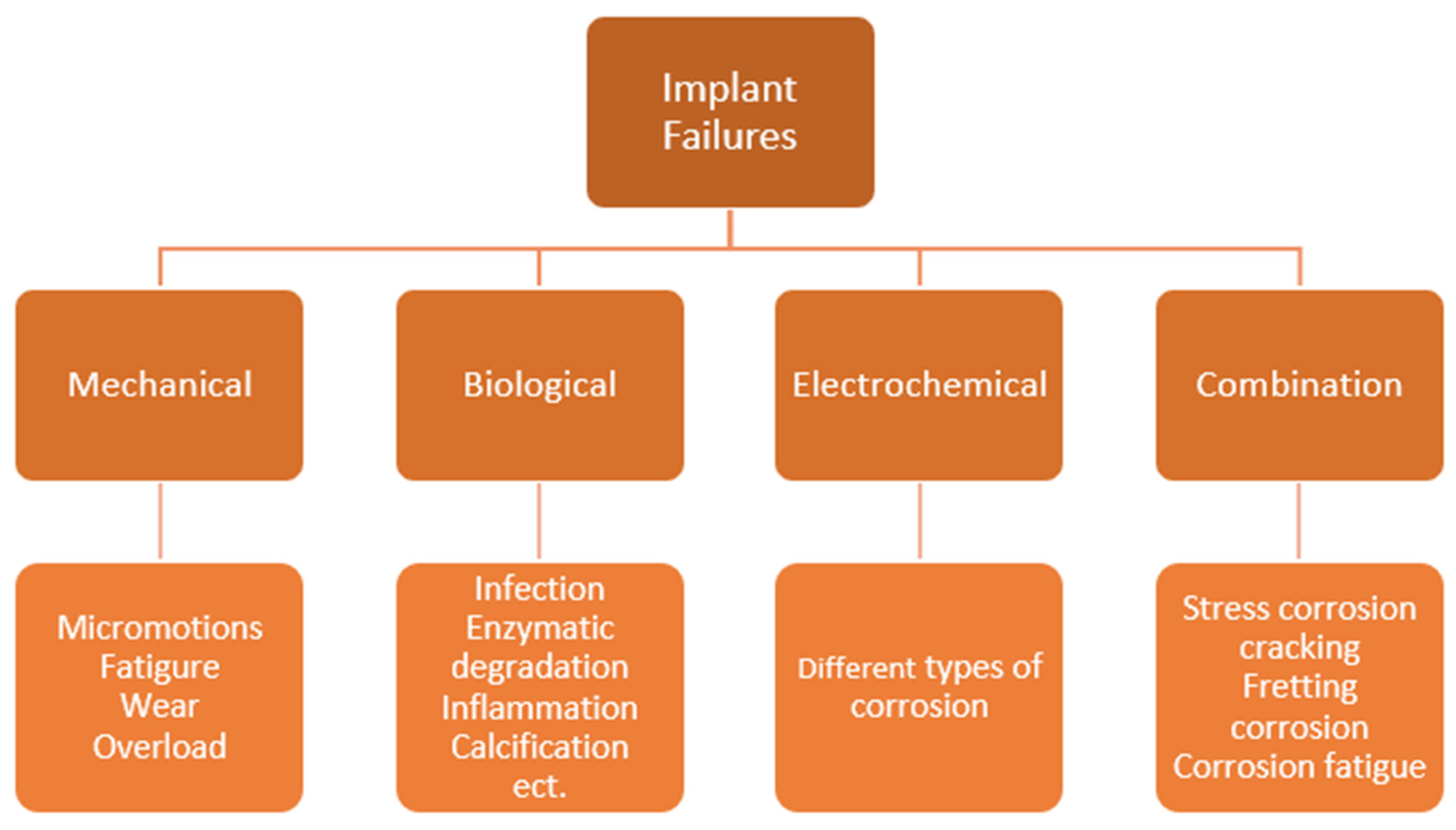
2.2. Implant Failures

The successful integration of an implant requires specific design and material selection for different applications. In order to prevent implant from failures, there are several areas that must be considered. They are good corrosion and wear resistance, high biocompatibility, appropriate mechanical properties and osseointegration [16].
The failure of implants can be caused from mechanical, biological, or electrochemical factors or a combination of these factors and therefore are classified into these four types, as shown in Figure 1. Failures caused by micromotions, fatigue, wear and overload are referred as mechanical failures. Biological failures arise from infection formation, enzymatic degradation, inflammation, calcification, etc. Electrochemical failures appear because of the variations of corrosion. Some failures, such as stress corrosion cracking, fretting corrosion and corrosion fatigue, are examples of combinations of failure classes. The biodegrading process in such cases plays a vital role. The implant may suffer because of a lack of structural integrity. It may also lead to periprosthetic bone loss, as in the example of osteolysis, which results from the formation of tiny polyethylene pieces caused by implant wear. In addition, released metals ion of implant can be also transferred by body fluids. When reaching various remote tissues, unpleasant biological reactions such as allergy, cytotoxicity and cancer can developed. An analysis of the implant failure reports of patients with orthopedic implants made of stainless steel and reveals the common failures originating from fatigue limit. Moreover, there are failures resulting from a combination of different failure classes, such as fatigue and corrosion [17].

2.3. Bone-Bonding Process

The bone formation process between bone and implant, which is also named osseointegration, plays a vital role in the successful integration of biomedical bone implants. It is still a challenge to achieve the proper bonding between implant and bone. Infections and implant loosening are the results of poor bonding. In addition, implants can affect the process by modifying its ability to provide biological cues, cells and growth factors, improving osseointegration [18].
The bone-bonding process consists of three main stages, which are the inflammatory response stage, the bone formation stage and the bone remodeling stage [19]. During the first stage, the fractured part of the bone is initially covered with blood, leading these tissues to first contact with the implant. Meanwhile, inflammatory cells migrate into the tissue, which surrounds the implant surface. During the contact, proteins are absorbed, and oxidation on the implant surface is undergone. Inflammations on implant surfaces are stimulated by immune and bone-healing reactions. Through continuous events on the contact zone, primarily the stability of implant is achieved. In the next stage, osteogenic cells are released onto the implant surface. These cells form the initial bone layer on the surface of implant, with a thickness of 0.5 µm, which is rich in phosphorus, calcium, bone sialoprotein and osteopontin. This layer is responsible for regulating adhesion and bonding with the implant surface. In the last stage, cavities with a depth of 100–500 µm are formed on the new bone thanks to the resorption of bone particles and the death of osteocytes. On the top of these cavities, new bone is formed by osteoclast cells. Because of the remodeled bone, implants generate secondary stability directly with the bone, obtaining stronger bone-to-implant adhesion [20,21]. Figure 2 shows the interaction between biomaterials and cells.
Titanium is so far one of the most preferred metal materials for orthopedic applications thanks to its inert behavior and high corrosion resistance. Compared with other metals, it has better direct bonding between the surface of implant and the bone. Its compatibility was revealed by observing the calcification, osteoblast activity, the formation of calcium phosphate in reproduced body fluid and the development of hard tissue on the implant surface of animals with implanted titanium. It was perceived that by implanting titanium in bone allows it to increase early contact between implant and bone, resulting in better bone-bonding strength. The surface morphology of an implant is also a vital factor because the compatibility of hard tissue depends on the adhesion and proliferation of its osteogenic cells.
Implant material must have good bonding with soft tissue, a lack of which may lead to bacterial invasion forming subsequent inflammation. This in turn results in the loosening of the implant, leading to implant failure. Titanium was also tested for its compatibility with blood cells. The formation of a thrombus in contact with titanium shows low compatibility. As a result, most titanium alloys are not used in places contacting with blood [22].

2.4. Mechanical Properties of Bone

Bone is a complex natural composite containing organic and inorganic components. The first group mainly consists of type-I collagen, although there is also type-III, type-IV collagens and fibrillin. The second type of components is hydroxyapatite. In combination of both components, bones form reinforced materials where organic materials provide the material with flexibility and inorganic materials provide strength. Under the dry conditions, about 95 wt.% of bones are type-I and hydroxyapatite [23].
The structure of a bone is hierarchical in that each level has different mechanical, chemical and biological performance levels. These levels are segregated as following: macroscale, microscale, submicroscale, nanoscale and subnanoscale. The general form of bone is represented at the macroscale level. The bone has two classifications, which are compact (cortical) bone and trabecular (cancellous) bone. Compact bone is located in diaphysis, forming a shell around trabecular bone [24]. By being almost solid, compact bone has only about 3–5% of space for blood vessels, cavities, osteocytes etc. Meanwhile, trabecular bone has large spaces that are filled with bone marrow. The porosity of trabecular bone varies in the range of 50–90% [23].
Mechanical properties of bone can vary significantly because of age, bone quality and the anatomical site. Because of that, it imposes a challenge in fully understanding the mechanics of bones. Properties of bone are important references that should be matched with implant material. In fact, one of the failures occurs because of the mechanical properties mismatch between bone and implant. Two of the vital mechanical characteristics of the biocompatibility of orthopedic implants are elastic modulus and strength. They are anisotropic. Cortical bone has more capacity for strength when loaded longitudinally, while it is weaker in radial and transverse directions. Moreover, it has better performance in compression compared with the one in tension. In addition, in two studies, the elastic modulus of cortical bone in the longitudinal direction was in the range of 14.0–21.8 GPa [23,25]. The values of tension and compression strength were in the ranges of 119.4–150.6 MPa and 187.7–222.3 MPa, respectively [24]. Meanwhile Poisson’s ration was between 0.24 and 0.56. Meanwhile, trabecular bone, during cyclic loading, exhibits time-dependent behavior and damage susceptibility. As well, its mechanical properties depend on the architectural disposition of each trabecula [23].

2.5. Surface Treatment Requirements

There are several aims that needs to be approached during the modification of implant surfaces. Table 1 gives an overview of the requirements.

2.5.1. Bone Formation

The ability of a surface to allow bone growth on the top of the implant surface is called osteointegration. Usually, this is also called a bone ingrowth, and it refers to bone formation in the porous surface of implants [26]. This factor plays a significant role in the successful integration of implants for long-term applications [21]. There are also desired surface characteristics of porous materials pertaining to treatment that provide the topology for good cell attachment, differentiation and proliferation. In addition, pore sizes should be appropriate to provide ingrowths capable of transferring metabolic waste and nutrients [23]. However, a high bone ingrowth rate is not always preferable. Some dental implant applications prefer to avoid bone formation on the surface of implants. The formation of an oxide layer on titanium-based alloys increases biocompatibility. It contributes to the higher bone formation. Eventually, the removal of an implant will require bone refracture [27].
The osteointegration of implants can be affected by different surface techniques and elements. By adding calcium and phosphorus on the oxide layer of titanium implants, the reaction with blood cells accelerates the osteointegration process [28]. Local blood cells such as osteoblasts and platelets react with the oxide layer of titanium implants, leading to the activation of platelets and the crosslinking of fibrins and providing osteogenic cells [38,39,40]. As a result, the process of the formation of structural and mechanical bonding between bone and implant surface occurs without fibroid connections, which is called osseointegration [38,40,41].

2.5.2. Adhesion with Soft Tissue

Second, the implant surface must have good adhesion to soft tissues. It forms a biological seal that protects the implant from bacterial infiltration. The absence of soft tissues can lead to the intrusion of different bacteria, which may result in inflammation such as implantitis [27,29]. In fact, when an implant is inserted into the body, a conditioning film is formed on its surface, consisting of proteins known as the extracellular matrix (ECM). Because of this film, bonding between the surface and host cells forms. However, this layer can also be colonized with bacteria [30]. The contact between bone and implant should be direct in order to form strong osteointegration, because an implant with a layer of soft tissue can cause an undesirable effect on bonding, leading to considerable loosening after several years [29].
Stainless steel and titanium alloys are of two main materials related to orthopedic applications. Among them, surfaces of stainless steel are more prone to forming infections [30].

2.5.3. Preventing the Formation of Biofilm

Third, the surface of implants needs to prevent the formation of biofilm. In the case of bacterial invasion, the surface of the material should still prevent the formation of multilayered biofilms, resulting in infections such as perimplantitis [27]. In fact, the formation of biofilms is one of the reasons for implant failures, contributing to the implant’s loosening or dislocation and poor vascularization [32]. The formation of biofilm is associated with the term “race for the surface” when a medical device is implanted into the human body, meaning that the host cells and bacteria are competing for surface adhesion. The first of them to colonize the surface prevents the colonizing of the second one [42]. The adhesion of bacteria ensues thanks to the physicochemical forces on the implant surface, which absorbs macromolecules and tiny organic compounds. This process is divided into Phase I and Phase II. In Phase I, the interaction between implant surface and bacteria is reversible and is formed during the first 1–2 h after implantation. Meanwhile, after 2–3 h, the surface forms strong adhesion with the bacterial cells thanks to specific and nonspecific reactions with the proteins of the bacteria. Once Phase II has settled down, it become hardly reversible [32,33].
The share of infected orthopedic implants is still relatively high. In the United States, by 2006, there were over annual 112,000 orthopedic implants infected by bacteria, which was 4.3% of the overall orthopedic implant number. There are several methods that researchers offer in order to reduce the bacterial adhesion leading to the formation of biofilms, which are based on two approaches: surface modification and surface coating. Antibacterial coatings such as antibodies, antibiotics, nitric oxide and silver ions have proven their efficacy by affecting the physiochemical properties of substrates, thus preventing the formation of bonding between bacteria and substrates [33]. In addition, surface modification is another approach to prevent the development of infections on implant surfaces. It implies the modification of the textural-, atomic- and molecular-scaled properties of an implant’s existing surface layer, in contrast to the surface coating technique, which creates an additional surface [34].

2.5.4. Increased Wear Resistance

Last, the surface modification of an implant should enhance its wear resistance [27]. Unsatisfactory wear properties of implant surface can result in several unwanted consequences from implantation. Implant loosening can occur because of the wear loss between bone and implant [36]. Wear mechanisms of these materials are divided into three groups: adhesive, abrasive and fatigue wear. Adhesive wear is the removal of surface particles by their adhesion to an adjacent surface caused from loading, which creates stronger bonding than that of the original surface. Abrasive wear occurs from rubbing between two surfaces with different hardness levels. Fatigue wear results from exceeding fatigue limit of its surfaces, releasing particles [37]. It is imperative to pay attention to the environment where wear occurs. The wear behavior of metals differs significantly in air and in body. In body-simulated fluid, the wear of materials is considerably less than that in air. Meanwhile, comparing abrasive wear and adhesive wear, the domination of adhesive wear over abrasive wear in body-simulated fluid was observed [36]. An excessive release of metal ions can have several influences on the human body, leading to accumulations in human organs, changes in metabolism, allergic effects, etc. [35].
There are several approaches to improve wear resistance; however, focusing on surface hardness is the most effective method [36]. Several techniques were investigated in the literature, specifically oxidizing [43], electroplating [44], nitriding [45], thermal spraying [46] and physical [47] and chemical vapor deposition [48].

2.6. Remarks

Surfaces of implants require special attention because they play a huge role in the successful implantation of orthopedic metallic devices. Although the material properties must have attained lower-value density, elastic modulus and higher values of hardness compared with current implant materials, surface properties are responsible for proper interactions with body tissues and fluids, preventing the formation of biofilms, and should have excellent corrosion and wear resistance. Otherwise, implant failures will ensue, which are divided into four groups: mechanical, biological, electrochemical and a combination of them. There are several surface treatment techniques that can enhance the biocompatibility of implants, decreasing the probability of implant failure. Four principal criteria for proper surface treatment are the ability to stimulate optimal bone formation, the proper adhesion of soft tissue, preventing the formation of biofilms and providing high wear resistance.

3. Materials for Biomedical Alloys

In this section, the different biomedical materials are reviewed, starting with their classification into metals, polymers, ceramics and composites. Section 3.2 focuses on metal alloys. The mechanical properties of metal alloys are also discussed.

3.1. Classification of Biomedical Materials

Biomedical materials have developed a wide range of applications in biomedicine. They are used in dental care, skin tissue engineering, cardiovascular devices, drug delivery and orthopedics. Because each of these applications requires customized material properties and good biocompatibility, there are several types of biomaterials, which can be categorized into four classes: metals, polymers, ceramics and composites [49]. Among them, the most common biomaterial class for implant applications is metals [50]. Figure 3 provides a short overview of biomedical materials.

3.1.1. Metals

Because of their excellent thermal and electrical conductivity as well as mechanical properties, biomaterials made from metals are widely implemented in biomedicine. Because electrons in metals are independent, thermal energy and electric charges can be transferred quickly. These free electrons provide a strong bonding force, holding positively charged metal ions together. Because of the fundamental nondirectional nature of metallic bonds, metal ions can be displaced without breaking the crystal structure. This allows solid material to plastically deform. In addition, some metals have good corrosion resistance. Because of these capabilities, several metals are applied in orthopedics, replacing hard tissues such as hips and knees or are used in fracture-healing purposes for bones, such as spinal fixation devices, dental implants and bone screws and plates [51].
The first attempt to implement metallic materials as medical implants was witnessed in the 19th century, when the Industrial Revolution resulted in an expansion of applications from the metal industry. They were used as implant materials in order to provide the fixation of fractured long bones. However, implants made from silver, gold or iron did not provide efficacious outcomes. In 1860s, Lister introduced the aseptic surgical technique, which allowed to implement metallic materials as medical implants. After that, metallic materials became major materials in orthopedics [16]. Furthermore, the application of metal alloys improved their outcomes thanks to enhanced mechanical and tribological properties, which led to better biocompatibility for implants. The first metallic implant for the human body was Sherman Vanadium steel; however it was found to be toxic in long run because of its rapid corrosion. Alternatively, in 1920s, 18Cr-8Ni (wt%) stainless steel was developed as an alloy with better corrosion resistance [15]. Later, by adding molybdenum, 316 stainless steel was obtained, providing improved corrosion resistance. Then, in the 1940, titanium and its alloys were studied as implant materials for orthopedic applications. The reason behind that was the successful implementation of these materials in aircraft applications thanks to their superior corrosion resistance in salty seawater. Meanwhile, in the 1950s, the amount of carbon in 316 stainless steel was decreased from 0.08 wt% to 0.03 wt% to obtain increased corrosion resistance as well as weldability. This introduced a new 316 L stainless steel grade [17]. By containing Cr, the formation of rust in the human body was prevented. In addition, the low carbon percentage in the compound increases resistance to corrosion, preventing the formation of chromium carbides [15]. However, the risk of stress corrosion can lead to unexpected cracking failures in the implant. Thus, this material is used only as a temporary implant in some cases. In comparison, CoCr alloys have higher corrosion resistance. This is thanks to the formation of the Cr2O3 oxide layer in the environment of the human body [12]. However, the toxic effects are caused by releasing Ni, Cr and Co, leading to several diseases [14]. As an alternative, Ti alloys have gained enormous demand thanks to several advantages in terms of mechanical properties (high specific strength), good corrosion resistance from the formed oxide layer and excellent biocompatibility [50,52]. The density of Ti alloy is the lowest among the three mentioned alternatives that are closer to bone. In terms of corrosion resistance, the unique behavior of Ti is the rapid formation of an oxide layer. It can be useful in cases where a layer is to be broken (pitting corrosion). The release of toxic elements is prevented from the immediate reformation of a new layer. Thus, Ti and its alloys were found to be better options for implant material in orthopedic applications [12].
Currently, there are a variety of metals that can be manufactured, but only a few of them can be applied as biomedical material, because of their biocompatibility requirements. According to the mentioned criteria, there are different types of metals, and their alloys have been used as implant materials for bone replacement [12,15,49]. Nowadays, biocompatible metallic materials are separated into four groups. They are cobalt-based alloys, stainless steel metals, titanium-based alloys and miscellaneous metals [16]. The first three groups are frequently used as biomedical materials [14,15,49,53].

3.1.2. Polymers

Biomedical polymers are one of the materials used in biomedical applications. Their unique flexibility in physical properties and chemical composition have serious potential in biomedicine. These materials mainly gained attention thanks to their ability to degrade in the human body after a certain time, after its function had been achieved. In addition, this type of biomaterial has advantages over metallic materials and ceramic in giving various shapes and its low cost [51]. The degradability allows for the removal of the materials without surgical revision. The application of polymers in biomedicine is relatively recent. Since the 1960s, these materials have gained many successes in applications. Despite that, numerous challenges have been faced in the transportation of these elements. They are mainly used in drug delivery and tissue engineering. However, because of the process of the degradation of polymers in the human body, the material properties have witnessed changes over time. These changed properties have the ability to affect the body in a different ways with respect to their initial responses, which lead to challenges that need further study [54].

3.1.3. Ceramic

Another material group that was developed to use in biomedicine is ceramic biomaterials. Initially, their application in medicine was limited because the materials are brittle and their tensile strength is low. However, in this century, these materials have developed an application for the replacement of body parts, especially bones. The combination of high compressive strength and inertness within the body environment favors implementing the material in dentistry [51]. Bioceramics found their place in biomedicine thanks to the studies of Larry Hench. Because metallic and polymeric implant materials were bioinert, by discovering materials that can generate bonding with bone, Professor Hench invented the first bioactive glass in 1969. He conducted experiments with degradable glass and found that a glass, currently termed 45S5, generated bonding with bone strong enough that in order to remove the implant, bone breaking was required [55]. After this invention, biomedicine found a new biomaterial group of bioactive ceramics materials, which include ceramic, glass and glass-ceramic materials [56]. Currently, one of the major applications of bioceramic materials in biomedicine is bone tissue engineering because of their ability to facilitate the growth of bone tissues at contact zones. Because of that, ceramics implant materials are produced, providing a three-dimensional (3D) porous environment called a scaffold. This allows for the transfer of body cells, providing cell interactions while preventing toxicity and degradation at a certain rate [57].

3.1.4. Composite

Biomaterials that consist of two or more phases or materials larger than molecular scales are called composite biomaterials. By combining different materials into composites, several characteristics of materials, such as elastic modulus, can be significantly changed compared with homogeneous materials. As examples of composite materials, bone, skin and wood can be classified as composite materials. In detail, bone consists of carbonated apatite and collagen, where the first item provides reinforcement and stiffness and the second provides toughness and flexibility [58]. Composite materials have vast potential in biomedical applications thanks to the possibility of obtaining strong, lightweight and stiff material. However, all components of a composite implant are required to be biocompatible, and the degradation of interfaces between components needs to be avoided. Currently, composite biomaterials are used mostly in dental and orthopedic applications [51].

3.2. Compositions of Metal Alloys

The materials implemented for biomedical implants are commonly used with the purpose of improving quality of life as a replacement for a part in the human body that was lost due to an injury or removed because of some disease. However, as was mentioned, implant material requires good biocompatibility and nontoxicity. There are several elements, so far, found to be highly biocompatible: Ti, Mo, Nb, Ta, Au, Zr, Sn and W. In contrast, there are some materials that are toxic in implant applications: Al, Cr, V, Ni, etc. [12,14]. Despite that, a good match between implant materials is achieved not only by choosing nontoxic materials but also by a combination of different elements that can increase the similarity of the mechanical properties of implants to the ones of the bone. The most important properties other than biocompatibility are low elastic modulus and high tensile strength [12,14,53]. In the case where the elastic modulus of an implant is high, the stress transfer on an adjacent bone will be prohibited, resulting in dead cells in the bone [12,32]. Meantime, the lifetime of implants depends on the corrosion and wear resistance of their material. Material debris generated from low wear resistance can be incompatible, leading to allergic reactions or toxicity [12,14,15,49]. In addition, antibacterial activity and osseointegration are vital aspects in forming bonding between implant and tissue, preventing infections. In order to successfully achieve tissue ingrowth, no local displacements between the surface of an implant and tissues are a prerequisite [12]. These conditions can be maintained by using a surface topology with roughness values between 1 µm and 1.5 µm, which is obtained from the surface treatment [59]. Therefore, the chemical composition of the implant material and its surface topology are vital factors that are responsible for the biocompatibility of the implant. Figure 4 provides the requirements for implant materials. Figure 5 provides the range of elastic modulus values for metallic materials.
Table 2 summarizes the advantages and disadvantages of metal implant materials.

3.2.1. Stainless Steel

Stainless steel is a type of metal alloy based on iron. It has high amount of chromium (11%–30 wt%) and a varying percentage of nickel. In recent years, AISI 316 L stainless steel grades have been the main grades used in biomedicine, among other types of stainless steel. It is composed of a high amount of chromium (16–18%) and nickel (10–14%) and a low amount of carbon (below 0.03%). Moreover, some additional elements are added, such as molybdenum (2–3%) and manganese (2%), as well as a tiny amount of sulfur, phosphorous, silicon and nitrogen [63]. This grade has good ductility, fatigue properties and work hardenability. In addition, the attractive side of the material is its low cost, production ease and availability. Despite its good biocompatibility, stainless steel alloys lack blood compatibility, bioactivity and osteoconductivity [62]. According to their chemical composition, these metals are divided into two groups: the chromium type and the chromium-nickel type. Furthermore, based on molecular microstructure, they are classified into four groups: martensite, austenite, ferrite and duplex (mix of ferrite and austenite). The first three groups are used in medical applications. Ferritic stainless steels are used in medical devices. Meanwhile, martensite stainless steels have appropriate hardness characteristics reaching 97 HRB, which makes them suitable to be used in surgical and dental instruments. Austenitic stainless steels find their applications in nonimplantable devices, which require high corrosion resistance and strength. In addition to that, most of the implant materials made from stainless steel are austenitic [16].

3.2.2. CoCr-Based Alloys

Alloys based on cobalt were initially used in aircraft applications. Throughout the past century, their applications were developed in biomedical applications. In fact, compared with stainless steel alloys, cobalt-based alloys offer better corrosion resistance, less wear and less fatigue. CoCrMo alloys are currently the most common materials used in implants for hard-tissue replacement with high loads. Because of their high fatigue resistance, these materials can be used in permanent applications, which can last for more than 20 years, showing their high long-term biocompatibility. Nevertheless, there are still several issue that needs further investigation. Cobalt-based alloys suffer several failures in terms of fretting, corrosion fatigue, wearing and stress-shielding effects, causing aseptic loosening and releasing of particles such as Co, Ni and Co ions, which leads to biological toxicity [16]. In addition, according to Figure 5, the high elastic modulus of these alloys is another disadvantage. Currently, wrought CoNiCrMo and cast CoCrMo alloys are the main cobalt-chromium type of alloys used in biomedical applications as replacements for knee joints and hip stems. The high ultimate tensile strength (655–1172 MPa [17]) and fatigue strength (up to 107 cycles [16]) of wrought alloys allows them to be used in implants for long-term applications [17], upwards of 20 years. Moreover, a small amount of Cu is added in order to obtain better antibacterial and mechanical performance [64].

3.2.3. Titanium and Titanium-Based Alloys

It was reported that the molecular structure of Ti and that of its alloys have a significant effect on biocompatibility. Its general molecular structure is hexagonal and close packed, which is called the α phase. However, it also can be transformed into a body-centered cubic molecular structure called the β phase. These phase transformations can be manipulated by alloying them with specific elements. These additives are called α (Al, O, C) and β (Mo, Nb, Ta) stabilizers. By manipulating the amount of the composition of both stabilizers, Ti alloys can be divided into three main groups: α- and near-α-type, α+β-type and β-type alloys [12,14,15,49].

α-and Near-α-Type Ti Alloys

The molecular structure of α-type Ti alloys is formed from α-phase stabilizers. Moreover, α-type titanium alloys that contain tiny amounts of β stabilizers (5 vol% or less) are called near-α titanium alloys. In general, these alloys are CP-Ti grades containing 0.18–0.40 wt% oxygen (O) and 0.20–0.50 wt% iron (Fe). These types of alloys are good for corrosion, creep resistance and weldability. For those reasons, the implementation of these materials is suitable for a high-temperature environment [12]. Despite that, the lower capacity of mechanical and the fatigue strength of these materials make them less desirable for bone replacement implants [12,14,15,49]. Because of that, these materials are implemented as bone implants with low loads applied [12].

α+β Type Ti Alloys

Compared with α-type Ti alloys, α+β-type Ti alloys contain both types of stabilizers, where β stabilizers are dominant in content. The main advantage of these materials is their high strength. In addition to that, these alloys can be optimized in mechanical properties through heat treatment. In detail, the chemistry, cooling rate and temperature of the heat treatment can lead to improvements in mechanical strength by up to 50%, maintaining elastic modulus at the same level. Ti-6Al-4V is currently the most widely applied implant material. Originally designed for aircraft applications, the attractive properties of corrosion resistance and performance levels for fatigue and strength allow its application in biomedicine. Furthermore, this material was further improved by decreasing the number of impurities, such as N, O, H and C. The toxic V was then replaced with Nb (Ti-6Al-7Nb) and Fe (Ti-5Al-2.5Fe), increasing wear resistance as well [12]. Despite the mentioned advantages, there is a possibility of the occurrence of several diseases from the existing Al in alloys [12,14,15,49]. In addition, the relatively high mechanical strength of α+β alloys leads to bone resorption as well as the loosening of the implant, and therefore, the first generation of implant materials required an alternative solution.

β-Type Ti Alloys

A recent and more suitable solution for previous types of materials is β-type Ti alloys. They completely consist of β stabilizers. As was mentioned, β stabilizers can be treated by different techniques so that different aspects of the material can be improved. Compared with α+β-type Ti alloys, β-type alloys have significantly lower elastic modulus (according to Figure 5, the elastic modulus of α+β-type and β-type Ti alloys can reach down to 89 GPa and 55 GPa, respectively). Having similar properties makes this material the more preferred option for biomedical implant applications [12]. Moreover, these types of materials have higher density because the content of these compositions include elements with relatively higher density, such as Ta, Mo, Zr and Nb [12,14,15,49]. However, the cost of these materials is relatively high, which increases the cost of these alloys.

3.3. Effects of Alloying Elements

Metals made from a single element, such as commercially pure titanium (CP Ti), have good biocompatibility. However, this is not enough to successfully implement these materials, because of the lack of desired mechanical and chemical properties. For that reason, this field requires better alternatives to the existing implant biomaterials. Adding alloying elements to metals offers the possibility to enhance mechanical and chemical properties and biocompatibility.

3.3.1. Titanium Alloying Elements

Titanium-based alloys have better biocompatibility than cobalt-based alloys and stainless steel thanks to their excellent corrosion resistance. Common titanium alloy Ti-6Al-4V has controversial effects, though [65]. The release of ions comes with allergic effects on the human body. Alloying elements such as niobium, tantalum and zirconium are considered safer alternatives to vanadium and aluminum. Titanium alloys with beta stabilizers have improved biocompatibility over the currently implemented options. There are wide variations of titanium alloys that are implemented and that are studied for biomedical applications. Consequently, some of the most used alloying elements are Al, Nb, V, Mo, Ta and Zr [49].

Titanium

Titanium has been used in biomedical applications thanks to its safe and inert behavior in the human body [49] and for its good corrosion resistance [66]. This element is nontoxic at any doses and does not have any biological role in the human body. Moreover, it can form good connections with host bone [16]. However, by using pure titanium in vivo, its inertness is affected, which leads to corrosion over time. Emitted ions of titanium are harmful insofar as they increase internal exposure. By studying the influence of implants on animals, it was observed that through successful implant integration, the amount of titanium in lungs was 2.2–3.8 times and in local lymph nodes was 7–9.4 times higher than the mean amount of the animal that was not surged. Some patients with dental implants developed a titanium allergy, leading to high risks [49].

Vanadium

Vanadium is an alloying element that is added to implant materials. On one hand, this element has an antidiabetic effect. On the other hand, it can reduce body weight gain and cause gastrointestinal discomfort [49]. Moreover, this element is very toxic for human cells when released into the human body [66]. In addition, there are studies that were conducted on animals where inhalation or oral exposure to this element negatively affected the neurological system, blood parameters, the respiratory system and other organs. There is a connection between Ti-6Al-4V implant failure and vanadium release. For that reason, implant materials containing this element are expected to be replaced by biomedical materials that are less harmful to the human body [16].

Aluminum

Aluminum is an element that is present in the human body for its specific needs. However, high amounts are toxic for humans. In some cases, it can lead to neurotoxicity and cause several diseases related to kidney, breast cancer and digestive disorders [16]. From observations on mice, their capacity of spatial memory was reduced and motor functioning quality were reduced after an injection of aluminum hydroxide [67]. Large local concentrations of aluminum in the body can lead to Alzheimer disease [49,66]. Moreover, it was observed that this element is linked with kidney disease and osteomalacia [49].

Zirconium

Zirconium is a beta-stabilizing element that has good biocompatibility [66]. It can strengthen the solution thanks to its relatively high atomic radius. In addition, its ability to stabilize the beta phase of alloy enhances recrystallization. By adding zirconium, tantalum, molybdenum, iron and niobium, elastic modulus can be decreased while the ultimate tensile strength can be increased [49].

Niobium

It was found that niobium has a toxic effect on the human body. A study estimated that niobium is capable of altering DNA and can cause the death of immune cells [16]. However, this element has good corrosion resistance and biocompatibility [66].

Tantalum

Because of their high resistance to heat and wear, refractory metals are good for applications that involve high wear. However, tantalum was found to be an advantageous element thanks to its flexibility, corrosion resistance and biocompatibility in biomedical applications [16,66]. Its corrosion resistance is capable of withstanding numerous acids and most solutions. In terms of biological aspects, it does not have any positive of negative effects on the human body. This material is used mainly as an alloying element in titanium alloys [16].

3.3.2. Stainless Steel

It was identified that stainless steel suffered several failures in orthopedic devices. By adding nitrogen and titanium alloying elements, the metal was modified for higher corrosion resistance and strength. In addition, annealed 316 L stainless steel was alloyed with 0.05–0.22 wt% of nitrogen. In combination with cold working, this resulted in an increased resistance to pitting corrosion [17]. Moreover, stainless steel needs to have a minimum of 11 wt% of chromium in order to prevent the formation of rust. Its good attraction with oxygen enchases the healing abilities of the material, restoring damaged oxide layers. Meanwhile, nickel results in increased stability in austenite iron. Because of oxide’s forming ability, it increases corrosion resistance. However, this element was stated to have toxic effects on the human body. Adding molybdenum results in an increased resistance against pitting corrosion, compensating for the corrosion caused from the formation of chromium carbide. Nitrogen can also be added to increase corrosion-resistance capacity, but it can also replace nickel in some 316 L alloys in order to enhance the mechanical strength of the material. The application of these materials was in total hip replacement [16].

3.3.3. Cobalt-Chromium (CoCr)

Alloys based on cobalt-chromium elements offer better corrosion resistance comparing to stainless steels thanks to the high concentration of chromium. This element additionally forms a layer of passive oxide and therefore increases wear resistance. As well, molybdenum and nickel not only increase corrosion resistance but also improve solid-solution strengthening. Nickel as well as carbon results in better castability. Tungsten improves solid-solution strengthening and results in better microstructure, resulting in a reduction in cavity shrinkage, holes caused by gas blow and the segregation of the grain boundary. Despite that, this element has a negative impact on corrosion resistance and fatigue strength [16]. Table 3 gives a brief overview of alloying elements.

3.4. Mechanical Properties

The mechanical properties of biomedical implants vary depending on their applications. As was mentioned, they are found to be the most important factor responsible for good biocompatibility. In detail, they offer tensile strength, elastic modulus, hardness, elongation, fatigue strength and corrosion resistance. Each of these parameters contributes a unique part to successful implant surgeries, while their inadequate values may lead to failures. The detailed information of each implant parameter is provided below.
Comparisons of different implant materials in terms of mechanical properties is provided below. First, the most vital criterion for the successful biomedical integration of implant materials in orthopedical applications is elastic modulus. The elastic modulus of 316 L stainless steel is equal to 193 GPa [68]. The elastic modulus of cobalt-based alloys varies between 240 GPa and 260 GPa [69]. Meanwhile, the value varies from 55 to 110 GPa for titanium alloys [70]. In fact, there is a wide variety of titanium alloys being studied for biomedical implant applications. Through time, the elastic modulus of titanium alloys was decreased significantly by focusing on beta-type alloys. The commonly used Ti6Al4V alloy has an elastic modulus of 110 GPa [16,60], while newly developed beta alloys such as Ti-35Nb-7Zr-5Ta and Ti-35.5Nb-7.3Zr-5.7Ta have elastic modulus values of 55 GPa [60] and 55–66 GPa [12], respectively. Nevertheless, these elastic modulus values are still high for appropriate bone replacement applications.
Regarding the ultimate tensile strength, high strength is required to withstand the high loads applied from daily routines. The appropriate strength of a material prevents an implant from fracturing. The value of 316 L stainless steel varies between 540 MPa and 1000 MPa. The ultimate tensile strength of CoCrMo-based alloys ranges from 900 MPa to 1540 MPa and that of titanium alloys is 900 MPa. Compared with the ultimate tensile strength of cortical bone, 130–150 MPa, these implant materials offer higher strength [16].
The fatigue behavior is another important aspect that is responsible for the lifetime of an implant. In fact, despite the uniform feature of metallic alloys, the quality of the implant surface plays a big role on fatigue performance. Because of the existence of cracks and pits, crack propagation can lead to mechanical failure. Compared with cobalt-based and titanium-based alloys, fewer data are available on the practical implementation of stainless steel because of the temporary application of stainless steel. Because of its low corrosion resistance, the fatigue strength of stainless steel alloys is considerably lower in body fluid environments. This causes the release of ions, which is harmful for the body, making it the least desirable option for long-term orthopedic applications. The fatigue strength of 316 L stainless steel usually varies between 200 MPa and 300 MPa when subjected into biological solutions. Meanwhile, the performance of fatigue properties of casted and forged 316 L stainless steel alloys is better. Commonly, fatigue failures are caused from pits, which lead to the initiation of crack propagation. However, austenitic stainless steel alloys with added nitrogen and molybdenum have better resistance to pitting corrosion, which increases resistance to fatigue corrosion. Meanwhile, cobalt-based alloys have better fatigue strength than casted and forged austenite stainless steel. In addition, their sensitivity to notches is lower than that of titanium alloys and of stainless steels. Moreover, forged cobalt-based alloys have higher fatigue strength than its casted alloys. The fatigue strength of forged alloys can exceed 500 MPa in air conditions. In contrast, the fatigue strength is decreased in simulated body fluid environments. Casted CoCrMo alloys have 100 MPa of fatigue strength, and wrought CoCrMo alloys have 200 MPa. Given these values, casted cobalt-based alloys are not a reliable material for long-term usage, whereas wrought CoCrMo alloys can be safe for approximately 20 years of usage because the operating tensile strength reaches up to 200 MPa of loads in legs and arms. Because of the variety of titanium alloys, the fatigue strength of these alloys varies. However, high corrosion resistance results in better fatigue performance compared with stainless steel and cobalt-based alloys in the highly corrosive environment of the human body. Despite that, β titanium alloys have fatigue strength values less than α-β alloys. As an example, the limits of the smooth fatigue of Ti-13Nb-13Zr (β phase) and Ti-6Al-4V (α-β phase) are 500 MPa at 107 cycles. However, the fatigue limit of α-β-phase titanium alloys such as aged Ti–15Mo–5Zr–3Al can reach 560–640 MPa. Given the approximate requirements of bone replacement in terms of fatigue strength, joint implants should withstand the compression and the bending strength of approximately 50 MPa and 200 MPa, respectively, for the total number of 107 cycles estimated for 20 years of use [16].
As well, the corrosion resistance of implant materials is an important parameter for successful implant integration [71]. High corrosion resistance prevents the formation of debris, and this formation can cause the release of toxic metal ions, leading to allergic reactions [12]. Among stainless steel, cobalt-based and titanium-based alloys, both cobalt-and titanium-based alloys have the advantage of high corrosion resistance. On the other hand, stainless steel alloys lack corrosion resistance [49]. The formation of an oxide layer is one of the key aspects of this good corrosion resistance. In that way, CoCr-based alloys form a passive oxide layer (Cr2O3) from the Cr alloying element in the material. Despite that, these alloys still emit toxic elements such as Ni, Cr and Co into the body. Similarly, titanium alloys form a TiO2 oxide layer, which results in excellent corrosion resistance. In addition, titanium alloys tend to rapidly form a new oxide layer in the human body environment when the old one is removed or damaged from wear or loadings. β titanium alloys also provide better corrosion resistance than α-β titanium alloys in implants [12]. Table 4 gives the list of biomaterials.

3.5. Remarks

There are four classes of biomedical materials: metals, polymers, ceramics and composites. The main applications of metals are in bone fixation and replacement. Currently, their compositions vary for the purpose of decreasing undesirable effects on the human body and the number of failed results because no ideal implant material exist. There are three main groups of biomedical alloys: stainless steel alloys, cobalt-based alloys and titanium-based alloys. Their alloying elements change the properties of materials in order to enhance biocompatibility, bone ingrowth, mechanical properties and corrosion/wear resistance, which are vital parameters in successful implant applications.

4. Surface Modification Techniques

4.1. Laser Surface Modification

Enhancing the surface properties of the biocompatible metals can foster the needs of biomedical applications, such as good corrosion resistance, high-tribological performance, and high-temperature performance. This surface-enhancing method would ideally preserve the original properties of the substrate and modify only the surface of the metal, by achieving lower friction coefficients and higher hardness properties than the bulk material. One type of such tools, which operates on the same principles, are lasers [81]. Lasers have found use in various material processes, such as welding, forming, machining and surface modifications. Even though the utilization of lasers in surface modification makes up a minor percentage of application in comparison with other material processing, this tool is finding its popularity among biomaterials, light alloys and tribological materials [82]. Table 5 provides the advantages associated with laser surface modification.
Laser surface modification can be classified into five main categories, which are based on the interactions between laser and material and on the microstructural and compositional effects on the specimen surface: laser surface melting (LSM), laser surface cladding (LSC), laser surface heating (LSH), laser surface alloying (LSA) and laser shock peening (LSP). Apart from the LSP and LSH methods, most of these methods implicate the utilization of filling material and/or melted substrates. In order to eliminate or minimize the risk of surface cracking, the substrate is preheated either with a supplementary heat source or a defocused laser beam in advance of using one of the abovementioned laser techniques [85,86].

4.1.1. Laser Surface Melting (LSM)

LSM can be used in the extensive list of industries as it has high precision and high depth/width ratios in fusion zones. This results in the reduction of material affected by a laser beam. It operates on the principle of applying an adequately high-power-density laser on the surface of the material. The high-energy beam’s absorption causes the substrate temperature to quickly increase in order to induce the local melting of the substrate. As heat is transferred from the surface into the material’s bulk, the front between the liquid and solid phase moves deeper into the bulk material. The surface temperature rises until the melting and evaporation temperature balance the surface energy deposition [87,88]. The changed zone develops a finer grain structure as a result of the quick subsequent resolidification of the laser-melted area. The melting and resolidification influence only the material’s surface thanks to the very brief contact period, having little to no impact on the material’s mass. In order to increase the contact wideness, the laser can be scanned across the surface to create overlapping parallel tracks, as shown in the Figure 6 [84].
With the usage of LSM, homogeneous, hard, and ultrafine structures are created on the surface without changing the chemical composition of specimen material [88,89]. As a result, improvements in the surface layer features of the bulk material, such as erosive, wear and corrosion resistances, can be found. Up to 42% reduction in the wear volume of laser melted AM50 Mg alloy surface, along with 15–25 HV improvement in micro hardness, was reported [90]. The laser-melted surface layer undergoes metallurgical modifications in the form of finer-grain refinement, supersaturated solid solutions and particle dispersion [91,92]. All these approaches have the potential to reinforce and harden the top layer. Alloys that cannot be successfully hardened by laser transformation hardening can be hardened by laser melting. In addition, this technique has the capability to repair the damaged surface, which may be difficult by conventional approaches [93]. Table 6 represents the outcomes of LSM treatment on biomedical alloys.
Figure 6. Experimental setup of the LSM treatments [94].
Metals 13 00082 g006

4.1.2. Laser Surface Cladding (LSC)

The underlying idea of laser cladding is the melting of the coating material and a small layer of the substrate using a scanning laser beam aimed at the surface to create the coating. The procedure entails laser melting of the substrate’s cladding without the base material’s being considerably affected. Without significantly altering the microstructure of the substrate, the quick solidification of the cladding material can lead to an increase in solid solubility and in grain refinement and the production of nonequilibrium phases in the clad layer [101,102]. The coating material, which can be in the form of powder, thermally sprayed coating, foil and wire, is predeposited on the substrate in two-step laser cladding and then melted with the laser beam to produce the coating. However, if powder is used for the cladding, it faces the challenge of keeping the powder on the surface during the process [103]. In most applications, preplacing the desired material on the bulk material is followed by LSM [86]. Figure 7 describes the laser-cladding steps. LSC has the ability to deposit multiple layers to form complex geometric shapes [104]. Cladding results in an insignificant dilution of base materials owing to the short laser-interaction duration and can therefore retain the strength of the original material [105]. The resulting coating thickness varies from 50 µm to 2 mm and gives protection against wear, corrosion and oxidation [101,106,107]. Table 7 represents the outcomes of LSC treatment on biomedical alloys.

4.1.3. Laser Surface Heat Treatment (LSH)

LSH operates mainly on the principle of modifying mechanical and physical characteristics by restrained heating and cooling with a laser beam. Temperature has to rise higher than the critical transformation temperature, but it should not exceed the melting temperature [113]. LSH is often used when there is no need for shape changing and there is a necessity in material strength increase. Moreover, this method enhances wear and corrosion resistances [114] and fatigue properties [115,116]. The power, diameter and scanning velocity of the laser beam, as well as the absorptivity and thermal characteristics of the base material, are the key processing parameters for LSH. This method is convenient for finite area applications and does not require external quenching [117]. Low laser power and scanning speed can enhance the hardenability with a deeper hardened layer. A hardened depth of 1.05 mm in the case of carbon steel was reported for a laser power of 600 W with a 5 mm beam diameter and a scan speed of 4 mm/s without melting [118]. This hardness value increases up to some distance from the surface (690 HV, for a laser power of 2 kW, speed of 8 mm/s and beam radius of 3 mm) and then decreases to match the bulk material’s hardness [119]. Some studies also reported on the microstructure changes due to LSH, which eventually enhanced machinability [120], while others reported on the enhanced tribological properties as well as the enhanced service life [121]. However, a brittle hardened layer may cause the deterioration of the bending properties of the treated materials [122]. Table 8 represents the outcomes of LSH treatment on different alloys. Table 8 represents the outcomes of LSH treatment on different alloys.

4.1.4. Laser Surface Alloying (LSA)

The laser surface alloying process is similar to LSC, with a difference in the thickness of the modified layer. If in LSC, mixing between the substrate and coating happens only on the interface of the bulk material or above, then in LSA, considerable mixing happens deeper to the specimen, forming the new alloyed surface layer. The formed layer has new phases and an alternate composition [127]. There are different approaches to depositing alloying elements on the substrate: direct injection during the laser treatment, depositing preplaced adhesive and nonadhesive coating [128]. One of the main parameters for the LSA is the temperature gradient, which affects the convective movement of the liquid transport in the melted pool. Moreover, shear stress also plays an important role in the allocation of the alloying particles on the bulk material surface [129]. The literature reported on the corrosion-resistance characteristics of nonferrous metals with alloyed surfaces, such as Ti-Pd systems. Adding a noble metal such as Pd to Ti can shift the mixed potential into the passive zone [130,131]. Therefore, Pd-alloyed Ti alloy has demonstrated a significantly reduced corrosion rate by using LSA [132]. In addition, a Ti-6Al-4V alloy laser surface alloyed with titanium nitride through gaseous nitrogen offers noticeable wear resistance against a two-body abrasive (1.7 times of original one) as well as dry sliding wear [133]. The wear-resistance properties of pure titanium can also be improved by using laser-borided, laser-borocarburized and laser-carburized surface alloying [134]. In addition, LSA with Si compared with LSA with Al and LSA with Si + Al demonstrated increased wear resistance thanks to the presence of the uniform distribution of Ti5Si3 [135]. On the other hand, Si and Si + Al LSA provided relatively higher resistance to cyclic oxidation than the Al LSA [136]. Similar results of improved sliding wear resistance of AISI 316 L stainless steel was reported using chromium carbide and titanium carbide [137]. Moreover, enhanced cavitation erosion (30 times higher than that of a received alloy) was reported for WC LSA of same materials thanks to the increased value of W in the solid solution as well as dendritic carbides precipitation [138]. Table 9 represents the outcomes of LSC treatment on different alloys.

4.1.5. Laser Shock Peening (LSP)

Laser shock peening is an effective surface treatment approach for hardening, roughening and cleaning the material’s surface layer. LSP operates on a principle in which high-energy laser is irradiated on the surface for a few nanoseconds, which results in the vaporization and further transition of the surface to the plasma stage. As the plasma expands, an intense shock wave penetrates into the metal [145]. Laser energy, pulse width, overlap rate and pulse wavelength are considered critical process parameters [146]. studies have reported on the maximum fatigue life of Ti–6Al–4V alloy when using three overlapped laser-spot LSP compared with sample as received as well as the sample with one or two overlapped laser spots. Single LSP and successive double LSP improved the fatigue strength by up to 22.2% and 41.7% as compared with initial Ti–6Al–4V alloy when applied with three overlapped laser spots [147]. This results in an improvement in the fatigue life, and the fatigue strength of the workpiece was reported for titanium alloys [148,149,150,151,152], magnesium alloys [153,154,155], carbon steel alloys [156,157,158,159] and other supermalls [160,161,162]. Moreover, this method can be considered as a promising method to enhance the corrosion and mechanical properties of the metal [163]. Table 10 represents the outcomes of LSC treatment on different alloys.
Apart from the automotive and aerospace industries, laser surface modification techniques found their use in biomedical applications. Table 11 represents some of the research on this theme.

4.2. Other Surface Modification Methodologies

The typical surface modification techniques for biomedical applications are grafting, nanostructures and surface structuring.

4.2.1. Grafting

One of the most recent and promising developments on the theme of surface modification is the biochemical method. It impels an antibacterial impact on titanium surfaces, which would foster the growth of biocompatible properties. The main drawback of this method is the limitation in long-term utilization, as absorbed substances may be released from the implant surface. On the other hand, this desorption can be resolved by introducing covalent binding. However, this method may decrease the bioactive potential of metal alloys [170]. Recent grafting methods were developed in order to solve these issues.
Covalent grafting is the technique that induces a strong link between the coating and the sample biomaterial. It can be conducted either directly to the surface or by using “anchor” molecules. The latter method minimizes the desorption and sustains the original characteristics of titanium alloy [171]. The most common molecules are listed in Table 12.

4.2.2. Nanostructures and Surface Structuring

The nanostructuring of the biocompatible alloy is a new branch of surface modification, which allows the creation of nanoscale surface patterns. Depending on the contact angle and their diameter, anodized nanostructures have shown their ability to bring about antibacterial properties in the material [183,184,185,186]. In addition, it was discovered that depending on their diameter, nanopores demonstrated less bacterial adhesion in comparison with nanotubes. On the other hand, one of the research papers presented in this review demonstrated that varying the diameter size of the nanotube and applying heat treatment would assist in the decrease of live bacteria. A combination of these parameters was recommended to be a good candidate for implant design and would enhance the tissue growth and biocompatibility of the alloy [185]. A Ti surface with surface roughness at the nanolevel was reported to decrease the bacterial cell adhesion and improve bone tissue growth [187].

4.2.3. Coatings

This study also focuses on another antibacterial approach to the titanium surface: the coating method. This type of application affects both the chemical and physical parameters of the material. The modifications were divided according to the physical and chemical types [32].

Physical Modification

Using bacteriostatic materials as a coating layer alters the top layer of titanium oxide (TiO2), changing its chemical and physical parameters. These materials allow conjoining with bacteria with no harm, therefore improving the compatibility of the implant by applying electrostatic force, which repels them without killing them. Mostly, titanium surfaces are covered with either hydrophobic materials or negatively charged polymers. Bacteriostatic materials are divided into two types: polysaccharide and polycations coatings and “smart” polymers. PolyNIPAMs are known as “smart” polymers. Depending on the temperature, these polymers can regulate the attachments of bacteria as well as detach them from the surface thanks to their phase transition. Meanwhile, polysaccharide and polycations coatings are associated with Arginylglycylaspartic acid (RGD) peptides [188,189,190]. A combination of hydraulic acid (HA) and chitosan has resulted in preventing the adhesion of bacteria to the material surface. Because of their charging capacity, biostatic ability is maintained, thus repelling the bacteria. However, the surface can be easily removed from external forces such as screwing the implant, making it not the most practical option [32].
Bactericidal materials have common aspects as bacteriostatic materials. However, their difference is being able to kill bacteria through several methods. The reason for this is the introduction of antibiotics between the layers of polymers. They come in different types, such as polymer coating [191], antimicrobial peptides [191,192], ion-implanted surfaces [193,194], photoactivatable bioactive Ti, nanomaterials [195,196], citric acid [197,198] and antiseptic and antibiotic coatings (antibiotics, silver and chlorhexidine) [199,200].
Physical modification includes a variety of modification methods. They were listed in Table 13.

Chemical Modification

Similar to the physical modification, the modification based on the chemical principle comes in different methods. It includes three types: nitride coatings, sol-gel and chemical vapor deposition (CVD). Table 14 shows the details of the process. Figure 8 shows the research conducted in the area of coating technologies.

4.3. Electrical Discharge Machining Modification

This section summarized the possibilities of EDM, micro-EDM, powder-mixed EDM and electrical discharge coating for surface modification purposes.

4.3.1. Electrical Discharge Machining

In comparison to traditional machining methods, electric discharge machining (EDM) permits the manufacturing of difficult-to-machine materials with high precision and while providing a superior surface finish. It operates in terms of applying sequential sparks between conductive workpiece material and electrode tools. The advantage of this method is in its noncontact approach, which does not cause mechanical deformation [210]. EDM has several variations, such as wire EDM, dry EDM, micro-EDM, powder-micro-EDM and die-sinking EDM. Although each principle is the same, the material removal rate and value of discharge energy differs by variation. The implementation of these machining techniques allows for conducting small- and large-scale machining, leading to high flexibility in manufacturing.
The EDM process is based on transferring electrical energy into thermal energy. It operates on the basis of the principle of a sequential series of electrical discharges that occurs between the electrode and the conductive workpiece in dielectric fluid. Each electrical spark occurs in the smallest gap between the electrode and the workpiece, induced by a high voltage, which is enough to overcome the dielectric breakdown strength. As a result, a crater forms on the workpiece surface. After the surface modification process, there is an altered metal zone (AMZ), which can be broken down into three layers: the spattered surface layer, recast layer and heat-affected zone (HAZ). The spattered surface layer features the formation of melted metal and electrode materials in the shape of the spatters and spheres on top of the surface [211]. Next, the surface that is melted but not removed from the workpiece forms the recast layer. The quick solidification results in changes in the material characteristics and the structure. Its hardness is increased, but the layer becomes very brittle, which increases the risks of crack formation, leading to implant failure. Thus, it is suggested to decrease the thickness or remove the layer in order to prevent failures, in some applications. Lastly, the surface layer that was heated without its melting is the heat-affected zone. The depth of this layer depends on the machining power of EDM and the heat transfer capacity of the material. This layer is responsible for the quality of the machined surface in terms of its integrity [212]. Figure 9 and Figure 10 show the process schematic and the surface layer created during this process. Table 15 gives some of the key findings.

4.3.2. Micro-EDM

Micro-EDM is one of the nonconventional machining techniques that was discovered as one of the most successful methods for the microcomponents processing industry. The noncontact approach of micro-EDM can be applied in machining brittle, ductile or very hardened materials with minimum force required between workpiece and electrode. Micro-EDM is capable of producing high-quality and high-precision machining when the right parameters are used. Because EDM is noncontact, it is feasible to machine with a long and relatively thin electrode. Although there are micromilling cutters on the market with a diameter of as little as 50 µm, their length is often three to five times that of their diameter and thus are not appropriate for machining particularly hard die materials, which can be machined only with EDM. Despite having a significant impact on the micromachining industry, micro-EDM has drawbacks, such as low MRR and high electrode wear. It is necessary to adjust for electrode wear either by switching out the electrode, by preparing an elongated electrode from the start or by constructing it onsite for the next machining procedures. Moreover, it is advised to keep the same electrode during milling because changing them might result in decreased precision from a new setup or fixing a misalignment of the electrode [218,219].
Micro-EDM operates on the same principles as conventional EDM, with the exception of reduced discharge energy, a decreased radius of the plasma channel, a lower volumetric material removal rate and a smaller size of the tool. One of the forthcoming ways for the localized coating of microscopic components is surface modification utilizing the micro-EDM technique. Only a small number of publications are available on the topic of coating of solid-lubricating materials. Mohanty et al. (2019) [220] studied the micro-EDM technique to make a firm and solid-lubricating coating on a surface of a Ti6Al4V workpiece. The dielectric medium was deionized water with added tungsten disulfide powder; the average size of the particle was 15 µm; and the tool was a brass rod with a diameter of 800 µm. The findings reported that numerous intermetallic compounds were formed on the changed surface. At the maximum powder level, of 12 g/L, it was discovered that the deposition rate of material was greater (0.049 mm3/min). According to a comparison with the inner material, the micro hardness has increased from 421.38 HV to 881.34 HV. For the parameter values of 70% duty factor, 60 V and 12 g/L, the highest recast layer of thickness, of 13.11 µm, was attained. A wear test was conducted, and it was demonstrated that the particular wear rate is lower than that of the base material. Currently, the method of adding powder material into dielectric fluid was considered as an updated micro-EDM technique in order to make the technology widely available for industrial applications. Mixing conductive powder into the dielectric fluid offers many advantages, including wider spark gaps, which results in lower discharge intensity and better surface quality [220]. Figure 11 presents the process schematic.
The main criteria for the machine to be able to operate at the microscale are extremely accurate machinery and a small material removal unit. A tiny unit removal criterion in the context of micro-EDM typically indicates that the discharge energy W e created in the sparking gap during one pulse must be as low as feasible, which can be achieved with the use of criteria relaxation capacitance type of power generator.
W e = 0 t e u e t i e t d t
Here, ub is voltage across the gap, ib is the currents, and t is the pulse-on duration.
Pulse duration may be significantly decreased by employing modest capacitance and physically lowering the inductance of the discharge circuit [222].
Kieswetter et al. and Buser et al. claimed that a material’s surface is strongly related to its microstructure and may affect the nearby cells of biomedical implants, and therefore, it will speed up the development of tissues or bones [223,224]. Micro-EDM was used by Jahan et al. to investigate the surface of biomedical alloys such as shape-memory alloy NiTi and high strength Ti-6Al-4V. The microstructure of the processed surface demonstrates how microhardness has changed the topography. Before machining and after machining, NiTi was reported to have a mean hardness of 420.9 HV and 524.4 HV. Similar to this, Ti-6Al-4V has a mean hardness of 429.5 HV before machining and 481.6 HV after machining [225]. Table 16 presents the relevant literature, which shows the effect of the micro-EDM on the surface of the biomaterial.

4.3.3. Powder-Mixed EDM

In the section, the powder-mixed EDM (PMEDM) technique is described, along with its process variables and its biomedical applications.

PMEDM Process

One innovative application of EDM technology is powder-mixed EDM, which enables the use of additive powder materials in the dielectric fluid. These metallic particles enable a reduction in the dielectric fluid’s insulating strength and help widen the interelectrode gap. This manipulation results in EDM performance improvement and in a better surface finish in comparison with conventional EDM. PMEDM operates on the principle in which the voltage usage helps to generate an electric field in between positive and negative charges of metallic powder materials. Then, energized particles gain acceleration and commence moving in a zigzag manner. This helps to increase the interelectrode gap and results in a superior surface finish [231]. Figure 12 illustrates the schematics of the PMEDM.
The discharge gap can be increased by adding powder particles to the dielectric because it depends mostly on the electrical and physical characteristics of the powder. Because of the powder particles’ appearance at high temperatures, it is possible to reduce the dielectric resistance and produce sparks that can travel a great distance [233,234]. Moreover, as powder particles get energized after the first spark, they tend to start moving hastily together with ions and electrons. This movement helps to produce more electric charges, as more and more electrons and ions get involved. This phenomenon helps to reduce the hydrostatic pressure along the plasma channel and aids in increasing the discharge gap. This expanded column of discharges helps to create larger shallow cavities on the workpiece, thereby increasing the material removal rate. Moreover, PMEDM helps to improve the surface finish by reducing the number and the size of microcracks. It was also stated that the utilization of the powder in the dielectric helps to enhance biocompatibility by improving the cell adhesion and cell proliferation properties [235].

PMEDM Process Variables

There are several powder-mixed EDM process variables that affect the performance of the machining. They are categorized into electrical, powder, nonelectrical and electrode-based parameters.
Peak current, discharge voltage, pulse-on and pulse-off times, duty cycle, polarity, electrode gap and gap voltage are examples of electrical parameters. It is feasible to experiment with the machining performance, including material removal rate, tool wear rate and surface roughness by tweaking these parameters [236].
Nonelectrical parameters also play important roles in machining performance. Dielectric flushing helps to maintain the machining accuracy as well as aids in eliminating debris from the workpiece. Other types of nonelectrical parameters are the workpiece and electrode rotation. The former leads to improved surface roughness and material removal rate, whereas the latter helps in interelectrode flushing. The last type of nonelectrical parameter is the dielectric fluid. It has three main functions. It can serve as an insulating medium, it aids in flushing the debris from the workpiece, and it operates as a medium, which takes away heat from the electrode tool.
The powder material is a parameter that helps to improve the machining performance as well. Table 17 represents a relevant short summary of each powder material type.
One of the most crucial components of the PMEDM is the electrode tool, as performance is often assessed using four primary output characteristics: final surface roughness, electrode wear rate, wear ratio and material removal rate (MRR). The material and tool shape have the highest number of impacts on the electrode’s performance [244].

PMEDM for Biomedical Use

Apart from the powder materials listed above, there is another powder, and it is considered as one of the most novel additives in the industry. Hydroxyapatite (HA) is known to be one of the toughest molecules. It can be utilized as a bone, as it has a similar crystal structure to that of the human skeleton. Moreover, HA has superior osseointegration and biocompatibility features to other powder additives. It has a less flammable effect and a lower chance of facing adverse chemical reactions. The major drawback of this additive is that it comes with unsuitable load-bearing conditions [245]. However, these can be solved by mixing it with other powder materials.
Many studies have been conducted on using HA as an additive material to the EDM [246,247]. Three main parameters that affect biocompatibility have been reported: contact angle, structure pattern and surface roughness. All these parameters also assist in the attachment of the biomolecules to the surface of the implant. Several experiments showed that HA has a positive impact on all three of these parameters [244]. Table 18 represents the relevant research conducted by using PMEDM on biomedical alloys

4.3.4. Electrical Discharge Coating

The electrical discharge coating process requires the electrode to be connected to the positive terminal and the workpiece to be connected to the negative terminal so that it can allow more material to be removed from the tool than the workpiece. That is how this process involves the material-additive process and how it differs from the usual EDM process, where surface enhancement, such as hardness and resistance to wear and corrosion, can be performed by depositing a high-performance coating [254,255].
Increasing the peak current increases electrode erosion thanks to the higher energy associated with the EDM pulse. A higher tool wear rate and surface roughness corresponds to a higher peak current. Therefore, increasing the peak current can result in increased material deposition on workpiece surface. The higher energy associated with the higher peak current can cause a rougher surface owing to more molten material separation from the workpiece surface and increased microhardness owing to the frequent heating-and-quenching cycle [256,257]. Janmanee and Muttamara [258] reported the generation of the optimum coating layer, of 20.32 %wt, on the tungsten carbide (WC) surface while using a peak current of 20 A, which covers microcracks with Ti layers, but beyond 20 A, the bonding abilities of the coating deteriorates. Table 19 and Table 20 present the range of values for the peak current for various materials.
Janmanee and Muttamara [258] suggested that 50% duty factor at 20 A can provide the optimum weight percentage of the deposition layer of Ti coating on tungsten workpiece surfaces. Das and Misra [259] observed increased surface roughness, layer thickness and microhardness with increasing duty factor, which indicates increasing pulse-on time. Contrary to this observation, Gill and Kumar [257] observed decreased microhardness on the coated surface thanks to less idling time to cool down and deposition on the surface with an increasing duty factor. Increased duty factors allow suspended particles and other compound to be flushed out. Tyagi et al. [260] also corroborate this observation, where a 90% duty factor generated an irregular surface with voids and pores owing to the arcing process’s occurring on the accumulated molten material as a result of less pulse-off time. A higher duty factor does not allow debris to escape from the machining zone and results in arcing and a potential deteriorated coated surface. When the duty factor reduced to 30%, it offered a less thick coated surface with densely populated small pores.
Das et al. [259] reported a significant enhancement of microhardness on aluminum material using the EDC process with a TiC–Cu green compact electrode. In their study, a 51.24 μm thick coating layer with a hardness of 1800 HV was successfully generated, which can be applied to various industrial applications without the need of additional equipment. A similar observation was reported by Gill et al. [262], where a Cu–Mn PM electrode facilitated the enhanced microhardness of 1191.7 HV(93.7% increment) thanks to the presence of a high concentration of manganese and carbon on the hot die steel workpiece surface. The presence of copper on the surface after the EDC also increases the corrosion-resistance property. Because of an improvement in both hardness and corrosion resistance, the EDC process can be applied to die mold manufacturing. Wang et al. observed an increased hardness value on carbon steel while using a P/M Ti electrode and lower current and lower pulse-on time. The expected service life of the die can be increased by three to seven times by using a P/M Ti electrode [263]. Gangadhar et al. [264] also reported improved wear resistance for mild steel thanks to the presence of WC, and its derivative as well as iron carbide originated from the P/M tool material. Tsai et al. [265] also reported improved corrosion resistance thanks to the Cr migration on the surface while using a Cu-Cr composite tool during EDC.
Samuel and Philip [266] reported increased sensitivity for a powder metallurgy fabricated electrode toward electrical parameters such a pulse current and pulse duration and net material addition to the workpiece surface. Mohri et al. [267] reported improved wear and corrosion resistance on carbon steel material for different electrode usages, such as WC and Al, thanks to the material migration from tool to workpiece and the formation of WC and Fe-Al. In addition, a green Ti electrode also enhanced the hardness of Al materials thanks to the formation of TiC. Tsunekawa et al. [268] reported enhanced surface hardness (3.5–10.5 GPa) thanks to the formation of a 100 μm coating consisting of TiC on aluminum material using a Ti-Al green electrode.
In order to reduce the environmental pollution problem associated with dielectric usage in EDC, dry EDC, which uses gases such as argon and oxygen as dielectric fluids, sparks new attention [269]. Chen and Wu [270] conducted dry EDC on an aluminum work surface with an electrode made of high/low-density TiN, and found the generation of pure TiN layers. Their study suggested a dense but fine granular coating surface morphology for low peak current, contrary to the high peak current that results in a porous appearance with high surface roughness. High density TiN electrodes are reported to be less vulnerable at higher discharge energy thanks to their lower wear rate. Figure 13 shows the process flow for dry EDC.
Chen et al. [272] observed the generation of a low performance coating layer, along with an unstable process owing to the self-propagating synthesis of a PM electrode made of Ti during higher-discharge-energy dry EDC. In order to achieve stable dry EDC, three sets of optimum process parameters under a nitrogen gas pressure of 5 kg cm−2 and a tool rotation of 100 rpm are proposed. The first set is composed of IP = 5 A, TON = 18 μs, duty factor = 15%; the second set is composed of IP = 5 A, TON = 25 μs, duty factor = 11%; and the third set is composed of IP = 5 A, TON = 25 μs, duty factor = 11%. With these optimum parameters, the uniform deposition of the TiN coating can be formed thanks to the reaction between N2 gas and Ti particles.
In another study, Chen and Wu [271] removed the oxygen from the chamber and inserted pure nitrogen gas into a closed chamber of die sinker EDM at a pressure of 5 kg cm−2, along with a tool rotation of 100 rpm. They reported a very low material removal rate and tool wear rate while using T-6 and T-8 sintered electrodes at anode and the generation of a pure TiN layer on the surface with good resistance to spalling. Discharge current and pulse duration are identified to be important factors influencing the coating morphology, and optimum process parameter proposed was 1 A < peak current < 30 A, 6 μs < pulse-on time < 72 μs, duty factor = 16%.
In another study, dry EDC with a Ti electrode was conducted on shape-memory alloys under a nitrogen gas environment, and the results suggested the generation of a recast layer containing TiN and CrN with better wear and corrosion-resistance properties [273].

4.4. Electrochemical Modification

In this section, the electrochemical modifications techniques that can be used for biomedical applications are discussed.

Electrochemical Polishing

Electrochemical polishing works on the principle of submerging the workpiece, which acts as an anode, into a bath of electrolyte kept at a constant temperature. It is linked to the positive side of the DC power supply, whereas the negative terminal is connected to the cathode. The attached power supply passes the current from the anode, where the oxidation and dissolution occur on the metal surface to the cathode. Threshold current conditions are necessary for the anodic disintegration process to occur. The peaks of surface roughness are dissolved as a result of the variations in current density between peaks and indentations because of the poor electric conductivity and high viscosity of the solution that produce the anodic layer, which promotes the sustainable conditions of the process [274].
Electrochemical polishing has the following effects on the metallic components: macropolishing, micropolishing and the formation of protective oxide layer on the surface of the metal [275].
The geometry of the surface is altered in the micrometric range during the electrochemical polishing procedure, smoothing out the surface. This causes a single-direction, highly organized reflection of the light rays in the region below 10 µm, giving the surface mirror-like characteristics. The surface is smoothed and lustered during the electropolishing process without the need for any mechanical tools, preventing the outer layer from suffering structural changes and showing the crystalline structure of the treated element’s inner core. Additionally, the procedure leads to the formation of the homogeneous, passive oxide layer on the entire surface. This modification protects it from corrosion and from leaving places that are difficult to access and presents to the element a sophisticated look [274]. Figure 14 illustrates the schematics of electrochemical polishing.
After World War II, electrochemical polishing was probably used for the first time in industries to treat different kinds of carbon and alloy steels. Stainless steels, for which mechanical polishing did not produce the same outcomes as electropolishing, produced the best technical and financial results [276].
For example, EP was used as a type of surface treatment in an effort to increase stainless steel’s surface biocompatibility. The elimination of microstructure abnormalities and nonmetallic inclusions caused by the beginning of the corrosion processes, especially localized corrosion, during the EP of 316 L-SS might improve the material’s biocompatibility and functionality/durability. During this experiment, a naturally grown surface passive oxide coating on the 316 L-SS surface is likewise removed by EP, and a new, supposedly more compact and chemically homogenous one is formed. This solid passive layer serves as a stopper for the surface release of harmful ions [277]. Table 21 shows the advantages and disadvantages of the process.
However, not all metallic materials can be polished to the same degree. Metals or alloys with a fine-grained, homogenous structure and no nonmetallic intrusions produce good electrochemical polishing results.
It is feasible to reach a high level of surface sanitation and sterility because electrochemically polished surfaces are simple to clean. This is a critical component for the pharmaceutical and food industry sectors because metal components that directly come into contact with food cannot contaminate the food or alter its flavor or color. Table 22 represents the effect of the electropolishing method on several biomedical alloys.
These benefits of electropolishing encourage the adoption of this process for implantable devices and surgical tools as well. Implants made of AISI 316 L steel, such as plates, screws and compressive-distractive apparatuses, are electropolished and chemically passivated at the “Mikromed” factory in Dabrowa Górnicza as a result of research on the electrochemical polishing technique. According to the PN-EN ISO 14630 standard, implants fabricated from AISI 316 L steel were characterized by an average roughness coefficient of Ra 0.16 µm and extremely high corrosion resistance following the electrochemical polishing procedure [274].

4.5. Hybrid Processes

Different hybrid process such as electrochemical jet machining (EJM) and other hybrid techniques are described and discussed in this section.

4.5.1. EJM

Electrochemical jet machining (EJM) is special type of ECM in which an electrolyte column is ejected via a nozzle that strikes a workpiece, resulting in localized anodic dissolution. EJM does not require expensive tooling, and complicated geometries may be realized without the use of masking, in contrast to more well-established ECM approaches. By achieving larger current densities through the well-defined electrolyte column, localized material removal rates can be improved. A variety of visible surface textures can be produced by changing EJM parameters. However, when combined with a prehardening process, this diversity of surface textures might be utilized to produce surfaces that are more mechanically resistant [287].

4.5.2. Other Hybrid Processes

In an attempt for the researchers to create functional but still-stochastic surfaces, material pretreatments have been looked at in an effort to change the surface topography and consequent roughness of an EJM-processed surface while preserving pretreatment-induced hardness [288]. As a result, a laser-assisted jet electrochemical machining (LA-JECM) hybrid process was developed, as shown in Figure 15.
LA-JECM is a hybrid process that operates on the principle of Faraday’s law of electrolysis, where the removal of material occurs as a result of the metal’s being electrochemically dissolved with the aid of a laser beam. An electrolytic jet is concentrically employed with a low-power laser beam. The material absorbs the laser’s electromagnetic field energy, which is then transformed into thermal energy via an electrochemical reaction. Here, the thermal energy from the laser beam accelerates the pace of the electrochemical processes, increasing the rate of material removal [289,290].
Before entering the jet cell, the laser beam is focused with the aid of a lens. Together with the laser beam, an electrolytic jet emerges from the nozzle and strikes the precise area that has been designated for machining. The pace of the electrochemical processes is accelerated when a laser impacts a material because of the absorption of laser energy, which raises the substance’s temperature and warms the nearby electrolyte. As the demand for activation energy decreases at high temperatures, the electrochemical processes begin more quickly. The rise in temperature in the machining zone also causes an increase in product response movements. The increased temperature causes the optimum diffusion of the reaction, which minimizes reaction byproduct in the space between electrodes, simultaneously lowers the electrode polarization potential and improves current density [291]. Table 23 shows the pros and cons of LA-JECM.
It is essential that the laser beam in LA-JECM be focused on the particular location that has to be machined. The optical effects of the laser light include scattering, refraction and reflection. These phenomena might induce laser beam deviations on the workpiece’s surface and make it difficult to consistently localize the necessary electrochemical process. In LA-JECM, the material is removed using a low-power laser beam. A low-power laser aids in the thermal activation of the workpiece’s surface layer by increasing the temperature, which in turn speeds up the electrochemical processes and improves the rate of material removal. Table 24 represents the research conducted by using LA-JECM on biomedical alloys.

4.6. Remarks

This section outlined the most commonly used modification techniques, with an emphasis on biological applications. The brief process definition and the classification for each modification technique were outlined. Even though the main criteria for choosing these approaches were biocompatibility, also green manufacturing, minimal bio toxicity and controllable processing without exceedingly rigorous testing settings throughout the modification process were matters of great interest. Although some of these methods are still in the laboratory stage, they are potentially capable of extending to the industrial level as well.

5. Research Challenges and Trends

Although the current research trends in biomedical engineering is to develop biomimetic biomaterials that could imitate natural bone characteristics with accelerating regenerative process, these kinds of biomaterials still lack compatibility in terms of desired mechanical properties as well as biocompatibility. Hence, modifying surfaces in order to further engineer the biological responses of these biomaterials without changing the bulk properties of the materials remains a promising approach for bone-implant industries. The research challenges and directions are described as follows:
  • Despite the large number of biomaterials that have been reported in the literature over the years, most of them did not prove satisfactory in terms of cytocompatibility. Although the literature reported various techniques to modify the surface for biomedical usages, these processes are still in the research phase and are limited in terms of their application in real life.
  • The research trend can be represented by using the Scopus online database. In Figure 16, the number of publications for the terms “titanium”, “stainless steel” and “cobalt” with “bone metal implant” are shown. In recent years, interest in biomedical titanium alloys has been growing. The main reason is the focus of the research on new titanium alloys with higher potential in orthopedic applications. Figure 17 shows the gradual focus on titanium alloys from stainless steel and cobalt alloys.
  • Moreover, one study shows that improvements in one area come with compromises in other material properties. Therefore, subsequent processes may need to apply in order to overcome the limitation that arises from one process.
  • Studies relevant to the modification of osteoinductive surfaces are scarce, and therefore, it remains a research challenge to develop surface modification techniques for osteoinductive surfaces without comprising mechanical strength [296].
  • Because of the diverse nature of surface modification techniques, it is almost impossible to choose a single process that can provide all the necessary service requirements. Because some processes can compromise the load-carrying capacity of biomaterials, the end application of implants needs of be taken into consideration [296].
  • Antibacterial surface coating has found its application in bone implantation and has gained significant research interest. Nanoparticles, such as used in silver-mixed EDM, can be useful in reducing aureus bacterial clusters, and therefore, cell attachment and proliferation characteristics for implant surfaces should be researched using EDC [297].
  • Because some of the processes inherently contribute to environmental issues thanks to the usage of a chemical or oil or dielectric fluid, future research needs to consider the environmental aspects of those processes. In addition, research efforts need to be directed to optimize the coating-process parameters so that defect-free coated surfaces can be fabricated in an ecofriendly manner.
  • It is also imperative to develop in a controlled fashion a process-control algorithm for generating surfaces with a certain topology and a certain surface finish [261].

6. Summary

The application of metallic implants in biomedicine remains a challenging process in terms of biocompatibility. There are no ultimate criteria to estimate surface requirements, because each approach has advantages and disadvantages. For instance, the osteointegration of implants is required to form excellent bonding between implant and bone. However, these bonds will be broken when the implant is removed. Moreover, the studies on implant failures are limited owing to the nonregular shape of the bone and complex loadings on implants when implanted. The main focus is on the mechanical properties on the longitudinal axis. The reasons for that are the vertical loads applied on the implants. Nevertheless, human lifestyles sometimes load implants in different directions. The focuses of most papers are limited in this aspect.
On one hand, implants that are made for bone-fixation purposes are made mainly of stainless steel thanks to the price and capacity for short-term applications. On the other hand, the long-term applications of bone-replacing implants result in the implementation of titanium-based alloys as implant materials. Nevertheless, the changes in the composition of implant material come as a tradeoff between certain parameters in order to obtain the most appropriate characteristics for each surgery. Meanwhile, by studying the recent research works on biocompatible materials for orthopedic applications, most attention is paid to the new generation of β-type titanium alloys. The drastic decrease of elastic modulus led to approaches involving mechanical properties that were closer to the bone, which resulted in better performance for biomedical implants. Despite that, these types of materials are still not perfect and require further investigation. In addition, the current research field is limited in research studies on the new implant materials. In fact, bone-replacing implants are expected to last for 20–25 years. Because of that long duration, the complete practical observations require a lot of time, and research insights are not available.

Author Contributions

Conceptualization, A.P.; methodology, S.O., N.N. and A.P.; investigation, S.O., N.N. and A.P.; resources, A.P. and D.T.; writing—original draft preparation, S.O., N.N. and A.P.; writing—review and editing, A.P. and D.T.; project administration, A.P.; funding acquisition, A.P. All authors have read and agreed to the published version of the manuscript.

Funding

This research study was funded by Nazarbayev University under the project “Multiscale Powder-Mixed EDM-Induced Functional Surfaces on Biomedical Alloys for Enhanced Mechanical, Electrochemical Corrosion, Tribological and Biological Performances” (grant No. 11022021FD2917).

Institutional Review Board Statement

Not applicable.

Data Availability Statement

Data are available on request.

Conflicts of Interest

The authors declare no conflict of interest.

References

  1. Kumar, D.; Mittal, K.; Kataria, S.; Kadiyan, S.; Sharma, S. Experimental investigation on surface modification of Wc-Co by electric discharge coating process using SiC/Cu green compact tool-electrode. Int. J. Res. Mech. Eng. Technol. 2013, 3, 274–278. [Google Scholar]
  2. Prakash, C.; Uddin, M. Surface modification of β-phase Ti implant by hydroaxyapatite mixed electric discharge machining to enhance the corrosion resistance and in-vitro bioactivity. Surf. Coat. Technol. 2017, 326, 134–145. [Google Scholar] [CrossRef]
  3. Das, A.; Misra, J.P. Modelling and parametric optimisation of deposited layer thickness in electric discharge coating process. Int. J. Surf. Sci. Eng. 2016, 10, 253–271. [Google Scholar] [CrossRef]
  4. Bai, C.-Y.; Koo, C.-H. Effects of kerosene or distilled water as dielectric on electrical discharge alloying of superalloy Haynes 230 with Al–Mo composite electrode. Surf. Coat. Technol. 2006, 200, 4127–4135. [Google Scholar] [CrossRef]
  5. Ratner, B.D. Biomedical applications of synthetic polymers. Pergamon Press Plc Compr. Polym. Science. 1989, 7, 201–247. [Google Scholar]
  6. Gupta, B.; Saxena, S.; Grover, N.; Ray, A. Plasma-treated yarns for biomedical applications. In Technical Textile Yarns; Elsevier: Amsterdam, The Netherlands, 2010; pp. 452–494. [Google Scholar]
  7. Martin, P.M. Surface preparation for film and coating deposition processes Handbook of Deposition Technologies for Films and Coatings; Elsevier: Amsterdam, The Netherlands, 2010. [Google Scholar]
  8. Priadi, D.; Siradj, E.S.; Winarto, W. Surface modification of SKD 61 by electrical discharge coating (EDM/EDC) with multilayer cylindrical electrode and jatropha curcas as dielectric fluid. In Proceedings of the Applied Mechanics and Materials; Trans Tech Publications Ltd.: Bäch SZ, Switzerland, 2013; pp. 96–101. [Google Scholar]
  9. Makhlouf, A. Current and advanced coating technologies for industrial applications. In Nanocoatings and Ultra-Thin Films; Elsevier: Amsterdam, The Netherlands, 2011; pp. 3–23. [Google Scholar]
  10. Carlsson, J.-O.; Martin, P.M. Chemical vapor deposition. In Handbook of Deposition Technologies for Films and Coatings; Elsevier: Amsterdam, The Netherlands, 2010; pp. 314–363. [Google Scholar]
  11. Uddin, A.J. Coatings for technical textile yarns. In Technical Textile Yarns; Elsevier: Amsterdam, The Netherlands, 2010; pp. 140–184. [Google Scholar]
  12. Zhang, L.C.; Chen, L.Y. A review on biomedical titanium alloys: Recent progress and prospect. Adv. Eng. Mater. 2019, 21, 1801215. [Google Scholar] [CrossRef] [Green Version]
  13. Lai, B.W.; Chang, Y.Y.; Shieh, T.M.; Huang, H.L. Biocompatibility and microstructure-based stress analyses of tinbzrta composite films. Materials 2022, 15, 29. [Google Scholar] [CrossRef]
  14. Manam, N.; Harun, W.; Shri, D.; Ghani, S.; Kurniawan, T.; Ismail, M.H.; Ibrahim, M. Study of corrosion in biocompatible metals for implants: A review. J. Alloys Compd. 2017, 701, 698–715. [Google Scholar] [CrossRef] [Green Version]
  15. Ibrahim, M.Z.; Sarhan, A.A.; Yusuf, F.; Hamdi, M. Biomedical materials and techniques to improve the tribological, mechanical and biomedical properties of orthopedic implants–A review article. J. Alloys Compd. 2017, 714, 636–667. [Google Scholar] [CrossRef]
  16. Chen, Q.; Thouas, G.A. Metallic implant biomaterials. Mater. Sci. Eng. R. Rep. 2015, 87, 1–57. [Google Scholar] [CrossRef]
  17. Eliaz, N. Corrosion of metallic biomaterials: A review. Materials 2019, 12, 407. [Google Scholar] [CrossRef] [PubMed] [Green Version]
  18. Agarwal, R.; García, A.J. Biomaterial strategies for engineering implants for enhanced osseointegration and bone repair. Adv. Drug Deliv. Rev. 2015, 94, 53–62. [Google Scholar] [CrossRef] [PubMed]
  19. Elgali, I.; Omar, O.; Dahlin, C.; Thomsen, P. Guided bone regeneration: Materials and biological mechanisms revisited. Eur. J. Oral Sci. 2017, 125, 315–337. [Google Scholar] [CrossRef] [PubMed] [Green Version]
  20. Marco, F.; Milena, F.; Gianluca, G.; Vittoria, O. Peri-implant osteogenesis in health and osteoporosis. Micron 2005, 36, 630–644. [Google Scholar] [CrossRef] [PubMed]
  21. Souza, J.C.M.; Sordi, M.B.; Kanazawa, M.; Ravindran, S.; Henriques, B.; Silva, F.S.; Aparicio, C.; Cooper, L.F. Nano-scale modification of titanium implant surfaces to enhance osseointegration. Acta Biomater. 2019, 94, 112–131. [Google Scholar] [CrossRef]
  22. Hanawa, T. Titanium–tissue interface reaction and its control with surface treatment. Front. Bioeng. Biotechnol. 2019, 7, 170. [Google Scholar] [CrossRef] [PubMed] [Green Version]
  23. Wang, X.; Xu, S.; Zhou, S.; Xu, W.; Leary, M.; Choong, P.; Qian, M.; Brandt, M.; Xie, Y.M. Topological design and additive manufacturing of porous metals for bone scaffolds and orthopaedic implants: A review. Biomaterials 2016, 83, 127–141. [Google Scholar] [CrossRef]
  24. Morgan, E.F.; Unnikrisnan, G.U.; Hussein, A.I. Bone mechanical properties in healthy and diseased states. Annu. Rev. Biomed. Eng. 2018, 20, 119–143. [Google Scholar] [CrossRef]
  25. Rho, J.-Y.; Kuhn-Spearing, L.; Zioupos, P. Mechanical properties and the hierarchical structure of bone. Med. Eng. Phys. 1998, 20, 92–102. [Google Scholar] [CrossRef]
  26. Kienapfel, H.; Sprey, C.; Wilke, A.; Griss, P. Implant fixation by bone ingrowth. J. Arthroplast. 1999, 14, 355–368. [Google Scholar] [CrossRef]
  27. Kurup, A.; Dhatrak, P.; Khasnis, N. Surface modification techniques of titanium and titanium alloys for biomedical dental applications: A review. Mater. Today: Proc. 2020, 39, 84–90. [Google Scholar] [CrossRef]
  28. Hanawa, T. A comprehensive review of techniques for biofunctionalization of titanium. J Periodontal Implant Sci 2011, 41, 263–272. [Google Scholar] [CrossRef] [PubMed]
  29. Albrektsson, T.; Brånemark, P.I.; Hansson, H.A.; Lindström, J. Osseointegrated Titanium Implants: Requirements for Ensuring a Long-Lasting, Direct Bone-to-Implant Anchorage in Man. Acta Orthop. Scand. 1981, 52, 155–170. [Google Scholar] [CrossRef] [PubMed] [Green Version]
  30. Harris, L.G.; Richards, R.G. Staphylococci and implant surfaces: A review. Injury 2006, 37, S3–S14. [Google Scholar] [CrossRef] [PubMed]
  31. Gallo, J.; Holinka, M.; Moucha, C.S. Antibacterial Surface Treatment for Orthopaedic Implants. Int. J. Mol. Sci. 2014, 15, 13849–13880. [Google Scholar] [CrossRef] [PubMed] [Green Version]
  32. Chouirfa, H.; Bouloussa, H.; Migonney, V.; Falentin-Daudré, C. Review of titanium surface modification techniques and coatings for antibacterial applications. Acta Biomater. 2019, 83, 37–54. [Google Scholar] [CrossRef]
  33. Hetrick, E.; Schoenfisch, M. Reducing Implant-Related Infections: Active Release Strategies. Chem. Soc. Rev. 2006, 35, 780–789. [Google Scholar] [CrossRef]
  34. Nouri, A. Introduction to surface coating and modification for metallic biomaterials. In Surface Coating and Modification of Metallic Biomaterials; Elsevier: Amsterdam, The Netherlands, 2015; pp. 3–60. [Google Scholar]
  35. Hanawa, T. Metal ion release from metal implants. Mater. Sci. Eng. C 2004, 24, 745–752. [Google Scholar] [CrossRef]
  36. Niinomi, M. Mechanical biocompatibilities of titanium alloys for biomedical applications. J. Mech. Behav. Biomed. Mater. 2008, 1, 30–42. [Google Scholar] [CrossRef]
  37. Bauer, T.W.; Schils, J. The pathology of total joint arthroplasty. Skelet. Radiol. 1999, 28, 483–497. [Google Scholar] [CrossRef]
  38. Davies, J.E. Understanding Peri-Implant Endosseous Healing. J. Dent. Educ. 2003, 67, 932–949. [Google Scholar] [CrossRef] [PubMed]
  39. Albrektsson, T.; Johansson, C. Osteoinduction, osteoconduction and osseointegration. Eur. Spine J. 2001, 10, S96–S101. [Google Scholar] [CrossRef] [PubMed]
  40. Albrektsson, T.; Wennerberg, A. Oral implant surfaces: Part 1-review focusing on topographic and chemical properties of different surfaces and in vivo responses to them. Int. J. Prosthodont. 2004, 17, 536–543. [Google Scholar] [PubMed]
  41. Brånemark, P.I.; Breine, U.; Adell, R.; Hansson, B.O.; Lindström, J.; Ohlsson, Å. Intra-Osseous Anchorage of Dental Prostheses: I. Experimental Studies. Scand. J. Plast. Reconstr. Surg. 1969, 3, 81–100. [Google Scholar] [CrossRef] [PubMed]
  42. Gristina, A.G.; Naylor, P.; Myrvik, Q. Infections from biomaterials and implants: A race for the surface. Med Prog Technol 1988, 14, 205–224. [Google Scholar] [PubMed]
  43. Czarnowska, E.; Wierzchoń, T.; Maranda-Niedbała, A. Properties of the surface layers on titanium alloy and their biocompatibility in in vitro tests. J. Mater. Process. Technol. 1999, 92, 190–194. [Google Scholar] [CrossRef]
  44. Thiemig, D.; Bund, A. Characterization of electrodeposited Ni–TiO2 nanocomposite coatings. Surf. Coat. Technol. 2008, 202, 2976–2984. [Google Scholar] [CrossRef]
  45. Akahori, T.; Niinomi, M.; Nakai, M.; Nishimura, H.; Takei, Y.; Fukui, H.; Ogawa, M. Wear and mechanical properties, and cell viability of gas-nitrided beta-type Ti-Nb-Ta-Zr system alloy for biomedical applications. Mater. Trans. 2008, 49, 166–174. [Google Scholar] [CrossRef] [Green Version]
  46. Perumal, G.; Geetha, M.; Asokamani, R.; Alagumurthi, N. Wear studies on plasma sprayed Al2O3–40 wt% 8YSZ composite ceramic coating on Ti–6Al–4V alloy used for biomedical applications. Wear 2014, 311, 101–113. [Google Scholar] [CrossRef]
  47. Fu, Y.; Yan, B.; Loh, N.L.; Sun, C.Q.; Hing, P. Characterization and tribological evaluation of MW-PACVD diamond coatings deposited on pure titanium. Mater. Sci. Eng. A 2000, 282, 38–48. [Google Scholar] [CrossRef]
  48. Ceschini, L.; Lanzoni, E.; Martini, C.; Prandstraller, D.; Sambogna, G. Comparison of dry sliding friction and wear of Ti6Al4V alloy treated by plasma electrolytic oxidation and PVD coating. Wear 2008, 264, 86–95. [Google Scholar] [CrossRef]
  49. Kaur, M.; Singh, K. Review on titanium and titanium based alloys as biomaterials for orthopaedic applications. Mater. Sci. Eng. C 2019, 102, 844–862. [Google Scholar] [CrossRef] [PubMed]
  50. Baltatu, M.S.; Tugui, C.A.; Perju, M.C.; Benchea, M.; Spataru, M.C.; Sandu, A.V.; Vizureanu, P. Biocompatible titanium alloys used in medical applications. Rev. De Chim. 2019, 70, 1302–1306. [Google Scholar] [CrossRef]
  51. Park, J.B.; Bronzino, J.D. Biomaterials: Principles and Applications; crc press: Boca Raton, FL, USA, 2002. [Google Scholar]
  52. Niinomi, M.; Nakai, M.; Hieda, J. Development of new metallic alloys for biomedical applications. Acta Biomater. 2012, 8, 3888–3903. [Google Scholar] [CrossRef]
  53. Chen, J.; Tan, L.; Yu, X.; Etim, I.P.; Ibrahim, M.; Yang, K. Mechanical properties of magnesium alloys for medical application: A review. J. Mech. Behav. Biomed. Mater. 2018, 87, 68–79. [Google Scholar] [CrossRef]
  54. Ulery, B.D.; Nair, L.S.; Laurencin, C.T. Biomedical applications of biodegradable polymers. J. Polym. Sci. Part B Polym. Phys. 2011, 49, 832–864. [Google Scholar]
  55. Hench, L.L. The story of Bioglass®. J. Mater. Sci. Mater. Med. 2006, 17, 967–978. [Google Scholar] [CrossRef]
  56. Jones, J.R. Review of bioactive glass: From Hench to hybrids. Acta Biomater. 2013, 9, 4457–4486. [Google Scholar] [CrossRef]
  57. Ma, H.; Feng, C.; Chang, J.; Wu, C. 3D-printed bioceramic scaffolds: From bone tissue engineering to tumor therapy. Acta Biomater. 2018, 79, 37–59. [Google Scholar] [CrossRef]
  58. Kaur, G.; Kumar, V.; Baino, F.; Mauro, J.C.; Pickrell, G.; Evans, I.; Bretcanu, O. Mechanical properties of bioactive glasses, ceramics, glass-ceramics and composites: State-of-the-art review and future challenges. Mater. Sci. Eng. C 2019, 104, 109895. [Google Scholar] [CrossRef]
  59. Shalabi, M.; Gortemaker, A.; Hof, M.V.t.; Jansen, J.; Creugers, N. Implant surface roughness and bone healing: A systematic review. J. Dent. Res. 2006, 85, 496–500. [Google Scholar] [CrossRef] [PubMed]
  60. Geetha, M.; Singh, A.K.; Asokamani, R.; Gogia, A.K. Ti based biomaterials, the ultimate choice for orthopaedic implants–a review. Prog. Mater. Sci. 2009, 54, 397–425. [Google Scholar] [CrossRef]
  61. Niinomi, M. Mechanical properties of biomedical titanium alloys. Mater. Sci. Eng. A 1998, 243, 231–236. [Google Scholar] [CrossRef]
  62. Kanayeva, D.; Bekmurzayeva, A.; Duncanson, W.J.; Azevedo, H.S. Surface modification of stainless steel for biomedical applications: Revisitinga century-old material. Mater. Sci. Eng. C 2018, 93, 1073–1089. [Google Scholar]
  63. Szczęsny, G.; Kopec, M.; Politis, D.J.; Kowalewski, Z.L.; Łazarski, A.; Szolc, T. A Review on Biomaterials for Orthopaedic Surgery and Traumatology: From Past to Present. Materials 2022, 15, 3622. [Google Scholar] [CrossRef] [PubMed]
  64. Zhang, E.; Zhao, X.; Hu, J.; Wang, R.; Fu, S.; Qin, G. Antibacterial metals and alloys for potential biomedical implants. Bioact. Mater. 2021, 6, 2569–2612. [Google Scholar] [CrossRef] [PubMed]
  65. Kuroda, D.; Niinomi, M.; Morinaga, M.; Kato, Y.; Yashiro, T. Design and mechanical properties of new β type titanium alloys for implant materials. Mater. Sci. Eng. A 1998, 243, 244–249. [Google Scholar] [CrossRef]
  66. Matsuno, H.; Yokoyama, A.; Watari, F.; Uo, M.; Kawasaki, T. Biocompatibility and osteogenesis of refractory metal implants, titanium, hafnium, niobium, tantalum and rhenium. Biomaterials 2001, 22, 1253–1262. [Google Scholar] [CrossRef]
  67. Shaw, C.A.; Petrik, M.S. Aluminum hydroxide injections lead to motor deficits and motor neuron degeneration. J. Inorg. Biochem. 2009, 103, 1555–1562. [Google Scholar] [CrossRef] [Green Version]
  68. Li, J.; Qin, L.; Yang, K.; Ma, Z.; Wang, Y.; Cheng, L.; Zhao, D. Materials evolution of bone plates for internal fixation of bone fractures: A review. J. Mater. Sci. Technol. 2020, 36, 190–208. [Google Scholar] [CrossRef]
  69. Frosch, K.-H.; Stürmer, K.M. Metallic biomaterials in skeletal repair. Eur. J. Trauma 2006, 32, 149–159. [Google Scholar] [CrossRef]
  70. Muthaiah, V.S.; Indrakumar, S.; Suwas, S.; Chatterjee, K. Surface engineering of additively manufactured titanium alloys for enhanced clinical performance of biomedical implants: A review of recent developments. Bioprinting 2022, 25, e00180. [Google Scholar] [CrossRef]
  71. Harb, S.V.; Uvida, M.C.; Trentin, A.; Lobo, A.O.; Webster, T.J.; Pulcinelli, S.H.; Santilli, C.V.; Hammer, P. PMMA-silica nanocomposite coating: Effective corrosion protection and biocompatibility for a Ti6Al4V alloy. Mater. Sci. Eng. C 2020, 110, 110713. [Google Scholar] [CrossRef] [PubMed]
  72. Wang, Q.; Han, C.; Choma, T.; Wei, Q.; Yan, C.; Song, B.; Shi, Y. Effect of Nb content on microstructure, property and in vitro apatite-forming capability of Ti-Nb alloys fabricated via selective laser melting. Mater. Des. 2017, 126, 268–277. [Google Scholar] [CrossRef]
  73. Elias, L.M.; Schneider, S.G.; Schneider, S.; Silva, H.M.; Malvisi, F. Microstructural and mechanical characterization of biomedical Ti–Nb–Zr(–Ta) alloys. Mater. Sci. Eng. A 2006, 432, 108–112. [Google Scholar] [CrossRef]
  74. Matsumoto, H.; Watanabe, S.; Hanada, S. Beta TiNbSn alloys with low Young’s modulus and high strength. Mater. Trans. 2005, 46, 1070–1078. [Google Scholar] [CrossRef] [Green Version]
  75. Wang, L.; Xie, L.; Lv, Y.; Zhang, L.-C.; Chen, L.; Meng, Q.; Qu, J.; Zhang, D.; Lu, W. Microstructure evolution and superelastic behavior in Ti-35Nb-2Ta-3Zr alloy processed by friction stir processing. Acta Mater. 2017, 131, 499–510. [Google Scholar] [CrossRef] [Green Version]
  76. Wang, L.; Lu, W.; Qin, J.; Zhang, F.; Zhang, D. Influence of cold deformation on martensite transformation and mechanical properties of Ti–Nb–Ta–Zr alloy. J. Alloys Compd. 2009, 469, 512–518. [Google Scholar] [CrossRef]
  77. Cui, W.F.; Guo, A.H. Microstructures and properties of biomedical TiNbZrFe β-titanium alloy under aging conditions. Mater. Sci. Eng. A 2009, 527, 258–262. [Google Scholar] [CrossRef]
  78. Gordin, D.M.; Gloriant, T.; Nemtoi, G.; Chelariu, R.; Aelenei, N.; Guillou, A.; Ansel, D. Synthesis, structure and electrochemical behavior of a beta Ti-12Mo-5Ta alloy as new biomaterial. Mater. Lett. 2005, 59, 2936–2941. [Google Scholar] [CrossRef]
  79. Ho, W.F.; Ju, C.P.; Chern Lin, J.H. Structure and properties of cast binary Ti–Mo alloys. Biomaterials 1999, 20, 2115–2122. [Google Scholar] [CrossRef] [PubMed]
  80. Dercz, G.; Matuła, I.; Zubko, M.; Kazek-Kęsik, A.; Maszybrocka, J.; Simka, W.; Dercz, J.; Świec, P.; Jendrzejewska, I. Synthesis of porous Ti–50Ta alloy by powder metallurgy. Mater. Charact. 2018, 142, 124–136. [Google Scholar] [CrossRef]
  81. Kwok, C.; Man, H.C.; Cheng, F.; Lo, K. Developments in laser-based surface engineering processes: With particular reference to protection against cavitation erosion. Surf. Coat. Technol. 2016, 291, 189–204. [Google Scholar] [CrossRef]
  82. Singh, A.; Harimkar, S.P. Laser surface engineering of magnesium alloys: A review. J. Miner. Met. Mater. Soc. 2012, 64, 716–733. [Google Scholar] [CrossRef]
  83. Baker, T. Laser surface modification of titanium alloys. In Surface Engineering of Light Alloys; Elsevier: Amsterdam, The Netherlands, 2010; pp. 398–443. [Google Scholar]
  84. Kusinski, J.; Kac, S.; Kopia, A.; Radziszewska, A.; Rozmus-Górnikowska, M.; Major, B.; Major, L.; Marczak, J.; Lisiecki, A. Laser modification of the materials surface layer–a review paper. Bull. Pol. Acad. Sci. Tech. Sci. 2012, 60, 711–728. [Google Scholar] [CrossRef]
  85. Tian, Y.; Chen, C.; Li, S.; Huo, Q. Research progress on laser surface modification of titanium alloys. Appl. Surf. Sci. 2005, 242, 177–184. [Google Scholar] [CrossRef]
  86. Lepski, D.; Brückner, F. Laser cladding. In The theory of laser materials processing; Springer: Berlin/Heidelberg, Germany, 2009; pp. 235–279. [Google Scholar]
  87. Anthony, T.; Cline, H. Surface rippling induced by surface-tension gradients during laser surface melting and alloying. J. Appl. Phys. 1977, 48, 3888–3894. [Google Scholar] [CrossRef]
  88. Jeyaprakash, N.; Yang, C.-H.; Kumar, D.R. Laser Surface Modification of Materials. In Practical Applications of Laser Ablation; IntechOpen: London, UK, 2020. [Google Scholar]
  89. Khanna, A.; Sridhar, K. Corrosion and oxidation behavior of laser treated surfaces; ASM International, Member/Customer Service Center: Materials Park, OH, USA, 1998; pp. 395–429. [Google Scholar]
  90. Zhang, Y.; Chen, J.; Lei, W.; Xv, R. Effect of laser surface melting on friction and wear behavior of AM50 magnesium alloy. Surf. Coat. Technol. 2008, 202, 3175–3179. [Google Scholar] [CrossRef]
  91. Pinto, M.A.; Cheung, N.; Ierardi, M.C.F.; Garcia, A. Microstructural and hardness investigation of an aluminum–copper alloy processed by laser surface melting. Mater. Charact. 2003, 50, 249–253. [Google Scholar] [CrossRef]
  92. Zhang, L.; Yu, G.; Li, S.; He, X.; Xie, X.; Xia, C.; Ning, W.; Zheng, C. The effect of laser surface melting on grain refinement of phase separated Cu-Cr alloy. Opt. Laser Technol. 2019, 119, 105577. [Google Scholar] [CrossRef] [Green Version]
  93. Yang, S.; Wang, Z.; Kokawa, H.; Sato, Y.S. Reassessment of the effects of laser surface melting on IGC of SUS 304. Mater. Sci. Eng. A 2008, 474, 112–119. [Google Scholar] [CrossRef]
  94. Conde, A.; Colaço, R.; Vilar, R.; De Damborenea, J. Corrosion behaviour of steels after laser surface melting. Mater. Des. 2000, 21, 441–445. [Google Scholar] [CrossRef]
  95. Singh, R.; Dahotre, N.B. Tribology of laser modified surface of stainless steel in physiological solution. J. Mater. Sci. 2005, 40, 5619–5626. [Google Scholar] [CrossRef]
  96. Akgün, O.V.; Ürgen, M.; Çakir, A.F. The effect of heat treatment on corrosion behavior of laser surface melted 304L stainless steel. Mater. Sci. Eng. A 1995, 203, 324–331. [Google Scholar] [CrossRef]
  97. Sun, Z.; Annergren, I.; Pan, D.; Mai, T. Effect of laser surface remelting on the corrosion behavior of commercially pure titanium sheet. Mater. Sci. Eng. A 2003, 345, 293–300. [Google Scholar] [CrossRef]
  98. Braga, F.J.; Marques, R.F.; de A Filho, E.; Guastaldi, A.C. Surface modification of Ti dental implants by Nd: YVO4 laser irradiation. Appl. Surf. Sci. 2007, 253, 9203–9208. [Google Scholar] [CrossRef]
  99. Hao, L.; Lawrence, J.; Phua, Y.; Chian, K.; Lim, G.; Zheng, H. Enhanced human osteoblast cell adhesion and proliferation on 316 LS stainless steel by means of CO2 laser surface treatment. J. Biomed. Mater. Res. Part B Appl. Biomater. 2005, 73, 148–156. [Google Scholar] [CrossRef]
  100. Hao, L.; Lawrence, J.; Li, L. Manipulation of the osteoblast response to a Ti–6Al–4V titanium alloy using a high power diode laser. Appl. Surf. Sci. 2005, 247, 602–606. [Google Scholar] [CrossRef]
  101. Vilar, R. Laser cladding. J. Laser Appl. 1999, 11, 64–79. [Google Scholar] [CrossRef]
  102. Steen, W.M.; Mazumder, J. Laser Material Processing; Springer Science & Business Media: Berlin/Heidelberg, Germany, 2010. [Google Scholar]
  103. Steen, W. Laser Surface Cladding. In Laser Surface Treatment of Metals; Springer: Cham, Switzerland, 1986; pp. 369–387. [Google Scholar]
  104. Zhong, M.; Liu, W. Laser surface cladding: The state of the art and challenges. Proc. Inst. Mech. Eng. Part C J. Mech. Eng. Sci. 2010, 224, 1041–1060. [Google Scholar] [CrossRef]
  105. Kathuria, Y. Some aspects of laser surface cladding in the turbine industry. Surf. Coat. Technol. 2000, 132, 262–269. [Google Scholar] [CrossRef]
  106. Volovitch, P.; Masse, J.; Fabre, A.; Barrallier, L.; Saikaly, W. Microstructure and corrosion resistance of magnesium alloy ZE41 with laser surface cladding by Al–Si powder. Surf. Coat. Technol. 2008, 202, 4901–4914. [Google Scholar] [CrossRef]
  107. Hazra, M.; Mondal, A.; Kumar, S.; Blawert, C.; Dahotre, N.B. Laser surface cladding of MRI 153M magnesium alloy with (Al+ Al2O3). Surf. Coat. Technol. 2009, 203, 2292–2299. [Google Scholar] [CrossRef]
  108. Weerasinghe, V.; Steen, W.; West, D. Laser deposited austenitic stainless steel clad layers. Surf. Eng. 1987, 3, 147–153. [Google Scholar] [CrossRef]
  109. Li, R.; Ferreira, M.; Anjos, M.; Vilar, R. Localized corrosion of laser surface cladded UNS S31254 superaustenitic stainless steel on mild steel. Surf. Coat. Technol. 1997, 88, 90–95. [Google Scholar] [CrossRef]
  110. Dinda, G.; Song, L.; Mazumder, J. Fabrication of Ti-6Al-4V scaffolds by direct metal deposition. Metall. Mater. Trans. A 2008, 39, 2914–2922. [Google Scholar] [CrossRef] [Green Version]
  111. Molian, P.; Hualun, L. Laser cladding of Ti-6Al-4V with BN for improved wear performance. Wear 1989, 130, 337–352. [Google Scholar] [CrossRef]
  112. Meng, Q.; Geng, L.; Ni, D. Laser cladding NiCoCrAlY coating on Ti-6Al-4V. Mater. Lett. 2005, 59, 2774–2777. [Google Scholar] [CrossRef]
  113. Gregson, V.G. CHAPTER 4 - Laser Heat Treatment. In Materials Processing: Theory and Practices; Bass, M., Ed.; Elsevier: Amsterdam, The Netherlands, 1983; Volume 3, pp. 201–233. [Google Scholar]
  114. Al-Sayed, S.; Hussein, A.; Nofal, A.; Hassab Elnaby, S.; Elgazzar, H. Characterization of a laser surface-treated martensitic stainless steel. Materials 2017, 10, 595. [Google Scholar] [CrossRef] [Green Version]
  115. Kulka, M.; Pertek, A. Laser surface modification of carburized and borocarburized 15CrNi6 steel. Mater. Charact. 2007, 58, 461–470. [Google Scholar] [CrossRef]
  116. Mahmoudi, B.; Torkamany, M.; Aghdam, A.S.R.; Sabbaghzade, J. Laser surface hardening of AISI 420 stainless steel treated by pulsed Nd: YAG laser. Materials & Design (1980-2015) 2010, 31, 2553–2560. [Google Scholar]
  117. Mazumder, J. Laser heat treatment: The state of the art. J. Miner. Met. Mater. Soc. 1983, 35, 18–26. [Google Scholar] [CrossRef]
  118. Shi, B.; Attia, H. Integrated process of laser-assisted machining and laser surface heat treatment. J. Manuf. Sci. Eng. 2013, 135. [Google Scholar] [CrossRef]
  119. Steen, W.; Courtney, C. Surface heat treatment of EnS steel using a 2kW continuous-wave CO2 laser. Met. Technol. 1979, 6, 456–462. [Google Scholar] [CrossRef]
  120. Przestacki, D.; Kukliński, M.; Bartkowska, A. Influence of laser heat treatment on microstructure and properties of surface layer of Waspaloy aimed for laser-assisted machining. Int. J. Adv. Manuf. Technol. 2017, 93, 3111–3123. [Google Scholar] [CrossRef]
  121. Yoo, Y.-T.; Shin, H.-J.; Jang, W.-Y. Surface Heat treatment of Die material by means of CW Nd: YAG Laser. Trans. Korean Soc. Mach. Tool Eng. 2004, 13, 67–74. [Google Scholar]
  122. Li, Z.-x.; Chen, J.-s.; Wang, X.-n.; Shen, X.-j.; Cen, Y.-m.; Chen, J.; Chu, Y.-j.; Han, Y.-j. Microstructure distribution and bending fracture mechanism of 65Mn steel in the laser surface treatment. Mater. Sci. Eng. A 2022, 850, 143568. [Google Scholar] [CrossRef]
  123. Lakhkar, R.S.; Shin, Y.C.; Krane, M.J.M. Predictive modeling of multi-track laser hardening of AISI 4140 steel. Mater. Sci. Eng. A 2008, 480, 209–217. [Google Scholar] [CrossRef]
  124. Vuorinen, E.; David, P.; Lundmark, J.; Prakash, B. Wear characteristic of surface hardened ausferritic Si-steel. J. Iron Steel Res. Int. 2007, 14, 245–248. [Google Scholar] [CrossRef]
  125. Tianmin, S.; Meng, H.; Yuen, T.H. Impact wear behavior of laser hardened hypoeutectoid 2Cr13 martensite stainless steel. Wear 2003, 255, 444–455. [Google Scholar] [CrossRef]
  126. Liu, X.-b.; Gang, Y.; Jian, G.; Shang, Q.-y.; Zhang, Z.-g.; Gu, Y.-j. Analysis of laser surface hardened layers of automobile engine cylinder liner. J. Iron Steel Res. Int. 2007, 14, 42–46. [Google Scholar] [CrossRef]
  127. Chi, Y.; Gu, G.; Yu, H.; Chen, C. Laser surface alloying on aluminum and its alloys: A review. Opt. Lasers Eng. 2018, 100, 23–37. [Google Scholar] [CrossRef]
  128. Singh, J. Laser-beam and photon-assisted processed materials and their microstructures. J. Mater. Sci. 1994, 29, 5232–5258. [Google Scholar] [CrossRef]
  129. Didenko, T.; Kusinski, J.; Kusinski, G. Multiphase model of heat and mass transport during laser alloying of iron with electrodeposited chromium layer. In Proceedings of the AIP Conference Proceedings, Incheon, South Korea, 18’20 May 2008; pp. 640–645. [Google Scholar]
  130. Stern, M.; Wissenberg, H. The influence of noble metal alloy additions on the electrochemical and corrosion behavior of titanium. J. Electrochem. Soc. 1959, 106, 759. [Google Scholar] [CrossRef]
  131. Tomashov, N.; Altovsky, R.; Chernova, G. Passivity and corrosion resistance of titanium and its alloys. J. Electrochem. Soc. 1961, 108, 113. [Google Scholar] [CrossRef]
  132. Tomashov, N.; Chernova, G.; Fedoseeva, T. Increase in Passivity and Corrosion Resistance of Ti by Surface Alloying With Pd. Prot. Met. 1977, 13, 134–138. [Google Scholar]
  133. Jiang, P.; He, X.; Li, X.a.; Yu, L.; Wang, H. Wear resistance of a laser surface alloyed Ti–6Al–4V alloy. Surf. Coat. Technol. 2000, 130, 24–28. [Google Scholar] [CrossRef]
  134. Makuch, N.; Kulka, M.; Dziarski, P.; Przestacki, D. Laser surface alloying of commercially pure titanium with boron and carbon. Opt. Lasers Eng. 2014, 57, 64–81. [Google Scholar] [CrossRef]
  135. Majumdar, J.D.; Mordike, B.; Manna, I. Friction and wear behavior of Ti following laser surface alloying with Si, Al and Si+ Al. Wear 2000, 242, 18–27. [Google Scholar] [CrossRef]
  136. Majumdar, J.D.; Weisheit, A.; Mordike, B.; Manna, I. Laser surface alloying of Ti with Si, Al and Si+ Al for an improved oxidation resistance. Mater. Sci. Eng. A 1999, 266, 123–134. [Google Scholar] [CrossRef]
  137. Tassin, C.; Laroudie, F.; Pons, M.; Lelait, L. Improvement of the wear resistance of 316L stainless steel by laser surface alloying. Surf. Coat. Technol. 1996, 80, 207–210. [Google Scholar] [CrossRef]
  138. Lo, K.; Cheng, F.; Kwok, C.; Man, H.C. Improvement of cavitation erosion resistance of AISI 316 stainless steel by laser surface alloying using fine WC powder. Surf. Coat. Technol. 2003, 165, 258–267. [Google Scholar] [CrossRef]
  139. McCafferty, E.; Moore, P. Corrosion Behavior of Laser-Surface Melted and Laser-Surface Alloyed Steels. J. Electrochem. Soc. 1986, 133, 1090. [Google Scholar] [CrossRef]
  140. Anjos, M.; Vilar, R.; Li, R.; Ferreira, M.; Steen, W.; Watkins, K. Fe Cr Ni Mo C alloys produced by laser surface alloying. Surf. Coat. Technol. 1995, 70, 235–242. [Google Scholar] [CrossRef]
  141. Kwok, C.; Cheng, F.; Man, H.C. Laser surface modification of UNS S31603 stainless steel. Part I: Microstructures and corrosion characteristics. Mater. Sci. Eng. A 2000, 290, 55–73. [Google Scholar] [CrossRef]
  142. Kwok, C.; Cheng, F.; Man, H.C. Laser surface modification of UNS S31603 stainless steel. Part II: Cavitation erosion characteristics. Mater. Sci. Eng. A 2000, 290, 74–88. [Google Scholar] [CrossRef]
  143. Weerasinghe, V.; West, D.; De Damborenea, J. Laser surface nitriding of titanium and a titanium alloy. J. Mater. Process. Technol. 1996, 58, 79–86. [Google Scholar] [CrossRef]
  144. Nwobu, A.; Rawlings, R.; West, D. Nitride formation in titanium based substrates during laser surface melting in nitrogen–argon atmospheres. Acta Mater. 1999, 47, 631–643. [Google Scholar] [CrossRef]
  145. Ding, K.; Ye, L. Laser shock peening: Performance and Process Simulation; Woodhead Publishing: Sawston, UK, 2006. [Google Scholar]
  146. Gupta, R.K.; Pant, B.; Kain, V.; Kaul, R.; Bindra, K. Laser shock peening and its applications: A review. Lasers Manuf. Mater. Process. 2019, 6, 424–463. [Google Scholar]
  147. Zhang, X.; Zhang, Y.; Lu, J.; Xuan, F.; Wang, Z.; Tu, S. Improvement of fatigue life of Ti–6Al–4V alloy by laser shock peening. Mater. Sci. Eng. A 2010, 527, 3411–3415. [Google Scholar] [CrossRef]
  148. Yang, Y.; Zhou, W.; Chen, B.; Tong, Z.; Chen, L.; Ren, X. Fatigue behaviors of foreign object damaged Ti-6Al-4V alloys under laser shock peening. Int. J. Fatigue 2020, 136, 105596. [Google Scholar] [CrossRef]
  149. Jin, X.; Lan, L.; Gao, S.; He, B.; Rong, Y. Effects of laser shock peening on microstructure and fatigue behavior of Ti–6Al–4V alloy fabricated via electron beam melting. Mater. Sci. Eng. A 2020, 780, 139199. [Google Scholar] [CrossRef]
  150. Ren, X.; Chen, B.; Jiao, J.; Yang, Y.; Zhou, W.; Tong, Z. Fatigue behavior of double-sided laser shock peened Ti-6Al-4V thin blade subjected to foreign object damage. Opt. Laser Technol. 2020, 121, 105784. [Google Scholar] [CrossRef]
  151. Nalla, R.; Altenberger, I.; Noster, U.; Liu, G.; Scholtes, B.; Ritchie, R. On the influence of mechanical surface treatments—deep rolling and laser shock peening—on the fatigue behavior of Ti–6Al–4V at ambient and elevated temperatures. Mater. Sci. Eng. A 2003, 355, 216–230. [Google Scholar] [CrossRef]
  152. Altenberger, I.; Nalla, R.K.; Sano, Y.; Wagner, L.; Ritchie, R.O. On the effect of deep-rolling and laser-peening on the stress-controlled low-and high-cycle fatigue behavior of Ti–6Al–4V at elevated temperatures up to 550 C. Int. J. Fatigue 2012, 44, 292–302. [Google Scholar] [CrossRef]
  153. Sealy, M.; Guo, Y.; Caslaru, R.; Sharkins, J.; Feldman, D. Fatigue performance of biodegradable magnesium–calcium alloy processed by laser shock peening for orthopedic implants. Int. J. Fatigue 2016, 82, 428–436. [Google Scholar] [CrossRef] [Green Version]
  154. Zhang, R.; Zhou, X.; Gao, H.; Mankoci, S.; Liu, Y.; Sang, X.; Qin, H.; Hou, X.; Ren, Z.; Doll, G.L. The effects of laser shock peening on the mechanical properties and biomedical behavior of AZ31B magnesium alloy. Surf. Coat. Technol. 2018, 339, 48–56. [Google Scholar] [CrossRef]
  155. Ge, M.-Z.; Xiang, J.-Y. Effect of laser shock peening on microstructure and fatigue crack growth rate of AZ31B magnesium alloy. J. Alloys Compd. 2016, 680, 544–552. [Google Scholar] [CrossRef]
  156. Correa, C.; De Lara, L.R.; Díaz, M.; Gil-Santos, A.; Porro, J.; Ocaña, J. Effect of advancing direction on fatigue life of 316L stainless steel specimens treated by double-sided laser shock peening. Int. J. Fatigue 2015, 79, 1–9. [Google Scholar] [CrossRef]
  157. Rubio-González, C.; Felix-Martinez, C.; Gomez-Rosas, G.; Ocaña, J.; Morales, M.; Porro, J. Effect of laser shock processing on fatigue crack growth of duplex stainless steel. Mater. Sci. Eng. A 2011, 528, 914–919. [Google Scholar] [CrossRef]
  158. Peng, C.; Xiao, Y.; Wang, Y.; Guo, W. Effect of laser shock peening on bending fatigue performance of AISI 9310 steel spur gear. Opt. Laser Technol. 2017, 94, 15–24. [Google Scholar] [CrossRef]
  159. Ganesh, P.; Sundar, R.; Kumar, H.; Kaul, R.; Ranganathan, K.; Hedaoo, P.; Raghavendra, G.; Kumar, S.A.; Tiwari, P.; Nagpure, D. Studies on fatigue life enhancement of pre-fatigued spring steel specimens using laser shock peening. Mater. Des. 2014, 54, 734–741. [Google Scholar]
  160. Zhou, L.; Long, C.; He, W.; Tian, L.; Jia, W. Improvement of high-temperature fatigue performance in the nickel-based alloy by LSP-induced surface nanocrystallization. J. Alloys Compd. 2018, 744, 156–164. [Google Scholar] [CrossRef]
  161. Kattoura, M.; Mannava, S.R.; Qian, D.; Vasudevan, V.K. Effect of laser shock peening on residual stress, microstructure and fatigue behavior of ATI 718Plus alloy. Int. J. Fatigue 2017, 102, 121–134. [Google Scholar] [CrossRef]
  162. Sihai, L.; Weifeng, H.; Liucheng, Z.; Xiangfan, N.; Yinghong, L. Aluminizing mechanism on a nickel-based alloy with surface nanostructure produced by laser shock peening and its effect on fatigue strength. Surf. Coat. Technol. 2018, 342, 29–36. [Google Scholar] [CrossRef]
  163. Antunes, R.; de Oliveira, M. Effect of surface treatments on the fatigue life of magnesium and its alloys for biomedical applications. In Surface Modification of Magnesium and Its Alloys for Biomedical Applications; Elsevier: Amsterdam, The Netherlands, 2015; pp. 283–310. [Google Scholar]
  164. Zhang, R.; Mankoci, S.; Walters, N.; Gao, H.; Zhang, H.; Hou, X.; Qin, H.; Ren, Z.; Zhou, X.; Doll, G.L. Effects of laser shock peening on the corrosion behavior and biocompatibility of a nickel–titanium alloy. J. Biomed. Mater. Res. Part B Appl. Biomater. 2019, 107, 1854–1863. [Google Scholar] [CrossRef] [PubMed]
  165. Rotaru, H.; Armencea, G.; Spirchez, D.; Berce, C.; Marcu, T.; Leordean, D.; Kim, S.G.; Lee, S.-W.; Dinu, C.; Băciuţ, G. In vivo behavior of surface modified Ti6Al7Nb alloys used in selective laser melting for custom-made implants. A preliminary study. Rom. J. Morphol. Embryol. 2013, 54, 791–796. [Google Scholar]
  166. Souza, F.A.; Queiroz, T.P.; Guastaldi, A.C.; Garcia-Júnior, I.R.; Magro-Filho, O.; Nishioka, R.S.; Sisti, K.E.; Sonoda, C.K. Comparative in vivo study of commercially pure Ti implants with surfaces modified by laser with and without silicate deposition: Biomechanical and scanning electron microscopy analysis. J. Biomed. Mater. Res. Part B Appl. Biomater. 2013, 101, 76–84. [Google Scholar] [CrossRef]
  167. Chikarakara, E.; Fitzpatrick, P.; Moore, E.; Levingstone, T.; Grehan, L.; Higginbotham, C.; Vázquez, M.; Bagga, K.; Naher, S.; Brabazon, D.J.B.M. In vitro fibroblast and pre-osteoblastic cellular responses on laser surface modified Ti–6Al–4V. Biomed. Mater. 2014, 10, 015007. [Google Scholar] [CrossRef] [Green Version]
  168. Ma, C.; Peng, G.; Nie, L.; Liu, H.; Guan, Y. Laser surface modification of Mg-Gd-Ca alloy for corrosion resistance and biocompatibility enhancement. Appl. Surf. Sci. 2018, 445, 211–216. [Google Scholar] [CrossRef]
  169. Paital, S.R.; Bunce, N.; Nandwana, P.; Honrao, C.; Nag, S.; He, W.; Banerjee, R.; Dahotre, N.B. Laser surface modification for synthesis of textured bioactive and biocompatible Ca–P coatings on Ti–6Al–4V. J. Mater. Sci. Mater. Med. 2011, 22, 1393–1406. [Google Scholar] [CrossRef] [PubMed]
  170. Migonney, V. History of biomaterials. Biomaterials 2014, 1–10. [Google Scholar]
  171. Lefaix, H.; Galtayries, A.; Prima, F.; Marcus, P. Nano-size protein at the surface of a Ti–Zr–Ni quasi-crystalline alloy: Fibronectin adsorption on metallic nano-composites. Colloids Surf. A Physicochem. Eng. Asp. 2013, 439, 207–214. [Google Scholar] [CrossRef]
  172. Chen, R.; Willcox, M.D.; Ho, K.K.K.; Smyth, D.; Kumar, N. Antimicrobial peptide melimine coating for titanium and its in vivo antibacterial activity in rodent subcutaneous infection models. Biomaterials 2016, 85, 142–151. [Google Scholar] [CrossRef] [PubMed]
  173. Gerits, E.; Kucharíková, S.; Van Dijck, P.; Erdtmann, M.; Krona, A.; Lövenklev, M.; Fröhlich, M.; Dovgan, B.; Impellizzeri, F.; Braem, A. Antibacterial activity of a new broad-spectrum antibiotic covalently bound to titanium surfaces. J. Orthop. Res.® 2016, 34, 2191–2198. [Google Scholar] [CrossRef] [PubMed]
  174. Kucharíková, S.; Gerits, E.; De Brucker, K.; Braem, A.; Ceh, K.; Majdič, G.; Španič, T.; Pogorevc, E.; Verstraeten, N.; Tournu, H. Covalent immobilization of antimicrobial agents on titanium prevents Staphylococcus aureus and Candida albicans colonization and biofilm formation. J. Antimicrob. Chemother. 2016, 71, 936–945. [Google Scholar] [CrossRef] [PubMed] [Green Version]
  175. Chouirfa, H.; Evans, M.D.; Castner, D.G.; Bean, P.; Mercier, D.; Galtayries, A.; Falentin-Daudré, C.; Migonney, V. Grafting of architecture controlled poly (styrene sodium sulfonate) onto titanium surfaces using bio-adhesive molecules: Surface characterization and biological properties. Biointerphases 2017, 12, 02C418. [Google Scholar] [CrossRef] [PubMed]
  176. Chouirfa, H.; Evans, M.D.; Bean, P.; Saleh-Mghir, A.; Crémieux, A.C.; Castner, D.G.; Falentin-Daudré, C.L.; Migonney, V.r. Grafting of bioactive polymers with various architectures: A versatile tool for preparing antibacterial infection and biocompatible surfaces. ACS Appl. Mater. Interfaces 2018, 10, 1480–1491. [Google Scholar] [CrossRef]
  177. Peyre, J.; Humblot, V.; Méthivier, C.; Berjeaud, J.-M.; Pradier, C.-M. Co-grafting of amino–poly (ethylene glycol) and magainin I on a TiO2 surface: Tests of antifouling and antibacterial activities. J. Phys. Chem. B 2012, 116, 13839–13847. [Google Scholar] [CrossRef]
  178. Hu, X.; Neoh, K.-G.; Shi, Z.; Kang, E.-T.; Poh, C.; Wang, W. An in vitro assessment of titanium functionalized with polysaccharides conjugated with vascular endothelial growth factor for enhanced osseointegration and inhibition of bacterial adhesion. Biomaterials 2010, 31, 8854–8863. [Google Scholar] [CrossRef]
  179. Córdoba, A.; Hierro-Oliva, M.; Pacha-Olivenza, M.A.n.; Fernéndez-Calderén, M.a.C.; Perelló, J.; Isern, B.; González-Martín, M.L.; Monjo, M.; Ramis, J.M. Direct covalent grafting of phytate to titanium surfaces through ti–O–P bonding shows bone stimulating surface properties and decreased bacterial adhesion. ACS Appl. Mater. Interfaces 2016, 8, 11326–11335. [Google Scholar] [CrossRef] [PubMed]
  180. Moon, J.-H.; Park, J.-H.; Lee, J.-Y. Antibacterial action of polyphosphate on Porphyromonas gingivalis. Antimicrob. Agents Chemother. 2011, 55, 806–812. [Google Scholar] [CrossRef] [PubMed] [Green Version]
  181. Calliess, T.; Sluszniak, M.; Winkel, A.; Pfaffenroth, C.; Dempwolf, W.; Heuer, W.; Menzel, H.; Windhagen, H.; Stiesch, M. Antimicrobial surface coatings for a permanent percutaneous passage in the concept of osseointegrated extremity prosthesis. Biomed. Tech./Biomed. Eng. 2012, 57, 467–471. [Google Scholar] [CrossRef] [PubMed]
  182. Pfaffenroth, C.; Winkel, A.; Dempwolf, W.; Gamble, L.J.; Castner, D.G.; Stiesch, M.; Menzel, H. Self-assembled antimicrobial and biocompatible copolymer films on titanium. Macromol. Biosci. 2011, 11, 1515–1525. [Google Scholar] [CrossRef] [PubMed] [Green Version]
  183. Lee, K.; Mazare, A.; Schmuki, P. One-dimensional titanium dioxide nanomaterials: Nanotubes. Chem. Rev. 2014, 114, 9385–9454. [Google Scholar]
  184. Narendrakumar, K.; Kulkarni, M.; Addison, O.; Mazare, A.; Junkar, I.; Schmuki, P.; Sammons, R.; Iglič, A. Adherence of oral streptococci to nanostructured titanium surfaces. Dent. Mater. 2015, 31, 1460–1468. [Google Scholar] [CrossRef] [PubMed]
  185. Ercan, B.; Taylor, E.; Alpaslan, E.; Webster, T.J. Diameter of titanium nanotubes influences anti-bacterial efficacy. Nanotechnology 2011, 22, 295102. [Google Scholar] [CrossRef]
  186. Del Curto, B.; Brunella, M.F.; Giordano, C.; Pedeferri, M.; Valtulina, V.; Visai, L.; Cigada, A. Decreased bacterial adhesion to surface-treated titanium. Int. J. Artif. Organs 2005, 28, 718–730. [Google Scholar] [CrossRef] [Green Version]
  187. Puckett, S.D.; Taylor, E.; Raimondo, T.; Webster, T.J. The relationship between the nanostructure of titanium surfaces and bacterial attachment. Biomaterials 2010, 31, 706–713. [Google Scholar] [CrossRef]
  188. Harris, L.; Tosatti, S.; Wieland, M.; Textor, M.; Richards, R. Staphylococcus aureus adhesion to titanium oxide surfaces coated with non-functionalized and peptide-functionalized poly (L-lysine)-grafted-poly (ethylene glycol) copolymers. Biomaterials 2004, 25, 4135–4148. [Google Scholar] [CrossRef]
  189. Maddikeri, R.; Tosatti, S.; Schuler, M.; Chessari, S.; Textor, M.; Richards, R.; Harris, L. Reduced medical infection related bacterial strains adhesion on bioactive RGD modified titanium surfaces: A first step toward cell selective surfaces. J. Biomed. Mater. Res. Part A 2008, 84, 425–435. [Google Scholar] [CrossRef] [PubMed]
  190. Chua, P.-H.; Neoh, K.-G.; Kang, E.-T.; Wang, W. Surface functionalization of titanium with hyaluronic acid/chitosan polyelectrolyte multilayers and RGD for promoting osteoblast functions and inhibiting bacterial adhesion. Biomaterials 2008, 29, 1412–1421. [Google Scholar] [CrossRef] [PubMed]
  191. Schaer, T.P.; Stewart, S.; Hsu, B.B.; Klibanov, A.M. Hydrophobic polycationic coatings that inhibit biofilms and support bone healing during infection. Biomaterials 2012, 33, 1245–1254. [Google Scholar] [CrossRef] [PubMed]
  192. Holmberg, K.V.; Abdolhosseini, M.; Li, Y.; Chen, X.; Gorr, S.-U.; Aparicio, C. Bio-inspired stable antimicrobial peptide coatings for dental applications. Acta Biomater. 2013, 9, 8224–8231. [Google Scholar] [CrossRef] [PubMed] [Green Version]
  193. Grischke, J.; Eberhard, J.; Stiesch, M. Antimicrobial dental implant functionalization strategies—A systematic review. Dent. Mater. J. 2016, 35, 545–558. [Google Scholar] [CrossRef] [Green Version]
  194. Jin, G.; Cao, H.; Qiao, Y.; Meng, F.; Zhu, H.; Liu, X. Osteogenic activity and antibacterial effect of zinc ion implanted titanium. Colloids Surf. B: Biointerfaces 2014, 117, 158–165. [Google Scholar] [CrossRef] [PubMed]
  195. Visai, L.; De Nardo, L.; Punta, C.; Melone, L.; Cigada, A.; Imbriani, M.; Arciola, C.R. Titanium oxide antibacterial surfaces in biomedical devices. Int. J. Artif. Organs 2011, 34, 929–946. [Google Scholar] [CrossRef]
  196. Hashimoto, K.; Irie, H.; Fujishima, A. TiO2 photocatalysis: A historical overview and future prospects. Jpn. J. Appl. Phys. 2005, 44, 8269. [Google Scholar] [CrossRef]
  197. Ntrouka, V.; Hoogenkamp, M.; Zaura, E.; van der Weijden, F. The effect of chemotherapeutic agents on titanium-adherent biofilms. Clin. Oral Implant. Res. 2011, 22, 1227–1234. [Google Scholar] [CrossRef]
  198. Mudunkotuwa, I.A.; Grassian, V.H. Citric acid adsorption on TiO2 nanoparticles in aqueous suspensions at acidic and circumneutral pH: Surface coverage, surface speciation, and its impact on nanoparticle− nanoparticle interactions. J. Am. Chem. Soc. 2010, 132, 14986–14994. [Google Scholar] [CrossRef]
  199. Darouiche, R.O. Treatment of infections associated with surgical implants. N. Engl. J. Med. 2004, 350, 1422–1429. [Google Scholar] [CrossRef] [PubMed]
  200. Rams, T.E.; Degener, J.E.; van Winkelhoff, A.J. Antibiotic resistance in human peri-implantitis microbiota. Clin. Oral Implant. Res. 2014, 25, 82–90. [Google Scholar] [CrossRef] [PubMed]
  201. Li, J.; Zhao, Y. Biocompatibility and antibacterial performance of titanium by surface treatment. J. Coat. Technol. Res. 2012, 9, 223–228. [Google Scholar] [CrossRef]
  202. Li, P.; Tong, Z.; Huo, L.; Yang, F.; Su, W. Antibacterial and biological properties of biofunctionalized nanocomposites on titanium for implant application. J. Biomater. Appl. 2016, 31, 205–214. [Google Scholar] [CrossRef] [PubMed]
  203. Lu, T.; Qiao, Y.; Liu, X. Surface modification of biomaterials using plasma immersion ion implantation and deposition. Interface Focus 2012, 2, 325–336. [Google Scholar] [CrossRef] [Green Version]
  204. Brohede, U.; Forsgren, J.; Roos, S.; Mihranyan, A.; Engqvist, H.; Strømme, M. Multifunctional implant coatings providing possibilities for fast antibiotics loading with subsequent slow release. J. Mater. Sci. Mater. Med. 2009, 20, 1859–1867. [Google Scholar] [CrossRef]
  205. Kang, B.-M.; Jeong, W.-J.; Park, G.-C.; Yoon, D.-J.; Ahn, H.-G.; Lim, Y.-S. The characteristics of an antibacterial TiAgN thin film coated by physical vapor deposition technique. J. Nanosci. Nanotechnol. 2015, 15, 6020–6023. [Google Scholar] [CrossRef]
  206. Kośmider, K.; Fernández-Rossier, J. Electronic properties of the MoS 2-WS 2 heterojunction. Phys. Rev. B 2013, 87, 075451. [Google Scholar] [CrossRef] [Green Version]
  207. Xu, J.; Li, Y.; Zhou, X.; Li, Y.; Gao, Z.D.; Song, Y.Y.; Schmuki, P. Graphitic C3N4-Sensitized TiO2 Nanotube Layers: A Visible-Light Activated Efficient Metal-Free Antimicrobial Platform. Chem. –A Eur. J. 2016, 22, 3947–3951. [Google Scholar] [CrossRef] [Green Version]
  208. Coradin, T.; Boissière, M.; Livage, J. Sol-gel chemistry in medicinal science. Curr. Med. Chem. 2006, 13, 99–108. [Google Scholar] [CrossRef]
  209. Hardes, J.; Streitburger, A.; Ahrens, H.; Nusselt, T.; Gebert, C.; Winkelmann, W.; Battmann, A.; Gosheger, G. The influence of elementary silver versus titanium on osteoblasts behaviour in vitro using human osteosarcoma cell lines. Sarcoma 2007, 2007, 26539. [Google Scholar] [CrossRef] [PubMed] [Green Version]
  210. Abbas, N.M.; Solomon, D.G.; Bahari, M.F. A review on current research trends in electrical discharge machining (EDM). Int. J. Mach. Tools Manuf. 2007, 47, 1214–1228. [Google Scholar] [CrossRef]
  211. Kang, S.H.; Kim, D.E. Investigation of EDM characteristics of nickel-based heat resistant alloy. KSME Int. J. 2003, 17, 1475–1484. [Google Scholar] [CrossRef]
  212. Kumar, S.; Singh, R.; Singh, T.P.; Sethi, B. Surface modification by electrical discharge machining: A review. J. Mater. Process. Technol. 2009, 209, 3675–3687. [Google Scholar] [CrossRef]
  213. Tanjilul, M.; Ahmed, A.; Kumar, A.S.; Rahman, M. A study on EDM debris particle size and flushing mechanism for efficient debris removal in EDM-drilling of Inconel 718. J. Mater. Process. Technol. 2018, 255, 263–274. [Google Scholar] [CrossRef]
  214. Abu Qudeiri, J.E.; Mourad, A.-H.I.; Ziout, A.; Abidi, M.H.; Elkaseer, A. Electric discharge machining of titanium and its alloys. Int. J. Adv. Manuf. Technol. 2018, 96, 1319–1339. [Google Scholar] [CrossRef]
  215. Kumar, A.; Mandal, A.; Dixit, A.R.; Das, A.K. Performance evaluation of Al2O3 nano powder mixed dielectric for electric discharge machining of Inconel 825. Mater. Manuf. Process. 2018, 33, 986–995. [Google Scholar] [CrossRef]
  216. Muthuramalingam, T.; Mohan, B. A review on influence of electrical process parameters in EDM process. Arch. Civ. Mech. Eng. 2015, 15, 87–94. [Google Scholar] [CrossRef]
  217. Chakraborty, S.; Dey, V.; Ghosh, S. A review on the use of dielectric fluids and their effects in electrical discharge machining characteristics. Precis. Eng. 2015, 40, 1–6. [Google Scholar] [CrossRef]
  218. Pham, D.T.; Dimov, S.S.; Bigot, S.; Ivanov, A.; Popov, K. Micro-EDM—recent developments and research issues. J. Mater. Process. Technol. 2004, 149, 50–57. [Google Scholar] [CrossRef]
  219. Lim, H.; Wong, Y.; Rahman, M.; Lee, M.E. A study on the machining of high-aspect ratio micro-structures using micro-EDM. J. Mater. Process. Technol. 2003, 140, 318–325. [Google Scholar] [CrossRef]
  220. Mohanty, S.; Kumar, V.; Das, A.K.; Dixit, A.R. Surface modification of Ti-alloy by micro-electrical discharge process using tungsten disulphide powder suspension. J. Manuf. Process. 2019, 37, 28–41. [Google Scholar] [CrossRef]
  221. Raju, L.; Hiremath, S.S. A state-of-the-art review on micro electro-discharge machining. Procedia Technol. 2016, 25, 1281–1288. [Google Scholar] [CrossRef] [Green Version]
  222. Kunieda, M.; Lauwers, B.; Rajurkar, K.P.; Schumacher, B.M. Advancing EDM through fundamental insight into the process. CIRP Ann. 2005, 54, 64–87. [Google Scholar] [CrossRef]
  223. Kieswetter, K.; Schwartz, Z.; Dean, D.; Boyan, B. The role of implant surface characteristics in the healing of bone. Crit. Rev. Oral Biol. Med. 1996, 7, 329–345. [Google Scholar] [CrossRef] [Green Version]
  224. Buser, D.; Schenk, R.; Steinemann, S.; Fiorellini, J.; Fox, C.; Stich, H. Influence of surface characteristics on bone integration of titanium implants. A histomorphometric study in miniature pigs. J. Biomed. Mater. Res. Part A 1991, 25, 889–902. [Google Scholar] [CrossRef]
  225. Jahan, M.P.; Kakavand, P.; Alavi, F. A comparative study on micro-electro-discharge-machined surface characteristics of Ni-Ti and Ti-6Al-4V with respect to biocompatibility. Procedia Manuf. 2017, 10, 232–242. [Google Scholar] [CrossRef]
  226. Kiran, P.; Mohanty, S.; Das, A.K. Surface modification through sustainable micro-EDM process using powder mixed bio-dielectrics. Mater. Manuf. Process. 2022, 37, 640–651. [Google Scholar] [CrossRef]
  227. Wang, K.; Zhang, Q.; Zhu, G.; Zhang, J. Effects of tool electrode size on surface characteristics in micro-EDM. Int. J. Adv. Manuf. Technol. 2018, 96, 3909–3916. [Google Scholar] [CrossRef]
  228. Shah, M.S.; Saha, P. Investigation on performance characteristics of micro-EDM dressing for the fabrication of micro-rod (s) on Ti-6Al-7Nb biomedical material. Mach. Sci. Technol. 2021, 25, 398–421. [Google Scholar] [CrossRef]
  229. Murali, M.; Yeo, S. Rapid biocompatible micro device fabrication by micro electro-discharge machining. Biomed. Microdevices 2004, 6, 41–45. [Google Scholar] [CrossRef] [PubMed]
  230. Davis, R.; Singh, A.; Debnath, K.; Sabino, R.M.; Popat, K.; Soares, P.; Keshri, A.K.; Borgohain, B. Enhanced Micro-Electric Discharge Machining-Induced Surface Modification on Biomedical Ti-6Al-4V Alloy. J. Manuf. Sci. Eng. 2022, 144, 71002. [Google Scholar] [CrossRef]
  231. Joshi, A.Y.; Joshi, A.Y. A systematic review on powder mixed electrical discharge machining. Heliyon 2019, 5, e02963. [Google Scholar] [CrossRef] [PubMed] [Green Version]
  232. Al-Amin, M.; Abdul Rani, A.M.; Abdu Aliyu, A.A.; Abdul Razak, M.A.H.; Hastuty, S.; Bryant, M.G. Powder mixed-EDM for potential biomedical applications: A critical review. Mater. Manufac. Process. 2020, 35, 1789–1811. [Google Scholar] [CrossRef]
  233. Chow, H.-M.; Yang, L.-D.; Lin, C.-T.; Chen, Y.-F. The use of SiC powder in water as dielectric for micro-slit EDM machining. J. Mater. Process. Technol. 2008, 195, 160–170. [Google Scholar] [CrossRef]
  234. Zhao, F. Research on effecting mechanism of particles in powder-mixed EDM. J. Dalian Univ. Technol. 2005, 45, 668–671. [Google Scholar]
  235. Prakash, C.; Kansal, H.K.; Pabla, B.; Puri, S. Powder mixed electric discharge machining: An innovative surface modification technique to enhance fatigue performance and bioactivity of β-Ti implant for orthopedics application. J. Comput. Inf. Sci. Eng. 2016, 16, 41006. [Google Scholar] [CrossRef]
  236. Assarzadeh, S.; Ghoreishi, M. A dual response surface-desirability approach to process modeling and optimization of Al2O3 powder-mixed electrical discharge machining (PMEDM) parameters. Int. J. Adv. Manuf. Technol. 2013, 64, 1459–1477. [Google Scholar] [CrossRef]
  237. Hourmand, M.; Sarhan, A.A.; Farahany, S.; Sayuti, M. Microstructure characterization and maximization of the material removal rate in nano-powder mixed EDM of Al-Mg2Si metal matrix composite—ANFIS and RSM approaches. Int. J. Adv. Manuf. Technol. 2019, 101, 2723–2737. [Google Scholar] [CrossRef]
  238. Gudur, S.; Potdar, V. Effect of silicon carbide powder mixed EDM on machining characteristics of SS 316L materialexperimentation. Int. J. Innov. Res. Sci. Eng. Technol. 2015, 4, 8131–8141. [Google Scholar]
  239. Prakash, C.; Kansal, H.; Pabla, B.; Puri, S. Experimental investigations in powder mixed electric discharge machining of Ti–35Nb–7Ta–5Zrβ-titanium alloy. Mater. Manuf. Process. 2017, 32, 274–285. [Google Scholar] [CrossRef]
  240. Gosai, N.D.; Joshi, A.Y. Experimental investigation and optimization of process parameters used in the silicon powder mixed electro discharge machining of Ti-6Al-4V alloy using response surface methodology. J. Manuf. Sci. Prod. 2016, 16, 21–32. [Google Scholar] [CrossRef]
  241. Banh, T.-L.; Nguyen, H.-P.; Ngo, C.; Nguyen, D.-T. Characteristics optimization of powder mixed electric discharge machining using titanium powder for die steel materials. Proc. Inst. Mech. Eng. Part E J. Process Mech. Eng. 2018, 232, 281–298. [Google Scholar] [CrossRef]
  242. Kolli, M.; Kumar, A. Assessing the influence of surfactant and B4C powder mixed in dielectric fluid on EDM of titanium alloy. Silicon 2019, 11, 1731–1743. [Google Scholar] [CrossRef]
  243. Gugulothu, B.; Rao, G.K.M.; Rao, D.H. Influence of drinking water and graphite powder concentration on electrical discharge machining of Ti-6Al-4V alloy. Mater. Today Proc. 2020, 27, 294–300. [Google Scholar] [CrossRef]
  244. Khan, A.A.; Ali, M.Y.; Haque, M.M. A study of electrode shape configuration on the performance of die sinking EDM. Int. J. Mech. Mater. Eng. 2009, 4, 19–23. [Google Scholar]
  245. Öpöz, T.T.; Yaşar, H.; Murphy, M.F.; Ekmekci, N.; Ekmekci, B. Ti6Al4V Surface Modification by Hydroxyapatite Powder Mixed Electrical Discharge Machining for Medical Applications. In Proceedings of the The 18th International Conference on Machine Design and Production, Eskişehir, Turkey, 3–6 July 2018; pp. 787–802. [Google Scholar]
  246. Ekmekci, N.; Efe, Y. The effect of nano and micro hydroxyapatite powder additives on surface integrity in electrical discharge machining of Ti6Al4V alloy. Surf. Coat. Technol. 2022, 445, 128708. [Google Scholar] [CrossRef]
  247. Ekmekci, N.; Ekmekci, B. Hydroxyapatite deposition onto Ti-6Al-4V surface in powder mixed electrical discharge machining. In Proceedings of the Advanced Materials Research; Trans Tech Publications Ltd.: Bäch SZ, Switzerland, 2014; pp. 205–209. [Google Scholar]
  248. Lamichhane, Y.; Singh, G.; Bhui, A.S.; Mukhiya, P.; Kumar, P.; Thapa, B. Surface modification of 316L SS with HAp nano-particles using PMEDM for enhanced biocompatibility. Mater. Today Proc. 2019, 15, 336–343. [Google Scholar] [CrossRef]
  249. Sharma, G.; Kumar, K.; Satsangi, P.; Sharma, N. Surface modification of biodegradable Mg-4Zn alloy using PMEDM: An experimental investigation, optimization and corrosion analysis. Innov. Res. BioMedical Eng. 2021, 43, 456–469. [Google Scholar] [CrossRef]
  250. Prakash, C.; Singh, S.; Pabla, B.; Uddin, M. Synthesis, characterization, corrosion and bioactivity investigation of nano-HA coating deposited on biodegradable Mg-Zn-Mn alloy. Surf. Coat. Technol. 2018, 346, 9–18. [Google Scholar] [CrossRef]
  251. Bains, P.S.; Bahraminasab, M.; Sidhu, S.S.; Singh, G. On the machinability and properties of Ti–6Al–4V biomaterial with n-HAp powder–mixed ED machining. Proc. Inst. Mech. Eng. Part H J. Eng. Med. 2020, 234, 232–242. [Google Scholar] [CrossRef]
  252. Abdul-Rani, A.M.; Nanimina, A.M.; Ginta, T.L. Surface morphology and corrosion behavior in nano PMEDM. In Smart Materials Technologies; Trans Tech Publications Ltd.: Bäch SZ, Switzerland, 2017; Volume 724, pp. 61–65. [Google Scholar] [CrossRef]
  253. Biswal, S.; Tripathy, S.; Tripathy, D. Machining performance analysis for PMEDM of biocompatible material Ti-6Al-7Nb alloy: A machine learning approach. Mater. Lett. 2022, 320, 132337. [Google Scholar] [CrossRef]
  254. Murray, J.; Algodi, S.J.; Fay, M.W.; Brown, P.D.; Clare, A.T. Formation mechanism of electrical discharge TiC-Fe composite coatings. J. Mater. Process. Technol. 2017, 243, 143–151. [Google Scholar] [CrossRef]
  255. Singh, A.K.; Kumar, S.; Singh, V. Effect of the addition of conductive powder in dielectric on the surface properties of superalloy Super Co 605 by EDM process. Int. J. Adv. Manuf. Technol. 2015, 77, 99–106. [Google Scholar] [CrossRef]
  256. Das, A.; Jain, N.K. Investigations on tool wear and material deposition aspects of TiC coating on aluminium by electro discharge coating process. Int. J. Manuf. Technol. Manag. 2013, 27, 251–262. [Google Scholar] [CrossRef]
  257. Gill, A.S.; Kumar, S. Surface alloying of H11 die steel by tungsten using EDM process. Int. J. Adv. Manuf. Technol. 2015, 78, 1585–1593. [Google Scholar] [CrossRef]
  258. Janmanee, P.; Muttamara, A. Surface modification of tungsten carbide by electrical discharge coating (EDC) using a titanium powder suspension. Appl. Surf. Sci. 2012, 258, 7255–7265. [Google Scholar] [CrossRef]
  259. Das, A.; Misra, J.P. Experimental investigation on surface modification of aluminum by electric discharge coating process using TiC/Cu green compact tool-electrode. Mach. Sci. Technol. 2012, 16, 601–623. [Google Scholar] [CrossRef]
  260. Tyagi, R.; Mahto, N.K.; Das, A.K.; Mandal, A. Preparation of MoS2+ Cu coating through the EDC process and its analysis. Surf. Eng. 2020, 36, 86–93. [Google Scholar] [CrossRef]
  261. Liew, P.J.; Yap, C.Y.; Wang, J.; Zhou, T.; Yan, J. Surface modification and functionalization by electrical discharge coating: A comprehensive review. Int. J. Extrem. Manuf. 2020, 2, 012004. [Google Scholar] [CrossRef]
  262. Gill, A.S.; Kumar, S. Surface roughness and microhardness evaluation for EDM with Cu–Mn powder metallurgy tool. Mater. Manuf. Process. 2016, 31, 514–521. [Google Scholar] [CrossRef]
  263. Moro, T.; Goto, A.; Mohri, N.; Saito, N.; Matsukawa, K.; Miyake, H. Surface modification process by electrical discharge machining with TiC semi-sintered electrode. Jpn. Soc. Precis. Eng. 2001, 67, 114–119. [Google Scholar] [CrossRef]
  264. Gangadhar, A.; Shunmugam, M.; Philip, P. Surface modification in electrodischarge processing with a powder compact tool electrode. Wear 1991, 143, 45–55. [Google Scholar] [CrossRef]
  265. Tsai, H.; Yan, B.; Huang, F. EDM performance of Cr/Cu-based composite electrodes. Int. J. Mach. Tools Manuf. 2003, 43, 245–252. [Google Scholar] [CrossRef]
  266. Samuel, M.; Philip, P. Power metallurgy tool electrodes for electrical discharge machining. Int. J. Mach. Tools Manuf. 1997, 37, 1625–1633. [Google Scholar] [CrossRef]
  267. Mohri, N.; Saito, N.; Tsunekawa, Y.; Kinoshita, N. Metal surface modification by electrical discharge machining with composite electrode. CIRP Ann. 1993, 42, 219–222. [Google Scholar] [CrossRef]
  268. Tsunekawa, Y.; Okumiya, M.; Mohri, N.; Takahashi, I. Surface modification of aluminum by electrical discharge alloying. Mater. Sci. Eng. A 1994, 174, 193–198. [Google Scholar] [CrossRef]
  269. Aliyu, A.A.A.; Abdul-Rani, A.M.; Ginta, T.L.; Prakash, C.; Axinte, E.; Razak, M.A.; Ali, S. A review of additive mixed-electric discharge machining: Current status and future perspectives for surface modification of biomedical implants. Adv. Mater. Sci. Eng. 2017, 2017, 8723239. [Google Scholar] [CrossRef] [Green Version]
  270. Chen, H.-J.; Wu, K.-L. Electrical Discharge Coating Process with Sintered TiN Electrodes. Int. Conf. Mach. Mater. Mech. Technol. 2014, 1–16. [Google Scholar]
  271. Chen, H.-J.; Wu, K.-L.; Yan, B.-H. Characteristics of Al alloy surface after EDC with sintered Ti electrode and TiN powder additive. Int. J. Adv. Manuf. Technol. 2014, 72, 319–332. [Google Scholar] [CrossRef]
  272. Chen, H.-J.; Wu, K.-L.; Yan, B.-H. Dry electrical discharge coating process on aluminum by using titanium powder compact electrode. Mater. Manuf. Process. 2013, 28, 1286–1293. [Google Scholar] [CrossRef]
  273. Huang, T.-S.; Hsieh, S.-F.; Chen, S.-L.; Lin, M.-H.; Ou, S.-F.; Chang, W.-T. Surface modification of TiNi-based shape memory alloys by dry electrical discharge machining. J. Mater. Process. Technol. 2015, 221, 279–284. [Google Scholar] [CrossRef]
  274. Łyczkowska-Widłak, E.; Lochyński, P.; Nawrat, G. Electrochemical polishing of austenitic stainless steels. Materials 2020, 13, 2557. [Google Scholar] [CrossRef]
  275. Kao, P.; Hocheng, H. Optimization of electrochemical polishing of stainless steel by grey relational analysis. J. Mater. Process. Technol. 2003, 140, 255–259. [Google Scholar] [CrossRef]
  276. Basmaci, G.; Ay, M. Optimization of cutting parameters, condition and geometry in turning AISI 316L stainless steel using the Grey-based Taguchi method. Acta Phys. Pol. A 2017, 131, 354–359. [Google Scholar] [CrossRef]
  277. Habibzadeh, S.; Li, L.; Shum-Tim, D.; Davis, E.C.; Omanovic, S. Electrochemical polishing as a 316L stainless steel surface treatment method: Towards the improvement of biocompatibility. Corros. Sci. 2014, 87, 89–100. [Google Scholar] [CrossRef]
  278. Lee, S.-J.; Lai, J.-J. The effects of electropolishing (EP) process parameters on corrosion resistance of 316L stainless steel. J. Mater. Process. Technol. 2003, 140, 206–210. [Google Scholar] [CrossRef]
  279. Yang, G.; Wang, B.; Tawfiq, K.; Wei, H.; Zhou, S.; Chen, G. Electropolishing of surfaces: Theory and applications. Surf. Eng. 2017, 33, 149–166. [Google Scholar] [CrossRef]
  280. Tam, S.; Loh, N.; Mah, C.; Loh, N. Electrochemical polishing of biomedical titanium orifice rings. J. Mater. Process. Technol. 1992, 35, 83–91. [Google Scholar] [CrossRef]
  281. Alekseev, Y.G.; Korolyov, A.Y.; Niss, V.; Parshuta, A. Electrolytic-Plasma Treatment of Inner Surface of Tubular Products. Sci. Tech. 2016, 15, 61–68. [Google Scholar] [CrossRef]
  282. Aliakseyeu, Y.G.; Korolyov, A.Y.; Niss, V.; Parshuto, A.; Budnitskiy, S.A. The Use of Pulsed Modes in the Electrochemical Polishing of Corrosion-Resistant Steels. Sci. Tech. 2019, 18, 200–208. [Google Scholar] [CrossRef]
  283. Haidopoulos, M.; Turgeon, S.; Sarra-Bournet, C.; Laroche, G.; Mantovani, D. Development of an optimized electrochemical process for subsequent coating of 316 stainless steel for stent applications. J. Mater. Sci. Mater. Med. 2006, 17, 647–657. [Google Scholar] [CrossRef] [PubMed]
  284. Löber, L.; Flache, C.; Petters, R.; Kühn, U.; Eckert, J. Comparison of different post processing technologies for SLM generated 316l steel parts. Rapid Prototyp. J. 2013. [Google Scholar] [CrossRef]
  285. Lyczkowska-Widlak, E.; Lochynski, P.; Nawrat, G.; Chlebus, E. Comparison of electropolished 316L steel samples manufactured by SLM and traditional technology. Rapid Prototyp. J. 2018, 25, 566–580. [Google Scholar] [CrossRef]
  286. Shahryari, A.; Omanovic, S.; Szpunar, J.A. Electrochemical formation of highly pitting resistant passive films on a biomedical grade 316LVM stainless steel surface. Mater. Sci. Eng. C 2008, 28, 94–106. [Google Scholar] [CrossRef]
  287. Natsu, W.; Ikeda, T.; Kunieda, M. Generating complicated surface with electrolyte jet machining. Precis. Eng. 2007, 31, 33–39. [Google Scholar] [CrossRef]
  288. Speidel, A.; Lutey, A.H.A.; Mitchell-Smith, J.; Rance, G.A.; Liverani, E.; Ascari, A.; Fortunato, A.; Clare, A. Surface modification of mild steel using a combination of laser and electrochemical processes. Surf. Coat. Technol. 2016, 307, 849–860. [Google Scholar] [CrossRef]
  289. Malik, A.; Manna, A. Investigation on the laser-assisted jet electrochemical machining process for improvement in machining performance. Int. J. Adv. Manuf. Technol. 2018, 96, 3917–3932. [Google Scholar] [CrossRef]
  290. Rajurkar, K.P.; Zhu, D.; McGeough, J.; Kozak, J.; De Silva, A. New developments in electro-chemical machining. CIRP Ann. 1999, 48, 567–579. [Google Scholar] [CrossRef]
  291. Kozak, J.; Rajurkar, K.P. Selected problems of hybrid machining processes. Part 1. Electrochemical discharge machining (ECDM/ECAM). Adv. Manuf. Sci. Technol. 2000, 24, 25–50. [Google Scholar]
  292. Malik, A.; Manna, A. Study on precision microholes using pulsed laser with jet electrochemical machining. Arab. J. Sci. Eng. 2018, 43, 4593–4608. [Google Scholar] [CrossRef]
  293. Pajak, P.; Desilva, A.K.; Harrison, D.K.; Mcgeough, J. Precision and efficiency of laser assisted jet electrochemical machining. Precis. Eng. 2006, 30, 288–298. [Google Scholar] [CrossRef]
  294. Malik, A.; Manna, A.; Prakash, C.; Singh, S. Laser-Assisted Jet Electrochemical Machining of Titanium-Based Biomedical Alloy. In Biomanufacturing; Springer: Berlin/Heidelberg, Germany, 2019; pp. 185–203. [Google Scholar]
  295. Zhang, H.; Xu, J.; Wang, J. Investigation of a novel hybrid process of laser drilling assisted with jet electrochemical machining. Opt. Lasers Eng. 2009, 47, 1242–1249. [Google Scholar] [CrossRef]
  296. Kazimierczak, P.; Przekora, A. Osteoconductive and osteoinductive surface modifications of biomaterials for bone regeneration: A concise review. Coatings 2020, 10, 971. [Google Scholar] [CrossRef]
  297. Bui, V.D.; Mwangi, J.W.; Meinshausen, A.-K.; Mueller, A.J.; Bertrand, J.; Schubert, A. Antibacterial coating of Ti-6Al-4V surfaces using silver nano-powder mixed electrical discharge machining. Surf. Coat. Technol. 2020, 383, 125254. [Google Scholar] [CrossRef]
Figure 1. Different forms of implant failures.
Metals 13 00082 g001
Figure 2. Interaction between biomaterials and cells.
Metals 13 00082 g002
Figure 3. Biomedical materials.
Metals 13 00082 g003
Figure 4. Important parameters of implants [60].
Metals 13 00082 g004
Figure 5. Range of elastic modulus of metallic material groups [61].
Metals 13 00082 g005
Figure 7. Laser surface–cladding steps [3].
Metals 13 00082 g007
Figure 8. Research conducted in different areas of coating technologies since 2014, in Google Scholar database.
Metals 13 00082 g008
Figure 9. Basic principle of EDM.
Metals 13 00082 g009
Figure 10. Surface layers after EDM treatment.
Metals 13 00082 g010
Figure 11. Micro-EDM schematics [221].
Metals 13 00082 g011
Figure 12. Schematics of the PMEDM process [232].
Metals 13 00082 g012
Figure 13. The working principles of dry EDC [271,272,273].
Metals 13 00082 g013
Figure 14. Schematics of electrochemical polishing.
Metals 13 00082 g014
Figure 15. Schematic diagram of laser-assisted jet electrochemical machining (LA-JECM) [289].
Metals 13 00082 g015
Figure 16. Number of publications for each search term in the Scopus online database.
Metals 13 00082 g016
Figure 17. The share of the number of publications among three major metal classes in the Scopus online database.
Metals 13 00082 g017
Table 1. Surface treatment requirements.
RequirementsImportanceFailure ConsequencesSolution
Bone formationMaintain bone ingrowth [26]
Long-term application [21]
Excessive bone formation [27]Appropriate morphology [23]
Adding Ca and P [28]
Adhesion with soft tissuePrevent bone loss [27]Implantitis [27]
Bone loss [27]
Implant loosening [29]
Antibacterial coatings [30]
Modification of surface topography [30]
Prevent biofilm formationPrevent infections [31]Implant dislocation [32]
Poor vascularization [32]
Implant infection [33]
Surface modification [33]
Surface coating [34]
Increase wear resistancePrevent allergy and toxicity [35]Implant loosening [36]
Release of particles [37]
Increase of hardness [36]
Table 2. Advantages and disadvantages of metal implant materials.
MetalAdvantageDisadvantage
Stainless steelDuctility [62]
Fatigue [62]
Work hardenability [62]
Cost [49,62]
Availability [49,62]
Acceptable biocompatibility [49]
Blood compatibility [62]
Bioactivity [62]
Osteoconductivity [62]
High elastic modulus [49]
Allergic reactions [49]
Corrosion resistance [49]
CoCr-based alloysBetter corrosion resistance [16]
Better fatigue [16,49]
Better wear [16,49]
Long-term biocompatibility [16]
Fretting and corrosion fatigue [16]
Wearing [16]
Stress-shielding effect [16]
Toxic [16,49]
High elastic modulus [17,49]
Expensive [49]
Ti-based alloys (α phase)Corrosion and creep resistance [12,49]
Weldability [12]
Mechanical and fatigue strength [12]
Ti-based alloys (α+β phase)High strength [12]
Corrosion resistance [12,49]
Fatigue [12,49]
Toxic [12]
Ti-based alloys (β phase)Low elastic modulus [12,49]
High density [12]
Expensive [12]
Table 3. Advantages and disadvantages of different alloying elements.
Alloying ElementAdvantagesDisadvantages
TitaniumInert [49]
Good corrosion resistance [66]
Good connection with host bone [16]
Corrosion in long-term when pure titanium [49]
Internal exposure [49]
VanadiumAntidiabetic effects [49]Body weight gain reduction and gastrointestinal discomfort [49]
Effect on neurological system, blood parameters, respiratory system [16]
Strong cytotoxicity [66]
Aluminum Neurotoxicity (in excessive amount) [16]
Alzheimer disease [66]
Kidney disease and osteomalacia [49]
ZirconiumBiocompatibility [66]
Strengthens alloys [49]
Stabilize betta phase alloys, increase recrystallization [49]
Decrease in elastic modulus [49]
NiobiumGood corrosion resistance [66]
Good biocompatibility [66]
Toxic effect [16]
Alters DNA [16]
Death of immune cells [16]
TantalumGood biocompatibility [16]
Corrosion resistance [66]
Wear resistance [16]
Table 4. List of biomaterials.
MaterialElastic Modulus (GPa)Tensile Strength (MPa)Fatigue (107 Cycles/MPa)Source
Cortical bone14.0–21.8119.4–150.6 [24,25]
Ti-45Nb
cast
24.5
64.3
1030
522
[52,72]
[72]
Ti-41.1Nb-7.1Zr64–66463–517 [73]
Ti-36Nb-2Ta-3Zr-0.3O32835–1180 [12]
Ti-35.5Nb-7.3Zr-5.7Ta55–66
55–66
600–650
827
[16]
[12]
Ti-35.3Nb-7.1Zr-5.1Ta61–65540–560 [73]
Ti-35Nb-7Zr-5Ta55596 [60]
Ti-35Nb-5Ta-7Zr-0.4O661010 [60]
Ti-35Nb-4Sn42–55500 [52,74]
Ti-35Nb-2Ta-3Zr48500–800 [52,75,76]
Ti-29Nb-13Ta-7.1Zr55 [60]
Ti-29Nb-13Ta-6Sn74 [60]
Ti-29Nb-13Ta-4.6Zr aged80911 [12]
Ti-29Nb-13Ta-4.6Zr65911 [60]
Ti-29Nb-13Ta-4.6Sn66 [60]
Ti-29Nb-13Ta-4.5Zr65 [60]
Ti-29Nb-13Ta-4Mo74 [60]
Ti-29Nb-13Ta-2Sn62 [60]
Ti-28Nb-13Zr-0.5Fe58 [52,77]
Ti-24Nb-4Zr-8Sn (hot rolled)
(Hot forged)
46
55
830
755
[12]
[12]
Ti-24Nb-0.5O54810 [12]
Ti-24Nb-0.5N43665 [12]
Ti-23Nb-0.7Ta-2Zr-1.2O60880 [12]
Ti-23Nb-0.7Ta-2Zr55400 [12]
Ti-16Nb-13Ta-4Mo91 [60]
Ti-16Nb-10Hf81
81
850
851
[16]
[12]
Ti-15Mo-5Zr-3Al (ST) aged
(ST)
80
80
1060–1100
852
[12,60]
[12]
Ti-15Mo-5Zr-3Al82
75–88

880–980

560–640
[60]
[16]
Ti-15Mo-2.8Nb-0.2Si-0.26O (21SRx)
(annealed)
83
83
980–1000
979–999
[16]
[12]
Ti-15Mo
(annealed)
78
78
800
874
[16]
[12]
Ti-13Nb-13Zr

(aged)
77–84
79–84
79–84
973–1037
970–1040
973–1037

500
[60]
[16]
[12]
Ti-12Mo-6Zr-2Fe (annealed)74–851060–1100490[12,16,60]
Ti-12Mo-5Ta74490–1000 [52,78]
Ti-12Mo-3Nb105 [52]
Ti-12Cr65760 [12]
Ti-10Fe-10Ta-4Zr-1092 [12]
Ti-9Mn941048 [12]
Ti-7.5Mo55 [52,79]
Ti-6Al-7Nb110
105
900–1050
860

500–600
[60]
[12,16]
Ti-6Al-4V

(annealed)
ELI (mill annealed)
110–112
110
110–114
101–110
860–965
930
895–930
860–965

500/330/610
[60]
[16]
[12]
[61]
Ti-6Mn-4Mo110589 [12]
Ti-5Al-1.5B110 [60]
Ti-5Al-2.5Fe110
110
1020
900
580[60]
[12,16,49]
Ti-3Al-2.5V100690 [12,16]
Ti-(10-80)Nb65–93900–1030 [16]
Ti-(70-80)Ta80–100600–650 [16]
Ti-Ta67–69510–690 [80]
Ti-Ta-Nb/Nb/Sn40–100700–1000 [16]
Ti-Zr-Nb-Ta46–58650–1000 [16]
TNZT 265[16]
TNZT-0.4O 450[16]
CP Ti100240–550430[12,49,60]
CP Ta200 [60]
AISI 316L210 [60]
CoCr (Cast)240 [60]
CoCrMo alloys (Cast)
(Wrought)
240900–1540200–300
400–500
[16]
316L Stainless steel200540–1000300[16]
Ti alloys105–125900 [16]
Mg alloys40–45100–250 [16]
NiTi30–501355 [16,60]
Table 5. The main advantages of using laser surface modification [83,84,85].
AdvantagesDisadvantages
  • Simple, efficient and economically preferable
  • Strong metallurgical bond is formed between substrate and laser-fabricated layer
  • Obtained final surface has satisfactory chemical purity and excludes the need of quenching and of using a chemical medium
  • Insignificant heat-affected zone, which enables keeping the substrate properties unchanged
  • Process can be automated and is relatively easy to control
  • Almost no machining is required after
  • Process is environmentally friendly
  • Limited beam size reduces the efficiency of the large surface treatment. However, this issue could be solved by applying diode lasers together with rectangular spots
  • Laser has weaker absorptivity when operating with metal
  • Problems when operating with complexly shaped metal parts
Table 6. LSM treatment on biomedical alloys.
Authors AlloyOutcomes
Singh and Dahotre [95]AISI 316 L
stainless steel
  • Slight increase in surface hardness (from 131 HV to 143–171 HV)
  • Passivation current density was reduced from 8 µA/cm2 to 2.8 µA/cm2
  • Increase in the pitting potential with the increase of the beam power
Akgün et al. [96] AISI 304 L
stainless steel
  • Increase in the corrosion potential
  • Insignificant effect of after-annealing treatment
  • Increase in the pitting potential by 145 mV
Sun et al. [97] CP titanium (ASTM grade 2)
  • The microstructure transitioned from equiaxed-α-phase grains to acicular α′ martensite
  • Surface hardness increased from 170 HV to 280 HV
Braga et al. [98] Pure Ti
  • Formation of a variety of rough surface textures as well as a severely oxidized surface
  • The oxidation state of metallic Ti was increased as the laser fluence was increased
  • The morphology and composition of the laser-treated surfaces favored osseointegration
Hao et al. [99,100] Ti-6Al-4V
  • The wettability properties of Ti-6Al-4V increased
  • Increase in roughness, surface oxygen content
  • Cell response was better than that in untreated samples
Table 7. LSC treatment on biomedical alloys.
Authors AlloyOutcomes
Weerasinghe et al. [108] AISI 316 L stainless steel
  • Pitting corrosion and stress corrosion resistance were favorable to the bulk specimen
  • However, it has a reduced resistance to generalized corrosion in boiling acid solutions
Li et al. [109] UNS S31254 austenitic stainless
  • The distribution of alloying additives was uniform throughout the depth of the clad layer
  • Corrosion resistance was not altered
Dinda et al. [110] Ti-6Al-4V
  • The tensile strength and yield strength of the weld material exceeded the values needed by ASTM No. F136-79 for biomedical applications
  • The ductility was significantly below acceptable limits
Molian and Hualun [111] Ti-6Al-4V
  • Demonstrates exceptional wear resistance
  • Hardness of maximum 1600 HV was observed
Meng et al. [112] Ti-6Al-4V
  • Fine coating without cracks
  • Average microhardness varied from 800 HV to 1000 HV, which is two times that of a bulk material
Table 8. LSH treatment on metallic alloys.
Authors AlloyOutcomes
Lakhkar et al. [123]AISI 4140 steel
  • The hardness variation may be regulated by adjusting the degree to which the beam tracks are overlapped
  • LSH samples had a threefold improvement in wear resistance
E. Vuorinen al. [124]Silicon and chromium alloyed steel
  • LSH on Si-alloyed steel samples showed better wear-resistance properties than Cr-alloyed steels
Tianmin et al. [125]2Cr13 stainless steel
  • Demonstrated improved impact behavior on the surface of the alloy in comparison with bulk material
  • Size of the wear cracks decreased; surface hardness increased
Xiu-bo et al. [126] Gray cast iron
  • Increase in surface hardness from 200 HV to 700 HV
  • Favorable wear-resistance properties
Table 9. LSA treatment on biocompatible alloys.
Authors AlloyAlloying ElementOutcomes
McCafferty and Moore [139]304 stainless steelMolybdenum (Mo)Passivation current density decreased; chromium content was increased
Anjos et al. [140]Carbon steelMolybdenum (Mo)Worse homogeneous quality of coating and worse corrosion resistance than LSC treatment
Akgün et al. [96] 304 L austenitic stainless steel Molybdenum (Mo)
and tantalum (Ta)
Enhancement in pitting corrosion resistance
Reduction in sample’s weight, as a result of specimen’s immersion in FeCl3∙6H2O solution
Kwok et al. [141,142] UNS S31603 stainless steel C, Co, Cr, Mn, Mo, Ni and SiEnhancement in corrosion resistance, but it depends on the concentration of the additive material
Weerasinghe et al. [143]CP titanium and Ti-5.5AI3.5Snr3Zr Nitrogen and nitrogen–argon mixtures Favorable corrosion resistance, but rough and brittle surface
Nwobu et al. [144] CP Ti Ar–N2 gas mixturesHighest surface hardness values at 50–100 vol% N2 atmospheres; nonhomogeneous coating along the surface
Table 10. LSP treatment on biocompatible alloys.
Authors AlloyOutcomes
Zhang R. et al. [164]NiTi alloy
  • Reduced the rate of Ni ion release
  • Favorable cell adhesion properties
Sealy M. P. et al. [153]MgCa alloy
  • Enhancement in fatigue properties
  • Reduction in surface pileups
Ge, Mao-Zhong and
Xiang, Jian-Yun [155]
AZ31B Mg alloy
  • Enhancement in fatigue life
  • Decrease in crack propagation speed
Zhang R. et al. [154] AZ31B Mg alloy
  • Growth in yield, hardness and fatigue strength
  • Enhancement in wear resistance
  • Favorable cell adhesion properties
Table 11. In vivo/in vitro experiments conducted with laser surface modification techniques for biomedical purposes.
AuthorsMethod UsedIn Vivo/In VitroMaterial UsedOutcomes
Rotaru et al. [165]Selective laser melting (SLM)In vivoTi6Al7NbThis method does not produce any adverse reactions and can be considered as biologically tolerated over the course of 3 months
The addition of the hydroxyapatite powder showed better results in terms of improving the osteoconductive properties
Souza et al. [166]Laser beam (LS) combined with sodium silicate deposition (SS).In vivo Titanium alloysSurface results for these methods were compared with the commercially available titanium implant (MS) and with the surface modified by dual acid-etching method (AS). Topographic performance was done on 60 implants before and after the surgery; these implants were embedded to 30 rabbits, and in vivo tests were conducted at 30, 60, and 90 days after the operation; the results have shown that the LS and SS methods procure higher osseointegration degrees and present better bone-implant interaction
Chikarakara et al. [167]CO2 LSM in an argon gas atmosphereIn vitroTi6Al7NbCompared with the nonprocessed alloy, laser-fabricated Ti-6Al-4V alloy surfaces dramatically improved cellular growth, adhesion, and viability
Samples with an average roughness value in the range from 1.39 to 2.73 µm were created, as opposed to the nonlaser-treated samples’ roughness of 0.56 µm
Guan et al. [168]Laser beam In vitroMg-6Gd-0.6CaAfter laser beam treatment, mostly α Mg emerged as the new solidification microstructure. Moreover, the corrosion rate decreased drastically in comparison with the untreated surface; the number of galvanic couples have also reduced noticeably; lastly, laser surface modification demonstrated better biocompatibility in terms of improving adhesion property and showing good proliferation capacity
Paital et al. [169]Laser induced meltingIn vitroTi–6Al–4VLaser surface modification showed better biocompatibility, by enhanced cytoskeleton organization and the presence of biocompatible phases on the surface
Table 12. List of anchor molecules.
Silane AnchorCatechol AnchorPhosphor-Based Anchor
  • Method of surface modification, which enables covalently attaching peptide molecules with the hydroxyl groups on the surface of bioactive material
  • This method requires the utilization of crosslinking agents to obtain the necessary chemical reactivity
  • It has shown its ability to endure industrial sterilization and to sustain good biocompatibility [172]
  • Good for antibacterial purpose [172,173]
  • Supports osseointegration in vitro and in vivo [174]
  • Method of surface modification, which grafts polymers with catechol groups to the titanium surface, where catechol molecules are used as anchors for chemical bonding [175]
  • It was noticed that the molecular weight of polymers plays a crucial role: as the polymer gets bigger, the antibacterial property of the biomaterial is enhanced [172,176,177]
  • Carboxymethyl chitosan offers greater antibacterial properties than hyaluronic acid [178]
  • Method of surface modification, where utilization of phosphates as crosslinker agents would help to link to oxide surfaces of titanium alloys
  • Moreover, this method helps to tune the surface properties to those desired
  • Phosphonate linkers are more stable than other agents and can be used in aqueous environments [172]
  • It decreases bacterial adhesion [179,180,181] and prevents biofilm formation on implants [182]
Table 13. List of physical modification techniques.
Plasma spray technology
  • This method sprays the molten or semimolten material at high speed on the surface of the workpiece forming a surface coating
  • This allows the surface of pure titanium to be covered with more biocompatible and antibacterial materials
  • The following review mentions that silver nanoparticles as well as hydroxyapatite coatings offered noticeable bioactivity, biocompatibility and no-toxicity in several research works [201,202]
Plasma immersion ion implantation and deposition (PIII and D)
  • It is a widely popular coating method thanks to its enhancement of antibacterial properties on Ti surfaces; by applying electrical discharge in vacuum chambers, ion-oxide film is produced
  • The integration of a coating and the titanium surface is enhanced, as is bonding between tissue and coating; as an example of such antibacterial efficiency, the presence of F-ion extremely decreased the growth of bacteria such as A. actinomycetemcomitans and P. gingivalis
  • In addition, the duration of the coating was remarkably underlined by the author, having this effect last for more than a week [203]
Physical vapor deposition
  • It is a coating strategy that vaporizes solid metal under high vacuum conditions and deposits it on the surface of electrically conductive material
  • This method can be performed on some of the organic and all the inorganic materials; it is famous for its environmental friendliness and satisfactory corrosion resistance
  • The major drawback is the inability to operate with complex shapes [204,205]
Graphene and its derivatives
  • Graphene is a novel method of coating, which was first isolated in 2004; it has a hexagonal network between atoms
  • The main advantages of this method are great optical, electronic and mechanical properties; during the coating, it has the drawback of transferring onto the substrate
  • This issue may be solved by combining the classical wet technique transfer with chemical vapor deposition, which would stabilize the graphene coating [206]
Table 14. Chemical modification processes.
Chemical Vapor Deposition (CVD)Sol-GelNitride (N) Coating
-
It is applied by the principle of heating the surface of the material to initiate chemical reaction with a gaseous reactant
-
This allows for the controlled formation of the coating in terms of purity as well as surface parameters
-
By applying C3N4 on the TiO2 surface, the antibacterial performance was noticeable against E. coli under visible light
-
This approach was limited because the process required UV light to activate [207]
-
A sol-gel is a synthesis that is focused on obtaining mineral phases from the polymerization of tiny molecular precursors Forming a colloidal solution (sol) is achieved from monomer conversion
-
By the end of the process, a gel is formed, offering excellent coating control, which allows for creating different architectures
-
This method allows the implementation of different compositions in the mixture, which can include antibiotics
-
Not all of them offer good biocompatibility; despite that, there are several ways to obtain a good balance between biocompatibility and antibacterial properties, such as by using Ag/HA composite on a Ti surface [208]
-
Applying nitride (N) coating on a Titanium (Ti) surface leads to the formation of TiN layer, which is well known for its chemical stability and capacity for high temperature as well as corrosion
-
The layer is found to be highly inert, and it has low friction
-
These parameters cause little interaction between tissue and implant, enhancing its antibacterial characteristics
-
Many researchers observe different results on this material, thus making the application of Ni coating controversial [209]
Table 15. Key findings of research on the theme of EDM.
Authors Key Findings
Tanjilul et al. [213]
  • The novel debris removal method assisted with simultaneous flushing and vacuum for deep-hole EDM drilling allows for improved debris removal
  • The designed configuration resulted in a reduction in drilling time and an improvement in surface roughness
Abu et al. [214]
  • This publication provides a thorough assessment of the research done on titanium and titanium alloys utilizing various electro discharge machining methods
  • Summarizes practical and theoretical EDM investigations targeted at MRR (material removal rate), surface roughness, tool wear and other factors
Kumar et al. [215]
  • Various ways have been proposed, including the application of a cryogenic-cooled electrode, supplying a magnetic field to the sparking zone from the outside and imparting rotation to the tools
  • Maximum MRR of 47 mg/min and a minimum SR of 1.487 µm were obtained, which are 44% and 51% greater than the standard EDM technique
Muthuramalingam and Mohan [216]
  • This work looks at getting a better understanding of the EDM process; the authors consider various process parameters for modeling and the impact of them, namely pulse width and shape and input electrical variables; moreover, the influence of discharge energy on performance measures, such as MRR, surface roughness and electrode wear rate, were discussed
  • The authors also look at how to regulate electrical process parameters, as well as empirical correlations between process parameters and process parameter optimization in the EDM process
Chakraborty et al. [217]
  • Literature work on the usage of dielectric fluids and their implications on the features of electrical discharge machining
  • When a high-pulse energy range was employed, the experiment of machining in distilled water showed a greater MRR and significantly less wear compared with machining in kerosene
  • The machining precision was bad with pure water, but the surface finish was improved; for die sinking, hydrocarbon oils are preferable, whereas when a high-pulse energy range is applied, machining in distilled water produced a greater MRR and less wear than when using hydrocarbon oils
  • Microeffectiveness EDMs can be improved by using dielectric fluids with lower viscosities
  • Hydrocarbon lubricants have less of an effect on machining cycle time than low-viscosity dielectric oils do
Table 16. Micro-EDM surface modification results.
AuthorsSample MaterialsOutcomes
Jahan et al. [225] NiTi shape-memory alloy (SMA)
  • Mean hardness value increased from 420.9 HV to 524.4 HV
Kiran et al. [226]Ti6Al4V
  • Decrease in the thickness of recast layer
  • Microhardness values obtained were between 286.81 HV and 495.9 HV
  • The average surface roughness value of the samples decreased with the addition of the powder materials to the dielectric
Wang et al. [227]Ti6Al4V
  • An increase in the size of the electrode leads to growth in the surface roughness values
  • Usage of the deionized water as a dielectric instead of the EDM oil results in the surface roughness decrease, because of the lower viscosity
  • The number of surface defects, such as microvoids and microcracks, is decreased with a reduction in discharge energy
Shah and Saha [228] Ti-6Al-7Nb
  • The implementation of idle time significantly lowered the pulse frequency
  • MRR was lowered in conjunction with the machining length
  • Low surface roughness values were observed
Murali and Yeo [229] Ti6Al4V
  • A significant decrease in surface roughness of 20–40% was obtained
  • The modest feature size was obtained
Davis et al. [230]Ti6Al4V
  • Reductions in the machining time, thickness of recast layer (26.44 µm) and surface topography (Ra = 743.65 nm) values were observed, which is suitable for dental applications
Table 17. Summary for different powder materials.
Powder MaterialKey Findings
Aluminum (Al)Final shape improvement, TWR reduction, mirror surface finish [237]
Silicon Carbide (SiC)Surface roughness, TWR and MRR increase [238]
Chromium (Cr)Machining efficiency improvement, electrode WR reduction
Silicon (Si)Surface roughness reduction [239,240]
Titanium (Ti)Increase in surface hardness, less microcracks observed [241]
Tungsten (W)Increase in surface microhardness, part life increase
Boron Carbide (B4C)Machining efficiency and MRR improvement [242]
Graphite (Gr/C)Electrical conductivity increase, TWR reduction and MRR improvement [243]
Molybdenum (Mo)Increase in tensile strength and conductivity
Alumina (Al2O3)Improvement in topography and surface finish
Carbon nanotubes (CNTs)Surface roughness, surface-crack size and recast layer thickness reduction
Table 18. PMEDM for surface modifications.
Authors PowderBiomedical AlloyKey Findings
Lamichhane et al. [248]Hydroxyapatite (HA)316 L stainless steel
  • An increase in current parameter results in the improvement of the surface texture
  • SEM results have shown that the addition of the HA powder decreased the number of the voids, cracks, and craters
  • XRD analysis showed the development of various intermetallic compounds on the surface of the 316 L SS
Sharma et al. [249]Mg and Zr Mg-4Zn alloy
  • In a comparison between Zr and Mg powder, the former one exhibited better MRR results, as well as showed lower surface roughness values
  • The corrosion rate for the Mg-4Zn with the Zr powder was 40.74% less in comparison with an unpolished surface
Prakash et al. [250] Hydroxyapatite (HA)Mg-Zn-Mn alloy
  • Porous layer of Mg-Zn, CaMg, Mn-P, Ca-Mn-O and Mn-CaO was formed on the surface
  • Microhardness was increased from 134 HV to 234 HV
  • Degradation rate was lowered by 90.85%
Bains et al. [251]Hydroxyapatite (HA)Ti–6Al–4V
  • Wear rate was reduced by 82%
Abdul-Rani et al. [252]Ti-6Al-7Nb
  • Improvement in surface topography
  • Slight improvement in corrosion resistance
Biswal et al. [253]SiC powderTi-6Al-7Nb
  • MRR increase from 0.225 to 0.923 mm3/min
  • TWR reduction from 0.451 to 0.097 mm3/min
  • Surface roughness reduction from 4.51 to 3.51 µm
Table 19. Range of voltage, current, pulse-on and -off times and duty factor for various materials in EDC by PMEDM method [261].
Workpiece Material Discharge Voltage (V)Peak Current (A)Pulse-On Time (µs)Pulse-Off Time (µs)Duty Factor (%)
Alloy20–600.1–515–80 (ßTi)
64–200 (Ti–6Al–4V)
5–100 (Al)
50–150 (Ti-Ta)
4–12825–83
Steel 40–1001–146.4–150 (AISI)
25–400 (tool)
85–26550–67
Composite 4–2510–50 (MMC)15–4520–80
Pure metal25–35
Table 20. Range of voltage, current, pulse-on and pulse-off times and duty factor for various materials in EDC by powder metallurgical electrode method [261].
Workpiece Material Discharge Voltage (V)Peak Current (A)Pulse-On Time (µs)Pulse-Off Time (µs)Duty Factor (%)
Alloy40–2702–254–1010 (Al)4–26050–84
Steel 20–1351–192–64 (Stainless)
20–30 (Die)
20–135 (AISI)
20–39230–90
Composite 4–1050–250 (WC-Co)
Table 21. Advantages and disadvantages of electrochemical polishing.
AdvantagesDisadvantages
  • Offering an aesthetic look
  • Creating a passive oxide layer using the elements chromium oxide Cr2O3, nickel oxide NiO, molybdenum oxide Mo2O3 and iron oxide Fe2O3 achieving high anticorrosion qualities [278]
  • Making it simpler to wash and clean materials that have undergone electrochemical polishing [279]
  • Removing the surface layer’s micro stresses brought on by processing and re-establishing the native material’s consistent microhardness [280]
  • The potential to polish areas and flaws that are not reachable by mechanical polishing
  • High power consumption
  • Low processing productivity [281]
  • High cost of recycling waste
  • Expensive electrolyte, which requires special disposal techniques
  • Difficulty in choosing suitable electrolyte composition [282]
Table 22. Electropolishing method on several biomedical alloys.
Authors AlloyOutcomes
Haidopoulos et al. [283]316 stainless steel
  • Smoothening the specimens with complex shape
  • No alteration to the microstructure of the material
Lober et al. [284]316 stainless steel
  • Average surface roughness parameter reduced from 15.03 µm to 0.21 µm
Lyczkowska-Widlak et al. [285]316 stainless steel
  • Utilization of electrochemical polishing assisted with the sanding treatment resulted in the increase of the average surface microhardness up to 21.82% in comparison with the unpolished specimen
Tam et al. [280] Grade 2 ASTM 348–83 titanium
  • Surface roughness of 0.08 µm was achieved without surface defects
Shahryari et al. [286] 316 LVM stainless steel
  • Substantial increase in pitting resistance
Table 23. Advantages and disadvantages of LA-JECM [292,293].
AdvantagesDisadvantages
  • Allows machining of difficult-to-conduct materials
  • Enhancement on the quality of the surface
  • Electric field scattering can be reduced by removing the material from the undesirable location during this procedure
  • Improvement in machining accuracy
  • Dissolution of the material can be concentrated on the surface
  • The laser beam must essentially be kept coaxially with an electrolyte jet and in a single place on the workpiece. This is clearly challenging because of the hydrodynamic behavior of the jet and gas development at the cathode
  • Boiling of electrolytes and electrical discharge
  • Gas evolution can also cause a jet to be disrupted, making the electrolyte flow more turbulent and causing the laser-jet point to move
Table 24. LA-JECM method on several biomedical alloys.
Authors AlloyOutcomes
Malik et al. [294]
  • Ti-6Al-4V
  • Noticeable increase in the MRR was achieved
Pajak et al. [293]
  • Hastelloy
  • Titanium
  • Aluminum
  • Stainless steel
  • MRR increased by 20%, 25%, 54% and 33%, respectively
  • Taper reduction estimated to be 38%, 40%, 65% and 41%, respectively
Zhang et al. [295]
  • 321S20 stainless steel
  • There is no spattering or recasting around the perimeter of the hole
  • MRR was less than laser beam–machining (LBM) method
Disclaimer/Publisher’s Note: The statements, opinions and data contained in all publications are solely those of the individual author(s) and contributor(s) and not of MDPI and/or the editor(s). MDPI and/or the editor(s) disclaim responsibility for any injury to people or property resulting from any ideas, methods, instructions or products referred to in the content.

