Article

An Insight into the Varying Effects of Different Cryogenic Temperatures on the Microstructure and the Thermal and Compressive Response of a Mg/SiO2 Nanocomposite

1
Department of Mechanical Engineering, National University of Singapore, 9 Engineering Drive 1, Singapore 117575, Singapore
2
Department of Bioengineering, University of California, 306 Stanley Hall MC #1762, Berkeley, CA 94720-1762, USA
3
Department of Chemical Engineering, Faculty of Applied Sciences, Delft University of Technology, 2629 HZ Delft, The Netherlands
*
Author to whom correspondence should be addressed.
Metals 2024, 14(7), 808; https://doi.org/10.3390/met14070808
Submission received: 27 May 2024 / Revised: 27 June 2024 / Accepted: 6 July 2024 / Published: 11 July 2024
(This article belongs to the Special Issue Design and Development of Metal Matrix Composites)

Abstract

:
This study for the first time reports that insights into microstructure and thermal and compressive responses can be best achieved following exposure to different cryogenic temperatures and that the lowest cryogenic temperature may not always produce the best results. In the present study, a Mg-SiO2 biocompatible and environment-friendly nanocomposite was synthesized by using the Disintegrated Melt Deposition method followed by hot extrusion. Subsequently, it was subjected to four different sub-zero temperatures (−20 °C, −50 °C, −80 °C, and −196 °C). The results reveal the best densification at −80 °C, marginally improved ignition resistance at 50 °C, the best damping response at −80 °C, the best microhardness at −50 °C, and the best compressive response at −20 °C. The results clearly indicate that the cryogenic temperature should be carefully chosen depending on the property that needs to be particularly enhanced governed by the principal requirement of the end application.

1. Introduction

In recent times, magnesium-based materials have gained increasing traction in structural and biomedical contexts with green potential; this is by virtue of several desirable properties such as its light weight, high specific strength and stiffness, Young’s modulus comparable with human bone, and biocompatibility owing to its non-toxic nature [1,2]. Furthermore, magnesium is sustainable as a resource, with worldwide production near 1 million tons annually in past years and having reserves assessed by the United States Geological Survey as sufficient to meet the needs of today and the future, positioning it as a metal expected to play a key and strategic role in the future [3].
Considering its low density of 1.74 g/cm3 (distinctly less than that of aluminum and titanium) and subsequent light weight, its ease of processing and machining, as well as its suitability for recycling, which comes with significant energy savings (35 kWh/kg for primary production vs. 3 kWh/kg for remelting), magnesium is a very suitable material for multiple applications [4,5]. Combined with abundant reserves, as mentioned earlier, this allows for a green (sustainable, energy-saving) structural metal, critical in the resource- and energy-intensive landscape of today and for the future.
Magnesium-based materials are currently being actively explored as resorbable temporary implants and as fixation implants in orthopedic, craniofacial, and cardiovascular contexts [6]. These applications are currently being serviced with conventional metallic materials, such as those based on Ti, Fe, and Co, which require a secondary removal surgery. Moreover, these conventional materials possess shortcomings, namely their high density, biological incompatibility arising from alloying elements (Ni, Co, Cr) [7,8], and stress-shielding effects. As magnesium is a bioresorbable material, it serves as a temporary fracture recovery support while also avoiding the aforementioned secondary/revision surgery, reducing the risk of complications [9,10]. Further, more recent engineering uses of magnesium targeted to reduce greenhouse gas emissions and increase human comfort include the automobile, rail, marine, aerospace, space, sports, electronics, and defense sectors [11,12].
SiO2 (Silica) is attractive as a ceramic reinforcement; possessing strong covalent bonds, it is thermally stable up to 1300 °C [13] and also has a very high hardness (7.3 GPa) [14]. In the context of Mg materials, the use of such ceramic reinforcements (especially at the nano-scale) can be an inexpensive and enticing alternative to the use of alloys for improvements in mechanical properties via dispersion strengthening and the activation of non-basal slip systems [15,16]. Its usefulness also lies outside of structural and engineering contexts; also known as bioglass, it exhibits high biocompatibility [17] and bioactivity, encouraging bone growth and aiding in bone reconstruction due to its osteoinductive properties [18,19,20]. Furthermore, it also has positive effects on material properties, examples of which include improvements in the compressive strength, corrosion resistance, and thermal stability of SiO2-reinforced magnesium composites [13,21,22].
Further improvements in material properties can be achieved via cryogenic treatment, a less pursued area in the case of magnesium nanocomposites. This involves exposing the material to sub-zero temperatures, which historically has been conducted for ferrous materials such as steel, with the aim of improving the reliability and lifespan of components based on these materials [23,24], but research on non-ferrous (including Mg-based) materials is still relatively sparse [25]. Preliminary research on Mg-based alloys and nanocomposites has been conducted [26,27,28], exhibiting positive outcomes such as improved mechanical properties and microstructure. However, these studies were conducted at temperatures of −20 °C (refrigeration) and/or −196 °C (liquid nitrogen treatment); limited information exists on the outcomes of cryogenic treatment of Mg-based materials at other sub-zero temperatures.
Accordingly, this work seeks to study and determine the effects of different sub-zero temperatures (including those between −20 °C and −196 °C) and correlate them with the microstructural, thermal, and compressive responses of a Mg-based nanocomposite containing nano-sized SiO2.

2. Materials and Methods

2.1. Synthesis

A nanocomposite ingot was synthesized by using pure Mg turnings (99.9% purity, Acros Organics, Morris Plains, NJ, USA) and 2 wt%. SiO2 nanoparticles (10–20 nm in size, Sigma Aldrich, Alfa Aesar GmbH & Co. KG, Haverhill, MA, USA) were synthesized using the DMD (Disintegrated Metal Deposition) process with a target superheat temperature of 750 °C. Using a conventional lathe, billets of 35.5 mm and 45 mm in length were machined from this ingot. These were then subjected to hot extrusion with an extrusion ratio of 20.25 at a temperature of 350 °C, using an extrusion die of 8 mm in diameter and with prior soaking at 400 °C for 1 h.
Materials from the resulting extruded rods were then subjected to cryogenic treatment, with the conditions summarized in Table 1.
Exposure for 24 h to different cryogenic temperatures was selected as previous studies on the cryogenic treatment of metallic materials have demonstrated that this duration results in distinct, noticeable effects on material properties such as strength and hardness [23,24,26] and is thus appropriate for this study given the variation in exposure temperatures.
Where applicable, property results of cryogenically-treated materials will be compared against pre-treated counterparts. Otherwise, they will be compared against the AE counterpart.

2.2. Density and Porosity

The Archimedes principle was used to determine the density and porosity of the materials. A Density Determination Kit (AND Company, Limited, Tokyo, Japan) mounted on a GH-252 weighing scale (AND Company, Limited, Tokyo, Japan) was used to obtain mass readings of the samples, with five samples characterized for each treatment case. The density and the porosity values of the cryogenically treated samples were also compared with those of the same samples prior to treatment.

2.3. Microstructural Characterization

Samples were ground with 4000-grit emery paper and fine-finished using 1 micron-sized alumina suspension for microstructural characterization. Microstructure images were taken using the JEOL JSM-6010PLUS/LV Scanning Electron Microscope (SEM, JEOL USA Inc., Peabody, MA, USA) equipped with Energy Dispersive Spectroscopy (EDS) at an accelerating voltage of 20 kV, 2000× magnification, and a working distance of 10 mm.
X-ray Diffraction (XRD) analysis of the materials was conducted using a Shimadzu XRD-6000 automatic spectrometer (Shimadzu Corporation, Kyoto, Japan). Longitudinal surfaces of the extruded materials were exposed to Cu Kα X-rays at a wavelength λ of 1.5418 Å, with a scanning speed of 2°/min and a scanning range from 25° to 80°.

2.4. Thermal Characterization

A Shimadzu DTG-60H thermogravimetric analyzer (TGA, Shimadzu Corporation, Kyoto, Japan) was used to determine the ignition response and a Shimadzu DSC-60 digital scanning calorimeter (DSC, Shimadzu Corporation, Kyoto, Japan) was used to determine the thermal response of materials in this study. These characterizations were conducted on samples with approximate dimensions of 2 mm × 2 mm × 2 mm, each subjected to a temperature range from 30 °C to 1400 °C at a rate of 10 °C/min in purified air at a flow rate of 50 mL/min for TGA, and from 30 °C to 600 °C at a rate of 5 °C/min in argon flowing at 25 mL/min for DSC.
A sample of 5 mm in height of each material was exposed to a temperature range from 50 °C to 400 °C at a rate of 5 °C/min in argon gas at a flow rate of 0.1 L/min to determine the coefficient of thermal expansion (CTE) using a TMA PT1000 Thermomechanical Analyzer (Linseis Messgeraete GmbH, Selb, Germany).

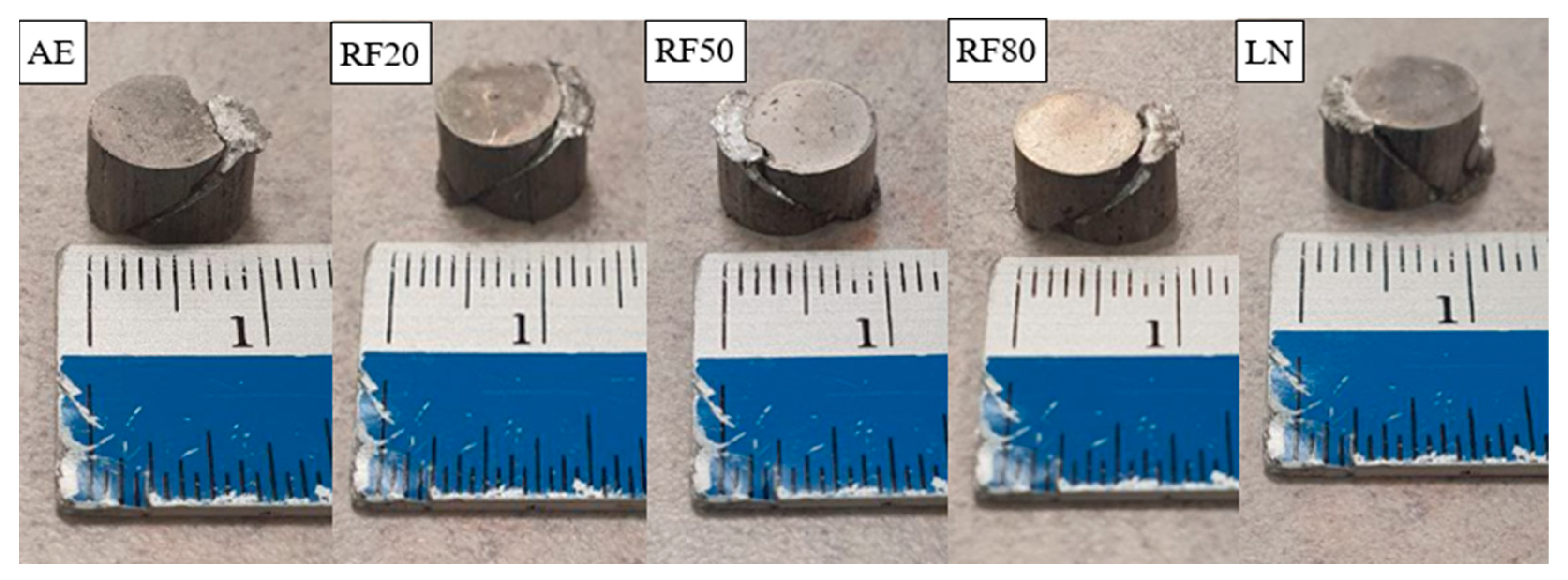
2.5. Mechanical Characterization

The damping/vibration response, including Young’s modulus, was characterized by subjecting samples with a length of 50 mm to impulse excitation. The recorded vibration signals were analyzed using the Resonance Frequency Damping Analyzer (RFDA) software (RFDA version 8.1.2, IMCE, Genk, Belgium) to glean insight into the materials’ damping response. One sample per material was analyzed.
Microhardness was characterized using an HMV 2 microhardness tester (Shimadzu Corporation, Kyoto, Japan) with an indenter force of 245.2 mN and a dwell time of 15 s, in accordance with procedures outlined in the standard ASTM E-384. A minimum of 15 hardness readings on one representative surface were taken per material for this test.
Flat and parallel samples with an aspect ratio of 1 were subject to quasi-static compressive loading using an MTS-E44 hydraulic tester (MTS Systems, Eden Prairie, MN, USA) at a strain rate of 0.000083 s−1 until failure. This analysis was conducted in accordance with the standard ASTM E9-09 [29]. Three representative samples were tested per material.

3. Results and Discussion

3.1. Density and Porosity
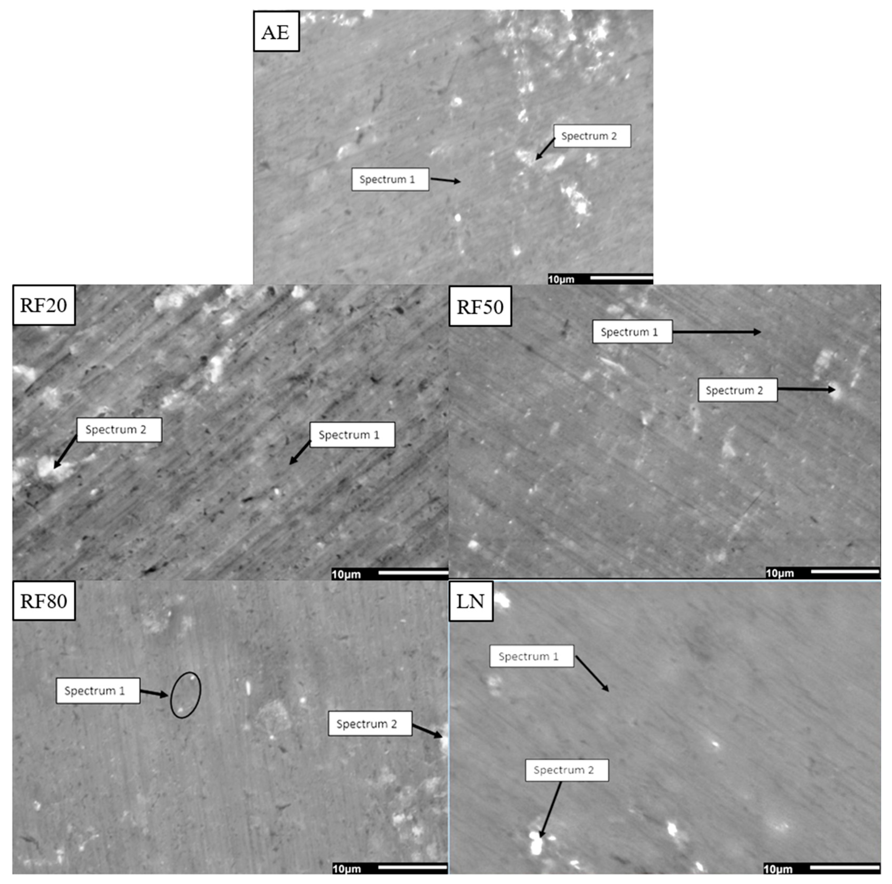
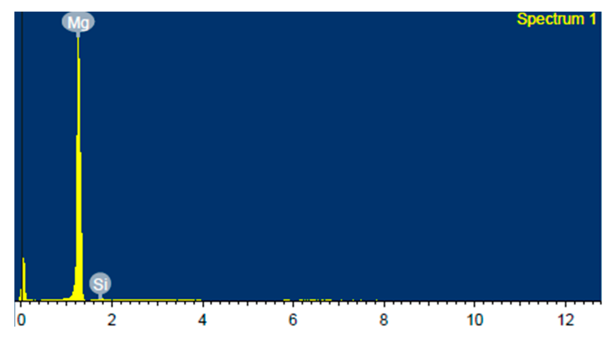
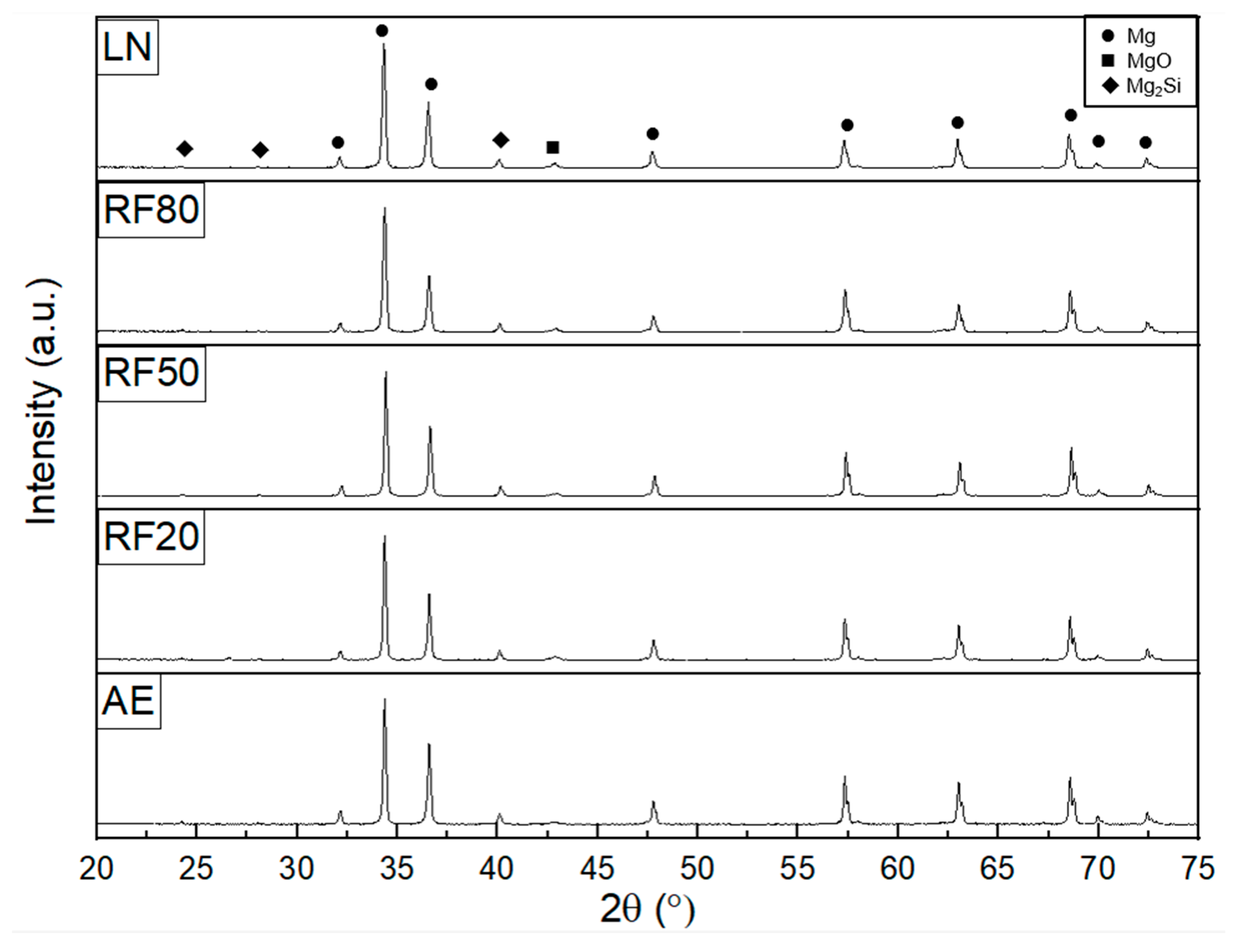
The density and porosity results of the Mg-2SiO2 samples subjected to different cryogenic temperatures are shown in Table 2. Porosity was determined by using the Archimedes method with the following formula (assuming that any voids were filled with air):
Porosity :   ρ t h e o r e t i c a l ρ e x p e r i m e n t a l ρ t h e o r e t i c a l ρ a i r
where ρtheoretical, ρexperimental, and ρair are material theoretical density, experimental density, and air density, respectively. Cryogenically treated materials exhibited, in general, elevated experimental densities and subsequently decreased porosity.
While no change in density was observed after exposure to −20 °C, lower exposure temperatures resulted in the densification of the materials, as indicated by the reduction in porosity. This was also observed in a study on CeO2-reinforced Mg which exposed materials to −20–196 °C temperatures (the latter of which was achieved by immersion in LN, similar to in this study) [26]. This observation can be attributed to two mechanisms, namely the internal compressive stresses acting on the materials as a result of sub-zero environments [23], as well as the sinking of dislocations into existing pores [30]. The best results were achieved at an exposure temperature of −80 °C.

3.2. Microstructure

Figure 1 shows the general microstructural morphology, while Table 3 shows the EDS results of selected locations within the microstructure of the Mg-2SiO2 materials in this work. Figure 2 shows one of the EDS plots/readings belonging to Mg-2SiO2 AE, showing the normalized nature of the results (i.e., the table only includes results above a certain threshold).
The X-ray diffractograms for all Mg-2SiO2 materials are shown in Figure 3. The peaks were verified against JCPDS cards (Mg: 00-004-0770, MgO: 00-084-0829, Mg2Si: 00-035-0073) present in the Powder Diffraction File (PDF-4+ 2023) [31], showing that the detected peaks were predominantly Mg, with some low-intensity peaks corresponding to MgO and Mg2Si. Furthermore, the relative intensities of the prismatic, basal, and pyramidal planes of the materials are also summarized in Table 4.
Cryogenic treatment did not appear to result in any discernible differences in the microstructure. The microstructure of all materials appeared to be predominantly a Mg matrix with small amounts of bright particles containing O; these are likely MgO and/or agglomerated SiO2 particles. Mg2Si was not visually detected despite some Si being detected in the Mg matrix regions. This lack of change in the general microstructural morphology and elemental composition is consistent with other Mg nanocomposites undergoing CT, such as those reinforced with CeO2 nanoparticles [26].
All materials exposed to sub-zero temperatures exhibited a stronger basal texture relative to their as-extruded counterparts, shown by the lower relative intensities of the prismatic and pyramidal peaks. This is in line with previous studies concerning Mg nanocomposites containing CeO2 where cryogenic treatment strengthened existing textures [26,32], such that the most dominant basal texture initially present in the extruded nanocomposites was further strengthened.

3.3. Thermal Response

The CTE and ignition temperatures of the materials studied in this work are summarized in Table 5. Only marginal changes in both properties were observed, indicating that cryogenic treatment does not result in significant alterations in the thermal properties of metallic composites.
Figure 4 shows the ignition responses of the materials, with the ignition temperature determined at the point just before the spike and subsequent recovery in temperature, as seen in the magnified inset. The results shown in Table 5 indicate only a marginal increase (+2 °C) in ignition temperature following cryogenic exposure at −20 °C and −80 °C. For LN exposure, the ignition temperature was even more marginally affected (1 °C increase). The most significant increase occurred after exposure to −50 °C (4 °C increase).
The DSC results in Figure 5 indicate that cryogenic treatment does not result in noticeable changes in thermal response as there are no discernable differences in the trends.

3.4. Mechanical Response

Cryogenic treatment was shown to variably change the damping capabilities of the materials, with a minimal effect on Young’s modulus (Table 6).
Typically, the reduction in the damping efficacy of cryogenically treated materials can be attributed to the resulting densification; this would result in reductions in porosity and, correspondingly, in air pockets and voids, which have been shown to enhance damping capabilities in aluminum and Mg-based materials [33,34]. However, a notable exception at −80 °C exposure was observed when both the attenuation coefficient and damping capacity improved. The underlying mechanisms behind such observations are not clear and require a more detailed study that may include dislocation density measurements by TEM experts. Nevertheless, improvements in the damping capacity and attenuation coefficient indicate an increase in the ability to dissipate vibrational energy, crucial in end applications where such loading is expected to occur (e.g., in components of vehicles).
Microhardness increased after cryogenic treatment, as seen in Table 7, with exposure to −50 °C conferring the greatest increase in hardness. The hardness results suggest that hardness variation is not solely governed by the extent of porosity (Table 2), but by other subtle changes in microstructural features, such as variations in texture and dislocation density. These results open the doors for a more detailed study by experts in this area.
Overall, improvements in compressive properties (combination of strength and failure strain) were observed for cryogenically treated materials (Table 8), most notably for RF20 and LN-treated materials which experienced more significant yield strength improvements. The variation in yield strength and failure strain with decreases in cryogenic temperature did not show a trend, while the average ultimate compressive strength increased after exposure to all cryogenic temperatures. The results reveal that the intrinsic microstructural factors that govern the friction stress of the matrix and subsequently the initiation of dislocation motion, affecting yield strength, evolve differently at different cryogenic temperatures. More detailed research is required in this area.
While improvements in compressive properties (especially work of fracture) have been explored with Mg materials subjected to sub-zero temperatures [26] in the past, relatively greater improvements in compressive properties (assessed through work of fracture—Table 8) were observed for cryogenically treated materials, attributable to a reduction in porosity which assists in the material’s ability to bear load and reduces the potential for the development of crack initiation sites [35,36]. Specifically, RF20 and LN materials showed the greatest improvement. The results suggest that for strength-based design, exposure at −20 °C is the best way forward. Considering that this temperature can be achieved through refrigeration, further energy or resource savings can be attained by forgoing the use of liquid nitrogen for the treatment of Mg materials in the future.
Macro-scale images of the fractured samples can be seen in Figure 6, with an approximate 45° angle of fracture. Figure 7 shows that microcracks were not present for all materials, with just the characteristic shear bands resulting from compressive loading apparent. No effect of cryogenic treatment was observed on the fracture surfaces of differently cryogenically treated samples which displayed similar fractographic characteristics.

4. Conclusions

Following the cryogenic treatment of Mg-2SiO2 nanocomposites exposed to different sub-zero temperatures, the following observations have been noted and conclusions made:
  • The greatest densification (33.6% reduction in porosity) was achieved with an exposure temperature of −80 °C.
  • Cryogenic treatment reinforced existing textures by increasing the relative intensity of the already dominant basal texture.
  • Exposure to −50 °C resulted in slight improvements in ignition resistance, with a 4 °C increase in ignition temperature.
  • The damping properties of Mg-2SiO2 were most improved through exposure to −80 °C, with the attenuation coefficient improving by 7.5% and the damping capacity by 19.8%.
  • The hardness of Mg-2SiO2 was most improved (31.8%) through exposure to −50 °C.
  • The greatest improvements in compressive response were achieved through exposure to −20 °C, with improvements in yield strength (8.2% increase), fracture strain (2.9% increase), and energy absorbed (5.3% increase).
The findings of the present work clearly indicate that exposure to differing cryogenic temperatures results in varying positive effects on the individual properties of Mg-2SiO2 nanocomposites. Thus, the cryogenic treatment temperature should be determined based on the requirements of the end application. The results presented in this work also emphasize that the cause of variations in properties resulting from cryogenic exposure requires more detailed study by experts (typically in-depth microstructural characterization using TEM and EBSD) and opens multiple pathways for future work.

Author Contributions

Conceptualization, M.G.; methodology, M.G.; validation, M.J. and M.G; formal analysis, M.J. and S.M.; investigation, M.J., S.M. and V.V.; resources, M.G.; data curation, M.J.; writing—original draft preparation, M.J.; writing—review and editing, M.J. and M.G.; visualization, M.J., S.M. and V.V.; supervision, M.J. and M.G.; project administration, M.G. All authors have read and agreed to the published version of the manuscript.

Funding

This research received no external funding.

Data Availability Statement

The original contributions presented in the study are included in the article, further inquiries can be directed to the corresponding author.

Acknowledgments

The authors would like to acknowledge Juraimi Bin Madon for the extrusion work and Ng Hong Wei for their assistance with DSC, CTE, and TGA testing.

Conflicts of Interest

The authors declare no conflict of interest.

References

  1. Bhat Panemangalore, D.; Shabadi, R.; Tingaud, D.; Touzin, M.; Ji, G. Biocompatible silica-based magnesium composites. J. Alloys Compd. 2019, 772, 49–57. [Google Scholar] [CrossRef]
  2. Liu, C.; Ren, Z.; Xu, Y.; Pang, S.; Zhao, X.; Zhao, Y. Biodegradable Magnesium Alloys Developed as Bone Repair Materials: A Review. Scanning 2018, 2018, 9216314. [Google Scholar] [CrossRef] [PubMed]
  3. Mineral Commodity Summaries 2024. In Mineral Commodity Summaries; Report; USGS: Reston, VA, USA, 2024. Available online: https://pubs.usgs.gov/publication/mcs2024 (accessed on 12 April 2024).
  4. Ditze, A.; Scharf, C. Recycling of Magnesium Alloys. In Magnesium—Alloys and Technology; Wiley Online Books: Hoboken, NJ, USA, 2003; pp. 254–278. [Google Scholar] [CrossRef]
  5. Lapovok, R.Y.; Thomson, P.F. Production of dense rod from magnesium swarf for re-melting. Magnes. Technol. 2004, 1, 149–154. [Google Scholar]
  6. Li, L.; Zhang, M.; Li, Y.; Zhao, J.; Qin, L.; Lai, Y. Corrosion and biocompatibility improvement of magnesium-based alloys as bone implant materials: A review. Regen. Biomater. 2017, 4, 129–137. [Google Scholar] [CrossRef]
  7. Pacheco, K.A. Allergy to Surgical Implants. Clin. Rev. Allergy Immunol. 2019, 56, 72–85. [Google Scholar] [CrossRef] [PubMed]
  8. Jin, W.; Chu, P.K. Orthopedic Implants. In Encyclopedia of Biomedical Engineering; Narayan, R., Ed.; Elsevier: Oxford, UK, 2019; pp. 425–439. [Google Scholar] [CrossRef]
  9. Selvaraj, D.; Raja, J.; Prasath, S. Interdisciplinary approach for bilateral maxillary canine: First premolar transposition with complex problems in an adult patient. J. Pharm. Bioallied Sci. 2013, 5, S190–S194. [Google Scholar] [CrossRef] [PubMed]
  10. Ibrahim, H.; Moghaddam, N.S.; Elahinia, M. Mechanical and In Vitro Corrosion Properties of a Heat-Treated Mg-Zn-Ca-Mn Alloy as a Potential Bioresorbable Material. Adv. Metall. Mater. Eng. 2017, 1, 1–7. [Google Scholar] [CrossRef]
  11. Ravi Kumar, N.V.; Blandin, J.J.; Suéry, M.; Grosjean, E. Effect of alloying elements on the ignition resistance of magnesium alloys. Scr. Mater. 2003, 49, 225–230. [Google Scholar] [CrossRef]
  12. Tekumalla, S.; Joo Yuan, N.; Haghshenas, M.; Gupta, M. Enhancing Properties of Aerospace Alloy Elektron 21 Using Boron Carbide Nanoparticles as Reinforcement. Appl. Sci. 2019, 9, 5470. [Google Scholar] [CrossRef]
  13. Wan, Y.; Cui, T.; Li, W.; Li, C.; Xiao, J.; Zhu, Y.; Ji, D.; Xiong, G.; Luo, H. Mechanical and biological properties of bioglass/magnesium composites prepared via microwave sintering route. Mater. Des. 2016, 99, 521–527. [Google Scholar] [CrossRef]
  14. Michel, M.D.; Serbena, F.C.; Lepienski, C.M. Effect of temperature on hardness and indentation cracking of fused silica. J. Non-Cryst. Solids 2006, 352, 3550–3555. [Google Scholar] [CrossRef]
  15. Umeda, J.; Kawakami, M.; Kondoh, K.; Ayman, E.L.S.; Imai, H. Microstructural and mechanical properties of titanium particulate reinforced magnesium composite materials. Mater. Chem. Phys. 2010, 123, 649–657. [Google Scholar] [CrossRef]
  16. Goh, C.S.; Gupta, M.; Wei, J.; Lee, L.C. The Cyclic Deformation Behavior of Mg—Y2O3 Nanocomposites. J. Compos. Mater. 2008, 42, 2039–2050. [Google Scholar] [CrossRef]
  17. Jaganathan, H.; Godin, B. Biocompatibility assessment of Si-based nano- and micro-particles. Adv. Drug Deliv. Rev. 2012, 64, 1800–1819. [Google Scholar] [CrossRef] [PubMed]
  18. Maquet, V.; Boccaccini, A.R.; Pravata, L.; Notingher, I.; Jérôme, R. Porous poly(α-hydroxyacid)/Bioglass® composite scaffolds for bone tissue engineering. I: Preparation and in vitro characterisation. Biomaterials 2004, 25, 4185–4194. [Google Scholar] [CrossRef]
  19. Roether, J.A.; Boccaccini, A.R.; Hench, L.L.; Maquet, V.; Gautier, S.; Jérôme, R. Development and in vitro characterisation of novel bioresorbable and bioactive composite materials based on polylactide foams and Bioglass® for tissue engineering applications. Biomaterials 2002, 23, 3871–3878. [Google Scholar] [CrossRef] [PubMed]
  20. Vergnol, G.; Ginsac, N.; Rivory, P.; Meille, S.; Chenal, J.-M.; Balvay, S.; Chevalier, J.; Hartmann, D.J. In vitro and in vivo evaluation of a polylactic acid-bioactive glass composite for bone fixation devices. J. Biomed. Mater. Res. Part B Appl. Biomater. 2016, 104, 180–191. [Google Scholar] [CrossRef] [PubMed]
  21. Bruckmann, F.D.; Nunes, F.B.; Salles, T.D.; Franco, C.; Cadoná, F.C.; Bohn Rhoden, C.R. Biological Applications of Silica-Based Nanoparticles. Magnetochemistry 2022, 8, 131. [Google Scholar] [CrossRef]
  22. Johanes, M.; Gupta, M. An Investigation into the Potential of Turning Induced Deformation Technique for Developing Porous Magnesium and Mg-SiO2 Nanocomposite. Materials 2023, 16, 2463. [Google Scholar] [CrossRef]
  23. Sonar, T.; Lomte, S.; Gogte, C. Cryogenic Treatment of Metal—A Review. Mater. Today Proc. 2018, 5, 25219–25228. [Google Scholar] [CrossRef]
  24. Zurecki, Z. Cryogenic Quenching of Steel Revisited. In Heat Treating, Proceedings of the 23rd ASM Heat Treating Society Conference, Pittsburgh, PA, USA, 25–28 September 2005; ASM: Almere, The Netherlands, 2006. [Google Scholar]
  25. Baldiserra, P.; Delprete, C. Deep Cryogenic Treatment: A Bibliographic Review. Open Mech. Eng. J. 2008, 2008, 1–11. [Google Scholar] [CrossRef]
  26. Gupta, S.; Parande, G.; Gupta, M. Comparison of Shallow (−20 °C) and Deep Cryogenic Treatment (−196 °C) to Enhance the Properties of a Mg/2wt.%CeO2 Nanocomposite. Technologies 2024, 12, 14. [Google Scholar] [CrossRef]
  27. Gupta, S.; Parande, G.; Tun, K.S.; Gupta, M. Enhancing the Physical, Thermal, and Mechanical Responses of a Mg/2wt.%CeO2 Nanocomposite Using Deep Cryogenic Treatment. Metals 2023, 13, 660. [Google Scholar] [CrossRef]
  28. Dong, N.; Sun, L.; Ma, H.; Jin, P. Effects of cryogenic treatment on microstructures and mechanical properties of Mg-2Nd-4Zn alloy. Mater. Lett. 2021, 305, 130699. [Google Scholar] [CrossRef]
  29. ASTM E9-09; Standard Test Methods of Compression Testing of Metallic Materials at Room Temperature. ASTM International: West Conshohocken, PA, USA, 2018.
  30. Xie, S.; Lv, Q.; Zhang, W.; Qu, Y.; Qi, H.; Yu, B.; Li, R.; Li, G.; Yang, F. Effect of Cryogenic Treatment on Microstructure and Mechanical Properties of Al0.6CrFe2Ni2 Dual-Phase High-Entropy Alloy. Metals 2023, 13, 195. [Google Scholar] [CrossRef]
  31. Gates-Rector, S.; Blanton, T. The Powder Diffraction File: A quality materials characterization database. Powder Diffr. 2019, 34, 352–360 (PDF-4+ 2023). [Google Scholar] [CrossRef]
  32. Gupta, S.; Johanes, M.; Parande, G.; Gupta, M. An Investigation into the Effect of Length Scale of Reinforcement on the Cryogenic Response of a Mg/2wt.%CeO2 Composite. Micro 2024, 4, 170–184. [Google Scholar] [CrossRef]
  33. Colakoglu, M. Factors effecting internal damping in aluminum. J. Theor. Appl. Mech. 2004, 42, 95–105. [Google Scholar]
  34. Xie, Z.-k.; Tane, M.; Hyun, S.-k.; Okuda, Y.; Nakajima, H. Vibration–damping capacity of lotus-type porous magnesium. Mater. Sci. Eng. A 2006, 417, 129–133. [Google Scholar] [CrossRef]
  35. Kováčik, J. Correlation between Young’s modulus and porosity in porous materials. J. Mater. Sci. Lett. 1999, 18, 1007–1010. [Google Scholar] [CrossRef]
  36. Rivera-Salinas, J.E.; Gregorio-Jáuregui, K.M.; Romero-Serrano, J.A.; Cruz-Ramírez, A.; Hernández-Hernández, E.; Miranda-Pérez, A.; Gutierréz-Pérez, V.H. Simulation on the Effect of Porosity in the Elastic Modulus of SiC Particle Reinforced Al Matrix Composites. Metals 2020, 10, 391. [Google Scholar] [CrossRef]
Figure 1. Scanning electron micrographs of Mg-2SiO2 materials in this work, with selected regions used for the EDS studies.
Metals 14 00808 g001
Figure 2. EDS results/chart of Mg-2SiO2 AE: matrix region.
Metals 14 00808 g002
Figure 3. X-ray diffractograms of Mg-2SiO2 materials in this work.
Metals 14 00808 g003
Figure 4. The ignition responses of Mg-2SiO2 materials in this work, showing the locations where ignition temperatures were evaluated.
Metals 14 00808 g004
Figure 5. Thermal response of Mg-2SiO2 materials in this work.
Metals 14 00808 g005
Figure 6. Macro-scale photographs of compressed Mg-2SiO2 materials in this work.
Metals 14 00808 g006
Figure 7. Fractographs of Mg-2SiO2 materials in this work.
Metals 14 00808 g007
Table 1. Material designations and treatments performed on Mg-2SiO2 nanocomposites studied in this work.
Material DesignationTreatment Performed
AEAs-extruded (no treatment)
RF20Refrigeration at −20 °C for 24 h
RF50Refrigeration at −50 °C for 24 h
RF80Refrigeration at −80 °C for 24 h
LNLiquid nitrogen exposure at −196 °C for 24 h
Table 2. Density and porosity results of Mg-2SiO2 materials studied in this work.
TreatmentAverage Experimental Density (g/cm3)Porosity Reduction (%)
Before TreatmentAfter Treatment
RF201.794 ± 0.0061.794 ± 0.0020
RF501.793 ± 0.0021.796 ± 0.003
(↑0.167%)
7.0
RF801.787 ± 0.0011.800 ± 0.004
(↑0.727%)
33.6
LN1.789 ± 0.0121.800 ± 0.006
(↑0.615%)
26.3
Table 3. The EDS results of Mg-2SiO2 materials in this work.
TreatmentSpectrumDetected Element (wt.%)
MgSiO
AE197.72.3-
288.62.49.0
RF201100--
288.02.39.7
RF50197.41.11.5
287.17.05.9
RF80196.01.82.2
292.91.85.4
LN1100--
295.51.53.1
Table 4. XRD results of Mg crystallographic planes in this work.
TreatmentPlaneI/Imax
AE10-10 prism0.1181
0002 basal1
10-11 pyramidal0.6450
RF2010-10 prism0.0854
0002 basal1
10-11 pyramidal0.5373
RF5010-10 prism0.0898
0002 basal1
10-11 pyramidal0.5670
RF8010-10 prism0.0825
0002 basal1
10-11 pyramidal0.4610
LN10-10 prism0.0965
0002 basal1
10-11 pyramidal0.5326
Table 5. CTE ignition temperatures of Mg-2SiO2 materials in this work.
TreatmentAverage CTE (×10−6/K)Ignition Temperature (°C)
AE22.95602
RF2023.63604
RF5023.07606
RF8022.79604
LN24.00603
Table 6. Results of damping properties and modulus.
TreatmentAttenuation CoefficientDamping CapacityE-Modulus (GPa)
AE41.80.00123847.54 ± 0.28
RF20 (pre-treatment)48.420.00143348.26 ± 0.28
RF20 (post-treatment)49.31
(↑1.8%)
0.001292
(↓9.6%)
49.06 ± 0.29
(↑1.7%)
RF50 (pre-treatment)38.730.00128848.15 ± 0.28
RF50 (post-treatment)35.19
(↓9.1%)
0.001174
(↓8.9%)
48.23 ± 0.38
(↑0.2%)
RF80 (pre-treatment)38.090.00110448.23 ± 0.28
RF80 (post-treatment)40.94
(↑7.5%)
0.001322
(↑19.8%)
48.23 ± 0.38
(no change)
LN (pre-treatment)44.540.00134448.71 ± 0.29
LN (post-treatment)44.88
(↑0.8%)
0.0001352
(↑0.6%)
48.67 ± 0.29
(↓0.1%)
Table 7. Microhardness values of Mg-2SiO2 materials in this work.
TreatmentAverage Microhardness (HV)
AE85 ± 7
RF2086 ± 6 (↑1.2%)
RF50112 ± 8 (↑31.8%)
RF80104 ± 5 (↑22.4%)
LN86 ± 5 (↑1.2%)
Table 8. Compressive properties of Mg-2SiO2 materials in this work.
TreatmentAverage 0.2% Yield Strength (MPa)Average Ultimate Compressive Strength (MPa)Average Failure Strain (%)Average Work of Fracture (MJ/m3)
AE158 ± 2380 ± 1127.6 ± 0.460.8 ± 1.8
RF20171 ± 1
(↑8.2%)
381 ± 12
(↑0.3%)
28.4 ± 1.2
(↑2.9%)
64.0 ± 3.7
(↑5.3%)
RF50152 ± 3
(↓3.8%)
389 ± 11
(↑2.4%)
27.6 ± 1.263.2 ± 3.1
(↑4.0%)
RF80154 ± 7
(↓2.5%)
385 ± 10
(↑1.3%)
27.5 ± 1.3
(↓0.4%)
60.9 ± 4.9
(↑0.2%)
LN164 ± 1
(↑3.8%)
383 ± 1.4
(↑0.8%)
28.4 ± 1.04
(↑2.9%)
63.9 ± 4.3
(↑5.1%)
Disclaimer/Publisher’s Note: The statements, opinions and data contained in all publications are solely those of the individual author(s) and contributor(s) and not of MDPI and/or the editor(s). MDPI and/or the editor(s) disclaim responsibility for any injury to people or property resulting from any ideas, methods, instructions or products referred to in the content.

