Article

Evaluation of Eco-Friendly Consolidating Treatments in Pugliese Tuff (Gravina Calcarenite) Used in Italian Heritage Buildings

by
Jose Antonio Huesca-Tortosa
1,*,
Yolanda Spairani-Berrio
1,
Cristiano Giuseppe Coviello
2,
Maria Francesca Sabbà
2,
Fabio Rizzo
2 and
Dora Foti
2,3
1
Department of Architectural Constructions, University of Alicante, Carretera de San Vicente del Raspeig, s/n, 03690 San Vicente del Raspeig, Spain
2
Department of Architecture, Construction and Design, Polytechnic University of Bari, Via Orabona 4, 70125 Bari, Italy
3
ITC—Construction Technologies Institute, CNR—Italian National Research, 70124 Bari, Italy
*
Author to whom correspondence should be addressed.
Buildings 2024, 14(4), 940; https://doi.org/10.3390/buildings14040940
Submission received: 14 February 2024 / Revised: 7 March 2024 / Accepted: 26 March 2024 / Published: 29 March 2024

Abstract

:
This work evaluates the effectiveness of various consolidating treatments applied to Pugliese tuff (Gravina Calcarenite). This type of stone has been used in numerous historic buildings in the Puglia area (southeast of Italy), which presents durability problems due to high porosity, low cohesion between clasts, and low mechanical resistance. Eco-friendly treatments that generate CaCO3 have been selected, specifically bioconsolidant KBYO biological and lime water, which a priori are capable of consolidating without occluding the pores or reducing them excessively, thereby creating compounds similar to those contained in the stone and being respectful of the environment. Nano-sized treatments have also been tested, including nanosilica and nanolime, to compare results with eco-friendly treatments. The bioconsolidating treatment has been applied in two different ways, the usual way consisting of two applications a day for 7 days, as well as a double treatment that is applied in two batches of 7 days with a rest of 7 days between applications. Double treatment has shown a great improvement in consolidation compared to the usual 7-day application; this treatment has obtained the best results in both mechanical and petrophysical properties. This study not only demonstrates the effectiveness of the bioconsolidant but also expands eco-friendly conservation strategies to improve the preservation of historical structures built in calcarenite.

1. Introduction

Gravina Calcarenite, also known as “Pugliese tuff”, is a type of limestone that has been used in architectural buildings in Italy since time immemorial [1,2], both in historical buildings and in dry stone constructions. It has even served as the basis for the famous Sassi in Matera excavated in the calcarenitic rock of Gravina [3,4,5]. This kind of stone is still in use today, as can be seen in the quarry in Puglia from which the base material for this study was obtained.
There is great concern for the conservation of the stone materials that make up our built cultural heritage, especially in the case of materials whose intrinsic characteristics are particularly weak against degradation, as is the case of calcarenites [6,7,8,9,10]. A large number of manuscripts have dealt with these questions for many decades, the most important of which are the Italian studies by Borea [11]. All the authors agree on the importance of understanding the properties of the material and its behavior before intervening.
Over the years, several authors have characterized Gravina calcarenite [5,6,9,12] and concluded that it is a biocalcarenite, consisting mainly of low-density calcite. Its petrophysical and mechanical properties have been carefully examined. The studies confirmed that Gravina calcarenite is a highly porous and heterogeneous rock with variations in both physical and mechanical characteristics depending on grain size, porosity, and cement content in the stone matrix. According to some authors [5,13] the compressive strength of this material decreases significantly as a function of the moisture content, with a maximum loss of 30% when fully saturated.
Gravina Calcarenite presents a typical “grainstone” texture, with a high porosity and medium-sized particles, exhibiting a massive structure [13,14,15]. In general, the characteristics of Gravina Calcarenite have a negative influence on its durability [16,17,18]. Its high porosity facilitates the absorption of water, which can transport salts that later crystallize, thereby causing serious damage [18,19,20,21,22,23,24] and allowing the entry of harmful substances. Nevertheless, this porosity can be beneficial for the absorption of consolidation treatments as it facilitates the penetration of treatments to inside the areas to be consolidated. In low-porosity stone, such as marbles, treatments tend to remain on the surface creating a film being an undesirable effect [25]. Consolidant treatments must be able to be introduced into the materials to be consolidated to improve their cohesion [26,27,28].
In most of the buildings made with Gravina Calcarenite, deterioration is mainly due to features induced by material loss, as defined in the ICOMOS-ISCS Glossary on Stone Deterioration Patterns [29]. Figure 1 shows damage to a Gravina Calcarenite masonry with abundant alveolizations and surface repairs with low-porosity coatings (made with cement mortar) that have increased the damage to the calcarenite.
For many decades, there has been great concern about the conservation of built heritage, which has led numerous researchers to work on these issues by developing and testing numerous consolidating treatments. In general, there is a high consensus regarding the improvements obtained by porous calcarenites when applying consolidating treatments [28,30,31,32,33,34]. However, even after having carried out tests prior to application, notable failures have been observed in the use of consolidating treatments, such as the use of some resins, polymers [35,36], or magnesium and/or zinc fluosilicates. That is the case of the church of San Michele Maggiore in Pavia, which is made of sandstone [37] and was restored in the 1970s by Piero Sanpaolesi using magnesium fluosilicate. A few years after its application, fragments of the façade detached as the treatment generated a very rigid surface although the treatment used was supported by previous studies [37]. This treatment had already been used for years.
This is one of the reasons why many researchers around the world continue to advance their knowledge about the effect that the different consolidation treatments have on stone materials; it is also the main objective of this work.
The intrinsic characteristics of each kind of stone lead the treatments applied to behave differently in each case [28,38,39], and therefore, before any intervention in the original building, it is very important to evaluate the behavior of each product. The applied consolidants must be able to improve the mechanical strength of the degraded areas, but they must not excessively modify other aspects, such as vapor permeability and porosity or, in aesthetic terms, color [38,39,40,41]. Consolidation treatments penetrate the porous network and modify it to a greater or lesser extent, and they can produce large petrophysical differences, particularly on the surface of the treated stone, such as the formation of hard surface crusts. Initially, this seems beneficial, but the moisture contained in the stone has greater difficulty in escaping when the treated surface of the stone has low water vapor permeability [28]. Water trapped within the materials can be harmful as it can generate pressure, accelerate the dissolution processes of the compounds present in the stone, or cause the loss of cohesion between particles. Trapped moisture can rise through capillary action, affecting higher levels or other areas and water can transport salts [42]. On the other hand, the reduction in pore size may mean that the salts crystallizing in the pore do not have enough space to crystallize, and further damage may be caused by haloclasts [42,43,44,45].
The main objective of this work is to advance the knowledge of possible eco-friendly treatments for use in future interventions on Gravina Calcarenite. Considering the composition of the stone, which is 95% calcite, treatments capable of producing CaCO3 were specially selected. One of the basic criteria has been to select products that do not obstruct the original porous structure of the stone, in other words, those that do not significantly reduce either the water vapor permeability or its porosity. It is also important that the treatments can penetrate the surface of the stone, that it generates good consolidation, that it has good durability and resistance to biological agents, and that it is safe for health [41]; in addition, nowadays, it is also very important that the treatments and advances in construction beenvironmentally friendly [46,47].
Two possible eco-friendly treatments have been selected, including “Lime Water” [48] from the waste from aerial lime slaking and bioconsolidant [49,50], which generates a biomineralisation by bacterial carbonatogenesis that produces calcium carbonate capable of consolidating the treated stone.
Bioconsolidant-treated specimens were tested with two different application methods as follows: the first with 7 days of application [50] (which is the usual method) and the second with 7 days of application, 7 days of rest and 7 days of application. This second method has been carried out because, with the normal 7-day application, not many improvements were observed. After consulting with other researchers who work with this same treatment, we decided to repeat the treatment after drying the specimens for 7 days in some samples.
Lime water usually generates little consolidation, which is due principally to the relatively low solubility of Ca(OH)2 in water (1.65 g/L at 20 °C) [51]. To achieve good results, it is necessary to successively repeat the applications for several weeks to consolidate the surface to be treated [52,53]. Considering these aspects and the high porosity of this calcarenite, it was decided to test enriched lime water with lime paste with a proportion of 20 g/L. This liquid treatment has been obtained by mixing lime water from the lime slaking residue with a small proportion (14 g/L) of paste lime aged for more than 3 years after slaking.
Currently, nanolimes are being used to consolidate numerous buildings because they generate calcium carbonate and does not clog the pores. This is because, having a very small particle size (diameter is quoted as 150 nm) [27,51,53,54,55], they can be introduced very well into the pores. It has been decided to also test this product only to have comparative data with those previously described.
Nanosilica is also selected only to serve as a comparative reference in terms of results. It has been widely demonstrated that nanosilica usually generates improvements in resistance [27,56,57], although in the case of calcarenites it introduces compounds very different from the nature of the stone itself.
Some authors point out that nanotechnology may have contraindications with nature [58], which is why it is important to know and try other eco-friendly alternative treatments, such as bioconolidants or lime water.

2. Materials and Methods

The Gravina Calcarenite stone block has been characterized, and its petrophysical and mechanical properties have been determined, which have been used as a basis for comparing the results obtained with the different treatments.
Nondestructive studies and tests, such as color determination, microscopic observation (SEM), water droplet absorption, capillary absorption, water vapor permeability, and mechanical properties using ultrasound, have been carried out on the same specimens before and after treatment. Destructive tests, such as compressive strength (Rmc), mercury intrusion porosimetry (MIP), and drilling resistance (DRMS), have been performed on different specimens, some of which were treated and others left untreated.

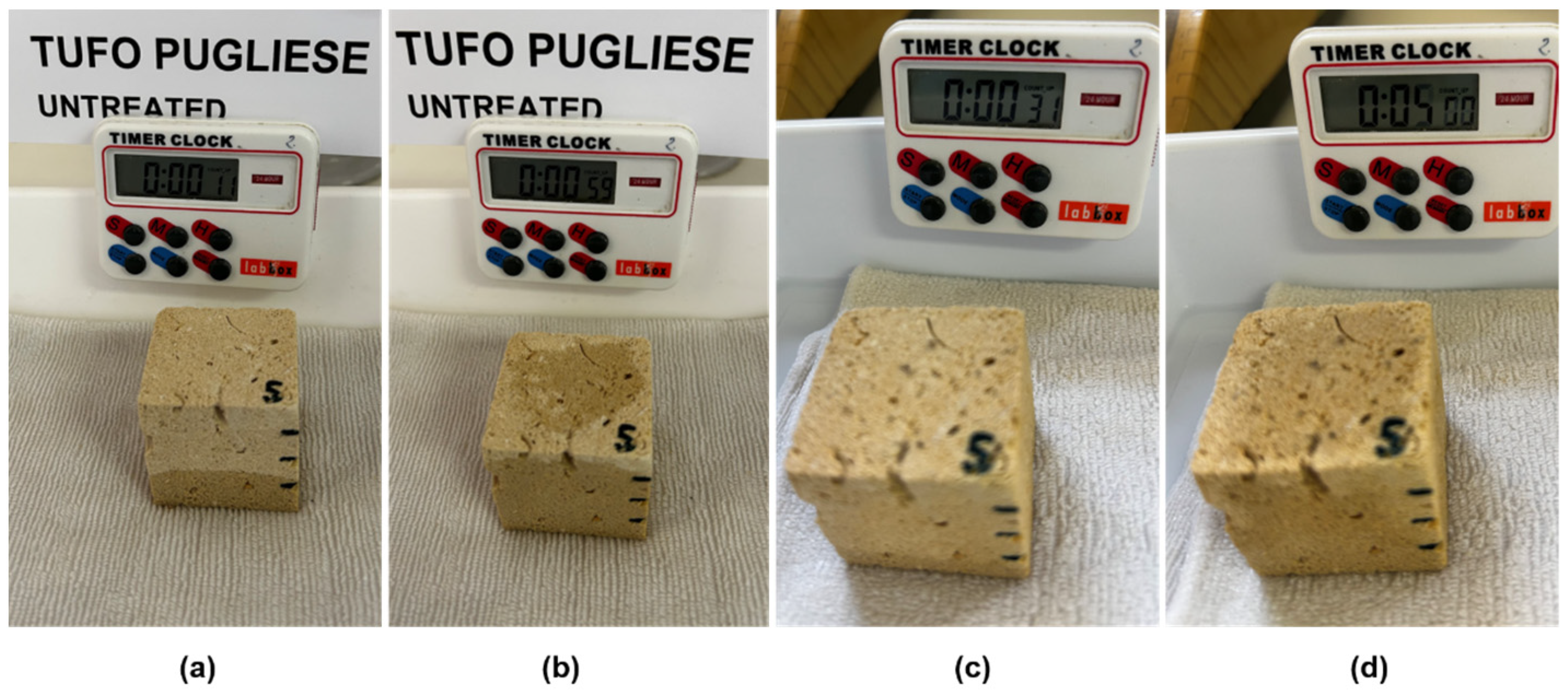
2.1. Materials and Treatments

Specimens of Pugliese tuff tested in this work were taken from the Gravina quarry in the Puglia region (Italy). From a 15 × 25 × 50 cm block, different specimen formats have been obtained according to the type of test to which they have been subjected. Specifically, 32 cubes with an edge of 4 cm, 15 discs with a diameter of 5.3 cm and a thickness of 1.8 cm, and 18 small cubes with an edge of 0.8–1 cm were tested.
The treatments tested have been:
  • Bioconsolidant: Mixostone M3P from the company KBYO Biological S.L. Product patented by the University of Granada (Spain) [59]. The composition of the M3P nutritional solution includes 1% Bacto-Casitone (a hydrolyzed casein), 1% Ca(CH3COO)2·4H2O (total calcium: 43.44 mM), 0.2% K2CO3·1/2H2O (total potassium: 35.6 mM; total carbonate: 17.8 mM), and 10 mM phosphate buffer in distilled water (pH 8).
This treatment has been applied in two different ways. The first called “Bio” uses two applications per day of the nutritional liquid for 7 days. The second method called “Double Bio” included 7 days with two applications per day, 7 days of rest without treating, and another 7 days of treatment with two applications per day. The samples were kept between 18 and 22 degrees Celsius and at a humidity of 50–60%. The nutritional product was applied to the specimens by spraying without reaching saturation; this implies that it was not able to absorb any more liquid.
  • Lime water enriched with lime putty with a concentration of 20 g/L from the company “Cales Pascual” was used. This product consolidates by the crystallization of calcite on the treated surface.
This treatment is referred to as “LW”. This treatment has been applied by spraying the samples until saturation. The specimens were kept between 18 and 22 °C and at a humidity of 50–60%.
  • Nanolime: Nanorestore from CTS was used. The product is based on nanophase calcium hydroxide dispersed in isopropyl alcohol that is capable of generating calcium carbonate nanocrystals.
This treatment is referred to as “N”. This treatment has been applied by spraying the samples until saturation. The specimens were kept between 18 and 22 °C and at a humidity of 50–60%.
  • Nanosilica: Nano Estel from CTS is an aqueous colloidal silica dispersion of nanometric dimensions (~10–20 nm).
This treatment is referred to as “E”. This treatment has been applied by spraying the samples until saturation. The specimens were kept between 18 and 22 °C and at a humidity of 50–60%.

2.2. Methods and Essays

The workflow in Figure 2 shows the different studies and tests carried out in this research work.

2.2.1. Instruments

  • Scanning electron microscope (SEM)
High resolution scanning electron microscope with EDS analysis Jeol brand model IT500HR/LA has been used. Small untreated and treated cubes with edge sizes less than 1 cm have been observed. This study was carried out at the University of Alicante.
  • Scanning electron microscope (FE-SEM)
ZEISS model Merlin VP Compact. The specimens studied are small and prismatic, less than 0.8 × 0.8 × 0.8 cm. Bioconsolidation specimens have been observed. This research was carried out at the University of Alicante.
  • Polarizing petrographic microscope (MOP):
Zeiss Axioskop transmitted light optical microscope has been used, with a Zeiss Stemi SV 6 magnifying glass and a Photometrics Cool SNAP-CF camera. This study was carried out at the University of Alicante.
  • X-ray diffractometers (DRX)
Bruker D8-Advance has been used. 5 gr of pulverized untreated calcarenite specimen have been analyzed. This study was carried out at the University of Alicante.
  • Mercury intrusion porosity (MIP):
The equipment used is the Micromeritics AutoPore IV 9500 with high pressure. The specimens studied are small and prismatic in shape, less than 1 × 1 × 1.1 cm. This study was carried out at the IGME (Madrid).
  • Handheld digital microscope
Pancellent Wireless Handheld Digital Microscope bult-in 2.0 Megapixel CMOS Sensor, support capture picture and video at 1920 × 1080 P. It has a magnification range from 50× to 1000×.
  • Compression testing machine
OMADISA Multipurpose testing machine with a 200 kN compression load cell on upper crosshead and a compression device with manual specimen. It includes digital readout and data handling. This machine is designed to carry out standard-compliant compressive strength. This study was carried out at the University of Alicante.
  • DRMS
A DRMS device to measure the drilling resistance of stone materials from SINT Technology, model DRMS Cord-less, Serial Nr 038, was used. This test was carried out at the Faculty of Science in Granada (Spain).
  • Colorimeter
A PCE colorimeter model CSM4 was used. This study was carried out at the University of Alicante.
  • Ultrasonic tester.
A STEINKAMP BP-5 Ultrasonic tester was used to determine the propagation time of the ultrasonic waves. This research was carried out at the University of Alicante.

2.2.2. Test Descriptions for Treatment Evaluation

  • DRMS
The cohesion of disaggregated particles is the most important stone property that should be improved after successful consolidation. Consolidation involves improving cohesion and therefore a significant increase in the resistance of the stone to external forces and environmental deterioration. Some researchers consider that the most appropriate method for the evaluation of the effectiveness of the consolidants is the application of the drilling resistance measurement system (DRMS) [60,61,62].
Untreated and treated stones, 12 cubes of 4 × 4 × 4 cm, 2 untreated and 2 with each treatment, were used. The system is equipped with software that allows continuous recording of the force, expressed in N, in relation to the drilling progress. The testing conditions were as follows:
1. 4.8 mm diameter flat edge diamond tipped drill bit;
2. Penetration speed set at 10 mm/min;
3. Rotation speed set at 300 rpm;
4. Drill depth to 20 mm.
During the tests, the rotation speed and penetration rate were kept constant. A total of 4–6 perforations were made for each specimen to obtain average values.
  • Compressive strength
The improvement in the compressive strength is also an indication of the effectiveness of the cohesion between particles generated by consolidation, so this test was carried out on untreated 4 cm cubes (5 specimens with different densities) and treated cubes (3 cubes of each type of treatment). The instructions of the UNE-EN 1926:2007 [63] standard have been followed, although due to a lack of sufficient material, the number of samples recommended by that standard has not been tested.
The test conditions were as follows:
▪ Uniform load increase of 200 N/s;
▪ Pressing speed of 10 mm/min;
  • Ultrasonic pulse velocity test.
Data were taken from the three faces of each cube before and after treatment. The values obtained were averaged. Vaseline was not applied to avoid contamination of the specimens [64,65].
  • Study of color
The objective is to see the color variation “∆E” in the same sample before and after applying the treatment according to the CIELAB color space, where ∆E = √(∆L*)2 + (∆a*)2 + (∆b*)2. Therefore, the measured parameters were lightness (L*), which varies from 0 (absolute black) to 100 (absolute white); a*, which represents the redness–greenness range (positive a*: red and negative a*: green); and b*, which is associated with the yellowness–blueness spectrum (positive b*: yellow and negative b*: blue).
The recommendations of the standard UNE-EN 15886:2011 have been followed [66]. Authors who have investigated color changes caused by surface treatments indicate that several readings of the color must be taken for it to be representative [67,68].
Four values were taken from each specimen, and the average was calculated. The data are processed in the PCE software cqcs3 version 4.4.6, and the values of the untreated specimen, which is called the “standard” measurement, can be compared with the treated specimen, which is called the “sample”.
  • Water absorptivity
A drop of water was dropped from a height of 4 cm on the surface of a specimen, and the time in seconds for the drop to be completely absorbed was measured. The instructions of authors who have worked on this type of trial have been followed [24].
  • Vapor permeability
Following the recommendations of the RILEM 25 PEM II2 test [69] and the UNE-EN 15803:2010 standard [70], this test was carried out on the same specimens before and after the application of each treatment. For this, three specimens of 5.3 cm in diameter and 1.8 cm in thickness were used. Each was carried at a constant weight. The contour of the specimen was waterproofed, and it was placed in glass container designed for this test with 35 g of completely dry silica gel inside.
The containers with the discs were placed in a climate chamber with a tray of water in the bottom. To determine the amount of water vapor that passed through the specimens, measurements of the weight of the set (container with silica gel and the stone specimen) were made once a day for 10 days. The increase in weight detected was due to the water vapor absorbed by the silica gel, which had passed through the pores of the stone.
  • Capillary absorption
The test has been carried out on each of the specimens separately before and after being treated. A total of 2–4 specimens of each type of treatment have been tested. They have been placed on a cloth moistened with water, and with a stopwatch, they have been weighed every 30 s until reaching a constant weight. The instructions of the UNE-EN 15801:2010 standard have been followed [71].

3. Results

3.1. Gravina Calcarenite Characterization

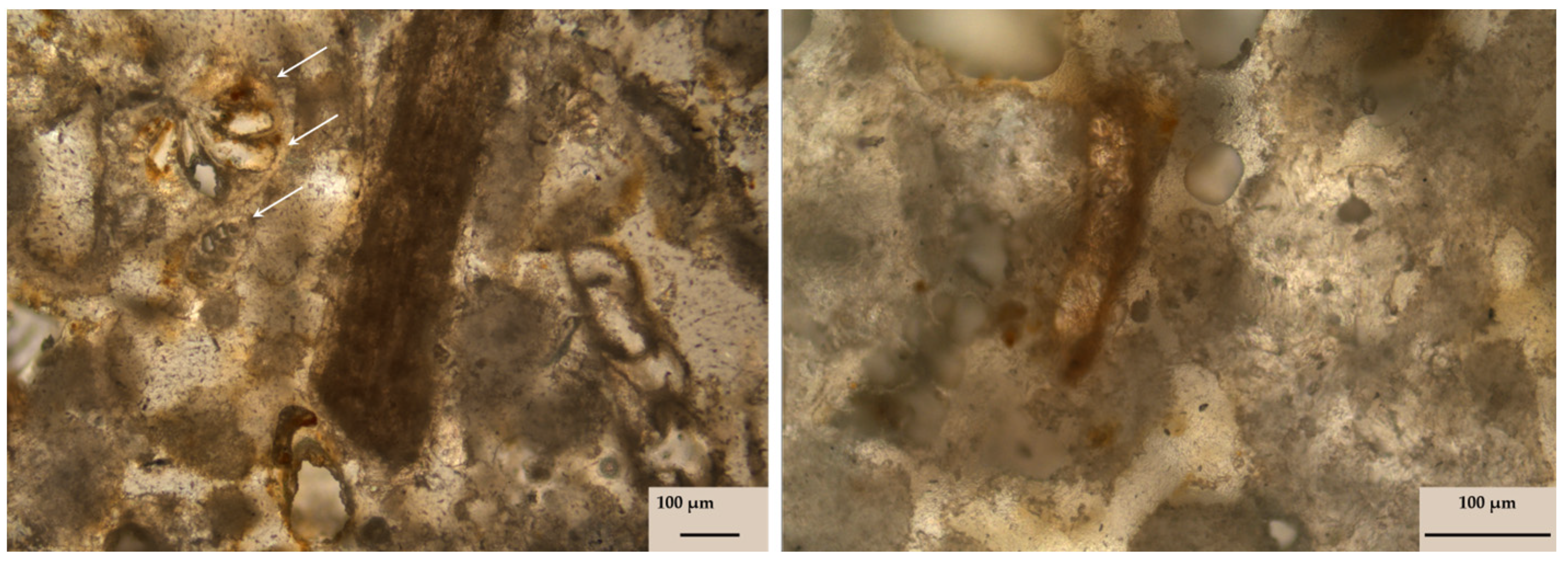
3.1.1. Petrographic Description

The observation by magnifying glass, SEM, and thin sections indicate that it is a grainstone type calcarenite with fossils of marine origin. XRD analysis shows that it is 95% calcite and 5% ankerite.
Figure 3 illustrates the thin section of the Gravina Calcarenite; the presence of foraminiferal fossils can be observed.

3.1.2. Petrophysical Properties
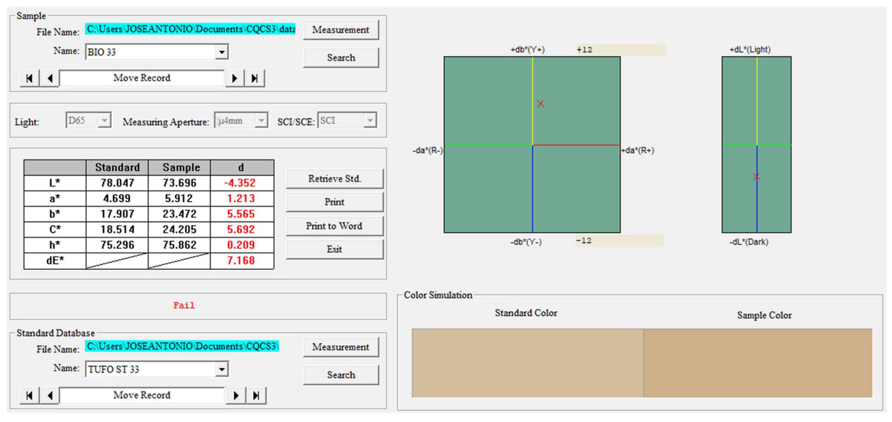
  • Color (C)
The color of the unaltered stone specimen is very light, but as soon as it gets wet or exposed to heat, it darkens rapidly, as observed during the tests. The average color parameters of the unaltered untreated specimens are as follows: L* = 78.544; a* = 4.568; b* = 17.236; c* = 17.832; and h* = 75.155. Figure 4a shows the color study process.
  • Densities (D) and porosity (P):
The average true density obtained with a helium pycnometer is 2.42 g/cm3.
The average bulk density of the untreated specimen is 1.32 g/cm3, ranging from 1.22 g/cm3 for the less dense specimen (due to occasional internal voids left by fossils) to 1.41 g/cm3 for the more compact specimen. These data coincide with those provided by other authors who have studied stones from the Gravina area [72,73].
The porosity obtained with MIP (from a single specimen studied) is 43%. It should be considered that this value may vary between samples because, as seen above, the bulk density of the samples studied can vary up to 7.5%.
The study using mercury porosimetry has shown that the average range of pores of the untreated sample is 1 to 100 μm. The average pore diameter is 1.984 μm.
  • Droplet absorption
The droplets are immediately absorbed and have an absorption time of less than 1 s. Some authors [24] have indicated that there is a direct relationship between the absorption of droplets and the porosity of the stones.
  • Capillary absorption
Capillary absorption of untreated specimens is high and very fast over time. The 4 cm cubic specimens are saturated by capillary absorption within two minutes. The water reaches the top face of the specimen in 1 min, i.e., it rises 4 cm in 1 min. In porous stones it is common for capillary absorption to be very high, thereby influencing the increase in the deterioration of stone materials [74,75]. The susceptibility of calcarenite to salt erosion is directly related to the structure of the pores and the mechanical properties. Salt crystallization is the most damaging in the pore range of 0.1 to 10 μm [22], coinciding with this case study, in which the average pore size is 1.984 μm.
  • Water vapor permeability
The average water vapor permeability of the 15 untreated specimens was 2.57 g after 10 days. The reference value for 100% permeability is 3.97 g (weight gain in 10 days of a container with only 35 g of dry silica gel and no stone specimen). Therefore, the Gravina Calcarenite is 64.7% permeable. These data correspond to its high porosity, also indicating the existence of great connectivity between pores [72,73,74,75].

3.1.3. Mechanical Properties
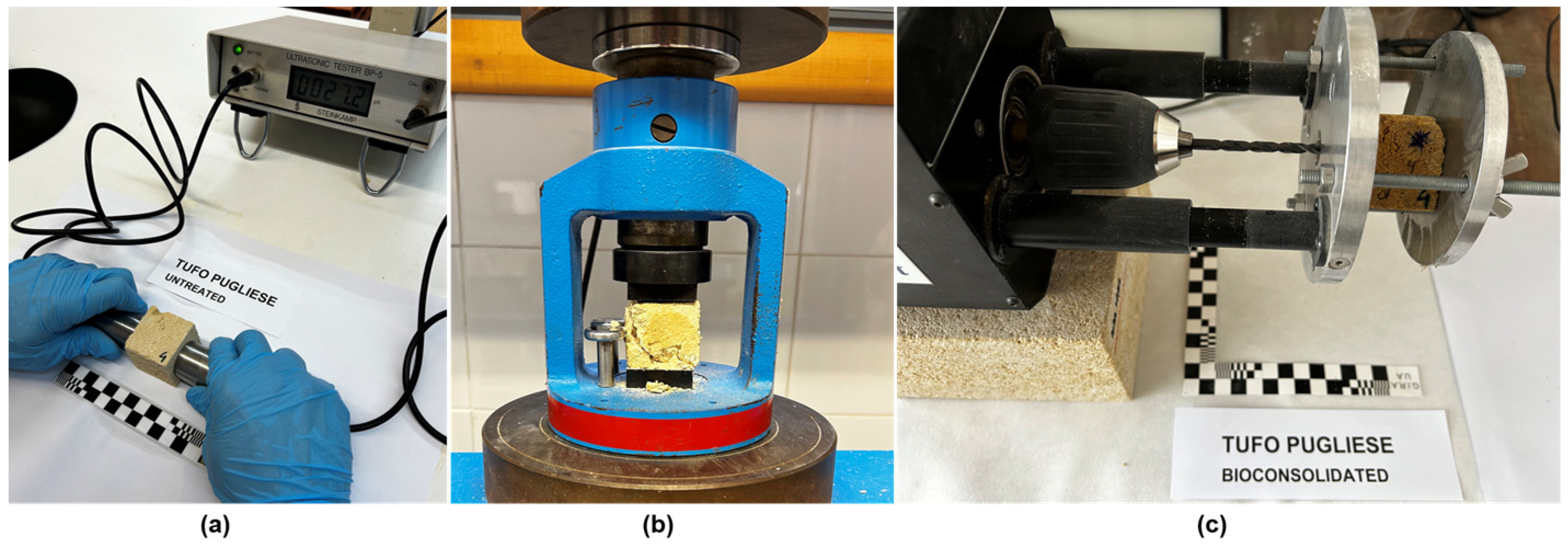
The average time taken for the pulse to pass through the three faces of all the untreated specimen was 1.772 ms, indicating the high porosity of Gravina Calcarenite [64].
The average mechanical compressive strength of the four specimens tested was 1.65 MPa, the minimum value was 1.25 MPa, and the maximum value was 1.97 MPa. These results indicate a very low mechanical resistance even for this type of stone. This makes Gravina Calcarenite especially susceptible to degradation [72,73,74,75].
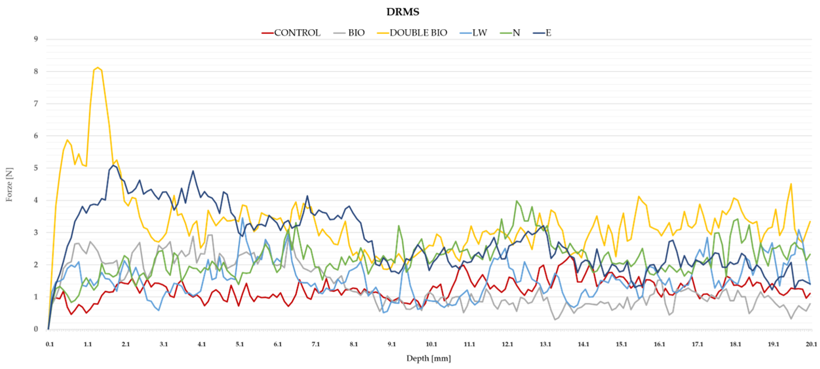
  • Drilling resistance measuring system (DRMS) (Figure 5c)
The drilling resistance results were heterogeneous, showing that within the test specimens, the resistance was variable in the case of drilling of fossils or cavities. The average value of the measurements has been assumed 1.24 N of force up to 2 cm depth. These results indicate a low mechanical resistance compared to other studies of similar calcarenites [50].

3.2. Treated Gravina Calcarenite

The applied treatments have worked differently because in the case of lime water, hardly any increase in resistance or improvement in properties was observed, while the bioconsolidant applied twice (7 days of treatment + 7 days of rest + 7 days of treatment) and the nanosilica (Nano Estel) are the treatments that generated the greatest consolidation. Nanolime (Nanorestore) and bioconsolidant applied only once (7 days of treatment) only slightly improved mechanical resistance and capillary absorption.
To obtain a comprehensive overview of the results obtained, each treatment includes a summary table with data from the color study (total color difference between untreated and treated samples, ΔE*), apparent density, porosity, pore diameter, drop absorption, capillarity, vapor permeability, ultrasonic, compression resistance, and DRMS.
The results shown in the tables of each treatment include the average value between the different types of specimens with different densities, as well as the minimum and maximum values obtained in each test or study. On those tables, the last columns compile quantified data regarding the difference compared to untreated specimens, and an assessment is provided.

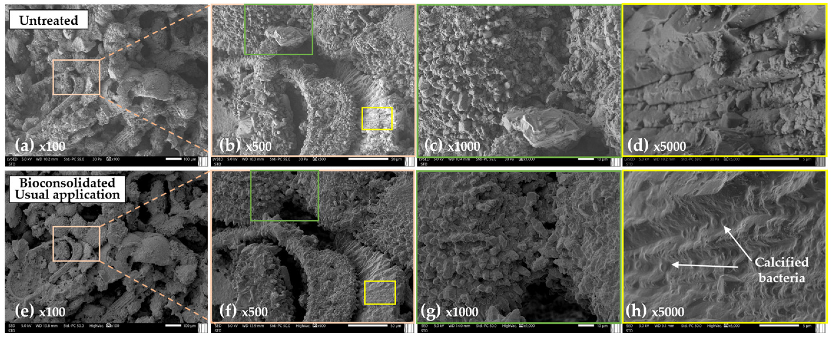
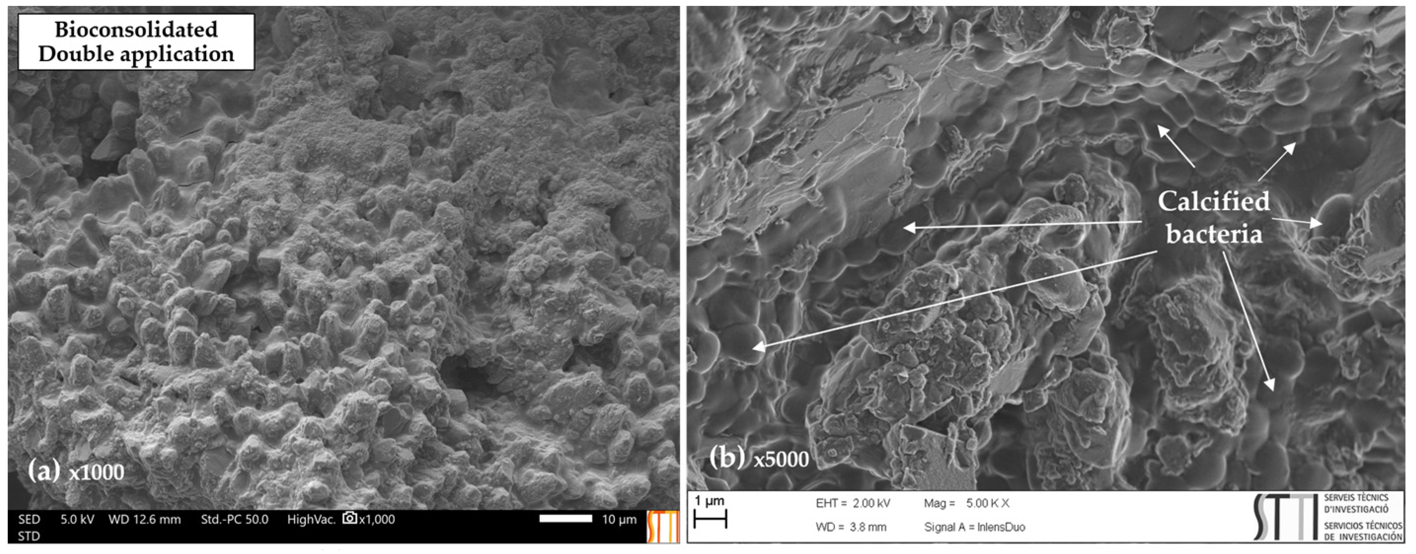
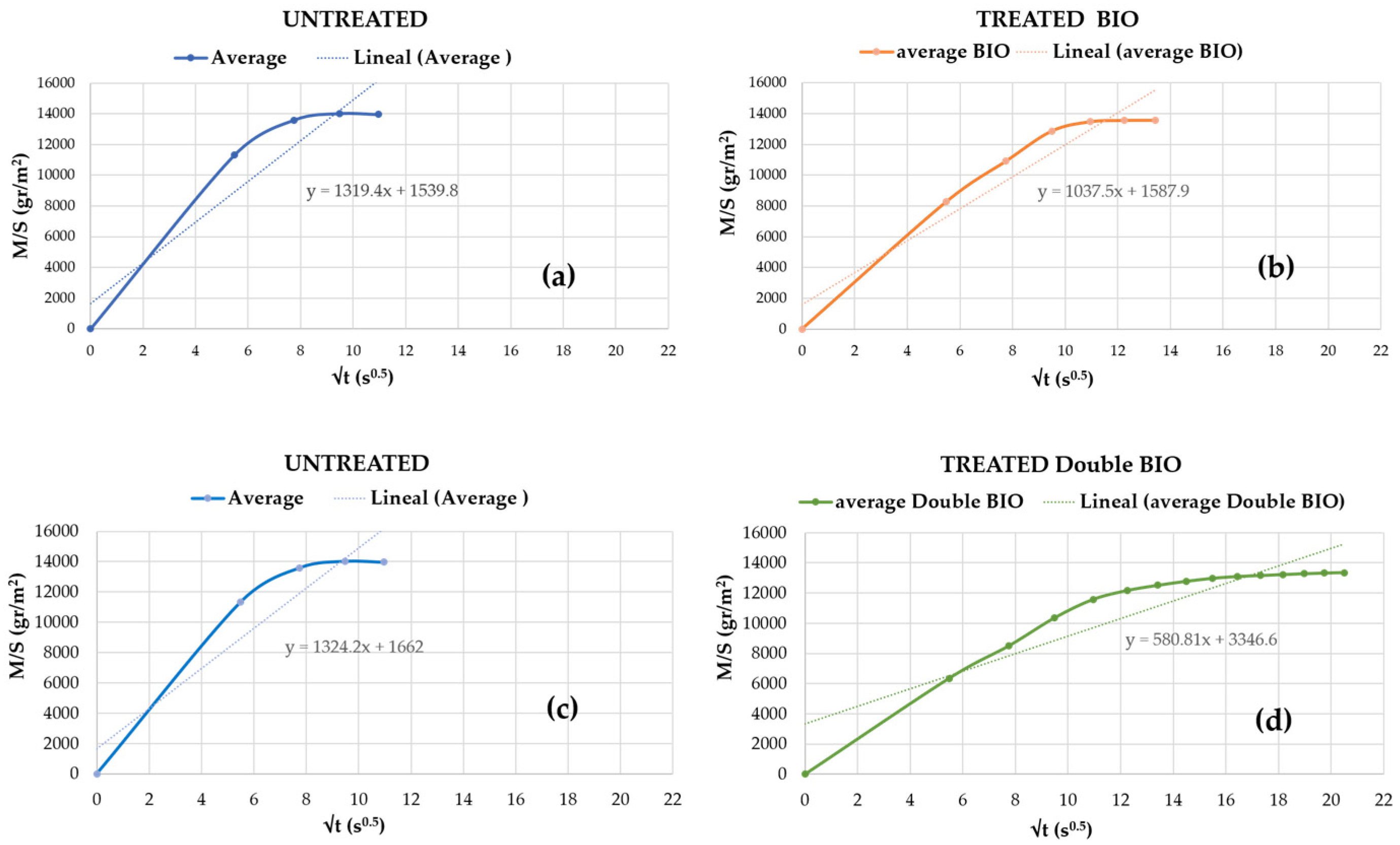
3.2.1. Treated with Bioconsolidant “Bio”

The bioconsolidated specimens macroscopically showed a slight change in color, darkening slightly in general to a more golden color compared to the same untreated specimens (Figure 6).
Color modification must be attributed in part to the oxidation of the ankerite in the stone, as the specimen was wetted 14 times over 7 days to apply the treatment and then dried at 50 °C to constant weight before testing.
The observation of all specimens with a low-magnification portable microscope has allowed us to see that there are no differences in the surface between the untreated and treated samples. The ankerite grains are seen in orange color. In the bioconsolidated specimens with double treatment (Figure 7), it can be observed that the clasts have more rounded edges than before treatment.
Regarding the behaviour at a microscopic level, it was seen that the porous structure of the bio specimen remains similar to the untreated specimen. However, the consolidation caused by carbonatogenesis can be observed.
Figure 8 shows the same areas, as seen in the SEM, of a 0.8 cm cube untreated (top row) and treated with KBYO for 7 days (bottom row).
The samples treated with double bioconsolidant treatment present a biofilm that binds the surface of the stone without occluding the pores (Figure 9a). The FESEM observation (Figure 9b) of the bioconsolidated samples at 5000× magnification shows that the microtopography of the stone is completely covered by a biofilm of calcified bacteria of about 2 microns in average size. This behavior is similar to that observed in other cases in which bioconsolidants have been used on calcarenitic rocks [49,50,76].
The bulk density before and after the test is similar for the usually bioconsolidation treatment, while in the double bioconsolidation, the density increased by 2.3% on average, which implies an improvement in the compactness of the treated specimen (data obtained from the increase in weight of the cubic test specimens).
Capillary absorption after bioconsolidation slowed down slightly, decreasing the slope of the curve, as shown in Figure 10 in which the graphs of water absorption with respect to the surface in relation to time are shown. Samples with double bioconsolidant treatment show greater improvements in their behavior. It can be seen that the curve has a much lower slope compared to the same untreated samples.
The droplet absorption time after treatment slightly increased. The water droplet remained on the surface for 1 to 2 s before penetrating the pores, and in the case of double bioconsolidation, it took approximately 3 s for the droplet to be absorbed. These data could indicate that the porosity has been modified; however, in this case, they are mainly related to a hydrophobic effect generated by the biofilms produced by this type of consolidation [49,50,76] because the porosity has hardly changed according to the data obtained with the porosimeter.
Figure 11 shows the same specimen during the testing process before and after being treated. Untreated samples become saturated in just 2 min, and moisture reaches the top surface in less than 1 min. The same specimen treated with double bioconsolidant have needed 5 min to rise by capillarity to the top of the cube. These same results have been obtained in other similar studies [50].
Regarding the mechanical properties, the speed of the ultrasonic test in all the treated specimens slightly increased. This behavior indicates greater compactness, although it should be noted that this test provides little information as the results on the different faces were somewhat heterogeneous. The compressive strength of the treated specimens increased with respect to the untreated control specimens by 29%. In the double-treated specimens, the compressive strength practically doubled, thereby increasing their resistance by 109%. This type of improvement has also been observed in other studies carried out on calcarenites [76].
The DRMS test showed that the consolidant penetrated to a depth of two centimetres (core of the specimen); the resistance to drilling was higher in the bioconsolidated specimens than in the untreated control specimens. In addition, the resistance to DRMS was much higher in the double-treated specimens than in the untreated specimens, and the resistances were multiplied. These data indicate that consolidation has occurred at a greater depth than in other studies carried out on other calcarenites with smaller pore size [50].
Table 1 presents the data for usually bioconsolidated specimens, while Table 2 contains data for double bioconsolidated specimens.

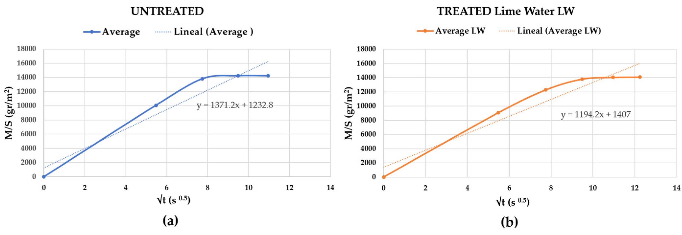
3.2.2. Treated with Lime Water “LW”

The specimens treated with lime water showed a slight macroscopic change in color, although this was not relevant to the naked eye. In this type of treatment very homogeneous results have been obtained in all the tests.
With respect to the behavior at a microscopic level, it could be seen that the porous structure remained similar to the untreated specimen. Furthermore, the consolidation produced by the carbonation of the Ca(OH)2 contained in the lime water could hardly be observed at 100× magnification.
Figure 12 shows the same area of a 0.8 cm untreated (in the upper row (a–d)) and treated cube with “LW” (in the lower row (e–h)). The images (a) and (e) present 100× magnification, the following images show the highlighted area inside the colored frames enlarged to 500× magnification, the following images show 1000× magnification, and the last images on the right show 5000× magnification.
The consolidated areas are indicated in the images; areas can be seen in which new CaCO3 crystals have appeared.
The bulk density before and after the test was similar. The droplet absorption time after treatment did not increase with respect to the same specimens.
The data obtained with the study of porosity are surprising because after the treatment, the porosity must be reduced by introducing matter into the pores. This is due to the fact that the sample treated with lime water studied using porosimetry should have greater porosity than the untreated reference sample. The Gravina Calcarenite, as mentioned above, is a somewhat heterogeneous stone.
The capillary absorption after consolidation slightly slowed down (Figure 13). The slope of the curve of the same treated samples is lower and capillary absorption is reduced by 1% in the first 2 min, which is time in which, without treatment, it reached constant weight.
Regarding the mechanical properties, the ultrasonic test speed slightly decreased after treatment in all the specimens, which is paradoxical because it indicates a heterogeneity of the samples and the difficulty of taking accurate data with an ultrasound without applying conductive material such as Vaseline. These materials were not used to avoid contaminating the samples since the same specimens have been tested before and after being treated.
The compressive strength of the treated specimens was not modified with respect to the untreated specimens. The DRMS test indicated that the lime water consolidated more in the first few millimeters, although the improvements were not very significant. In Table 3, the summary of results for specimens treated with enriched lime water is displayed.
Some authors [28,41,52,53] indicate that the treatment with lime water must be repeated over several weeks to obtain good results in consolidation. In this case, the treatment has been applied only once, although with a higher percentage of CaCO3 per liter, so we consider that the results obtained agree with the need to repeat the application for it to be effective.

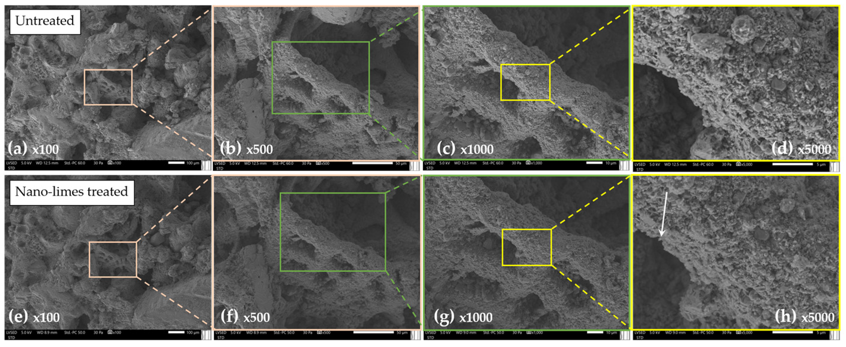
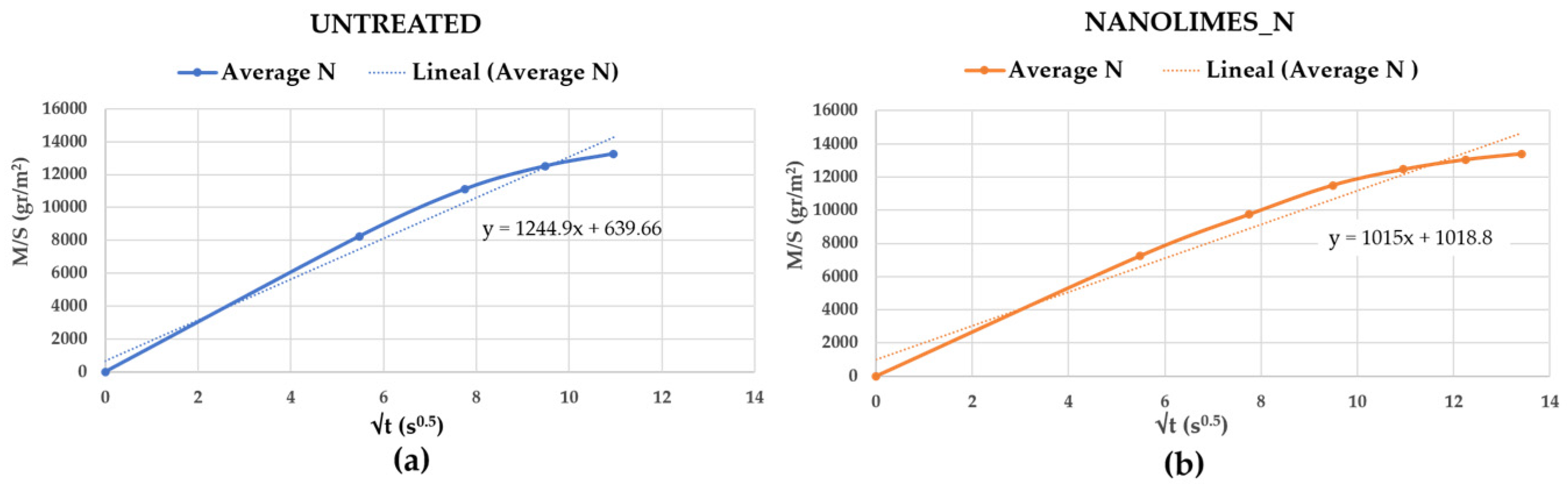
3.2.3. Treated with Nanolimes “N”

The specimens treated with nanolimes (Nanorestore) at the macroscopic level do not exhibit significant color changes and nor modification of the stone surface.
Regarding the behavior at a microscopic level, it can be seen that the porous structure remained similar to the untreated specimen seen at 100x magnification. As the magnification increased, a slight consolidation produced by the carbonation of the Ca(OH)2 present in the treatment could be observed. The surface of the stone was covered by a layer of small CaCO3 crystals [32,51,55].
Figure 14 shows the same area of a 0.8 cm untreated (in the upper row) and treated cube with N (in the lower row). It can be seen that the macroporous pattern is maintained, but in detail, small nano-sized CaCO3 crystals can be observed; these are indicated with an arrow in the last images.
The bulk density before and after the test was similar. The droplet absorption time after treatment was not modified with respect to the same specimens.
The data obtained with the study of porosity shows that after the treatment the pore size is slightly reduced, obtaining an average diameter of 1.87 µm. These data coincide with those of other investigations carried out by other authors [32]. Although the heterogeneity of this type of stone must always be considered.
The capillary absorption after consolidation slightly slowed down, decreasing the slope of the curve as seen in Figure 15.
The compressive strength of the treated samples has increased by 48% compared to the untreated samples. The DRMS test indicated that the nanolime consolidated even the center of the sample improving its drilling resistance. Other authors who have worked on these aspects agree on the improvement of mechanical resistance [32,51,53,54].
In Table 4, the summary of results for specimens treated with nanolimes is displayed.

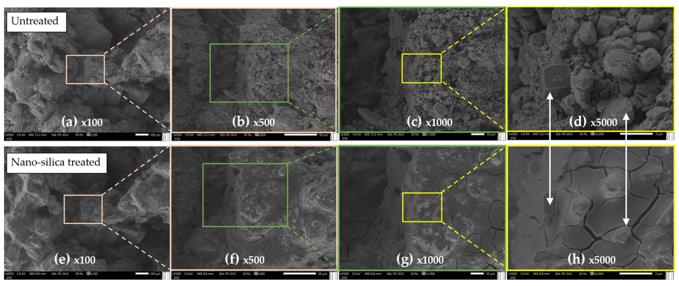
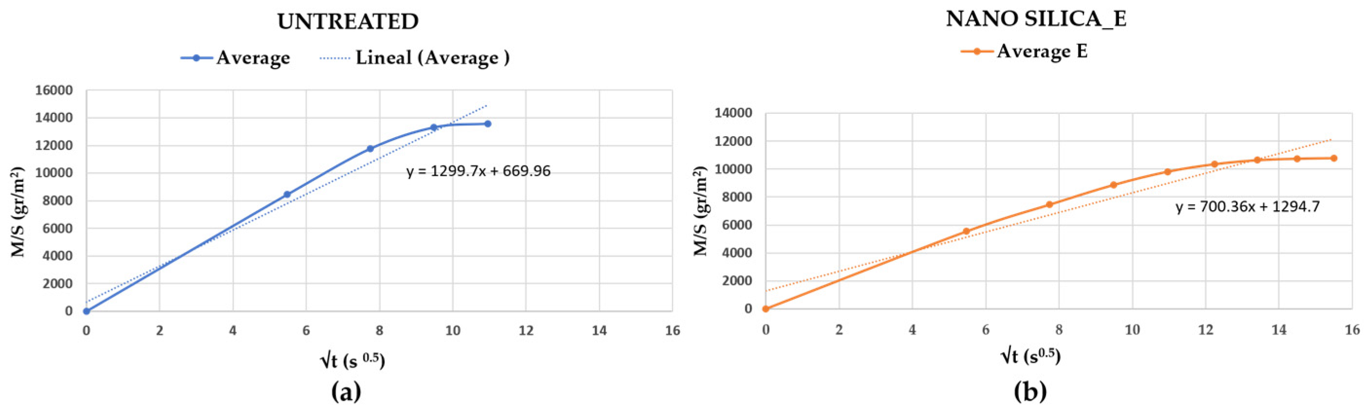
3.2.4. Treated with Nanosilica “E”

The specimens treated with nanosilica (Nano Estel) presented a slight change in color at the macroscopic level, darkening with respect to the untreated specimen. Regarding to the microscopic behavior, it was seen that the porous structure remained similar to the untreated specimen seen at 100x magnification, although at this magnification, the consolidation was already visible. At a higher magnification, the crackle layer of nanosilica produced by the treatment was detected. The surface of the stone has been completely covered by a crackle layer. This behavior is common in this type of treatment, as reported in other research works [27,35,56,57].
Figure 16 shows the same area of a 0.8 cm untreated (top row) and E-treated cube (bottom row).
The bulk density after the test increased by 9.5%, and the porosity reduced by 25%. These data indicate that the consolidant has increased the mass of the specimens studied.
The droplet absorption time after treatment did not increase compared to the same specimens. The capillary absorption after consolidation slightly slowed down, thereby decreasing the slope of the curve (Figure 17).
Regarding the mechanical properties, the ultrasonic test speed slightly increased after treatment in all the specimens, indicating a slight improvement in compactness. The compressive strength of the treated specimens also increased by a factor of two. The DRMS test indicated that this type of treatment has consolidated more in the first 8 millimeters, although it also improved in the interior of the treated specimens compared to the untreated ones.
In Table 5, the summary of results for specimens treated with nanosilica is displayed.

4. Discussion

Gravina Calcarenite (Pugliese tuff) is a very porous stone (43%) with a presence of macrofossils and low mechanical strengths. They cause variations in the bulk density values of the specimens studied, thereby influencing the results of the different treatments. The variations in the bulk density of the untreated samples vary by ±7.5% with respect to the average value. These possible differences were considered when selecting the specimens because each treatment was applied to specimens with different densities. The heterogeneity of the specimens studied means that there were differences in some results for the same treatment. That was initially expected, just as other authors who have investigated this type of stone had already indicated [5,12,13,72].
The SEM observation shows none of the treated specimens had the porous network occluded. Nanosilica and the bioconsolidant with double applications were the treatments that formed the greatest bonds at a microscopic level.
All treatments allowed water vapor permeability, and improvements were observed after the application of nanosilica and the double bioconsolidant treatment. This fact is apparently contradictory, but these results could be related to the effect of creating capillary tubes with less friction when air passes through [77]. It has been observed in the SEM that the microtopography generated with both the nanosilica and the bioconsolidant has enveloped the surface, thereby creating a film without edges, which rounds the internal surface of the stones.
The average values of the results obtained indicate that all treatments, except lime water, improved the compressive strength and DRMS resistances (Figure 18). These high improvements in mechanical resistance coincide with the results shown by other authors who have tested the effectiveness of this type of consolidants on calcarenites [27,32,35,50,53,54,56,57,76]. The results obtained with ultrasound have been heterogeneous, although with the exception of lime water, the other treatments slightly increased the speed of wave transmission, which implies improvements in the cohesion between particles.
The results obtained with the DRMS indicate that the treatments that have generated the greatest consolidations have been nanosilica “E” and double bioconsolidant. The DRMS graph in Figure 18 shows the average results of all treatments comparatively. In general, the specimens are homogeneous with specific anomalies. Peaks outside the midrange indicate the occasional presence of fossils/clasts with greater mechanical resistance than the rest of the stone. The presence of pores has caused the graph line to indicate zero resistance.
The untreated specimen called “control” is represented with the red line and shows how it behaved homogeneously in the 200 mm depth of the test. It can be seen that the resistance is low (1.24 N) compared to other calcarenites of similar composition and porosity [50].
The bioconsolidated specimen with one application is the gray line. It can be seen that in the first 9 mm, there was improvement compared to the untreated specimens, although its resistance to drilling has barely improved.
The double bioconsolidated is represented with the yellow line showing much improvement in the entire 200 mm of the test with a peak in the first millimeters, possibly due to fossil anomalies. The drilling resistance has doubled, which is a fact that coincides with the improvements in compression resistance. The treatment has penetrated all of the specimens tested, thereby consolidating the interior. These data, according to some authors [28,50,72,78], are considered favorable for the effectiveness of the consolidative treatment.
The lime water in light blue color had a similar behavior, with similar results to the untreated specimens.
The nanolime, in green color, has slightly improved the drilling resistance; this agrees with the results obtained in the other tests.
The nanosilica, in dark blue, generally improved DRMS resistance, although to a greater extent in the first 8 mm.
These results agree with those obtained in other tests, such as capillary rise humidity and compression resistance. The increase in density in the double bioconsolidated specimens and in those consolidated with nanosilica also indicates the effectiveness of these treatments of the specimens.
The double bioconsolidant worked very well, creating a large community of bacteria capable of creating CaCO3 that consolidated the stone [79]; this result is probably due to the composition of Gravina Calcarenite, which is 95% calcite. Carbonate productivity is strongly dependent on the mineralogy of the substrate. According to Rodríguez-Navarro [80], calcitic substrates offer a greater affinity for bacterial binding than silicate substrates, thus promoting bacterial growth and metabolic activity, resulting in greater production of calcium carbonate cement. Bacterial calcite grows coherently on the calcitic substrate, being very chemically and mechanically stable.
Furthermore, this treatment has been the only one capable of delaying the absorption of water droplets and the one that most delayed and reduced the absorption of water by capillarity. The treatment generated a bond with the original matrix, strengthening the bonds between mineral grains and clasts, while the bacterial exopolymeric substances (EPS) generated hydrophobicity [81].
The results obtained with the mercury porosimeter (MIP) agree with SEM observations and with the results of the different tests (Figure 19). It is observed that the curve that changes the most is with the nanosilica treatment called “E”.
The average pore size in the untreated specimens was 1.984 µm, similar to the specimens studied treated with lime water, nanolime, bioconsolidant, and double bioconsolidant. The specimen treated with nanosilica lowered its diameter considerably, achieving an average of 0.339 µm, thereby reducing the pore diameter by 83% compared to the untreated samples. The specimens treated with nanosilica are those with the lowest percentage of porosity. The reduction in pore size is a priori negative for behavior against haloclasty, according to some authors [22,41,42,43,44,45,75,82]. The crystallization of the salts inside the pores causes damage if they do not have enough space to contain the formed crystal, and pressures are produced capable of fracturing the stone.
On the other hand, consolidating treatments are usually applied to the already deteriorated surface, which therefore has higher percentages of porosity than the healthy stone tested in this work. It is recommended to carry out tests in situ. To do this, it is recommended to select inconspicuous areas of the building and apply the treatments previously selected in the laboratory tests to a small area of the altered surface (approx. 20 × 20 cm). In this case, it is proposed to apply the double bioconsolidant in situ. Follow-up is carried out for a preestablished time, not less than 1 year. Data are taken on color, surface humidity, and observation with a portable microscope before and after and DRMS before and after. A small specimen can be extracted before and after the in situ testing time has ended. With this little specimen, SEM, MOP, or porosimetry observations can be made [82].
Regarding the variations in the color of the treated specimens, it should be noted that the buildings made with Gravina Calcarenite present a natural aging patina that makes them darker in color, as can be seen in Figure 1. This is mainly due to the oxidation of the iron of the ankerite [83] present in the composition of this calcarenite. The investigation was carried out on “healthy zone” stones that were not exposed to weathering and had no preexisting natural patina of ageing. Therefore, the color changes caused by the treatments in the specimens should not be a condition for rejecting the use of this treatment. The nanolimes did not modify the color of the treated specimens, being below the range established by some authors who place it in ΔE* ≤ 3 [84].

5. Conclusions

Gravina Calcarenite is a very porous stone (43%) with a low mechanical strength that makes it extremely weak against deterioration due to weather agents. In these conditions, the application of certain consolidating treatments can be an effective solution to extend its durability. The mineralogical composition (95% calcite) makes it an ideal stone to be treated with bioconsolidants that activate the macrobiota present in the stone, thereby creating a high community of calcified bacteria capable of consolidating it and multiplying its mechanical compressive strength and DRMS by two.
Treatments with lime water did not improve the mechanical compressive strength, although the resistance to DRMS increased by only 25% compared to the untreated specimens. The average pore diameter did not reduce, although the permeability to water vapor reduced by 8%. In summary, the results obtained in this study do not show improvements with this type of treatment with a single application. They should probably be applied for several days or weeks until good results are obtained. This opens other lines for future research.
The nanosilica treatment improved mechanical strength, especially in the first 8 mm depth of the treated surface, without reducing vapor permeability. This treatment introduces different chemical compounds into the base stone to be consolidated. The pore diameter has been reduced by 83% compared with the untreated specimen. These dates can generate greater damage due to haloclasty [42,43,44,45], so it is considered advisable to carry out complementary tests prior to the application of these treatments. Specimens of the salts present in each specific building should be taken, and preliminary tests for resistance to salt crystallization should be carried out using salts similar to those existing in each construction.
Treatments with nanolimes (Nanorestore) improved the mechanical compressive strength of the treated specimens by 42%, and the resistance to DRMS increased by 76% compared to the untreated specimens. The average pore diameter did not reduce, although the water vapor permeability reduced by only 8.5%. The treatment with nanolimes is proposed as effective in consolidating damaged areas, although it is less effective than the double-applied bioconsolidant in this case under study. However, this type of treatment with nanoparticles can present risk of toxicity, according to some authors [58].
In this study, it was shown that the double application of the bioconsolidating treatment, leaving 7 days without treatment between the two applications, considerably improved consolidation compared to a single application. The FE-SEM images show high bioconsolidation with a high presence of calcified bacteria. The average pore diameter did not change, and the water vapor permeability increased. The promising results of this ecofriendly treatment presented in this article are positive, a priori, for improving damaged areas in Gravina Calcarenite.

Author Contributions

Conceptualization, J.A.H.-T., D.F. and Y.S.-B.; methodology, J.A.H.-T. and Y.S.-B.; software, J.A.H.-T. and Y.S.-B.; validation, C.G.C., M.F.S., F.R. and D.F.; formal analysis, C.G.C., M.F.S. and D.F.; investigation, J.A.H.-T., C.G.C., F.R. and Y.S.-B.; data curation, J.A.H.-T. and Y.S.-B.; writing—original draft preparation, J.A.H.-T., C.G.C. and Y.S.-B.; writing—review and editing, J.A.H.-T., Y.S.-B., C.G.C., M.F.S., F.R. and D.F.; visualization, J.A.H.-T., Y.S.-B., C.G.C. and D.F; supervision, C.G.C. and D.F. All authors have read and agreed to the published version of the manuscript.

Funding

This research received no external funding.

Data Availability Statement

The original contributions presented in the study are included in the article, further inquiries can be directed to the corresponding author.

Acknowledgments

The authors would like to thank Carlos Rodriguez Navarro for his collaboration in the DRMS tests at the University of Granada, David Benavente García for his support in DRX and capillarity tests, Elena Andrés Abián for her collaboration in the translation and to Pepe Cabello from KBYO Biological.

Conflicts of Interest

The authors declare no conflict of interest.

References

  1. Vitiello, V.; Castelluccio, R.; Ramondini, M. Conservation of Cultural Heritage. Evaluation of Archaeological Digs Effect on the Degradation of the Structures Found. In Proceedings of the 4th International Conference on Protection of Historical Constructions (PROHITECH 2020/21), Athens, Greece, 25–27 October 2021. [Google Scholar] [CrossRef]
  2. Bonomo, A.; Acito, A.; Prosser, G.; Rizzo, G.; Munnecke, A.; Koch, R.; Bentivenga, M. Matera’s Old Quarries: Geological and Historical Archives That Need Protection and Valorization. Geoheritage 2019, 11, 1603–1619. [Google Scholar] [CrossRef]
  3. Artiola, V.; La Verde, G.; D’Avino, V.; Pugliese, M. Sassi of Matera Building Material: High-Resolution Gamma-Ray Spectroscopy Characterization for Radioprotection. Buildings 2021, 11, 258. [Google Scholar] [CrossRef]
  4. Corbi, I.; Baratta, A.; Corbi, O.; Li, H. Existing Mono-Cell and Multi-Cell Low-Rise Dry, Hollowed Constructions. Buildings 2023, 13, 340. [Google Scholar] [CrossRef]
  5. Bonomo, A.E.; Rizzo, G.; Prosser, G. Calcarenite di Gravina formation a row material for the lime production. In Proceedings of the International Conference on Metrology for Archaeology and Cultural Heritage, Florence, Italy, 4–6 December 2019; pp. 84–89. [Google Scholar]
  6. Andriani, G.F.; Walsh, N. Petrophysical and mechanical properties of soft and porous building rocks used in Apulian monuments (south Italy). Geol. Soc. Lond. Spec. Publ. 2010, 333, 129–141. [Google Scholar] [CrossRef]
  7. Martínez Martínez, J.; Berrezueta, E.; Aguilera, H.; Fusi, N.; Gómez Heras, M. Cálculo de la (micro)tasa de alteración en calcarenitas expuestas en clima semiárido costero. Geogaceta 2023, 74, 27–30. [Google Scholar] [CrossRef]
  8. Fort, R.; Varas, M.J.; Alvarez de Buergo, M.; Martin-Freire, D. Determination of anisotropy to enhance the durability of natural stone. J. Geophys. Eng. 2011, 8, S132–S144. [Google Scholar] [CrossRef]
  9. Bonomo, A.; Amodio, A.; Prosser, G.; Sileo, M.; Rizzo, G. Evaluation of soft limestone degradation in the Sassi UNESCO site (Matera, Southern Italy): Loss of material measurement and classification. J. Cult. Herit. 2020, 42, 191–201. [Google Scholar] [CrossRef]
  10. Weiss, T.; Siegesmund, S.; Kirchner, D.; Sippel, J. Insolation weathering and hygric dilatation: Two competitive factors in stone degradation. Environ. Geol. 2004, 46, 402–413. [Google Scholar] [CrossRef]
  11. Borea, E. (Coord). Materiali Lapidei. Problemi Relativi Allo Studio del Degrado e Della Conservazione. I-II. Bolletino D’arte (supplementi al n 41). Ministero per I Beni Culturali e Ambientali, Italia. 1987. Available online: http://www.bollettinodarte.beniculturali.it/opencms/multimedia/BollettinoArteIt/documents/1580393362188_volume_I_intero.pdf (accessed on 25 March 2024).
  12. Lupo, M.; Genevois, R.; Tecca, P.R. Physical and mechanical characteristics of the Gravina Calcarenite (southern Italy). Ital. J. Eng. Geol. Environ. 2019, 2, 37–54. [Google Scholar] [CrossRef]
  13. Caputo, M.C.; De Carlo, L.; Masciale, R.; Perkins, K.; Turturro, A.C.; Nimmo, J.R. Detection and quantification of preferential flow using artificial rainfall with multiple experimental approaches. Hydrogeol. J. 2023, 32, 467–485. [Google Scholar] [CrossRef]
  14. Tropeano, M.; Caldara, M.A.; De Santis, V.; Festa, V.; Parise, M.; Sabato, L.; Spalluto, L.; Francescangeli, R.; Iurilli, V.; Mastronuzzi, G.A. Geological Uniqueness and Potential Geotouristic Appeal of Murge and Premurge, the First Territory in Puglia (Southern Italy) Aspiring to Become a UNESCO Global Geopark. Geosciences 2023, 13, 131. [Google Scholar] [CrossRef]
  15. Moretti, M.; Owen, G.; Tropeano, M. Soft-sediment deformation induced by sinkhole activity in shallow marine environments: A fossil example in the Apulian Foreland (Southern Italy). Sediment. Geol. 2011, 235, 331–342. [Google Scholar] [CrossRef]
  16. Grassi, D.; Grimaldi, S.; Simeone, V. On the causes of the instability affecting rupestrian urban centres of Apulia Region (Southerrn Italy). In 32° Intentional Geological Congress Firenze, Paper A32IGC9KF3, Session T16.06—Geoscience for Cultural Heritage; Natural Hazard and Cultural Heritages: Firenze, Italy, 2004. [Google Scholar]
  17. D’Angeli, I.M.; Lacalamita, M.; Sasso, C.; Schingaro, E.; Parise, M. Morphological and mineralogical characterization of surficial weathering on calcarenite rocks in the rupestrian system of “San Michele delle Grotte” at Gravina in Puglia (Bari, Apulia). CATENA 2022, 216, 106382. [Google Scholar] [CrossRef]
  18. Simeone, V.; Doglioni, A.; Lacertosa, R.M.; Sdao, F. Environmental and Geological Characters and Stability Problems in the Historic Centre of Matera (South Italy). In IAEG/AEG Annual Meeting Proceedings; Shakoor, A., Cato, K., Eds.; Springer: San Francisco, CA, USA, 2019; Volume 2, pp. 161–168. [Google Scholar] [CrossRef]
  19. Siegesmund, S.; Gross, C.J.; ·Dohrmann, R.; Marler, B.; Ufer, K.; Koch, T. Moisture expansion of tuf stones and sandstones. Environ. Earth Sci. 2023, 82, 146. [Google Scholar] [CrossRef]
  20. Mecca, I. In situ experimentations for the compatibility and durability of the restorations: The case study of the Sassi of Matera. VITRUVIO—Int. J. Archit. Technol. Sustain. 2016, 1, 35–48. [Google Scholar] [CrossRef]
  21. Benavente, D.; Martinez-Martinez, J.; Cueto, N.; Ordoñez, S.; García del Cura, M.A. Impact of salt and frost weathering on the physical and durability properties of travertines and carbonate tuffs used as building material. Environ. Earth Sci. 2018, 77, 147. [Google Scholar] [CrossRef]
  22. Benavente, D.; De Jongh, M.; Cañaveras, J.C. Weathering processes and mechanisms caused by capillary waters and pigeon droppings on porous limestones. Minerals 2021, 11, 18. [Google Scholar] [CrossRef]
  23. Smith, B.; Gómez-Heras, M.; McCabe, S. Understanding the decay of stone-built cultural heritage. Prog. Phys. Geogr. Earth Environ. 2008, 32, 439–461. [Google Scholar] [CrossRef]
  24. Alonso, F.J.; Alonso, L.; Vázquez, P. Hydric properties and anisotropy of porous sedimentary rocks. In Proceedings of the VIII Congreso Geológico de España, Oviedo, España, 17–19 July 2012. [Google Scholar]
  25. Toniolo, L.; Gherardi, F. The protection of marble surfaces: The challenge to develop suitable nanostructured treatments. In Advanced Materials for the Conservation of Stone; Springer: Cham, Switzerland, 2018; pp. 57–78. [Google Scholar] [CrossRef]
  26. Lazzarini, L.; Tabasso, M. Il Restauro Della Pietra; CEDAM: Padova, Italy, 1980. [Google Scholar]
  27. Zornoza-Indart, A.; López-Arce, P.; Leal, N.; Simão, J.; Zoghlami, K. Consolidation of a Tunisian bioclastic calcarenite: From conventional ethyl silicate products to nanostructured and nanoparticle based consolidants. Constr. Build. Mater. 2016, 116, 188–202. [Google Scholar] [CrossRef]
  28. Gherardi, F. Current and Future Trends in Protective Treatments for Stone Heritage. In Conserving Stone Heritage. Cultural Heritage Science; Gherardi, F., Maravelaki, P.N., Eds.; Springer: Cham, Switzerland, 2022. [Google Scholar] [CrossRef]
  29. ICOMOS-ISCS: Illustrated Glossary on Stone Deterioration Patterns 2008. Available online: http://www.international.icomos.org/publications/monuments_and_sites/15/pdf/Monuments_and_Sites_15_ISCS_Glossary_Stone.pdf (accessed on 25 March 2024).
  30. Jimenez-Lopez, C.; Rodriguez-Navarro, C.; Piñar, G.; Carrillo-Rosúa, F.J.; Rodriguez-Gallego, M.; Gonzalez-Muñoz, M.T. Consolidation of degraded ornamental porous limestone stone by calcium carbonate precipitation induced by the microbiota inhabiting the stone. Chemosphere 2007, 68, 1929–1936. [Google Scholar] [CrossRef]
  31. Ripoll, A.; Rojo, A.; Ruiz de Argandoña, V.G. Evaluation of nanoparticulate consolidants applied to Novelda Stone (Spain). Mater. Constr. 2022, 72, e294. [Google Scholar] [CrossRef]
  32. Wen, Y.; Qing, H.; Shu, H.; Liu, Q. Evaluating the Protective Effects of Calcium Carbonate Coating on Sandstone Cultural Heritage. Coatings 2021, 11, 1534. [Google Scholar] [CrossRef]
  33. Fernandez, F.; Germinario, S.; Montagno, R.; Basile, R.; Borgioli, L.; Laviano, R. Experimental Procedures of Accelerated Aging and Evaluation of Effectiveness of Nanostructured Products for the Protection of Volterra (Italy) Panchina Stone. Buildings 2022, 12, 1685. [Google Scholar] [CrossRef]
  34. Chen, W.; Dai, P.; Yuan, P.; Zhang, J. Effect of inorganic silicate consolidation on the mechanical and durability performance of sandstone used in historical sites. Constr. Build. Mater. 2016, 121, 445–452. [Google Scholar] [CrossRef]
  35. Koestler, R. Polymers and resins as food for microbes. In Of Microbes and Art: The Role of Microbial Communities in the Degradation and Protection of Cultural Heritage; Ciferri, O., Tiano, P., Mastromei., G., Eds.; Springer: Boston, MA, USA, 2000; pp. 153–167. [Google Scholar] [CrossRef]
  36. Melo, M.; Bracci, S.; Camaiti, M.; Chiantore, O.; Piacenti, F. Photodegradation of acrylic resins used in the conservation of stone. Polym. Degrad. Stab. 1999, 66, 23–30. [Google Scholar] [CrossRef]
  37. Lombardini, N. Piero Sanpaolesi e il restauro della facciata della basílica di San Michele Maggiore a Pavia. In Piero Sanpaolesi. Restauro e Método; Tampone, G., Gurrieri, F., Girgi, L., Eds.; Nardini: Firenze, Italy, 2012; pp. 325–352. [Google Scholar]
  38. Fort González, R.; Pérez-Monserrat, E.M.; Layuno Rosas, M.d.l.Á.; Varas, M.J.; Álvarez de Buergo, M.; Martínez-Ramírez, S.; Blanco-Varela, M.T.; Villegas Broncano, M.Á.; García Heras, M.; López-Arce, P.; et al. La Con-servación de Los Geomateriales Utilizados en el Patrimonio, Universidad Complutense de Madrid. 2012. Available online: http://hdl.handle.net/10261/46731 (accessed on 25 March 2024).
  39. UNE 41810; Conservación del Patrimonio Cultural. Criterios de Intervención en Materiales Pétreos. AENOR: Madrid, Spain, 2007.
  40. Clifton, J. Stone Consolidating Materials: A Status Report; National Bureau of Standards: Washington, DC, USA, 1980. [Google Scholar] [CrossRef]
  41. Hansen, E.; Doehne, E.; Fidler, J.; Larson, J.; Martin, B.; Matteini, M.; Rodriguez-Navarro, C.; Sebastian Pardo, E.; Price, C.; de Tagle, A.; et al. A review of selected inorganic consolidants and protective treatments for porous calcareous materials. Estud. Conserv. 2003, 48 (Suppl. 1), 13–25. [Google Scholar] [CrossRef]
  42. Martínez-Martínez, J.; Arizzi, A.; Benavente, D. The Role of Calcite Dissolution and Halite Thermal Expansion as Secondary Salt Weathering Mechanisms of Calcite-Bearing Rocks in Marine Environments. Minerals 2021, 11, 911. [Google Scholar] [CrossRef]
  43. Benavente, D. Why Pore Size Is Important in the Deterioration of Porous Stones Used in the Built Heritage. Macla 2011, 15, 41–42. Available online: http://hdl.handle.net/10045/19869 (accessed on 25 March 2024).
  44. Rodriguez-Navarro, C.; Doehne, E. Salt weathering: Influence of evaporation rate, supersaturation and crystallization pattern. Earth Surf. Process. Landf. 1999, 24, 191–209. [Google Scholar] [CrossRef]
  45. Yu, S.; Oguchi, C. Role of pore size distribution in salt uptake, damage, and predicting salt susceptibility of eight types of Japanese building stones. Eng. Geol. 2010, 115, 226–236. [Google Scholar] [CrossRef]
  46. Coviello, C.G.; Lassandro, P.; Sabbà, M.F.; Foti, D. Mechanical and Thermal Effects of Using Fine Recycled PET Aggregates in Common Screeds. Sustainability 2023, 15, 16692. [Google Scholar] [CrossRef]
  47. Lerna, M.; Foti, D.; Petrella, A.; Sabbà, M.F.; Mansour, S. Effect of the Chemical and Mechanical Recycling of PET on the Thermal and Mechanical Response of Mortars and Premixed Screeds. Materials 2023, 16, 3155. [Google Scholar] [CrossRef] [PubMed]
  48. Fiedler, J. Lime treatments: An overview of lime watering and shelter coating of friable historical limestone masonries. In Stone: Stone Building Materials, Construction and Associated Component Systems: Their Decay and Treatment; English Heritage Research Transactions: London, UK, 2002; pp. 19–28. [Google Scholar]
  49. Jroundi, F.; Schiro, M.; Ruiz-Agudo, E.; Elert, K.; Martín-Sánchez, I.; González-Muñoz, M.T.; Rodriguez-Navarro, C. Protection and consolidation of stone heritage by self-inoculation with indigenous carbonatogenic bacterial communities. Nat. Commun. 2017, 8, 279. [Google Scholar] [CrossRef] [PubMed]
  50. Spairani-Berrio, Y.; Huesca-Tortosa, J.A.; Rodriguez-Navarro, C.; Gonzalez-Muñoz, M.T.; Jroundi, F. Bioconsolidation of Damaged Construction Calcarenites and Evaluation of the Improvement in Their Petrophysical and Mechanical Properties. Materials 2023, 16, 6043. [Google Scholar] [CrossRef] [PubMed]
  51. Rodriguez-Navarro, C.; Ruiz-Agudo, E. Nanolimes: From synthesis to application. Pure Appl. Chem. 2018, 90, 523–550. [Google Scholar] [CrossRef]
  52. Slížková, Z.; Drdácký, M. Effects of Various Chemical Agents on Mechanical Characteristics of Weak Lime Mortar. In Historic Mortars; Springer: Cham, Switzerland, 2018. [Google Scholar] [CrossRef]
  53. D’Armada, P.; Hirst, E. Nano-Lime for Consolidation of Plaster and Stone. J. Archit. Conserv. 2012, 18, 63–80. [Google Scholar] [CrossRef]
  54. Borsoi, G.; Lubelli, B.; Hees, R.; Veiga, R.; Silva, A. Application Protocol for the Consolidation of Calcareous Substrates by the Use of Nanolimes: From Laboratory Research to Practice. Restor. Build. Monum. 2018, 22, 109–199. [Google Scholar] [CrossRef]
  55. Navarro-Moreno, D.; Martínez-Arredondo, A.; García-Vera, V.E.; Lanzón, M. Uso de nanomateriales para la conservación de la piedra del Teatro Romano de Cartagena. Loggia Arquit. Restauración 2023, 36, 106–119. [Google Scholar] [CrossRef]
  56. Dziadkowiec, J.; Cheng, H.; Ludwig, M.; Ban, M.; Tausendpfund, T.; Klitzing, R.; Mezger, M.; Valtiner, M. Cohesion Gain Induced by Nanosilica Consolidants for Monumental Stone Restoration. Langmuir 2022, 38, 6949–6958. [Google Scholar] [CrossRef] [PubMed]
  57. Iucolano, F.; Colella, A.; Liguori, B.; Calcaterra, D. Suitability of silica nanoparticles for tuff consolidation. Constr. Build. Mater. 2019, 202, 73–81. [Google Scholar] [CrossRef]
  58. Gomez-Villalba, L.S.; Salcines, C.; Fort, R. Application of Inorganic Nanomaterials in Cultural Heritage Conservation, Risk of Toxicity, and Preventive Measures. Nanomaterials 2023, 13, 1454. [Google Scholar] [CrossRef] [PubMed]
  59. Gonzalez-Muñoz, M.T.; Rodriguez-Navarro, C.; Jimenez-Lopez, C.; Rodriguez-Gallego, M. Method and Product for Protecting and Reinforcing Construction and Ornamental Materials. Spanish Patent WO 2008/009771 A1, January 2008. [Google Scholar]
  60. Delgado-Rodrigues, J.; Ferreira Pinto, A.; da Costa, D.R. Tracing of decay profiles and evaluation of stone treatments by means of microdrilling techniques. J. Cult. Herit. 2002, 3, 117–125. [Google Scholar] [CrossRef]
  61. Fratini, F.; Rescic, S.; Tiano, P. A new portable system for determining the state of conservation of monumental stones. Mater. Struct. 2005, 39, 139–147. [Google Scholar] [CrossRef]
  62. Pamplona, M.; Kocher, M.; Snethlage, R.; Aires Barros, L. Drilling resistance: Overview and Outlook. Z. Der. Dtsch. Ges. Für. Geowissenschaften. J. Appl. Reg. Geol. 2007, 158, 665–679. [Google Scholar] [CrossRef]
  63. UNE-EN 1926:2007; Natural Stone Test Methods-Determination of Uniaxial Compressive Strength. AENOR: Madrid, Spain, 2007.
  64. Eljufout, T.; Alhomaidat, F. Evaluation of natural building stones’ characterizations using ultrasonic testing technique. Arab. J. Sci. Eng. 2021, 46, 11415–11424. [Google Scholar] [CrossRef]
  65. UNE-EN ISO 16809:2020; Non-Destructive Testing-Ultrasonic Thickness Measurement (ISO 16809:2017). AENOR: Madrid, Spain, 2020.
  66. UNE-EN 15886:2011; Conservation of Cultural Property-Test Methods-Colour Measurement of Surfaces. AENOR: Madrid, Spain, 2011.
  67. Pérez-Ema, N.; Buergo, M.; Bustamante, R.; Gómez-Heras, M. Changes in Petrophysical Properties of the Stone Surface due to Past Conservation Treatments in Archaeological Sites of Merida (Spain). In Engineering Geology for Society and Territory; Lollino, G., Giordan, D., Marunteanu, C., Christaras, B., Yoshinori, I., Margottini, C., Eds.; Springer: Berlin/Heidelberg, Germany, 2015; Volume 8. [Google Scholar] [CrossRef]
  68. Pozo-Antonio, J.S.; Noya, D.; Montojo, C. Aesthetic Effects on Granite of Adding Nanoparticle TiO2 to Si-Based Consolidants (Ethyl Silicate or Nano-Sized Silica). Coatings 2020, 10, 215. [Google Scholar] [CrossRef]
  69. RILEM. Commission 25-PEM Protection et érosion des Monuments. Recomandations provisoires. Mater. Constr. 1980, 13, 175–252. [Google Scholar] [CrossRef]
  70. UNE-EN 15803:2010; Conservation of Cultural Property-Test Methods-Determination of Water Vapour Permeability (dp). AENOR: Madrid, Spain, 2010.
  71. UNE-EN 15801:2010; Conservation of Cultural Property-Test Methods-Determination of Water Absorption by Capillarity. AENOR: Madrid, Spain, 2010.
  72. Ciantia, M.O.; Castellanza, R.; Crosta, G.B.; Hueckel, T. Effects of mineral suspension and dissolution on strength and compressibility of soft carbonate rocks. Eng. Geol. 2014, 184, 1–18. [Google Scholar] [CrossRef]
  73. Ciantia, M.O.; Castellanza, R.; di Prisco, C. Experimental Study on the Water-Induced Weakening of Calcarenites. Rock. Mech. Rock. Eng. 2015, 48, 441–461. [Google Scholar] [CrossRef]
  74. de Jongh, M.; Benavente, D.; Young, M.; Graham, C.; Lee, M. The Long-Term Efficiency and Compatibility of Hydrophobic Treatments in Protecting Vulnerable Sandstone at Arbroath Abbey (Scotland). Heritage 2023, 6, 4864–4885. [Google Scholar] [CrossRef]
  75. Benavente, D.; García del Cura, M.A.; Fort, R.; Ordóñez, S. Durability estimation of porous building stones from pore structure and strength. Eng. Geol. 2004, 74, 113–127. [Google Scholar] [CrossRef]
  76. Spairani, Y.; Cisternino, A.; Foti, D.; Lerna, M.; Ivorra, S. Study of the Behavior of Structural Materials Treated with Bioconsolidant. Materials 2021, 14, 5369. [Google Scholar] [CrossRef] [PubMed]
  77. Reinemann, D.; Parlange, J.; Timmons, M. Theory of small-diameter airlift pumps. Int. J. Multiph. Flow. 1990, 16, 113–122. [Google Scholar] [CrossRef]
  78. Ban, M.; Aliotta, L.; Gigante, V.; Mascha, E.; Sola, A.; Lazzeri, A. Distribution depth of stone consolidants applied on-site: Analytical modelling with field and lab cross-validation. Constr. Build. Mater. 2020, 259, 120394. [Google Scholar] [CrossRef]
  79. Rodriguez-Navarro, C.; Jroundi, F.; Gonzalez-Muñoz, M. Stone Consolidation by Bacterial Carbonatogenesis: Evaluation of in situ Applications. Restor. Build. Monum. 2015, 21, 9–20. [Google Scholar] [CrossRef]
  80. Rodriguez-Navarro, C.; Jroundi, F.; Schiro, M.; Ruiz-Agudo, E.; Gonzalez-Muñoz, M.T. Influence of Substrate Mineralogy on Bacterial Mineralization of Calcium Carbonate: Implications for Stone Conservation. Appl. Environ. Microbiol. 2012, 78, 4017–4029. [Google Scholar] [CrossRef]
  81. Elert, K.; Ruiz-Agudo, E.; Jroundi, F.; Gonzalez-Muñoz, M.T.; Fash, B.W.; Fash, W.L.; Valentin, N.; de Tagle, A.; Rodriguez-Navarro, C. Degradation of ancient Maya carved tuff stone at Copan and its bacterial bioconservation. NPJ Mater. Degrad. 2021, 5, 44. [Google Scholar] [CrossRef]
  82. Di Benedetto, C.; Cappelletti, P.; Favaro, M.; Graziano, S.F.; Langella, A.; Calcaterra, D.; Colella, A. Porosity as key factor in the durability of two historical building stones: Neapolitan yellow tuff and vicenza stone. Eng. Geol. 2015, 193, 310–319. [Google Scholar] [CrossRef]
  83. Ellwood, B.; Burkart, B.; Rajeshwar, K.; Darwin, R.; Neeley, R.; McCall, A.; Long, G.; Buhl, M.; Hickcox, C. Are the iron carbonate minerals, ankerite and ferroan dolomite, like siderite, important in paleomagnetism? J. Geophys. Res. 1989, 94, 7321–7331. [Google Scholar] [CrossRef]
  84. Rodrigues, J.D.; Grossi, A. Indicators and ratings for the compatibility assessment of conservation actions. J. Cult. Herit. 2007, 8, 32–43. [Google Scholar] [CrossRef]
Figure 1. Building in Bari (Italy) realized with Gravina Calcarenite stone. It is appreciated features induced by material loss like alveolizations.
Buildings 14 00940 g001
Figure 2. Workflow.
Figure 2. Workflow.
Buildings 14 00940 g002
Figure 3. Thin-section micrographs of Gravina Calcarenite used in these studies. Marine fossils are marked with white arrows. It has been observed through a polarizing petrographic microscope.
Buildings 14 00940 g003
Figure 4. (a) Color study with colorimeter. (b) Glass containers with the specimens after water vapor permeability test. (c) Droplet absorption test.
Buildings 14 00940 g004
Figure 5. (a) Ultrasonic test. (b) Compression test. (c) DRMS testing process.
Buildings 14 00940 g005
Figure 6. Colorimetric data of color difference between the same specimen (disk nº 3) untreated (standard) and treated with bioconsolidant for 7 days (sample). It can be seen that the total difference is ΔE* = 7.168, a value higher than 3, the limit at which it is invisible to the naked eye.
Buildings 14 00940 g006
Figure 7. Handheld microscope view of the same specimen of Gravina Calcarenite, untreated and bioconsolidated with double application of the treatment. The orange areas correspond to the ankerite. The common zones are indicated in the images with the arrows.
Buildings 14 00940 g007
Figure 8. SEM observation of the same untreated and treated specimen. Colored frames indicate enlarged areas. The (top) row shows the untreated specimen, and the (bottom) row shows the bioconsolidated specimen with 7 days of treatment. The biofilm is visible in the last picture on the right.
Buildings 14 00940 g008
Figure 9. (a) SEM view of the specimen treated with double application of bioconsolidant showing at 1000× magnification the entire surface enveloped by biofilm. (b) FE-SEM observation of Gravina Calcarenite with double bioconsolidant treatment. At 5000 magnification, the calcified bacteria can be distinguished with rounded shapes that surround and consolidate the original stone matrix.
Buildings 14 00940 g009
Figure 10. Capillary absorption graph at the same specimens (before and after being treated): (a) untreated specimens (before applying the bioconsolidant); (b) treated specimens with bioconsolidant; (c) untreated specimens (before applying the double bioconsolidant); (d) treated specimens with double bioconsolidant. Less capillary absorption is seen in the double-treated specimens.
Buildings 14 00940 g010
Figure 11. Photographs of the capillary absorption test of specimen 5. (a) Untreated at 11 s. (b) Untreated at 59 s. (c) Bioconsolidated with double treatment at 31 s. (d) Bioconsolidated with double treatment after 5 min.
Buildings 14 00940 g011
Figure 12. SEM observation of the same specimen untreated and treated with lime water. Colored frames indicate enlarged areas. The (top) row shows the untreated specimen and the (bottom) row the treated specimen. The arrows indicate the consolidated areas.
Buildings 14 00940 g012
Figure 13. Capillary absorption graph of the average of the same specimens: (a) untreated; (b) treated with lime water.
Buildings 14 00940 g013
Figure 14. SEM observation of the same specimen untreated and treated with nanolimes. In the (upper) row is the untreated specimen and in the (lower) row the treated specimen. Colored frames indicate enlarged areas. The arrow indicates the consolidated areas.
Buildings 14 00940 g014
Figure 15. Capillary absorption graph of the same specimens: (a) untreated; (b) treated with nanolimes.
Buildings 14 00940 g015
Figure 16. SEM observation of the same specimen untreated and treated with nanosilica (Nano Estel). In the (upper) row is the untreated specimen and in the (lower) row the treated specimen. Colored frames indicate enlarged areas. The arrows point to the same areas before and after being treated.
Buildings 14 00940 g016
Figure 17. Capillary absorption graph of the same specimens: (a) untreated; (b) treated with nanolimes.
Buildings 14 00940 g017
Figure 18. DRMS resistance graph. In the vertical axis, the resistance in N and in the horizontal axis, the depth of the test up to 200 mm.
Buildings 14 00940 g018
Figure 19. Pore size distribution graph. (a) Untreated. (b) Lime water. (c) Double bioconsolidation. (d) Bioconsolidation. (e) Nanolimes. (f) Nanosilica.
Buildings 14 00940 g019
Table 1. Effects of the bioconsolidation treatment (usual application for 7 days).
Treatment Middle ValueMin. ValueMax. ValueDifference from UntreatedConclusions
Behavior
BioColor ΔE*10.0907.16813.60910.090Modification.
Obscured *
Ap. Density g/cm3)1.29 1.261.320.8%Same density *
Porosity %39--−4.5%Slight reduction **
Ø Pore (µm)2.71 --+36%No reduction **
Absorption drops (s)≤2 ≤1 ≤2≤2Increased absorption time *
Capillary (g/m2 120 s0.5)122612021250−2%Low reduced *
Permeability (g/10 days)2.69 1.972.1+4%Increased
Permeability *
Ultrasounds (m/s)1.817 1.6941.977+0.6%Same speed *
Rmc (Mp)2.13 1.92 2.24 +29%Increased
Resistances **
DRMS (N)1.36 1.141.58+9.7%Increased
Resistances **
* Data compared to the same untreated specimens, ** data compared to untreated control specimens.
Table 2. Effect of the double bioconsolidation treatment.
Treatment Middle ValueMin. ValueMax. ValueDifference from UntreatedConclusions
Behavior
Bio
Double
application
Color ΔE*10.2588.80711.71010.258Modification *
Obscured
Ap. Density (g/cm3)1.34 1.25 1.43 +2.3%Increased density *
Porosity (MIP)42%--−2%Slight reduction **
Ø Pore (µm)2.70--+36%No reduction **
Absorption drops (s)≤3≤1≤4+3Increased absorption time *
Capillary (g/m2 120 s0.5)10428451239−16%Reduced capillary *
Permeability (g/10 days)3.052.12.5+18%Increased
Permeability *
Ultrasounds (m/s)1.8601.7181.951+4%Increased speed *
Rmc (Mp)3.45 3.123.75+109%Increased
Resistances **
DRMS (N)3.302.584.02+166%Increased
Resistances **
* Data compared to the same untreated specimens, ** data compared to untreated control specimens.
Table 3. Effects of the lime water (LW) treatment.
Treatment Middle ValueMin. ValueMax. ValueDifference from UntreatedConclusions
Behavior
LW Color (ΔE*)1.8550.6513.3691.855Low Modification *
Obscured *
Ap. Density (g/cm3)1.295 1.255 1.332 0.1%Same density *
Porosity (%)44--+1%No reduction **
Ø Pore (µm)1.91 --−3%Low reduced **
Absorption drops (s)≤1≤1≤10No absorption time modification *
Capillary (g/m2 120 s0.5)104212281282−1%Low reduced *
Permeability (g/10 days)2.362.312.41−8.5%Reduced
Permeability *
Ultrasounds (m/s)1.67015111821−3%Reduced speed *
Rmc (Mp)1.65 1.281.900No increased
Resistances **
DRMS (N)1.55 1.721.38+25%Increased DRMS **
* Data compared to the same untreated specimens, ** data compared to untreated control specimens.
Table 4. Effects of the nanolime treatment “N”.
Treatment Middle ValueMin. ValueMax. ValueDifference from
Untreated
Conclusions
Behavior
Nanolimes Color (ΔE*)0.9930.2502.3400.993No modification *
Whitish *
Ap. Density (g/cm3)1.2951.2551.332+0.1%Same density *
Porosity (%)44--+1%No reduction **
Ø Pore (µm)1.87 --−5%Low reduced **
Absorption drops (s)≤1≤1≤10 No modification *
Capillary (g/m2 120 s0.5)112610081284−6%Low reduced *
Permeability (g/10 days)2.37 2.242.54−8%Low reduced *
Ultrasounds (m/s)177615672027+3%Increased speed *
Rmc (Mp)2.351.563.32+48%Increased **
DRMS (N) 2.18 1.263.36+76%Increased **
* Data compared to the same untreated specimens, ** data compared to untreated control specimens.
Table 5. Summary of conclusions on the behavior of nanosilica treatment.
Treatment Middle ValueMin. ValueMax. ValueDifference from UntreatedConclusions
Behavior
Nanosilica
E
Color ΔE*4.0933.0684.7134.093Low modification *
Ap. Density (g/cm3)1.44 1.42 1.48 +9.5%Increased density *
Porosity (%)32%--−25%High reduction **
Ø Pore (µm)0.34 −83%High reduction **
Absorption drops (s)≤1 ≤1 ≤1 0No modification time *
Capillary (g/m2 120 s0.5)878805982−28%Reduced capillary *
Permeability (g/10 days)3.14 3.083.19+23%Increased
Permeability *
Ultrasounds (m/s)1.809 16711.929+2% Increased speed *
Rmc (Mp)3.453.123.75+109%Increased Rmc **
DRMS (N) 2.762.313.21+123%Increased DRMS **
* Data compared to the same untreated specimens, ** data compared to untreated control specimens.
Disclaimer/Publisher’s Note: The statements, opinions and data contained in all publications are solely those of the individual author(s) and contributor(s) and not of MDPI and/or the editor(s). MDPI and/or the editor(s) disclaim responsibility for any injury to people or property resulting from any ideas, methods, instructions or products referred to in the content.

