Article

Biomaterial-Assisted Self-Healing for Crack Reduction in High-Performance Centrifugal Concrete Piles

by
Arian Adibinia
1,
Hesam Dehghan Khalili
1,*,
Mohammad Mehdi Mohebbi
1,
Mohammad Momeni
1,*,
Pezhman Moradi
2,
Soleiman Ghouhestani
1 and
Ali Poorkarimi
1
1
Department of Civil Engineering, Faculty of Engineering, Fasa University, Fasa 7461686131, Iran
2
Department of Civil and Environmental Engineering, Louisiana State University, Baton Rouge, LA 70803, USA
*
Authors to whom correspondence should be addressed.
Buildings 2025, 15(7), 1064; https://doi.org/10.3390/buildings15071064
Submission received: 11 February 2025 / Revised: 21 March 2025 / Accepted: 24 March 2025 / Published: 26 March 2025
(This article belongs to the Section Building Materials, and Repair & Renovation)

Abstract

:
Cracks in reinforced concrete structures compromise strength and durability, particularly in high-performance centrifugal concrete (HPC) piles, where degradation can become irreversible. Despite their high density and low permeability, HPC piles remain vulnerable to cracking, sulfate attack, and chloride penetration, necessitating innovative durability solutions. While self-healing concrete has been widely studied, its application in HPC piles remains unexplored, representing a critical research gap. This study investigates the synergistic use of Bacillus sphaericus bacteria and flax fibers to enhance crack healing, permeability reduction, and mechanical performance in HPC piles. In this research, HPC specimens were fabricated using a specialized centrifugal device and casting process. During the mixing phase, bacteria and flax fibers were incorporated into the concrete. The fresh mix was then spun to form the final specimens. To evaluate bacterial self-healing performance of specimens, controlled random cracks were induced using a compression testing machine. Thereafter, a series of compressive strength tests, 30 min water absorption tests (BS 1881), scanning electron microscopy (SEM) combined with energy dispersive X-ray spectroscopy (EDS), and EDS mapping (MAP) were conducted to evaluate self-healing efficiency. Results demonstrated that bacterial activation upon cracking led to calcium carbonate precipitation, effectively sealing cracks, reducing permeability, and enhancing compressive strength. Optimizing bacterial and fiber content further influenced water absorption and mechanical properties in both cubic and centrifugally cast specimens. This study bridges a critical gap by introducing biomaterial-based self-healing in HPC piles, offering a sustainable, cost-effective, and long-term strategy for enhancing the durability of deep foundation systems in aggressive environments.

1. Introduction

Microcracks in concrete, which can arise during construction or develop over time, are often disregarded until they lead to significant structural issues that compromise the material’s durability. Once these cracks occur, they can further deteriorate the structure, leading to a reduction in its load-bearing capacity. Additionally, routine maintenance to address these cracks can result in significant expenses for the owners of these structures. Therefore, there is an urgent need for the development of a sustainable approach that not only enhances crack resistance but also reduces the cost of maintenance and eliminates the need for physical intervention to repair these cracks [1,2].
Corrosion is a widespread and critical issue that affects concrete structures in various environments, although the rate of corrosion can vary depending on environmental factors. Several contributing factors are responsible for the onset of corrosion, including exposure to different forms of water (fresh, distilled, saline, and mineral water), water vapor, and various aggressive gases, such as chlorine, ammonia, hydrogen sulfide, sulfur dioxide, and combustible gases, as well as exposure to mineral acids [3]. When corrosion occurs in reinforced concrete, it can significantly reduce the load-bearing capacity of the structure. Among the various forms of corrosion in concrete, chloride-induced corrosion represents the most severe threat. Once the concentration of chlorides in the concrete reaches a critical threshold, the protective properties of the concrete that prevent corrosion of the rebar are compromised, leading to the onset of corrosion [4]. While the breakdown of sulfate is a major cause of concrete corrosion, other environmental and structural factors can also accelerate the corrosion process and further contribute to the material’s degradation [5].
In response to these challenges, self-healing methods have been developed to prevent the formation of cracks in concrete structures. These innovative techniques involve incorporating specific materials into the concrete mixture before it sets, thereby enabling the concrete to heal itself when cracks appear. One common method involves embedding healing agents, such as bacteria or chemicals, within protective capsules, which release the agents when the concrete cracks [6].
An exciting development in self-healing concrete is the use of bacteria-based self-healing, which employs dormant bacteria embedded in the concrete. These bacteria remain inactive until cracks form, at which point the cracks provide an ideal environment for the bacteria to become active. Once activated, the bacteria absorb moisture and generate large amounts of calcium carbonate, which effectively fills the cracks and restores the integrity of the concrete. In this process, the bacteria are introduced into the concrete matrix along with a calcium-based nutrient solution that sustains the bacteria. When the cracks form, the bacteria feed on the calcium solution, precipitating calcium carbonate that fills the cracks and repairs the damage. This method not only helps repair cracks but also plays a crucial role in corrosion prevention, as the calcium carbonate precipitation consumes oxygen, preventing the reinforcement from corroding [7].
Recent research has demonstrated that the incorporation of bacteria into concrete can significantly enhance its compressive strength, reduce water permeability, and improve its resistance to chloride ions, thereby improving the concrete’s durability and sustainability [8]. The most common chemical reaction involved in the self-healing process is the precipitation of calcium carbonate, which fills the cracks and restores the structural integrity of the concrete [9,10,11]. However, the success of bacterial self-healing concrete depends on the bacteria’s ability to remain dormant until they are needed, which requires carefully controlled environmental conditions, including temperature, moisture levels, and nutrient availability. To ensure that the bacteria can survive until they are activated, protective materials such as microcapsules, ceramics, porous resins, and fibers are used to shield the bacteria from harsh environmental elements [12].
The study of self-healing concrete has garnered significant attention from researchers over the years, leading to a wide range of investigations aimed at improving the healing agents and delivery methods suitable for different environmental conditions and applications. Furthermore, for the practical application of self-healing concrete in real-world scenarios, it is imperative to assess the long-term durability and effectiveness of the self-healing mechanisms.
In recent studies, flax fibers have shown promise as a material that can improve bacterial protection in self-healing concrete. Rauf et al. [13] evaluated the performance of flax fibers in harsh concrete environments, specifically their ability to protect Bacillus sphaericus bacteria. The flax fibers used in this study were 5 to 25 mm long, with a diameter of 0.04 mm and a density of 1.5 g per cubic centimeter. The bacteria were cultivated in a nutrient broth medium and incorporated onto the flax fibers, which served as carriers. When comparing the compressive strength of the various samples, the non-bacterial, non-fiber sample showed a compressive strength of 306 kg/cm2, whereas the sample containing both fibers and bacteria exhibited a significant improvement, with a compressive strength of 408 kg/cm2. The experiment used a mixture of 2.1 kg of fibers per cubic meter, and the resulting cement mixture achieved a compressive strength of 385 kg/cm2 [13]. Similarly, Dabbagh et al. [14] investigated the effects of polypropylene fibers combined with Bacillus megaterium and Sporosarcina pasteurii bacteria on concrete. In contrast to the previous study, bacteria were directly added to the concrete without any additional protective carriers. Their results demonstrated a notable increase in compressive strength by 11 to 15 percent and a reduction in water absorption, thus enhancing the longevity and durability of the concrete. The compressive strength values recorded were 275 kg/cm2 for the control sample, 317 kg/cm2 for the concrete containing Bacillus megaterium fibers and bacteria, and 306 kg/cm2 for the concrete with Sporosarcina fibers and bacteria. Additionally, Xiao et al. [15] explored the use of bio-grouting to improve the bearing capacity of precast concrete piles in calcareous sands. Their model pile load tests revealed that bio-grouted piles exhibited 4.4 times higher total bearing capacity than unimproved piles, thanks to an increase in toe bearing resistance. SEM images showed four key mechanisms of calcium carbonate precipitation that contributed to the improved strength. This research provides an environmentally friendly solution for enhancing concrete piles in foundation systems. Yamasamit et al. [16] demonstrated that incorporating Bacillus subtilis in concrete enhances self-healing capabilities and improves mechanical properties through microbial-induced calcium carbonate precipitation, leading to increased compressive, tensile, and flexural strengths compared to normal mortar. Several studies have demonstrated the effectiveness of bacteria-based self-healing concrete in enhancing durability, strength, and crack repair. Wani et al. [17] reported that Bacillus megaterium and Bacillus sphaericus significantly improved crack healing, achieving up to 92% surface repair for cracks ranging from 0.1 to 0.8 mm. Adil et al. [18] investigated Bacillus subtilis in concrete cubes of different grades (M20, M25, M30) and found that a 75 mL bacterial dosage provided the best compressive strength improvement, while a 90 mL dosage yielded the most effective self-healing. Vishal et al. [19] examined bacterial concrete with varying cell counts, showing a 25–40% increase in strength compared to conventional concrete, along with enhanced resistance to high temperatures. Elshazly et al. [20] demonstrated that bacterial mortar effectively filled pores and healed cracks, producing significant calcium carbonate deposits, with Bacillus megaterium and Bacillus sphaericus showing superior performance even in subzero temperatures. These findings collectively highlight the potential of bacterial self-healing concrete in improving structural sustainability and longevity.
Although bacterial self-healing concrete has demonstrated significant potential in repairing cracks and extending the lifespan of reinforced structures, there remains a critical gap in applying bio-based self-healing methods in high-performance centrifugal (HPC) concrete piles. HPC piles, renowned for their high density and strength due to the centrifugal casting process, are vital in deep foundation systems. They are particularly well-suited for high-corrosion environments, such as marine and nearshore structures, where the risk of structural damage and cracking is a major concern. However, one of the major challenges with these piles is that they are not accessible for inspection or repair once installed. Additionally, it remains uncertain whether the centrifugal casting process influences the performance of bacteria in the concrete and whether they perform similarly to conventional concrete. Despite their widespread use, the integration of self-healing bio-materials into these piles has not been studied, highlighting an essential gap in the current body of research. The incorporation of self-healing agents in HPC could lead to substantial improvements in pile durability, reduce maintenance costs, and extend the service life of foundation systems. To fill this gap, the present study aims to evaluate the self-healing behavior of bacteria in HPC piles by conducting a series of tests, including compressive strength tests, a 30 min water absorption test (BS 1881) [21], scanning electron microscopy (SEM) combined with energy dispersive X-ray spectroscopy (EDS), and EDS mapping (MAP). This research seeks to optimize the self-healing process in specialized concrete applications and assess its performance under different environmental conditions. The successful incorporation of self-healing bio-materials in HPC could revolutionize sustainable infrastructure practices, offering durable, cost-effective, and environmentally friendly solutions for foundational concrete structures.

2. Materials and Methods

In this study, a self-healing technique was employed, which incorporated Bacillus sphaericus bacteria for the generation of calcium carbonate precipitates. These precipitates were subsequently combined with natural flax fibers to provide protection for the bacteria in the centrifuge concrete.

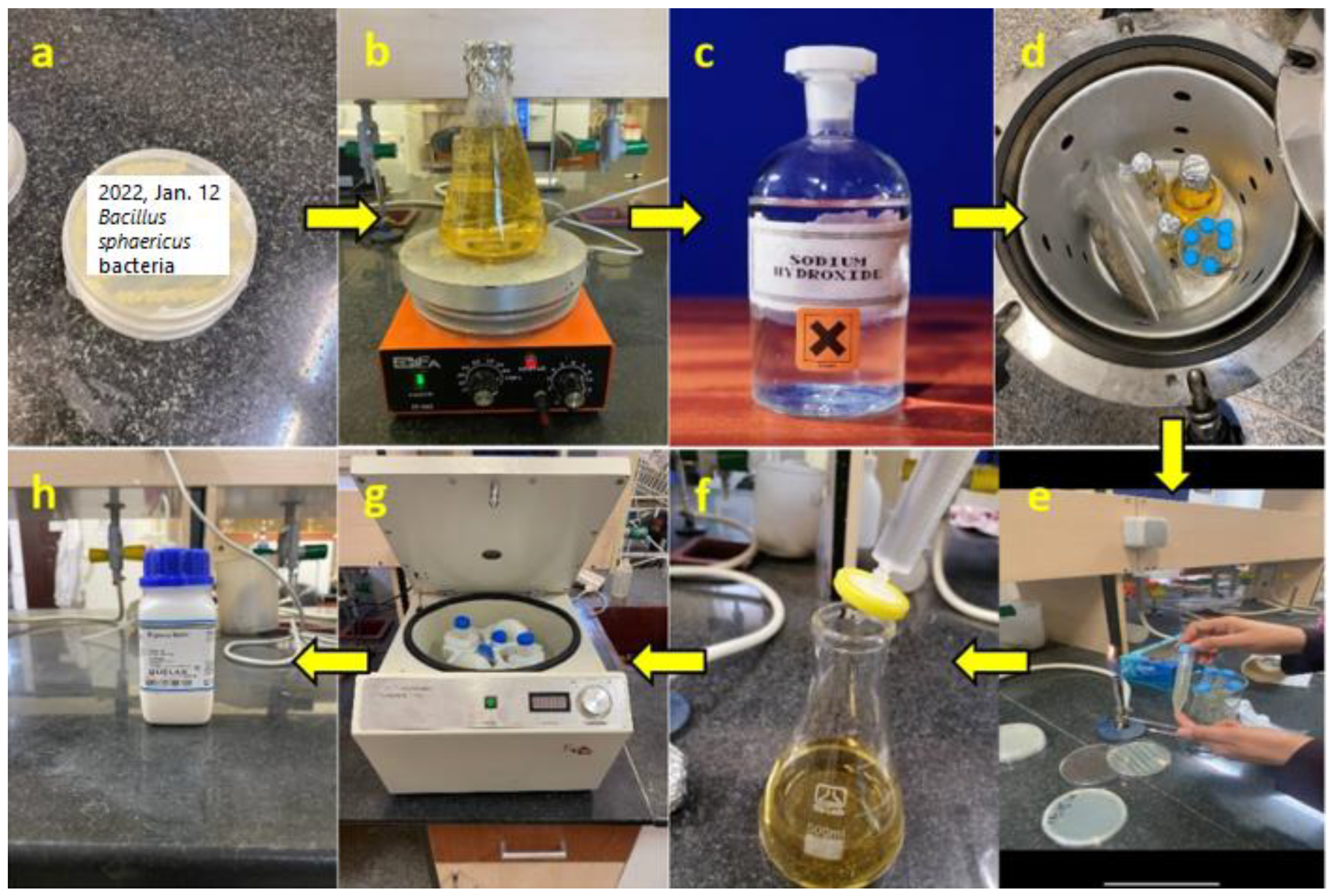
2.1. Bacterial Culture

The implemented self-healing technique in this study is inspired by refs. [13,22]. This technique involves the incorporation of Bacillus sphaericus bacteria into the concrete mix. In this regard, the Bacillus sphaericus PTCC 1490 was provided from the Persian Type Culture Collection of the Iranian Research Organization for Science and Technology (Tehran, Iran). The selected Bacillus sphaericus was activated in nutrient broth (Oxoid Ltd., Altrincham, UK), respectively, at 37 °C for 24 h.
For the successful cultivation and application of bacteria, a well-defined preparation process is required to ensure optimal growth conditions and maintain sterility. The bacterial preparation process begins with the growth of the bacterial strain on nutrient agar and nutrient broth. The culture medium is then prepared by dissolving 6.5 g of nutrient broth in 500 cc of distilled water and stirring it at 90 °C. To adjust the pH, a molar sodium hydroxide solution is added. All containers and the culture medium are then autoclaved at 120 °C to ensure sterility. The bacteria are inoculated into the culture medium under sterile conditions, followed by the addition of urea to the solution. After colony formation, the samples are centrifuged to separate the bacteria. Peptone water is then added to suspend the microorganisms, and finally, the solution is applied onto the flax fibers which are used to prepare the bacterial concrete specimens. These preparation stages are illustrated in Figure 1.

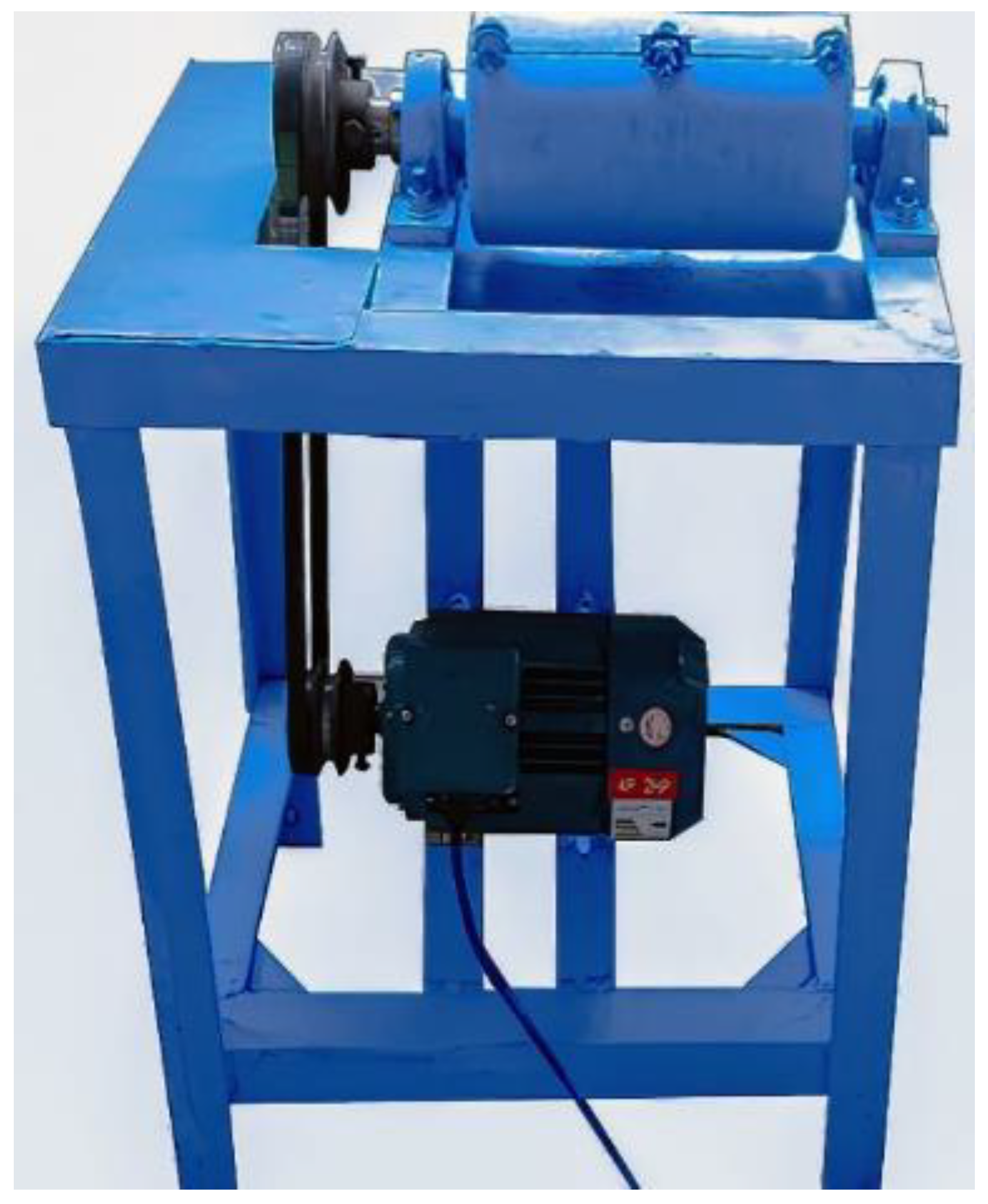
2.2. Creating Small-Scale Cylindrical Specimens of HPC Pile

To study the behavior of HPC pile, small-scale samples were created using a specialized centrifuge concrete machine (Figure 2). This machine was designed and built in the Structural and Geotechnical Engineering Laboratories at Fasa University, specifically for producing hollow-shaped centrifuge specimens. The specimens have dimensions of 30 cm in length and 15 cm in diameter, making them suitable for modeling small-scale HPC pile samples. To create these samples, the required materials were mixed according to the specified design and poured into a movable concrete mold. The mold was then placed inside the fixed mold of the centrifuge machine and securely fastened with screws. Table 1 provides details on the speed and time required to prepare the HPC pile specimens. It outlines the number of rotations necessary for the centrifuge to effectively mix the concrete. As shown in the table, the samples are rotated in four stages, with durations ranging from 3 to 5 min and rotation speeds varying between approximately 75 and 520 revolutions per minute. These parameters ensure the proper formation of the hollow HPC pile specimens. The rotation speed was controlled using a laser tachometer to maintain accuracy throughout the process.
After completing the rotation process, the sample was left stationary inside the machine for 8 h to allow for initial setting. Subsequently, the sample was carefully removed from the movable mold and placed in a steam chamber set at a temperature of 60 degrees Celsius. This step ensured proper curing of the concrete. Finally, the centrifuge sample was transferred to a water tank maintained at a temperature of 25 degrees Celsius for 28 days to complete the concrete setting process. It is worth noting that, according to the centrifuge concrete construction protocol, the temperature inside the steam chamber should ideally be 70 degrees Celsius. However, the temperature was reduced to 60 degrees Celsius to protect the bacteria present in the concrete.

2.3. Preparing Bacterial HPC Specimens

In harsh concrete environments, flax fibers have demonstrated superior performance compared to other natural fibers [13]. Thus, this study utilized flax fibers with specific characteristics: a required length of 25 ± 5 mm, a diameter of 0.4 mm, and a specific weight of 1.5 g per cubic centimeter. Initially, the necessary quantity of urea for this research is dissolved in 50 cc of water. It should be noted that urea is essential for cultivating bacteria to effectively carry out the self-healing process, as shown in relevant studies [13,23,24]. Subsequently, in a completely sterile environment, it is filtered through a syringe filter near the flame. Following this, the designated amount of calcium carbonate is carefully weighed and prepared. To ensure minimal bacterial contamination, the fibers undergo sterilization within a specialized plastic container using an autoclave next to the flame before the addition of bacteria. Two grams of fibers are then individually separated and placed in falcon tubes, which are subsequently positioned in a centrifuge. At the end of the falcon tubes, bacteria are collected while the supernatant is discarded. Peptone water is added to each falcon tube, and gentle shaking is employed. Subsequently, each falcon tube is uniformly poured over 4 g of fibers. After a waiting period of 24 h for complete fiber drying, the bacteria are fully absorbed by the fibers. At this point, the fibers and bacteria are ready for inclusion in the mix design.
For this study, Type II cement is utilized, and pure polycarboxylate is employed as a superplasticizer due to the low water-to-cement ratio. The physical and chemical specifications of the superplasticizer are described in Table 2.
Table 3 outlines the mixing plan for each HPC sample. The samples labeled F and FB consist of varying percentages of fibers, with FB samples additionally containing bacteria. The fiber percentages for each sample are 0.4%, 0.9%, and 1.3%, corresponding to fiber amounts of 26.2, 52.4, and 79.6 kg/m3. The FB samples possess the same fiber content as their corresponding F samples but with the addition of bacteria. The fiber-free sample serves as the control against which the other samples are compared. The table presents the quantities of cement, aggregate, sand, water, superplasticizer, fibers, urea (in the form of urea by weight percentage of cement), and calcium carbonate (in the form of calcium carbonate by weight percentage of cement) used in each sample, along with the water-to-cement ratio in liters per cubic meter (L/m3). In the bacterial samples (FB), each gram of fiber contains 107 colony-forming units (cfu) of bacteria. It should be noted that due to the low specific gravity of the fibers and their coating with cement slurry, segregation in the concrete mix was not a significant concern. In other words, despite the high-speed nature of the centrifugal casting process, the fibers did not migrate toward the outer edges of the specimen. As a result, the fiber distribution in the examined samples was uniform, and segregation could be disregarded.

3. Tests and Analyses

The following sections present the tests and analyses conducted to evaluate the effectiveness, durability, and strength of the bacterial concrete repair method. Several tests were performed, encompassing compressive strength testing (ASTM C39) [25], artificial crack generation, 30 min water absorption test (BS 1881), scanning electron microscopy (SEM) combined with energy dispersive X-ray spectroscopy (EDS), along with EDS mapping (MAP). The following sections will provide a detailed explanation of these methods.

3.1. Compressive Strength Test

To determine the compressive strength of the specimens, tests were performed on cube specimens measuring 15 × 15 × 15 cm and centrifugal specimens at both 7-day and 28-day intervals. The testing was conducted in accordance with the standard procedures outlined in ASTM C39. During the test, a force was applied to the upper surface of the sample, specifically targeting the center. The loading process was executed continuously, adhering to a predetermined rate of 153–383 KN/min.

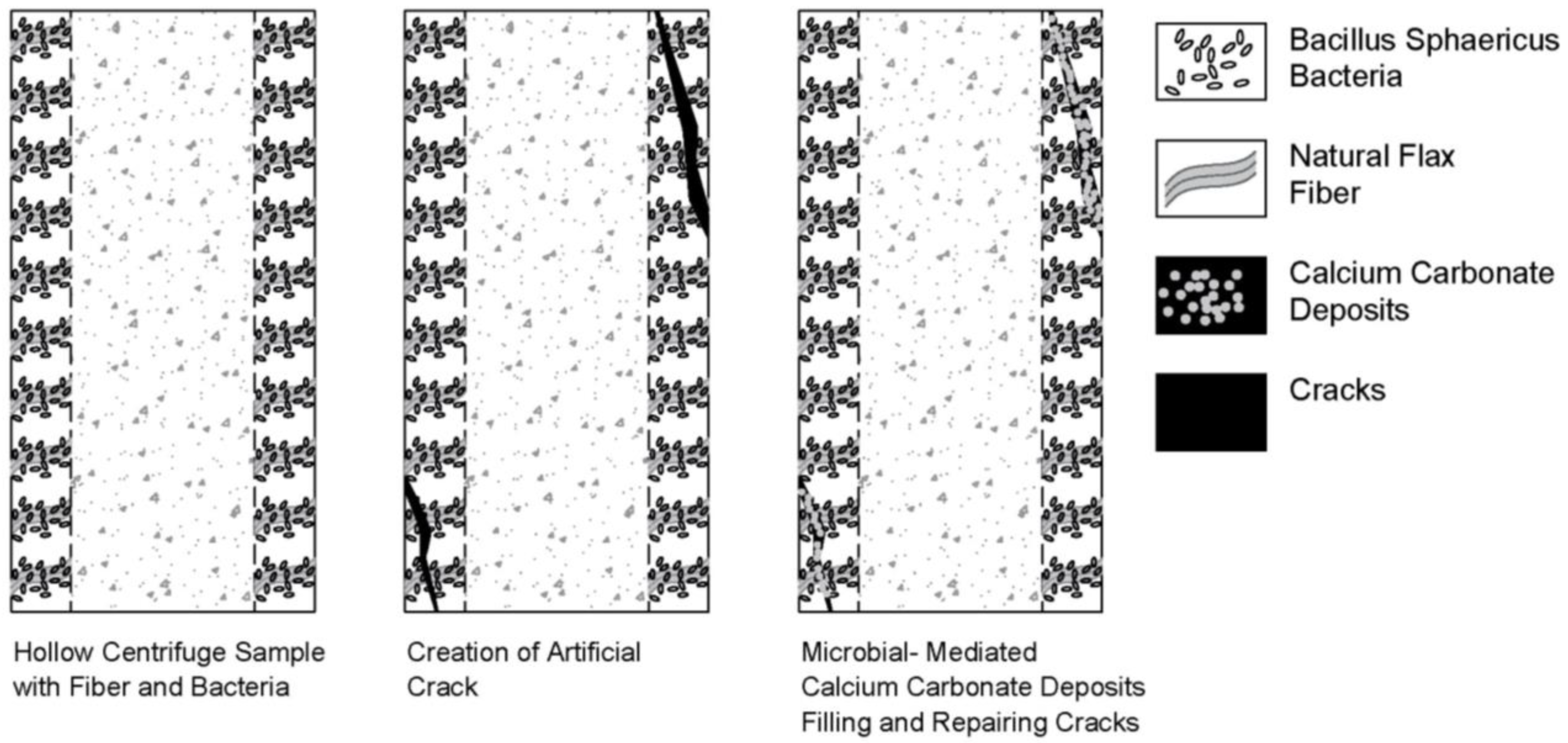
3.2. Artificial Crack Creation

To induce artificial cracks on centrifugal samples, the Lou method, which was developed in 2015, was utilized after conducting compressive strength tests [26]. The process involved placing the sample in a concrete breaker jack machine and carefully generating a crack while restraining the sample before it fractured. This method led to the formation of minute hairline cracks on the sample. To examine larger cracks for this study, the centrifugal samples were positioned horizontally in the concrete breaker jack machine, and the loading process was immediately stopped upon detecting a crack. Figure 3 illustrates the centrifugal samples after undergoing the artificial crack creation process.

3.3. 30-min Water Absorption Test

The 30 min water absorption test plays a crucial role in evaluating the durability of concrete structures. To ensure the required strength, the specimens undergo an initial curing period of 28 days in accordance with the BS 1881 standard. After this curing period, the specimens are subjected to a drying phase by exposing them to a temperature of at least 110 degrees for a minimum of 24 h. Then, the dry specimen is initially weighed and then submerged in water for a duration of 30 min. The percentage of water absorbed by concrete during this period is subsequently calculated. This test is commonly performed after repairing concrete cracks because the temperature of 110 degrees eliminates bacteria. It should be noted that other methods have been proposed in the literature for liquid absorption analysis. For instance, automated weight measurement techniques have been introduced to facilitate absorption testing [27], while computer vision-based techniques have been applied to enhance the accuracy of liquid absorption assessment [28]. These methods provide alternative means for evaluating self-healing efficiency and could be considered for future studies and investigations.

3.4. SEM/EDS Analysis of Bacteria-Induced Crack Repair

SEM is a powerful imaging technique that employs electrons to scan specimen surfaces, resulting in highly magnified images. When the electrons interact with the atoms in the specimen, unique signals are generated, carrying information about the sample’s surface topography and constituent materials. This technology enables observation of specimens under various conditions, including vacuum, weak vacuum, and even in environmental electron microscopes with humid environments. This study employed SEM with EDS to verify the effectiveness of bacteria-produced calcium carbonate in repairing cracks. To conduct the analysis, 1 × 1 cm samples were extracted from the crack site. For the SEM imaging and analysis, the specimens were affixed to a surface that could be positioned within the microscope chamber using an adhesive. The TESCAN SEM VEGA3 model, manufactured by TESCAN in Brno, Czech Republic, was utilized for this experiment.

3.5. Enhancing SEM Analysis of Small-Scale Samples: EDS and MAP Techniques

EDS and MAP play vital roles in the analysis of small-scale samples using SEM. EDS serves as an analytical method for identifying and quantifying the elemental composition of a sample. It offers broad applicability across various materials, including biological, metallurgical, mineral, and ceramic samples. By analyzing the X-ray energy emitted from the sample, EDS provides valuable insights into the chemical elements present. On the other hand, MAP is an imaging technique that visualizes the spatial distribution of elements within a sample. Since the elements observed in SEM images are not easily discernible, MAP becomes an indispensable tool for displaying the element distribution. By utilizing the same SEM device, MAP depicts the abundance distribution of each element through colored dots, effectively separating and highlighting them in the images.

3.6. Research Schematic

In Figure 4, a clear and concise schematic is presented, outlining the conducted research. The image portrays a centrifuged sample, exhibiting visible cotton fibers and bacteria. It visually demonstrates the sequence of events: initially, a crack develops in the sample, subsequently followed by the infiltration of air and water. Consequently, the bacteria precipitate calcium carbonate, effectively sealing the crack. This schematic offers a comprehensive visual representation of the research process.
Upon the initiation of a crack in the sample, the bacteria commence the precipitation of calcium carbonate in the presence of air and water. This process gives rise to the formation of diverse phases of calcium carbonate, such as calcite, aragonite, and vaterite. Among these phases, vaterite and calcite are the predominant forms of calcium carbonate deposits.
In Figure 5 (adapted from ref. [29]), various forms of calcium carbonate resulting from bacterial activity are depicted. The different forms of calcium carbonate precipitates can be distinguished by their morphology. Calcite is the most common form of calcium carbonate and has a rhombohedral crystal structure. Aragonite has a needle-like crystal structure, while vaterite has an amorphous crystal structure. These forms exhibit clear visual distinctions and can be readily identified. The existence of these diverse forms implies that bacteria play a crucial role in the process of calcium carbonate precipitation and deposition.

4. Results and Discussion

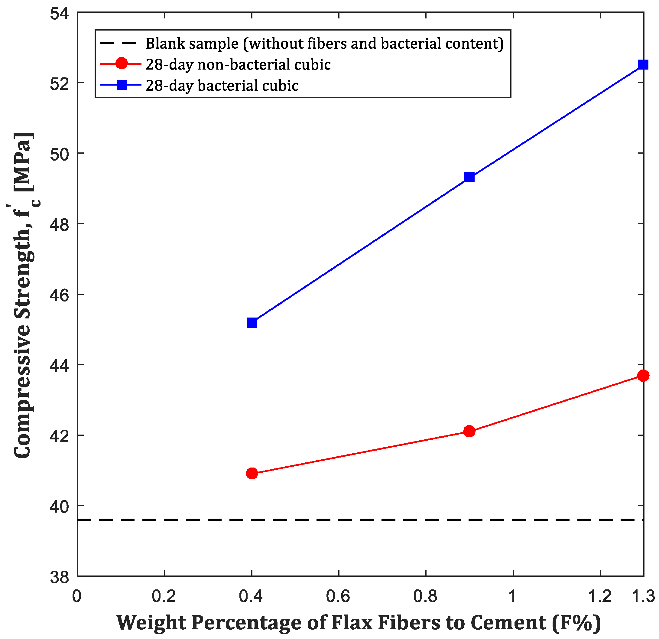
4.1. Compressive Strength Test Results

The compressive strength test was conducted on both cubic and centrifuged cylindrical samples. Active bacteria were incorporated into the concrete mixture, and the compressive strength of the specimens was evaluated accordingly. For cubic samples, testing was performed at 28 days. However, due to the reduced effectiveness of active bacteria in calcium carbonate production over time, the compressive strength of centrifuged samples was assessed only at 7 days, with no evaluation at 28 days. To compare the effects of bacterial and non-bacterial samples, flax fibers were incorporated into all cubic samples except for the control specimen.
Figure 6 presents the compressive strength of 28-day cubic samples with and without bacteria, compared to the blank sample, which has a compressive strength of 39.6 MPa (without fibers or bacterial content). As shown in the figure, both bacterial and non-bacterial samples exhibit higher compressive strength than the blank sample, indicating the positive effect of fibers in enhancing strength. Furthermore, non-bacterial samples generally show lower compressive strength than their bacterial counterparts, emphasizing the role of Bacillus sphaericus in strength improvement through calcium carbonate precipitation. This microorganism facilitates calcium carbonate deposition within the concrete matrix, effectively filling pores and increasing both density and overall strength. Additionally, the compressive strength curve of 28-day non-bacterial cubic samples exhibits a noticeably steeper slope compared to that of bacterial centrifuged samples. This result confirms that the bacteria remained active for 28 days following artificial cracking and contributed to the enhancement of the compressive strength of the samples.
Figure 7 illustrates the compressive strength of bacterial centrifuged cylindrical samples at 7 days, compared to the blank centrifuged sample with a compressive strength of 41.5 MPa. As can be seen, like cubic samples, increasing the flax fiber-to-cement ratio enhances compressive strength. Notably, a higher fiber content correlates with a greater bacterial concentration in the specimen, accelerating the self-healing process of the concrete. This further highlights the role of Bacillus sphaericus in strength enhancement through calcium carbonate production.
It should be noted that to assess bacterial behavior over longer periods, further investigations are needed. If this technology proves effective, future research could explore bacterial species with enhanced survival capabilities under harsh environmental conditions. Some bacteria, for instance Bacillus subtilis [30], can form spores or cysts, enabling them to withstand unfavorable conditions until the environment becomes suitable for reproduction. However, this aspect falls beyond the scope of the present study and requires further research to fully explore its potential.
The findings of this study are consistent with previous research studies (such as refs. [7,13,14]), which demonstrated improvements in concrete compressive strength through the use of various bacterial strains and mix designs. Variations in the results can be attributed to differences in bacterial types, mix formulations, and the use of fibers as bacterial carriers, highlighting the potential of bacteria and fiber combinations in enhancing concrete strength.

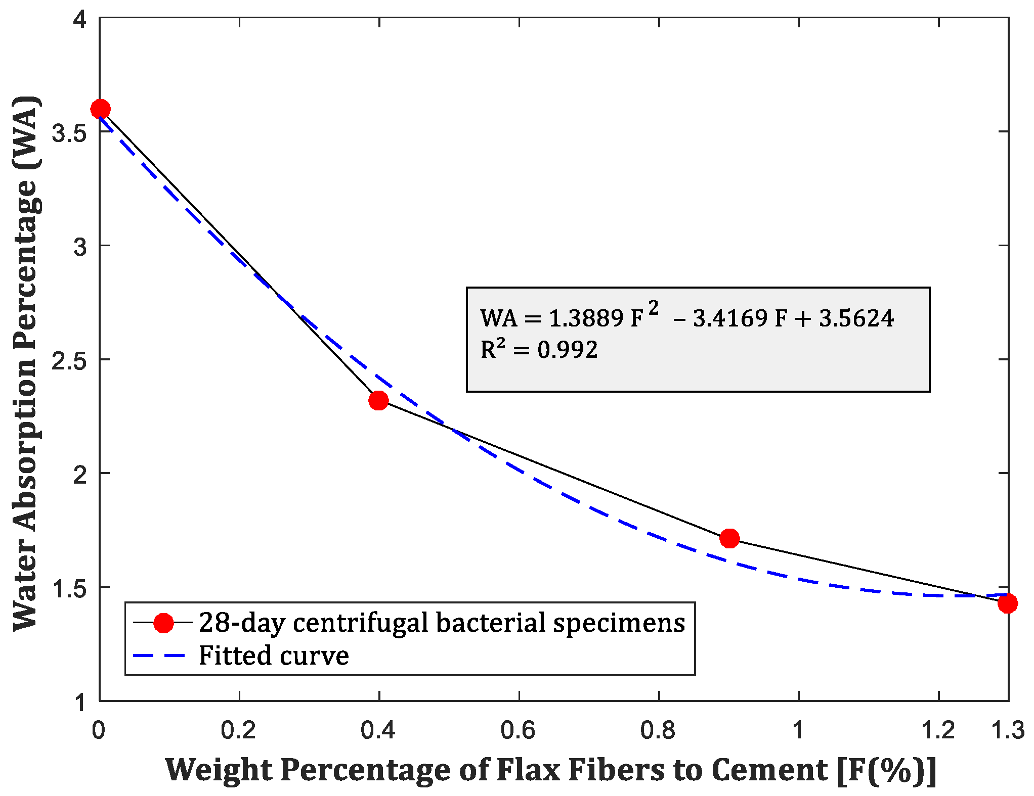
4.2. Results of 30-min Water Absorption Test

Improving water resistance in concrete structures is a critical consideration to enhance their overall longevity and durability. In this section, the findings from a 30 min water absorption test performed on 28-day-old bacterial centrifuged specimens that underwent self-repair prior to testing are presented. This analysis investigates the impact of Bacillus sphaericus and flax fibers on water absorption in concrete samples. For every gram of flax fiber integrated into the concrete, an equivalent of 107 cfu of Bacillus cereus bacteria was added.
Significantly, the combination of fibers and bacteria in the centrifuge samples led to a substantial reduction in the rate of water absorption. This study highlights the substantial influence of Bacillus sphaericus on water absorption percentages and corrosion within concrete. Figure 8 presents a graph depicting the relationship between water absorption percentage and fiber content. The control sample, without any flax fibers or bacteria, serves as the reference point. The graph consistently demonstrates a decrease in water absorption as the fiber content increases, clearly illustrating that the inclusion of flax fibers along with bacteria effectively diminishes the concrete’s capacity to absorb water. This reduction in water absorption is most pronounced at the highest fiber content (1.3%), highlighting the effectiveness of this treatment in improving the concrete’s resistance to water infiltration. Furthermore, according to Figure 8, the reduction in water absorption due to flax fibers tends to slow down by increasing the weight percentage of flax fibers to cement. In other words, the initial rate of reduction may be significant, but it gradually diminishes as more flax fibers are added.

4.3. Results of SEM, EDS, and MAP Analysis of Centrifuge Bacterial Samples

To assess the capacity of bacteria to generate calcium carbonate for sealing and mending cracks, the centrifuged bacterial samples were initially fractured using a jackhammer and then allowed to undergo a 28-day self-healing process in a water basin. Subsequently, the extent of healing was determined through SEM, EDS, and MAP analyses, and the results are presented in the following sections. It should be noted due to the operational constraints of advanced analyses (i.e., SEM, EDS, and MAP), small pieces (approximately 1 × 1 cm) were extracted from the specimens examined using mechanical tools (disk grinder). These pieces were carefully selected to include the crack site, ensuring the representation of healed areas filled with calcium carbonate from bacterial activity. In addition, the hollow cylindrical structure of the samples facilitated the extraction process.

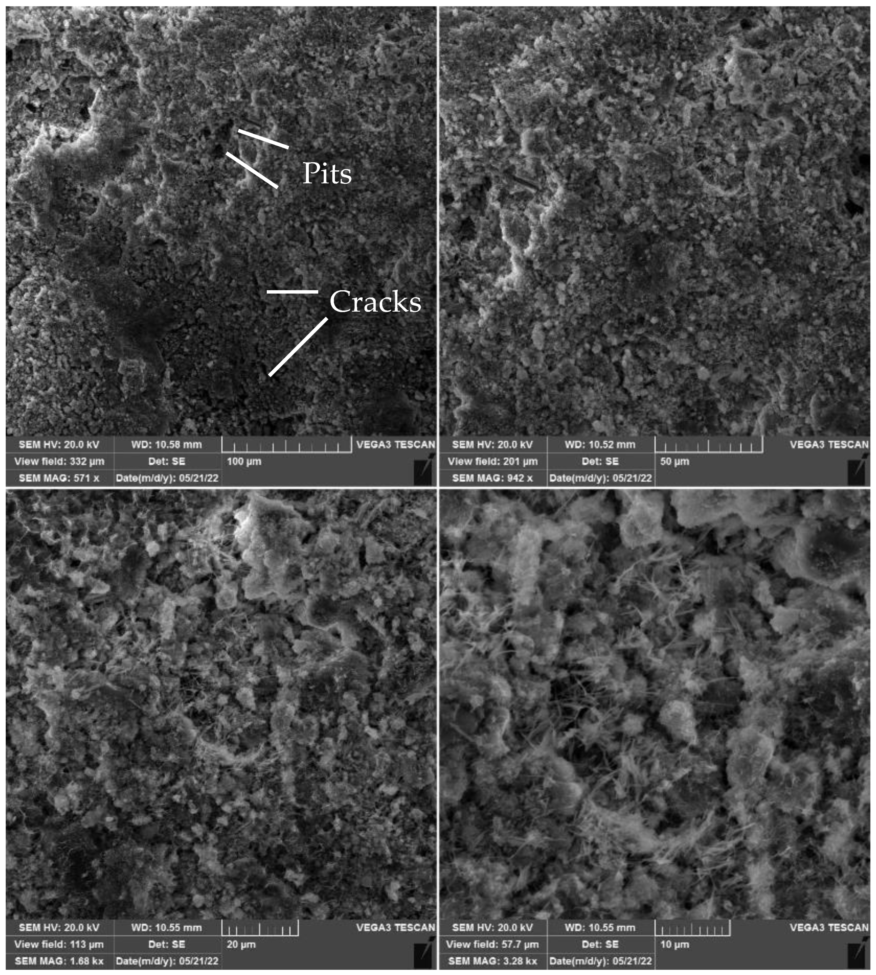
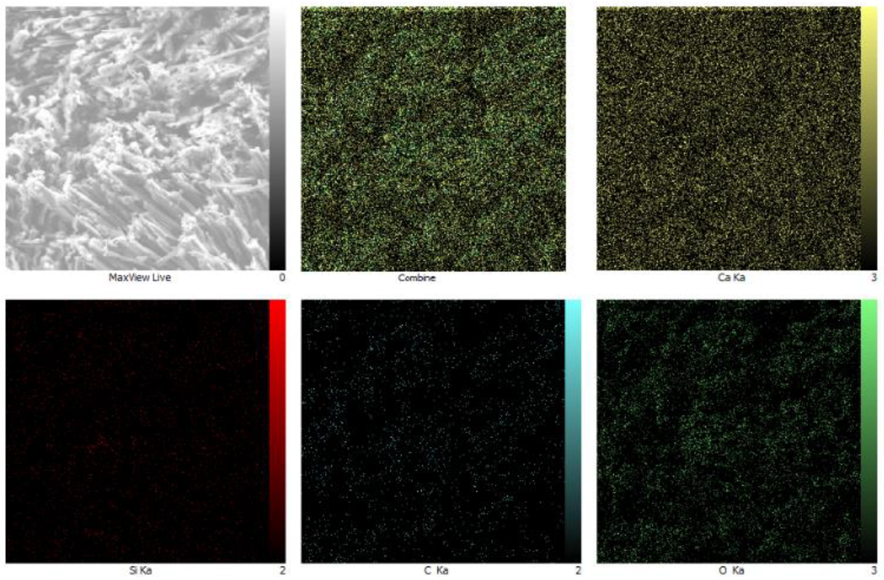
4.3.1. SEM Analysis Results

The SEM images reveal that the addition of bacteria and fibers to the concrete has led to a noticeable increase in the amount of calcium carbonate produced and the variety of forms it takes, such as calcite, aragonite, and vaterite. Notably, even the control sample, without added bacteria, displayed self-curing properties, as evident from the presence of calcium carbonate precipitates on its surface.
At 28 days, the SEM image of the control sample (Figure 9) exhibited visible microcracks and surface pitting due to the jackhammering process. Despite these imperfections, the control sample’s surface appeared smooth, attributed to the concrete being centrifuged during preparation. The presence of various calcium carbonate precipitates in this sample indicates ongoing chemical reactions within the concrete.
Figure 10 displays an SEM image of a centrifuge sample containing 0.4% weight percentage of flax fibers to cement. The image indicates fewer calcium carbonate precipitates compared to other bacterial samples. The cement fibers hinder bacterial growth and calcium carbonate formation, yet some calcite formation still occurs, driven by calcium ions reacting with atmospheric carbon dioxide. Despite the inclusion of fibers, visible cracks persist on the concrete surface, indicating that the low fiber percentage is insufficient to entirely prevent cracking.
Moreover, the even distribution of cement fibers throughout the sample plays a crucial role in their effectiveness in crack prevention. An uneven distribution would diminish their ability to impede crack formation.
Figure 11 presents an SEM image of a centrifuge sample containing 0.9% weight percentage of flax fibers to cement. The image reveals a thick layer of aragonite crystals covering the concrete surface, forming a dense, fibrous network with a hexagonal shape. The high percentage of aragonite indicates the substantial production of this mineral, facilitated by the presence of a high concentration of calcium ions in the concrete, providing the necessary raw materials for aragonite synthesis. This dense layer of aragonite offers several benefits, including enhanced protection against weathering, corrosion, and potential improvements in strength and durability. It should be noted that incorporating bacteria and fibers into the mix design of centrifuged concrete piles can enhance their durability under corrosion and cracks in centrifuged piles (which, generally speaking, may not be easily visible). Therefore, when cracks and corrosion develop on the outer surface, bacteria fill these cracks, preventing further deterioration and deepening. The gradual filling of cracks confirms that the bacteria are actively performing their self-healing function. Therefore, it can be concluded that this method can be effective in extending the lifespan of centrifuged piles.
The SEM images in Figure 12 demonstrate the production of calcium carbonate in the form of vaterite in a centrifuge sample containing 1.3% weight percentage of flax fibers to cement. These images reveal that the vaterite crystals are small and needle-like. The higher percentage of vaterite is attributed to the increased number of bacteria present, producing urea that reacts with calcium ions in the cement to form vaterite. The images also display four different crystal types, indicating the bacteria’s ability to produce a variety of calcium carbonate minerals, influenced by different environmental conditions within the sample.
The insights provided by the SEM analysis offer promising potential for enhancing cement-based materials’ performance through better control of bacterial growth and activity, facilitating the targeted formation of specific calcium carbonate minerals. Furthermore, the presence of both vaterite and aragonite in the examined bacterial samples provides valuable insights into the role of bacteria and flax fibers in calcium carbonate production. In other words, the findings from the SEM analysis offer a clearer understanding of how bacteria and flax fibers influence calcium carbonate formation.
In conclusion, the SEM analysis has provided a deeper understanding of the influence of bacteria and fibers on calcium carbonate production and concrete characteristics. The presence of various forms of calcium carbonate, such as calcite, aragonite, and vaterite, highlights the complexity of the chemical reactions occurring within the concrete matrix. It should be noted that while calcium carbonate is not a primary product of cement hydration, some secondary reactions, such as carbonation, can lead to its formation over time. The key distinction is that the calcium carbonate filling cracks in bacterial concrete primarily originates from microbial activity rather than standard cement hydration. In other words, the incorporation of bacteria and fibers not only enhances calcium carbonate production but also contributes to crack repair and prevention. SEM images revealed that, 28 days after crack formation, bacteria survived and proliferated, producing a diverse range of calcium carbonate forms that partially repaired the cracks. These findings are consistent with the results from Dhami et al. [29], further validating the potential of bacteria and fibers in improving the design and performance of concrete materials. Furthermore, it was observed that most of the fine cracks were filled with calcium carbonate in the examined specimens. However, the extent of healing varied for deeper and larger cracks. As an example, in the sample containing 0.9% by weight of flax fibers relative to the cement, a 32.1 cm crack was initially formed, with 17.1 cm (approximately 53%) healed. In the sample with 1.3% by weight of flax fibers to cement, a 34.5 cm crack was created, and 23.8 cm (approximately 69%) of it was healed. Therefore, increasing the weight percentage of flax fibers relative to cement leads to a higher bacterial presence, which in turn enhances the healing process.

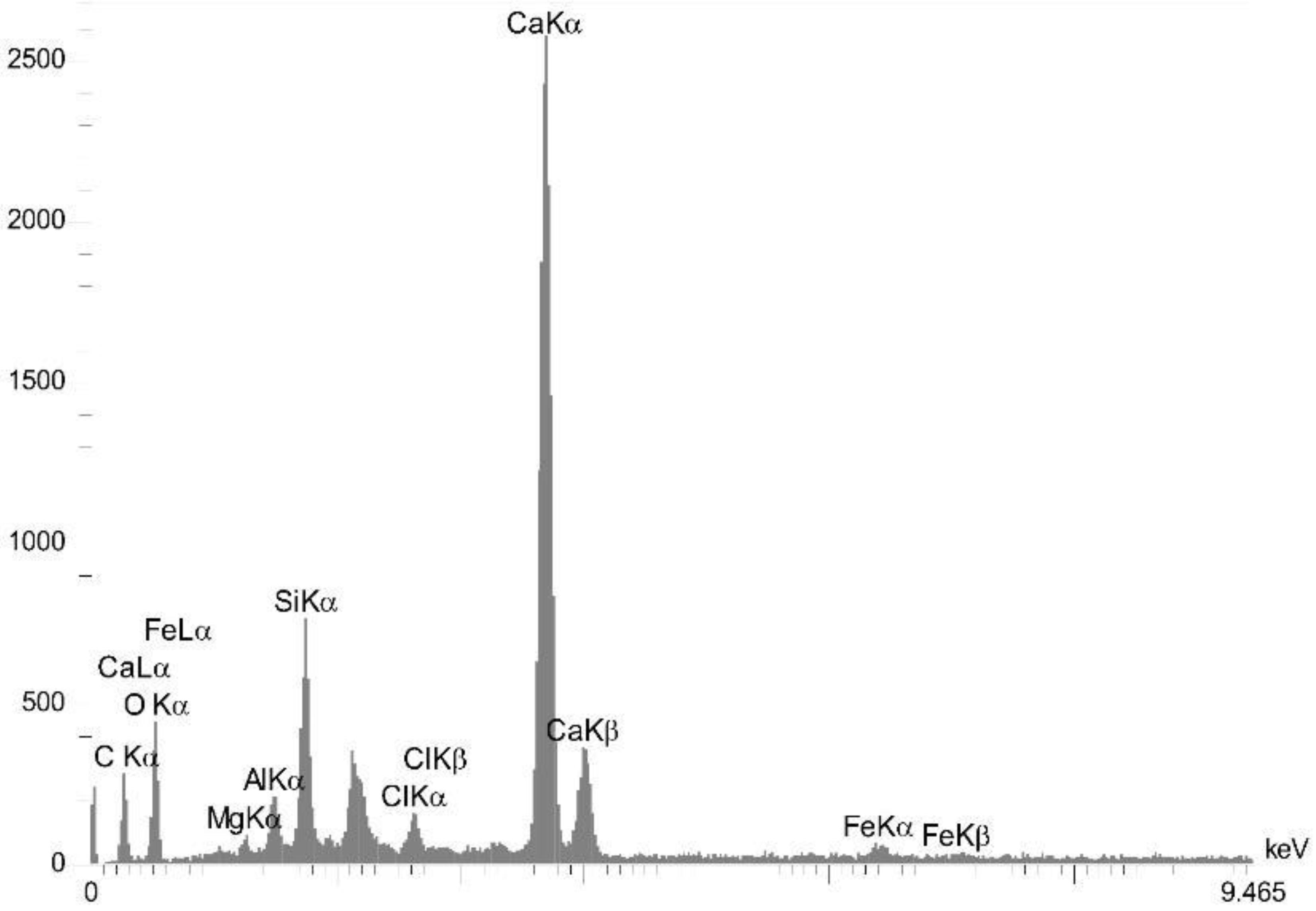
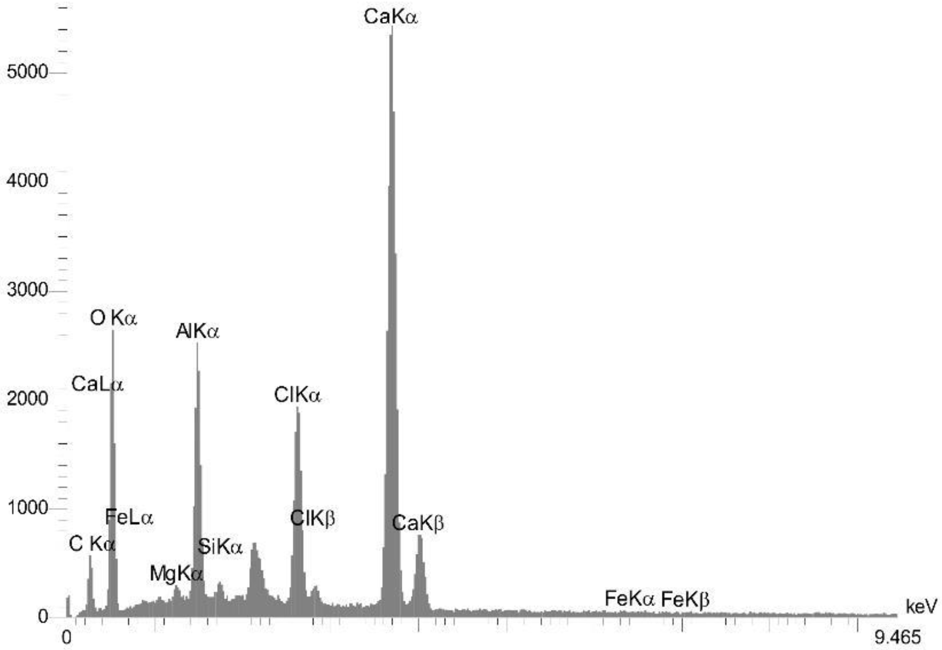
4.3.2. EDS Analysis Results

The EDS analysis was performed on centrifuged samples containing 12 and 36 g of fibers and bacteria at 28 days of age to determine the weight percentage of elements present. The results outlined in Table 4 are visually presented in Figure 13 and Figure 14 for the FB0.4% and FB1.3% samples at the age of 28 days. Figure 13 shows EDS analysis outcomes for a centrifuged sample with 0.4% flax fibers in cement, and Figure 14 illustrates the EDS analysis for a sample containing 1.3% flax fibers in cement, both at the age of 28 days. In these figures, the X-ray peaks represent the emission of X-rays from the elements within the sample, and the intensity of the peaks indicates the relative abundance of each element.
To interpret the graphs, the x-axis represents the energy of the X-rays in kiloelectronvolts (keV), and the y-axis represents the intensity of the emitted X-rays, corresponding to the quantity of X-rays emitted from the sample. The peaks in the graph correspond to the characteristic X-rays emitted by different elements, with each element having a unique energy signature. Furthermore, peak height alone does not directly translate to weight percentage because different elements have varying X-ray emission efficiencies, matrix effects such as absorption and fluorescence influence the measurements, and detector sensitivity differs for each element. Therefore, the peaks in the graph allow us to identify the elements present in the sample.
For instance, in Figure 14, the peak at 3.7 keV corresponds to the characteristic X-Ray emitted by calcium (Ca), while the peak at 0.5 keV represents the characteristic X-Ray emitted by oxygen (O), and so on for other elements.
The results of the EDS analysis are presented in Table 4, showcasing the weighted frequency analysis of various elements in the samples.
From the EDS analysis, it was observed that the samples with FB0.4% and FB1.3% contained 85.37% and 97.04% weight percentage of calcium carbonate (CaCO3), respectively. Notably, the content of calcium carbonate increased as the percentage of bacteria and fibers increased. It should be noted that the table presents processed, quantitative data in terms of weight percentage, derived from the raw spectra after applying calibration, normalization, and correction factors. Therefore, a tall peak in Figure 13 and Figure 14 does not necessarily indicate a high weight percentage in the table. For example, elements such as oxygen (O) and carbon (C) may produce lower-intensity peaks in the graph due to their lower atomic numbers and weaker X-ray emissions, even if their weight percentages are relatively high. In contrast, heavier elements like calcium (Ca) or iron (Fe) may exhibit sharper or taller peaks due to stronger X-ray emissions, even when their concentrations are lower than those of lighter elements.

4.3.3. MAP Analysis Results

The MAP analysis results, shown in the images of this section, provide a comprehensive understanding of the dispersion of calcium carbonate (CaCO3) within the bacterial samples. The analysis was conducted on each bacterial sample separately, and the elements Ca, Si, C, and O were represented as colored dots. The colors of the dots correspond to the relative abundance of each element in the sample. For example, yellow dots represent Ca, red dots represent Si, blue dots represent C, and green dots represent O.
The image shows that the CaCO3 is distributed unevenly within the bacterial samples. In some samples, the CaCO3 is concentrated in small clusters, while in other samples, it is more evenly distributed. The distribution of the CaCO3 may be affected by several factors, including the fiber content and bacteria percentages, the growth conditions, and the age of the sample.
Figure 15, Figure 16 and Figure 17 depict the constituents of calcium carbonate as observed through MAP analysis, with a specific focus on the dispersion of calcium carbonate within the crack. The results indicate that the bacteria are evenly distributed within the concrete, and the production of calcium carbonate dispersion is not concentrated in one point. This dispersion is observed within the crack, as revealed by the MAP analysis after the formation of a crack. Overall, the MAP analysis provides a detailed insight into the dispersion of calcium carbonate within the bacterial samples, crucial for understanding the impact of bacteria on concrete. The uniform distribution of bacteria and the observable production of calcium carbonate dispersion within the crack provide valuable information for the design of future concrete structures capable of self-healing.

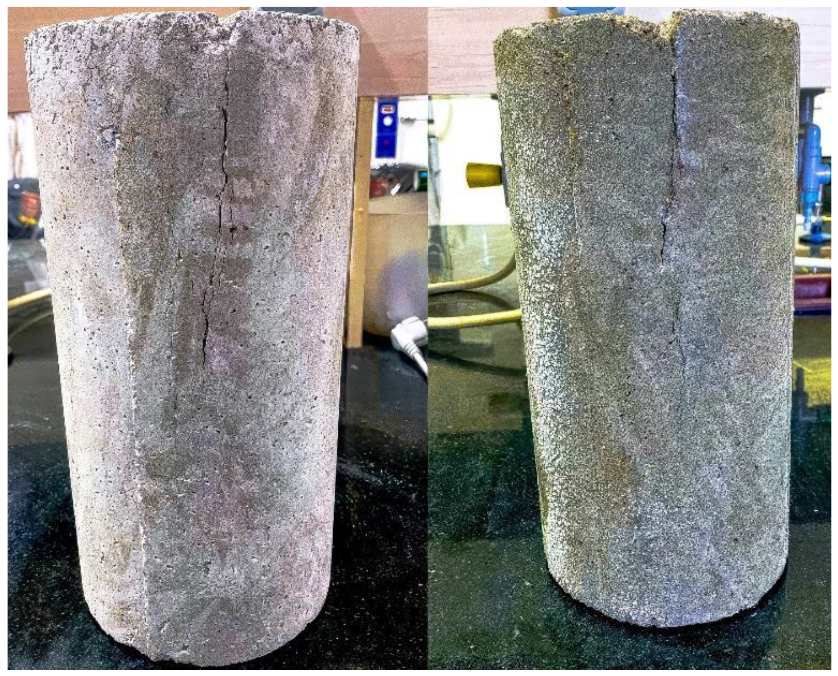
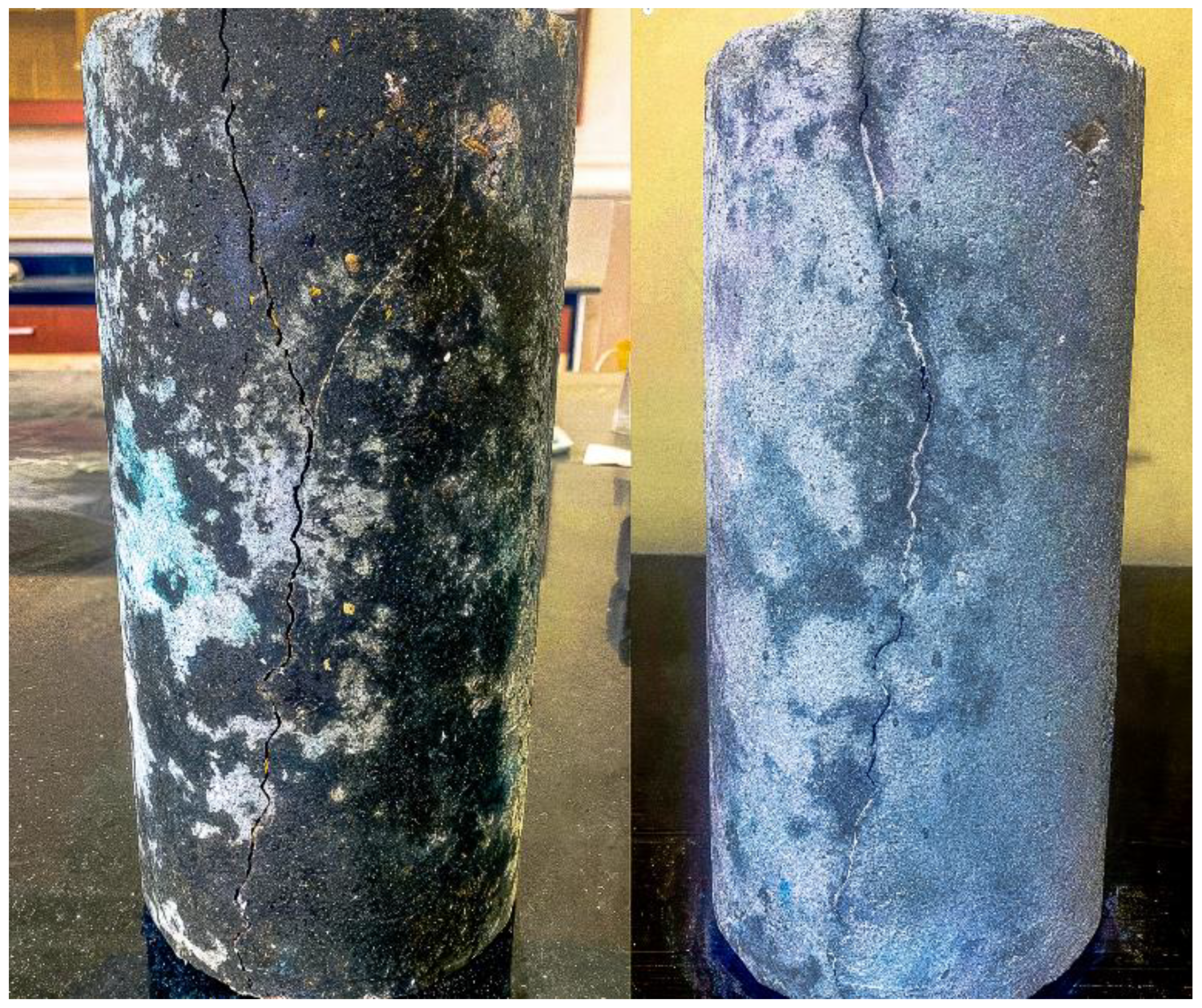
4.3.4. Examples of Bacterial Centrifuge Samples Before and After Self-Healing

In this section, visual illustrations of bacterial centrifuge samples are presented before and after the self-healing process. Figure 18, Figure 19 and Figure 20 present the samples with bacteria, capturing their condition immediately after the crack (left side) and at 28 days, depicting the ongoing self-healing process (right side).
Upon closer examination of Figure 18, subtle hairline fractures are observable, but no substantial repair of significant cracks is evident. In contrast, Figure 19 and Figure 20 reveal the formation of calcium carbonate crystals in the forms of aragonite and vaterite on samples with fiber contents of 0.9 and 1.3 percent, leading to more comprehensive repairs in some fractured areas. In regions where cracks remained unfilled, it can be inferred that either no fibers were present in those sections or bacterial activity was diminished. Additionally, not every crack can be fully sealed, as some may have widths exceeding the self-healing capacity of the material. Another possible explanation is that the study duration was insufficient for complete self-regeneration, suggesting that a longer observation period may be necessary for a more comprehensive analysis.
In summary, the visual examples underscore the efficacy of utilizing fibers and bacteria to mend fractures in sample materials. While complete repair may not be achieved in all cases, the incorporation of calcium carbonate crystals alongside fibers and bacteria demonstrates significant enhancements in the overall repair process. It should be noted that in this study, small-scale specimens were investigated to examine material behavior, bacterial activation, and self-healing properties in a controlled and cost-effective environment. Element testing was chosen as it allows for reproducible conditions before scaling up to real-world applications or full-scale physical testing. While the specimens were smaller than full-scale piles, the testing parameters—such as rotational speed, fiber content, and bacterial activity—were designed to reflect those of larger systems. However, further investigations on larger-scale specimens are needed to fully assess the applicability of these findings to real-world foundation systems.

5. Conclusions

This study explored the use of Bacillus sphaericus bacteria and flax fibers in precast high-performance centrifugal concrete (HPC) piles to address corrosion issues and enhance the durability and strength of concrete. A series of compressive strength tests, 30 min water absorption tests (BS 1881), scanning electron microscopy (SEM) combined with energy dispersive X-ray spectroscopy (EDS), and EDS mapping (MAP) were conducted to evaluate self-healing efficiency. The main results obtained in this study are as follows:
  • SEM images confirmed that calcium carbonate sediment is produced in calcite, aragonite, and vaterite forms within centrifugal samples.
  • The addition of Bacillus sphaericus bacteria and fibers resulted in a 14.21% increase in compressive strength in centrifugal samples and up to a 20% increase in cubic samples compared to those without bacteria.
  • The inclusion of fibers and bacteria led to reduced water absorption, facilitating increased calcium carbonate sediment production, further strengthening the concrete.
  • Visual observations showed that most of the cracks in centrifugal pile foundations were successfully restored, improving the overall integrity of the concrete. Therefore, the self-healing concrete method can be considered as an effective solution for deep foundations, as any cracks and corrosion that may form are not visually detectable.
  • The combination of reduced water absorption, increased compressive strength and crack removal has shown that the combination of bacteria and fibers can mitigate corrosion of piles.
  • MAP analyses revealed that calcium carbonate deposits were distributed throughout the cracks, ensuring effective healing across the concrete structure.
  • In summary, the biological self-healing method using Bacillus sphaericus bacteria and flax fibers can improve the durability and life of HPC piles. This study provides important insights into the potential applications of this method for mitigating corrosion and enhancing concrete performance in various construction projects.

Author Contributions

Conceptualization, A.A., H.D.K., M.M.M., M.M., S.G. and A.P.; methodology, A.A., H.D.K., M.M.M., M.M., S.G. and A.P.; validation, A.A., H.D.K., M.M.M., M.M., S.G. and A.P.; formal analysis, A.A., H.D.K., M.M.M., M.M., P.M., S.G. and A.P.; investigation, A.A., H.D.K., M.M.M., M.M., P.M., S.G. and A.P.; resources, A.A., H.D.K. and M.M.M.; data curation, A.A., H.D.K. and M.M.M.; writing—original draft preparation, A.A., H.D.K., M.M.M., M.M. and P.M.; writing—review and editing, A.A., H.D.K., M.M.M., M.M., P.M., S.G. and A.P.; visualization, A.A., H.D.K., M.M.M., M.M., P.M., S.G. and A.P.; supervision, H.D.K., M.M.M., M.M., S.G. and A.P. All authors have read and agreed to the published version of the manuscript.

Funding

This research received no external funding.

Data Availability Statement

All data and models that support the findings of this study are available from the corresponding author upon reasonable request.

Conflicts of Interest

The authors declare no conflicts of interest.

References

  1. Qaidi, S.; Al-Kamaki, Y.S.; Al-Mahaidi, R.; Mohammed, A.S.; Ahmed, H.U.; Zaid, O.; Althoey, F.; Ahmad, J.; Isleem, H.F.; Bennetts, I. Investigation of the effectiveness of CFRP strengthening of concrete made with recycled waste PET fine plastic aggregate. PLoS ONE 2022, 17, e0269664. [Google Scholar]
  2. Qaidi, S.M.; Mohammed, A.S.; Ahmed, H.U.; Faraj, R.H.; Emad, W.; Tayeh, B.A.; Althoey, F.; Zaid, O.; Sor, N.H. Rubberized geopolymer composites: A comprehensive review. Ceram. Int. 2022, 48, 24234–24259. [Google Scholar] [CrossRef]
  3. AliPour, I.S.P. Factors Affecting the Corrosion of Concrete and Steel Reinforcement in Reinforced Concrete Structures. 2021. In Proceedings of the 4th National Conference on New Technologies in Architectural, Civil and Urban Engineering of Iran, Teheran, Iran, 26 August 2021. [Google Scholar]
  4. Mirsayapov, I.; Yakupov, S.; Hassoun, M. About concrete and reinforced concrete corrosion. In Proceedings of the International Scientific Conference on Socio-Technical Construction and Civil Engineering (STCCE—2020), Kazan, Russian, 29 April–15 May 2020; p. 012061. [Google Scholar]
  5. Zhang, W.; Zhao, C.; Zhang, C.; Guo, C. Concrete Corrosion Mechanism and Durability Design of Municipal Sewerage. In Proceedings of the IOP Conference Series: Earth and Environmental Science, Zhuhai, China, 15–17 January 2021; p. 012021. [Google Scholar]
  6. Li, V.C.; Herbert, E. Robust self-healing concrete for sustainable infrastructure. J. Adv. Concr. Technol. 2012, 10, 207–218. [Google Scholar] [CrossRef]
  7. Reddy, S.V.; Satya, A.K.; Rao, S.M.; Azmatunnisa, M. A biological approach to enhance strength and durability in concrete structures. Int. J. Adv. Eng. Technol. 2012, 4, 392–399. [Google Scholar]
  8. Althoey, F.; Zaid, O.; Arbili, M.M.; Martínez-García, R.; Alhamami, A.; Shah, H.A.; Yosri, A.M. Physical, strength, durability and microstructural analysis of self-healing concrete: A systematic review. Case Stud. Constr. Mater. 2023, 18, e01730. [Google Scholar]
  9. Qian, C.; Wang, R.; Cheng, L.; Wang, J. Theory of microbial carbonate precipitation and its application in restoration of cement-based materials defects. Chin. J. Chem. 2010, 28, 847–857. [Google Scholar]
  10. Anbu, P.; Kang, C.-H.; Shin, Y.-J.; So, J.-S. Formations of calcium carbonate minerals by bacteria and its multiple applications. Springerplus 2016, 5, 250. [Google Scholar]
  11. Wiktor, V.; Jonkers, H.M. Quantification of crack-healing in novel bacteria-based self-healing concrete. Cem. Concr. Compos. 2011, 33, 763–770. [Google Scholar] [CrossRef]
  12. Wang, J.; Soens, H.; Verstraete, W.; De Belie, N. Self-healing concrete by use of microencapsulated bacterial spores. Cem. Concr. Res. 2014, 56, 139–152. [Google Scholar] [CrossRef]
  13. Rauf, M.; Khaliq, W.; Khushnood, R.A.; Ahmed, I. Comparative performance of different bacteria immobilized in natural fibers for self-healing in concrete. Constr. Build. Mater. 2020, 258, 119578. [Google Scholar]
  14. Dabbagh, H.; Ashengroph, M.; Zare, P.; Salehi, P. Investigating the Effect of Two Types of Bacteria on the Compressive Strength and Water Absorption of Lightweight Fiber-Reinforced Concrete. In Proceedings of the 11th National Congress of Civil Engineering, Shiraz, Iran, 30 April 2019. [Google Scholar]
  15. Xiao, Y.; Stuedlein, A.W.; Pan, Z.; Liu, H.; Matthew Evans, T.; He, X.; Lin, H.; Chu, J.; Van Paassen, L.A. Toe-bearing capacity of precast concrete piles through biogrouting improvement. J. Geotech. Geoenviron. Eng. 2020, 146, 06020026. [Google Scholar] [CrossRef]
  16. Yamasamit, N.; Sangkeaw, P.; Jitchaijaroen, W.; Thongchom, C.; Keawsawasvong, S.; Kamchoom, V. Effect of Bacillus subtilis on mechanical and self-healing properties in mortar with different crack widths and curing conditions. Sci. Rep. 2023, 13, 7844. [Google Scholar] [CrossRef]
  17. Wani, S.; Gęca, M.J.; Selvaraj, T.; Priya, T.S. Assessing the influence of Bacillus megaterium and Bacillus sphaericus in cementitious materials: Promoting sustainability towards strength, durability and crack repair. Ain Shams Eng. J. 2024, 15, 102748. [Google Scholar] [CrossRef]
  18. Adil, M.R.; Khatun, S.; Jamal, M.; Mondal, A. Investigation on Self-Healing Concrete Using Bacillus subtilis Bacteria. MATEC Web Conf. 2024, 400, 01002. [Google Scholar] [CrossRef]
  19. Vishal, A.; Chepuri, A.; Chandana, N. Assessment of bacteria-based self-healing concrete through experimental investigations—A sustainable approach. J. Mater. Sci. Mater. Eng. 2025, 20, 15. [Google Scholar] [CrossRef]
  20. Elshazly, M.A.; Elakhras, A.A.; Elshami, A.A.; Ahmad, S.S.; Elmahdy, M.A. Investigating the effectiveness of a bacterial self-healing mechanism for repairing cracks in sustainable cement mortar at low temperatures. Results Eng. 2025, 25, 103907. [Google Scholar] [CrossRef]
  21. BS 1881; Testing Concrete-Part 124: Methods for Analysis of Hardened Concrete. British Standards Institution: London, UK, 1998.
  22. Ahmed, I.; Yokota, A.; Yamazoe, A.; Fujiwara, T. Proposal of Lysinibacillus boronitolerans gen. nov. sp. nov., and transfer of Bacillus fusiformis to Lysinibacillus fusiformis comb. nov. and Bacillus sphaericus to Lysinibacillus sphaericus comb. nov. Int. J. Syst. Evol. Microbiol. 2007, 57, 1117–1125. [Google Scholar] [CrossRef]
  23. Chen, H.-J.; Peng, C.-F.; Tang, C.-W.; Chen, Y.-T. Self-healing concrete by biological substrate. Materials 2019, 12, 4099. [Google Scholar] [CrossRef]
  24. Konstantinou, C.; Wang, Y.; Biscontin, G.; Soga, K. The role of bacterial urease activity on the uniformity of carbonate precipitation profiles of bio-treated coarse sand specimens. Sci. Rep. 2021, 11, 6161. [Google Scholar] [CrossRef]
  25. ASTM C 39/C 39M; Standard Test Method for Compressive Strength of Cylindrical Concrete Specimens. ASTM International: West Conshohocken, PA, USA, 2006.
  26. Luo, M.; Qian, C.-X.; Li, R.-Y. Factors affecting crack repairing capacity of bacteria-based self-healing concrete. Constr. Build. Mater. 2015, 87, 1–7. [Google Scholar] [CrossRef]
  27. Sabir, B.; Wild, S.; O’farrell, M. A water sorptivity test for martar and concrete. Mater. Struct. 1998, 31, 568–574. [Google Scholar]
  28. Kabir, H.; Wu, J.; Dahal, S.; Joo, T.; Garg, N. Automated estimation of cementitious sorptivity via computer vision. Nat. Commun. 2024, 15, 9935. [Google Scholar] [CrossRef] [PubMed]
  29. Dhami, N.K.; Reddy, M.S.; Mukherjee, A. Biomineralization of calcium carbonates and their engineered applications: A review. Front. Microbiol. 2013, 4, 314. [Google Scholar]
  30. Tie, Y.; Ji, Y.; Zhang, H.; Jing, B.; Zeng, X.; Yang, P. Investigation on the mechanical properties of Bacillus subtilis self-healing concrete. Heliyon 2024, 10, e34131. [Google Scholar]
Figure 1. Bacterial preparation process: (a) strain grown, (b) preparation of the culture medium, (c) pH adjustment, (d) autoclaving, (e) bacterial inoculation, (f) adding urea, (g) centrifuging, (h) adding peptone water.
Buildings 15 01064 g001
Figure 2. Centrifugal device for creating cylindrical concrete pile samples.
Buildings 15 01064 g002
Figure 3. Centrifuge sample after artificial crack formation.
Buildings 15 01064 g003
Figure 4. Schematic of calcium carbonate deposition in a bacterial hollow-centrifugal concrete sample.
Buildings 15 01064 g004
Figure 5. Calcium carbonate morphologies in SEM, classified by their shape types (adapted from ref. [29]).
Buildings 15 01064 g005
Figure 6. Compressive strength for different flax fiber–cement weight ratios in bacterial and non-bacterial cubic samples at 28 days.
Buildings 15 01064 g006
Figure 7. Compressive strength for different flax fiber–cement weight ratios in 7-day centrifugal bacterial samples.
Buildings 15 01064 g007
Figure 8. Water absorption percentage for various weight percentages of flax fibers to cement in 28-day-old centrifugal bacterial specimens.
Buildings 15 01064 g008
Figure 9. Magnified Scanning Electron Microscopy (SEM) image of a centrifuge cement sample at 28 days of age (control sample) with a smooth surface, visible cracks, and pits.
Buildings 15 01064 g009
Figure 10. SEM Image of a centrifuge sample containing 0.4% weight percentage of flax fibers to cement at day 28, displaying fewer calcium carbonate precipitates formed compared to other bacterial samples, with some calcite present and evident cracks on the concrete surface.
Buildings 15 01064 g010
Figure 11. SEM Image of a centrifuge sample containing 0.9% weight percentage of flax fibers to cement at day 28, displaying a higher amount of calcium carbonate precipitates (in the form of aragonite) produced by bacteria, which cover the concrete surface with a layer of the precipitate.
Buildings 15 01064 g011
Figure 12. SEM image of a centrifuge sample containing 1.3% by weight percentage of flax fibers to cement at day 28, exhibiting the production of calcium carbonate (in the form of vaterite).
Buildings 15 01064 g012
Figure 13. EDS analysis results of centrifuge sample containing 0.4% by weight percentage of flax fibers to cement at day 28.
Buildings 15 01064 g013
Figure 14. EDS analysis results of centrifuge sample containing 1.3% by weight percentage of flax fibers to cement at day 28.
Buildings 15 01064 g014
Figure 15. Map distribution of elements in centrifuge sample containing 0.4% by weight percentage of flax fibers to cement at day 28.
Buildings 15 01064 g015
Figure 16. Map distribution of elements in centrifuge sample containing 0.9% by weight percentage of flax fibers to cement at day 28.
Buildings 15 01064 g016
Figure 17. Map distribution of elements in centrifuge sample containing 1.3% by weight percentage of flax fibers to cement at day 28.
Buildings 15 01064 g017
Figure 18. Photograph of before (left) and after (right) self-healing process in centrifuge sample containing 0.4% by weight percentage of flax fibers to cement at day 28.
Buildings 15 01064 g018
Figure 19. Photograph of before (left) and after (right) self-healing process in centrifuge sample containing 0.9% by weight percentage of flax fibers to cement at day 28.
Buildings 15 01064 g019
Figure 20. Photograph of before (left) and after (right) self-healing process in centrifuge sample containing 1.3% by weight percentage of flax fibers to cement at day 28.
Buildings 15 01064 g020
Table 1. Concrete mold rotation phases and durations.
Cycle StagesFirst StageSecond StageThird StageFourth Stage
RPM (Revolutions per minute)75–85195–205420–430510–520
Duration (minutes)5433
Table 2. Components comprising concrete superplasticizer.
Chemical CompoundChemical NatureColorPhysical StatepHSpecific Weight (kg/L)Chloride (PPM)
Modified Poly Carboxylic Acid PolymersAnionicLight BrownLiquid7 ± 11/1 ± 0.02 at 20 °C500
Table 3. Concrete sample mixing plan.
SampleCement
(kg/m3)
Aggregate (kg/m3)Sand
(kg/m3)
Water
(kg/m3)
Water to
Cement Ratio (L/m3)
Superplasticizer (L)Fibers
(kg/m3)
Urea
(W/Wc)
Calcium Carbonate (W/Wc)
Fiber-Free50012007601410.282.8---
F0.4%50012007601410.282.82.26--
F0.9%50012007601410.282.84.52--
F1.3%50012007601410.282.86.79--
FB0.4%50012007601410.282.82.262.81.2
FB0.9%50012007601410.282.84.522.81.2
FB1.3%50012007601410.282.86.792.81.2
Table 4. Results of Weighted Frequency Analysis of Elements.
Sample NameAlCCaClFeMgOSi
FB0.4%1.9713.8223.841.231.890.9947.718.55
FB1.3%0.6414.6833.330.260.220.4949.041.34
Disclaimer/Publisher’s Note: The statements, opinions and data contained in all publications are solely those of the individual author(s) and contributor(s) and not of MDPI and/or the editor(s). MDPI and/or the editor(s) disclaim responsibility for any injury to people or property resulting from any ideas, methods, instructions or products referred to in the content.

