Article

Immune Response and Apoptosis-Related Pathways Induced by Aeromonas schubertii Infection of Hybrid Snakehead (Channa maculata♀ × Channa argus♂)

1
Guangzhou Key Laboratory of Aquatic Animal Diseases and Waterfowl Breeding, Innovative Institute of Animal Healthy Breeding, College of Animal Sciences and Technology, Zhongkai University of Agriculture and Engineering, Guangzhou 510225, China
2
Key Laboratory of Aquatic Animal Immune Technology of Guangdong Province and Key Laboratory of Fishery Drug Development of Ministry of Agriculture, Pearl River Fisheries Research Institute, Chinese Academy of Fishery Sciences, Guangzhou 510380, China
*
Authors to whom correspondence should be addressed.
Pathogens 2021, 10(8), 997; https://doi.org/10.3390/pathogens10080997
Submission received: 20 May 2021 / Revised: 27 July 2021 / Accepted: 4 August 2021 / Published: 7 August 2021
(This article belongs to the Special Issue Relevant and Recurring Bacterial Pathogens in Aquaculture)

Abstract

:
Aeromonas schubertii is the etiological pathogen of internal organ nodules in snakehead fish. Infections with A. schubertii produce a significant economic loss in aquaculture. Therefore, it is important to examine the immune mechanisms by which snakeheads defend against A. schubertii infection. In this study, we established a hybrid snakehead infection model by intraperitoneal injection of A. schubertii that produced internal organ nodules. The splenic immune response of infected fish was examined at the transcriptome level by Illumina-seq analysis. Results showed 14,796 differentially expressed genes (DEGs) following A. schubertii infection, including 4441 up-regulated unigenes and 10,355 down-regulated unigenes. KEGG analysis showed 2084 DEGs to be involved in 192 pathways, 14 of which were immune-related. Twelve DEGs were used to validate quantitative real-time PCR results with RNA-seq data. Time-course expression analysis of six genes demonstrated modulation of the snakehead immune response by A. schubertii. Furthermore, transcriptome analysis identified a substantial number of DEGs that were involved in the apoptosis signaling pathway. TUNEL analysis of infected spleens confirmed the presence of apoptotic cells. This study provided new information for a further understanding of the pathogenesis of A. schubertii in snakeheads, which can be used to prevent and possibly treat A. schubertii infections.

1. Introduction

Channa argus and Channa maculata, commonly known as the snakehead fishes, are native to most areas of China and belong to the family Channidae. Because of their delicious taste and traditional Chinese medicinal properties, snakeheads have for a long time been economically important and widely cultivated in ponds and reservoirs throughout China [1]. Hybrid snakehead (C. maculata× C. argus♂) is the first filial generation of C. argus (male parent) and C. maculata (female parent). Due to the advantages of heterosis in commercial fish farming (high growth rate and strong disease resistance) these crossbred fish are a valuable commodity and are one of the most popular species cultivated in most areas of China. Snakehead production exceeds 510,000 tons (worth~1.6 billion US dollars) annually [2], with hybrid snakehead being the predominant fish.
With intensive aquaculture farming within the last few years, snakehead infections by Aeromonas schubertii have become significant. This bacterium belongs to the moderate Aeromonas complex group and is a Gram-negative rod present in freshwater, seawater, and soil [3]. It was first isolated in 1988 from human clinical specimens including abscesses, wounds, skin, pleural fluid, and blood [4]. Accumulated clinical evidence demonstrated A. schubertii to be involved in the etiology of a variety of human diseases such as skin and soft-tissue infections, diarrhea, and necrotizing fasciitis [5]. Recently, A. schubertii has been reported to be the causative agent for outbreaks of “white spots disease” in snakehead fish: C. argus [6], C. maculata [7], and the hybrid snakehead [8]. The typical symptoms of the disease are severe multiple white nodules scattered throughout the spleen, liver, and kidney [6,9]. The disease has a high mortality rate of approximately 45%, resulting in serious economic loss, which jeopardizes the development of snakehead aquaculture [6].
As in most microbial infections, hosts are highly dependent on the immune system for protection from infection. Teleosts are one of the earliest evolutionary groups with both innate and adaptive forms of immunity [10]. Numerous immune-related genes have been identified in teleosts [11]. In the past few years, transcriptome sequencing technology has been used to identify immune-related genes, which has substantially improved the understanding of the host immune defense against various pathogens [12]. For example, Li et al. used RNA-seq to analyze the mucosal immune response of Ictalurus punctatus and found signatures for intestinal barrier disruption and pathogen entry following Edwardsiella ictaluri infection [13]. Transcriptome analysis by Wang et al. identified changes in multiple immune-related genes in Larimichthys crocea during Cryptocaryon irritans infection [14]. Transcriptome profiling analysis has also been used for Epinephelus coioides [15], Cynoglossus semilaevis [16], and Ctenopharyngodon idella [17]. Although white spots disease, caused by A. schubertii, is one of the most serious diseases in snakehead fish [18], few reports have considered the host immune response to this pathogen during infection.
Apoptosis is considered a host immune defense mechanism [19,20,21]. Apoptotic death of infected cells is thought to limit the spread of pathogens by protecting the integrity of surrounding tissues [22,23,24]. In some infections, pathogenic bacteria can either induce or inhibit host cell apoptosis [25,26]. For example, bacteria-induced apoptosis promotes an inflammatory response that causes cell death and tissue damage, which furthers bacterial colonization [27,28]. In contrast, some bacteria inhibit apoptosis to prevent efferocytosis, thus evading innate host defense [29,30,31]. Therefore, apoptosis can play an important role in host-pathogen interactions [32]. In this study, we have focused on tissue apoptosis in the hybrid snakehead after A. schubertii infection.
In vivo transcriptome profiling of snakehead A. schubertii infection was performed by RNA sequencing. Apoptosis was demonstrated by a TUNEL assay. The expression patterns of several genes involved in the innate immune response pathway and the apoptotic pathway were confirmed by RT-qPCR. The purpose of this study was to systematically and comprehensively understand the molecular immune mechanisms and apoptosis-related pathways that underlie the host response to A. schubertii.

2. Results

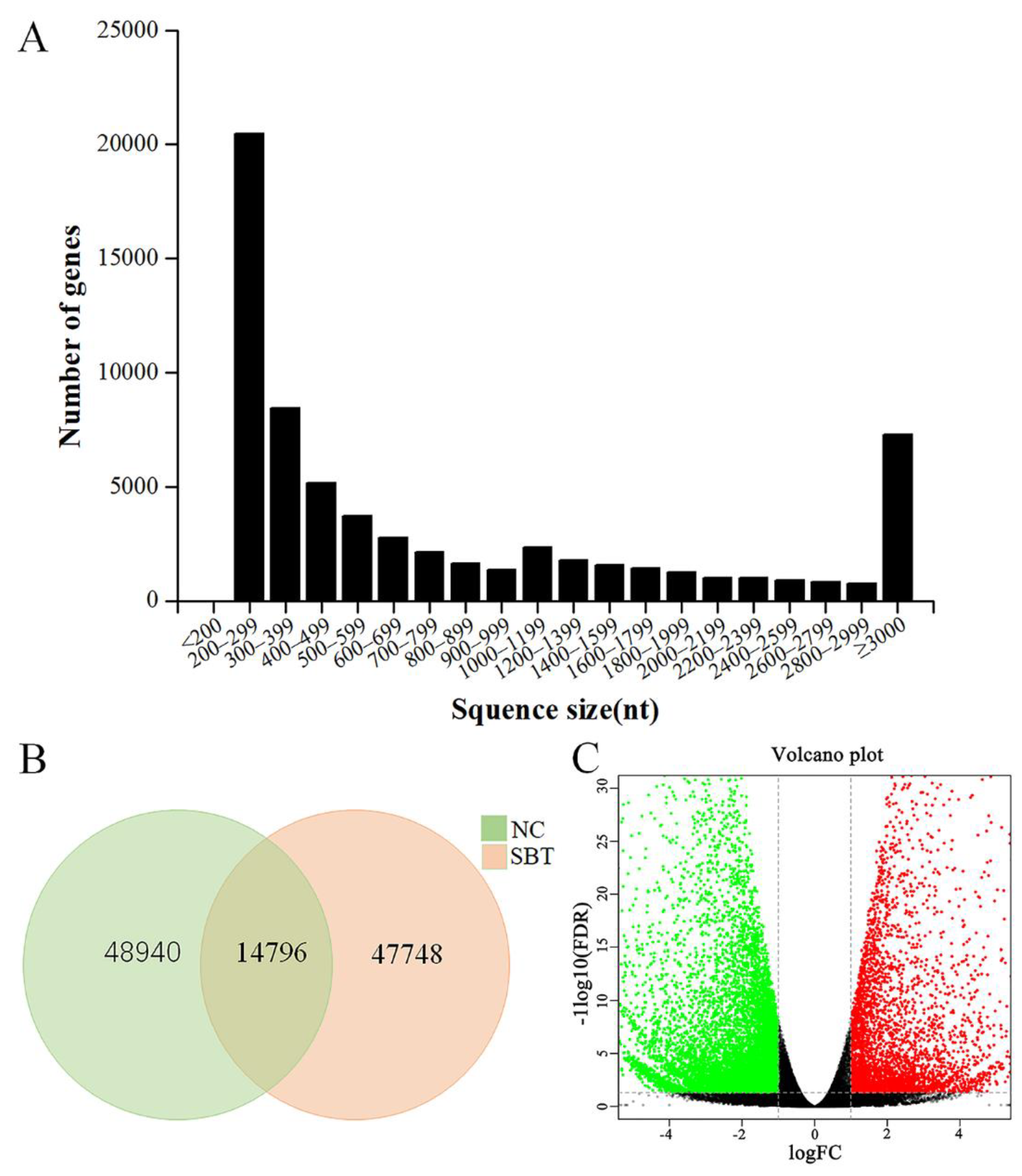
2.1. Transcriptome Sequencing and De Novo Assembly

For transcriptome sequencing, an infectious model of A. schubertii in juvenile hybrid snakehead was established by intraperitoneal injection of the pathogenic bacteria. Three days post-infection, numerous apparent white nodules, approximately 1.0–2.0 mm in diameter, were observed particularly on the spleen and on several other internal organs (Figure 1). Spleen samples were collected 8 h post-infection and used for transcriptomic sequencing. A total of 195,057,214 raw reads [103,354,170 and 91,703,044 reads generated from control (NC) and A. schubertii infected (SBT), respectively] were obtained. After cleaning of low-quality reads, there were 102,015,252 and 90,423,428 clean reads for the NC and SBT groups, respectively (Table 1). All cleaned data were assembled with Trinity software by de novo assembly, which generated a total of 65,884 unigenes, with a mean length of 1159 base pairs (bp). The range was from 201 to 26,244 bp with an N50 value of 2708 bp. Among these unigenes, 34,096 (51.75%) were 150–500 bp, 11,618 (17.63%) were 500–1000 bp, 8363 (12.69%) were 1000–2000 bp, with 11,807 (17.92%) over 2000 bp (Figure 2A). Levels of gene expression were standardized to RPKM. A total of 63,736 and 62,544 contigs were detected for NC and SBT, respectively, with 14,796 common contigs (Figure 2B). All unigenes were compared against protein databases including; the NCBI nonredundant protein database (Nr), the Swiss-Prot database, the KOG database, and the KEGG database by BLAST (Table 2).

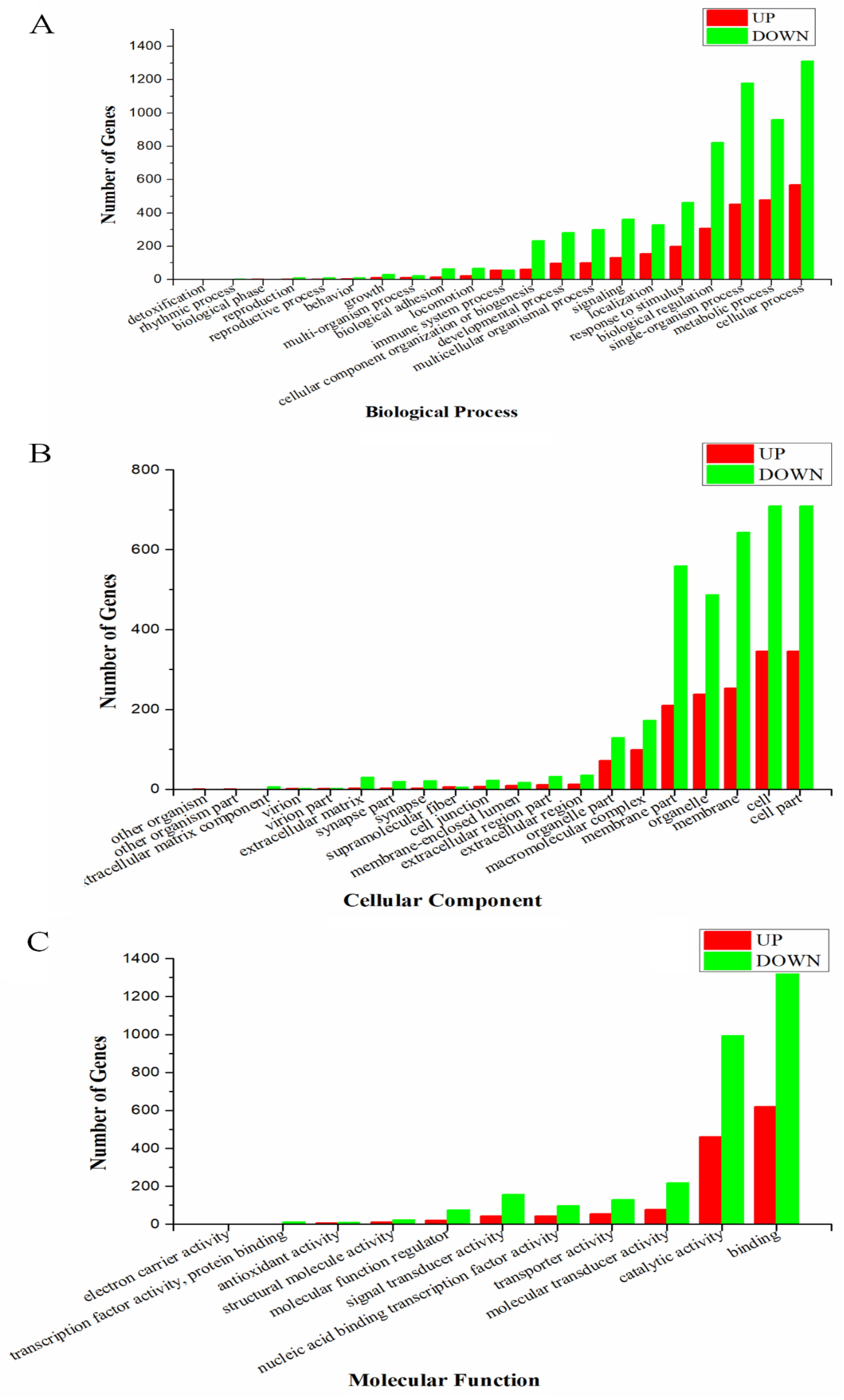
2.2. Functional Explanation of Differentially Expressed Genes (DEGs)

To search DEGs from NC and SBT, the threshold was set for log2 (fold-change) | > 1 and p-value < 0.01. A total of 14,796 DEGs were identified, including 4441 (accounting for 30.01% of all DEGs) up-regulated genes and 10,355 (accounting for 69.99%) down-regulated genes. DEGs after A schubertii infection were visualized by volcano plot. Down-regulated DEGs (green dots) were greater than up-regulated DEGs (red dots) (Figure 2C).
To investigate the potential biological function of DEGs, GO, and KEGG analyses were executed for all DEGs. GO analysis found that all DEGs could be classified into 52 groups and three categories; biological process (21), cellular component (20), and molecular function (11) (Figure 3). A total of 663 DEGs were categorized into “response to stimulus”, including 199 up-regulated and 464 down-regulated DEGs. There were 114 (including 56 up-regulated and 58 down-regulated DEGs) in the “immune system process”, 493 (131 up-regulated and 362 down-regulated DEGs) in “signaling”, and 1131 (307 up-regulated and 824 down-regulated DEGs) in “biological regulation”. KEGG analysis found a total of 2084 DEGs were enriched into 192 pathways, the top 20 of which are shown in Figure 4. In particular, 14 immune-related pathways and one apoptosis-related pathway were screened including 127 DEGs (6.31%) in “cytokine-cytokine receptor interaction”, 82 (3.93%) in “phagosome”, and 43 (2.06%) in “toll-like receptor signaling pathway” (Table 3). There were 37 significant DEGs (including 22 up-regulated and 15 down-regulated) involved in the apoptosis-related signaling pathway. These DEGs are represented in the pathway diagram in different colors (Figure 5).

2.3. RT-qPCR Validation of DEGs

Transcriptome and RNA-seq data were verified with 12 randomly selected DEGs for RT-qPCR analysis. These DEGs were chemokine (C-X-C motif) 13 (CXCL-13), interferon-gamma (IFNγ), toll-like receptor 5 (TLR-5), interleukin-1 (IL-1), interleukin-1 receptor1 (IL-1R1), interleukin-6 (IL-6), interferon regulatory factor-3 (IRF-3), tumor necrosis factor alpha2 (TNFα-2), major histocompatibility complex class II (MHC-II), T cell receptor alpha (TCRα), TNF-related apoptosis-inducing ligand (TRAIL), and integrin subunit beta 1 (ITGB-1). As shown in Figure 6, the fold-change values by RT-qPCR were consistent with the values obtained by RNA-seq for all selected genes.

2.4. Detection of Immune-Related DEGs after Challenge with A. schubertii

To further assess immune-related DEGs, hybrid snakeheads were challenged with A. schubertii and mRNA expression levels of six immune-related genes were examined at multiple time points post-infection (6, 12, 24, and 48 h). The results are shown in Figure 7. Gene expression levels are roughly clustered into two patterns. The most common pattern was displayed by five genes; IL-1R1, TLR-5, CXCL-13, IFNγ, and IL-6. The mRNA levels of these genes were significantly higher than control (p < 0.01) at 6, 12, 24, and 48 h after infection. A second pattern was observed for TCRα, which were significantly up-regulated at 6 and 12 h following infection, then decreased at 24 h, but returned to a near-normal level by 48 h.

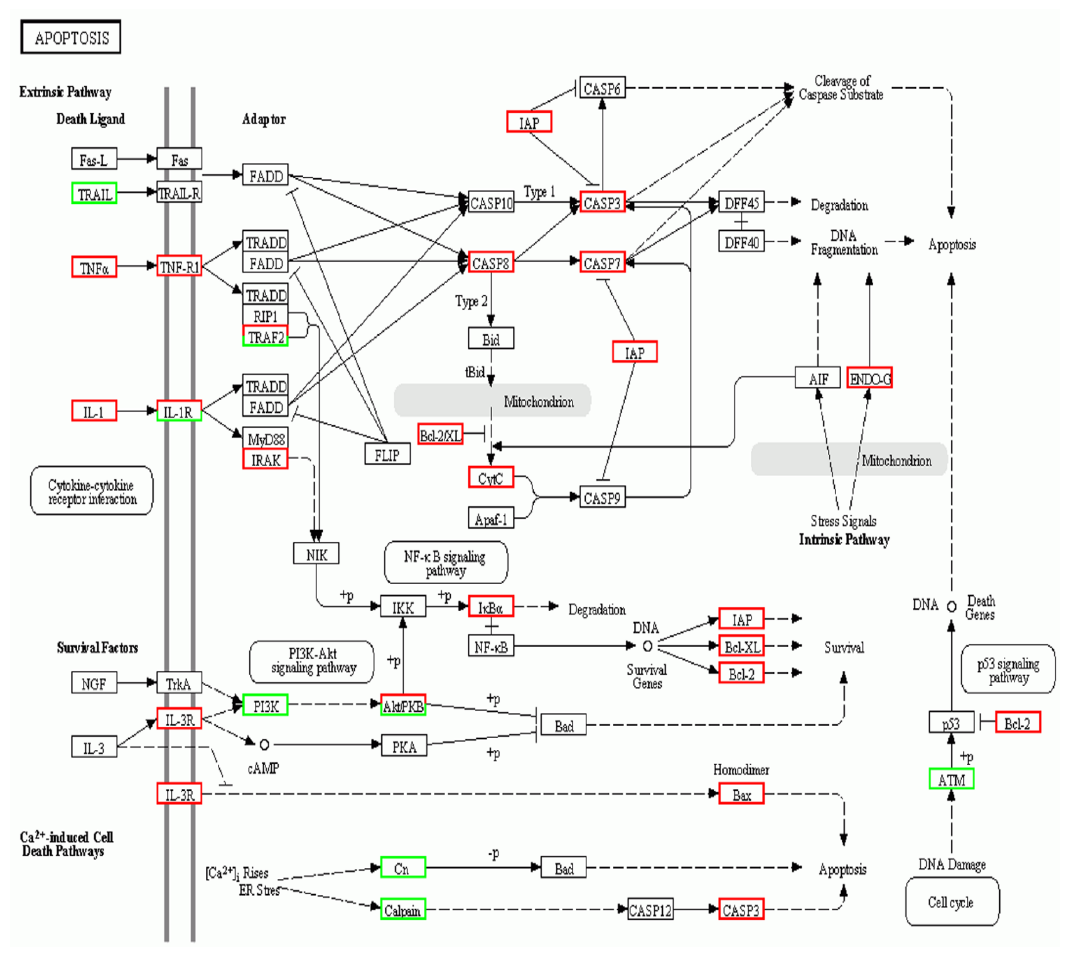
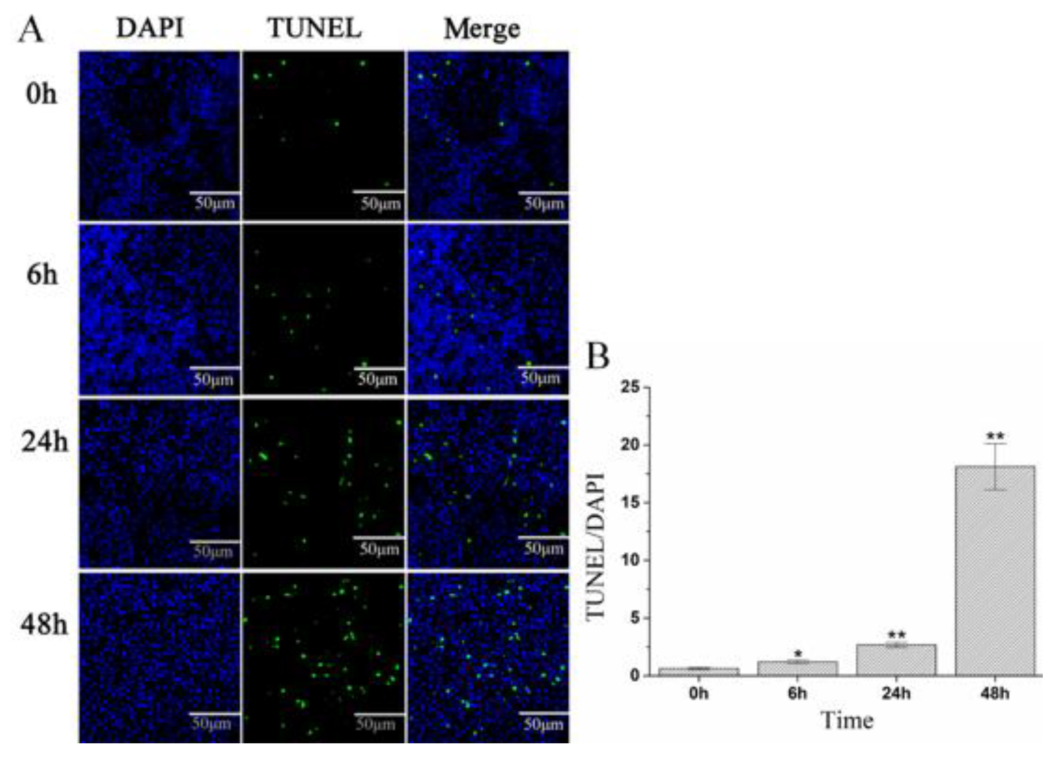
2.5. A. schubertii Infection Induced Apoptosis in Hybrid snakehead

Based on KEGG enrichment pathways analysis, A. schubertii infection significantly activated the cell apoptosis pathway. A TUNEL apoptosis detection kit was used to assess apoptosis of hybrid snakehead splenocytes at 0, 6, 24, and 48 h after A. schubertii infection. The TUNEL assay attaches green fluorescein to fragmented DNA, indicating an apoptotic cell. Nuclei have a blue fluorescent signal when labeled with DAPI. The number of green apoptotic cells in the spleen gradually increased with infection (Figure 8A). The apoptotic cell rate significantly increased 0–48 h post-infection. The apoptotic cell rate at 48 h (18.12) was more than 28 times greater than at 0 h (0.63) (Figure 8B). To further investigate apoptosis, the expression of four apoptosis-related genes (TNFα, CASP3, CASP7, and CASP8) was measured following infection by RT-qPCR. As shown in Figure 9, mRNA levels of CASP3, CASP7, and CASP8 were significantly up-regulated compared to control (p < 0.05) at 6, 12, 24, and 48 h post-infection. TNFα mRNA levels were significantly up-regulated (p < 0.05) at 6 h, decreasing to nearly normal levels at 12 and 24 h, and then significantly up-regulated at 48 h.

3. Discussion

For the snakehead fish aquaculture industry, A. schubertii is an important pathogen. The infection produces typical nodules throughout the internal organs of diseased fish. These symptoms rarely occur with other Aeromonas infections. This observation implied that A. schubertii pathogenesis was distinct from common Aeromonas species [9]. Nodule formation has traditionally been considered a dangerous strategy by which to limit infection, protecting the host by walling off resistant pathogens [33,34]. However, some nodules (such as granuloma caused by pathogenic mycobacteria) paradoxically allow the pathogens to multiply and disseminate infection, promoting disease progression [35,36]. In our previous studies, we reported similar nodules in tilapia infected with A. schubertii. Histopathological analysis showed the center of the nodules to contain collections of bacteria surrounded by degenerate and necrotic tissue, which is similar to the granuloma caused by mycobacteria [9]. Few studies have reported interactions between A. schubertii and hybrid snakehead. Furthermore, the host immune response to A. schubertii during infection is still largely unknown.
Transcriptome analysis has proven to be a useful tool by which to elucidate pathogenic mechanisms. Herein, we established an A. schubertii infection model in juvenile hybrid snakeheads. The clinical signs of the challenged fish were similar to those already described for other A. schubertii infected fish such as snakehead and tilapia [6,9,37]. Sequencing of the splenic transcriptome response of hybrid snakehead with or without A. schubertii infection allowed for analysis of the pathogen-host interaction during infection. In total, 14,796 DEGs including 4441 (accounting for 30.01% of all DEGs) up-regulated genes and 10,355 (accounting for 69.99%) down-regulated genes were found to differ between groups. This number is greater than the number found in peripheral blood of hybrid snakehead after Nocardia seriolae infection [38], or in dissected muscle tissue of C. striatus after Aphanomyces invadans infection [39]. The spleen is an important immune organ in fish. Previous comparative transcriptome analysis of hybrid sturgeon (Huso dauricus × Acipenser schrenckii) spleens identified 283 DEGs (168 up-regulated genes and 115 down-regulated genes) between A. hydrophila infected fish and control. Various anti-bacterial immune-relevant pathways were identified [40]. Therefore, we focused on DEGs related to the splenic immune response after infection. Herein, functional enrichment analysis showed 371 DEGs (196 up-regulated genes and 175 down-regulated genes) in 14 immune-related KEGG pathways that were closely involved in anti-bacterial immune responses, indicating a complicated immune system in hybrid snakeheads.
The innate immune system operates the first line of defense against invading pathogens by potential pathogen recognition [41]. Pattern recognition receptors (PRRs) recognize microbial pathogen-associated molecular patterns (PAMPs) that trigger the innate immune system [42]. The major families of PRRs are the Toll-like receptors (TLRs), nucleotide-binding and oligomerization domain (NOD)-like receptors (NLRs), and retinoic acid-inducible gene I (RIG-I)-like receptors (RLRs). In vivo challenge experiments in teleost species have shown tissue activation of PRR signaling pathways following bacterial infection [40,43,44]. Herein, we observed the number of up-regulated DEGs to be greater than those down-regulated in the TLR signaling pathway, the RIG-I-like receptor signaling pathway, the NLR signaling pathway, and the cytosolic DNA-sensing pathway (Table 3). Furthermore, the time course RT-qPCR analysis demonstrated A. schubertii infection to progressively increase mRNA levels of two PRRs (TLR-5 and IL-1R1). TLR-5 is known to recognize bacterial flagellin of invading bacteria, resulting in activation of the key NF-κB signaling pathway that leads to proinflammatory gene program activation [45]. IL-1R1 recognizes and binds to the extracellular domain of IL-1 related cytokines that are involved in multiple immunological and inflammatory processes [46]. The time course RT-qPCR analysis demonstrated a dramatic increase in the expression of the proinflammatory cytokine mRNAs for CXCL-13, IFNγ, and IL-6 with infection. These cytokine genes have been associated with the innate immune response, playing an important role in defense against pathogenic microorganisms in many species of fish [47,48,49]. Therefore, their up-regulation indicated that PRRs and the related proinflammatory response may represent an important anti-infection mechanism during the early stage of A. schubertii infection. In addition, transcriptome analysis identified a small number of DEGs in other immune-related signaling pathways including Fc gamma R-mediated phagocytosis (two down-regulated genes), B cell receptor signaling pathway (two down-regulated genes), natural killer cell-mediated cytotoxicity (two down-regulated genes), Fc epsilon RI signaling pathway (two down-regulated genes), hematopoietic cell lineage (two down-regulated genes), and complement and coagulation cascades (one up-regulated gene). These results are inconsistent with other studies of bacterial infections [41,50] but are partially consistent with the analysis of grass carp intestines following infection with A. hydrophila [12]. Thus, we propose A. schubertii may have evolved highly sophisticated mechanisms to modulate host immune responses that ensure successful infection.
Apoptosis is a primary form of programmed cell death that plays an important role in the regulation of growth, development, and immune responsiveness [51]. Oligo Nucleosomal DNA fragmentation and chromatin condensation are two hallmarks of apoptosis [52]. The TUNEL technique can sensitively detect DNA fragmentation and is a standard means by which to detect apoptosis in tissue sections [53]. TUNEL has been widely used to detect apoptotic cells during pathogen invasion. Pathogenic bacteria known to trigger host cell apoptosis are A. hydrophila [54], Mycoplasma gallisepticum [55], and Mycobacterium tuberculosis [56]. In a previous study, TUNEL found 50 to 70% apoptotic cells in the periphery of mycobacterial granulomas in clinical cases of tuberculosis, which implied that apoptosis may be involved in the formation of granulomas [57]. The major histological feature of A. schubertii infection is the nodule, which has a structure similar to that of mycobacterial granulomas [9]. In this study, TUNEL demonstrated the rate of apoptosis to significantly increase in the spleens of fish after A. schubertii infection, reaching 18.12 at 48 h. This result showed that prolonged infection induced severe cellular apoptosis. We speculate that A. schubertii-induced apoptosis is an important cause of nodule formation in hybrid snakeheads.
As an indispensable defense mechanism for host resistance to pathogen invasion, apoptosis mainly occurs in three basic ways: death receptor, endoplasmic reticulum, and mitochondrial pathways [58,59]. The signaling pathways (intrinsic or extrinsic) of apoptosis are complex but always rely on the activation of caspases (cysteine proteases) [60]. In this study, transcriptome analysis demonstrated apoptosis-related pathway molecules to be involved in A. schubertii infection (Figure 5). For example, death receptor pathway-related genes: TNFα, TNF-R1, TRAF2, IRAK, CAPS8, IAP, CASP3, and CASP7 were up-regulated, while TRAIL was down-regulated, suggesting that the death receptor signaling pathway may be crucial to A. schubertii infection. The expression of TNFα, CASP8, CASP7, and CASP3 genes was assessed at 6, 12, 24, and 48 h post A. schubertii challenge. TNFα induces CASP8-dependent apoptosis through the formation of a death-inducing signaling complex comprised of the TNF receptor-associated death domain (TRADD) and the death domain (FADD) [61]. CASP8 is a cysteine protease best known for mediating the downstream caspase (CASP 3, 6, 7) death receptor signaling pathway that results in apoptosis [61,62]. CASP8, CASP 7, and CASP3 are thought to be key factors in the caspase cascade of apoptosis execution by cleaving multiple structural and repair proteins [63]. Herein, the expression of CASP8, CASP7, and CASP 3 was significantly up-regulated after infection, with the expression of TNFα significantly up-regulated at 6 and 24 h post-infection. This observation suggested that A. schubertii infection activated the TNFα-related death receptor apoptosis pathway in hybrid snakeheads.

4. Materials and Methods

4.1. Fish Sample, Bacteria Preparation, and Ethics

Healthy juvenile hybrid snakeheads weighing 30 ± 2 g were obtained from a local fish farm in Guangzhou City, Guangdong Province, China. Fish were kept in aerated tap water at 28 °C with commercial feed for 2 weeks. The bacterial strain of A. schubertii used for the experiments was isolated from a diseased snakehead fish [8]. Bacteria were cultured in Brain-Heart infusion agar (BHIA) at 28 °C for 12 h with constant moderate shaking (180 rpm), then harvested by centrifugation at 4000× g for 10 min, washed once with phosphate-buffered saline (PBS, pH 7.2), and centrifuged (4000× g, 10 min). Bacterial pellets were resuspended in PBS and the concentration adjusted.

4.2. Bacterial Challenge, Sample Collection, and Total RNA Extraction

To collect samples for transcriptome sequencing, ten healthy snakeheads (water temperature at 28 ± 2 °C) were injected intraperitoneally with a suspension of A. schubertii (3 × 10⁶ CFU/fish). Ten fish treated with 0.2 mL of PBS were used as control. Eight h post-infection, three fish from each group were anesthetized with MS-222 and their spleens collected (SBT-1, SBT-2, and SBT-3 from the A. schubertii infected group) and (NC-1, NC-2, and NC-3 from the control group) for RNA-seq. The samples were stored in liquid nitrogen until RNA extraction. The remaining fish were continuously monitored for clinical signs until 7 days post-infection.
To investigate fish apoptosis and immune response, 120 healthy snakeheads were randomly divided into two groups. Sixty test fish (water temperature 28 ± 2 °C) were intraperitoneally inoculated with A. schubertii (3 × 10⁵ CFU/fish). Another sixty fish were treated with 0.2 mL of PBS as control. In genes expression experiments, nine fish from each group were anesthetized with MS-222, and spleen samples were obtained at 6, 12, 24, and 48 h post-infection. Each sample consisted of a mixture of tissues from three fish. Three replicates of each sample were assessed for each time point. All samples were frozen in liquid nitrogen until RNA extraction. For apoptosis experiments, three fish from the infected group were anesthetized with MS-222, and spleen samples were taken at 0, 6, 24, and 48 h post-infection for analysis by the TUNEL assay.
Total RNA was extracted using TRIzol Reagent (Invitrogen, Carlsbad, CA, USA). The mRNA was reverse-transcribed using PrimeScript RT reagent Kit with gDNA Eraser (Takara, Dalian, China), following the manufacturer’s recommendations.

4.3. Library Preparation and RNA-seq

Total RNA quality was examined using an Agilent 2100 Bioanalyzer (Agilent Technologies, Palo Alto, CA, USA). RNA-seq was conducted using an Illumina HiSeq 4000 platform (Gene Denovo, China). Briefly, mRNA was purified using oligo (dT) magnetic beads and fragmented into short fragments of 300–400 nucleotides using an RNA fragmentation reagent (Thermo Fisher Scientific, San Francisco, CA, USA). Subsequently, 3’ and 5’ adaptors were ligated into unique RNA fractions, adaptor-ligated RNA fragments were reverse-transcribed and amplified by qPCR, and then the RNA library was sequenced. RNA-seq raw data have been submitted to the NCBI SRA database and the accession number is SRP108948.

4.4. Transcriptome Assembly and Unigene Annotation

Raw Illumina paired-end reads were filtered using internal software to remove reads without adaptors; those reads in which unknown bases (N) comprised greater than 5% of the read, and low quality reads. Post-filtered reads (clean reads) were stored in FASTQ format. Trinity was used to perform de novo assembly with clean reads. Next, TGICL gene indices clustering tools were used to cluster transcripts into unigenes. All unigenes were annotated with the following databases: Nr (NCBI non-redundant protein database), Swiss-Prot, KOG (Clusters of Orthologous Groups), and KEGG (Kyoto Encyclopaedia of Genes and Genomes) with E-values less than 10−5.

4.5. Enrichment Analysis of DEGs

Expression data from two libraries (NC and SBT) were determined by mapping to the transcriptome assembly using Bowtie 2 software. The reads per kb of transcripts per million fragments mapped (RPKM) values were analyzed using RESM and differentially expressed genes were obtained using edgeR NC and SBT spleens. Furthermore, to determine the threshold p-value in multiple tests, a false discovery rate (FDR) was used. Significant enrichment was calculated when FDR was <0.05 and RPKM values showed at least a two-fold difference between the two samples. GO was performed to identify the functional annotations of the DEGs. Goatools software (Available online: https://github.com/tanghaibao/Goatools, accessed on 13 September 2017) was used to analyze the functional enrichment terms. Significant enrichment was considered when FDR was <0.05. We also used the KEGG Orthology Based Annotation System (KOBAS; Available online: http://kobas.cbi.pku.edu.cn/home.do, accessed on 15 September 2017) to perform the KEGG pathway analysis, with enriched pathways determined by hypergeometric test (filtered with FDR < 0.05).

4.6. Real-Time Fluorescence Quantitative PCR (RT-qPCR) Analysis

To validate the RNA-seq data, 12 randomly selected genes were chosen from all of the DEGs. The mRNA expression levels of these selected genes were then determined by RT-qPCR. RT-qPCR analysis was performed as described previously [64]. RT-qPCR was performed using an SYBR Premix Ex Taq kit (Takara, Dalian, China) with a QuantStudio 7 Flex Real-Time machine (Applied Biosystems, Waltham, MA, USA). All reactions were performed in triplicate. Relative expression levels of mRNAs were calculated using the 2ΔΔCt method. β-actin was used as a housekeeping gene for the normalization of mRNA.
The same procedure was used to measure the expression profiles of 10 selected genes involved in immune response and apoptosis pathways (KEGG pathway: hsa04210). The oligonucleotide primer pairs used for all RT-qPCR reactions are listed in Table S1.

4.7. Apoptosis Analysis

Apoptosis was evaluated using TUNEL staining. Hybrid snakeheads were infected with A. schubertii as described above. After 0, 6, 24, and 48 h post-infection, spleens were fixed in 4% (w/v) paraformaldehyde for at least 24 h, dehydrated through a graded alcohol series to xylene, embedded in paraffin wax, and sectioned at 3–5 μm. The spleen tissue sections were subjected to TUNEL staining with a TUNEL assay kit (Roche, Switzerland) as described previously [65]. After staining, the sections were viewed by Ortho-Fluorescent Microscopy (Nikon Eclipse C1, Tokyo, Japan) with cell apoptosis and apoptosis rate judged by Image-pro plus 6.0 (Media Cybernetics, Bethesda, MD, USA) Three fields were examined for each sample and statistically analyzed.

4.8. Statistical Analysis

Statistical analysis was performed using SPSS 19.0 software (SPSS Inc., Chicago, IL, USA). All data are presented as means and standard error. One-way ANOVA and Duncan’s t-tests were used to analyze the data. The minimum level of significance level was set as p < 0.05.

5. Conclusions

In conclusion, we have shown that A. schubertii infection induced sophisticated immunological reactions in hybrid snakeheads. These reactions included up-regulation of cytokine genes, increased expression of PRRs, a pro-inflammatory response, and activation of the death receptor apoptosis pathway. Our results provided valuable information about the immune response of hybrid snakeheads during the early stages of a bacterial infection. Furthermore, these findings provided new insights into anti-A. schubertii immunity in hybrid snakehead, which can be used to unravel A. schubertii infections in fish at the molecular level.

Supplementary Materials

The following are available online at https://www.mdpi.com/article/10.3390/pathogens10080997/s1, Table S1. Primers used for qPCR in this study.

Author Contributions

Conceptualization, C.L. and Y.S.; methodology, C.L. and J.M.; software, Z.Q.; validation, J.M., D.Z. and C.L.; formal analysis, C.L.; investigation, B.J.; resources, C.L.; data curation, W.L. and J.M.; writing—original draft preparation, C.L. and Q.W.; writing—review and editing, C.L. and Q.W.; visualization, C.L.; supervision, Y.S., L.L. and Q.W.; project administration, C.L. and L.L.; funding acquisition, C.L. and Y.S. All authors have read and agreed to the published version of the manuscript.

Funding

The study was supported by the Science and Technology Planning Project of Guangdong Province (Project #2016A030303028), China Agriculture Research System (Project #CARS-45).

Institutional Review Board Statement

The study was conducted according to the guidelines of the Declaration of Helsinki, and approved by the Animal Ethics Committee of Pearl River Fisheries Research Institute (protocol code: LAEC-PRFRI-2017-04-13; date of approval: 28 April 2017).

Data Availability Statement

The raw data reads were deposited in NCBI SRA database (https://www.ncbi.nlm.nih.gov/sra, accessed on 6 August 2021) under the accession number SRP108948.

Conflicts of Interest

The authors declare no conflict of interest.

References

  1. Williams, J.D.; Courtenay, W.R. Snakeheads (Pisces, Channidae)—A Biological Synopsis and Risk Assessment; U.S. Geological Survey: Reston, VA, USA, 2004.
  2. Xu, J.; Bian, C.; Chen, K.; Liu, G.; Jiang, Y.; Luo, Q.; You, X.; Peng, W.; Li, J.; Huang, Y.; et al. Draft genome of the Northern snakehead, Channa argus. Gigascience 2017, 6, 1–5. [Google Scholar] [CrossRef]
  3. Kokka, R.P.; Lindquist, D.; Abbott, S.L.; Janda, J.M. Structural and pathogenic properties of Aeromonas schubertii. Infect. Immun. 1992, 60, 2075–2082. [Google Scholar] [CrossRef] [Green Version]
  4. Hickmanbrenner, F.W.; Fanning, G.R.; Arduino, M.J.; Brenner, D.J.; Rd, F.J. Aeromonas schubertii, a new mannitol-negative species found in human clinical specimens. J. Clin. Microbiol. 1988, 26, 1561–1564. [Google Scholar] [CrossRef] [Green Version]
  5. Carnahan, A.M.; Marii, M.A.; Fanning, G.R.; Pass, M.A.; Joseph, S.W. Characterization of Aeromonas schubertii strains recently isolated from traumatic wound infections. J. Clin. Microbiol. 1989, 27, 1826–1830. [Google Scholar] [CrossRef] [Green Version]
  6. Liu, J.Y.; Li, A.H. First case of Aeromonas schubertii infection in the freshwater cultured snakehead fish, Ophiocephalus argus (Cantor), in China. J. Fish Dis. 2012, 35, 335–342. [Google Scholar] [CrossRef]
  7. Chen, Y.F.; Liang, R.S.; Zhuo, X.L.; Wu, X.T.; Zou, J.X. Isolation and characterization of Aeromonas schubertii from diseased snakehead, Channa maculata (Lacepède). J. Fish Dis. 2012, 35, 421. [Google Scholar] [CrossRef] [PubMed]
  8. Liu, C.; Li, K.B.; Wang, Q.; Chang, O.Q.; Liang, H.L.; Wang, F.; Pan, D.B.; Shi, C.B.; Wu, S.Q. Isolation, identification and characterization of Aeromonas schubertii from hybrid snakehead (Channa maculata♀ × C. Argus♂). J. Fish. China 2012, 36, 1119–1125. [Google Scholar] [CrossRef]
  9. Liu, C.; Chang, O.Q.; Zhang, D.F.; Li, K.B.; Wang, F.; Lin, M.H.; Shi, C.B.; Jiang, L.; Wang, Q.; Bergmann, S.M. Aeromonas shuberti as a cause of multi-organ necrosis in internal organs of Nile tilapia, Oreochromis niloticus. J. Fish Dis. 2018, 41, 1529–1538. [Google Scholar] [CrossRef] [PubMed]
  10. Whyte, S.K. The innate immune response of finfish—A review of current knowledge. Fish Shellfish Immunol. 2007, 23, 1127–1151. [Google Scholar] [CrossRef] [PubMed]
  11. Ahn, D.H.; Kang, S.; Park, H. Transcriptome analysis of immune response genes induced by pathogen agonists in the Antarctic bullhead notothen Notothenia coriiceps. Fish Shellfish Immunol. 2016, 55, 315–322. [Google Scholar] [CrossRef]
  12. Song, X.; Hu, X.; Sun, B.; Bo, Y.; Wu, K.; Xiao, L.; Gong, C. A transcriptome analysis focusing on inflammation-related genes of grass carp intestines following infection with Aeromonas hydrophila. Sci. Rep. 2017, 7, 40777. [Google Scholar] [CrossRef] [Green Version]
  13. Li, C.; Zhang, Y.; Wang, R.; Lu, J.; Nandi, S.; Mohanty, S.; Terhune, J.; Liu, Z.; Peatman, E. RNA-seq analysis of mucosal immune responses reveals signatures of intestinal barrier disruption and pathogen entry following Edwardsiella ictaluri infection in channel catfish, Ictalurus punctatus. Fish Shellfish Immunol. 2012, 32, 816–827. [Google Scholar] [CrossRef] [PubMed]
  14. Wang, P.; Wang, J.; Su, Y.Q.; Mao, Y.; Zhang, J.S.; Wu, C.W.; Ke, Q.Z.; Han, K.H.; Zheng, W.Q.; Xu, N.D. Transcriptome analysis of the Larimichthys crocea liver in response to Cryptocaryon irritans. Fish Shellfish Immunol. 2016, 48, 1–11. [Google Scholar] [CrossRef]
  15. Huang, Y.; Huang, X.; Yan, Y.; Cai, J.; Ouyang, Z.; Cui, H.; Wang, P.; Qin, Q. Transcriptome analysis of orange-spotted grouper (Epinephelus coioides) spleen in response to Singapore grouper iridovirus. BMC Genom. 2011, 12, 556. [Google Scholar] [CrossRef] [PubMed] [Green Version]
  16. Zhang, X.; Wang, S.; Chen, S.; Chen, Y.; Liu, Y.; Shao, C.; Wang, Q.; Lu, Y.; Gong, G.; Ding, S.; et al. Transcriptome analysis revealed changes of multiple genes involved in immunity in Cynoglossus semilaevis during Vibrio anguillarum infection. Fish Shellfish Immunol. 2015, 43, 209–218. [Google Scholar] [CrossRef] [PubMed]
  17. Chen, J.; Li, C.; Huang, R.; Du, F.; Liao, L.; Zhu, Z.; Wang, Y. Transcriptome analysis of head kidney in grass carp and discovery of immune-related genes. BMC Vet. Res. 2012, 8, 108. [Google Scholar] [CrossRef] [Green Version]
  18. Liu, C.; Guo, Y.M.; Cao, J.Z.; Zhang, D.F.; Chang, O.Q.; Li, K.; Wang, F.; Shi, C.B.; Jiang, L.; Wang, Q.; et al. Detection and quantification of Aeromonas schubertii in Channa maculata by TaqMan MGB probe fluorescence real-time quantitative PCR. J. Fish Dis. 2019, 42, 109–117. [Google Scholar] [CrossRef]
  19. Everett, H.; Mcfadden, G. Apoptosis: An innate immune response to virus infection. Trends Microbiol. 1999, 7, 160–165. [Google Scholar] [CrossRef]
  20. Kim, S.H.; Cho, S.N.; Lim, Y.J.; Choi, J.A.; Lee, J.; Go, D.; Song, C.H. Phagocytosis influences the intracellular survival of Mycobacterium smegmatis via the endoplasmic reticulum stress response. Cell Biosci. 2018, 8, 52. [Google Scholar] [CrossRef] [PubMed]
  21. Hughes, F.M.; Foster, B.; Grewal, S.; Sokolova, I.M. Apoptosis as a host defense mechanism in Crassostrea virginica and its modulation by Perkinsus marinus. Fish Shellfish Immunol. 2010, 29, 247–257. [Google Scholar] [CrossRef]
  22. Birge, R.B.; Ucker, D.S. Innate apoptotic immunity: The calming touch of death. Cell Death Differ. 2008, 15, 1096–1102. [Google Scholar] [CrossRef] [Green Version]
  23. Yeretssian, G.; Labbe, K.; Saleh, M. Molecular regulation of inflammation and cell death. Cytokine 2008, 43, 380–390. [Google Scholar] [CrossRef] [PubMed]
  24. Behar, S.M.; Martin, C.J.; Booty, M.G.; Nishimura, T.; Zhao, X.; Gan, H.X.; Divangahi, M.; Remold, H.G. Apoptosis is an innate defense function of macrophages against Mycobacterium tuberculosis. Mucosal Immunol. 2011, 4, 279–287. [Google Scholar] [CrossRef] [PubMed] [Green Version]
  25. Lancellotti, M.; Pereira, R.F.; Cury, G.G.; Hollanda, L.M. Pathogenic and opportunistic respiratory bacteria-induced apoptosis. Braz. J. Infect. Dis. 2009, 13, 226–231. [Google Scholar] [CrossRef] [PubMed] [Green Version]
  26. Zhou, Z.J.; Sun, L. Edwardsiella tarda-Induced inhibition of apoptosis: A strategy for intracellular survival. Front. Cell Infect. Microbiol. 2016, 6, 76. [Google Scholar] [CrossRef] [PubMed] [Green Version]
  27. Zychlinsky, A.; Sansonetti, P.J. Apoptosis as a proinflammatory event: What can we learn from bacteria-induced cell death? Trends Microbiol. 1997, 5, 201–204. [Google Scholar] [CrossRef]
  28. Chen, Y.; Zychlinsky, A. Apoptosis induced by bacterial pathogens. Microb. Pathog. 1994, 17, 203–212. [Google Scholar] [CrossRef] [PubMed]
  29. Behar, S.M.; Briken, V. Apoptosis inhibition by intracellular bacteria and its consequence on host immunity. Curr. Opin. Immunol. 2019, 60, 103–110. [Google Scholar] [CrossRef]
  30. Van Schaik, E.J.; Chen, C.; Mertens, K.; Weber, M.M.; Samuel, J.E. Molecular pathogenesis of the obligate intracellular bacterium Coxiella burnetii. Nat. Rev. Microbiol. 2013, 11, 561–573. [Google Scholar] [CrossRef] [Green Version]
  31. Rikihisa, Y. Molecular Pathogenesis of Ehrlichia chaffeensis Infection. Annu. Rev. Microbiol. 2015, 69, 283–304. [Google Scholar] [CrossRef]
  32. Matty, M.A.; Roca, F.J.; Cronan, M.R.; Tobin, D.M. Adventures within the speckled band: Heterogeneity, angiogenesis, and balanced inflammation in the tuberculous granuloma. Immunol. Rev. 2015, 264, 276–287. [Google Scholar] [CrossRef] [PubMed] [Green Version]
  33. Cronan, M.R.; Beerman, R.W.; Rosenberg, A.F.; Saelens, J.W.; Johnson, M.G.; Oehlers, S.H.; Sisk, D.M.; Jurcic, S.K.; Medvitz, N.A.; Miller, S.E.; et al. Macrophage epithelial reprogramming underlies mycobacterial granuloma formation and promotes infection. Immunity 2016, 45, 861–876. [Google Scholar] [CrossRef] [Green Version]
  34. Saunders, B.M.; Frank, A.A.; Orme, I.M. Granuloma formation is required to contain bacillus growth and delay mortality in mice chronically infected with Mycobacterium tuberculosis. Immunology 1999, 98, 324–328. [Google Scholar] [CrossRef] [PubMed]
  35. Yang, C.T.; Cambier, C.J.; Davis, J.M.; Hall, C.J.; Crosier, P.S.; Ramakrishnan, L. Neutrophils exert protection in the early tuberculous granuloma by oxidative killing of mycobacteria phagocytosed from infected macrophages. Cell Host Microbe 2012, 12, 301–312. [Google Scholar] [CrossRef] [Green Version]
  36. Martin, C.J.; Carey, A.F.; Fortune, S.M. A bug’s life in the granuloma. Semin. Immunopathol. 2016, 38, 213–220. [Google Scholar] [CrossRef]
  37. Ren, Z.; Cai, Y.; Wang, S.; Liu, S.; Li, A.; Xiong, Y.; Tang, J.; Sun, Y.; Guo, W.; Zhou, Y. First case of Aeromonas schubertii infection in brackish water wild Nile tilapia, Oreochromis niloticus, in China. Aquaculture 2018, 501, 247–254. [Google Scholar] [CrossRef]
  38. Chen, J.; Li, Y.; Wang, W.; Xia, L.; Wang, Z.; Hou, S.; Huang, J.; Lu, Y. Transcriptome analysis of immune-related gene expression in hybrid snakehead (Channa maculata female symbol × Channa argus male symbol) after challenge with Nocardia seriolae. Fish Shellfish Immunol. 2018, 81, 476–484. [Google Scholar] [CrossRef]
  39. Kumaresan, V.; Pasupuleti, M.; Arasu, M.V.; Al-Dhabi, N.A.; Arshad, A.; Amin, S.; Yusoff, F.M.; Arockiaraj, J. A comparative transcriptome approach for identification of molecular changes in Aphanomyces invadans infected Channa striatus. Mol. Biol. Rep. 2018, 45, 2511–2523. [Google Scholar] [CrossRef]
  40. Jiang, N.; Fan, Y.; Zhou, Y.; Wang, W.; Ma, J.; Zeng, L. Transcriptome analysis of Aeromonas hydrophila infected hybrid sturgeon (Huso dauricus × Acipenser schrenckii). Sci. Rep. 2018, 8, 17925. [Google Scholar] [CrossRef] [PubMed] [Green Version]
  41. Eggestol, H.O.; Lunde, H.S.; Ronneseth, A.; Fredman, D.; Petersen, K.; Mishra, C.K.; Furmanek, T.; Colquhoun, D.J.; Wergeland, H.I.; Haugland, G.T. Transcriptome-wide mapping of signaling pathways and early immune responses in lumpfish leukocytes upon in vitro bacterial exposure. Sci. Rep. 2018, 8, 5261. [Google Scholar] [CrossRef] [PubMed]
  42. Cao, X. Self-regulation and cross-regulation of pattern-recognition receptor signalling in health and disease. Nat. Rev. Immunol. 2016, 16, 35–50. [Google Scholar] [CrossRef]
  43. Li, J.; Gao, Y.; Xu, T. Comparative genomic and evolution of vertebrate NOD1 and NOD2 genes and their immune response in miiuy croaker. Fish Shellfish Immunol. 2015, 46, 387–397. [Google Scholar] [CrossRef] [PubMed]
  44. Du, Z.; Yang, H.; Tan, Y.; Tian, G.; Zhang, Q.; Cui, Y.; Yanfeng, Y.; Wu, X.; Chen, Z.; Cao, S.; et al. Transcriptomic response to Yersinia pestis: RIG-I like receptor signaling response is detrimental to the host against plague. J. Genet. Genom. 2014, 41, 379–396. [Google Scholar] [CrossRef]
  45. Tallant, T.; Deb, A.; Kar, N.; Lupica, J.; de Veer, M.J.; Didonato, J.A. Flagellin acting via TLR5 is the major activator of key signaling pathways leading to NF-kappa B and proinflammatory gene program activation in intestinal epithelial cells. BMC Microbiol. 2004, 4, 33. [Google Scholar] [CrossRef] [PubMed] [Green Version]
  46. Dinarello, C.A. Interleukin-1 in the pathogenesis and treatment of inflammatory diseases. Blood 2011, 117, 3720–3732. [Google Scholar] [CrossRef] [PubMed] [Green Version]
  47. Tian, C.; Chen, Y.; Ao, J.; Chen, X. Molecular characterization and bioactivity of a CXCL13 chemokine in large yellow croaker Pseudosciaena crocea. Fish Shellfish Immunol. 2010, 28, 445–452. [Google Scholar] [CrossRef]
  48. Li, L.; Chen, S.N.; Laghari, Z.A.; Huang, B.; Huo, H.J.; Li, N.; Nie, P. Receptor complex and signalling pathway of the two type II IFNs, IFN-gamma and IFN-gammarel in mandarin fish or the so-called Chinese perch Siniperca chuatsi. Dev. Comp. Immunol. 2019, 97, 98–112. [Google Scholar] [CrossRef]
  49. Huang, P.; Cai, J.; Yu, D.; Tang, J.; Lu, Y.; Wu, Z.; Huang, Y.; Jian, J. An IL-6 gene in humphead snapper (Lutjanus sanguineus): Identification, expression analysis and its adjuvant effects on Vibrio harveyi OmpW DNA vaccine. Fish Shellfish Immunol. 2019, 95, 546–555. [Google Scholar] [CrossRef]
  50. Chen, J.L.; Li, Y.Q.; Wang, W.J.; Xia, L.Q.; Wang, Z.W.; Hou, S.Y.; Huang, J.H.; Lu, Y.S. Transcriptome analysis of immune-related gene expression in hybrid snakehead (Channa maculata ♀ × Channa argus ♂) after challenge with Nocardia seriolae. Fish Shellfish Immunol. 2019, 91, 405. [Google Scholar] [CrossRef]
  51. Gerschenson, L.E.; Rotello, R.J. Apoptosis: A different type of cell death. FASEB J. 1992, 6, 2450–2455. [Google Scholar] [CrossRef]
  52. Joselin, A.P.; Schulze-Osthoff, K.; Schwerk, C. Loss of Acinus inhibits oligonucleosomal DNA fragmentation but not chromatin condensation during apoptosis. J. Biol. Chem. 2006, 281, 12475–12484. [Google Scholar] [CrossRef] [PubMed] [Green Version]
  53. Gavrieli, Y.; Sherman, Y.; Ben-Sasson, S.A. Identification of programmed cell death in situ via specific labeling of nuclear DNA fragmentation. J. Cell Biol. 1992, 119, 493–501. [Google Scholar] [CrossRef]
  54. Majumdar, T.; Chattopadhyay, P.; Saha, D.R.; Sau, S.; Mazumder, S. Virulence plasmid of Aeromonas hydrophila induces macrophage apoptosis and helps in developing systemic infection in mice. Microb. Pathog. 2009, 46, 98–107. [Google Scholar] [CrossRef] [PubMed]
  55. Hu, W.; Zhang, W.; Shah, S.; Ishfaq, M.; Li, J. Mycoplasma gallisepticum infection triggered histopathological changes, oxidative stress and apoptosis in chicken thymus and spleen. Dev. Comp. Immunol. 2021, 114, 103832. [Google Scholar] [CrossRef]
  56. Fairbairn, I.P. Macrophage apoptosis in host immunity to mycobacterial infections. Biochem. Soc. Trans. 2004, 32, 496–498. [Google Scholar] [CrossRef] [Green Version]
  57. Keane, J.; Balcewicz-Sablinska, M.K.; Remold, H.G.; Chupp, G.L.; Meek, B.B.; Fenton, M.J.; Kornfeld, H. Infection by Mycobacterium tuberculosis promotes human alveolar macrophage apoptosis. Infect. Immun. 1997, 65, 298–304. [Google Scholar] [CrossRef] [Green Version]
  58. Wang, H.; Liu, H.; Zheng, Z.M.; Zhang, K.B.; Wang, T.P.; Sribastav, S.S.; Liu, W.S.; Liu, T. Role of death receptor, mitochondrial and endoplasmic reticulum pathways in different stages of degenerative human lumbar disc. Apoptosis 2011, 16, 990–1003. [Google Scholar] [CrossRef]
  59. Jorgensen, I.; Rayamajhi, M.; Miao, E.A. Programmed cell death as a defence against infection. Nat. Rev. Immunol. 2017, 17, 151–164. [Google Scholar] [CrossRef] [PubMed]
  60. Savitskaya, M.A.; Onishchenko, G.E. Mechanisms of apoptosis. Biochemistry 2015, 80, 1393–1405. [Google Scholar] [CrossRef] [PubMed]
  61. Fan, T.J.; Han, L.H.; Cong, R.S.; Liang, J. Caspase family proteases and apoptosis. Acta Biochim. Biophys. Sin. 2005, 37, 719–727. [Google Scholar] [CrossRef] [Green Version]
  62. Gunther, C.; Martini, E.; Wittkopf, N.; Amann, K.; Weigmann, B.; Neumann, H.; Waldner, M.J.; Hedrick, S.M.; Tenzer, S.; Neurath, M.F.; et al. Caspase-8 regulates TNF-alpha-induced epithelial necroptosis and terminal ileitis. Nature 2011, 477, 335–339. [Google Scholar] [CrossRef] [PubMed] [Green Version]
  63. Slee, E.A.; Adrain, C.; Martin, S.J. Executioner caspase-3, -6, and -7 perform distinct, non-redundant roles during the demolition phase of apoptosis. J. Biol. Chem. 2001, 276, 7320–7326. [Google Scholar] [CrossRef] [PubMed] [Green Version]
  64. Cao, J.; Liu, C.; Wang, Q.; Zhang, D.; Chang, O.; Wang, Y.; Shi, C.; Wang, L. Investigating of type IV pili to the pathogenicity of Aeromonas schubertii. Aquaculture 2021, 530, 735800. [Google Scholar] [CrossRef]
  65. Kyrylkova, K.; Kyryachenko, S.; Leid, M.; Kioussi, C. Detection of apoptosis by TUNEL assay. Methods Mol. Biol. 2012, 887, 41–47. [Google Scholar]
Figure 1. The Aeromonas schubertii infection model in hybrid snakehead. (A) White nodules (indicated by white arrows) were distributed in an infected spleen. (B) Spleen from the negative control.
Pathogens 10 00997 g001
Figure 2. (A) Overview of A. schubertii infected hybrid snakehead transcriptome sequence length distribution for all unigenes. The x-axis indicates the interval length of the transcript and the y-axis indicates the number of transcripts with different lengths. (B) Venn diagram of control and A. schubertii intergroup expression. (C) Volcano plot of DEGs identified in NC and SBT spleens. Green and red dots indicate down-regulated and up-regulated DEGs, respectively.
Pathogens 10 00997 g002
Figure 3. Gene ontology enrichment analysis of DEGs in A. schubertii infected hybrid snakehead. The differentially expressed genes (DEGs) were classified into three subclasses, including biological process (A), cellular component (B), and molecular function (C).
Pathogens 10 00997 g003
Figure 4. Scatter plots of the top 20 enriched KEGG pathways. The rich factor is the ratio of DEGs to all the genes annotated for this pathway term.
Pathogens 10 00997 g004
Figure 5. Significant differentially expressed genes identified by KEGG as involved in the apoptosis signaling pathway. Green boxes indicate significantly decreased expression. Red boxes indicate significantly increased expression. White boxes indicate unchanged expression.
Pathogens 10 00997 g005
Figure 6. Comparison of the fold change expression of twelve putative differentially expressed genes (DEGs) between RNA-seq and qRT-PCR. The expression levels of selected genes by qRT-PCR were each normalized to that of the β-actin gene. The relative expression levels from the RNA-seq analysis were calculated as RPKM values. Each value is the mean of three samples. Bars represent mean ± S.E. ** p < 0.01; * p < 0.05.
Pathogens 10 00997 g006
Figure 7. The relative expression levels of immune-related genes in the spleen of hybrid snakehead after infection with A. schubertii. Gene expression was analyzed by qRT-PCR with the 2ΔΔCt method, using β-actin as a reference. (A) IL-1R1, (B) TLR-5, (C) CXCL-13, (D) IFNγ, (E) IL-6, (F) TCRα. Each value is the mean of nine samples. Bars represent mean ± S.E. ** p < 0.01; * p < 0.05.
Pathogens 10 00997 g007
Figure 8. A. schubertii infection-induced apoptosis in spleens of hybrid snakehead at 0, 6, 24, and 48 h post-infection. Splenic tissue sections were analyzed by TUNEL staining. Apoptotic cells are stained green and nuclei are stained blue (A). The apoptotic cell rate during 0–48 h post-infection (B). Each value is the mean of three samples. Bars represent mean ± S.E. ** p < 0.01; * p < 0.05.
Pathogens 10 00997 g008
Figure 9. The relative expression levels of apoptosis-related genes in the spleen of hybrid snakehead after infection with A. schubertii. Gene expression was analyzed by qRT-PCR with the 2ΔΔCt method, using β-actin as a reference. (A) TNFα, (B) CASP8, (C) CASP7, (D) CASP3. Each value is the mean of nine samples. Bars represent mean ± S.E. ** p < 0.01; * p < 0.05.
Pathogens 10 00997 g009
Table 1. Overview of RNA-seq.
SampleRaw ReadsClean ReadsClean BaseGC
NC-133,306,40232,889,1744.92G47.02%
NC-233,820,16233,376,3064.99G46.84%
NC-336,227,60635,749,7725.34G47.11%
NC-total103,354,170102,015,25215.25G46.99%
SBT-130,868,93630,479,5704.57G46.76%
SBT-229,255,26828,832,8164.32G46.49%
SBT-331,578,84031,111,0424.67G47.12%
SBT-total91,703,04490,423,42814.55G46.79%
Note: NC and SBT represent the control and infection groups, respectively.
Table 2. Summary annotations of assembled unigenes.
DatabaseAnnotated UnigenesPercentage
Nr26,98140.95%
Swiss-Prot21,48432.61%
GO12,78619.40%
KEGG15,00122.77%
KOG16,38524.87%
All databases11,80917.92%
At least one database27,53341.79%
Total unigenes65,884100%
Table 3. Immune-related and apoptosis pathways identified after A. schubertii infection.
Pathway IDKEGG PathwaysDEGsTotal
UpDown
ko04060Cytokine-cytokine receptor interaction6766133 (6.38%)
ko04145Phagosome334982 (3.93%)
ko04620Toll-like receptor signaling pathway331043 (2.06%)
ko04672Intestinal immune network for IgA production122234 (1.63%)
ko04622RIG-I-like receptor signaling pathway19827 (1.30%)
ko04621NOD-like receptor signaling pathway13821 (1.01%)
ko04623Cytosolic DNA-sensing pathway16117 (0.82%)
ko04062Chemokine signaling pathway213 (0.14%)
ko04666Fc gamma R-mediated phagocytosis022 (0.10%)
ko04662B cell receptor signaling pathway022 (0.10%)
ko04650Natural killer cell-mediated cytotoxicity022 (0.10%)
ko04664Fc epsilon RI signaling pathway022 (0.10%)
ko04640Hematopoietic cell lineage022 (0.10%)
ko04610Complement and coagulation cascades101 (0.05%)
ko04210Apoptosis221537 (1.78%)
Publisher’s Note: MDPI stays neutral with regard to jurisdictional claims in published maps and institutional affiliations.

