pH-Dependent Structural Dynamics of Cathepsin D-Family Aspartic Peptidase of Clonorchis sinensis
Abstract
1. Introduction
2. Results and Discussion
2.1. Homology Models and Features of Mature CsCatD2
2.2. Four Systems for MD Simulations According to the Enzyme Conformations and pH Values
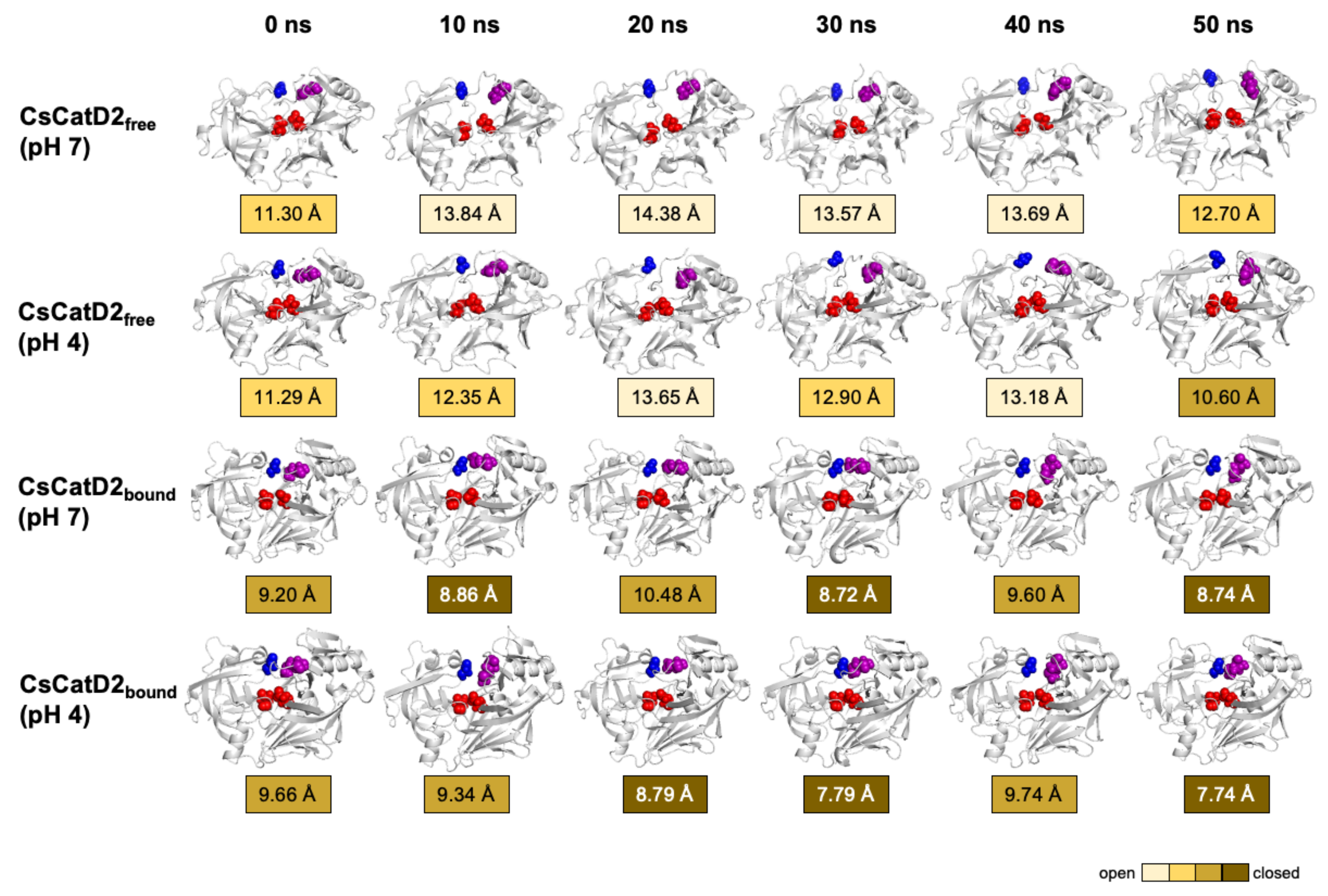
2.3. CsCatD2free Adopts a Semi-Closed Conformation for Ligand-Binding at Acidic pH
2.4. Both Ligand-Binding and Acidic pH Limit the Conformational Flexibility
2.5. Neutral pH May Induce the Opening of Potential Allosteric Inhibitory Sites
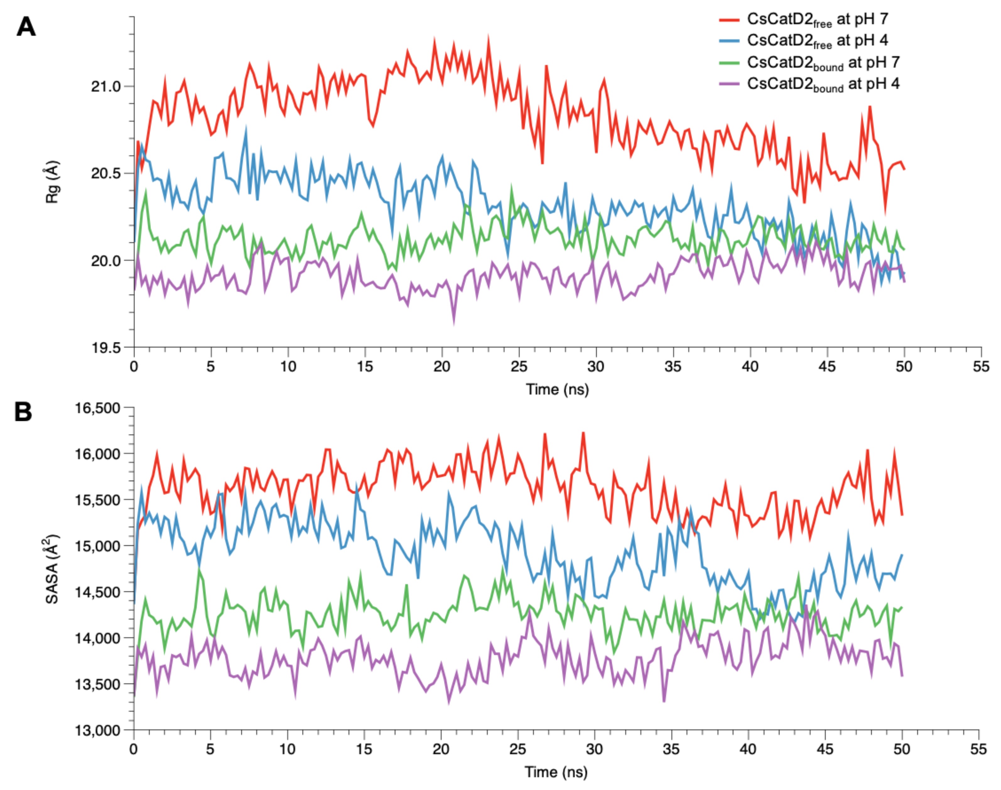
2.6. Acidic pH Enhances the Compactness of CsCatD2free to Converge to the Level of Bound Forms
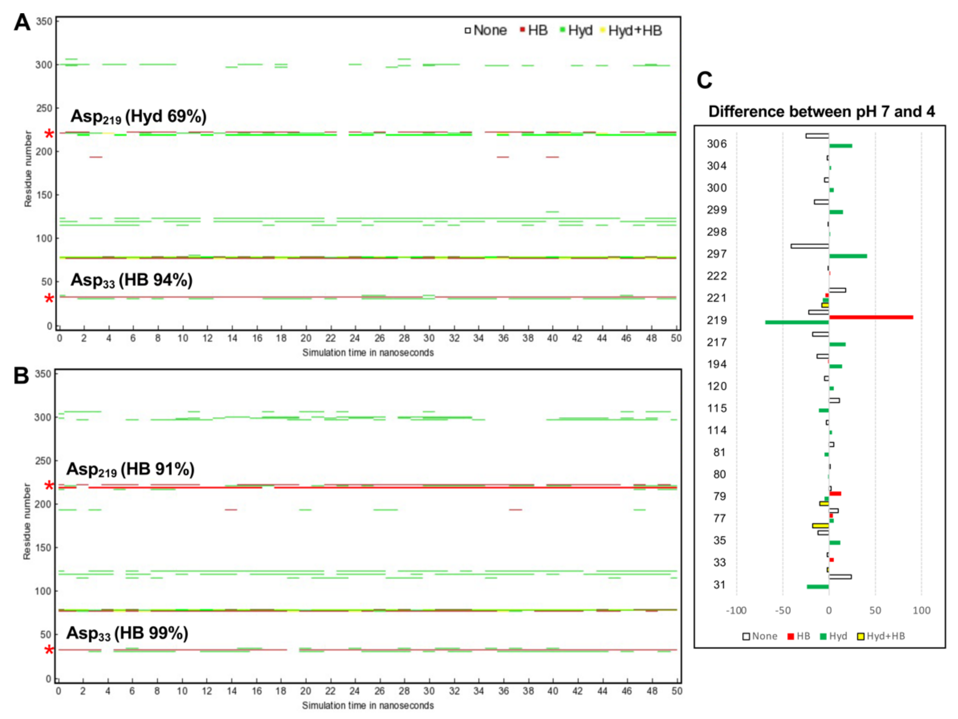
2.7. Acidic pH Alters Correlated Motions and Hydrogen Bonds Occupancy
2.8. Twisting of the Active Site Pocket by the Bound Inhibitor and Acidic pH
2.9. Effect of Acidic pH on Flap Dynamics and Its Correlation with the Twisting Motion of the Active Site Pocket
3. Materials and Methods
3.1. Homology Modeling and Verification
3.2. Structural Comparison
3.3. MD Simulation
3.4. Post-MD Analysis
4. Conclusions
Supplementary Materials
Author Contributions
Funding
Institutional Review Board Statement
Informed Consent Statement
Data Availability Statement
Acknowledgments
Conflicts of Interest
References
- Lun, Z.R.; Gasser, R.B.; Lai, D.H.; Li, A.X.; Zhu, X.Q.; Yu, X.B.; Fang, Y.Y. Clonorchiasis: A key foodborne zoonosis in China. Lancet Infect. Dis. 2005, 5, 31–41. [Google Scholar] [CrossRef]
- Na, B.K.; Pak, J.H.; Hong, S.J. Clonorchis sinensis and clonorchiasis. Acta Trop. 2020, 203, 105309. [Google Scholar] [CrossRef]
- Choi, B.I.; Han, J.K.; Hong, S.T.; Lee, K.H. Clonorchiasis and cholangiocarcinoma: Etiologic relationship and imaging diagnosis. Clin. Microbiol. Rev. 2004, 17, 540–552. [Google Scholar] [CrossRef] [PubMed]
- Vennervald, B.J.; Polman, K. Helminths and malignancy. Parasite Immunol. 2009, 31, 686–696. [Google Scholar] [CrossRef] [PubMed]
- Bouvard, V.; Baan, R.; Straif, K.; Grosse, Y.; Secretan, B.; El Ghissassi, F.; Benbrahim-Tallaa, L.; Guha, N.; Freeman, C.; Galichet, L.; et al. A review of human carcinogens-Part B: Biological agents. Lancet Oncol. 2009, 10, 321–322. [Google Scholar] [CrossRef]
- Delcroix, M.; Sajid, M.; Caffrey, C.R.; Lim, K.C.; Dvorak, J.; Hsieh, I.; Bahgat, M.; Dissous, C.; McKerrow, J.H. A multienzyme network functions in intestinal protein digestion by a platyhelminth parasite. J. Biol. Chem. 2006, 281, 39316–39329. [Google Scholar] [CrossRef]
- Sajid, M.; McKerrow, J.H. Cysteine proteases of parasitic organisms. Mol. Biochem. Parasitol. 2002, 120, 1–21. [Google Scholar] [CrossRef]
- Na, B.K.; Kim, S.H.; Lee, E.G.; Kim, T.S.; Bae, Y.A.; Kang, I.; Yu, J.R.; Sohn, W.M.; Cho, S.Y.; Kong, Y. Critical roles for excretory-secretory cysteine proteases during tissue invasion of Paragonimus westermani newly excysted metacercariae. Cell. Microbiol. 2006, 8, 1034–1046. [Google Scholar] [CrossRef]
- Na, B.K.; Kang, J.M.; Sohn, W.M. CsCF-6, a novel cathepsin F-like cysteine protease for nutrient uptake of Clonorchis sinensis. Int. J. Parasitol. 2008, 38, 493–502. [Google Scholar] [CrossRef] [PubMed]
- Robinson, M.W.; Dalton, J.P.; Donnelly, S. Helminth pathogen cathepsin proteases: It’s a family affair. Trends Biochem. Sci. 2008, 33, 601–608. [Google Scholar] [CrossRef]
- Pearson, M.S.; Ranjit, N.; Loukas, A. Blunting the knife: Development of vaccines targeting digestive proteases of blood-feeding helminth parasites. Biol. Chem. 2010, 391, 901–911. [Google Scholar] [CrossRef] [PubMed]
- Cwiklinski, K.; Donnelly, S.; Drysdale, O.; Jewhurst, H.; Smith, D.; De Marco Verissimo, C.; Pritsch, I.C.; O’Neill, S.; Dalton, J.P.; Robinson, M.W. The cathepsin-like cysteine peptidases of trematodes of the genus Fasciola. Adv. Parasitol. 2019, 104, 113–164. [Google Scholar]
- Horn, M.; Fajtova, P.; Rojo Arreola, L.; Ulrychova, L.; Bartosova-Sojkova, P.; Franta, Z.; Protasio, A.V.; Opavsky, D.; Vondrasek, J.; McKerrow, J.H.; et al. Trypsin- and Chymotrypsin-like serine proteases in Schistosoma mansoni—‘The undiscovered country’. PLoS Negl. Trop. Dis. 2014, 8, e2766. [Google Scholar] [CrossRef] [PubMed]
- Kang, J.M.; Ju, H.L.; Ju, J.W.; Sohn, W.M.; Kim, T.S.; Bahk, Y.Y.; Hong, S.J.; Na, B.K. Comparative biochemical and functional properties of two leucine aminopeptidases of Clonorchis sinensis. Mol. Biochem. Parasitol. 2012, 182, 17–26. [Google Scholar] [CrossRef]
- Williamson, A.L.; Brindley, P.J.; Abbenante, G.; Datu, B.J.; Prociv, P.; Berry, C.; Girdwood, K.; Pritchard, D.I.; Fairlie, D.P.; Hotez, P.J.; et al. Hookworm aspartic protease, Na-APR-2, cleaves human hemoglobin and serum proteins in a host-specific fashion. J. Infect. Dis. 2003, 187, 484–494. [Google Scholar] [CrossRef] [PubMed]
- Caffrey, C.R.; Placha, L.; Barinka, C.; Hradilek, M.; Dostal, J.; Sajid, M.; McKerrow, J.H.; Majer, P.; Konvalinka, J.; Vondrasek, J. Homology modeling and SAR analysis of Schistosoma japonicum cathepsin D (SjCD) with statin inhibitors identify a unique active site steric barrier with potential for the design of specific inhibitors. Biol. Chem. 2005, 386, 339–349. [Google Scholar] [CrossRef]
- Sojka, D.; Hartmann, D.; Bartosova-Sojkova, P.; Dvorak, J. Parasite Cathepsin D-Like Peptidases and Their Relevance as Therapeutic Targets. Trends Parasitol. 2016, 32, 708–723. [Google Scholar] [CrossRef]
- Dougall, A.M.; Skwarczynski, M.; Khoshnejad, M.; Chandrudu, S.; Daly, N.L.; Toth, I.; Loukas, A. Lipid core peptide targeting the cathepsin D hemoglobinase of Schistosoma mansoni as a component of a schistosomiasis vaccine. Hum. Vaccin. Immunother. 2014, 10, 399–409. [Google Scholar] [CrossRef]
- Hartman, A.M.; Mondal, M.; Radeva, N.; Klebe, G.; Hirsch, A.K. Structure-Based Optimization of Inhibitors of the Aspartic Protease Endothiapepsin. Int. J. Mol. Sci. 2015, 16, 19184–19194. [Google Scholar] [CrossRef] [PubMed]
- Brinkworth, R.I.; Prociv, P.; Loukas, A.; Brindley, P.J. Hemoglobin-degrading, aspartic proteases of blood-feeding parasites: Substrate specificity revealed by homology models. J. Biol. Chem. 2001, 276, 38844–38851. [Google Scholar] [CrossRef]
- Li, J.; Chi, Z.; Liu, Z.; Yue, L.; Peng, Y.; Wang, L. Cloning and characterization of a novel aspartic protease gene from marine-derived Metschnikowia reukaufii and its expression in E. coli. Appl. Biochem. Biotechnol. 2009, 159, 119–132. [Google Scholar] [CrossRef]
- Williamson, A.L.; Brindley, P.J.; Loukas, A. Hookworm cathepsin D aspartic proteases: Contributing roles in the host-specific degradation of serum proteins and skin macromolecules. Parasitology 2003, 126, 179–185. [Google Scholar] [CrossRef] [PubMed]
- Kang, J.M.; Yoo, W.G.; Le, H.G.; Thai, T.L.; Hong, S.J.; Sohn, W.M.; Na, B.K. Partial Characterization of Two Cathepsin D Family Aspartic Peptidases of Clonorchis sinensis. Korean J. Parasitol. 2019, 57, 671–680. [Google Scholar] [CrossRef]
- Boehr, D.D.; D’Amico, R.N.; O’Rourke, K.F. Engineered control of enzyme structural dynamics and function. Protein Sci. 2018, 27, 825–838. [Google Scholar] [CrossRef] [PubMed]
- McGillewie, L.; Ramesh, M.; Soliman, M.E. Sequence, Structural Analysis and Metrics to Define the Unique Dynamic Features of the Flap Regions Among Aspartic Proteases. Protein J. 2017, 36, 385–396. [Google Scholar] [CrossRef]
- Berman, H.M.; Westbrook, J.; Feng, Z.; Gilliland, G.; Bhat, T.N.; Weissig, H.; Shindyalov, I.N.; Bourne, P.E. The Protein Data Bank. Nucleic Acids Res. 2000, 28, 235–242. [Google Scholar] [CrossRef]
- Marciniszyn, J., Jr.; Hartsuck, J.A.; Tang, J. Mode of inhibition of acid proteases by pepstatin. J. Biol. Chem. 1976, 251, 7088–7094. [Google Scholar] [CrossRef]
- Hanova, I.; Brynda, J.; Houstecka, R.; Alam, N.; Sojka, D.; Kopacek, P.; Maresova, L.; Vondrasek, J.; Horn, M.; Schueler-Furman, O.; et al. Novel Structural Mechanism of Allosteric Regulation of Aspartic Peptidases via an Evolutionarily Conserved Exosite. Cell Chem. Biol. 2018, 25, 318–329.e4. [Google Scholar] [CrossRef]
- Mancilla-Olea, M.I.; Ortega-Lopez, J.; Figueroa-Angulo, E.E.; Avila-Gonzalez, L.; Cardenas-Guerra, R.E.; Miranda-Ozuna, J.F.T.; Gonzalez-Robles, A.; Hernandez-Garcia, M.S.; Sanchez-Ayala, L.; Arroyo, R. Trichomonas vaginalis cathepsin D-like aspartic proteinase (Tv-CatD) is positively regulated by glucose and degrades human hemoglobin. Int. J. Biochem. Cell Biol. 2018, 97, 1–15. [Google Scholar] [CrossRef]
- Minarowska, A.; Gacko, M.; Karwowska, A.; Minarowski, L. Human cathepsin D. Folia HistoChem. CytoBiol. 2008, 46, 23–38. [Google Scholar] [CrossRef]
- Karubiu, W.; Bhakat, S.; McGillewie, L.; Soliman, M.E. Flap dynamics of plasmepsin proteases: Insight into proposed parameters and molecular dynamics. Mol. Biosyst. 2015, 11, 1061–1066. [Google Scholar] [CrossRef]
- Arodola, O.A.; Soliman, M.E. Molecular Dynamics Simulations of Ligand-Induced Flap Conformational Changes in Cathepsin-D-A Comparative Study. J. Cell Biochem. 2016, 117, 2643–2657. [Google Scholar] [CrossRef]
- Berjanskii, M.V.; Wishart, D.S. A simple method to predict protein flexibility using secondary chemical shifts. J. Am. Chem. Soc. 2005, 127, 14970–14971. [Google Scholar] [CrossRef]
- Dostal, J.; Pecina, A.; Hruskova-Heidingsfeldova, O.; Mareckova, L.; Pichova, I.; Rezacova, P.; Lepsik, M.; Brynda, J. Atomic resolution crystal structure of Sapp2p, a secreted aspartic protease from Candida parapsilosis. Acta Crystallogr. D Biol. Crystallogr. 2015, 71, 2494–2504. [Google Scholar] [CrossRef]
- Masa, M.; Maresova, L.; Vondrasek, J.; Horn, M.; Jezek, J.; Mares, M. Cathepsin D propeptide: Mechanism and regulation of its interaction with the catalytic core. Biochemistry 2006, 45, 15474–15482. [Google Scholar] [CrossRef]
- Lee, A.Y.; Gulnik, S.V.; Erickson, J.W. Conformational switching in an aspartic proteinase. Nat. Struct. Biol. 1998, 5, 866–871. [Google Scholar] [CrossRef]
- Lobanov, M.; Bogatyreva, N.S.; Galzitskaia, O.V. [Radius of gyration is indicator of compactness of protein structure]. Mol. Biol. 2008, 42, 701–706. [Google Scholar] [CrossRef]
- Richmond, T.J. Solvent accessible surface area and excluded volume in proteins. Analytical equations for overlapping spheres and implications for the hydrophobic effect. J. Mol. Biol. 1984, 178, 63–89. [Google Scholar] [CrossRef]
- Kumalo, H.M.; Soliman, M.E. A comparative molecular dynamics study on BACE1 and BACE2 flap flexibility. J. Recept. Signal. Transduct. Res. 2016, 36, 505–514. [Google Scholar] [CrossRef] [PubMed]
- Krieger, E.; Nabuurs, S.B.; Vriend, G. Homology modeling. Methods Biochem. Anal. 2003, 44, 509–523. [Google Scholar]
- Lovell, S.C.; Davis, I.W.; Arendall, W.B., 3rd; de Bakker, P.I.; Word, J.M.; Prisant, M.G.; Richardson, J.S.; Richardson, D.C. Structure validation by Calpha geometry: Phi, psi and Cbeta deviation. Proteins 2003, 50, 437–450. [Google Scholar] [CrossRef]
- Colovos, C.; Yeates, T.O. Verification of protein structures: Patterns of nonbonded atomic interactions. Protein Sci. 1993, 2, 1511–1519. [Google Scholar] [CrossRef] [PubMed]
- Wiederstein, M.; Sippl, M.J. ProSA-web: Interactive web service for the recognition of errors in three-dimensional structures of proteins. Nucleic Acids Res. 2007, 35, W407–W410. [Google Scholar] [CrossRef] [PubMed]
- Zhang, Y.; Skolnick, J. TM-align: A protein structure alignment algorithm based on the TM-score. Nucleic Acids Res. 2005, 33, 2302–2309. [Google Scholar] [CrossRef] [PubMed]
- Krieger, E.; Vriend, G. New ways to boost molecular dynamics simulations. J. Comput. Chem. 2015, 36, 996–1007. [Google Scholar] [CrossRef] [PubMed]
- Mukherjee, B.; Tessaro, F.; Vahokoski, J.; Kursula, I.; Marq, J.B.; Scapozza, L.; Soldati-Favre, D. Modeling and resistant alleles explain the selectivity of antimalarial compound 49c towards apicomplexan aspartyl proteases. EMBO J. 2018, 37, e98047. [Google Scholar] [CrossRef]
- McGillewie, L.; Soliman, M.E. Flap flexibility amongst plasmepsins I, II, III, IV, and V: Sequence, structural, and molecular dynamics analyses. Proteins 2015, 83, 1693–1705. [Google Scholar] [CrossRef] [PubMed]
- Seeber, M.; Felline, A.; Raimondi, F.; Muff, S.; Friedman, R.; Rao, F.; Caflisch, A.; Fanelli, F. Wordom: A user-friendly program for the analysis of molecular structures, trajectories, and free energy surfaces. J. Comput Chem. 2011, 32, 1183–1194. [Google Scholar] [CrossRef]
- Grant, B.J.; Rodrigues, A.P.; ElSawy, K.M.; McCammon, J.A.; Caves, L.S. Bio3d: An R package for the comparative analysis of protein structures. Bioinformatics 2006, 22, 2695–2696. [Google Scholar] [CrossRef]


Publisher’s Note: MDPI stays neutral with regard to jurisdictional claims in published maps and institutional affiliations. |
© 2021 by the authors. Licensee MDPI, Basel, Switzerland. This article is an open access article distributed under the terms and conditions of the Creative Commons Attribution (CC BY) license (https://creativecommons.org/licenses/by/4.0/).
Share and Cite
Kang, J.-M.; Lê, H.G.; Na, B.-K.; Yoo, W.G. pH-Dependent Structural Dynamics of Cathepsin D-Family Aspartic Peptidase of Clonorchis sinensis. Pathogens 2021, 10, 1128. https://doi.org/10.3390/pathogens10091128
Kang J-M, Lê HG, Na B-K, Yoo WG. pH-Dependent Structural Dynamics of Cathepsin D-Family Aspartic Peptidase of Clonorchis sinensis. Pathogens. 2021; 10(9):1128. https://doi.org/10.3390/pathogens10091128
Chicago/Turabian StyleKang, Jung-Mi, Hương Giang Lê, Byoung-Kuk Na, and Won Gi Yoo. 2021. "pH-Dependent Structural Dynamics of Cathepsin D-Family Aspartic Peptidase of Clonorchis sinensis" Pathogens 10, no. 9: 1128. https://doi.org/10.3390/pathogens10091128
APA StyleKang, J.-M., Lê, H. G., Na, B.-K., & Yoo, W. G. (2021). pH-Dependent Structural Dynamics of Cathepsin D-Family Aspartic Peptidase of Clonorchis sinensis. Pathogens, 10(9), 1128. https://doi.org/10.3390/pathogens10091128


