Review

Control Strategies to Cope with Late Wilt of Maize

1
Plant Sciences Department, MIGAL—Galilee Research Institute, Tarshish 2, Kiryat Shmona 11016, Israel
2
Faculty of Sciences, Tel-Hai College, Upper Galilee, Kiryat Shemoneh 12210, Israel
Pathogens 2022, 11(1), 13; https://doi.org/10.3390/pathogens11010013
Submission received: 2 December 2021 / Revised: 15 December 2021 / Accepted: 20 December 2021 / Published: 23 December 2021

Abstract

:
Control of maize late wilt disease (LWD) has been at the forefront of research efforts since the discovery of the disease in the 1960s. The disease has become a major economic restraint in highly affected areas such as Egypt and Israel, and is of constant concern in other counties. LWD causes dehydration and collapsing at a late stage of maize cultivation, starting from the male flowering phase. The disease causal agent, Magnaporthiopsis maydis, is a seed- and soil-borne phytoparasitic fungus, penetrating the roots at sprouting, colonizing the vascular system without external symptoms, and spreading upwards in the xylem, eventually blocking the water supply to the plant’s upperparts. Nowadays, the disease’s control relies mostly on identifying and developing resistant maize cultivars. Still, host resistance can be limited because M. maydis undergoes pathogenic variations, and virulent strains can eventually overcome the host immunity. This alarming status is driving researchers to continue to seek other control methods. The current review will summarize the various strategies tested over the years to minimize the disease damage. These options include agricultural (crop rotation, cover crop, no-till, flooding the land before sowing, and balanced soil fertility), physical (solar heating), allelochemical, biological, and chemical interventions. Some of these methods have shown promising success, while others have contributed to our understanding of the disease development and the environmental and host-related factors that have shaped its outcome. The most updated global knowledge about LWD control will be presented, and knowledge gaps and future aims will be discussed.

Graphical Abstract

1. Introduction

Zea mays L. (maize, corn) is one of the world’s leading crops for food, feed, and fuel and as a raw material for different industrial products [1]. Worldwide annual maize production is expanding at a rate of 1.6%. It was forecast that this rate will not meet the global demand in 2050 [2]. Among many diseases threatening this cultivar [3,4], late wilt disease (LWD) has been reported so far in 10 countries and is considered a major concern in highly infected countries such as Egypt [5], Israel [6], India [7], Spain, and Portugal [8]. Economic losses due to LWD were up to 40% in Egypt [9], 50–100% in Israel [10,11], and 51% in India [12]. Incidences of the disease can reach 100% in Egypt and Israel, and 70% in India. Although the disease has not been reported in the United States, M. maydis is regarded as a potentially high-risk phytopathogen [13,14]. LWD harms yield production by erupting at the flowering growth phase, resulting in severe dehydration and plant death.
Since the discovery of LWD in Egypt in the early 1960s [15], worldwide scientific efforts have led to much progress in understanding the disease mode and the pathogen causing it, Magnaporthiopsis maydis [13]. Moreover, specific research tools for the study of LWD were developed and applied in the lab, in growth room experiments under controlled conditions, and in field trials. A significant part of these efforts was dedicated to creating diverse control methods to restrict the disease’s burst and spread and minimize its effect on commercial maize manufacture. Previously, we reviewed the techniques developed over the years to study LWD and monitor its causal agent [16]. A follow-up review summarized the accumulated scientific knowledge and future perspectives. These aspects include the geographic disease distribution, the pathogenesis (including the environmental factors affecting it), the symptoms’ evolvement, and their outcome effect on commercial production [17]. All the updated information regarding the pathogen itself, M. maydis, was also summarized. This includes the fungus life cycle, primary and secondary hosts, interactions with other phytopathogens, and the fungus’ ability to survive under different environmental conditions. These aspects will be summarized here briefly.
The current review focuses on the vast efforts dedicated in the past 60 years to late wilt disease control. The inspected control methods produced different degrees of success and include agricultural options (flood fallowing and balanced soil fertility) [18,19], biofriendly approaches [20], physical (solar heating) [21], allelochemical [22], and chemical pesticide [6,23,24] practices. Recently, the tillage system’s impact, the cover crop, and the crop rotation have been shown to serve as bioprotective factors against M. maydis [25,26].
A targeted research effort led to advancement in our capability to eradicate LWD chemically. A practical, efficient, and economic Azoxystrobin-based control protocol [10,24,27,28] was developed, which can be applied commercially to protect LWD-susceptible maize cultivars. Notwithstanding this recent encouraging achievement, the intensive chemical intervention has several short- and long-term drawbacks. In the short term, an intensive chemical application may cause the emergence of resistance to the fungicide. Such situations are becoming more and more common [29]. In the long term, phytoparasitic fungi chemical eradication may result in environmental, human, and animal hazards.
The limitation of chemical fungicides has become critical and is currently a global priority [30]. Hence, considerable research efforts in the past two decades were dedicated to seeking alternative methods of LWD control. Most of these efforts focus on eco-friendly substitutions to traditional chemical approaches. These consist of using Trichoderma spp. or other beneficial microorganisms as a biocontrol agent (see, for example, [20,31]). Late wilt green control studies also aimed at developing soil conservation practices that promote antagonizing mycorrhizal fungi (summarized by [31]). Even though this scientific course has been extensively explored against many harmful plant pathogens [32], in regard to LWD, substantial knowledge gaps exist. Consequently, the potential of green approaches to control M. maydis has only now been revealed.
Currently, the most eco-friendly, cost-effective, and efficient method to restrict M. maydis is by using highly resistant maize varieties [33,34]. Yet, the discovery of M. maydis highly aggressive isolates [8,35,36] is a constant problem. These fungal strains may threaten resistant maize cultivars. Indeed, growing resistance cultivars for extended periods in the same location may lead to gradual LWD susceptibility weakening [16,24]. This concerning situation pushes researchers to continue seeking new methods to control LWD.
The current review will summarize the many approaches tested to restrain the disease’s spread and damage, stating their advantages and limitations. It will also recommend the application of these methods in high- or low-risk scenarios (resulting from host resistance degree and soil infection load). Ultimately, the review will highlight knowledge gaps and future research focus points that should be addressed to advance LWD-safe commercial maize production capability.

2. Late Wilt Disease

2.1. The Pathogen

The late wilt causal agent, M. maydis, is a seed-borne and soil-borne vascular wilt fungal pathogen that penetrates the host roots and colonizes the xylem tissue [37,38]. The taxonomic tree of this fungus is: phylum: Ascomycota, subphylum: Pezizomycotina, class: Sordariomycetes, subclass: Sordariomycetidae, family: Magnaporthaceae, genus: Magnaporthiopsis, species: Magnaporthiopsis maydis (the most updated scientific name of the pathogen [39,40], Index Fungorum database, website: http://www.indexfungorum.org/names/NamesRecord.asp?RecordID=810225, accessed on date 30 November 2021). Former scientific names are Cephalosporium maydis (Samra, Sabet, & Hing, 1963) [41] and Harpophora maydis (Samra, Sabet, & Hing, 1963; Gams, 2000) [42].
To date, no perfect stage for M. maydis has been identified [43], and M. maydis reproduces asexually through sclerotia and spores [41]. This fungus may be considered necrotrophic because it thrives on the remains of dead plant tissues after killing its host. Yet, it is able to survive for a lengthy period (through the whole maize growth period) on living susceptible genotypes and asymptomatically in resistant genotypes and alternative species hosts. So, it may be better defined as a hemibiotrophic fungus.
M. maydis spread as sclerotia, spores, or hyphae on the plants’ residuals [38]. The pathogen can persist in the stubble and maize debris; no-till systems may help maintain it [13]. M. maydis can survive in the ground for lengthy periods or by thriving inside diverse host plants, such as lupine (Lupinus termis L.) [44], cotton (Gossypium hirsutum L.) [45,46], watermelon (Citrullus lanatus), and green foxtail (Setaria viridis) [45,47].

2.2. Geographic Distribution

LWD has been reported so far in 10 countries: Egypt (1961) [48], India (1970) [49], Hungary (1998) [50], Spain and Portugal (2011) [51], Israel (2013) [11], and possibly Nepal (2015) [52]. The report in Nepal referred to Cephalosporium acremonium, a synonym of the black bundle disease agent [38,53]. Acremonium maydis, the black bundle disease agent, had mistakenly been referred to as M. maydis in the past. There are also unconfirmed reports (summarized by Johal et al., 2004 [13]) that LWD was discovered in Italy, Romania, and Kenya.
Global LWD spreading was attributed mainly to infected seeds’ transmission. Indeed, Pecsi and Nemeth (1998) [50] presumed that late wilt spread to Hungary by importing infected seeds. M. maydis was detected in 39 out of 42 seed samples in Egypt [54]. In Hungary, Michail et al. (1999) [37] detected the fungus at a higher proportion in white maize cultivar seeds (1–9%) than in yellow cultivar seeds (1–3%). The pathogen was detected in the embryo, the endosperm, and the seed coat in 12 of the 13 seed samples tested.

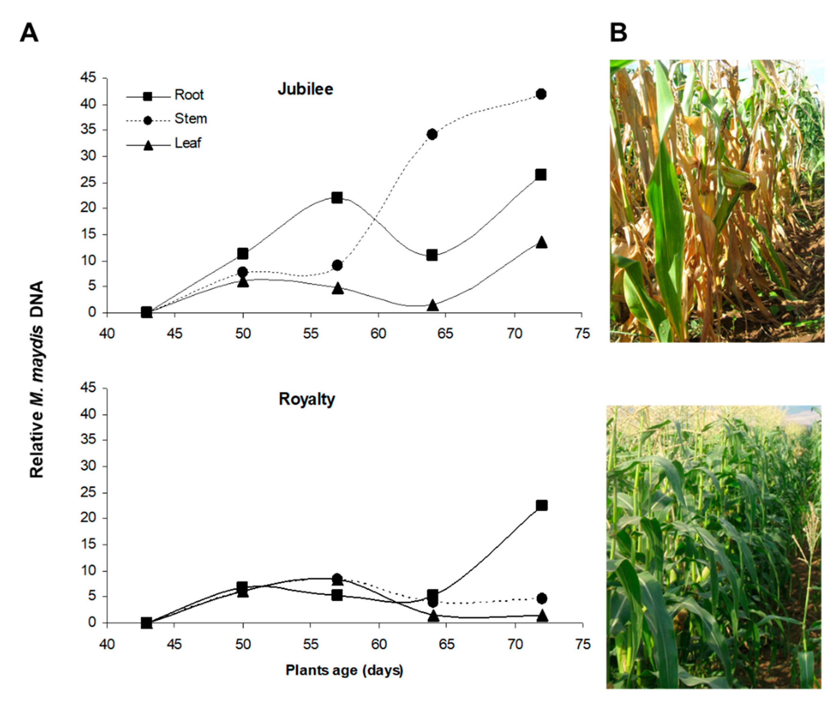
2.3. Disease Cycle and Pathogenesis

The disease mode in LWD-sensitive maize cultivars is well detailed in the scientific literature. M. maydis infects maize seedlings mainly during the first three weeks by sowing through their roots or mesocotyl (the seed-coleoptile connecting tissue). As the plants grow, they are less infected and become LWD-resistant about 50 days after sowing [38]. The disease cycle starts when the fungus grows on the roots’ surface, producing hyphae with brown, short, thick-walled, and swollen cells [38]. After root penetration, M. maydis colonizes xylem tissue (identified 21 days after sowing) and is rapidly transferred to the upper parts of the plant. M. maydis may occasionally cause seed rot or pre-emergence damping-off under high inoculum pressure [55].
The second critical infection phase starts when tassels first emerge (ca. day 55–65, R1 silking, silks visible outside the husks). At this stage, the fungus hyphae and conidia appear throughout the stalk [38], pathogen DNA levels reach their highest point in the stems [11] (Figure 1), and the first aboveground symptoms are revealed. Later, when M. maydis colonizes the entire stalk, avascular tissue occlusion by hyphae and gum-like secreted materials occurs, resulting in water supply suffocation, rapid dehydration, and death [13,38]. At the end of the growing season (12–13 weeks after planting), the fungus is identified in different ear parts [37]. It is detected mainly in the ear branch but also in the cob, seeds, ear husks, and silk of naturally infected maize cultivars. The symptoms are intensified under drought stress [56,57]. Although the disease appears as patches scattered in the field in many cases [16], LWD may result in total field infection and total yield loss in heavily infected areas planted with susceptible maize cultivars [10,24]. A parallel asymptomatic infection mode, with some delay, occurs in resistant cultivars (Figure 1). This process can result in infected seeds that enhance the pathogen spread [10,11].

2.4. Disease Symptoms

The degree of disease symptoms is directly affected by soil infection load, maize genotype susceptibility, and environmental growing conditions, particularly the watering regime [18,38,56,57,58]. The first disease signs of LWD can be seen in the seedlings phase. M. maydis can destructively affect the seedlings’ aboveground emergence [55,59]. It may also cause reduced development of the sprouts’ roots [6,15], color alteration, and necrosis [22]. Small necrotic lesions (2–4 mm long) on the roots were documented three weeks post-inoculation [22]. Their size increased gradually to lengths of 10–14 mm. Intriguingly, similar dry dark-red local lesions on the roots near the soil surface were observed in M. maydis-inoculated cotton seedlings [46].
The first aboveground wilt symptoms appear approximately 50–60 days after sowing, near the flowering stage (from the R1 silking to the R2 blister) [38]. These first symptoms are parallel to the fungus’ establishment in the plant vascular system (see Section 2.3). They consist of rapid wilting of the first aboveground leaves that spread upwards during the subsequent two weeks [6]. As the disease advances, the leaves gradually alter their color and wilt [60]. Yellowish to brown-reddish streaks may be seen on the lower internode (Figure 2A–C) and a color alteration of the vascular bundles appears (Figure 2D). At this stage, the lower stem dehydrates (mainly at the internodes) and has a shrunken and hollow appearance (Figure 2E).
The roots and stalk vascular bundles become blocked, and their color in the stalk alters from light-yellow or white to dark-yellow and to brown [6,10,28,38]. Plants’ diseased leaves have a high proline content, probably linked with dryness stress due to limited water flow caused by tracheary elements plugging [61]. In addition, the infection may reduce the number of vascular bundles (seen in a cross-section of the internode). Late wilt infection is frequently associated with secondary pathogens infection that enhances the stem symptoms [62]. Eventually, fewer ears are produced, and if kernels do develop, they are often immature and damaged [6,11] (Figure 3) and infested with the pathogen. Seed quantity [63] and quality [28] are negatively correlated to disease severity. The infected kernels can result in seed rot and pre-emergence damping-off [64]. Finally, these disease processes can lead to the plant’s death (Figure 3).

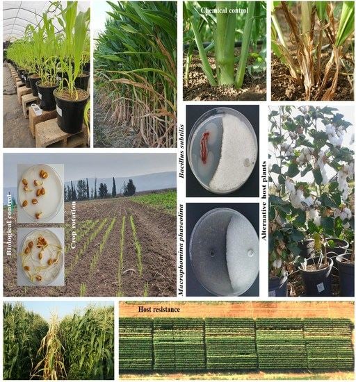
3. Control Strategies

3.1. Host Resistance

The magnitude of LWD losses largely depends upon the degree of soil infestation and the susceptibility of the grown cultivars. The use of resistant genotypes is considered the best, most practical, eco-friendly, and cost-effective method of controlling the disease [65,66]. This method is preferred even though resistant hybrids to LWD are often low-yielding or have other undesirable agronomic characteristics [67]. A program to develop and identify new hybrid strains resistant to LWD has been operating in Egypt since the 1980s [68], in Israel for more than a decade (R&D North, MIGAL—Galilee Research Institute, Kiryat Shmona, Israel) [28] (Figure 4), and was also reported in India [7].
Significant efforts were directed towards using specific genetic markers for LWD to identify resistant germplasm and subsequently develop genetically resistant maize inbred lines [43]. Several studies suggest that many genes control LWD resistance in maize [70]. Yet, little scientific data exist regarding LWD resistance alongside high yield in maize [67]. LWD resistance inheritance is complex, having significant genotype × environment interactions. Hence, directed selection for LWD tolerance is probably less effective [71].
Nonetheless, DNA markers could be a suitable substitute for such traits in maize. To this end, identification and validation of closely linked markers are needed. Indeed, quantitative trait locus/loci (QTL) that confer LWD resistance have been identified and validated [71,72]. Further research investigation is necessary to detect stable QTL with considerable phenotypic variation explained [71].
Despite these meaningful efforts, the reasons for LWD susceptibility differences among maize cultivars remain obscure. It was gradually revealed that the infection process’ outcome results from chemical and histological differences between cultivars [73]. Phenols as phytochemical compounds have many functions; one is to protect plants against pathogens [74]. It was shown earlier that resistant maize plants contained higher total and free phenolic compounds than susceptible plants [75,76,77]. Phenolic acids (ferulic acids and cinnamic) found in vitro in resistant maize cultivars could suppress the growth of M. maydis. The phenolic content in all maize cultivars increased after infection. This increase was higher in resistant cultivars compared to susceptible cultivars. Lately, it was proven that under artificial infection stress, from 30 to 90 days after sowing (DAS), a linear increase in phenolic content was more pronounced in the resistant cultivar compared to the susceptible one [73].
In contrast, LWD-susceptible cultivars have a higher total soluble sugar content that may be favored by the pathogen as a carbon source of nutrition. The stalks’ sugar content generally decreased when the plants matured. Infection with M. maydis increased sugar content in both resistant and susceptible cultivars [78]. The tendency for the gradual increase in phenolic substance concentrations and the gradual decrease in sugar content in the plant tissues, along with the growth session, may be the reason for the plant’s acquired immunity to late wilt infection. Sabet et al. supported these findings and discovered that most plants were infected when the inoculum was applied to young plants. The M. maydis infection became less frequent until no maize plants were infected after seven weeks from sowing [38].
Furthermore, the pathogen was found in the resistant maize cultivars, mostly in the infected roots and rarely in the vessels of the stem’s lower internodes [79]. Laccase secretion in M. maydis was recently investigated in vitro, in response to different host tissues [80]. This enzyme can catalyze the oxidation of phenolic substrates and may act as a fungal defense against antifungal phenols secreted by the plant. Studying the laccase role in the pathogen’s interactions with diferent hosts would be very interesting and can contribute to our understanding of the resistance mechanism.
Particularly intriguing, in a field trial, in 72 old plants of a resistant cultivar, the M. maydis DNA levels in the roots, stems, and leaves were similar to the fungal DNA spreading in a susceptible cultivar on day 57 [11] (Figure 1). This may explain why resistance is sometimes limited to a specific growth period. Eventually, some maize genotypes that are non-symptomatic in the harvesting stage (typically the R3–R4 growth stages three to four weeks after fertilization) will dehydrate and die after an additional one or two weeks [5].
Another aspect that may contribute to the immunity of LWD tolerance by some maize genotypes is their tissue structure. Indeed, the roots of resistant cultivars had a different structure of the tissue surrounding the xylem vessels [76,77]. Resistant LWD cultivars, in comparison to susceptible ones, had increased thickness of sheath bundles surrounding the vascular bundles [73] and more layers of sclerenchymatous cells surrounding the xylem vessels. These tissue layers may act as mechanical barriers protecting bundles against pathogen invasion. Indeed, the fungus appears to penetrate directly through the host cells to ramify in the cortex. After that, the hypha progresses both in and between cells towards the xylem [38]. Hyphae aggregate at the endodermis prior to the fungus breaking into the vascular bundle, three weeks after sowing.
Vascular bundles of resistant inbred lines revealed a higher number of xylem vessels than susceptible ones [73]. The pith area was also much larger in resistant inbred lines than in susceptible ones. This supports the conclusion that occlusion plays a pivotal role in causing LWD symptoms [81]. Indeed, the M. maydis virulent isolates’ ability to plug the host plant xylem vessels was proven [73]. Thus, mechanical suppression of water uptake through such blocked vessels could be concluded. Supporting this conclusion are the growing pieces of evidence that high water potential reduces LWD damage in the field [18,82].
Finally, plant hormones may influence M. maydis pathogenesis, the severity of the disease symptoms, and host susceptibility [83]. It was shown that the plant growth hormones, auxin (indole-3-acetic acid), cytokinin (kinetin), and gibberellin (gibberellic acid), inhibit the development of the LWD pathogen in vitro [84] (Figure 5).
Despite auxin’s marked restricting effect on the pathogen’s growth in vitro, treatments based on dimethylamine salt of 2,4-D (dicots’ herbicide that mimics auxin influence, 96.9% active ingredient, Aminobar, Luxembourg Industries Ltd., Tel Aviv, Israel) were inefficient. Applying this compound in a plate assay (O. Degani, personal communication) and in the field using the driplines irrigation system [84] failed to suppress late wilt or prevent its symptoms. So, do plant hormones play a vital role in the tolerance mechanism in some maize cultivars? This question should be explored in future studies.
Alongside these efforts, the development of new methods for tracking the pathogen [7,85,86], estimating its distribution and damage [8,14,87], and controlling it in various ways [22,25,65,88,89] remain major goals.

3.2. Chemical Control

3.2.1. From In Vitro Evaluation to a Field Assay of Selected Fungicides

It is important to locate LWD antagonists’ fungicides while evaluating their phytotoxicity and efficiency against M. maydis and other associated fungi (the stalk-rot complex) involved in LWD [90]. Together with this effort, developing rapid and efficient screening approaches to assess the potential of these fungicides is needed [10,91]. Preliminary tests on growth media plates aimed at screening many chemical preparations rapidly and indicating their efficiency can be conducted with minimal investment and can save time and effort. After eliminating inadequate potential fungicides, selected compounds will be verified in seeds, detached roots, and sprouts’ assays. Only in the final phase will a limited number of high-potential selected pesticides be tested in a field experiment throughout the growth season (Figure 6). While this method is important to reduce the high investment and the long time required for field trials, it is not flawless.
It is necessary to acknowledge that culture plate screening, seeds’ and detached roots’ assay, and even potted young plant trials under controlled conditions have partial ability to forecast field results [10]. Nonetheless, potted seedling experiments for chosen fungicides are essential. Such experiments do not depend on the year or season required for field growth and can be carried out throughout the year. They are also not affected by environmental high variability conditions accompanying open-air experiments.

3.2.2. Seed Coating

Seed coating is a common technique to protect emerging maize sprouts from soil-borne fungal pathogens. It has the potential of playing a vital role in shielding against M. maydis. Since the initial infection occurs during the seedling development period [38], the LWD can be managed efficiently with a fungicide seed treatment. Indeed, such attempts have been made in the past. In India, Begum et al. (1989, 1996) showed that seed treatments with captan, carboxin, carbendazim, and thiram significantly reduced late wilt severity and increased yield in the field [92,93]. Yield increased by 25.9% for carbendazim and 34.1% for captan (at 1 g/kg seed).
In contrast, seed treatments failed to prevent late wilt in Egyptian trials [23,94]. According to Johal et al. (2014) [13], such differences can result from variances in the virulence, chemical sensitivity of M. maydis isolates, or the consequences of the stalk-rot disease complex in Egyptian soils [90]. Nonetheless, non-chemical seed treatments were tested successfully in Egypt [95]. It was demonstrated that the usage of some rhizobacterial strains (Bacillus subtilis and Pseudomonas fluorescens) and organic compounds, such as compost and humic substances, as a seed treatment can prevent maize infection with LWD. In addition, under field conditions in regular and saline soil, rhizobacterial seed coating with B. subtilis and P. fluorescens either alone or in combination significantly decreased disease infection and enhanced plant yield compared to the control [88].
In Israel, in relatively resistant maize with only minor LWD symptoms, the Azoxystrobin seed coating (0.0025 mg active ingredient/seed) prevented fungal development and increased plant and cob weight [10]. Yet, this treatment could not defend a susceptible maize cultivar in heavily infested soil at the disease’s wilting burst (60 DAS) and later on. In follow-up work [28], the Azoxystrobin + Difenoconazole seed coating (AS + DC, “Ortiva-Top”, manufactured by Syngenta, Basel, Switzerland, supplied by Adama Makhteshim, Airport City, Israel) was applied. This treatment was efficient in the initial growth stages (up to 50 DAS). At the later growth stages, the AS-DC seed coating provided an additional layer of protection when combined with the injection of fungicides into the irrigation system, as elaborated in the following Section 3.2.3. This combination significantly reduced disease symptoms and the pathogen DNA in the plants’ lower stem to near-zero levels, resulting in high yield (quantity and quality) production.

3.2.3. Soil Treatments

Systemic fungicides and their fungi-toxic products translocate to maize leaves within two days and can last in maize roots for 90 days [23], so M. maydis may be repressed by these chemicals within the root, as well as in the soil. Thus, soil treatments with systemic fungicides, such as Benomyl (Benlate, methyl 1-(Butylcarbamoy l)-1H-1,3-benzimidazol-2-yl methylcarbamate, CAS no. 17804-35-2), were the preferred method of dealing with the disease.
In pot experiments with 15 fungicides conducted by Singh and Siradhana (1989) [23,96], LWD was significantly reduced by soil applications of Benomyl and Bavistin (CAS no. 63278-70-6, carbendazim), both at 0.1% and by 2% Bayleton (CAS no. 43121-43-3, triadimefon). Yet, over the years, during LWD research, it has become clear that fungicides that were successfully proven in pots may fail in field experiments. An example is the fungicide Benomyl, introduced in 1968 by DuPont (Wilmington, DE, USA). It is a systemic benzimidazole that is selectively toxic to microorganisms and invertebrates, especially earthworms, but non-toxic to mammals. It was found that Benomyl at 2.5 to 100 ppm concentrations completely inhibits LWD in pots (but not by applications 30 days after sowing or by seed treatments) [96]. Still, the application of 10 kg Benomyl/4200 m2 in the field failed to prevent the disease [94]. Lack of success has been attributed to reduced absorption of systemic fungicides by maize in the field soil compared to pot experiments [94].
The application method can be game-changing in this regard, as was proven in Egypt [23] and Israel (see Section 3.2.4). Abd-el-Rahim and colleagues (1982) found that the systemic fungicide Benylate applied at four 15-day intervals (2.5 kg/acre) after sowing resulted in the best LWD control [13]. In Israel, the application of fungicides at 15-day time intervals from sowing is an obligatory condition to achieve effective LWD control [24,27,28], as discussed in detail below. Nevertheless, it was estimated that the cost and labor required for frequent fungicide applications in the United States make this control method prohibitively expensive [13].

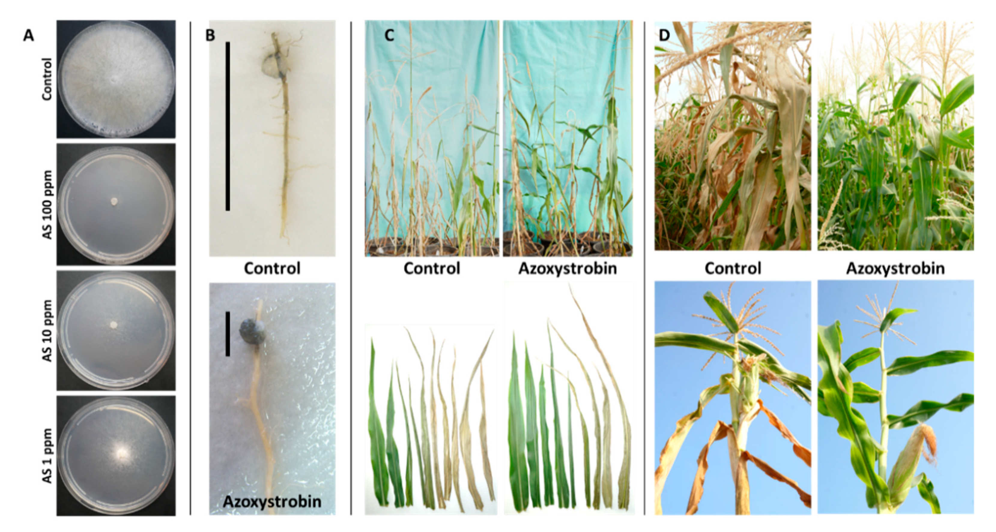
3.2.4. Azoxystrobin Irrigation-Based Treatments

Several chemicals were examined for their ability to restrict M. maydis, the causal disease agent in recent years [24,28]. Fast plate assay proved the success of Azoxystrobin against the pathogen (Figure 6), but applying its based commercial preparation in the field by spraying was ineffective [24]. In contrast, injecting Azoxystrobin directly into a dripline assigned for each row 15, 30, and 45 DAS inhibited wilt symptom development and recovered cob yield by 100%. However, this method is not always feasible, and to the best of our knowledge, has never been tested commercially due to the high cost of the irrigation system.
More recently, an efficient and more economically applicable solution to LWD was suggested that could be applied on a large scale to shield-susceptible corn varieties in commercial fields [28]. This application is based on antifungal mixtures having a different mode of action to prevent resistance development. The method involved seed coating and injection of Azoxystrobin and Difenoconazole mixture (AS+DC) into the irrigation system at three 15-day intervals from sowing. Economic efficacy was reached using one dripline for two adjacent rows (a row spacing of 50 cm instead of 96 cm). The short row spacing guaranteed the effective concentration of the antifungal compound near the roots. A quantitative real-time PCR (qPCR)-based molecular detection method, proven earlier [10], showed that due to the AS + DC treatment, M. maydis DNA levels in the host tissue dropped to near-zero values. In the coupled-row-based cultivation, this treatment reduced wilt symptoms by 41%. At the same time, it recovered yield to the common level in healthy fields (1.6 times more than the non-protected control). In addition, yield quality (A-class cobs at a weight exceeding 250 g) increased from 58% to 75% in this treatment.
Nowadays, drip irrigation is considered one of the most effective chemical methods to restrict maize LWD but it is the most expensive of the present alternatives [28]. The value of drip extension lines (without installation) is 50–60% of total costs, so reducing the extension number by half could save about 30–40% of the irrigation expenses and lead to savings in labor and time. Consequently, this successful treatment is now more cost effective and can be applied commercially to protect susceptible maize hybrids in infested fields against late wilt.

3.2.5. Fungicide Resistance

Azoxystrobin, a member of the Qo-inhibiting fungicides (QoIs) class, is considered one of the most important agricultural antifungals [97]. However, growing resistance to these antifungals and the resulting control failure are become a significant problem (see, for example, such cases in Magnaporthe grisea [98]). In the past two decades, ca. 30 phytopathogen species distributed across 20 genera were reported to exhibit field resistance toward QoI fungicides [99]. QoI fungicides disrupt respiration in the mitochondria by binding to the cytochrome bc1 enzyme complex Qo site, impairing electron transfer and thus inhibiting mitochondrial respiration and ATP production [97]. Since their mode of action is based on a single site, QoI fungicides pose a high risk for revealing fungicide resistance. Indeed, Azoxystrobin targets site mutations in cyt b gene (G143A, F129L) and additional mechanisms have been reported [100].
Under such circumstances, the suggestion is to avoid using QoI antifungals or to apply anti-resistance-emergence strategies such as mixing low-risk and high-risk antifungal compounds. These steps may help delay the emergence of resistance to the high-risk fungicide [101]. In addition, cross-resistance has been documented among all members of the QoI group (Fungicide Resistance Action Committee (FRAC) Code List© 2021). Consequently, mixing Azoxystrobin with other antifungal agents is critical for LWD control.

3.3. Biological Control

3.3.1. Strengthening Beneficial Microorganism Communities in the Soil and Their Secreted Metabolites

Since fungicide treatment limitation exerts increasing pressure in many countries due to environmental and potential health risks, searching for alternatives to cope with LWD is a continuous effort. Biopesticides are environmentally friendly. Therefore, this approach occupies an increasingly central place in worldwide scientific research to this end. To address this challenge, many studies were directed towards LWD biological control [20,31,102,103,104]. These methods include operating and strengthening beneficial microorganism communities in the soil (for example, by compost addition [102]) or direct intervention using antagonistic bacteria and fungi or their secreted metabolites. An example is B. subtilis MF497446 and Pseudomonas koreensis (two plant-growth-promoting rhizobacteria [31]). This microorganism’s mixture resulted in siderophore production and M. maydis repression. Moreover, the treatment prevented pre- and post-emergence damping-off and promoted the growth of greenhouse plants, proving to be highly efficient in the field in reducing infection and increasing the yield [31].
Seed treatments with biocontrol formulations (B. subtilis, Bacillus pumilus, P. fluorescens, Epicoccum nigrum) were suggested for maize LWD control and tested in the field with encouraging results [89]. The seed treatments were tested in two growing seasons, reducing the pathogen impact on pre-emergence damping-off, wilt incidences, and crop yield. In an additional study, the influence of maize root colonization by microorganisms was also practical for this purpose [104]. In this study, the rhizosphere actinomycetes (Streptomyces rochei, S. graminofaciens, S. gibsonii, S. annulatus, Candida maltosa, C. glabrata, C. slooffii) and the fungi Rhodotorula rubra significantly reduced M. maydis growth in vitro. These treatments were also highly influential in seed dressing under greenhouse-controlled conditions. Adding these species in the absence of the LWD pathogen significantly increased maize plant growth parameters [104].
Another example is the use of a filtrate of mixed strains of the cyanobacteria Anabaena oryzae, Nostocmuscorum, and N. calcicolawere [102]. An alternative approach was to use marine algae and cyanobacteria A. oryzae extracts exhibiting antifungal activities to target the LWD pathogen [103]. These algae include Corallina elongata, Jania rubens, Laurencia obtusa, Gelidium crinale, Enteromorpha compressa, and Ulva fasciata. The results are encouraging, with U. fasciata and A. oryzae exhibiting high antifungal activity against M. maydis [103].

3.3.2. Trichoderma spp. Maize Late Wilt Biocontrol

Late wilt disease can also be biologically controlled using Trichoderma spp. This genus’ species can form endophytic mutualistic relationships with various plant species [105]. Other Trichoderma species have been identified to possess biocontrol potential against plants’ fungal pathogens [106]. For instance, Trichoderma cutaneum reduced the incidence of LWD of maize under greenhouse conditions by 89% compared to the non-protected control (from 94% to 11%) [104]. Likewise, Trichoderma harzianum treatment in the field reduced the pre-emergence damping-off from 47% to 32% and increased the plants’ survival by 59% [64]. The application of Trichoderma viride alone, or even better with chitosan NPs combined with the mycorrhizae Glomus mosseae, controlled late wilt in the greenhouse and in field trials, and enhanced the plants’ growth indices [107]. Using the same combination of T. viride and mycorrhizae without chitosan NPs led to a low-efficiency LWD control. To maximize the impact of Trichoderma-based treatments, Elshahawy and El-Sayed (2018) [20] showed that extracts of the microalgae, Chlorella vulgaris, with each of the Trichoderma species, T. virens and Trichoderma koningii, led to effective LWD control in the greenhouse and in the field. These combinations led to a 72% reduction in disease frequency in the greenhouse and a 2.5-fold higher grain yield in the field [20].
The potential for using Trichoderma-based treatment against Israeli M. maydis strains has only recently been tested [108]. Examining eight marine [109] and soil isolates of Trichoderma spp. known for their high mycoparasitic potential revealed that Trichoderma longibrachiatum (T7407) and Trichoderma asperelloides (T203) isolates have solid antagonistic activity against the Israeli M maydis strain. These eco-friendly agents were tested in a series of experiments in the laboratory (Figure 7) and in a growth room under controlled conditions until their final examination in pots under field conditions throughout an entire growing season [108]. The T. longibrachiatum (T7407) green treatment significantly improved growth and yield indices to healthy plants’ levels, reduced pathogen DNA in the plants’ tissues by 98%, and prevented disease symptoms (Figure 8).
In solid and submerged media culture growth assays, these species secrete soluble metabolites that inhibit or kill the maize pathogen [108]. Such a metabolite was recently isolated and identified as pyrone 6-pentyl-2H-pyran-2-one (6-pentyl-α-pyrone or 6-PP) [110]. This potent M. maydis antifungal compound is secreted by Trichoderma asperellum (P1), an endophyte separated in our laboratory from maize seeds of a cultivar susceptible to LWD [111]. The 6-PP metabolite was previously identified as one of the key bioactive compounds of several Trichoderma species [112].
To test the bioprotective potential of these isolates for commercial maize production, follow-up work was performed. T. asperellum (P1) significantly improved the infected sprouts’ growth parameters and reduced M. maydis DNA in their roots [111]. The application of this endophytic species also excels in the field over an entire growing season [58]. At the season’s end, the T. asperellum treatment resulted in 1.6- and 1.3-fold improvement in the LWD symptoms in the lower stem and cob, respectively. Furthermore, this treatment led to 4.9-fold lower M. maydis DNA levels in the plants.
It was reported that the fungus T. longibrachiatum (T7407) produced 29% more fungal inhibitory effects than T. asperellum (P1) in solid growth media [110]. Active ingredients that will be isolated from T7407 could probably be more potent. The active ingredient isolation and identification in this and other potential Trichoderma spp.-secreted antifungal metabolites are of great importance and should be the focus of follow-up works.

3.3.3. Manipulating the Plant Microbiome

The natural microorganisms’ communities inhabiting the plant’s phyllosphere (the plant’s aboveground habitat) or the rhizosphere (the roots’ nearby habitat) comprise pathogenic and non-pathogenic members that may protect the plant from pathogens. The soil pathogen, M. maydis, interacts with the maize endophytes, which may provide the plant’s first defense line. Recently, such endophytes were isolated from six sweet and fodder maize hybrids with deferent sensitivity to LWD [111]. These include ten fungal species and one bacterial species, B. subtilis. The fungal species belonged to Chaetomium, Trichoderma, Penicillium, Rhizopus, Alternaria, and Fusarium genera. Some are plant pathogens (Alternaria alternata and Fusarium proliferatum), so they play complex roles in their interactions with the host plant. Enriching seeds with either Chaetomium subaffine or T. asperellum significantly promoted the infected plants’ growth parameters 42 days past sowing. The fungal species Chaetomium cochliodes, T. asperellum, Penicillium citrinum, and the bacteria B. subtilis treatments reduced the LWD pathogen DNA in the host plant’s roots [111].
In cotton plants, interactions between M. maydis and F. oxysporum (the cotton wilt agent) led to an interesting result—reduced symptoms of the cotton wilt disease [46]. The infection reduction was maximized when M. maydis preceded F. oxysporum in the soil compared to simultaneous inoculation. Co-infection of the plants with both fungi resulted in immunity compared to infection with F. oxysporum alone. In contrast, there was little or no protective outcome when M. maydis was added to the soil after F. oxysporum [46].
Similar antagonistic relationships in cotton plants were found between M. maydis and Macrophomina phaseolina, the charcoal rot disease agent [45]. In a plate confrontation assay, the fungi formed a clear line between the colonies at the meeting area, representing strong mutual antagonism. Under field conditions, M. phaseolina markedly recovered the plants’ health while eliminating the severe late wilt symptoms [45].

3.4. Agrotechnical Measures

3.4.1. Various Cultural Methods

Agrotechnical applications were also reported in varying degrees of maturity in field experiments. These methods, which had a beneficial impact on LWD suppression, include excessive irrigation (pot experiments conducted in an open-air enclosure) [56] and applying plant extracts (Lycium europaeum [22], aloe vera (Barbados Aloe) fleshy leaves, onion bulbs, garlic cloves, jimsonweed, and peppermint leaves [113]). In addition, soil solarization to increase temperatures above 35 °C with transparent polyethylene film [21], balanced soil fertility [19,114], and avoiding drought stress [56,61] can reduce LWD severity and yield losses. Magnaporthiopsis maydis survival is restricted to the top 20 cm of soil [115], and survival depends mainly on infected crop residues. Thus, sanitation measures such as deep tillage and annual plowing may significantly impact LWD (Dr. J. Leslie, personal communication) [14]. The use of no-till maize systems could eventually result in increased virulence of the pathogen or inoculum build-up in the soil. Finally, it was shown that organic compounds (compost tea, olive mill wastewater, and humic acid as seed treatment) [95] and non-traditional methods such as adding nanosilica and zinc oxide nanoparticles [89] have promising potential to reduce late wilt in the field.

3.4.2. Beneficial Mycorrhizal Communities

Preserving soil mycorrhizal fungi between growth periods has been essential in crop protection (summarized by [31]). Results from other crop diseases suggest that under no-till cropping, crops in a rotation or selected cover crops could support building mycorrhizal communities that function through a sequence of several crops [116]. Intensive tillage combined with extended periods where the field is unprocessed results in damaging the integrity of mycorrhizal networks. Preserving the integrity and continuity of the soil mycorrhizal networks may provide the plant with higher resistance to soil diseases [117], including late wilt disease [25]. Indeed, arbuscular mycorrhizal fungi (AMF) can improve plants’ resistance to biotic and abiotic stresses. This results from activating the plant’s local and systemic defense mechanisms [118].
Although global scientific efforts are focusing on seeking solutions to LWD based on eco-friendly biological methods, a lack of information exists on maize performance under LWD stress in reduced tillage and crop rotation. In Egypt, crop rotation with rice (Oryza sativa) provides some control [119]. Maize did not develop late wilt following paddy-cultivated rice, which increases the availability of Mn for subsequent crops [60]. In India, M. maydis remained viable in stem residues on the soil’s surface for one year, but the pathogen could not be recovered after ten months from stem pieces buried at 10 cm [82]. High soil moisture enhances LWD development, but saturated soil reduces it [119]. In Portugal, Patanita et al. (2020) [25] showed that M. maydis presence and grain production were significantly reduced when both minimum tillage and cover crop were applied. It was also found that arbuscular root colonization was higher following these practices.
In Israel, agricultural practice based on conserving soil microflora integrity (by avoiding tillage) and influencing its nature (by cultivating specific crops in a dual-season growth) was applied [26]. When maize was seeded on wheat soil, a significant improvement in the shoot’s fresh weight (47–54%) and cob (36–46%) was achieved compared to the other treatments (clover soil, commercial mycorrhiza preparation, and the control). This achievement was not affected drastically by tillage. It was followed by a sharp decrease in disease symptoms (73%) and the pathogen’s presence (82–64%) in the plants’ tissues. It was concluded that since wheat and maize are more closely related (they are both Poaceae) than clover and maize, they might share similar mycorrhizal networks adapted to perform better with these crops. Indeed, the growth promotion mentioned above and LWD tolerance were reduced in the clover-maize sequence, even more so when tillage was applied. To support this hypothesis, it was found that crop plants acquired a mycorrhizal community closely related to that of the former host plant and different from that found when the soil was disturbed by tillage or not cropped before the growth [116].
With the novelty of such studies, it is essential to indicate that crop rotation may have a long-term impact on soil fungus populations, which may only be evident after an extended period. In addition, no single cropping system is preferred for all fungi [120]. Thus, a tailored solution should be planned wisely to address the cultivar that needs to be protected, the crops in the rotation, the tillage system, and the pathogen/disease stresses. Such situations should be inspected in subsequent studies. Closely related crops such as maize and wheat may benefit from the same control strategy.

4. Future Challenges and Opportunities

Late wilt disease of maize is a challenging disease that imposes a significant economic price in infected areas. The primary method of LWD restriction, the use of resistance germline, is an efficient, economical, and environmentally friendly solution that should be maintained and improved. Breeding for resistance is a continuous effort and is becoming especially essential when new virulent isolates of M. maydis are recognized [13]. However, limited information is available on the inheritance of resistance. Sources of maize genetic resistance must be pursued, the resistance mechanisms should be explored and clarified, and methods to rapidly identify new and stable LWD resistance maize cultivars should be developed (or those already set could be improved). The development of specific genetic markers for resistance to late wilt would greatly facilitate the incorporation of resistance into adapted hybrids [14].
Alongside this primary purpose, the continued development of other solutions to efficiently control the LWD pathogen, M. maydis, for commercial maize production is urgently needed in Israel, Egypt, Spain, India, and other countries [7,8,59,121]. Chemical control of LWD is considered the most effective solution in highly infected areas to protect susceptible maize cultivars. In recent years, research efforts devoted to identifying and applying chemical pesticides against LWD in Israel produced encouraging results [24,27,28]. Azoxystrobin-based commercial mixtures can be applied at a timetable adjusted to M. maydis pathogenesis and shield-susceptible maize cultivars, even in a highly infected area. Yet, such a method is dependent on dripline irrigation that is not suitable in many maize-growing areas and is less economical. In addition, intensive chemical treatment has several drawbacks in the short and long term. In the short term, an intensive chemical application may result in the appearance of fungicide resistance. Such cases have gradually become more common [29]. Fungicides that restrain plant parasitic fungi may lead to human, animal, and environmental risks in the long term.
Thus, future efforts devoted to this direction should focus on searching for new, hazard-free chemicals that are highly effective against M. maydis. Varied, effective application techniques of successfully tested pesticides should be developed to meet the growers’ needs. There is also a strong necessity to minimize the pesticides’ dosages and to produce and inspect blends of fungicides to prevent the emergence of fungal resistance lines.
One option that could solve many of the problems posed by chemical crop protection is combining chemical and biological approaches [30]. This solution has been proposed to reduce fungicide doses (and their residues’ impact on harvested crops). In addition, combining antifungal chemical and biological treatments reduces the selection pressure on pathogens and thereby the chances of resistance development. However, to enable this method of implementation, many knowledge gaps need to be addressed.
Today, biopesticides have gained considerable attention in the scientific world because they are a vital alternative to traditional chemical pesticides used to protect field crops. While the development of new eco-friendly options is at the forefront of many novel research studies, there is still a requirement for improved existing protocols, as demonstrated by Elshahawy and El-Sayed, 2018 [20]. Their work suggests maximizing the efficacy of Trichoderma against M. maydis using freshwater microalgae extracts. Such a research direction is opening the door for many similar solutions that, if adequately developed and tested, may produce highly effective and economical solutions to LWD.
Bio-friendly protective microorganisms produce secondary metabolites with strong antifungal activity. Such a metabolite is 6-pentyl-α-pyrone (6-PP) [110]. This ingredient plays a pivotal role in the biological control of several important phytopathogens and thus may provide a broad defense to the plant. The clean T. asperellum 6-PP-secreted product has a highly effective antifungal activity against M. maydis. Such an isolation and identification process is the first step in discovering its commercial potential as a new fungicide. Future studies should confirm this purified component’s effectiveness as a seed coating or other preventive treatments to shield highly susceptible maize hybrids against LWD.
Any in vitro work to identify new metabolites with M. maydis antagonism must be followed by in vivo work, which would include M. maydis-infected seedlings and mature plants over an entire growth period under field conditions. These further steps, the sprouts, and the field trials will ultimately enable reaching a concluding decision—will the metabolite be practical on a commercial field scale? If so, an identified and validated chemical or mixture of a few materials having high efficiency against the LWD causal agent could be proposed as a new pesticide’s main ingredient and commercially developed into a future product.
As recently shown, one important source of potential microorganisms exhibiting powerful activity against M. maydis is the maize plants themselves [111]. The roots or seeds of maize plants (apparently LWD-susceptible cultivars are preferred) are inhabited by many beneficial fungi and bacteria that shield the plant from outside invading pathogens. Identification of these members of the plant microbiome and exploring their potential may open a vast array of new possibilities to control M. maydis. So, we are encouraged to widen and deepen our understanding to reveal the true potential of the maize microbiome in the plant survival struggle against the pathogen. A better understanding of these interactions under natural conditions will help us understand, influence, and take advantage of the endophyte-based biocontrol potential.

Funding

This work presents studies funded by research grants from the Israel Ministry of Agriculture and Rural Development, Chief Scientist (grants number 21-35-0003 and 21-07-0004); the Israel Plant Council, Ministry of Agriculture and Rural Development; the Israel Falcha Workers Organization; the Israel Cotton Board Ltd.; the Jewish National Fund (Keren Kayemeth LeIsrael); the Jewish Charitable Association (ICA)—Galilee Research Institute (MIGAL) accelerator, Israel; the Galilee Research Institute MIGAL) internal grant, Israel; and the Tel-Hai College, Israel, Science Relations Foundation. The APC was funded by MIGAL—Galilee Research Institute, Kiryat Shmona, Israel.

Institutional Review Board Statement

Not applicable.

Informed Consent Statement

Not applicable.

Data Availability Statement

The data presented in this study are available on request from the corresponding author.

Acknowledgments

I would like to thank the many partners who supported and assisted in this research over the years, especially Ran Drori, Yuval Goldblat, Gilad Cernica, Shlomit Dor, Daniel Movshovitz, Onn Rabinovitz, Shaul Graph, Assaf Chen, Danielle Regev, Dekel Abraham, Asaf Gordani and Paz Becher (MIGAL—Galilee Research Institute). I would also like to thank Netafim Ltd. (Tel Aviv, Israel) for their support in the field experiments and Asaf Solomon and David Katsav for the aerial photos.

Conflicts of Interest

The author declares no conflict of interest.

References

  1. Gálvez Ranilla, L. The Application of Metabolomics for the Study of Cereal Corn (Zea mays L.). Metabolites 2020, 10, 300. [Google Scholar] [CrossRef] [PubMed]
  2. Ray, D.K.; Mueller, N.D.; West, P.C.; Foley, J.A. Yield trends are insufficient to double global crop production by 2050. PLoS ONE 2013, 8, e66428. [Google Scholar] [CrossRef] [PubMed] [Green Version]
  3. Mueller, D.S.; Wise, K.A.; Sisson, A.J.; Allen, T.W.; Bergstrom, G.C.; Bissonnette, K.M.; Bradley, C.A.; Byamukama, E.; Chilvers, M.I.; Collins, A.A. Corn yield loss estimates due to diseases in the united states and ontario, canada, from 2016 to 2019. Plant Health Prog. 2020, 21, 238–247. [Google Scholar] [CrossRef]
  4. Pratap, A.; Kumar, J. Alien Gene Transfer in Crop Plants, Volume 2: Achievements and Impacts; Springer: New York, NY, USA, 2014; Volume 2. [Google Scholar] [CrossRef]
  5. El-Naggarr, A.A.A.; Sabryr, A.M.; Yassin, M.A. Impact of late wilt disease caused by Harpophora maydis on maize yield. J. Biol. Chem. Environ. Soc. 2015, 10, 577–595. [Google Scholar]
  6. Degani, O.; Cernica, G. Diagnosis and Control of Harpophora maydis, the Cause of Late Wilt in Maize. Adv. Microbiol. 2014, 4, 94–105. [Google Scholar] [CrossRef] [Green Version]
  7. Sunitha, N.; Gangappa, E.; Gowda, R.V.; Ramesh, S.; Biradar, S.; Swamy, D.; Hemareddy, H. Assessment of Impact of Late Wilt Caused by Harpophora maydis (Samra, Sabet and Hing) on Grain Yield and its Attributing Traits in Maize (Zea mays L.). Mysore J. Agric. Sci. 2020, 54, 30–36. [Google Scholar]
  8. Ortiz-Bustos, C.M.; Testi, L.; García-Carneros, A.B.; Molinero-Ruiz, L. Geographic distribution and aggressiveness of Harpophora maydis in the Iberian peninsula, and thermal detection of maize late wilt. Eur. J. Plant Pathol. 2015, 144, 383–397. [Google Scholar] [CrossRef]
  9. Samra, A.; Sabet, K.; Kamel, M.; Abd El-Rahim, M. Further Studies on the Effect of Field Conditions and Cultural Practices on Infection with Stalk-Rot Complex of Maize; Arab Republic of Egypt, Min. of Agriculture, Plant Protection Department: Hanoi, Viet Nam, 1971.
  10. Degani, O.; Movshowitz, D.; Dor, S.; Meerson, A.; Goldblat, Y.; Rabinovitz, O. Evaluating Azoxystrobin Seed Coating Against Maize Late Wilt Disease Using a Sensitive qPCR-Based Method. Plant Dis. 2019, 103, 238–248. [Google Scholar] [CrossRef] [Green Version]
  11. Drori, R.; Sharon, A.; Goldberg, D.; Rabinovitz, O.; Levy, M.; Degani, O. Molecular diagnosis for Harpophora maydis, the cause of maize late wilt in Israel. Phytopathol. Mediterr. 2013, 52, 16–29. [Google Scholar]
  12. Payak, M.; Sharma, R. Research on Diseases of Maize; Indian Council of Agricultural Research: New Delhi, India, 1978; p. 228. [Google Scholar]
  13. Johal, L.; Huber, D.M.; Martyn, R. Late wilt of corn (maize) pathway analysis: Intentional introduction of Cephalosporium maydis. In Pathways Analysis for the Introduction to the U.S. of Plant Pathogens of Economic Importance; Purdue University: West Lafayette, IN, USA, 2004. [Google Scholar]
  14. Bergstrom, G.; Leslie, F.J.; Huber, D.; Lipps, P.; Warren, H.; Esker, P.; Grau, C.; Botratynski, T.; Bulluck, R.; Floyd, J.; et al. Recovery Plan For Late Wilt of Corn Caused by Harpophora maydis Syn. Cephalosporium maydis; National Plant Disease Recovery System (NPDRS): Washington, DC, USA, 2008; p. 24.
  15. Samra, A.S.; Sabet, K.A.; Hingorani, M.K. A new wilt disease of maize in Egypt. Plant Dis. Rep. 1962, 46, 481–483. [Google Scholar]
  16. Degani, O.; Dor, S.; Movshovitz, D.; Rabinovitz, O. Methods for Studying Magnaporthiopsis maydis, the Maize Late Wilt Causal Agent. Agronomy 2019, 9, 181. [Google Scholar] [CrossRef] [Green Version]
  17. Degani, O. A Review: Late Wilt of Maize—The Pathogen, the Disease, Current Status and Future Perspective. J. Fungi 2021, 7, 989. [Google Scholar] [CrossRef] [PubMed]
  18. Samra, A.S.; Sabet, K.A.; Abdel-Rahim, M.F. Effect of Soil Conditions and Cultural Practices on Infection with Stalk Rots; U.A.R. Ministry of Agric. Government Printing Offices: Cairo, Egypt, 1966; pp. 117–164.
  19. Singh, S.D.; Siradhana, B.S. Effect of macro and micronutrients on the development of late wilt of maize induced by Cephalosporium maydis. Summa Phytopath 1990, 16, 140–145. [Google Scholar]
  20. Elshahawy, I.E.; El-Sayed, A.E.-K.B. Maximizing the efficacy of Trichoderma to control Cephalosporium maydis, causing maize late wilt disease, using freshwater microalgae extracts. Egypt. J. Biol. Pest Control 2018, 28, 48. [Google Scholar] [CrossRef] [Green Version]
  21. Fayzalla, E.; Sadik, E.; Elwakil, M.; Gomah, A. Soil solarization for controlling Cephalosporium maydis, the cause of late wilt disease of maize in Egypt. Egypt J. Phytopathol. 1994, 22, 171–178. [Google Scholar]
  22. Tej, R.; Rodríguez-Mallol, C.; Rodríguez-Arcos, R.; Karray-Bouraoui, N.; Molinero-Ruiz, L. Inhibitory effect of Lycium europaeum extracts on phytopathogenic soil-borne fungi and the reduction of late wilt in maize. Eur. J. Plant Pathol. 2018, 152, 249–265. [Google Scholar] [CrossRef] [Green Version]
  23. Abd-el-Rahim, M.F.; Sabet, K.A.; El-Shafey, H.A.; El-Assiuty, E.M. Chemical control of the late-wilt disease of maize caused by Cephalosporium maydis. Agric. Res. Rev. 1982, 60, 31–49. [Google Scholar]
  24. Degani, O.; Weinberg, T.; Graph, S. Chemical control of maize late wilt in the field. Phytoparasitica 2014, 42, 559–570. [Google Scholar] [CrossRef]
  25. Patanita, M.; Campos, M.D.; Félix, M.d.R.; Carvalho, M.; Brito, I. Effect of Tillage System and Cover Crop on Maize Mycorrhization and Presence of Magnaporthiopsis maydis. Biology 2020, 9, 46. [Google Scholar] [CrossRef] [Green Version]
  26. Degani, O.; Gordani, A.; Becher, P.; Dor, S. Crop Cycle and Tillage Role in the Outbreak of Late Wilt Disease of Maize Caused by Magnaporthiopsis maydis. J. Fungi 2021, 7, 706. [Google Scholar] [CrossRef]
  27. Degani, O.; Dor, S.; Chen, A.; Orlov-Levin, V.; Stolov-Yosef, A.; Regev, D.; Rabinovitz, O. Molecular Tracking and Remote Sensing to Evaluate New Chemical Treatments Against the Maize Late Wilt Disease Causal Agent, Magnaporthiopsis maydis. J. Fungi 2020, 6, 54. [Google Scholar] [CrossRef]
  28. Degani, O.; Dor, S.; Movshowitz, D.; Fraidman, E.; Rabinovitz, O.; Graph, S. Effective chemical protection against the maize late wilt causal agent, Harpophora maydis, in the field. PLoS ONE 2018, 13, e0208353. [Google Scholar] [CrossRef] [PubMed]
  29. Massi, F.; Torriani, S.F.; Borghi, L.; Toffolatti, S.L. Fungicide Resistance Evolution and Detection in Plant Pathogens: Plasmopara viticola as a Case Study. Microorganisms 2021, 9, 119. [Google Scholar] [CrossRef] [PubMed]
  30. Ons, L.; Bylemans, D.; Thevissen, K.; Cammue, B.P.A. Combining Biocontrol Agents with Chemical Fungicides for Integrated Plant Fungal Disease Control. Microorganisms 2020, 8, 1930. [Google Scholar] [CrossRef] [PubMed]
  31. Ghazy, N.; El-Nahrawy, S. Siderophore production by Bacillus subtilis MF497446 and Pseudomonas koreensis MG209738 and their efficacy in controlling Cephalosporium maydis in maize plant. Arch. Microbiol. 2020. [Google Scholar] [CrossRef]
  32. Sood, M.; Kapoor, D.; Kumar, V.; Sheteiwy, M.S.; Ramakrishnan, M.; Landi, M.; Araniti, F.; Sharma, A. Trichoderma: The “secrets” of a multitalented biocontrol agent. Plants 2020, 9, 762. [Google Scholar] [CrossRef] [PubMed]
  33. Gazala, P.; Gangappa, E.; Ramesh, S.; Swamy, D. Comparative Breeding potential of two crosses for response to late wilt disease (LWD) in maize (Zea mays L.). Genet. Resour. Crop Evol. 2021, 68, 1751–1757. [Google Scholar] [CrossRef]
  34. Rakesh, B.; Gangappa, E.; Sonali, G.; Gowda, R.P.V.; Swamy, S.D.; Ramesh, S.; Hemareddy, H.B.; Nagaraju, N. Modified method of screening maize inbred lines to late wilt disease caused by Harpophora maydis. Mysore J. Agric. Sci. 2016, 50, 684–690. [Google Scholar]
  35. Agag, S.H.; Sabry, A.M.; EL-Samman, M.G.; Mostafa, M.H. Pathological and Molecular Characterization of Magnaporthiopsis maydis Isolates Causing Late Wilt in Maize. Egypt. J. Phytopathol. 2021, 49, 1–9. [Google Scholar] [CrossRef]
  36. Zeller, K.A.; Ismael, A.M.; El-Assiuty, E.M.; Fahmy, Z.M.; Bekheet, F.M.; Leslie, J.F. Relative Competitiveness and Virulence of Four Clonal Lineages of Cephalosporium maydis from Egypt Toward Greenhouse-Grown Maize. Plant Dis. 2002, 86, 373–378. [Google Scholar] [CrossRef] [PubMed] [Green Version]
  37. Michail, S.H.; Abou-Elseoud, M.S.; Nour Eldin, M.S. Seed health testing of corn for Cephalosporium maydis. Acta Phytopathol. Entomol. Hung. 1999, 34, 35–42. [Google Scholar]
  38. Sabet, K.A.; Zaher, A.M.; Samra, A.S.; Mansour, I.M. Pathogenic behaviour of Cephalosporium maydis and C. acremonium. Ann. Appl. Biol. 1970, 66, 257–263. [Google Scholar] [CrossRef]
  39. Klaubauf, S.; Tharreau, D.; Fournier, E.; Groenewald, J.Z.; Crous, P.W.; de Vries, R.P.; Lebrun, M.H. Resolving the polyphyletic nature of Pyricularia (Pyriculariaceae). Stud. Mycol. 2014, 79, 85–120. [Google Scholar] [CrossRef]
  40. Luo, J.; Zhang, N. Magnaporthiopsis, a new genus in Magnaporthaceae (Ascomycota). Mycologia 2013, 105, 1019–1029. [Google Scholar] [CrossRef]
  41. Samra, A.S.; Sabet, K.A.; Hingorani, M.K. Late wilt disease of maize caused by Cephalosporium maydis. Phytopathology 1963, 53, 402–406. [Google Scholar]
  42. Gams, W. Phialophora and some similar morphologically little-differentiated anamorphs of divergent ascomycetes. Stud. Mycol. 2000, 45, 187–200. [Google Scholar]
  43. Zeller, K.A.; Jurgenson, J.E.; El-Assiuty, E.M.; Leslie, J.F. Isozyme and amplified fragment length polymorphisms from Cephalosporium maydis in Egypt. Phytoparasitica 2000, 28, 121–130. [Google Scholar] [CrossRef]
  44. Sahab, A.F.; Osman, A.R.; Soleman, N.K.; Mikhail, M.S. Studies on root-rot of lupin in Egypt and its control. Egypt. J. Phytopathol. 1985, 17, 23–35. [Google Scholar]
  45. Degani, O.; Dor, S.; Abraham, D.; Cohen, R. Interactions between Magnaporthiopsis maydis and Macrophomina phaseolina, the Causes of Wilt Diseases in Maize and Cotton. Microorganisms 2020, 8, 249. [Google Scholar] [CrossRef] [PubMed] [Green Version]
  46. Sabet, K.; Samra, A.; Mansour, I. Interaction between Fusarium oxysporum f. vasinfectum and Cephalosporium maydis on cotton and maize. Ann. Appl. Biol. 1966, 58, 93–101. [Google Scholar] [CrossRef]
  47. Dor, S.; Degani, O. Uncovering the Host Range for Maize Pathogen Magnaporthiopsis maydis. Plants 2019, 8, 259. [Google Scholar] [CrossRef] [Green Version]
  48. Sabet, K.A.; Samra, A.S.; Hingorani, M.K.; Mansour, I.M. Stalk and root rots of maize in the United Arab Republic. FAO Plant Prot. Bull. 1961, 9, 121–125. [Google Scholar]
  49. Payak, M.M.; Lal, S.; Lilaramani, J.; Renfro, B.L. Cephalosporium maydis—A new threat to maize in India. Indian Phytopathol. 1970, 23, 562–569. [Google Scholar]
  50. Pecsi, S.; Nemeth, L. Appearance of Cephalosporium maydis Samra Sabet and Hingorani in Hungary. Mededelingen Faculteit Landbouwkundige en Toegepaste Biologische Wetenschappen. Univ. Gent. Ghent Belgium 1998, 63, 873–877. [Google Scholar]
  51. Molinero-Ruiz, M.L.; Melero-Vara, J.M.; Mateos, A. Cephalosporium maydis, the cause of late wilt in maize, a pathogen new to Portugal and Spain. Plant Dis. 2011, 94, 379. [Google Scholar] [CrossRef] [PubMed] [Green Version]
  52. Subedi, S. A review on important maize diseases and their management in Nepal. J. Maize Res. Dev. 2015, 1, 28–52. [Google Scholar] [CrossRef]
  53. Veerabhadraswamy, A.; Garampalli, R.H. Effect of arbuscular mycorrhizal fungi in the management of black bundle disease of maize caused by Cephalosporium acremonium. Sci. Res. Rep. 2011, 1, 96–100. [Google Scholar]
  54. Mohamed, H.A.; Moneim, A.; Fathi, S. Reaction of corn inbred lines, varieties, and hybrids to four fungi causing stalk rots. Plant Dis. Rep. 1966, 50, 401–402. [Google Scholar]
  55. Sabet, K.; Samra, A.; Abd-el-Rahim, M. Seed Transmission of Stalk-Rot Fungi and Effect of Seed Treatment. In Investigations on Stalk-Rot Disease of Maize in UAR; Ministry of Agriculture, Government Printing Offices: Cairo, Egypt, 1966. [Google Scholar]
  56. Ortiz-Bustos, C.; López-Bernal, A.; Testi, L.; Molinero-Ruiz, L. Environmental and irrigation conditions can mask the effect of Magnaporthiopsis maydis on growth and productivity of maize. Plant Pathol. 2019. [Google Scholar] [CrossRef]
  57. Degani, O.; Goldblat, Y. Ambient Stresses Regulate the Development of the Maize Late Wilt Causing Agent, Harpophora maydis. Agric. Sci. 2014, 5, 571–582. [Google Scholar] [CrossRef] [Green Version]
  58. Degani, O.; Rabinovitz, O.; Becher, P.; Gordani, A.; Chen, A. Trichoderma longibrachiatum and Trichoderma asperellum Confer Growth Promotion and Protection against Late Wilt Disease in the Field. J. Fungi 2021, 7, 444. [Google Scholar] [CrossRef] [PubMed]
  59. Degani, O.; Regev, D.; Dor, S.; Rabinovitz, O. Soil Bioassay for Detecting Magnaporthiopsis maydis Infestation Using a Hyper Susceptible Maize Hybrid. J. Fungi 2020, 6, 107. [Google Scholar] [CrossRef]
  60. Sabet, K.A.; Samra, A.S.; Mansour, I.M. Late-Wilt disease of maize and a study of the causal organism. In Investigations on Stalk-Rot Disease of Maize in U.A.R. (Egypt); Ministry of Agriculture Editing, Publication and Bibliography Control: Washington, DC, USA, 1966; pp. 8–45. [Google Scholar]
  61. Abd El-Rahim, M.F.; Fahmy, G.M.; Fahmy, Z.M. Alterations in transpiration and stem vascular tissues of two maize cultivars under conditions of water stress and late wilt disease. Plant Pathol. 1998, 47, 216–223. [Google Scholar] [CrossRef]
  62. Farahat, G.A.; Barakat, E.H.; El-Bana, M. Effects of late wilt disease on infection development of ear rot disease, phenolic compounds, trypsin and α-amylase inhibitors of some maize hybrids grains and quality characteristics of fortified cookies. Middle East J. Agric. Res. 2020, 9, 515–532. [Google Scholar]
  63. Shehata, F.A. The Inheritance of Resistence to Late Wilt Caused by Cephalosporium maydis in Some Corn Lines. Master’s Thesis, Al-Azhar University, Cairo, Egypt, 1976. [Google Scholar]
  64. Shalaby, M.; El-Moghazy, S.; Mehesen, A.A. Biological control of maize late wilt disease caused by Cephalosporium maydis. J. Agric. Res. Kafrelsheikh Univ. 2009, 35, 1–19. [Google Scholar]
  65. Sunitha, N.; Gangappa, E.; GOWDA, R.V.; Ramesh, S.; SWAMY, S.D.; Hemareddy, H. Effectiveness of one cycle of phenotype-based backcross breeding for resistance to late wilt disease in maize (Zea mays L.). Mysore J. Agric. Sci. 2020, 54, 47–50. [Google Scholar]
  66. Galal, O.A.; Aboulila, A.A.; Motawei, A.; Galal, A. Biochemical and molecular diversity and their relationship to late wilt disease resistance in yellow maize inbred lines. Egypt. J. Genet. Cytol. 2019, 47, 203–227. [Google Scholar]
  67. Kamara, M.M.; Ghazy, N.A.; Mansour, E.; Elsharkawy, M.M.; Kheir, A.M.S.; Ibrahim, K.M. Molecular Genetic Diversity and Line × Tester Analysis for Resistance to Late Wilt Disease and Grain Yield in Maize. Agronomy 2021, 11, 898. [Google Scholar] [CrossRef]
  68. El-Shafey, H.A.; El-Shorbagy, F.A.; Khalil, I.I.; El-Assiuty, E.M. Additional sources of resistance to the late-wilt disease of maize caused by Cephalosporium maydis. Agric. Res. Rev. Egypt 1988, 66, 221–230. [Google Scholar]
  69. Amir, N.; Bosek, A.; Rabinovitz, O. Summary of cultivars resistance test for late wilt disease—Yavne (Israel central district) 2014. Nir Vatelem 2015, 58, 17–19. [Google Scholar]
  70. El-Hosary, A.; El-Fiki, I. Diallel cross analysis for earliness, yield, its components and resistance to late wilt in maize. Int. J. Agric. Sci. Res. 2015, 5, 199–210. [Google Scholar]
  71. Sunitha, N.; Gangappa, E.; Gowda, R.V.; Ramesh, S.; Biradar, S.; Swamy, D.; Hemareddy, H. Discovery of Genomic Regions Associated with Resistance to Late Wilt Disease Caused by Harpophora Maydis (Samra, Sabet and Hing) in Maize (Zea mays L.). J. Appl. Genet. 2021. [Google Scholar] [CrossRef]
  72. Al Taweel, H. Mapping of QTLs for Resistance to Late Wilt of Maize in Egypt Using SNP; The American University in Cairo Digital Archive and Research Repository (DAR Repository): New Cairo, Egypt, 2013; Available online: http://dar.aucegypt.edu/handle/10526/3595 (accessed on 30 November 2021).
  73. Ghazy, N.; El-Gremi, S.; Belal, E.-S. Chemical and Histological Differences of Corn (Zea mays L.) Responsive to Harpophora maydis Infection. Environ. Biodivers. Soil Secur. 2017, 1, 3–7. [Google Scholar] [CrossRef]
  74. Nicholson, R.L.; Hammerschmidt, R. Phenolic compounds and their role in disease resistance. Annu. Rev. Phytopathol. 1992, 30, 369–389. [Google Scholar] [CrossRef]
  75. Nazim, M.; El-Assiuty, E.; El-Shennawy, Z.; El-Shanawani, M.; Zein El-Abedeen, A. Source of variation in Cephalosporium maydis; the causal of late wilt of maize. 4Th. In Proceedings of the International Mycological Congress (Imc4), Regensburg, Germany, 28 August–3 September 1990. [Google Scholar]
  76. Sadik, E. Studies on Cephalosporium maydis the Incitant of Late Wilt of Maize. Master’s Thesis, Faculty of Agriculture, Assiut University, Assiut, Egypt, 1973. [Google Scholar]
  77. Rushdi, M.; Sadik, E. Studies on the nature of resistance in maize [Keystone 203 inbred, DC 67 hybrid varieties] to Cephalosporium maydis infection. Assiut J. Agric. Sci. 1975, 6, 69–76. [Google Scholar]
  78. Abd El-Ghani, H. Studies on Stalk Rot Disease of Corn in Egypt. Ph.D. Thesis, Faculty of Agriculture, Ain Shams Univresity, Cairo, Egypt, 1987. [Google Scholar]
  79. Fadle, F. Studies on Some Factors Affecting Late Wilt and Black Bundle Disease of Maize in UAR. Ph.D. Thesis, Faculty of Agriculture, Ain Shams Univresity, Cairo, Egypt, 1968. [Google Scholar]
  80. Degani, O.; Goldblat, Y. Potential Role of Laccases in the Relationship of the Maize Late Wilt Causal Agent, Magnaporthiopsis maydis, and Its Host. J. Fungi 2020, 6, 63. [Google Scholar] [CrossRef] [PubMed]
  81. Saeed, F.; Abou-Elseoud, M.; Darwish, A. Anatomical structure of corn roots in relation to their resistance to late wilt disease. Assiut J. Agric. Sci. 1990, 21, 179–191. [Google Scholar]
  82. Singh, S.D.; Siradhana, B.S. Date of sowing in relation to late wilt disease of maize. Indian Phytopathol. 1988, 41, 489–491. [Google Scholar]
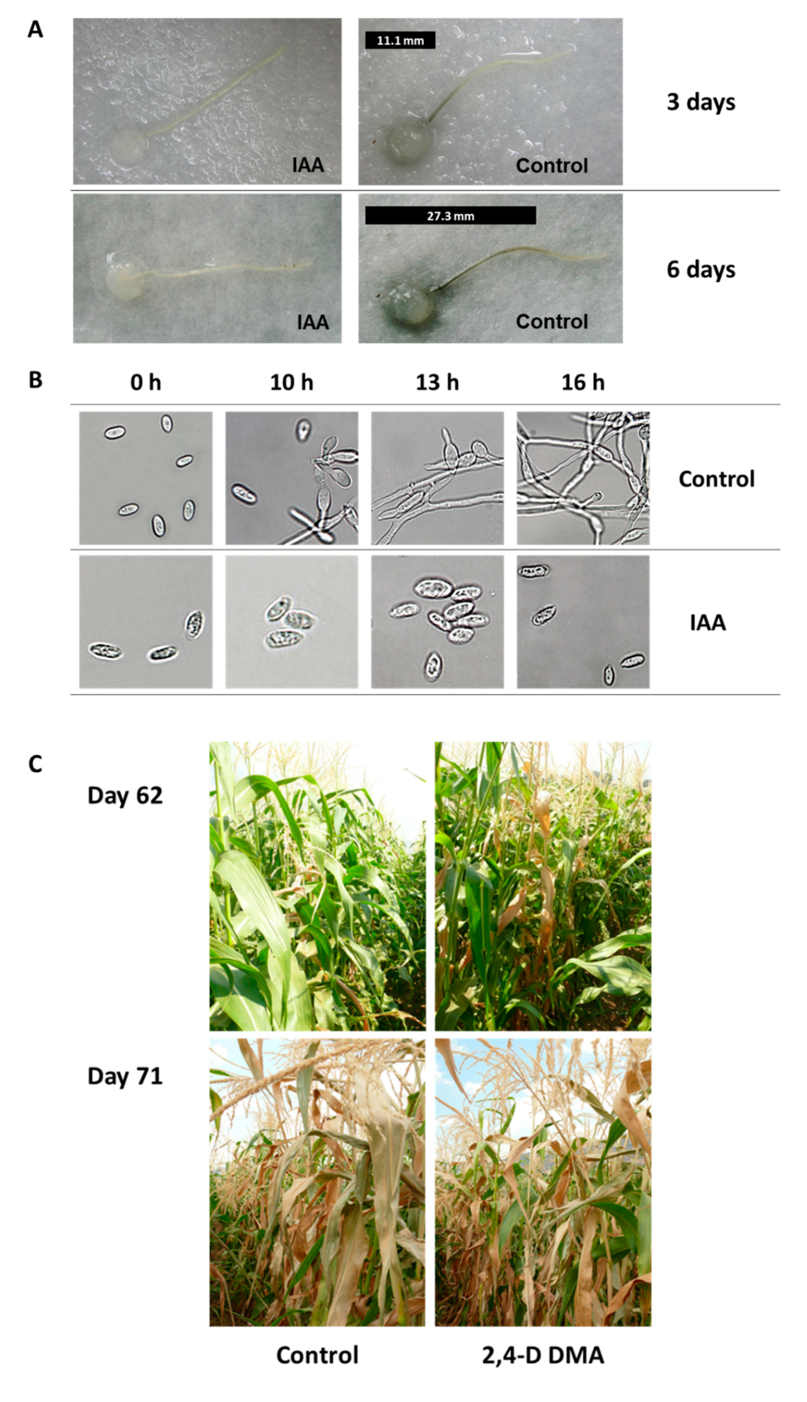
  83. Bari, R.; Jones, J.D. Role of plant hormones in plant defence responses. Plant Mol. Biol. 2009, 69, 473–488. [Google Scholar] [CrossRef] [PubMed]
  84. Degani, O.; Drori, R.; Goldblat, Y. Plant growth hormones suppress the development of Harpophora maydis, the cause of late wilt in maize. Physiol. Mol. Biol. Plants 2015, 21, 137–149. [Google Scholar] [CrossRef] [Green Version]
  85. Awad, A.M.; El-Abbasi, I.H.; Shoala, T.; Youssef, S.A.; Shaheen, D.; Amer, G.A.E.-A. PCR and Nanotechnology Unraveling Detection Problems of the Seed-borne Pathogen Cephalosporium maydis, the Causal Agent of Late Wilt Disease in Maize. Int. J. Nanotechnol. Allied Sci. 2019, 3, 30–39. [Google Scholar]
  86. Campos, M.D.; Patanita, M.; Campos, C.; Materatski, P.; Varanda, C.M.; Brito, I.; Félix, M.d.R. Detection and Quantification of Fusarium spp. (F. oxysporum, F. verticillioides, F. graminearum) and Magnaporthiopsis maydis in Maize Using Real-Time PCR Targeting the ITS Region. Agronomy 2019, 9, 45. [Google Scholar] [CrossRef] [Green Version]
  87. Magarey, R.D.; Borchert, D.M.; Engle, J.S.; Colunga-Garcia, M.; Koch, F.H.; Yemshanov, D. Risk maps for targeting exotic plant pest detection programs in the United States. EPPO Bull. 2011, 41, 46–56. [Google Scholar] [CrossRef] [Green Version]
  88. El-Shabrawy, E.-S.; Shehata, H. Controlling maize late-wilt and enhancing plant salinity tolerance by some rhizobacterial strains. Egypt. J. Phytopathol. 2018, 46, 235–255. [Google Scholar] [CrossRef]
  89. Hamza, A.M.; El-Kot, G.; El-Moghazy, S. Non-traditional methods for controlling maize late wilt disease caused by Cephalosporium maydis. Egypt. J. Biol. Pest Control 2013, 23, 87–93. [Google Scholar]
  90. Khokhar, M.K.; Hooda, K.S.; Sharma, S.S.; Singh, V. Post flowering stalk rot complex of maize-Present status and future prospects. Maydica 2014, 59, 226–242. [Google Scholar]
  91. Degani, O.; Kalman, B. Assessment of Commercial Fungicides against Onion (Allium cepa) Basal Rot Disease Caused by Fusarium oxysporum f. sp. cepae and Fusarium acutatum. J. Fungi 2021, 7, 235. [Google Scholar] [CrossRef] [PubMed]
  92. Begum, H.; Mohammad, S.; Rao, G.K.; Raj, R.B. Influence of seed dressing fungicides on the incidence of post flowering stalk rot (late wilt and charcoal rot), yield and profitability of maize. Crop Res. Hisar 1989, 2, 142–146. [Google Scholar]
  93. Satyanarayana, E.; Begum, H. Relative efficacy of fungicides (seed dressers) and irrigation schedule for the control of late wilt of maize. Curr. Res. Univ. Agric. Sci. Bangalore 1996, 25, 59–60. [Google Scholar]
  94. Sabet, K.A.; Samra, A.S.; Abdel-Rahim, M.F. Systemic action of benomyl against late-wilt disease of maize. Ann. Appl. Biol. 1972, 71, 211–218. [Google Scholar] [CrossRef]
  95. Ashour, A.; Sabet, K.; El-Shabrawy, E.-S.; Alhanshoul, A. Control of maize late wilt and enhancing plant growth parameters using rhizobacteria and organic compounds. Egypt. J. Phytopathol. 2013, 41, 187–207. [Google Scholar] [CrossRef]
  96. Singh, S.D.; Siradhana, B.S. Chemical control of late wilt of maize induced by Cephalosporium maydis. Indian J. Mycol. Plant Path. 1989, 19, 121–122. [Google Scholar]
  97. Fernández-Ortuño, D.; Torés, J.A.; De Vicente, A.; Pérez-García, A. Mechanisms of resistance to QoI fungicides in phytopathogenic fungi. Int. Microbiol. 2010, 11, 1–9. [Google Scholar]
  98. Avila-Adame, C.; Koller, W. Characterization of spontaneous mutants of Magnaporthe grisea expressing stable resistance to the Qo-inhibiting fungicide azoxystrobin. Curr. Genet. 2003, 42, 332–338. [Google Scholar] [CrossRef]
  99. Fungicide Resistance Action Committee. List of Plant Pathogenic Organisms Resistant to Disease Control Agents. Available online: https://www.frac.info/docs/default-source/working-groups/sdhi-fungicides/group/list-of-resistant-plant-pathogens_2012-edition.pdf (accessed on 30 November 2021).
  100. Gisi, U.; Sierotzki, H.; Cook, A.; McCaffery, A. Mechanisms influencing the evolution of resistance to Qo inhibitor fungicides. Pest Manag. Sci. 2002, 58, 859–867. [Google Scholar] [CrossRef]
  101. Castroagudín, V.L.; Ceresini, P.C.; de Oliveira, S.C.; Reges, J.T.; Maciel, J.L.; Bonato, A.L.; Dorigan, A.F.; McDonald, B.A. Resistance to QoI fungicides is widespread in Brazilian populations of the wheat blast pathogen Magnaporthe oryzae. Phytopathology 2015, 105, 284–294. [Google Scholar] [CrossRef] [PubMed] [Green Version]
  102. El-Moghazy, S.; Shalaby, M.E.; Mehesen, A.A.; Elbagory, M. Fungicidal effect of some promising agents in controlling maize late wilt disease and their potentials in developing yield productivity. Environ. Biodivers. Soil Secur. 2017, 1, 129–143. [Google Scholar] [CrossRef]
  103. Doleib, N.M.; Farfour, S.A.; Al-shakankery, F.M.; Ammar, M.; Hamouda, R.A. Antifungal activates of cyanobacteria and some marine algae against Cephalosporium maydis, the cause of Maize Late Wilt disease In Vitro. Biosci. Res. 2021, 18, 536–543. [Google Scholar]
  104. El-Mehalowy, A.A.; Hassanein, N.M.; Khater, H.M.; Daram El-Din, E.A.; Youssef, Y.A. Influence of maize root colonization by rhizosphere actinomycetes and yeast fungi on plant growth and on the biological control of late wilt disease. Inter. J. Agric. Biol. 2004, 6, 599–605. [Google Scholar]
  105. Harman, G.E.; Howell, C.R.; Viterbo, A.; Chet, I.; Lorito, M. Trichoderma species-opportunistic, avirulent plant symbionts. Nat. Rev. Microbiol. 2004, 2, 43–56. [Google Scholar] [CrossRef]
  106. Harman, G.E. Overview of Mechanisms and Uses of Trichoderma spp. Phytopathology 2006, 96, 190–194. [Google Scholar] [CrossRef] [PubMed] [Green Version]
  107. El-Gazzar, N.; El-Bakery, A.M.; Ata, A.A. Influence of some bioagents and chitosan nanoparticles on controlling maize late wilt and improving plants characteristics. Egypt. J. Phytopathol. 2018, 46, 243–264. [Google Scholar] [CrossRef]
  108. Degani, O.; Dor, S. Trichoderma Biological Control to Protect Sensitive Maize Hybrids Against Late Wilt Disease in the Field. J. Fungi 2021, 7, 315. [Google Scholar] [CrossRef]
  109. Gal-Hemed, I.; Atanasova, L.; Komon-Zelazowska, M.; Druzhinina, I.S.; Viterbo, A.; Yarden, O. Marine isolates of Trichoderma spp. as potential halotolerant agents of biological control for arid-zone agriculture. Appl. Environ. Microbiol. 2011, 77, 5100–5109. [Google Scholar] [CrossRef] [PubMed] [Green Version]
  110. Degani, O.; Khatib, S.; Becher, P.; Gordani, A.; Harris, R. Trichoderma asperellum Secreted 6-Pentyl-α-Pyrone to Control Magnaporthiopsis maydis, the Maize Late Wilt Disease Agent. Biology 2021, 10, 897. [Google Scholar] [CrossRef] [PubMed]
  111. Degani, O.; Danielle, R.; Dor, S. The microflora of maize grains as a biological barrier against the late wilt causal agent, Magnaporthiopsis maydis. Agronomy 2021, 11, 965. [Google Scholar] [CrossRef]
  112. Hamrouni, R.; Molinet, J.; Dupuy, N.; Taieb, N.; Carboue, Q.; Masmoudi, A.; Roussos, S. The Effect of Aeration for 6-Pentyl-alpha-pyrone, Conidia and Lytic Enzymes Production by Trichoderma asperellum Strains Grown in Solid-State Fermentation. Waste Biomass Valorization 2020, 11, 5711–5720. [Google Scholar] [CrossRef] [Green Version]
  113. El-Moghazy, S.; Shalaby, M. Effects of some aqueous plant extracts and sulphur compounds on the control of maize late wilt disease caused by Cephalosporium maydis. J. Agric. Res. Tanta Univ. 2006, 32, 758–775. [Google Scholar]
  114. Mosa, H.; Motawei, A.; El-Aal, A.A. Nitrogen fertilization influence on combining ability for grain yield and resistance to late wilt disease in maize. J. Agric. Res. Kafrelsheikh Univ. 2010, 36, 278–291. [Google Scholar]
  115. Sabet, K.A.; Samra, A.S.; Mansour, I.M. Saprophytic behaviour of Cephalosporium maydis and C. acremonium. Ann. Appl. Biol. 1970, 66, 265–271. [Google Scholar] [CrossRef]
  116. Brígido, C.; van Tuinen, D.; Brito, I.; Alho, L.; Goss, M.J.; Carvalho, M. Management of the biological diversity of AM fungi by combination of host plant succession and integrity of extraradical mycelium. Soil Biol. Biochem. 2017, 112, 237–247. [Google Scholar] [CrossRef] [Green Version]
  117. Sharma, M.; Sudheer, S.; Usmani, Z.; Rani, R.; Gupta, P. Deciphering the omics of plant-microbe interaction: Perspectives and new insights. Curr. Genom. 2020, 21, 343–362. [Google Scholar] [CrossRef] [PubMed]
  118. Diagne, N.; Ngom, M.; Djighaly, P.I.; Fall, D.; Hocher, V.; Svistoonoff, S. Roles of arbuscular mycorrhizal fungi on plant growth and performance: Importance in biotic and abiotic stressed regulation. Diversity 2020, 12, 370. [Google Scholar] [CrossRef]
  119. El-Shafey, H.A.; Claflin, L.E. (Eds.) Late Wilt; APS Press: St. Paul, MN, USA, 1999; pp. 43–44. [Google Scholar]
  120. Smit, E.; Van Rensburg, G.D.J.; Rijkenberg, F. Number of isolates of maize root fungi in different crop rotation systems. S. Afr. J. Plant Soil 1997, 14, 127–130. [Google Scholar] [CrossRef]
  121. Galal, A.A.; El-Rouby, M.M.; Gad, A.M. Genetic analysis of resistance to late wilt (Cephalosporium maydis) in variety crosses of maize. Z. Planzenzucht. 1979, 83, 176–183. [Google Scholar]
Figure 1. Molecular diagnostic of Magnaporthiopsis maydis pathogenesis in susceptible and resistant maize (Zea mays L.) cultivars. The experiment is described in [11]. (A) The progression of late wilt disease (LWD) from 43 days after seeding until maturity was estimated using a PCR-based method. The LWD-sensitive Jubilee cv. (upper panel) and the relatively LWD-resistant Royalty cv. (lower panel) were inspected (both sweet maize cultivars from Pop Vriend Seeds B.V., Andijk, The Netherlands, supplied by Eden Seeds, Reut, Israel). A semi-quantitative analysis of DNA isolated from the maize plants’ roots, stems, and leaves was performed weekly. The optical density of the M. maydis-specific primer bands was measured and normalized against the optical density of the control—ribosomal DNA (rDNA) bands. (B) A representative photograph of Jubilee cv. (upper panel) and Royalty (lower panel) plants on day 76 of growth.
Pathogens 11 00013 g001
Figure 2. Lower stem symptoms of late wilt disease (LWD) of maize in a susceptible maize cultivar. All photos were taken of the lower stems (near the first internode) at harvest (ca. 80 days from sowing). (A) Wilt symptoms of lower plants’ leaves and stems in the field. (B) Cross-section magnification showing the vascular bundles’ color alteration to red-brown. Insert: cross-section in which a green frame marks the magnification area. The green arrow points to the surface color change. (C) The color alteration of the vascular bundles appears as red strips on the lower stem surface. (D) Cross-section of field LWD-infected plants. Late wilt diseased plants have a tissue color alteration to a yellow-brown hue and vascular bundle occlusion (adapted from [16]). Insert: photo taken from a healthy plant. (E) Cross-section of a severe late wilt diseased plant showing the breakdown of the parenchyma tissue.
Pathogens 11 00013 g002
Figure 3. Maize late wilt disease (LWD) symptoms in the field 75 days after sowing (adapted from [6,11]). An observation for assessing cultivar resistance to Magnaporthiopsis maydis infection was conducted in a sweet corn field in the Hula Valley (Neot Mordechai, Upper Galilee, northern Israel). LWD-sensitive Jubilee cv. and relatively LWD-resistant Royalty cv. are shown (see Figure 1). (A) Aerial photograph (taken by Asaf Solomon) of the maize field, in which the two cultivars are marked: wilted Jubilee cv. plants (red box) and healthy Royalty cv. plants (blue box). (B) Ground photograph of the two representative cultivars: Jubilee (left) and Royalty (right). (C) Cob samples of the cultivars tested: Jubilee (left) and Royalty (right).
Pathogens 11 00013 g003
Figure 4. Cultivars’ resistance test for late wilt disease (adapted from [69]). A semi-commercial examination of fodder corn genotypes was conducted in the southern coastal plain of Israel (Yavne field) in 2014. The experiment included 14 maize cultivars in 3 repetitions, with each plot containing 6 rows measuring about 200 m in length. The experimental area photo was taken close to harvesting (day 99 from sowing) by David Katsav. The brown lines are late wilt diseased cultivars with severe dehydration, while the green lines are healthy cultivars. The photos on the right: resistance cultivar—Pan 33–031 (Eden Seeds, Hatzav, Israel); reduced sensitivity cultivar—Colossus from HSR Seeds (CTS, Hod Hasharon, Israel); susceptible cultivar—Avgaro (Hazera Seeds Ltd., Berurim MP Shikmim, Israel).
Pathogens 11 00013 g004
Figure 5. The impact of auxin (hormone indole-3-acetic acid, IAA) on Magnaporthiopsis maydis growth in vitro and in the field (adapted from [84]). (A) Detached root pathogenicity assay for the influence of auxin. Young, white side-roots, ca. 2 cm long, were cut from 20-day-old potted maize (Zea mays L., Jubilee cv.) seedlings and inoculated by placing a 6 mm diameter M. maydis culture agar disk taken from the margins of a 4–6-day-old colony (grown at 28 ± 1 °C in the dark) on each root’s cut end. The inoculated roots were positioned separately in Petri dishes that contain auxin (IAA, 100 mg/L) or distilled and deionized water, DDW (control). The palates were incubated at 28 ± 1 °C in the dark. Development of M. maydis infection thread (a dark filament) inside the roots was qualitatively evaluated after three and six days. The infection thread length is marked in the photo by a black line placed near each root. (B) Effect of plant hormones on M. maydis spore germination. Spores were washed from the surface of 4-day-old colonies and suspended in watery solutions containing IAA. The spores’ suspensions were then incubated at 28 ± 1 °C in the dark, in a rotary shaker (at 150 rpm) for the indicated times. The length of each spore is ca. 10 µm. (C) Field experiments to assess the efficiency of dimethylamine salt of 2,4-dichlorophenoxyacetic acid (2,4-D DMA, mimics auxin’s influence) in controlling late wilt. The experiment was conducted in an infested sweet corn field in the Hula Valley (upper Galilee, northern Israel). The Jubilee cv. plants were treated separately with 2,4-D DMA (96.9% active ingredient, Aminobar, Luxembourg Industries Ltd., Israel). The treatment was applied at a dosage of 150 cm3/0.1 ha, 15, 30, and 45 days from sowing. The treated and control (untreated) plants were photographed 62 and 71 days past sowing.
Pathogens 11 00013 g005
Figure 6. From in vitro evaluation to a field assay of Azoxystrobin. (A) Agar plate assay to evaluate the inhibition of Magnaporthiopsis maydis mycelial growth by Azoxystrobin (AS, CAS no. 131860-33-8, Amistar, Makhteshim Agan, Airport City, Israel) (adapted from [6]). Photos were taken five days after incubation at 28 °C in the dark. The fungicide was evaluated at a rate of 1, 10, and 100 mg/L active ingredients. Control—potato dextrose agar (PDA) plate without fungicide. (B) Detached root pathogenicity assay (adapted from [6]). The main (longest) inner feeder root was cut from a potted 3-week-old Jubilee cv. maize sprout. The detached root was inoculated by placing an M. maydis culture disk (6 mm diameter) taken from the margins of a 4–6-day-old colony, two cm away from its cut end. The inoculated roots were incubated in a moist atmosphere, in Petri dishes, at 28 °C in the dark for six days. The pathogen infection thread (a dark filament) within the root is marked in the photos by a black line near each root. Control—root inoculated with M. maydis without fungicides. (C) Effect of Azoxystrobin seed coating (0.002 cm3/seed) on plant development in a greenhouse (adapted from [28]). A photograph of all the plants’ aboveground parts (upper panel) and leaves (lower panel) in the treatment and the non-protected control groups 72 days after sowing (DAS), 13 days after fertilization (DAF). (D) Late wilt disease symptoms in a field experiment 71 DAS, 16 DAF (adapted from [27]). Upper panel—wilt symptoms of a non-protected plot (control) compared to an Azoxystrobin-treated plot. Lower panel—representative plants. The fungicide was applied in seed coating (0.002 cm3/seed, Azoxystrobin + Difenoconazole, “Ortiva-Top”, Syngenta, Basel, Switzerland, supplier Adama Makhteshim, Airport City, Israel) and at three intervals, 18, 31, and 45 DAS, 2.25 L/hectare. Disease symptoms include drying-out that progresses upwards in the plant, stem and leaf yellowing, and dehydration.
Pathogens 11 00013 g006
Figure 7. In vitro estimation of Trichoderma asperelloides (T203)-secreted metabolites-based biological control against Magnaporthiopsis maydis (adapted from [108]). (A) T203-submerged cultures grown with shaking (150 rpm) for isolating secreted metabolites. (B) Static shallow media cultures of M. maydis on potato dextrose broth (PDB) medium containing T203-secreted metabolites filtrate. Control is PDB medium M. maydis cultures maintained under the same conditions. (C) Effect of growth media of T203 isolate on corn seed germination. The seeds were germinated in Petri dishes soaked in 4 mL of PDB (control) or PDB + secretion products (growth medium filtrate six days after T203 growth). All images are displayed after 5–6 days incubation at 28 ± 1 °C in the dark.
Pathogens 11 00013 g007
Figure 8. Trichoderma longibrachiatum (T7407) biological control against Magnaporthiopsis maydis in the lab and the field (adapted from [58,108]). (A) Plate mycoparasitism assay to identify interactions between Magnaporthiopsis maydis and T7407 in a potato dextrose agar (PDA)-rich medium. The two fungi were placed opposite each other, T7407 on the left and M. maydis on the right. Photos were taken after 3 and 10 days of growth. (B) Field inoculation of 20-day-old seedlings by an M. maydis-infected toothpick. The toothpicks were used for stabbing each plant at the near-surface portion of the stem. (C) The lower stem (first aboveground internode) disease symptoms. (D) The cobs’ spathes disease symptoms. (E) The experiment’s plots. Representative images of the field plants were taken 82 days after sowing. Controls are unprotected diseased plants.
Pathogens 11 00013 g008
Publisher’s Note: MDPI stays neutral with regard to jurisdictional claims in published maps and institutional affiliations.

