Article

Development of Single Nucleotide Polymorphism (SNP)-Based Triplex PCR Marker for Serotype-specific Escherichia coli Detection

1
Division of Forest Science, Kangwon National University, Chuncheon 24341, Korea
2
Faculty of Biological Science, Department of Biotechnology and Genetic Engineering, Islamic University, Kushtia 7003, Bangladesh
3
Institute of Forest Science, Kangwon National University, Chuncheon 24341, Korea
*
Author to whom correspondence should be addressed.
Pathogens 2022, 11(2), 115; https://doi.org/10.3390/pathogens11020115
Submission received: 19 November 2021 / Revised: 29 December 2021 / Accepted: 14 January 2022 / Published: 19 January 2022
(This article belongs to the Special Issue Spatial Epidemiology and Surveillance of Foodborne Pathogens)

Abstract

:
Single-nucleotide polymorphisms (SNPs) are one of the most common forms of genetic variation and as such are powerful tools for the identification of bacterial strains, their genetic diversity, phylogenetic analysis, and outbreak surveillance. In this study, we used 15 sets of SNP-containing primers to amplify and sequence the target Escherichia coli. Based on the combination of the 15-sequence primer sets, each SNP site encompassing forward and reverse primer sequences (620–919 bp) were aligned and an SNP-based marker was designed. Each SNP marker exists in at least two SNP sites at the 3′ end of each primer; one natural and the other artificially created by transition or transversion mutation. Thus, 12 sets of SNP primers (225–488 bp) were developed for validation by amplifying the target E. coli. Finally, a temperature gradient triplex PCR kit was designed to detect target E. coli strains. The selected primers were amplified in three genes (ileS, thrB, and polB), with fragment sizes of 401, 337, and 232 bp for E. coli O157:H7, E. coli, and E. coli O145:H28, respectively. This allele-specific SNP-based triplex primer assay provides serotype-specific detection of E. coli strains in one reaction tube. The developed marker would be used to diagnose, investigate, and control food-borne E. coli outbreaks.

1. Introduction

Single-nucleotide polymorphisms (SNPs) are single-base differences in DNA between individual organisms [1,2,3,4]. They enable the distinction of very closely related organisms or very limited allelic differences on similar genomic structures, such as different serotypes of Escherichia coli bacteria [5,6]. SNPs are one of the most useful molecular markers because of their stability and abundance in all organisms. The advent of whole-genome sequencing (WGS) technologies [7,8], wider open-source websites, i.e., PubMLST [9], and the availability of reference genome sequences of many bacteria has allowed for the wider implementation of SNP-based detection [10,11,12]. Nucleotide substitutions during DNA replication of bacteria, with mutation rates of approximately 10−9 changes per nucleotide per generation, are important biologically informative markers in bacteria [13,14]. In addition, SNP data have also been used in epidemiological studies of field outbreaks [15]. However, the differentiation of bacteria at the serovar level remains challenging, and SNP-based approaches for differentiating specific strains have been applied to various bacterial species, such as Escherichia [16,17], Salmonella [18], Brucella [19,20], and Bacillus species [21]. To date, several molecular methods have been used for the detection of E. coli [3,10,11,17,22,23]. However, SNP-based techniques have become increasingly attractive for the efficient detection of E. coli compared to other recognized molecular techniques [13]. Using differences in SNPs of genes between isolates is also a promising technique for the identification of geographical origins of E. coli [13,24,25,26]. SNPs are abundant in microbes and are often considered the optimum choice for genetic studies, having several advantages such as flexibility, reduced error rate, speed, and cost-effectiveness [27]. SNPs within different E. coli genes may be useful markers for the development of rapid surveillance of food-borne diseases, outbreak tracing [26,28] and typing methods, routine diagnostics, and the typing of Salmonella [29]. This approach has already been useful in retrospective investigations [27,28,30,31].
Shiga toxin-producing E. coli (STEC), E. coli O157 has been a major food-borne pathogen since the early 1980s; however, early and accurate diagnosis presents preventive measures to minimize the risk of food-borne pathogens and their outbreaks [32,33]. E. coli O157:H7 has been reported in several outbreaks worldwide, in both developing and developed countries, including in the USA [34], Canada [35], and Europe [36]. Currently, there several molecular techniques are commonly used for food-borne disease surveillance and the subtyping of E. coli, such as the PulseNet [37,38], Multilocus Sequence Typing [39], and Multilocus Variable-Number Tandem-Repeat Analysis [40] techniques. PulseNet is based on the characterization of whole bacterial genomes using enzymatic digestion patterns that are separated by pulsed-field gel electrophoresis [12,20,38]. This technique can be biased because of the subjective interpretation of band patterns [37,41]. Similarly, Multilocus Sequence Typing (MLST) is widely used as a genotyping method and has been applied successfully to E. coli [22,23]; however, this sequencing procedure is expensive and time-consuming for routine monitoring [42]. Multilocus Variable-Number Tandem-Repeat analysis is another subtyping technique used for E. coli, but it has limitations in the development of a universal panel for MLVA [40]. Nevertheless, SNP analysis enables the classification of very similar genomes and the detailed classification of genomes of E. coli that are difficult to distinguish using conventional typing techniques [43]. To overcome the limitations of the abovementioned molecular methods, SNP-based molecular methods have been developed. Therefore, by comparing it with the existing methods, the SNP-based method can be used to develop accurate, rapid, and molecularly meaningful typing methods that may lead to the more efficient detection of serotype-specific food-borne pathogenic E. coli. In this study, we altered a mismatched nucleotide within the three bases closest to the 3′ end (SNP site) in each primer sequence to detect serotype-specific E. coli. The aim of this study was to investigate the interrogation of informative SNPs in genes of WGS sequences of E. coli, along with the development of SNP marker-based detection.

2. Results

2.1. Acquisition and Alignment of WGS of E. coli from GenBank

A total of six E. coli strains, including three O157 and non-O157 sequences, was downloaded from GenBank. From these, E. coli str. K-12 contained the most known genome sequences and was therefore selected as the reference strain, whereas the remaining five E. coli strains were considered for comparison. The accession numbers of all six E. coli strains are provided in Table S1.

2.2. Search of SNP Sites from NGS of E. coli Genomes in GenBank

The six E. coli genome sequences were downloaded and compared to the reference genome sequence. A total of 2160 SNPs were identified from the alignment of E. coli WGS (Table S1). From them, the high-quality, useful, and abundant SNP sites were selected based on nonsynonymous mutation with bioinformatics software, and 15 sets of SNP sites encompassing forward and reverse primers were thus selected. These 15 primer sets were chosen from 11 different genes of the E. coli genome (thrB/C, nhaR, ileS, dapB, carB, caiB/C, polB, araB, yabI, thiP, and leuD) (Table 1). The ambiguous codes and positions of SNPs with a nonsynonymous amino acid of a specific gene and the reference genome of non-O157 E. coli (NC_000913) are shown in Table 1.

2.3. Amplification of SNP Sites and Sequencing of Target E. coli Strains Using Newly Designed Primers

A total of 15 SNP markers with a size of 620–919 bp were identified from E. coli genomes via the bioinformatics software analysis. Within the SNP markers, SNP sites, bold ambiguous codes, and the positioning of the amino acid codes in each gene of the reference E. coli genome and with the respective genes are marked in Table 1. All 15 primers were tested for the amplification of the SNP markers with the target E. coli strains (Table 1). The amplified product sizes and an image of the PCR results are shown in Figure 1.

2.4. Confirmation of SNP Sites Based on Aligned Sequences of Target E. coli and Design of E. coli Serotype-Specific SNP Primers Using Sequences Containing SNP Sites

During the first PCR amplification, seven primer sets (01-, 02-, 07, 08-, 09-, 16-, and 22-ecoli) were amplified on the target genes of the tested E. coli strains (Tables S2 and S3), whereas four primer sets (12-, 21-, 23-, 24-ecoli) were not amplified on any target bands (Figure 1) and three primer sets (14-, 15-, and 26-ecoli) were partially amplified on the target genes of four E. coli strains (Figure 1, Tables S1 and S3). Moreover, the primers of 20- and 22-ecoli were amplified with all the tested E. coli strains during the second PCR, and their sequences are shown in Table S2. Thus, we tested all primers with all target E. coli strains with the first and second PCR amplification and each primer was amplified the specific gene (i.e., 01-primer amplified specific gene, homoserine kinase, thrB) of the four target E. coli strains.
The amplified products of all 15 primers, their sequences, and the detailed information of each primer with the four target E. coli strains are shown in Tables S2 and S3. The specific gene target amplified sequences (n = 38 out of 60) were submitted to the NCBI database with the following accession numbers (OL589326–OL589363). The chimeric, off-target sequences with non-specific genes and dimer sequences (n = 22) were not used for further analysis of the SNP-based primer design (Table S4). Moreover, the three positions of SNPs were 22,945 > C, 23,104 > A, and 23,137 > A of ileS gene in reference non-O157 E. coli str. K-12 (NC_000913) (Table 1 and Table S1). The amplified ileS gene products of the target E. coli strains were sequenced and aligned. We then checked one or multiple SNP positions on the aligned sequences. Thus, 12 sets of SNP primers of serotype-specific E. coli were designed based on five genes (nhaR, thrB/C ileS caiB, and polB) where at least one natural SNP was present (Table 2).

2.5. E. coli-Specific SNP Primer Design Using Aligned Gene Sequences with Availability of SNP Sites

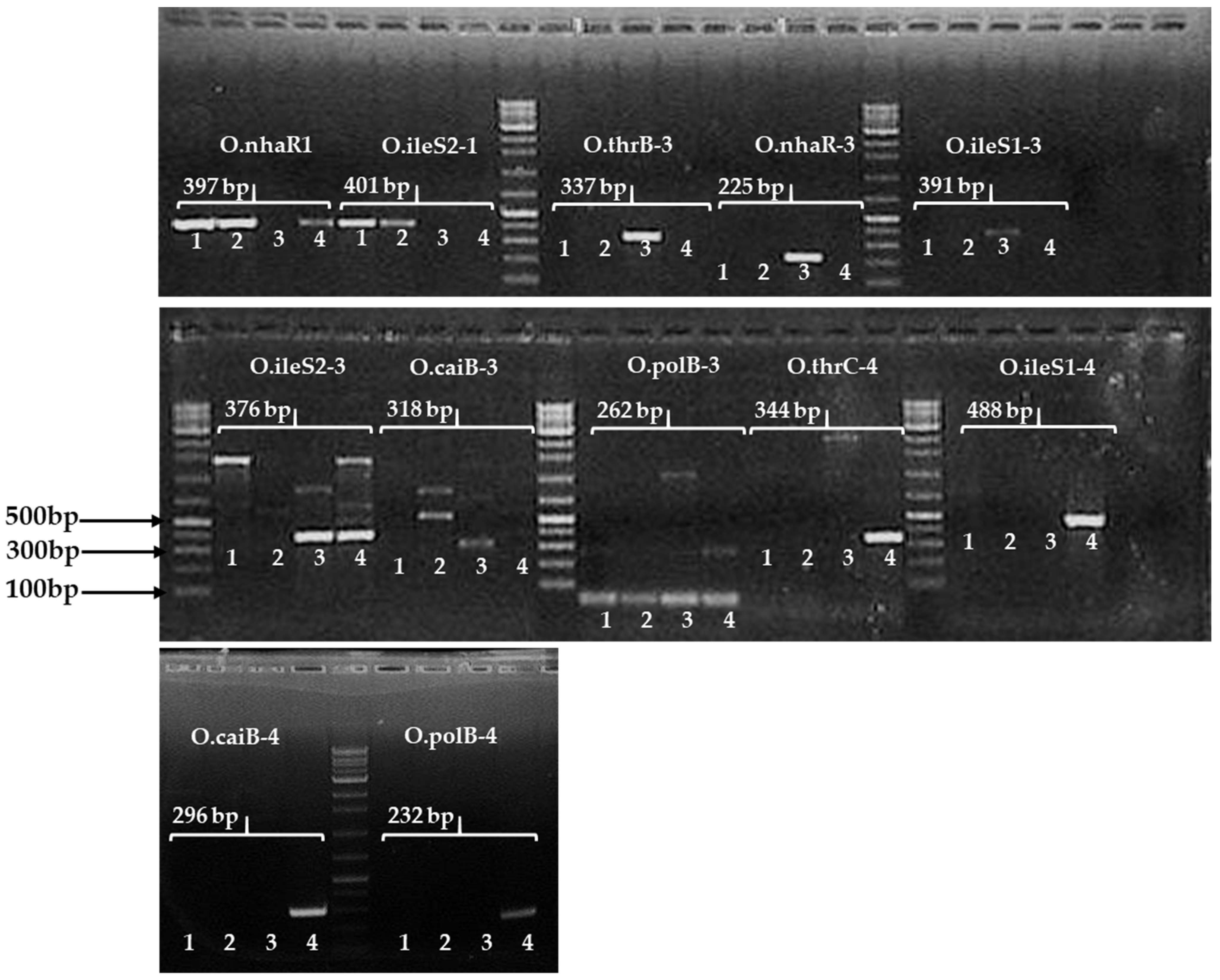
Based on previous research [44], the triplet base of the 3′ end primer of each primer sequence and the last triplet codon at the 3′ end of the first and third position base altered within all primer sets so that they could hybridize very strongly during PCR amplification. Based on this principle, we designed 12 sets of SNP primers (225–488 bp) developed for validation by amplifying target E. coli strains. Two examples of these primers are shown in Figures S1 and S2. The marker “01”-amplified homoserine kinase (thrB) genes of four E. coli strains were aligned. It was found that the number of the natural SNP position was 342 and the artificial transition (purine to purine)-mutated SNP position number was 340 on the aligned gene sequences. Thus, we designed the SNP based marker ‘O-thrB-3-F/R’ to include at least two SNPs, e.g., the forward primer length was 18 bp from 325 to 342 (the position of the natural SNP ‘G = 342′ and the transition-altered SNP site was “G = 340; A > G”) and the reverse primer length was 20 bp from 642 to 661 (the position of two natural SNPs ‘T = 642′ and ‘C = 643’, respectively, and the transition-mutated SNP site was “G = 645; A > G”). The amplified target product size was 337 bp (Figure S1). Another forward primer ‘ileS1-4-F’ was 19 bp long (141–159) and the reverse primer ‘ileS1-4-R’ was 20 bp long (609–628), as shown in Figure S2. The target amplicon size was 488 bp. Moreover, the amplicon size (391 bp) and the position of the forward and reverse primers of the primer “ileS1-3” are shown in Figure S2. The natural SNP is marked by a square shape and the altered SNP is marked by a black shaded italicized square shape (Figures S1 and S2). Thus, 12 SNP-based markers of E. coli serotype-specific primers were used for amplification with the four E. coli strains, and finally, it was possible to confirm selection with the efficiency of the SNP marker of the positive target band of all E. coli strain-specific SNP primers for PCR amplification (Table 3 and Figure 2).

2.6. SNP Marker-Based Triplex PCR

To determine the strain specificity of the confirmed SNPs, we aligned the re-sequences containing the variable positions of the target E. coli sequences (Table S1). To detect the target serotypes of E. coli-specific SNP markers in a single reaction, a temperature gradient SNP marker-based triplex PCR kit was developed. This efficient test was performed with target E. coli strains via PCR amplification of the SNP-based triplex PCR kit (Figure 3 and Table 4).
In the encompassing primer sequences (Table 1), the red-colored bases indicate natural SNPs and the blue-colored bases indicate artificial SNPs (altered by transition or transversion mutation) (Table 2). For example, for the primer of O.ileS2-1F “GATCATCTTCCGCGCAGCG”, which consists of three SNP bases, the underlined nucleotide positions are the 16th, ‘A’, and the 19th, ‘G’, whereas the blue color base, the 17th, was changed from ‘A’ to ‘G’ (transition). Thus, in each primer sequence of the SNP sites, we altered an artificial base, except for O-polB-4-F, so that SNP-based primers improved their ability to bind the PCR template sequence, thereby improving the allele-specific detection of target E. coli strains (Table 2, Figure 2). The 12 sets of SNP-based primers were designed to amplify the desired band of serotype-specific E. coli. Finally, SNPs containing three genes (ileS, thrB, and polB) with three markers (O.ileS2-1, O.thrB-3, and O.polB-4) were selected to detect three target E. coli strains (Figure 3 and Table 4). In the case of E. coli O157:H7, two SNP-based primers (O.nhaR1 and O.ileS2-1) produced the target band, whereas the other two markers (O.ileS2-3 and O.caiB-3) produced the off-target bands (Table 3). The primer ‘O.ileS2-1′ detected two E. coli O157:H7 strains, but it did not detect the other two non-O157 E. coli strains. Thus, this marker (O.ileS2-1) is a good candidate for E. coli O157:H7. However, if we want to know the serotypes of an unknown E. coli, we have to PCR-amplify each of 12 primer pairs, which is a limitation of the developed SNP-based (n = 12) marker. We overcame the cost and time involved in repeated PCR amplification of the SNP-based marker through the development of a triplex SNP marker (the developed triplex PCR marker is indicated as “O1”) (Figure 3 and Table 4).
Serotype-specific E. coli strains were detected in a single reaction with the developed assay (Figure 3 and Table 4). In addition, the marker ileS2-1 was able to detect the most pathogenic O157, whereas the other two primer pairs, O.thrB-3 and O.polB-4, could detect E. coli (KVCC-BA1800069) and E. coli O145:H28 (KVCC-BA1800090), respectively (Table 4).

2.7. Cross Reaction and Validation Test

The developed markers were investigated by means of the cross-reaction test with other gram-positive and gram-negative pathogenic bacteria. The developed assay did not produce cross-reactions with any of the tested bacteria, indicating the specificity of the primer sets. However, the assay sometimes produced an off-target band with the tested bacterial strains (data not shown). For more validation, 23 identified E. coli from wild-animal fecal samples were tested and validated with SNP-based markers. The generating band was marked on the isolate lanes numbered from one to eight, where lane 1 is a positive band of reference E. coli O157 (401 bp) and the tested E. coli (lane no. 2–7) was matched with E. coli O157:H7. Lanes no. 9–11 were matched to the target E. coli (KVCC-BA1800069, lane no. 13, amplicon length 337 bp). Lanes no. 14–19 were matched to the target E. coli O145:H28 (KVCC-BA1800090; lane no. 14, amplicon length 232 bp). Five of the isolates (lanes no. 20–25) were not exactly matched to the three target E. coli strains. They might have originated from the animal fecal samples as different E. coli strains (Figure S3). Sometimes, the tested E. coli was produced multiple off-target bands. To obtain a clearer resolution, we should conduct further analysis with a variety of E. coli isolates with diverse sources, such as foods.

3. Discussion

To date, several different methodological approaches have been used to detect E. coli, for instance, PCR bands [45], PFGE [46], phage typing [47], and MLST [18,23]. There are some limitations to the current molecular typing methods; however, SNP-based techniques have recently been suggested as a cost-effective alternative typing method for various bacterial species, as well as E. coli [6,27,28,48]. The SNPs in the WGS have discriminative power that enables the comparison of genetic bases not only between bacterial subspecies but also at the serotype level. This provides an easy method of determining the position of SNPs from the WGS of bacteria using different bioinformatics software tools. Thus, various polymorphisms can be used to detect very similar strains from different sources [2,3]. In addition, SNPs could be used as an accurate and convenient method to detect disease outbreaks, for the surveillance of food-borne pathogens [13,26] and their source detection [28,34], to develop risk models for outbreaks, and even to map the phylogenetic and evolutionary relationships between similar strains [49].
In this study, fifteen primer sets were chosen from 11 different genes of the E. coli genome (Table 1). Primers were designed based on suitable abundant SNP sites on the aligned WGS of the 11 abovementioned genes, approximately 620–919 bp (Table 1). Grish and Burbudae [50] conducted a similar study and found that nine markers were considered to be the best candidate markers in terms of their target band patterns out of the 30 SNP markers tested. In this study, SNP-based primers were designed based on natural and artificial SNPs or by introducing a mismatched nucleotide within the three bases at the 3′ end SNP sites (transition or transversion mutations). In addition, the artificial mismatched (transition or transversion) bases that were varied in the 3rd position of a codon at the 3′ end of each primer might have altered the codon, resulting in an amino acid substitution, which represents a target for PCR methods [51,52,53]. Moreover, the introduction of a mismatched (A-T transversion or A-G transition) base pair at the third base from the 3′ end could increase the allele-specific amplification during PCR [44,54,55]. Thus, transitions (A-G and T-C), as well as transversions (A-T, A-C, G-C, and G-T), were useful as base pair mismatches in improving the allele-specific amplification. Based on natural and artificial bases (transition/transversion), we developed 12 sets of SNP primers (232–488 bp) for validation by amplifying the four target E. coli genes including three different serotype strains (Table 2). Moreover, the melting temperature (Tm) sometimes depends on the GC content of the designed primer sequences, which is important for the adjustment of PCR conditions. By introducing transverse mismatched bases, the melting temperature of allele-specific SNP-based primers can be fixed or adjusted to standardized PCR conditions [44]. Similarly, the E. coli O157 and non-O157 detected primers were designed based on natural and artificial SNPs or a mismatched nucleotide introduced within the three bases (except for primer ‘O.polB-4-F’) at the 3′ end SNP sites (Table 2, Figure 2). A temperature gradient SNP marker-based triplex PCR kit was developed for the target serotypes of E. coli specific SNP markers in a single reaction. An efficient test was performed with the target E. coli serovar by means of PCR amplification of the SNP-triplex PCR kit and was adjusted to standardized PCR conditions (Table S5). In addition, the E. coli detected in wild-animal fecal samples were tested with the efficiency and validation of the designed primer (O1), but all the isolates were not exactly matched to the three target E. coli serotype strains (Figure S3). We should analyze more isolates for the validation of the SNP-based triplex O1 primer.
Recently, software algorithms and parameters have been used to search for SNP positioning from raw or assembled genome sequences [56,57]. SNP-based techniques have become increasingly attractive for the efficient detection of E. coli, compared to other molecular techniques. In some previous investigations, the SNP-based technique has already been useful in retrospective research studies based on SNPs of WGS that can detect different isolates of similar bacterial strains [2,30,31,58]. In one study, two highly homologous serovars were distinguished based on 20 SNPs compared to the sequence of a reference strain [59]. Nonetheless, another study showed that up to four SNPs were required for the precise identification of all E. coli-investigated serovars. Dallman et al. showed three SNP differences in outbreak-associated isolates compared to non-outbreak isolates. It was possible to detect outbreak isolates using SNP-based primers [60]. Current SNP-based typing methods can be applied for the detection and typing of STEC and other organisms. [24]. Desphande et al. showed that 29 SNPs were used as a signature sequence (either synonymous or non-synonymous amino acid changes) for E. coli strains from other pathogenic serotyping E. coli strains present in the NCBI database [61]. In our research, we mention the varying nucleotide positions (SNPs) between the following strains: IAI39, 2011C-3493, Sakai, NRG 857C, and UMN026, and the reference strain (K-12) (Table S1). A study by Camprubí-Font et al. compared 286 polymorphic sites in adherent-invasive E. coli (AIEC-associated SNPs). Sixty SNPs were selected for re-sequencing, and 20 of the confirmed SNPs in 11 genes of AIEC strains were used for the identification of E. coli [2]. Similarly, we found 2160 SNPs on the aligned genome of six serotype-specific E. coli strains, among which the SNP-flanking regions of aligned sequences on 11 genes were selected for further sequencing and finally only three SNPs containing three genes (ileS2, polB, and thrB) were confirmed for the SNP-based triplex PCR marker. However, Moorhead et al. speculated that PCR enzymes are capable of three to five proofreading activities to correct an artificial mismatch [51] but DNA polymerase can extend primers less efficiently (100- to 10,000-fold) compared to matched with mismatched 3′ bases [62]. The simultaneous introduction of mismatched bases in the third position prevented the amplification of fragments from the targeted E. coli genome (Table 2). Therefore, altered SNPs could be used for developing an alternative, accurate, rapid, and cost-effective typing method that may lead to significant improvements in the diagnosis of E. coli. To the best of our knowledge, this study is the first to detect serotype-specific E. coli strains using the developed (O1) SNP triplex PCR method.

4. Materials and Methods

4.1. General Overview of SNP-Based Marker Design and Validation with PCR Amplification of Target E. coli

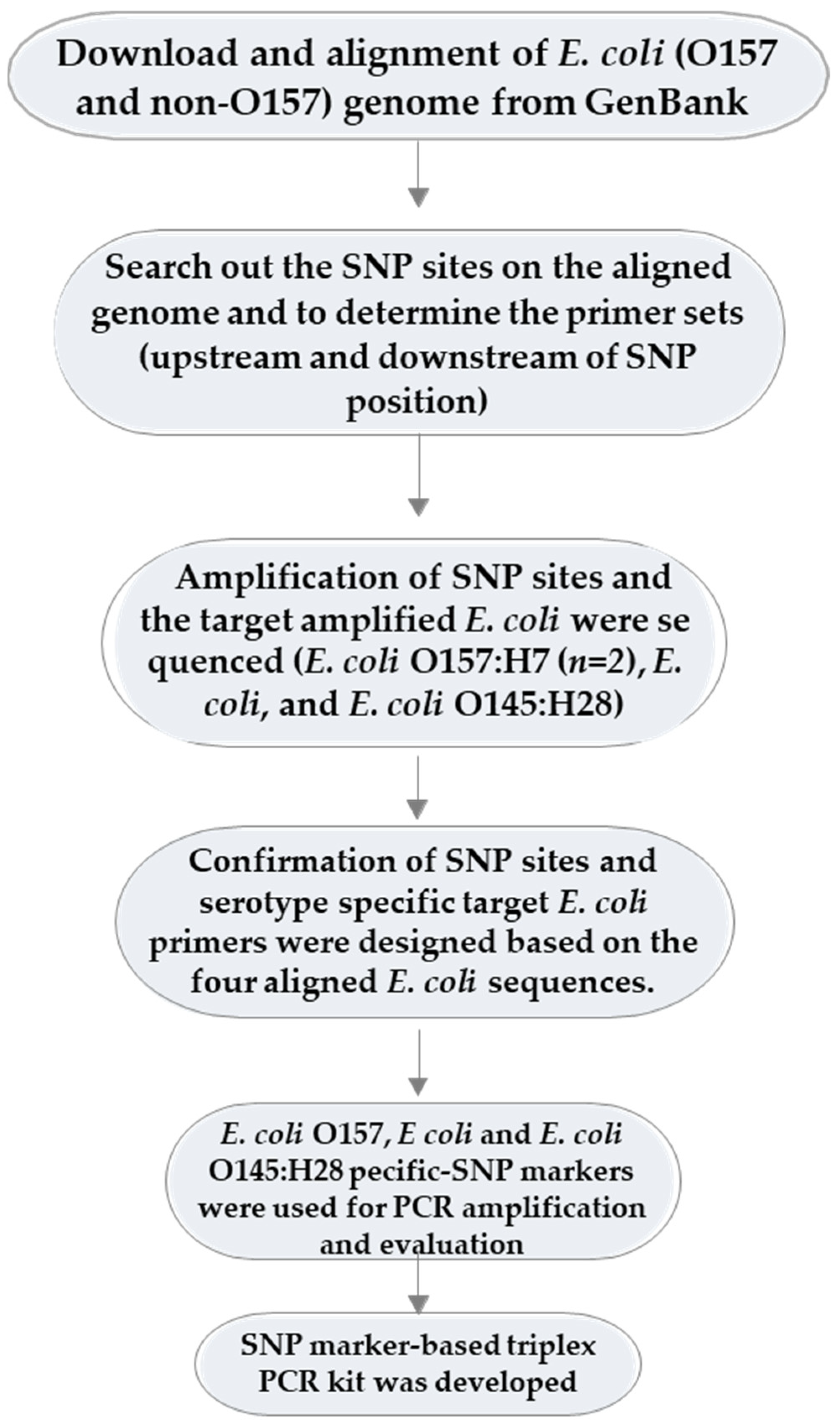
There were six steps involved in the development of SNP-based markers. They were as follows: (i) the whole-genome sequences of six E. coli strains, including three E. coli O157 and three non-O157 strains, were retrieved from the GenBank database and each was aligned with the respective reference (K-12 substr. MG1655). (ii) We used the NUCmer program [63] to search for the SNP positions on the aligned WGS and to determine the primer sets encompassing each SNP site (upstream and downstream of each SNP position). The SNP-matrix files were shown in variant call format (VCF) files by generating them from VCFtools v.4.1.0 (https://vcftools.github.io/index.html, accessed on 29 December 2021), and a filtered SNP matrix was constructed from the output VCF files [63]. (iii) The designed primers were used for the amplification of SNP sites, and subsequently, the four amplified E. coli were sequenced (E. coli O157 and non-O157). (iv) Then, E. coli primers were designed based on the four aligned E. coli sequences. (v) E. coli O157 and non-O157 specific-SNP markers were used for PCR amplification and evaluation. (vi) Finally, the SNP marker-based triplex PCR kit was designed. A schematic flow diagram of the development of the SNP-based marker for E. coli detection is shown in Figure 4.

4.2. Culture and Isolation of Genomic DNA from Serotype-Specific E. coli (O157 and Non-O157) Strains

The four E. coli strains, including three different serotype strains, were selected for DNA isolation, PCR amplification (with the designed SNP-encompassing primers), and sequencing. The two E. coli O157 strains were O157:H7 (ATCC-95150, NCCP-15739) and the non-O157 strains were E. coli (KVCC-BA1800069) and E. coli O145:H2 (KVCC-BA1800090). The target bacterial colonies were streaked onto nutrient agar media, and a single colony was collected using a sterilized toothpick. The colonies were incubated at 35 °C for 18 h in a 5 mL lactose broth (LB) solution. Genomic DNA was extracted from 1 mL of LB culture fluid using a DNeasy Blood and Tissue Kit, according to the manufacturer’s instructions (Qiagen, Valencia, CA, USA).

4.3. Acquisition and Alignment of WGS of E. coli from GenBank

The complete whole-genome sequences of E. coli based on NGS data were downloaded from GenBank (ftp://ftp.ncbi.nlm.nih.gov/genomes/refseq/, accessed on 10 July 2021), including one reference strain of the most studied and best-annotated genome (E. coli str. K-12), which was used as a reference genome. E. coli str. K-12 substr. MG1655 (NC-000913.3) was downloaded along with five query strains, E. coli IAI39 (NC_011750), E. coli O104:H4 str. 2011C-3493 (NC-018658), E. coli O157:H7 str. Sakai (NC-002695), E. coli O83:H1 str. NRG 857C (NC-017634), and E. coli UMN026 (NC-011751). Among them, three were E. coli O157 and three were non-O157. We selected the most suitable SNPs based on the allelic diversity of the aligned sequences with SNP sites encompassing a primer compared to the reference and query sequences of E. coli (Table S1). The “selected SNPs” are defined as the homologous gene sequences of aligned pairs of WGS. In addition, the SNPs in the highly variable region end of the contigs or synonymous amino acid changes in the coding regions were not selected.

4.4. Search for SNP Sites on WGS Alignment and Design with Suitable Primers Encompassing SNP Sites (Upstream and Downstream of SNP Sites)

We used the NUCmer software of MUMer v4.0.0 (https://mummer4.github.io/, accessed on 12 July 2021) to determine the SNP positions on WGS alignments and to design primers both upstream and downstream of SNP sites [42,63,64]. The construction of the indel matrix and the detection of SNPs were completed using VCFtools v.4.1.0 (https://vcftools.github.io/index.html, accessed on 15 July 2021), and variant annotation was performed using SnpEff v.4.3.0 (http://pcingola.github.io/SnpEff/, accessed on 17 July 2021). The final output of the workflow was a filtered SNP matrix. SNP positions were inferred using show-snp programs [63]. Eleven genes were selected based on the suitable SNP sites. These genes (homoserine kinase-thrB, threonine synthase-thrC, transcriptional activator protein-nhaR, isoleucine-tRNA ligase-ileS, 4-hydroxy-tetrahydrodipicolinate-dapB, carbamoyl-phosphate synthase-carB, DNA polymerase-polB, ribulokinase-araB, inner membrane protein-yabI, thiamine transport system permease protein-thiP, and 3-isopropylmalate dehydratase small subunit-leuD) were amplified with four E. coli strains, which were selected as targets for sequence analysis for the desired SNPs (Table 1). The gene sequences were selected based on non-synonymous changes in amino acids. The “selected SNPs” were validated by re-sequencing with Sanger sequencing. The primers were designed upstream and downstream of the selected SNP position to achieve good sequence quality containing SNPs, using Primer3 software v0.4.0 (http://bioinfo.ut.ee/primer3-0.4.0/, accessed on 19 July 2021). Multiple primer sets were produced but the suitable primer sets were selected based on corresponding SNP positions and amplicon length (approximately 750–1000 bp).

4.5. Amplification of DNA Fragments Encompassing the SNP Sites of Interest and Re-Sequencing of the Amplified PCR Products of the Target Strains Using the Newly Designed Primers

The target E. coli and laboratory-detected E. coli from wild-animal fecal samples were streaked onto nutrient agar media, and a single colony was then incubated at 35 °C for 18 h in a 5 mL lactose broth (LB) solution. Genomic DNA was extracted from 1 mL of LB culture fluid using a DNeasy Blood and Tissue Kit, according to the manufacturer’s instructions (QIAGEN Inc, Valencia, CA, USA). Then the target E. coli strains were selected for re-sequencing (Table S2) using newly designed primers for the amplification of the SNP sites that were aligned with six WGS GenBank datasets (https://www.ncbi.nlm.nih.gov/genome/?term=E.+coli, accessed on 10 July 2021). Information on the whole genome of E. coli is provided in Supplementary Table S1. Each PCR reaction consisted of 1 μL (5 ng/μL) of template DNA, 0.5 μL each of forward and reverse primers, 3 μL 10× HS buffer, 3 μL dNTP, 0.3 μL Hot Star Taq DNA polymerase (Qiagen), and 21.7 μL distilled water to a final volume 30 μL. The first and second cycles of PCR consisted of amplification at 95 °C for 5 min, followed by 30–35 cycles of denaturation for 30 s, annealing at 55 °C for the 1st PCR cycle and 50 °C for the 2nd PCR cycle with 30 s, polymerization at 72 °C for 1 min 30 s, and a final elongation at 72 °C for 10 min. The amplified product sequences are provided in Table S2 and Figure 1. The amplified primer products were purified (Gel & PCR Purification Kit; Biomedic Co., Ltd., Seoul, Korea) and sequenced using a BigDye Terminator v3.1 Cycle Sequencing Kit (Applied Biosystems, Foster City, CA, USA) and an ABI 3730 DNA Analyzer (Applied Biosystems, Foster City, CA, USA). All 11 genes, including SNP sites, were amplified using the target E. coli. The amplified PCR products of the target E. coli were re-sequenced (Table S2) and aligned using BioEdit Sequence Alignment Editor, version 7.0.0 (Tom Hall, North Carolina State University, Raleigh, NC, USA). However, none of the SNP-encompassing primers produced the desired SNPs of the expected bands or sequences due to the presence of short or chimeric sequences. The SNP sites on the aligned target E. coli of 11 gene sequences were confirmed. The E. coli detected primers were designed based on natural and altered SNPs or a mismatched nucleotide introduced within the three bases at the 3′ end SNP sites (transition or transversion mutations). Thus, all the designed primers were further analyzed using NetPrimer (https://www.premierbiosoft.com/netprimer/, accessed on 22 July 2021) to select the optimal primer pairs. The validated polymorphisms are referred to as “confirmed SNPs”.

4.6. PCR Amplification of E. coli O157 and Non-O157 Specific SNP Markers and Their Efficient Testing

Standard PCR primers targeting numerous SNP locations were initially designed through an in silico approach, and the primers were optimized via repeated PCR testing. To improve detection efficiency, we introduced a mismatched base within the triplet base of the 3′ end primer of each primer sequence. It was observed that there were no non-specific bands during PCR amplification [55]. Optimization was accomplished through repeated PCR cycling through variations in primer design, assay conditions, reagent concentrations, and the selection of alternative SNP targets. For this, the designed SNP primers were used to amplify the target E. coli strains. When SNPs were found with ambiguous or overlapping peaks, they were removed from further analysis. Finally, it was possible to confirm efficient SNP-based primers with the expected target bands of E. coli strains. Therefore, allele-specific primers were designed to discriminate single base changes through experimental optimization [54,55].

4.7. Development of a Triplex SNP-Based PCR Marker

For the detection of all target E. coli strain-specific SNP markers in a single reaction, a temperature gradient SNP-based triplex PCR kit was developed. Each PCR reaction consisted of 1 μL (5 ng/μL) of template DNA, 3 μL each of forward and reverse primers, DSbio Hot Start Taq mixture 10 μL, and 6 μL distill water to a final volume 20 μL. (Table S4). The PCR reaction was performed for a period of 5 min at 95 °C, followed by denaturation for 30–35 cycles for 30 s, then annealing at 55 °C for 35 s, polymerization at 72 °C for 1 min 30 s, and a final elongation at 72 °C for 10 min. The amplified product size and PCR results are shown in Figure 2 and Table S1.

4.8. Validation and Cross Reaction Test with SNP-Based Triplex PCR

Wild-animal fecal samples were collected from various agricultural regions across South Korea (unpublished data). From these fecal samples, E. coli isolates were detected based on cultural, serological, and molecular approaches as per the methods described previously [65]. For efficiency tests, the laboratory-isolated selective E. coli (n = 23) was tested with the triplex PCR marker (Figure S3). In addition, possible cross-reactions were also investigated with other closely related Gram-negative and Gram-positive bacteria (data not shown). The Gram-positive bacteria were Staphylococcus aureus (NCCP-14780); Bacillus cereus (NCCP-14579) and the Gram-negative bacteria were Salmonella enterica (NCCP-15756), E. coli (NCCP-14034), Pseudomonas aeruginosa (NCCP-16099), Shigella dysenteriae (NCCP-14746), Klebsiella pneumoniae (NCCP-14631), and Enterobacter cloacae (NCCP-14621). These bacteria were checked with the target bacteria (E. coli O157:H7, E. coli, and E. coli O145:H28) during the triplex PCR assay, and cross-reactivity was observed via PCR amplification with the specific length of the target band.

5. Conclusions

SNPs are the most common form of genetic variation, not only in E. coli but also in a wide variety of other bacterial species. SNP-based markers represent a target for PCR methods to easily and rapidly differentiate between different E. coli strains [51,66]. STEC pathogens still pose a major threat to public health, not only in developing countries but also in developed countries. To limit their spread and prevent infectious food-borne disease outbreaks, accurate and rapid diagnosis and classification of isolates are of great importance. SNPs are the most popular tool for the detection and study of genetic diversity and the phylogenetic analysis of any kind of genetic resource. This could be used in the medical sector for rapid and easy identification and typing of E. coli. In addition, the mutations can serve as phylogenetic markers for strain classification. Despite the importance of SNPs in our understanding of the diversity of E. coli populations, the research community is currently lacking a comprehensive database, even though multiple frontline laboratories in the USA and Canada have applied SNP analysis for the typing of STEC for clinical public health purposes [13]. SNP-based markers are also used for spatial epidemiology, typing, traceability, genetic information, determination, and genetic evolution analysis of food-borne pathogenic E. coli. Therefore, SNP-based studies promote food-borne disease outbreak monitoring and prevention, and the analysis and control of pathogenic E. coli. In this study, we used only E. coli obtained from wild-animal fecal samples for the evaluation of detection efficiency. However, further analysis and investigation should be conducted with a number of variable sources of E. coli for the evaluation of the efficiency of the developed SNP-based triplex marker assay.

6. Patents

Yung Chul Park, M.M. Rahman, and S.J. Lim. Development of multiplex PCR kit and detection of pathogenic Escherichia coli (E. coli O157:H7). Kangwon National University, Korea. Patent application No HP-17-076.

Supplementary Materials

The followings are available online at https://www.mdpi.com/article/10.3390/pathogens11020115/s1, Figure S1: The alignment of 01 ecoli_F/R primer amplified gene sequences (homoserine kinase-thrB) of four target E. coli strains. Primer pair is indicated by underline and italicized region, natural SNP base is marked by a square shape, and artificial mutated SNP base is marked by a black shaded square shape. Figure S2: The alignment of 08 ecoli_F/R primer amplified gene sequences (isoleucine-tRNA transfer ligase, ileS) of four target E. coli strains. Primer pair is indicated by underline. The natural SNP base is marked by a square shape, and artificial mutated SNP base is marked by a black shaded square shape. Here two primer pairs are presented (iles “1-4-F/R” and “iles3F/R”) Figure S3: PCR amplification of Escherichia coli strain-specific triplex primer set (O1) with laboratory-isolated E. coli. Table S1: The position of single nucleotide polymorphism (SNP) bases, accession numbers of E. coli strains for reference and comparison. Table S2: The amplification of target Escherichia coli strains with 15 flanking primers, their sequences, and alignment, and SNP-based primer design on the aligned sequences. Table S3: Information on SNP-encompassing primer amplification success rate. Table S4: The successful amplification of all SNP-encompassing primers with target E. coli strains, their gene bank accession numbers, with similarity tests. Table S5: Information on the triplex SNP-based PCR marker (O1) for serotype-specific E. coli detection.

Author Contributions

Conceptualization, Y.-C.P. and M.-M.R.; methodology, Y.-C.P. and M.-M.R.; software, M.-M.R.; validation, M.-M.R.; formal analysis, Y.-C.P. and M.-M.R.; investigation, M.-M.R.; resources, S.-J.L.; data curation, Y.-C.P. and M.-M.R.; writing-original draft preparation, M.-M.R.; writing review and editing, Y.-C.P. and M.-M.R.; visualization, Y.-C.P. and M.-M.R.; supervision, Y.-C.P.; project administration, Y.-C.P. and S.-J.L.; supervision, Y.-C.P.; funding acquisition, Y.-C.P. All authors have read and agreed to the published version of the manuscript.

Funding

This research was funded by the Cooperative Research Program for Agricultural Science & Technology Development (grant number PJ010859012016).

Institutional Review Board Statement

Not applicable.

Informed Consent Statement

Not applicable.

Data Availability Statement

In this study, the genetic sequence data used were deposited into the NCBI with the following GenBank accession numbers (OL589326-OL589363). Moreover, the data will be available on request to the corresponding author.

Acknowledgments

We would like to acknowledge the support for collecting samples from Kwang Bae Yoon, senior researcher of the National Institute of Ecology. We acknowledge Mohammad Shakhawat Hussain (Dept. Food Science and Biotechnology, Kangwon National University), who provided an ATCC E. coli strain for his kind co-operation. Special thanks to the Korean Veterinary culture collection center (KVCC) and National Culture Collection of Pathogens (NCCP) institute for providing and depositing our laboratory-isolated E. coli. We pay special thanks to members of our laboratory of wildlife genomics at Kangwon National University, South Korea.

Conflicts of Interest

The authors declare no conflict of interest.

References

  1. Bakker, H.C.; Switt, A.I.; Cummings, C.A.; Hoelzer, K.; Degoricija, L.; Rodriguez-Rivera, L.D.; Wright, E.M.; Fang, R.; Davis, M.; Root, T.; et al. Whole-genome single-nucleotide polymorphism-based approach to trace and identify outbreaks linked to a common Salmonella enterica subsp. enterica serovar Montevideo pulsed-field gel electrophoresis type. Appl. Environ. Microbiol. 2011, 77, 8648–8655. [Google Scholar] [CrossRef] [Green Version]
  2. Camprubí-Font, C.; Lopez-Siles, M.; Ferrer-Guixeras, M.; Niubó-Carulla, L.; Abellà-Ametller, C.; Garcia-Gil, L.J.; Martinez-Medina, M. Comparative genomics reveals new single-nucleotide polymorphisms that can assist in identification of adherent-invasive Escherichia coli. Sci. Rep. 2018, 8, 2695. [Google Scholar] [CrossRef]
  3. Uelze, L.; Grützke, J.; Borowiak, M.; Hammerl, J.A.; Juraschek, K.; Deneke, C.; Tausch, S.H.; Malorny, B. Typing Methods Based on Whole Genome Sequencing Data. One Health Outlook 2020, 2, 3. [Google Scholar] [CrossRef] [PubMed] [Green Version]
  4. Jian, Y.; Li, M. A narrative review of single-nucleotide polymorphism detection methods and their application in studies of Staphylococcus aureus. J. Bio-X Res. 2021, 4, 1–9. [Google Scholar] [CrossRef]
  5. Shakya, M.; Ahmed, S.A.; Davenport, K.W.; Flynn, M.C.; Lo, C.C.; Chain, P.S.G. Standardized phylogenetic and molecular evolutionary analysis applied to species across the microbial tree of life. Sci. Rep. 2020, 10, 1723. [Google Scholar] [CrossRef] [PubMed]
  6. Kim, T.W.; Jang, Y.H.; Jeong, M.K.; Seo, Y.; Park, C.H.; Kang, S.; Lee, Y.J.; Choi, J.S.; Yoon, S.S.; Kim, J.M. Single-nucleotide polymorphism-based epidemiological analysis of Korean Mycobacterium bovis isolates. J. Veter. Sci. 2021, 22, e24. [Google Scholar] [CrossRef]
  7. Pightling, A.W.; Pettengill, J.B.; Luo, Y.; Baugher, J.D.; Rand, H.; Strain, E. Interpreting Whole-Genome Sequence Analyses of Foodborne Bacteria for Regulatory Applications and Outbreak Investigations. Front. Microbiol. 2018, 9, 1482. [Google Scholar] [CrossRef] [Green Version]
  8. Zhang, P.; Essendoubi, S.; Keenliside, J.; Reuter, T.; Stanford, K.; King, R.; Lu, P.; Yang, X. Publisher Correction: Genomic analysis of Shiga toxin-producing Escherichia coli O157:H7 from cattle and pork-production related environments. NPJ Sci. Food 2021, 5, 21. [Google Scholar] [CrossRef] [PubMed]
  9. Jolley, K.A.; Bray, J.E.; Maiden, M. Open-access bacterial population genomics: BIGSdb software, the PubMLST.org website and their applications. Wellcome Open Res. 2018, 3, 124. [Google Scholar] [CrossRef]
  10. Sahl, J.W.; Sistrunk, J.R.; Baby, N.I.; Begum, Y.; Luo, Q.; Sheikh, A.; Qadri, F.; Fleckenstein, J.M.; Rasko, D.A. Insights into enterotoxigenic Escherichia coli diversity in Bangladesh utilizing genomic epidemiology. Sci. Rep. 2017, 7, 3402. [Google Scholar] [CrossRef]
  11. Quainoo, S.; Coolen, J.; van Hijum, S.; Huynen, M.A.; Melchers, W.; van Schaik, W.; Wertheim, H. Whole-Genome Sequencing of Bacterial Pathogens: The Future of Nosocomial Outbreak Analysis. Clin. Microbiol. Rev. 2017, 30, 1015–1063. [Google Scholar] [CrossRef] [PubMed] [Green Version]
  12. Im, S.B.; Gupta, S.; Jain, M.; Chande, A.T.; Carleton, H.A.; Jordan, I.K.; Rishishwar, L. Genome-Enabled Molecular Subtyping and Serotyping for Shiga Toxin-Producing Escherichia coli. Front. Sustain. Food Syst. 2021, 5, 752873. [Google Scholar] [CrossRef]
  13. Parsons, B.D.; Zelyas, N.; Berenger, B.M.; Chui, L. Detection, characterization, and typing of Shiga toxin-producing Escherichia coli. Front. Microbiol. 2016, 7, 478. [Google Scholar] [CrossRef] [Green Version]
  14. Alberts, B.; Johnson, A.; Lewis, J.; Roberts, K.; Walter, P. The Maintenance of DNA Sequences. In Molecular Biology of the Cell, 4th ed.; Routledge: New York, NY, USA, 2002; Volume 4, p. 1616. Available online: https://www.ncbi.nlm.nih.gov/books/NBK26881/ (accessed on 29 November 2021).
  15. Yokoyama, E.; Hirai, S.; Ishige, T.; Murakami, S. Application of Whole Genome Sequence Data in Analyzing the Molecular Epidemiology of Shiga Toxin-Producing Escherichia coli O157:H7/H. Int. J. Food Microbiol. 2018, 264, 39–45. [Google Scholar] [CrossRef]
  16. Bletz, S.; Bielaszewska, M.; Leopold, S.R.; Köck, R.; Witten, A.; Schuldes, J.; Zhang, W.; Karch, H.; Mellmann, A. Evolution of enterohemorrhagic Escherichia coli O26 based on single-nucleotide polymorphisms. Genome Biol. Evol. 2013, 5, 1807–1816. [Google Scholar] [CrossRef] [Green Version]
  17. Leekitcharoenphon, P.; Johansson, M.H.K.; Munk, P.; Malorny, B.; Skarżyńska, M.; Wadepohl, K.; Moyano, G.; Hesp, A.; Veldman, K.T.; Bossers, A.; et al. Genomic evolution of antimicrobial resistance in Escherichia coli. Sci. Rep. 2021, 11, 15108. [Google Scholar] [CrossRef] [PubMed]
  18. Pearce, M.E.; Alikhan, N.F.; Dallman, T.J.; Zhou, Z.; Grant, K.; Maiden, M. Comparative analysis of core genome MLST and SNP typing within a European Salmonella serovar Enteritidis outbreak. Int. J. Food Microbiol. 2018, 274, 1–11. [Google Scholar] [CrossRef] [PubMed]
  19. Piranfar, V.; Sharif, M.; Hashemi, M.; Vahdati, A.R.; Mirnejad, R. Detection and discrimination of two Brucella species by multiplex real-time PCR and high-resolution melt analysis curve from human blood and comparison of results using RFLP. Iran J. Basic Med. Sci. 2015, 18, 909–914. [Google Scholar] [PubMed]
  20. Koylass, M.S.; King, A.C.; Edwards-Smallbone, J.; Gopaul, K.K.; Perrett, L.L.; Whatmore, A.M. Comparative performance of SNP typing and ‘Bruce-ladder’ in the discrimination of Brucella suis and Brucella canis. Vet. Microbiol. 2010, 142, 450–454. [Google Scholar] [CrossRef] [PubMed]
  21. Van Ert, M.N.; Easterday, W.R.; Simonson, T.S.; U’Ren, J.M.; Pearson, T.; Kenefic, L.J.; Busch, J.D.; Huynh, L.Y.; Dukerich, M.; Trim, C.B.; et al. Strain-specific single-nucleotide polymorphism assays for the Bacillus anthracis Ames strain. J. Clin. Microbiol. 2007, 45, 47–53. [Google Scholar] [CrossRef] [PubMed] [Green Version]
  22. Alcaine, S.D.; Soyer, Y.; Warnick, L.D.; Su, W.L.; Sukhnanand, S.; Richards, J.; Fortes, E.D.; McDonough, P.; Root, T.P.; Dumas, N.B.; et al. Multilocus sequence typing supports the hypothesis that cow- and human-associated Salmonella isolates represent distinct and overlapping populations. Appl. Environ. Microbiol. 2006, 72, 7575–7585. [Google Scholar] [CrossRef] [Green Version]
  23. Yan, W.; Zhang, Q.; Zhu, Y.; Jing, N.; Yuan, Y.; Zhang, Y.; Ren, S.; Hu, D.; Zhao, W.; Zhang, X.; et al. Molecular Mechanism of Polymyxin Resistance in Multidrug-Resistant Klebsiella pneumoniae and Escherichia coli Isolates from Henan Province, China: A Multicenter Study. Infect Drug Resist. 2021, 14, 2657–2666. [Google Scholar] [CrossRef]
  24. Joensen, K.G.; Tetzschner, A.M.; Iguchi, A.; Aarestrup, F.M.; Scheutz, F. Rapid and Easy In Silico Serotyping of Escherichia coli Isolates by Use of Whole-Genome Sequencing Data. J. Clin. Microbiol. 2015, 53, 2410–2426. [Google Scholar] [CrossRef] [PubMed] [Green Version]
  25. Strachan, N.J.; Rotariu, O.; Lopes, B.; MacRae, M.; Fairley, S.; Laing, C.; Gannon, V.; Allison, L.J.; Hanson, M.F.; Dallman, T.; et al. Whole Genome Sequencing demonstrates that Geographic Variation of Escherichia coli O157 Genotypes Dominates Host Association. Sci. Rep. 2015, 5, 14145. [Google Scholar] [CrossRef] [PubMed] [Green Version]
  26. Liu, W.; Zhao, H.; Qiu, Z.; Jin, M.; Yang, D.; Xu, Q.; Feng, H.; Li, J.; Shen, Z. Identifying geographic origins of the Escherichia coli isolates from food by a method based on single-nucleotide polymorphisms. J. Microbiol. Methods 2020, 168, 105807. [Google Scholar] [CrossRef]
  27. Dallman, T.J.; Greig, D.R.; Gharbia, S.E.; Jenkins, C. Phylogenetic structure of Shiga toxin-producing Escherichia coli O157:H7 from sub-lineage to SNPs. Microb. Genom. 2021, 7, mgen000544. [Google Scholar] [CrossRef]
  28. Singh, N.; Lapierre, P.; Quinlan, T.M.; Halse, T.A.; Wirth, S.; Dickinson, M.C.; Lasek-Nesselquist, E.; Musser, K.A. Whole-Genome Single-Nucleotide Polymorphism (SNP) Analysis Applied Directly to Stool for Genotyping Shiga Toxin-Producing Escherichia coli: An Advanced Molecular Detection Method for Foodborne Disease Surveillance and Outbreak Tracking. J. Clin. Microbiol. 2019, 57, e00307-19. [Google Scholar] [CrossRef] [PubMed] [Green Version]
  29. Wilson, M.R.; Brown, E.; Keys, C.; Strain, E.; Luo, Y.; Muruvanda, T.; Grim, C.; Jean-Gilles Beaubrun, J.; Jarvis, K.; Ewing, L.; et al. Whole Genome DNA Sequence Analysis of Salmonella subspecies enterica serotype Tennessee obtained from related peanut butter foodborne outbreaks. PLoS ONE 2016, 11, e0146929. [Google Scholar] [CrossRef] [PubMed]
  30. Mellmann, A.; Harmsen, D.; Cummings, C.A.; Zentz, E.B.; Leopold, S.R.; Rico, A.; Prior, K.; Szczepanowski, R.; Ji, Y.; Zhang, W.; et al. Prospective genomic characterization of the German enterohemorrhagic Escherichia coli O104:H4 outbreak by rapid next generation sequencing technology. PLoS ONE 2011, 6, e22751. [Google Scholar] [CrossRef]
  31. Reuter, S.; Harrison, T.G.; Köser, C.U.; Ellington, M.J.; Smith, G.P.; Parkhill, J.; Peacock, S.J.; Bentley, S.D.; Török, M.E. A pilot study of rapid whole-genome sequencing for the investigation of a Legionella outbreak. BMJ Open 2013, 3, e002175. [Google Scholar] [CrossRef] [PubMed]
  32. Todd, E. Food-Borne Disease Prevention and Risk Assessment. Int. J. Environ. Res. Public Health 2020, 17, 5129. [Google Scholar] [CrossRef]
  33. Rani, A.; Ravindran, V.B.; Surapaneni, A.; Mantri, N.; Ball, A.S. Review: Trends in point-of-care diagnosis for Escherichia coli O157:H7 in food and water. Int. J. Food Microbiol. 2021, 349, 109233. [Google Scholar] [CrossRef] [PubMed]
  34. Capps, K.M.; Ludwig, J.B.; Shridhar, P.B.; Shi, X.; Roberts, E.; DebRoy, C.; Cernicchiaro, N.; Phebus, R.K.; Bai, J.; Nagaraja, T.G. Identification, Shiga toxin subtypes and prevalence of minor serogroups of Shiga toxin-producing Escherichia coli in feedlot cattle feces. Sci. Rep. 2021, 11, 1–12. [Google Scholar] [CrossRef]
  35. Varga, C.; John, P.; Cooke, M.; Majowicz, S.E. Area-Level Clustering of Shiga Toxin-Producing Escherichia coli Infections and Their Socioeconomic and Demographic Factors in Ontario, Canada: An Ecological Study. Foodborne Pathog. Dis. 2021, 18, 438–447. [Google Scholar] [CrossRef] [PubMed]
  36. Majowicz, S.E.; Scallan, E.; Jones-Bitton, A.; Sargeant, J.M.; Stapleton, J.; Angulo, F.J.; Yeung, D.H.; Kirk, M.D. Global incidence of human Shiga toxin-producing Escherichia coli infections and deaths: A systematic review and knowledge synthesis. Foodborne Pathog. Dis. 2014, 11, 447–455. [Google Scholar] [CrossRef] [PubMed] [Green Version]
  37. Hyytiä-Trees, E.; Smole, S.C.; Fields, P.A.; Swaminathan, B.; Ribot, E.M. Second generation subtyping: A proposed PulseNet protocol for multiple-locus variable-number tandem repeat analysis of Shiga toxin-producing Escherichia coli O157 (STEC O157). Foodborne Pathog. Dis. 2006, 3, 118–131. [Google Scholar] [CrossRef] [PubMed]
  38. Ribot, E.M.; Freeman, M.; Hise, K.B.; Gerner-Smidt, P. PulseNet: Entering the age of next-generation sequencing. Foodborne Pathog. Dis. 2019, 16, 451–456. [Google Scholar] [CrossRef] [PubMed] [Green Version]
  39. Jenke, C.; Harmsen, D.; Weniger, T.; Rothgänger, J.; Hyytiä-Trees, E.; Bielaszewska, M.; Karch, H.; Mellmann, A. Phylogenetic Analysis of Enterohemorrhagic Escherichia coli O157, Germany, 1987–2008. Emerg. Infect. Dis. 2010, 16, 610–616. [Google Scholar] [CrossRef] [PubMed]
  40. Wakabayashi, Y.; Harada, T.; Kawai, T.; Takahashi, Y.; Umekawa, N.; Izumiya, H.; Kawatsu, K. Multilocus Variable-Number Tandem-Repeat Analysis of Enterohemorrhagic Escherichia coli Serogroups O157, O26, and O111 Based on a De Novo Look-Up Table Constructed by Regression Analysis. Foodborne Pathog. Dis. 2021, 18, 647–654. [Google Scholar] [CrossRef] [PubMed]
  41. Lindstedt, B.A. Multiple-locus variable number tandem repeats analysis for genetic fingerprinting of pathogenic bacteria. Electrophoresis 2005, 26, 2567–2582. [Google Scholar] [CrossRef]
  42. Sheludchenko, M.S.; Huygens, F.; Hargreaves, M.H. Highly Discriminatory Single-Nucleotide Polymorphism Interrogation of Escherichia coli by Use of Allele-Specific Real-Time PCR and EBURST Analysis. Appl. Environ. Microbiol. 2010, 76, 4337–4345. [Google Scholar] [CrossRef] [Green Version]
  43. Kim, J.S.; Lee, M.S.; Kim, J.H. Recent Updates on Outbreaks of Shiga Toxin-Producing Escherichia coli and Its Potential Reservoirs. Front. Cell Infect. Microbiol. 2020, 10, 273. [Google Scholar] [CrossRef] [PubMed]
  44. Hirotsu, N.; Murakami, N.; Kashiwagi, T.; Ujiie, K.; Ishimaru, K. Protocol: A simple gel-free method for SNP genotyping using allele-specific primers in rice and other plant species. Plant Methods 2010, 6, 12. [Google Scholar] [CrossRef] [Green Version]
  45. Zhang, W.; Zhao, M.; Ruesch, L.; Omot, A.; Francis, D. Prevalence of virulence genes in Escherichia coli strains recently isolated from young pigs with diarrhea in the US. Vet. Microbiol. 2007, 123, 145–152. [Google Scholar] [CrossRef]
  46. Dong, H.J.; Lee, S.; Kim, W.; An, J.U.; Kim, J.; Kim, D.; Cho, S. Prevalence, virulence potential, and pulsed-field gel electrophoresis profiling of Shiga toxin-producing Escherichia coli strains from cattle. Gut Pathog. 2017, 9, 22. [Google Scholar] [CrossRef] [PubMed]
  47. Yim, J.H.; Seo, K.H.; Chon, J.W.; Jeong, D.; Song, K.Y. Status and Prospects of PCR Detection Methods for Diagnosing Pathogenic Escherichia coli: A Review. J. Dairy Sci. Biotechnol. 2021, 39, 51–56. [Google Scholar] [CrossRef]
  48. Dias, R.C.; Moreira, B.M.; Riley, L.W. Use of FimH Single-Nucleotide Polymorphisms for Strain Typing of Clinical Isolates of Escherichia coli for Epidemiologic Investigation. J. Clin. Microbiol. 2010, 48, 483–488. [Google Scholar] [CrossRef] [PubMed] [Green Version]
  49. EFSA Panel on Biological Hazards (EFSA BIOHAZ Panel); Koutsoumanis, K.; Allende, A.; Alvarez-Ordóñez, A.; Bolton, D.; Bover-Cid, S.; Chemaly, M.; Davies, R.; De Cesare, A.; Hilbert, F.; et al. Whole-genome sequencing and metagenomics for outbreak investigation, source attribution and risk assessment of food-borne microorganisms. EFSA J. Eur. Food Saf. Auth. 2019, 17, e05898. [Google Scholar]
  50. Girish, P.S.; Barbuddhe, S.B.; Biswas, A.K.; Mandal, P. (Eds.) Single Nucleotide Polymorphism Genotyping Methods. Meat Quality Analysis: Advanced Evaluation Methods, Techniques, and Technologies Editors: Ashim Kumar Biswas, Prabhat Mandal eBook, 1st ed.; Academic Press: London, UK, 2019; ISBN 978-0128192337. [Google Scholar]
  51. Moorhead, S.M.; Dykes, G.A.; Cursons, R.T. An SNP-based PCR assay to differentiate between Listeria monocytogenes lineages derived from phylogenetic analysis of the sigB gene. J. Microbiol. Methods 2003, 55, 425–432. [Google Scholar] [CrossRef]
  52. Tartof, S.Y.; Solberg, O.D.; Riley, L.W. Genotypic Analyses of Uropathogenic Escherichia coli Based on FimH Single Nucleotide Polymorphisms (SNPs). J. Med. Microbiol. 2007, 56, 1363–1369. [Google Scholar] [CrossRef] [Green Version]
  53. Shiraiwa, K.; Ogawa, Y.; Eguchi, M.; Hikono, H.; Kusumoto, M.; Shimoji, Y. Development of an SNP-Based PCR Assay for Rapid Differentiation of a Japanese Live Vaccine Strain from Field Isolates of Erysipelothrix Rhusiopathiae. J. Microbiol. Methods 2015, 117, 11–13. [Google Scholar] [CrossRef] [PubMed]
  54. Gaudet, M.; Fara, A.G.; Beritognolo, I.; Sabatti, M. Allele-Specific PCR in SNP Genotyping. Single Nucleotide Polymorphisms. Methods Mol. Biol. 2009, 578, 415–424. [Google Scholar]
  55. Liu, J.; Huang, S.; Sun, M.; Liu, S.; Liu, Y.; Wang, W.; Zhang, X.; Wang, H.; Hua, W. An improved allele-specific PCR primer design method for SNP marker analysis and its application. Plant Methods 2012, 8, 34. [Google Scholar] [CrossRef] [Green Version]
  56. Kisand, V.; Lettieri, T. Genome sequencing of bacteria: Sequencing, de novo assembly and rapid analysis using open source tools. BMC Genom. 2013, 14, 211. [Google Scholar] [CrossRef]
  57. Petkau, A.; Mabon, P.; Sieffert, C.; Knox, N.C.; Cabral, J.; Iskander, M.; Iskander, M.; Weedmark, K.; Zaheer, R.; Katz, L.S.; et al. SNVPhyl: A single nucleotide variant phylogenomics pipeline for microbial genomic epidemiology. Microb. Genom. 2017, 3, e000116. [Google Scholar] [CrossRef] [PubMed]
  58. Octavia, S.; Lan, R. Single nucleotide polymorphism typing of global Salmonella enterica serovar Typhi isolates by use of a hairpin primer real-time PCR assay. J. Clin. Microbiol. 2020, 48, 3504–3509. [Google Scholar] [CrossRef] [PubMed] [Green Version]
  59. Taylor, A.J.; Lappi, V.; Wolfgang, W.J.; Lapierre, P.; Palumbo, M.J.; Medus, C.; Boxrud, D. Characterization of foodborne outbreaks of Salmonella enterica serovar enteritidis with whole genome sequencing single nucleotide polymorphism-based analysis for surveillance and outbreak detection. J. Clin. Microbiol. 2015, 53, 3334–3340. [Google Scholar] [CrossRef] [Green Version]
  60. Dallman, T.J.; Byrne, L.; Ashton, P.M.; Cowley, L.A.; Perry, N.T.; Adak, G.; Petrovska, L.; Ellis, R.J.; Elson, R.; Underwood, A.; et al. Whole-genome sequencing for national surveillance of Shiga toxin-producing Escherichia coli O157. Clin. Infect. Dis. Off. Publ. Infect. Dis. Soc. Am. 2015, 61, 305–312. [Google Scholar] [CrossRef] [Green Version]
  61. Deshpande, N.P.; Wilkins, M.R.; Mitchell, H.M.; Kaakoush, N.O. Novel genetic markers define a subgroup of pathogenic Escherichia coli strains belonging to the B2 phylogenetic group. FEMS Microbiol. Lett. 2015, 362, fnv193. [Google Scholar] [CrossRef] [PubMed] [Green Version]
  62. Chen, X.; Sullivan, P. Single nucleotide polymorphism genotyping: Biochemistry, protocol, cost, and throughput. Pharm. J. 2003, 3, 77–96. [Google Scholar] [CrossRef]
  63. Urtz, S.; Phillippy, A.; Delcher, A.L.; Smoot, M.; Shumway, M.; Antonescu, C.; Salzberg, S.L. Versatile and open software for comparing large genomes. Genome Biol. 2004, 5, 1–9. [Google Scholar]
  64. Hommais, F.; Pereira, S.; Acquaviva, C.; Escobar-Páramo, P.; Denamur, E. Single-Nucleotide Polymorphism Phylotyping of Escherichia coli. Appl. Environ. Microbiol. 2005, 71, 4784–4792. [Google Scholar] [CrossRef] [PubMed] [Green Version]
  65. Rahman, M.M.; Lim, S.J.; Kim, W.H.; Cho, J.Y.; Park, Y.C. Prevalence data of diarrheagenic E. coli in the fecal pellets of wild rodents using culture methods and PCR assay. Data Brief 2020, 33, 106439. [Google Scholar] [CrossRef] [PubMed]
  66. Manning, S.D.; Motiwala, A.S.; Springman, A.C.; Qi, W.; Lacher, D.W.; Ouellette, L.M.; Mladonicky, J.M.; Somsel, P.; Rudrik, J.T.; Dietrich, S.E.; et al. Variation in virulence among clades of Escherichia coli O157:H7 associated with disease outbreaks. Proc. Natl. Acad. Sci. USA 2008, 105, 4868–4873. [Google Scholar] [CrossRef] [Green Version]
Figure 1. PCR amplification of three different serotypes including four target Escherichia coli strains with 15 primer sets of first PCR amplification {(01-ecoli-F/R (919 bp); 02-(669 bp); 07-(674 bp); 08-(839 bp); 09-(852 bp); 12-(649 bp); 14-(713 bp); 15-(745 bp); 16-(702 bp); 20-(738 bp); 21-(796 bp); 22-(742 bp); 23-(620 bp); 24-(804 bp) and 26-(671 bp)}. PCR band ‘M’ indicates DNA 100 bp marker. The gel lane numbers are shown in each section (1–4): lane No. 1 = E. coli O157:H7 (ATTC-95150); No. 2 = E. coli O157:H7 (NCCP-15739); No. 3 = E. coli (KVCC-BA1800069); No. 4 = E. coli O145:H28 (KVCC-BA1800090). The four primers (12-, 21-, 23-, and 24-ecoli primer sets) were not produced with any desired bands with the target genes of E. coli, whereas the three primers (14-, 15- and 26-ecoli primer sets) were not produced with the target genes of E. coli. The detailed information of all primers was provided in Table 1, Tables S2 and S3.
Pathogens 11 00115 g001
Figure 2. PCR amplification of SNP-based markers with four target E. coli strains including two E. coli O157, E. coli, and E. coli O145. PCR band ‘M’ indicates DNA of 100 bp. The gel lane numbers are provided in each section (1–4): lane No. 1 = E. coli O157:H7 (ATTC-95150); No. 2 = E. coli O157:H7 (NCCP-15739); No. 3 = E. coli (KVCC-BA1800069); No. 4 = E. coli O145:H28 (KVCC-BA1800090). The nine primers (O.nhaR1, O.ileS2-1, O.thrB-3, O.nhaR-3, O.ileS1-3, O.thrC-4, O.ileS1-4, O.caiB-4, and O.polB-4) produced a single target band, whereas three primers (ileS2-3, O.caiB-3, O.polB-3) produced dimers or multiple bands. The two primers (O.nhaR1 and O.ileS2-1) produced the target band of serotype specific E. coli O157:H7 but O.nhaR1 also produced the target band of E. coli. Moreover, the primers (ileS1-3, O.caiB-3, O.polB-3, and O.polB-4) produced a light band during the 1st PCR and these three primers produced a strong band with the 2nd PCR amplification (images are not shown in here but 1st and 2nd PCR amplification sequences are provided in Tables S2 and S4). Moreover, the detailed primer information is provided in Table 2.
Pathogens 11 00115 g002
Figure 3. PCR amplification of serotype-specific triplex E. coli primer set (O1). The gel lane numbers are shown in each section (1–4): No. 1 = E. coli O157:H7 (ATTC-95150); No. 2 = E. coli O157:H7 (NCCP-15739); No. 3 = E. coli (KVCC-BA1800069); No. 4 = E. coli O145:H28 (KVCC-BA1800090), and No. 5 = Genomic DNA without PCR of E. coli. (A) A strong band was produced by 35 PCR cycles with the serotype-specific E. coli; (B) a weak band was produced by 30 PCR cycles with the serotype-specific E. coli; (C) band with isolated genomic DNA of three serotype-specific E. coli without PCR amplification. In each lane with a band, the image was produced by using the concentration of 5 μL PCR products.
Pathogens 11 00115 g003
Figure 4. A schematic flow diagram of the development of the SNP-based marker for serotype-specific E. coli detection.
Pathogens 11 00115 g004
Table 1. Information on natural SNP-containing primer sets based on whole-genome sequences (WGS) of Escherichia coli strains (E. coli O157 and non-O157).
P. Code CForward Primer (5’–3′)Reverse Primer (5’–3′)Amplicon SizeFlanking Sequence with Ambiguous Code and Position of SNP of a Reference E. coli Genome aAlter Amino Acid-SNP Position in a Respective Gene-Amino Acid of Reference E. coli bGene
01GACGTTACAGCTGCCGGTACCCAACCAGTCGGCAAC919thrB (3196: C) GCTGGAAGGCVGTATCTCCGG.(S/G)-396-RHomoserine kinase (thrB)
02TCGGCGGTCGCTTTATGGCCACGGCTGCATAACCCA669thrC (4363: G) GTTAAACTCGDCTAACTCGAT.(S/T)-630-AThreonine synthase (thrC)
07GCGAGCTGGGAGAACTGGGATTCGCTGTACCGCCGG674nhaR (19293: C) AGAACGGCGABTTTTGATTCC.(V/F)-579-LTranscriptional activator protein (nhaR)
08TGACTCGCCGTATGTGCCTGCCCGGCAGATAAGTGC839ileS (22945: C) GAAGCCAGTTVACTGGTGCGT.(N/D)-485-H; (V/F)-714-I; (Y/D)-747-NIsoleucine--tRNA ligase (ileS)
ileS (23104: A) CTCGCTGGTADTCTGGACCAC.
ileS (23137: A) TCTGCCTGCCDACCGCGCAAT.
09CGGCCTGGAAACCGCTAATCGGTTGATGCCACCCAC852ileS (23980: T) GCCGGACACAKTGGATGTATG.V-1590-LIsoleucine--tRNA ligase (ileS)
12GGCATTAGAGGGCGTGCATGTCATACGGCTGGACGC649dapB (28751: G) TGTCTTTGCTVCCAATTTTAG.(T/P)-378-A4-hydroxy-tetrahydrodipicolinate (dapB)
14TTGCTAAAGTGGCGGCGAAGACGGATTCGCTTCGCA713carB (32142: T) CGCCGATGCGYTCCGTGCGGG.L-1326-FCarbamoyl-phosphate synthetase subunit beta (carB)
15TATGCAGCCAGCCATCGGAGATGTGGCACTTCCCGC785caiC (36991: A) AGGCGGCTGTVCCATCAACGT.(G/A)-721-VCarnitine-CoA ligase (caiC)
16GGCGGTATACGGGAAGGCCGTCTCCGGGGGATCTGA702caiB (39005: C) AACTTCCGCGMCCCATTCTGC.V-1108-GCarnitine-CoA transferase (caiB)
20CCCAAGTTGCCCGGTCATGCACAGGGCTACGACGTT738polB (63783: A) GGTTTCGCGTVCATATTCCTG.(G/A)-355-VDNA polymerase II (polB)
polB (63825: A) GCGCAGGTATMGCTCCTGCTG.R-397-LDNA polymerase II (polB)
21CAAATGCCGCCACCGAACCGTCCGCCAGAACGCTAT796polB (65142: G) ATCGTAGTTGBCAAACCAGGC.(G/D)-1714-ADNA polymerase II (polB)
22CCGATTGGCCTGCTTCCAGCAGCTGTGGTCGGGATT742araB (69326: T) AGTCCAAGTGWCAGTGAACAG.V-979-DaraB—Ribulokinase (araB)
23TTGGCAGCGGCGAGTTAAAAGCGTCGCATCAGGCAA620yabI (71824: G) CTTCCTGCCAKGGATTCTGGC.W-474-GInner membrane protein (yabI)
24CCATCTGGCGGGCGATAGGGTGCTGGAGATGAGCGG804thiP (73115: G) CAGCACGCATRCAAAGGCCAG.V-205-AThiamine transport system permease protein (thiP)
26CAGTGGCGGCAGGAGTACCCCTGGGCATTGACCGAC671leuD (78863: G) CATAAACGCASGTTGTTTTGC.L-16-P3-isopropylmalate dehydratase subunit (leuD)
a Reference (non-O157) genome of Escherichia coli str. K-12 substr. MG1655 (NC_000913) and ambiguous code indicate, B = C/G/T; D = A/G/T; H = A/C/T; V = A/C/G; W = A/T, S = C/G; M = A/C; K = G/T; R = A/G; Y = C/T; b the position of amino acid codes of respective genes of a reference E. coli str. K-12 substr. with changed amino acids due to SNP-based changes. The amino acid codes, S = Serine, G = Glycine, R = Arginine, T = Threonine, A = Alanine, V = Valine, F = Phenylalanine, D = Aspartate, N = Asparagine, Y = Tyrosine, Z = Glutamine, W = Tryptophan, P = Proline, M = Methionine, K = Lysine, L = Leucine, I = Isoleucine. C refers to primer code numbers (i.e., 01 ecoli, 02 ecoli, and 07 ecoli, and so on), which were originated from the designed multiple primers based on the encompassing SNPs of the reference E. coli genome.
Table 2. List of developed single-nucleotide polymorphism (SNP)-based primers based on five gene sequences.
PrimerSequence (5′–3′) aLength (bp)Amplicon Size (bp)Gene Description
O.nhaR-1-FTTGTTTGACGTTGGCGTGACT21397Transcriptional activator protein (nhaR)
O.nhaR-1-RCGGCATCATCAAACTCACCA20
O.nhaR-3-FGCGTACTTAACGCCGCATTG20225
O.nhaR-3-RCCGGGAACGGTTTTTCTAGT20
O.thrB-3-FTGTTCGGTGGTCGCGACG18337Homoserine kinase (thrB/C)
O.thrB-3-RCGTGAATGAAGCCAGCTAGA20
O.thrC-4-FCCATTCTGACCGCGACCCCT20344
O.thrC-4-RAACCAGCTGGTTGCGCACC19
O.ileS1-4-FTCTGGGCGTGCTGGGCAAT19488Isoleucine–tRNA ligase (ileS)
O.ileS1-4-RAGCGCAGCAGCTCAAGTTCT20
O.ileS1-3-FACAAAGGCGCGAAGCCAATT20391
O.ileS1-3-RGGATGGGTAAAGCGCAGTAA20
O.ileS2-1FGATCATCTTCCGCGCAGCG19401
O.ileS2-1RCAACAACAGAAGAGTGAGTATAG23
O.ileS2-3-FCGATCATCTTCCGCGCGCCG20376
O.ileS2-3-RGAGTCAAACCATACATCCAATTTG24
O.caiB-3-FGCAAATTGCGGCGGGAGGGC20318Carbamoyl-phosphate synthase large chain (caiB)
O.caiB-3-RCTGCCTGATGCGACGTTAAT20
O.caiB-4-FATAACCAGTTTCGGGTTGCGC21296
O.caiB-4-RATCGAAATCGCCGGACCGGTT21
O.polB-3-FCAAGGGGCGACCGCGCTTCGA21262DNA polymerase II (polB)
O.polB-3-RGCTGGAAACCGTGCGCCCC19
O.polB-4-FTAATGGTGCCGCGGTTCTGG20232
O.polB-4-RCTTTACCTGCGTATCTTCAGT21
a Red color indicates natural SNP and blue color indicates artificial SNP with transition or transversion mutated.
Table 3. Selection of SNP primers with the target band pattern of serotype-specific E. coli strains.
Primer E. coli O157:H7 (ATCC-95150)E. coli O157:H7 (NCCP-15739)E. coli (KVCC-BA1800069)E. coli O145:H28 (KVCC-BA1800090)
O.nhaR1
O.ileS2-1
O.thrB-3
O.nhaR-3
O.ileS1-3
O.ileS2-3+
O.caiB-3 ++
O.polB-3 +
O.thrC-4
O.ileS1-4
O.caiB-4
O.polB-4
“√” indicates a positive target band, “+” indicates an off-target single band, and “++” indicates the off-target double band.
Table 4. Information on triplex SNP primers (O1) and their target strains.
Primer NameSequence (5′–3′)Length (bp)Amplicon Size (bp)Target Strains
O.ileS2-1FGATCATCTTCCGCGCAGCG19401E. coli O157:H7 (ATTC-95150; NCCP-15739)
O.ileS2-1RCAACAACAGAAGAGTGAGTATAG23
O.thrB-3-FTGTTCGGTGGTCGCGACG18337E. coli (KVCC-BA1800069)
O.thrB-3-RCGTGAATGAAGCCAGCTAGA20
O.polB-4-FTAATGGTGCCGCGGTTCTGG20232E. coli O145:H28 (KVCC-BA1800090)
O.polB-4-RCTTTACCTGCGTATCTTCAGT21
Publisher’s Note: MDPI stays neutral with regard to jurisdictional claims in published maps and institutional affiliations.