Article

Development of a Real-Time Recombinase Polymerase Amplification Assay for the Rapid Detection of African Swine Fever Virus Genotype I and II

1
Department of Biomedical Laboratory Science, College of Software and Digital Healthcare Convergence, Yonsei University, Wonju 26493, Korea
2
Laboratory of Molecular Genetic Research, Federal Research Center for Virology and Microbiology, 601125 Volginskii, Russia
3
Chungbuk Veterinary Service and Research, Cheongju 28153, Korea
4
One Health Frontier Inc., Wonju 26493, Korea
*
Author to whom correspondence should be addressed.
Pathogens 2022, 11(4), 439; https://doi.org/10.3390/pathogens11040439
Submission received: 21 February 2022 / Revised: 1 April 2022 / Accepted: 1 April 2022 / Published: 5 April 2022

Abstract

:
African swine fever (ASF) is a contagious viral disease in pigs and wild boars which poses a major threat to the pig industry. Rapid and accurate diagnosis is necessary to control ASF. Hence, we developed a rapid diagnostic method using a recombinase polymerase amplification (RPA) assay targeting the conserved sequences of CP204L (p30) thatcan rapidly detect ASF virus (ASFV) genotype strains I and II. The lower detection limit of the real-time RPA assay was 5 × 101 copies per reaction. The real-time RPA assay effectively detected ASFV isolates and clinical specimens belonging to ASFV genotypes I and II. The sensitivity and specificity of the assay were 96.8% (95% confidence interval (CI): 83.3–99.9) and 100% (95% CI: 88.4–100.0), respectively. The agreement between the real-time RPA assay and a reference commercial real-time quantitative polymerase chain reaction (qPCR) was 100%. The real-time RPA assay had a detection time of 6.0 min (95% CI: 5.7–6.2), which was significantly shorter than that of qPCR (49 min; 95% CI: 47.4–50.6; p < 0.001). Thus, the developed real-time RPA assay is a rapid and accurate diagnostic tool for detecting ASFV genotypes I and II.

1. Introduction

African swine fever (ASF), a contagious disease in pigs and wild boars, is caused by the ASF virus (ASFV), which is a large double-stranded DNA virus belonging to the genus Asfarviridae. As the mortality rate among pigs affected by ASF is 100%, this disease poses a great threat to porcine husbandry. Moreover, no effective commercial vaccines are available for this disease. ASF continues to spread across European and Asian countries despite the implementation of preventive measures [1]. The disease has already spread across Germany and is transmitted among wild boars [2]. Furthermore, wild boar migration has led to the spread of ASF to other European countries. ASF outbreaks were reported in previously unaffected areas of Liguria and Piedmont, Italy in 2022, which may have been transmitted by wild boars, where the ASFV genotype II was detected. This further raises serious concerns about the spread of ASF in Europe. The situation in Asian countries is worse than that in European countries. Since the first outbreak of ASF in China in 2018, the disease has spread across China [3], Vietnam, Cambodia, Laos, Thailand, and the Republic of Korea, greatly affecting the agricultural industry [4]. In the Republic of Korea, since the first outbreak of ASF in pig farms near the demilitarized zone in September 2019, ASF outbreaks have continued to occur in Hwacheon in 2020 and Yeongwol in 2021 [5]. ASF appears to have been transmitted through wild boars in forested areas, and the occurrence of additional outbreaks is only a matter of time [6].
Thus, rapid and efficient detection of diseased pigs is important because the diagnosis-based implementation of appropriate and timely measures has a significant impact on quarantine management or surveillance in specific areas. In particular, the success of control measures depends on the availability of appropriate diagnostic tests that are both fast and accurate. Additionally, the interpretation of ASF diagnostic results could be complicated depending on the clinical presentation of ASF, the epidemiological situation, and the characteristics of the epidemic virus [7]. Another important requirement for the diagnosis of ASF is that the results should be available rapidly. Real-time quantitative polymerase chain reaction (qPCR) is a widely used molecular diagnostic method, which has the advantages of high sensitivity and accuracy. However, it requires expensive equipment and may take ~2–3 h to deliver results. Thus, it is suitable for specialized laboratories equipped with professional facilities. Therefore, there is a need to develop a rapid, reliable, and sensitive diagnostic method that can be used in fields.
The recombinase polymerase amplification (RPA) assay is a promising isothermal PCR method. It is a novel method using recombinase and isothermal polymerase, and its results can be obtained within 20 min without the need for thermal cycling [8]. Furthermore, because the RPA assay requires high-quality specimens, it can be used as an on-site test using a specific probe for amplified products [9]. Most PCR-based methods for detecting the ASFV genome are based on the amplification of the B646L gene, which encodes the outer capsid protein of p72 [10,11,12,13,14]. The B646L gene is used as a marker for ASFV genotyping because of its high number of mutations. Therefore, it is necessary to identify conserved target genes for the detection of ASFV. The CP204L gene encodes p30, a 30 kDa phosphoprotein of ASFV, which is involved in the internalization of the virus into the cell. p30 is released both at an early stage [15] and during the latency phase of ASFV infection [16]. It is one of the most immunogenic proteins for detecting ASFV infection and a suitable target for diagnostic assays [17]. We found that the CP204 gene is highly conserved among the currently prevalent ASFV strains, including genotypes I and II, and that it could be utilized to detect ASFV.
Therefore, we developed a diagnostic method that can detect ASFV using the RPA method to diagnose ASF quickly and effectively by targeting the CP204L (p30) gene. We evaluated its performance using ASFV isolates and clinical specimens from pigs experimentally challenged with ASFV.

2. Results

2.1. Analytical Sensitivity of Real-Time RPA Assay

Appropriate primers and probes for ASFV detection were selected in a pilot study for real-time RPA. The primer pairs and probes used in the subsequent experiments are listed in Table 1. The synthesized double-stranded DNA fragment, which corresponded to the amplification target sequence of the CP204L gene, was used as a standard to determine the detection limit of the real-time RPA assay for ASFV. The synthesized DNA standard was serially diluted in 10-fold increments to achieve DNA concentrations ranging from 5 × 10⁶ to 5 × 100 copies/μL. Based on the results of three independent real-time RPA assays with the diluted DNA standard, the detection limit of the real-time RPA assay was found to be 5 × 101 copies per reaction, which could be detected within 12 min (684 s) (Figure 1).

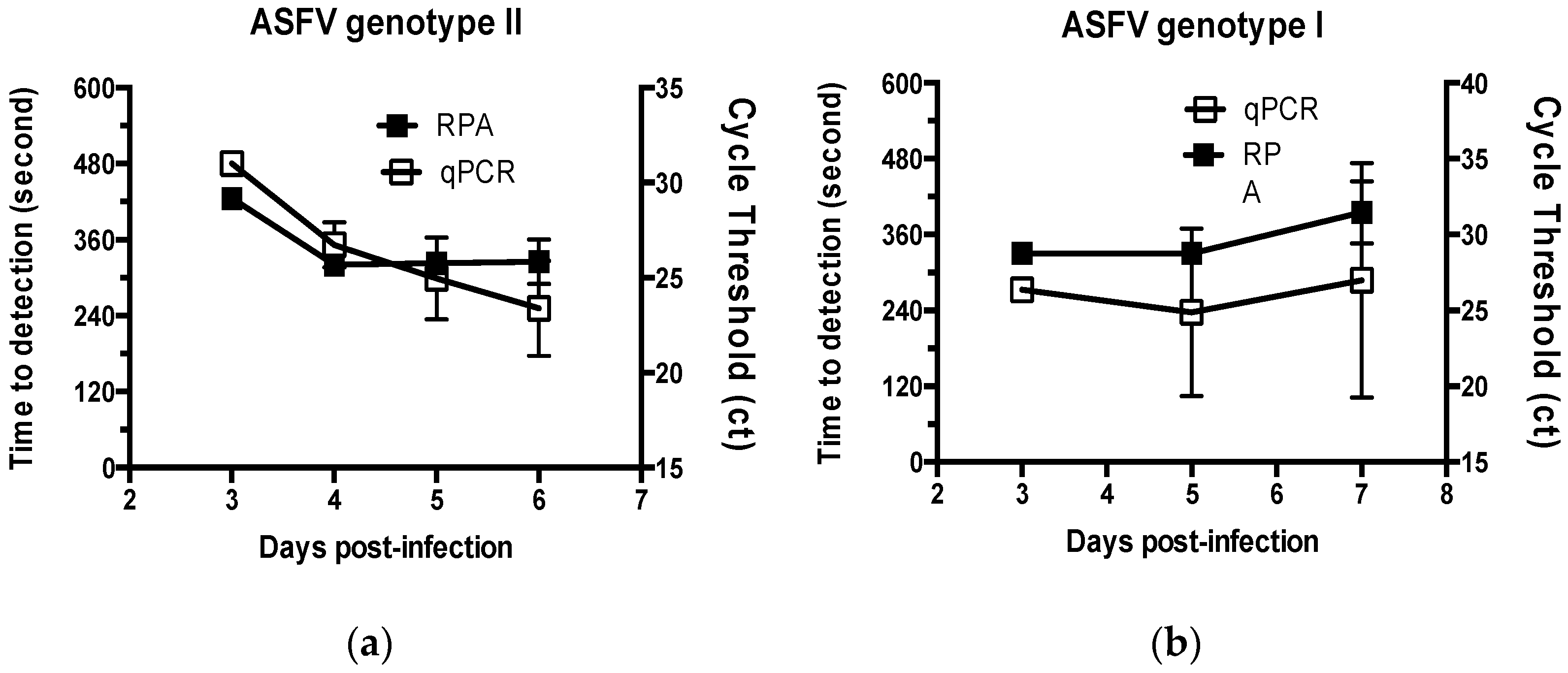
2.2. Performance of the Real-Time RPA Assay on ASFV Specimens

Of the 31 ASFV specimens, 11 ASFV isolates were tested; all ASFV isolates were positive in the real-time RPA assay (Table 2). The mean detection time for the real-time RPA assay was ~6 min (364 s) (data not shown).
The performance of the real-time RPA assay was tested using specimens from pigs challenged with virulent strains of ASFV. Of the four pigs challenged with ASFV Volgograd-v (ASFV genotype II), three specimens collected 3-days post-infection were detected as positive in the real-time RPA assay, and the average detection time of the positive results was ~7 min (435 s). After that time point, specimens collected from pigs were positive in the real-time RPA assay, and the detection time was slightly decreased to ~5–6 min (316–360 s) (Figure 2). In addition, blood specimens obtained from pigs challenged with ASFV Congo-v (ASFV genotype I) were used for analysis. Specimens collected from pigs 3-days post-challenge were detected as positive in the real-time RPA assay, and the average detection time was 5.5 min (330 s). For specimens collected at 5- and 7-days post-challenge, all were detected as positive in the real-time RPA assay, and the detection time was ~5–6 min. Of the 31 ASFV specimens, including ASFV isolates and clinical samples from infected pigs, 30 (96.8%) were positive in the real-time RPA assay (Table 2).
ASF specimens were also analyzed using a commercially available reference qPCR kit (VDx ASFV qPCR kit) (Figure 3). Of the 31 ASFV specimens, 30 (96.8%) were positive in the qPCR analysis, which was identical to that of the real-time RPA assay (Table 2 and Table 3). Interestingly, the real-time RPA assay and qPCR yielded a negative result for the same specimen, presumably because the virus concentration was lower than the detection limit in the pig challenged with ASFV at an early stage of infection. Both assays yielded negative results for the other tested viruses, with a specificity of 100%. Therefore, the concordance rate between the two assays was 100% (Table 3).
The mean detection time of qPCR was 49 min (95% CI: 47.4–50.6), whereas that of the real-time RPA assay was 6 min (95% CI: 5.7–6.2). Therefore, compared to qPCR, the real-time RPA assay could detect ASFV significantly faster (p < 0.001). Furthermore, the real-time RPA assay could effectively detect both ASFV genotypes I and II not only in clinical isolates but also in specimens derived from infected animals. Together, these findings suggest that the real-time RPA assay can be used to detect ASFV in a variety of specimens.

3. Discussion

qPCR has been used as a standard method for ASF diagnosis owing to its excellent sensitivity and specificity for detecting ASFV in clinical specimens derived from domestic pigs and wild boars [7]. However, qPCR has limitations in its application to on-site diagnosis because it requires expensive equipment and prolonged time to yield results. In recent years, several isothermal DNA amplification methods have been developed as simple and rapid alternatives to PCR-based amplification for detecting various animal pathogens [18,19,20,21,22,23]. The RPA assay does not require initial heating for DNA denaturation and the results can be obtained within 20 min. There are few or no non-specific reactions that can be problematic in other isothermal amplification methods, such as loop-mediated isothermal amplification. In addition, the RPA assay can be applied to a wide range of specimens because it shows tolerance to PCR inhibitors, which may be present in some samples [24].
In the present study, we developed a real-time RPA assay based on an exo-probe for the rapid and sensitive detection of ASFV genotypes I and II. ASFV genotype II circulates in many countries and ASFV genotype I was recently reported in China [25]. Several PCR-based methods have been developed to identify the ASFV genome; however, most are based on the amplification of the B646L gene fragment, which encodes the outer capsid protein of ASFV (p72) [10,11,12,13,14]. The B646L gene was also used to determine the genotype of ASFV, indicating the presence of a genotype-specific mutation in the B646L gene. However, we selected the CP204L gene as an amplification target because it is highly conserved among ASFV strains belonging to genotypes I and II. Additionally, we found sequences that are highly conserved and contain few nucleotides variations in the CP204L gene of ASFV strains, including the representative strains of genotypes I and II, the “Congo”and“Georgia/2007” strains, respectively, as well as other genotypes, and designed primers and probes for these sequences. We found that a real-time RPA assay using the prepared primers and probe could effectively detect ASFV isolates and clinical specimens of ASFV genotypes I and II. The detection limit of the RPA targeting CP204L (p30) developed in this study was 50 copies per reaction. In contrast, in the case of other RPAs developed with B646L (p72) as a target, the detection limit was 93.4–150 copies per reaction [11,12,13]. In light of these results, the RPA developed in the present study was much more sensitive than the existing ones. The developed RPA showed 100% sensitivity for the ASFV isolates, 96.8% sensitivity for the ASFV-challenged specimen, and 100% concordance when compared to the World Organization for Animal Health (OIE) standard qPCR. The sensitivities of other RPAs ranged from 92.3% to 100%, which were calculated based on the OIE qPCR results for suspected blood, tissue, or environmental specimens but not including ASFV isolates as a standard reference [10,11,12,13]. However, our study could be considered a meaningful advancement because ASFV isolates were used as a reference and the results are shown for the specimens from ASFV-challenged pigs according to the selected time point post-challenge. Although this real-time RPA assay was designed to detect known ASFV genotypes, it is necessary to validate the diagnostic utility of the real-time RPA assay using other ASFV genotypes in further studies. Additionally, this assay was evaluated with the highly virulent ASFV genotype I from Congo and genotype II from Russia, which cause an acute type of infections, but not with the attenuated strain such as the genotype I strain from China. However, the fact that the CP204L(P30) gene, the target of the RPA assay in this study, is highly conserved among ASFV strains indicates that this assay could detect both virulent and attenuated ASFV strains. Further evaluation of the performance of the developed real-time RPA assay over a wide range of clinical specimens including blood, tissue extract, spleen, and bone marrow at various stages of infection will advance the application of this assay to help control ASF through rapid and accurate detection of ASFV.

4. Materials and Methods

4.1. ASFV DNA Specimens

ASFV DNA specimens were provided by the Federal Research Center for Virology and Microbiology (FRCVM), Volginskii, Russia (Table 4). Because the recent ASFV strains that occurred in Asia were closely related to Georgia/2007 (NC_044959.2) [3], ASFV strains belonging to genotype II and genetically close to Georgia/2007 were selected for use in this study. ASFV DNA specimens were obtained from ASFV field isolates and collected from pigs experimentally challenged with virulent ASFV; the specimens included both ASFV genotypes I and II. Viruses were propagated using primary swine macrophages, which were prepared from pig blood and cultured in RPMI 1640 medium (Gibco, Waltham, MA, USA) supplemented with 30% (v/v) autologous porcine plasma, 10% (v/v) fetal bovine serum (Gibco), and antibiotics at 37 °C with 5% CO2. The virus titer was determined using tissue culture infectious dose (TCID50) assays as previously described [26].
All animal experiments were approved by the Research Ethics Committee of the Federal Research Center of Virology and Microbiology, Russia (No3/2021) and were conducted in accordance with Russian legislation. Large white piglets were obtained from a commercial pig farm in Russia. The pigs were not vaccinated against any infectious diseases and were bred under biosafety level 3AG conditions in the animal facilities at the Federal Research Center of Virology and Microbiology (FRCVM). The piglets were 2–2.5 months in age (weighing 15–18 kg) and were diagnosed as free of specific pathogens (data not shown). Three piglets were injected intramuscularly with 1 × 10³ TCID50 of ASFV for each genotype for the ASFV challenge. The ASFV Volgograd-v strain was isolated from outbreaks in Russia. The strain belongs to genotype II and is closely related to the reference strain “Georgia/2007 (NC_044959.2)”. The virulent ASFV Congo-v strain (strain K49, MZ202520.1) was isolated from Africa in 1949. It belongs to genotype I and causes an acute form of ASF. Blood samples were collected from the jugular vein of pigs at 3, 4-, 5-, 6-, or 7-days post-infection. DNA was extracted from the infected cell culture and blood samples using a blood and tissue kit (Qiagen, Hilden, Germany) according to the manufacturer’s instructions. The extracted DNA was stored at −80 °C until use.

4.2. Synthesis of Standard DNA Samples

High-performance liquid chromatography-pure oligonucleotides corresponding to an amplification target sequence of the CP204L gene were custom-synthesized by Integrated DNA Technologies Inc. (IDT Inc., Coral Ville, IA, USA). The synthesized target sequence of CP204L was 183 base pairs (bp) in length: (2P30, 5′-TCA TCT TCA AAA CGG ATT TAA GAT CAT CTT CAC AAG TTG TGT TTC ATG CGG GTA GCC TGT ATA ATT GGT TTT CTG TTG AGA TTA TCA ATA GCG GTA GAA TTG TTA CGA CCG CTA TAA AAA CAT TGC TTA GTA CTG TTA AGT ATG ATA TTG TGA AAT CTG CTC GTA TAT ATG CAG GGC AAG GGT-3′). DNA oligonucleotide was dissolved in DNase/RNase-free distilled water to a concentration of 100 pmol/μL, equivalent to ~5 × 10¹⁰ DNA molecules. The copy number of the synthesized DNA was calculated according to “Calculation for determining the number of copies of a template” (http://cels.uri.edu/gsc/cdna.html, accessed on 5 August 2021) [27]. The synthesized standard DNA was aliquoted and stored at −80 °C until use.

4.3. Primers and Exo-Probes

Nucleotide sequences of the ASFV strains from GenBank were aligned to identify the conserved regions of the C204L gene. Primers and exo-probes were designed based on the conserved region of the C204L gene according to the appendix of the TwistAmp® reaction manual (www.twistdx.co.uk) (accessed on 22 June 2021). Primers and exo-probes with reference sequences of ASFV genotypes were designed, and the probe contained tetrahydrofuran flanked by a dT-fluorophore and dT-quencher. The set of primers and probes that respond to ASFV DNA are shown in Table 1. Primers and exo-probes were synthesized by Macrogen Co. (Seoul, Korea) and IDT, Inc. (Coralville, IA, USA), respectively.

4.4. Real-Time RPA Assay

Real-time RPA reactions were performed in a total volume of 25 μL using the TwistAmp™ RT exo kit (TwistDX, Cambridge, UK). The reaction mixture included 0.56 pM of each RPA primer, 0.12 pM exo probe, 19 mM magnesium acetate, and 1 μL of viral or sample DNA template. All reagents, except for the viral template and magnesium acetate, were prepared in a master mix, which was distributed into 0.2 mL tubes. One microliter of viral DNA was then added to each tube. Subsequently, magnesium acetate was pipetted into the tube lids, the lids were carefully closed, and the tubes were centrifuged. After brief vortexing and centrifugation, the tubes were immediately placed in a T-8 device (Axxin, Fairfield, Australia) to start the reaction at 39 °C for 20 min with mixing after 4 min of amplification. The fluorescence signal was measured in real-time and markedly increased upon successful amplification.

4.5. qPCR

qPCR was performed using the VDx ASFV qPCR kit (Median Diagnostics, Chuncheon, Korea) on a CFX96 PCR system(BioRad Lab. Ltd., Hercules, CA, USA) according to the manufacturer’s instructions. The reactions were performed as follows: 50 °C for 2 min and 95 °C for 10 min, followed by 40 cycles of 95 °C for 15 s and 58 °C for 60 s.

4.6. Performance Assessment of the Real-Time RPA Assay

To analyze the detection limit of the real-time RPA assay, a series of 10-fold serial dilutions of the synthesized standard DNA was prepared to achieve DNA concentrations ranging from 1 × 10⁶ to 1 × 100 copies/reaction. A volume of 1 μL of each DNA dilution was used as the template. The real-time RPA assay was performed in triplicate using standard DNA. Threshold time was plotted against the corresponding amount of DNA detected.
ASFV DNA specimens containing ASFV genotypes I and II from FRCVM, Russia were used to evaluate the sensitivity of the real-time RPA assay (Table 4). In addition, the detection was investigated at timepointspost-infection by using specimens collected from animals challenged with ASFV. To evaluate the specificity, other viruses were used as templates for the assay. A commercially available qPCR kit (VDx ASFV qPCR kit, Median Diagnostics, Chuncheon, Korea) was used to compare the performance of real-time RPA.

4.7. Statistical Analysis

Data were analyzed using GraphPad Prism (ver. 6; GraphPad Software, San Diego, CA, USA) program. The sensitivity, specificity, positive predictive value (PPV), and negative predictive value (NPV) of the assays were determined using the number of viruses or specimens obtained from FRCVM as a reference. Differences between experimental groups were analyzed using the Student’s t-test and were considered significant at p-values less than 0.05.

5. Conclusions

We developed a real-time RPA assay targeting the CP204 gene to rapidly detect ASFV genotypes I and II. This real-time RPA assay could detect ASFV within 15 min, including virus isolates and clinal specimens from pigs challenged with virulent ASFV genotypes I and II strains.

Author Contributions

Conceptualization, T.I. and B.-Y.J.; methodology, T.I.; software, H.-S.B.; validation, T.I., S.M. and S.Y.; formal analysis, S.-S.K.; investigation, T.I.; resources, S.M. and S.Y.; data curation, T.I. and S.M.; writing—original draft preparation, T.I. and N.M.; writing—review and editing, B.-Y.J., S.-S.K. and N.M.; visualization, B.-Y.J.; supervision, B.-Y.J. and S.-S.K.; project administration, B.-Y.J.; funding acquisition, B.-Y.J. All authors have read and agreed to the published version of the manuscript.

Funding

This work was supported by a grant from the Animal Disease Management Technology Development Program (grant no. 320071-02) and Animal Disease Management Technology Advancement Support Program (grant no. 122011-2) of the Ministry of Agriculture, Food, and Rural Affairs, Republic of Korea.

Institutional Review Board Statement

The study was conducted in accordance with the Declaration of Helsinki, and approved by the Research Ethics Committee of the Federal Research Center of Virology and Microbiology, Russia (No3/2021) (protocol code 03/06 from 18 June 2021).

Informed Consent Statement

Not applicable.

Data Availability Statement

The original data used for the analyses can be obtained from the authors after approval by the responsible institutions in Korea and Russia.

Acknowledgments

We would like to thank the staff of FRCVM, who were involved in the preparation and documentary support of the ASF DNA samples during their transportation.

Conflicts of Interest

The authors declare no conflict of interest.

References

  1. Gaudreault, N.N.; Madden, D.W.; Wilson, W.C.; Trujillo, J.D.; Richt, J.A. African Swine Fever Virus: An Emerging DNA Ar-bovirus. Front. Vet. Sci. 2020, 13, 215. [Google Scholar] [CrossRef] [PubMed]
  2. Sauter-Louis, C.; Conraths, F.J.; Probst, C.; Blohm, U.; Schulz, K.; Sehl, J.; Fischer, M.; Forth, J.H.; Zani, L.; Depner, K.; et al. African Swine Fever in Wild Boar in Europe—A Review. Viruses 2021, 13, 1717. [Google Scholar] [CrossRef] [PubMed]
  3. Ge, S.; Li, J.; Fan, X.; Liu, F.; Li, L.; Wang, Q.; Ren, W.; Bao, J.; Liu, C.; Wang, H.; et al. Molecular Characterization of African Swine Fever Virus, China, 2018. Emerg. Infect. Dis. 2018, 24, 2131–2133. [Google Scholar] [CrossRef] [PubMed] [Green Version]
  4. Zhou, L.; Yu, E.Y.W.; Wang, S.; Sun, C. African swine fever epidemic in China. Veter. Rec. 2019, 184, 713. [Google Scholar] [CrossRef]
  5. Yoo, D.; Kim, H.; Lee, J.Y.; Yoo, H.S. African swine fever: Etiology, epidemiological status in Korea, and perspective on control. J. Veter. Sci. 2020, 21, e38. [Google Scholar] [CrossRef] [PubMed] [Green Version]
  6. Kim, S.-H.; Kim, J.; Son, K.; Choi, Y.; Jeong, H.-S.; Kim, Y.-K.; Park, J.-E.; Hong, Y.-J.; Lee, S.-I.; Wang, S.-J.; et al. Wild boar harbouring African swine fever virus in the demilitarized zone in South Korea, 2019. Emerg. Microbes Infect. 2020, 9, 628–630. [Google Scholar] [CrossRef] [PubMed]
  7. Gallardo, C.; Fernández-Pinero, J.; Arias, M. African swine fever (ASF) diagnosis, an essential tool in the epidemiological in-vestigation. Virus Res. 2019, 271, 197676. [Google Scholar] [CrossRef] [PubMed]
  8. Piepenburg, O.; Williams, C.H.; Stemple, D.; Armes, N.A. DNA Detection Using Recombination Proteins. PLOS Biol. 2006, 4, e204. [Google Scholar] [CrossRef] [PubMed]
  9. James, A.; Macdonald, J. Recombinase polymerase amplification: Emergence as a critical molecular technology for rapid, low-resource diagnostics. Expert Rev. Mol. Diagn. 2015, 15, 1475–1489. [Google Scholar] [CrossRef] [PubMed] [Green Version]
  10. Zhang, S.; Sun, A.; Wan, B.; Du, Y.; Wu, Y.; Zhang, A.; Jiang, D.; Ji, P.; Wei, Z.; Zhuang, G.; et al. Development of a Directly Visualized Recombinase Polymerase Amplification–SYBR Green I Method for the Rapid Detection of African Swine Fever Virus. Front. Microbiol. 2020, 11, 22. [Google Scholar] [CrossRef] [PubMed]
  11. Miao, F.; Zhang, J.; Li, N.; Chen, T.; Wang, L.; Zhang, F.; Mi, L.; Zhang, J.; Wang, S.; Wang, Y.; et al. Rapid and Sensitive Recombinase Polymerase Amplification Combined with Lateral Flow Strip for Detecting African Swine Fever Virus. Front. Microbiol. 2019, 15, 1004. [Google Scholar] [CrossRef] [PubMed]
  12. Fan, X.; Li, L.; Zhao, Y.; Liu, Y.; Liu, C.; Wang, Q.; Dong, Y.; Wang, S.; Chi, T.; Song, F.; et al. Clinical Validation of Two Recombinase-Based Isothermal Amplification Assays (RPA/RAA) for the Rapid Detection of African Swine Fever Virus. Front. Microbiol. 2020, 21, 1696. [Google Scholar] [CrossRef] [PubMed]
  13. Zhai, Y.; Ma, P.; Fu, X.; Zhang, L.; Cui, P.; Li, H.; Yan, W.; Wang, H.; Yang, X. A recombinase polymerase amplification combined with lateral flow dipstick for rapid and specific detection of African swine fever virus. J. Virol. Methods 2020, 285, 113885. [Google Scholar] [CrossRef] [PubMed]
  14. Wang, J.; Wang, J.; Geng, Y.; Yuan, W. A recombinase polymerase amplification-based assay for rapid detection of African swine fever virus. Can. J. Vet. Res. 2017, 81, 308–312. [Google Scholar] [PubMed]
  15. Afonso, C.L.; Alcaraz, C.; Brun, A.; Sussman, M.D.; Onisk, D.V.; Escribano, J.M.; Rock, D.L. Characterization of P30, a highly antigenic membrane and secreted protein of African Swine Fever Virus. Virology 1992, 189, 368–373. [Google Scholar] [CrossRef]
  16. Pornthummawat, A.; Truong, Q.L.; Hoa, N.T.; Lan, N.T.; Izzati, U.Z.; Suwanruengsri, M.; Nnueangphuet, P.; Hirai, T.; Ymaguchi, R. Pathological lesions and presence of viral antigens in four surviving pigs in African swine fever outbreak farms in Vietnam. J. Vet. Med. Sci. 2021, 83, 1653–1660. [Google Scholar] [CrossRef]
  17. Petrovan, V.; Yuan, F.; Li, Y.; Shang, P.; Murgia, M.V.; Misra, S.; Rowland, R.R.; Fang, Y. Development and characterization of monoclonal antibodies against p30 protein of African swine fever virus. Virus Res. 2019, 269, 197632. [Google Scholar] [CrossRef] [PubMed]
  18. Yan, L.; Zhou, J.; Zheng, Y.; Gamson, A.S.; Roembke, B.T.; Nakayama, S.; Sintim, H.O. Isothermal amplified detection of DNA and RNA. Mol. BioSyst. 2014, 10, 970–1003. [Google Scholar] [CrossRef] [Green Version]
  19. Davi, S.D.; Kissenkötter, J.; Faye, M.; Böhlken-Fascher, S.; Stahl-Hennig, C.; Faye, O.; Faye, O.; Sall, A.A.; Weidmann, M.; Ademowo, O.G.; et al. Recombinase polymerase amplification assay for rapid de-tection of Monkeypox virus. Diagn. Microbiol. Infect. Dis. 2019, 95, 41–45. [Google Scholar] [CrossRef] [PubMed]
  20. Zhang, J.; Liu, J.; An, D.; Fan, Y.; Cheng, Z.; Tang, Y.; Diao, Y. A novel recombinase polymerase amplification assay for rapid detection of epidemic fowl adenovirus. Poult. Sci. 2020, 99, 6446–6453. [Google Scholar] [CrossRef] [PubMed]
  21. Zhu, Y.; Zeng, F.; Sun, J.; Liu, X.; Wu, M.; Huang, B.; Lian, Y.; Xiao, L.; Ma, L.; Zhang, S.; et al. Application of recombinase polymerase amplification method for rapid detection of infectious laryngotracheitis virus. Mol. Cell. Probes 2020, 54, 101646. [Google Scholar] [CrossRef]
  22. El-Tholoth, M.; Branavan, M.; Naveenathayalan, A.; Balachandran, W. Recombinase polymerase amplification–nucleic acid lateral flow immunoassays for Newcastle disease virus and infectious bronchitis virus detection. Mol. Biol. Rep. 2019, 46, 6391–6397. [Google Scholar] [CrossRef] [PubMed] [Green Version]
  23. Daher, R.K.; Stewart, G.; Boissinot, M.; Bergeron, M.G. Recombinase Polymerase Amplification for Diagnostic Applications. Clin. Chem. 2016, 62, 947–958. [Google Scholar] [CrossRef] [PubMed]
  24. Wang, J.; Wang, J.; Li, R.; Liu, L.; Yuan, W. Rapid and sensitive detection of canine distemper virus by real-time reverse tran-scription recombinase polymerase amplification. BMC Vet. Res. 2017, 15, 241. [Google Scholar] [CrossRef]
  25. Sun, E.; Huang, L.; Zhang, X.; Zhang, J.; Shen, D.; Zhang, Z.; Wang, Z.; Huo, H.; Wang, W.; Huangfu, H.; et al. Genotype I African swine fever viruses emerged in domestic pigs in China and caused chronic infection. Emerg. Microbes Infect. 2021, 10, 2183–2193. [Google Scholar] [CrossRef] [PubMed]
  26. Koltsova, G.; Koltsov, A.; Krutko, S.; Kholod, N.; Tulman, E.; Kolbasov, D. Growth Kinetics and Protective Efficacy of Attenuated ASFV Strain Congo with Deletion of the EP402 Gene. Viruses 2021, 13, 1259. [Google Scholar] [CrossRef] [PubMed]
  27. Andrew, S. Calculation for Determining the Number of Copies of a Template. URI Genomics & Sequencing. Available online: http://cels.uri.edu/gsc/cdna.html (accessed on 5 August 2021).
Figure 1. Analytical performance of the real-time RPA assay for ASFV. The synthesized DNA standards in concentrations ranging from 5 × 10⁶ to 5 × 100 copies were amplified as templates for the real-time RPA assay. A representative of three experiments is shown.
Pathogens 11 00439 g001
Figure 2. Comparison of the real-time RPA assay and qPCR for ASFV isolates and clinical specimens. The time to detection of the real-time RPA assay and qPCR were measured using11 ASFV isolates and clinical specimens collected from pigs challenged with ASFV (a) Volgograd-v (genotype II) or (b) Congo-v (genotype I). The time to a positive result for the tested specimens is expressed in seconds.
Pathogens 11 00439 g002
Figure 3. Results of the real-time RPA assay with clinical specimens from pigs challenged with ASFV genotypes I and II. Comparison between the performance of real-time RPA and qPCR on ASFV DNA samples (a). Ct values for real-time RPA and qPCR are shown in (b,c), respectively. Large white piglets were challenged with 1 × 10³ TCID50 of ASFV Volgograd-v (genotype II) or Congo-v (genotype I), and the blood samples were collected at the designated time point after infection. DNA was extracted from the collected blood samples and used as a template for the real-time RPA assay.
Pathogens 11 00439 g003
Table 1. Sequences of primers and the probe used in this study.
NameNucleotide Sequence (5′-3′)Amplicon Size
(bp)
2RPAP30FAGATCATCTTCACAAGTTGTGTTTCATGCGGGTAG141
2RPAP30RCGAGCAGATTTCACAATATCATACTTAACAGTACT
2P30Exo v1TAGCGGTCGTAACAATTCTACCGCTATTGA(Fam dT)A
(THF)TC(BHQ1-dT)CAACAGAAAACCAAT-C3spacer
Table 2. Detection of the ASFV genome in clinical samples using real-time RPA and qPCR assays.
Virus/SpecimensReal-Time RPAqPCR
No. of PositivesNo. of NegativesNo. of PositivesNo. of Negatives
ASFV GII isolates (n = 11)110110
ASFV GII animal specimens (n = 14)131131
ASFV GI animal specimens (n = 6)6060
Other viruses
(n = 11)
011011
Sensitivity96.8% (95% CI: 83.3–99.9)96.8% (95% CI: 83.3–99.9)
Specificity100% (95% CI: 71.5–100.0) 100% (95% CI: 71.5–100.0)
Positive predictive value100% (95% CI: 88.4–100.0)100% (95% CI: 88.4–100.0)
Negative predictive value91.7% (95% CI: 61.5–99.8)91.7% (95% CI: 61.5–99.8)
Table 3. Comparison between the real-time RPA assay and qPCR for the detection of ASFV.
qPCR
No. of PositivesNo. of NegativesTotal
Real-time RPANo. of positive30030
No. of negative011
Total30131
Table 4. List of viruses used in the present study.
NameIsolates/OriginGenotype/SerotypeSource
D 1ASFV Stavropol 01/8IIFRCVM
D 2ASFV Irkutsk 2017IIFRCVM
D 3ASFV StavropolIIFRCVM
D 4ASFV Omsk #1IIFRCVM
D 5ASFV Omsk #2IIFRCVM
D 6ASFV Nizhny Novgorod #1IIFRCVM
D 7ASFV Nizhny Novgorod #2IIFRCVM
D 8ASFV Nizhny Novgorod #3IIFRCVM
D 9ASFV Nizhny Novgorod #4IIFRCVM
D 10ASFV KrasnodarIIFRCVM
D 11ASFV SaratovIIFRCVM
D 12ASFV DNA from pig # 1-Volgograd-v (3 dpi)IIFRCVM
D 13ASFV DNA from pig # 1-Volgograd-v (5 dpi)IIFRCVM
D 14ASFV DNA from pig #2-Volgograd-v (3 dpi)IIFRCVM
D 15ASFV DNA from pig #2-Volgograd-v (5 dpi)IIFRCVM
D 16ASFV DNA from pig #3-Volgograd-v (3 dpi)IIFRCVM
D 17ASFV DNA from pig #3-Volgograd-v (4 dpi)IIFRCVM
D 18ASFV DNA from pig #3-Volgograd-v (5 dpi)IIFRCVM
D 19ASFV DNA from pig #3-Volgograd-v (6 dpi)IIFRCVM
D 20ASFV DNA from pig #4-Volgograd-v (4 dpi)IIFRCVM
D 21ASFV DNA from pig #4-Volgograd-v (5 dpi)IIFRCVM
D 22ASFV DNA from pig #4-Volgograd-v (6 dpi)IIFRCVM
D 23ASFV DNA from pig #5-Volgograd-v (3 dpi)IIFRCVM
D 24ASFV DNA from pig #5-Volgograd-v (4 dpi)IIFRCVM
D 25ASFV DNA from pig #5-Volgograd-v (5 dpi)IIFRCVM
D 26ASFV DNA from pig #1-Congo-v (3 dpi)IFRCVM
D 27ASFV DNA from pig #1-Congo-v (5 dpi)IFRCVM
D 28ASFV DNA from pig #1-Congo-v (7 dpi)IFRCVM
D 29ASFV DNA from pig #2- Congo-v (3 dpi)IFRCVM
D 30ASFV DNA from pig #2-Congo-v (5 dpi)IFRCVM
D 31ASFV DNA from pig #2-Congo-v (7 dpi)IFRCVM
V 1Influenza A virusH1N1KBPV
V 2Influenza A virusH3N2KBPV
V 3Parainfluenza virus 2 KBPV
V 4Parainfluenza virus 4 KBPV
V 5AdenovirusSerotype 1KBPV
V 6AdenovirusSerotype 3KBPV
V 7AdenovirusSerotype 7KBPV
V 8Enterovirus AEnterovirus 71KBPV
V 9Rota virus AType 1KBPV
V 10Rota virus AType 3KBPV
V 11Cytomegalovirus KBPV
FRCVM: Federal Research Center for Virology and Microbiology, Volginskii, Russia. KBPV: Korea Bank for Pathogenic Viruses, Korea University, Seoul, Korea. Blood was collected at the designated time points (12–31 days) from pigs challenged with virulent ASFV, either the Volgograd-v or Congo-v strain, and DNA was extracted from the blood.
Publisher’s Note: MDPI stays neutral with regard to jurisdictional claims in published maps and institutional affiliations.

