Review

Role of Vaginal Mucosa, Host Immunity and Microbiota in Vulvovaginal Candidiasis

by
Subatrra Nair Balakrishnan
1,
Haizat Yamang
1,
Michael C. Lorenz
2,
Shu Yih Chew
1,* and
Leslie Thian Lung Than
1,*
1
Department of Medical Microbiology, Faculty of Medicine and Health Sciences, University Putra Malaysia, Serdang 43300, Selangor, Malaysia
2
Department of Microbiology and Molecular Genetics, University of Texas McGovern Medical School, Houston, TX 77030, USA
*
Authors to whom correspondence should be addressed.
Pathogens 2022, 11(6), 618; https://doi.org/10.3390/pathogens11060618
Submission received: 30 January 2022 / Revised: 10 May 2022 / Accepted: 11 May 2022 / Published: 25 May 2022

Abstract

:
Vulvovaginal candidiasis (VVC) is a prevalent gynaecological disease characterised by vaginal wall inflammation that is caused by Candida species. VVC impacts almost three-quarters of all women throughout their reproductive years. As the vaginal mucosa is the first point of contact with microbes, vaginal epithelial cells are the first line of defence against opportunistic Candida infection by providing a physical barrier and mounting immunological responses. The mechanisms of defence against this infection are displayed through the rapid shedding of epithelial cells, the presence of pattern recognition receptors, and the release of inflammatory cytokines. The bacterial microbiota within the mucosal layer presents another form of defence mechanism within the vagina through acidic pH regulation, the release of antifungal peptides and physiological control against dysbiosis. The significant role of the microbiota in maintaining vaginal health promotes its application as one of the potential treatment modalities against VVC with the hope of alleviating the burden of VVC, especially the recurrent disease. This review discusses and summarises current progress in understanding the role of vaginal mucosa and host immunity upon infection, together with the function of vaginal microbiota in VVC.

1. Introduction

Vulvovaginal candidiasis (VVC) is a mucosal infection at the vaginal tissue and the vulva of the lower female reproductive tract, which is caused predominantly by Candida albicans, an opportunistic fungal pathogen. Over 90% of VVC cases are caused by C. albicans and the remaining 10% are caused by the non-albicans Candida (NAC) species, such as C. glabrata, C. krusei, C. tropicalis and C. parapsilosis [1]. Ascending genital tract infections and their association with direct and indirect economic costs make VVC a global health concern [2]. Seventy-five percent of women are reported to experience this vaginal yeast infection at least once in their lifespan [3]. Following anaerobic bacterial vaginosis, candidiasis is the second most prevalent root cause of vaginal inflammation that is associated with symptoms, such as itching, irritation, dysuria, dyspareunia, cottage cheese-like vaginal discharges, or soreness of the vulva that may persist for days or even weeks which can be aggravated by sexual intercourse [4]. In addition, the normal human vaginal microenvironment is more acidic (pH of 3.8–4.5) compared to other mammals (pH of 5.4–7.8), with Lactobacilli as the dominant organisms in the human vaginal microbiota—more than 70% of the human vaginal microbiota in some women [5]. Diagnosis of VVC is made by direct microscopy from vaginal swabs or smears, in which yeast cells, pseudohyphae and hyphae can be observed. An increase in vaginal pH of about 4.5, erythema of the vaginal mucosa, vulval oedema, and a thick white discharge are also common signs of VVC [6].
Recurrent VVC (RVVC) occurs if a woman experiences more than three episodes within 12 months. The majority of infections in women with RVVC are caused by C. albicans [7]. Women with RVVC have been identified as having predisposing genetic factors or hypersensitivity to Candida that make them susceptible to this infection [8]. According to Foxman et al. (2013), based on their study in five European countries and the United States, a global prevalence of 3871 RVVC cases per 100,000 women have been documented with the highest frequency (9%) observed in women aged between 25 and 34 years old [9]. Moreover, VVC and RVVC tend to infect women primarily during their reproductive years, as they experience an elevated endogenous oestrogen level [10]. In addition, there are two types of risk factors for VVC, namely host factors and behavioural factors. Pregnancy, menopausal hormone replacement (MHR), uncontrolled diabetes mellitus, immunosuppression, antibiotic and glucocorticoid usage as well as genetic effects are host-related risk factors. Meanwhile, contraceptive usage, personal hygiene practices and sexual behaviour fall under the behavioural risk factors for VVC [11].
Candida possesses a plethora of virulence factors which include the ability to evade host defence, adherence to the surface epithelium, biofilm formation and invasion by the secretion of extracellular enzymes [12]. Candida adherence to host surfaces is essential for the primary colonisation of human tissues, which is a prerequisite for infections. A key mechanism in the onset of VVC is the adherence of Candida to vaginal epithelial cells (VEC) [13], via adhesins, which are unique cell surface proteins that play their role by mediating Candida spp. cell adhesion to biotic and abiotic surfaces by cell surface physicochemical features [14,15]. They also contribute to biofilm development on host or abiotic surfaces, which is linked to a majority of infections caused by Candida spp. and contribute to treatment failures, as these structures are more resistant to antifungal drugs [16,17]. Biofilm formation on implanted medical devices (e.g., vascular or urinary catheters, artificial heart valves, dental implants, etc.) of Candida, especially in C. albicans, has made it one of the most common causes of nosocomial infections, which are becoming extremely hard to treat due to both inherent and acquired antifungal resistance [18]. Moreover, infection-induced production of hydrolytic enzymes improves organism adherence, invasion and elimination of immunological components in the host, and has a role in nutrition acquisition. Hydrolytic enzymes, such as secreted aspartyl proteinases (SAP), phospholipases (PL) and candidialysin [19] play a role in VVC pathogenesis. A recent in vitro study carried out by Ali et al. (2018) reported that the deletion of SAP1SAP3 genes has reduced the C. albicans vaginopathic capacity [20]. The expression of SAP1SAP3 genes has been associated with vaginal infection that may implicate the yeast-to-hyphal switching in the virulence of C. albicans [21]. The SAP1SAP3 genes are the first SAP genes to be expressed by C. albicans during the development of lesions in VVC [22]. In addition, Roselletti et al. (2017) reported that SAP1 and SAP3 genes were expressed in almost half of the symptomatic women, while SAP2 expression was elevated in all of the patients [23]. According to Pericolini et al. (2015), SAP2 expression has also led to an increase in caspase-1 expression in murine and human VEC [24]. On the other hand, candidalysin is required for the activation of immunopathological signalling in the vaginal mucosa. The ability of candidalysin to destroy epithelia and potentially promote free vaginal heparin sulphate may contribute to the fungal fitness strategy to protect against PMN-mediated clearance at the mucosal surface [25].
The vaginal mucosa is the most significant and adaptable structure in the female reproductive system and is the surface upon which Candida species adhere to initiate infections, whilst vaginal immunity confers protection to the host upon VVC occurrence. In addition, the composition of the vaginal microbiota also plays a significant role in the onset of VVC. This review summarises the role of three crucial components: the vaginal mucosa, vaginal immunity and the vaginal microbiota in VVC.

2. Vaginal Mucosa

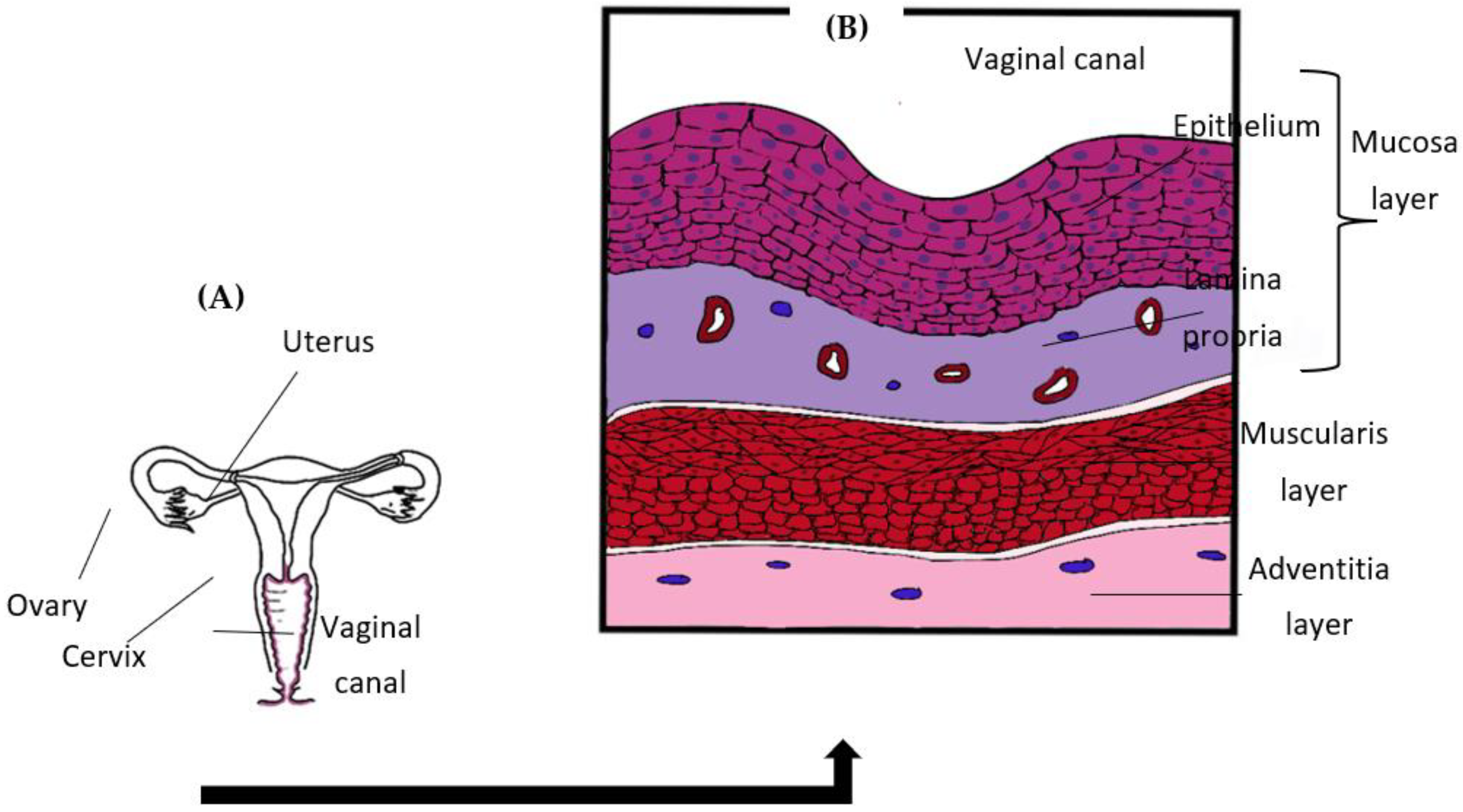
2.1. Vaginal Structure

The vagina is an elastic and muscular organ of the female genital tract that connects the uterus and cervix to the outside of the body (Figure 1A). An outermost mucosa layer, the interstitial muscularis layer, and the deeper adventitia layer are the three different layers that make up the vaginal wall structure (Figure 1B). The mucosal layer is a structure that continues from the uterine lining and can be divided into two sublayers; the first sublayer is the epithelial layer which rests on an underlying second layer of lamina propria. The vaginal epithelial layer or vaginal epithelium (VE) is a multi-layered stratified squamous structure which undergoes differentiation into several distinct and parallel layers known as strata [26]. The uppermost layer of VE is also known as the stratum corneum and is closest to the lumen. It undergoes rapid shredding and regeneration throughout the menstruation cycle [27]. The exfoliation of VE cells serves as the initial defence mechanism of the vagina as this action removes potential pathogenic Candida bound to it [28].

2.2. VEC and Its Significance

As the first cell lining to be in contact with Candida, the apical VEC layer plays an important role as the protective barrier and the first line of defence. This apical VEC layer together with the other underlying VEC layers is held together by intercellular transmembrane proteins which include F11R, desmosomal adhesion proteins (JAM3), tight junction protein 1 (TJP1), claudin-1 and E-cadherin. These junction proteins are found occupying the multi-layered structure of the VE that assists in maintaining the overall cellular integrity of the layer and simultaneously restricts the dissemination of Candida [29]. In fact, it was proposed that the mechanism by which C. albicans invades mucosal epithelial tissue is mediated through proteolytic degradation of adherence junctions [30,31], although other routes of entry, including candidalysin-mediated epithelial lysis, have also been suggested. The VEC also contributes significantly to the activation of innate immunity through expressed pattern recognition receptors (PRRs) on the apical surface of the cells. PRRs are capable of detecting the presence of pathogenic Candida upon binding (ligation) with pathogen-associated molecular patterns (PAMPs) secreted by or presented on the surface of the pathogen [19]. Previous research revealed various PRRs are responsible for Candida recognition, dominated by the Toll-like receptors (TLRs), C-type lectin receptors (CLRs), the nucleotide-binding oligomerisation domain (NOD)-like receptor (NLR) and the retinoic acid-inducible gene I (RIG-I) -like receptors (RLRs) [32]. Although each receptor portrays different functions, TLRs are known to be expressed within VEC, which mediate the release of antimicrobial peptides (AMP), pro-inflammatory and anti-inflammatory cytokines, along with co-stimulatory molecules to promote adaptive immunity [33]. Pro-inflammatory cytokines, such as TNF-α, IL-1β, and IL-6, are released by VEC, and this promotes the migration of local immune cells to affected sites. The release of these cytokines activates and mediates the proliferation of innate immune cells, primarily macrophages and neutrophils, to the affected site [34]. A study conducted by Diletta et al. (2020) shows that, upon exposure to C. albicans hyphae, significantly higher production of TNF-α among RVVC patients was observed compared with the control group (healthy individuals), indicating its possible role in acute local immune response [35]. Anti-inflammatory cytokines (IL-10 and TGF-β) are also released by VEC and are believed to regulate inflammatory responses, which prevent excessive damage to local cells [36]. This dynamic release of cytokines gives an impact on the balance of vaginal response toward the pathogen.
Pathogen recognition by PRRs activates NF-κB and MAPK cascades leading to the production of AMPs by VEC [37], including lysozyme, lactoferrin, serine leukocyte protease inhibitor (SLPI), calprotectin, and elafin as well as α- and β-defensins throughout the vaginal tract that exhibits a wide range of innate immunity, including antifungal properties [38,39]. Calprotectin, a metal-binding protein, was proven to be an effective antifungal agent as it induces iron starvation and consequently inhibits iron availability to C. albicans [40]. Research on the antifungal effect of SLPI shows cytoplasmic deflation of C. albicans upon treatment with SLPI peptides, suggesting a penetrative effect of SLPI through the cell wall of the fungus [41]. Meanwhile, Chairatan et al. (2016) found that α-defensin exhibits antifungal activities by blocking the adhesion of C. albicans within the epithelial cells of human intestines and inhibits its biofilm formation [42]. An in vivo study using a mouse model of VVC caused by C. albicans revealed a significant increase in murine β-defensin 1 expression during early infection (acute phase), paired with polymorphonuclear leukocyte (PMN; neutrophils) infiltration, demonstrating its role in first-line defence against fungal infection and as inducers of innate inflammatory mediators [43].
Host-pathogen interplay within the mucosa is undeniably unique, considering the ability of VEC to discriminate between disease-causing microbes and commensal. As VEC is exposed to diverse microorganisms within the mucosa interface, it is crucial to understand the tolerance mechanism of VEC towards different types of organisms. In a review, Naren et al. (2010) suggested that microbial homeostasis within mucosa is highly influenced by the nature of the resident microbes [44]. As we know, Candida is a benign part of the normal vaginal microbiota in many women. However, due to vaginal dysbiosis, it increases the fungal burden within the vagina, triggers yeast-to-hyphal switching and releases virulence factors of C. albicans [19]. The virulence factors of Candida, together with the presence of PAMPs, make them recognisable as a “threat” and thus, they stimulate inflammatory immune responses [44]. In addition, another review by McLure et al. (2014) suggested that the differential responses are affected by the cellular localisation of TLRs. Unlike commensals, pathogenic microorganisms can breach the epithelial barrier and invade the deeper layer of mucosa, resulting in a pro-inflammatory response that can precipitate the pathology associated with VVC [37].

3. Vaginal Host Immunity

The initial line of defence against Candida infections at most body sites involves the innate immune system. The cells of various epithelial surfaces coordinate the innate response to counteract proliferating pathogens by recruiting additional cellular mediators, like neutrophils, and inducing the expression of AMPs. The adaptive immune response is defined by specificity, and it plays a role in pathogen removal in the late stages of infection and in the development of immunological memory [45]. The vaginal mucosa, both its tissue layers together with the cervicovaginal fluids, contain both innate and acquired immune responses. The antifungal activity mediated by epithelial cells has been proven to be both fungistatic and non-inflammatory, implying that it functions as a basic defence mechanism to prevent excessive tissue damage and to maintain asymptomatic commensalism, perhaps via broad-specificity AMPs [46]. Adaptive immunity, in contrast, engenders pathogen-specific defence mechanisms where the antigen-presenting cells (APC) digest the pathogen antigens and deliver them to T cells, resulting in T-cell activation. Following that, the activation of cytokine production and antibody synthesis will take place where B cells will produce antibodies and the T cells will mediate the protective mechanism [47].

3.1. Role of PRRs in Immune Recognition

Generally, the innate immune responses are mediated by the interplay of the innate immune cells of the host that express PRRs on their surface together with the PAMPs of the pathogens [48]. PRRs expressed by immune cells can be classified into four different classes, such as Toll-like receptors (TLRs), C-type lectin receptors (CLRs), the nucleotide-binding oligomerisation domain (NOD)-like receptor (NLR) and the retinoic acid-inducible gene I (RIG-I) -like receptors (RLRs) [1]. In general, monocytes and neutrophils in the circulation, as well as the macrophages in affected tissues, are the major cells of the host’s innate immune response that are involved in identifying invading pathogens [48]. Neutrophils are the main immune cells involved in the regulation of VVC and, thus, neutropenic women are more susceptible to this infection [49,50]. In support of that statement, a study using the murine VVC model by Ibrahim et al. (2013) has reported that vaccination with rAls3p-N protein of C. albicans, formulated with alum adjuvant (NDV-3), elicited protection against VVC by which anti-rAls3p-N antibodies work as an opsonin to increase IFN-γ primed neutrophil activity in killing C. albicans [51]. TLRs and CLRs families are the most common receptors that identify Candida-associated molecular patterns which are mostly expressed by monocytes on their cell membranes. While in neutrophils, TLRs expression is minimal, whereas phagocytic receptors, such as complement receptor 3 (CR3) and Fcγ receptors (FcγRs), are highly expressed [52]. TLRs can be classified into cell-membrane associated receptors (TLR1, TLR2, TLR4, TLR5 and TLR6) or intracellular receptors (TLR3, TLR7, TLR8 and TLR9). TLRs mainly recognise fungal components; for instance, TLR2 is involved in the identification of phospholipomannan, TLR4 is involved in the recognition of O-linked mannans, TLR6 is involved in the recognition of zymosan (essentially a crude mixture of fungal cell wall components), while TLR9 is involved in the detection of fungal DNA [53]. On the other hand, CLRs are responsible for identifying polysaccharide structures from C. albicans, i.e., Dectin-1 detects β-glucans, whereas the macrophage mannose receptor (MR) and dendritic cell-specific ICAM-3-grabbing nonintegrin (DC-SIGN) as well as Dectin-2 recognise the N-linked mannans [52]. During infection, the detection of PAMPs by PRRs can induce gene expression in the inflammatory responses by initiating the production of pro-inflammatory cytokines, type I interferons (IFNs), chemokines together with antimicrobial proteins, and the proteins that participated in PRRs signalling modulation [54].

3.2. Role of PAMPs in Immune Recognition

Fungal PAMPs are conserved and constitute primarily cell wall carbohydrate structures specific to these microbes. In C. albicans, the outer cell wall consists of proteins glycosylated with O-linked and N-linked and phosphorylated mannans that eventually prevent the immune recognition of the inner cell wall components. This layer is thinner at the level of the bud scar caused by yeast division, allowing PAMPs from the inner cell wall to be recognised by the immune system [55]. Candida species often mask the inner cell wall layer, such as β-glucan and chitin, from recognition by Dectin-1, one of the well-characterised CLRs and other PRRs [56]. Therefore, recognition of PAMPs by the innate immune system allows the distinction between ‘self’ and the microbial ‘non-self’ [57]. Neutrophils have been demonstrated to be able to create extracellular traps and trigger the cell wall remodelling in Candida, which leads to better β-glucan exposure and Dectin-1 binding [58]. The majority of fungal pathogens, including C. albicans, have a core structure of β-(1,3)-glucan covalently bonded to β-(1,6)-glucan and chitin (a β-(1,4)-linked polymer of N-acetylglucosamine (GlcNAc)) [59]. β-glucans are polymeric carbohydrates that are disclosed to regulate inflammatory responses in both in vitro and in vivo models [60]. The degree of branching, polymer length and tertiary structure of β-glucans impact their immunomodulatory effects, but no consensus has been achieved on the basic structural requirements for biological activity, and various forms of glucans have distinct biological effects [61]. When Dectin-1 binds to β-glucan, an Src-Syk-CARD9-dependent pathway is initiated, which results in the triggering of the NF-κB and the transcription of antifungal cytokines, such as IL-6, IL-1β, along with IL-23 [58]. Dectin-1 stimulates cytokine production in a synergistic manner with TLR2 and TLR4, but it also generates IL-1β, IL-17 and IL-10 independently of these TLRs. The activation of signalling pathways within dendritic cells (DCs), as a result of PRR activation, leads to the initiation of a specific adaptive cellular immune response. Dendritic cells are found throughout the vaginal and cervical epithelia, where they adhere to Candida and initiate cell differentiation together with activation of cell signals that cause inflammation, cell proliferation and induce adaptive immunity [62]. The cells of the innate immune system detect portions of the C. albicans cell wall skeletal and matrix components. The complicated interaction between host receptors and C. albicans cell wall components, together with the released compounds, can activate various inflammasomes [63]. Fungal cell wall components can cause inflammation by activating epithelial and immunological receptors, resulting in the release of pro-inflammatory cytokines and chemokines. However, pathogenic fungi can mask these cell wall components to remain undetected [64].

3.3. Antimicrobial Defence Mechanism against C. albicans

The cationic AMPs of innate immunity have received a lot of interest in the last two decades as prospective candidates for developing new antimicrobials [65]. Direct targeting of AMPs to microbial membranes and subsequent disruption suggests that resistance is less likely to be developed, which is critical in the fight against antifungal-resistant vaginal infections. The reasons put forth for this theory are because AMPs are more likely to be cidal, as opposed to static, and they kill cells quickly through mechanisms that are hard to bypass, such as membrane disruption. AMPs often possess several targets, such as the cell membrane, specific proteins and nucleic acids, hence, even if resistance to one develops, others remain to take the task forward [66]. For instance, beyond AMPs binding to membranes, there is evidence that they can adhere to target ribosomal subunits, inhibit DNA, RNA, protein, and macromolecule synthesis, as well as interfere with respiration, intracellular protein folding, and iron regulation [67]. These natural peptides are used by species throughout evolution as a first line of defence [68]. These AMPs are produced by professional phagocytes, as well as epithelial cells of the skin and the respiratory, gastrointestinal, and genitourinary tracts [69]. Human β-defensins have broad antifungal activity and chemotactic functions and are some of the AMPs expressed within the VE [70]. The fungicidal activity of the human β-defensins is mainly displayed through membrane permeabilisation, resulting in intracellular ion leakage, which inhibits planktonic and biofilm growth alike [71,72]. Complementing their direct antifungal effects, β-defensins also exhibit immunomodulatory function via pro-inflammatory cytokines stimulation and immune cell recruitment [73]. The cathelicidin LL-37 (CRAMP in mice), is a linear cationic alpha-helical peptide produced by various epithelial cells, including those of the gut as well as those in the squamous epithelium of the cervix and vagina, as well as in cervicovaginal secretions, and is one of the best-studied AMPs in human [74]. LL-37 associates with the cell wall and cell membrane where in C. albicans, the cell wall β-1,3-exoglucanase Xog1 has been identified as an LL-37 receptor. The Xog1–LL-37 interactions lead to cell wall remodelling that results in elevation of Xog1 enzyme activity, thus lowering the C. albicans adhesion [75]. Levinson et al. (2009) reported that increased levels of LL-37 and a few other AMPs were discovered in the vaginal fluids of women with various persistent vaginal infections, including VVC [76]. In addition, AMPs have also been found to bind to the cell wall glucan, potentially interfering with cell wall functions, including adhesion [77]. A study by Woodburn et al. (2019) have used designed antimicrobial peptides (dAMP) i.e., RP504, RP544, RP556, and RP557 and evaluated them on fluconazole-sensitive and resistant C. albicans, C. glabrata, C. tropicalis, and C. parapsilosis, as well as inherently resistant C. krusei, where these AMPs were reported to be fungicidal since the minimum inhibitory concentration (MIC) values were similar to their minimum fungicidal concentration (MFC) values. Combined with activity in a rodent VVC model, the findings support the clinical evaluation of dAMPs for topical treatment of VCC and recurrent VVC infections [78].

3.4. Adaptive Immune Responses upon VVC Occurrence

Adaptive immunity includes the fungus-specific defence mechanisms that are developed directly or indirectly through cell-mediated immunity (T cells). Most of the vaginal T cells are thought to move to the VE as a result of local antigenic stimulation and/or inflammatory chemokines [62]. Since mucosal candidiasis is more common in people who have T cell immunosuppression, therefore some experimental models have shown that T cells can protect humans against Candida infections and the decrease in T cell-mediated immunity has been linked to increased susceptibility to VVC in women who are immunocompromised [79]. Among HIV-infected women, lower CD4+ T cell counts and higher HIV viral loads were associated with VVC [80]. The isolation and detection of antibodies in vaginal washings have been used to demonstrate humoral immunity in the vaginal mucosa [81]. In response to pathogens, B cells and immunoglobulin-secreting plasma cells were found to move into the vaginal epithelium [38]. IgG and IgA secreting plasma cells are abundant in the endocervix lamina propria but limited in the vagina, indicating that immune microenvironments exist in the female genital tract [82]. TGF-β, a well-known anti-inflammatory cytokine, has been found to be expressed constitutively in the mucosal surfaces of the vaginal mucosa regardless of the existence of a Th1 specific anti-Candida response [83]. Secretion of CCL20 and β-defensin 2 by VEC attract CCR6-expressing dendritic cells to the mucosa, where they process fungal antigens and activate Th responses, including Th17 cells. In conjunction with IL-1β and IL-6, TGF-β may also trigger Th17 differentiation where the Th17 cell production of IL-17 enhances neutrophil activity, while IL-22 strengthens epithelial barrier function [84]. These released cytokines induce the expression of anti-Candida peptides and, hence, it has been suggested that they have the potential to halt the development of VVC [85]. There is increasing evidence suggesting that IL-17A and IL-17F-mediated immunity may play a role in host defence in the mouse vagina [86]. According to Peter et al. (2020) induction of a strong IL-17-related gene signature in the vagina during estrogen-dependent murine VVC has also been observed [87], but how consequential this signalling is in preventing the symptoms of VVC remains under debate.

4. Vaginal Microbiota

4.1. Vaginal Microbiota Plays a Predominant Role in VVC Occurrence

Apart from its unique structure, the human vagina is colonised by numerous distinct microorganisms, collectively known as vaginal microbiota and which can encompass microorganisms with potential commensal, symbiotic or pathogenic relationships with the host [88]. The mucosal layer of the vagina is predominantly occupied by Lactobacillus species [89], facultative anaerobic, Gram-positive, catalase-negative, non spore-forming, rod-shaped bacteria. They rely on VEC for resources. They can utilise glycogen produced by endocervical and fallopian tube cells, using α-amylase to convert it into glucose, then fermenting that to produce lactic acid that reduces the vaginal pH [89,90]. Acidic pH inhibits the yeast-to-hyphal transition in C. albicans and thus maintains the fungus in a less-immunostimulatory state. The quantity of cell-free glycogen available in vaginal discharge is likely to be a significant influence on their growth and acid generation that makes them the dominant microflora of the human vagina [91]. They are also vital for vaginal health, and a reduction in their population is known to increase the risk of vaginal infections [92].
Some of the most common Lactobacillus species found are L. crispatus, L. plantarum, L. gasseri, L. jensenii, L. fermentum, L. brevis, L. iners, L. casei, L delbrueckii, L. vaginalis, and L. salivarius [89,93]. Among reproductive-age women, there can be up to 10 different species of Lactobacillus found within the epithelium layer of the vagina. Ravel et al. (2011) studied the vaginal microbiota profile of asymptomatic women from different ethnicities and divided them into five different clusters. These clusters are referred to as community state types (CST) that reflect the dominance of microbiota in each individual. They found that most of the women belong to the four clusters of CSTs (Lactobacillus-dominated). These microbial communities are classified as CST I, CST II, CST III and CST V which are dominantly occupied by L. crispatus, L. gasseri, L. iners, and L. jensenii, respectively. CST IV, on the other hand, is a diverse and polymicrobial which is dominated by other types of anaerobic bacteria, including Aerococcus, Mobiluncus, Atopobium, Megasphaera, Peptoniphilus, Sneathia, Gardnerella, Eggerthella, Prevotella, Finegoldia, together with Dialister [94]. All of these microorganisms make up the vaginal microbiota and colonise in a dynamic environment [95]. Though each woman exhibits a different vaginal microbiota community, those with Lactobacillus as the dominant member are more often associated with healthy vaginal homeostasis [96].
Fungal communities are also a part of the vaginal microbiota that exists within the mucosa layer and the microflora of its species are known as vaginal mycobiota [97]. The vaginal mycobiota of women is mainly dominated by Candida species, together with a minimal representation of other fungal species, including Cladosporium, Eurotium and Alternaria genera [98]. C. albicans exists in the urogenital tract of healthy women asymptomatically and exists within eubiotic communities in the vaginal microbiota [97]. However, it is one of the leading causes of vaginitis in humans, by far the most common fungal cause, and the differential immunogenicity of the yeast and hyphal forms can trigger symptomatic VVC [97,98]. A wide range of contributing factors, such as quorum sensing between cells, nutrient starvation, vaginal pH, vaginal dysbiosis and the presence of N-acetylglucosamine (GlcNAc) can promote the morphological transitions and pathogenicity of C. albicans [18,99]. This illustrates a combination of behavioural (sexual practices, antibiotic use, hormonal contraception) and innate factors (immune responses, genetic conditions) that may also influence the overgrowth of C. albicans in vaginal mucosa [100].

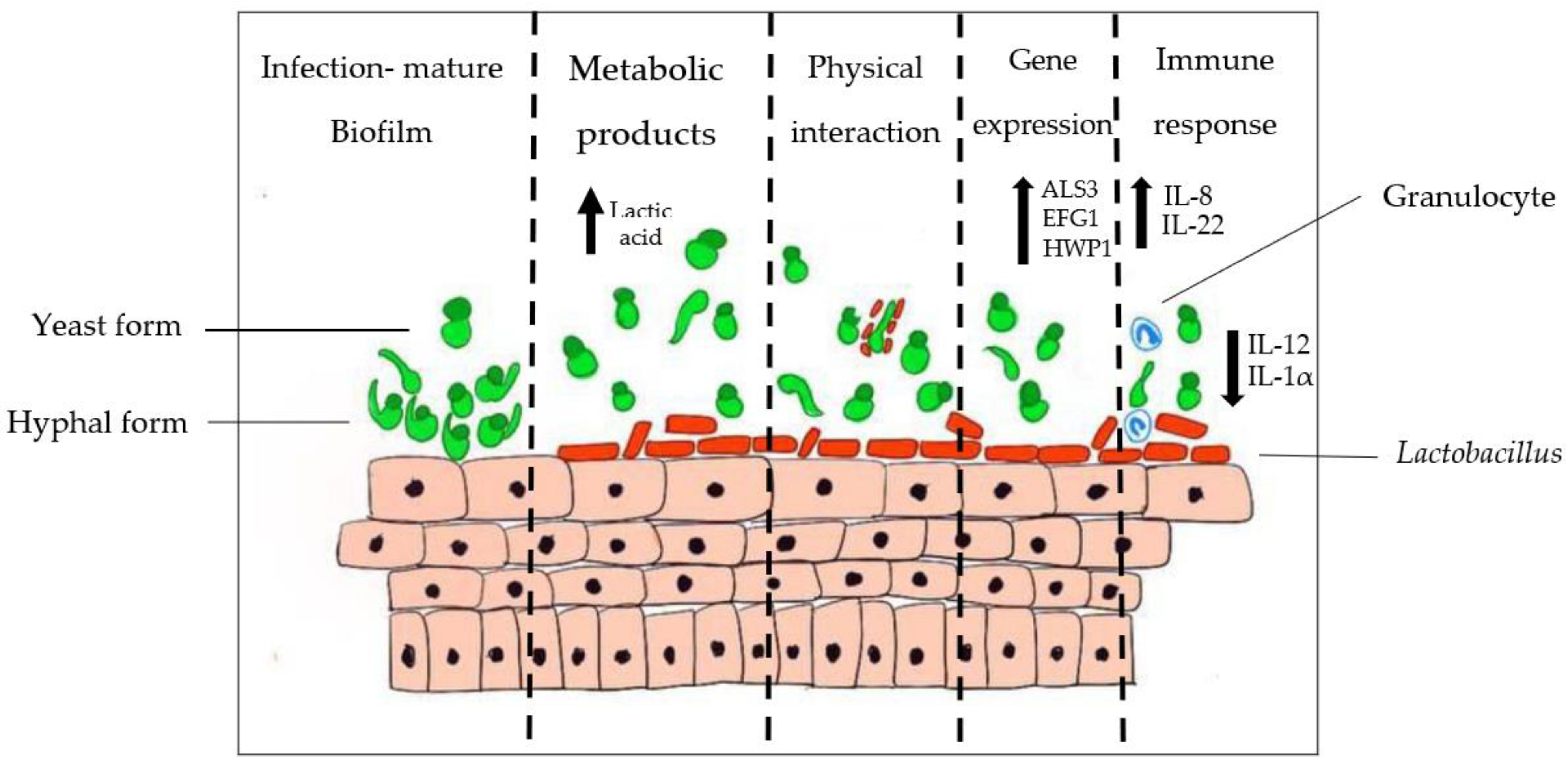
4.2. Interaction between Lactobacillus and Candida

The vaginal microbiota plays a crucial role in shielding the lower genital tract of women against Candida invasion. The bacterial microbiota within the vagina provides the first-line barrier protection as it creates competition for space and resources for Candida and, hence, prevents them from invading the vaginal epithelia [19]. Moreover, some Lactobacillus spp. exhibit specific and direct antifungal effects against Candida. For instance, L. rhamnosus GR-1 and L. reuteri RC-14 were found to exhibit antagonistic effects against an NCAC species, the vaginal pathogen C. glabrata [101]. Additionally, Li et al. (2019) demonstrated that both L. crispatus and L. delbrueckii inhibited 60% to 70% of C. albicans in a VVC Sprague-Dawley rat model compared with non-treated controls [102]. Production of lactic acid by Lactobacillus is proven to inhibit the hyphal formation of C. albicans [103]. A study by Camile Nina et al. (2019) revealed that lactic acid, in combination with major peptidoglycan hydrolase released by L. rhamnosus, GG successfully inhibits the hyphal formation of C. albicans [104]. Paniágua et al. (2021) also reported that C. albicans’ hyphae development was significantly hampered by L. casei Shirota [105]. C. albicans was found to modulate low pH into an alkaline condition to undergo hyphal formation, thus, in a way, supporting the idea that vaginal acidic condition is an inhibitory mechanism against the morphological transition in C. albicans [106]. Lactic acid also stimulates the release of anti-inflammatory cytokines. Hearps et al. (2017) found that lactic acid induces anti-inflammatory cytokine IL-1RA release and suppresses pro-inflammatory mediators production by cervicovaginal epithelial cell lines [107]. However, not only with lactic acid; it was also proven that Lactobacillus conditioned supernatant (LCS), isolated from L. fermentum and L. crispatus, was revealed to downregulate the expression of hypha-related genes of C. albicans, including ALS3, ECE1, SAP5 and HWP1 [103].
Lactobacillus also produces antimicrobial metabolites, such as hydrogen peroxide and biosurfactants, which are highly effective against Candida [108]. It was found that hydrogen peroxide-producing Lactobacillus, like L. acidophilus, was able to inhibit the growth of C. albicans compared with the non-hydrogen peroxide-producing strain [109]. However, C. albicans is quite resistant to ROS, so the antifungal activity might be due to overlapping mechanisms contributed by Lactobacillus [110]. On the other hand, Prisicila et al. (2020) reported that biosurfactants produced by L. crispatus were able to reduce the adhesion of C. albicans to HeLA cells with an inhibitory effect on biofilm formation [111]. Biosurfactants produced by L. gasseri together with L. jensenii were also able to suppress biofilms produced by C. albicans, C. tropicalis, and C. krusei by 25% to 35% [112]. A study by Chew et al. (2015) found that cell-free supernatants (CFS) of L. rhamnosus GR-1 and L. reuteri RC-14 were able to inhibit biofilm formation and suppress biofilm-related genes (EPA6 and YAK1) of C. glabrata [113].
Figure 2 below shows the series of events that take place during the interaction of C. albicans with Lactobacillus spp. As illustrated, upon infection and formation of Candida biofilm on vaginal mucosae, the normal microflora that are present (e.g., Lactobacillus) regulate themselves to produce metabolites such as lactic acid to inhibit Candida overgrowth via acidification of the vaginal mucosae. The metabolites produced by Lactobacillus inhibit C. albicans colonisation, either by preventing adherence to the epithelial cell wall or by exerting a fungistatic effect due to the surge in concentrations of organic acids. Adherence of Candida species is prevented by the co-aggregation action and saturation of adhesion sites by lactobacilli. After the interaction, the presence of lactobacilli suppresses the expression of virulence genes of C. albicans, namely ALS3, EFG1 and HWP1 genes that are related to hyphal production and adhesion. A recent study by Wang et al. (2017) reported that L. crispatus CFS significantly down-regulates the expression of hyphae-specific genes ALS3 (0.140-fold), HWP1 (0.075-fold), and ECE1 (0.045-fold), while it up-regulates the expression of the negative transcriptional regulator gene NRG1 with (1.911-fold) [114]. In the event of Candida colonisation, Lactobacillus can change the host immunological response, attract granulocytes and promote immune defence.
The composition of the vaginal microbiota naturally dominates the mucosal layer in a commensal and balanced ecosystem. However, several external stimuli and intrinsic factors can cause disruption to the ecosystem of the vaginal microbiota. Factors, such as sexual hormonal changes, douching, menstruation, sexual activity, menopause, stress, pregnancy, lactation, antibiotics usage and oral contraceptive pills may result in dysbiosis in the vaginal microbiota [89,115]. This is characterised by the depletion of healthy vaginal microflora causing overgrowth of dysbiosis-associated pathogens [116]. As the vaginal microflora is commonly dominated by Lactobacillus, dysbiosis-induced neutralisation of vaginal pH favours Candida overgrowth by diminishing the microbiota-derived protective mechanism within the vaginal mucosa, allowing the morphogenesis and adherence of pathogenic Candida to the VE [117]. Such dysbiosis has been shown to contribute to vulnerability towards vaginal infection and the possibility of developing VVC [116,118,119].

5. Future Perspectives

One of the promising treatments in VVC intervention is vaginal microbiota transplantation (VMT). This method involves the administration of microbiota from a healthy individual to the vagina of VVC patients as replacement therapy, restoring the imbalance microbiota community [120]. The continuous study of VMT is largely influenced by the success of faecal microbiota transplantation (FMT) in treating gastrointestinal diseases [121]. The effectiveness of FMT has been established through several clinical trials, including treatment for irritable bowel syndrome and Clostridioides difficile infection (previously known as Clostridium difficile infection) that show significant alleviation of symptoms, with fewer adverse side effects and an improvement of life quality among patients [122,123]. A recent study by Iriana et al. (2020) reported that FMT targeting gut microbiota in ulcerative colitis patients shows an increase in anti-Candida antibodies relative to placebo controls [124]. Because a Lactobacillus-dominated microbiota has been considered indicative of vaginal health, further study on its potential as a prophylactic agent or adjuvant in women with gynaecological diseases is warranted. A study by Lev-Sagie et al. (2019) shows that VMT in patients with bacterial vaginosis shifted the microbiota profile closer to that of the healthy donors [125]. The therapeutic effects of probiotics can be extended to cases of candidiasis. Xie et al. (2017) concluded that patients treated with exogenous probiotics exhibit a better short-term clinical cure and mycological cure rates when prescribed together with conventional antifungal therapy [126]. This correlates with a study conducted by Kovachev et al. (2015), as they found that patients who were administered probiotics in combination with azole treatment showed a significant reduction in clinical complaints, an improvement in vaginal microbiota and a healthier vaginal environment [127]. This research on VMT provides a platform to apply VMT not only as a sole treatment but as a supporting intervention in treating VVC.

6. Conclusions

In conclusion, the vaginal mucosa is the primary contact point with microorganisms in VVC and therefore it plays a primary role in the development of symptomatic infections as a physical barrier, by triggering immunological responses, and as the host of a surface-associated community of potentially antagonistic bacteria. The detection of PRRs by PAMPs contributes to the immune recognition upon VVC occurrence. The vaginal microbiota within the mucosal layer enhances the defence mechanism of the vagina through acidic pH regulation, the release of AMPs and physiological control against vaginal dysbiosis. The significant role of vaginal microbiota in maintaining vaginal health also promotes its application, i.e., vaginal microbiota transplantation (VMT) as a potential adjuvant treatment to alleviate, especially the recurrent form of VVC, alongside the use of probiotics and their metabolites. Hence, a further understanding of the vaginal microenvironment and host-pathogen interactions against Candida is crucial to initiate and sustain the long-term development of improved VVC intervention.

Author Contributions

Conceptualisation, L.T.L.T. and S.Y.C.; Writing—original draft preparation, S.N.B. and H.Y.; Writing—review and editing, L.T.L.T., S.Y.C. and M.C.L.; Visualisation, S.N.B. and H.Y.; Funding acquisition, L.T.L.T. and S.Y.C. All authors have read and agreed to the published version of the manuscript.

Funding

This work was supported by Geran Putra Berimpak (GP-GPB) from Universiti Putra Malaysia (Grant number: GP-GPB/2021/9702200).

Institutional Review Board Statement

Not applicable.

Informed Consent Statement

Not applicable.

Data Availability Statement

Not applicable.

Conflicts of Interest

The authors declare no conflict of interest.

References

  1. Kalia, N.; Singh, J.; Kaur, M. Immunopathology of recurrent vulvovaginal infections: New aspects and research directions. Front. Immunol. 2019, 10, 2034. [Google Scholar] [CrossRef] [PubMed]
  2. Bitew, A.; Abebaw, Y. Vulvovaginal candidiasis: Species distribution of Candida and their antifungal susceptibility pattern. BMC Women’s Health 2018, 18, 94. [Google Scholar] [CrossRef] [PubMed]
  3. Sasani, E.; Rafat, Z.; Ashrafi, K.; Salimi, Y.; Zandi, M.; Soltani, S.; Hashemi, F.; Hashemi, S.J. Vulvovaginal candidiasis in Iran: A systematic review and meta-analysis on the epidemiology, clinical manifestations, demographic characteristics, risk factors, etiologic agents and laboratory diagnosis. Microb. Pathog. 2021, 154, 104802. [Google Scholar] [CrossRef] [PubMed]
  4. Abdullahi Nasir, I.; Uchenna, E.; Onyia, J.; Ifunanya, A.L. Prevalence of vulvovaginal candidiasis among nonpregnant women attending a tertiary health care facility in Abuja, Nigeria. Res. Rep. Trop. Med. 2015, 6, 37. [Google Scholar] [CrossRef] [Green Version]
  5. O’Hanlon, D.E.; Come, R.A.; Moench, T.R. Vaginal pH measured in vivo: Lactobacilli determine pH and lactic acid concentration. BMC Microbiol. 2019, 19, 13. [Google Scholar] [CrossRef] [PubMed] [Green Version]
  6. Zeng, X.; Zhang, Y.; Zhang, T.; Xue, Y.; Xu, H.; An, R. Risk Factors of Vulvovaginal Candidiasis among Women of Reproductive Age in Xi’an: A Cross-Sectional Study. Biomed Res. Int. 2018, 2018, 9703754. [Google Scholar] [CrossRef] [Green Version]
  7. Lírio, J.; Giraldo, P.C.; Amaral, R.L.; Sarmento, A.C.A.; Costa, A.P.F.; Goncalves, A.K. Antifungal (oral and vaginal) therapy for recurrent vulvovaginal candidiasis: A systematic review protocol. BMJ Open 2019, 9, e027489. [Google Scholar] [CrossRef] [Green Version]
  8. Sobel, J.D. Recurrent vulvovaginal candidiasis. Am. J. Obstet. Gynecol. 2016, 214, 15–21. [Google Scholar] [CrossRef]
  9. Foxman, B.; Muraglia, R.; Dietz, J.P.; Sobel, J.D.; Wagner, J. Prevalence of recurrent vulvovaginal candidiasis in 5 European countries and the United States: Results from an internet panel survey. J. Low. Genit. Tract Dis. 2013, 17, 340–345. [Google Scholar] [CrossRef]
  10. Bernstein, J.A.; Seidu, L. Chronic Vulvovaginal Candida Hypersensitivity: An Underrecognized and Undertreated Disorder by Allergists. Allergy Rhinol. 2015, 6, 44–49. [Google Scholar] [CrossRef] [Green Version]
  11. Dovnik, A.; Golle, A.; Novak, D.; Arko, D.; Takač, I. Treatment of vulvovaginal candidiasis: A review of the literature. Acta Dermatovenerol. Alp. Pannonica Adriat. 2015, 24, 5–7. [Google Scholar] [CrossRef] [PubMed]
  12. Mohandas, V.; Ballal, M. Distribution of Candida Species in different clinical samples and their virulence: Biofilm formation, proteinase and phospholipase production: A study on hospitalized patients in Southern India. J. Glob. Infect. Dis. 2011, 3, 4–8. [Google Scholar] [CrossRef] [PubMed]
  13. Silva, S.; Henriques, M.; Martins, A.; Oliveira, R.; Williams, D.; Azeredo, J. Biofilms of non-Candida albicans Candida species: Quantification, structure and matrix composition. Med. Mycol. 2009, 47, 681–689. [Google Scholar] [CrossRef] [PubMed] [Green Version]
  14. Chaffin, W.L. Candida albicans Cell Wall Proteins. Microbiol. Mol. Biol. Rev. 2008, 72, 495–544. [Google Scholar] [CrossRef] [Green Version]
  15. Timmermans, B.; Las Peñas, A.D.; Castaño, I.; Van Dijck, P. Adhesins in candida glabrata. J. Fungi 2018, 4, 60. [Google Scholar] [CrossRef] [PubMed] [Green Version]
  16. Kalaiarasan, K.; Singh, R.; Chaturvedula, L. Changing virulence factors among vaginal non-albicans Candida species. Indian J. Med. Microbiol. 2018, 36, 364–368. [Google Scholar] [CrossRef]
  17. Nett, J.; Andes, D. Candida albicans biofilm development, modeling a host-pathogen interaction. Curr. Opin. Microbiol. 2006, 9, 340–345. [Google Scholar] [CrossRef]
  18. Rodríguez-Cerdeira, C.; Martínez-Herrera, E.; Carnero-Gregorio, M.; López-Barcenas, A.; Fabbrocini, G.; Fida, M.; El-Samahy, M.; González-Cespón, J.L. Pathogenesis and Clinical Relevance of Candida Biofilms in Vulvovaginal Candidiasis. Front. Microbiol. 2020, 11, 544480. [Google Scholar] [CrossRef]
  19. Ardizzoni, A.; Wheeler, R.T.; Pericolini, E. It Takes Two to Tango: How a Dysregulation of the Innate Immunity, Coupled With Candida Virulence, Triggers VVC Onset. Front. Microbiol. 2021, 12, 692491. [Google Scholar] [CrossRef]
  20. Ali, A.; Azab, M.; Abdelrahman, A. Distribution of secreted aspartyl protease (SAP) virulence genes and antifungal resistance genes at vulvovaginal candidiasis isolates. GSC Biol. Pharm. Sci. 2018, 5, 86–94. [Google Scholar] [CrossRef]
  21. Morrow, B.; Srikantha, T.; Soll, D.R. Transcription of the gene for a pepsinogen, PEP1, is regulated by white-opaque switching in Candida albicans. Mol. Cell. Biol. 1992, 12, 2997–3005. [Google Scholar] [PubMed] [Green Version]
  22. Naglik, J.R.; Rodgers, C.A.; Shirlaw, P.J.; Dobbie, J.L.; Fernandes-Naglik, L.L.; Greenspan, D.; Agabian, N.; Challacombe, S.J. Differential expression of Candida albicans secreted aspartyl proteinase and phospholipase B genes in humans correlates with active oral and vaginal infections. J. Infect. Dis. 2003, 188, 469–479. [Google Scholar] [CrossRef] [PubMed] [Green Version]
  23. Roselletti, E.; Perito, S.; Gabrielli, E.; Mencacci, A.; Pericolini, E.; Sabbatini, S.; Cassone, A.; Vecchiarelli, A. NLRP3 inflammasome is a key player in human vulvovaginal disease caused by Candida albicans. Sci. Rep. 2017, 7, 17877. [Google Scholar] [CrossRef] [PubMed] [Green Version]
  24. Pericolini, E.; Gabrielli, E.; Amacker, M.; Kasper, L.; Roselletti, E.; Luciano, E.; Sabbatini, S.; Kaeser, M.; Moser, C.; Hube, B.; et al. Secretory aspartyl proteinases cause vaginitis and can mediate vaginitis caused by Candida albicans in mice. MBio 2015, 6, e00724-15. [Google Scholar] [CrossRef] [PubMed] [Green Version]
  25. Richardson, J.P.; Willems, H.M.E.; Moyes, D.L.; Shoaie, S.; Barker, K.S.; Tan, S.L.; Palmer, G.E.; Hube, B.; Naglik, J.R.; Peters, B.M. Candidalysin drives epithelial signaling, neutrophil recruitment, and immunopathology at the vaginal mucosa. Infect. Immun. 2018, 86, e00645-17. [Google Scholar] [CrossRef] [PubMed] [Green Version]
  26. Anderson, D.J.; Marathe, J.; Pudney, J. The Structure of the Human Vaginal Stratum Corneum and its Role in Immune Defense. Am. J. Reprod. Immunol. 2014, 71, 618–623. [Google Scholar] [CrossRef] [PubMed] [Green Version]
  27. Patton, D.L.; Thwin, S.S.; Meier, A.; Hooton, T.M.; Stapleton, A.E.; Eschenbach, D.A. Epithelial cell layer thickness and immune cell populations in the normal human vagina at different stages of the menstrual cycle. Am. J. Obstet. Gynecol. 2000, 183, 967–973. [Google Scholar] [CrossRef]
  28. Linhares, I.M.; Sisti, G.; Minis, E.; de Freitas, G.B.; Moron, A.F.; Witkin, S.S. Contribution of Epithelial Cells to Defense Mechanisms in the Human Vagina. Curr. Infect. Dis. Rep. 2019, 21, 30. [Google Scholar] [CrossRef]
  29. Blaskewicz, C.D.; Pudney, J.; Anderson, D.J. Structure and function of intercellular junctions in human cervical and vaginal mucosal epithelia. Biol. Reprod. 2011, 85, 97–104. [Google Scholar] [CrossRef] [Green Version]
  30. Villar, C.C.; Kashleva, H.; Nobile, C.J.; Mitchell, A.P.; Dongari-Bagtzoglou, A. Mucosal tissue invasion by Candida albicans is associated with E-cadherin degradation, mediated by transcription factor Rim101p and protease Sap5p. Infect. Immun. 2007, 75, 2126–2135. [Google Scholar] [CrossRef] [Green Version]
  31. Frank, C.F.; Hostetter, M.K. Cleavage of E-cadherin: A mechanism for disruption of the intestinal epithelial barrier by Candida albicans. Transl. Res. 2007, 149, 211–222. [Google Scholar] [CrossRef] [PubMed]
  32. Fazeli, A.; Bruce, C.; Anumba, D.O. Characterization of Toll-like receptors in the female reproductive tract in humans. Hum. Reprod. 2005, 20, 1372–1378. [Google Scholar] [CrossRef] [PubMed] [Green Version]
  33. Kwissa, M.; Nakaya, H.I.; Oluoch, H.; Pulendran, B. Distinct TLR adjuvants differentially stimulate systemic and local innate immune responses in nonhuman primates. Blood 2012, 119, 2044–2055. [Google Scholar] [CrossRef] [PubMed]
  34. Fernando, M.R.; Reyes, J.L.; Iannuzzi, J.; Leung, G.; McKay, D.M. The pro-inflammatory cytokine, interleukin-6, enhances the polarization of alternatively activated macrophages. PLoS ONE 2014, 9, e94188. [Google Scholar] [CrossRef]
  35. Rosati, D.; Bruno, M.; Jaeger, M.; Kullberg, B.-J.; van de Veerdonk, F.; Netea, M.G.; ten Oever, J. An Exaggerated Monocyte-Derived Cytokine Response to Candida Hyphae in Patients With Recurrent Vulvovaginal Candidiasis. J. Infect. Dis. 2020, 23, jiaa444. [Google Scholar] [CrossRef]
  36. Steele, C.; Fidel, P.L. Cytokine and chemokine production by human oral and vaginal epithelial cells in response to Candida albicans. Infect. Immun. 2002, 70, 577–583. [Google Scholar] [CrossRef] [Green Version]
  37. McClure, R.; Massari, P. TLR-dependent human mucosal epithelial cell responses to microbial pathogens. Front. Immunol. 2014, 5, 386. [Google Scholar] [CrossRef] [Green Version]
  38. Pudney, J.; Quayle, A.J.; Anderson, D.J. Immunological Microenvironments in the Human Vagina and Cervix: Mediators of Cellular Immunity Are Concentrated in the Cervical Transformation Zone. Biol. Reprod. 2005, 73, 1253–1263. [Google Scholar] [CrossRef] [Green Version]
  39. Madanchi, H.; Shoushtari, M.; Kashani, H.H.; Sardari, S. Antimicrobial peptides of the vaginal innate immunity and their role in the fight against sexually transmitted diseases. New Microbes New Infect. 2020, 34, 100627. [Google Scholar] [CrossRef]
  40. Besold, A.N.; Gilston, B.A.; Radin, J.N.; Ramsoomair, C.; Culbertson, E.M.; Li, C.X.; Cormack, B.P.; Chazin, W.J.; Kehl-Fie, T.E.; Culotta, V.C. Role of Calprotectin in Withholding Zinc and Copper from Candida albicans. Infect. Immun. 2018, 86, e00779-17. [Google Scholar] [CrossRef] [Green Version]
  41. Curvelo, J.A.D.R.; Barreto, A.L.S.; Portela, M.B.; Alviano, D.S.; Holandino, C.; Souto-Padrón, T.; Soares, R.M.D.A. Effect of the secretory leucocyte proteinase inhibitor (SLPI) on Candida albicans biological processes: A therapeutic alternative? Arch. Oral Biol. 2014, 59, 928–937. [Google Scholar] [CrossRef] [PubMed] [Green Version]
  42. Chairatana, P.; Chiang, I.L.; Nolan, E.M. Human α-Defensin 6 Self-Assembly Prevents Adhesion and Suppresses Virulence Traits of Candida albicans. Biochemistry 2017, 56, 1033–1041. [Google Scholar] [CrossRef] [PubMed]
  43. Miró, M.S.; Caeiro, J.P.; Rodriguez, E.; Vargas, L.; Vigezzi, C.; Icely, P.A.; Castillo, G.D.V.; Azcurra, A.I.; Abiega, C.D.; Riera, F.O.; et al. Candida albicans Modulates Murine and Human Beta Defensin-1 during Vaginitis. J. Fungi 2022, 8, 20. [Google Scholar] [CrossRef] [PubMed]
  44. Srinivasan, N. Telling apart friend from foe: Discriminating between commensals and pathogens at mucosal sites. Innate Immun. 2010, 16, 391–404. [Google Scholar] [CrossRef]
  45. Swidergall, M. Candida albicans at Host Barrier Sites: Pattern Recognition Receptors and Beyond. Pathogens 2019, 8, 40. [Google Scholar] [CrossRef] [Green Version]
  46. Yano, J.; Peters, B.M.; Noverr, M.C.; Fidel, P.L., Jr. Novel mechanism behind the immunopathogenesis of vulvovaginal candidiasis:“neutrophil anergy. Infect. Immun. 2018, 86, e00684-17. [Google Scholar] [CrossRef] [Green Version]
  47. Wira, C.R.; Fahey, J.V.; Sentman, C.L.; Pioli, P.A.; Shen, L. Innate and adaptive immunity in female genital tract: Cellular responses and interactions. Immunol. Rev. 2005, 206, 306–335. [Google Scholar] [CrossRef]
  48. Bojang, E.; Ghuman, H.; Kumwenda, P.; Hall, R.A. Immune sensing of Candida albicans. J. Fungi 2021, 7, 119. [Google Scholar] [CrossRef]
  49. Son, H.J.; Kim, M.J.; Lee, S.; Choi, S.; Jung, K.H.; Jung, J.; Chong, Y.P.; Kim, S.H.; Choi, S.H.; Kim, Y.S.; et al. Risk factors and outcomes of patients with ocular involvement of candidemia. PLoS ONE 2019, 14, e0222356. [Google Scholar] [CrossRef]
  50. Fradin, C.; De Groot, P.; MacCallum, D.; Schaller, M.; Klis, F.; Odds, F.C.; Hube, B. Granulocytes govern the transcriptional response, morphology and proliferation of Candida albicans in human blood. Mol. Microbiol. 2005, 56, 397–415. [Google Scholar] [CrossRef]
  51. Ibrahim, A.S.; Luo, G.; Gebremariam, T.; Lee, H.; Schmidt, C.S.; Hennessey, J.P.; French, S.W.; Yeaman, M.R.; Filler, S.G.; Edwards, J.E. NDV-3 protects mice from vulvovaginal candidiasis through T- and B-cell immune response. Vaccine 2013, 31, 5549–5556. [Google Scholar] [CrossRef] [Green Version]
  52. Cambi, A.; Gijzen, K.; de Vries, I.J.M.; Torensma, R.; Joosten, B.; Adema, G.J.; Netea, M.G.; Kullberg, B.J.; Romani, L.; Figdor, C.G. The C-type lectin DC-SIGN (CD209) is an antigen-uptake receptor for Candida albicans on dendritic cells. Eur. J. Immunol. 2003, 33, 532–538. [Google Scholar] [CrossRef] [PubMed]
  53. Netea, M.G.; Brown, G.D.; Kullberg, B.J.; Gow, N.A.R. An integrated model of the recognition of Candida albicans by the innate immune system. Nat. Rev. Microbiol. 2008, 6, 67–78. [Google Scholar] [CrossRef] [PubMed]
  54. Takeuchi, O.; Akira, S. Pattern recognition receptors and inflammation. Cell 2010, 140, 805–820. [Google Scholar] [CrossRef] [PubMed] [Green Version]
  55. Hatinguais, R.; Willment, J.A.; Brown, G.D. PAMPs of the Fungal Cell Wall and Mammalian PRRs. In The Fungal Cell Wall: An Armour and a Weapon for Human Fungal Pathogens, 2nd ed.; Latgé, J.P., Ed.; Springer Nature: Cham, Switzerland, 2020; Volume 425, pp. 187–223. [Google Scholar]
  56. Chen, S.M.; Shen, H.; Zhang, T.; Huang, X.; Liu, X.Q.; Guo, S.Y.; Zhao, J.J.; Wang, C.F.; Yan, L.; Xu, G.T.; et al. Dectin-1 plays an important role in host defense against systemic Candida glabrata infection. Virulence 2017, 8, 1643–1656. [Google Scholar] [CrossRef] [PubMed] [Green Version]
  57. Medzhitov, R. Toll-Like Receptors and Innate Immunity. Curr. Biol. 2017, 27, R577–R578. [Google Scholar] [CrossRef] [Green Version]
  58. Jaeger, M.; Plantinga, T.S.; Joosten, L.A.B.; Kullberg, B.J.; Netea, M.G. Genetic basis for recurrent vulvo-vaginal candidiasis. Curr. Infect. Dis. Rep. 2013, 15, 136–142. [Google Scholar] [CrossRef]
  59. Gantner, B.N.; Simmons, R.M.; Underhill, D.M. Dectin-1 mediates macrophage recognition of Candida albicans yeast but not filaments. EMBO J. 2005, 24, 1277–1286. [Google Scholar] [CrossRef] [Green Version]
  60. Nakagawa, Y.; Ohno, N.; Murai, T. Suppression by Candida albicans β-glucan of cytokine release from activated human monocytes and from T cells in the presence of monocytes. J. Infect. Dis. 2003, 187, 710–713. [Google Scholar] [CrossRef] [Green Version]
  61. Brown, G.D.; Gordon, S. Fungal β-glucans and mammalian immunity. Immunity 2003, 19, 311–315. [Google Scholar] [CrossRef] [Green Version]
  62. Gonçalves, B.; Ferreira, C.; Alves, C.T.; Henriques, M.; Azeredo, J.; Silva, S. Vulvovaginal candidiasis: Epidemiology, microbiology and risk factors. Crit. Rev. Microbiol. 2016, 42, 905–927. [Google Scholar] [CrossRef] [PubMed] [Green Version]
  63. Cutler, J.E. N-glycosylation of yeast, with emphasis on Candida albicans. Med. Mycol. Suppl. 2001, 39, 75–86. [Google Scholar] [CrossRef] [Green Version]
  64. Pericolini, E.; Perito, S.; Castagnoli, A.; Gabrielli, E.; Mencacci, A.; Blasi, E.; Vecchiarelli, A.; Wheeler, R.T. Epitope unmasking in vulvovaginal candidiasis is associated with hyphal growth and neutrophilic infiltration. PLoS ONE 2018, 13, e0201436. [Google Scholar] [CrossRef] [PubMed]
  65. Fox, J.L. Antimicrobial peptides stage a comeback. Nat. Biotechnol. 2013, 31, 379–382. [Google Scholar] [CrossRef] [PubMed]
  66. Nuti, R.; Goud, N.S.; Saraswati, A.P.; Alvala, R.; Alvala, M. Antimicrobial Peptides: A Promising Therapeutic Strategy in Tackling Antimicrobial Resistance. Curr. Med. Chem. 2017, 24, 4303–4314. [Google Scholar] [CrossRef]
  67. Ageitos, J.M.; Sánchez-Pérez, A.; Calo-Mata, P.; Villa, T.G. Antimicrobial peptides (AMPs): Ancient compounds that represent novel weapons in the fight against bacteria. Biochem. Pharmacol. 2017, 133, 117–138. [Google Scholar] [CrossRef]
  68. Cairns-Smith, A.G. Antimicrobial peptides of multicellular organisms. Life Puzzle 2017, 415, 51–64. [Google Scholar]
  69. Nakatsuji, T.; Gallo, R.L. Antimicrobial peptides: Old molecules with new ideas. J. Investig. Dermatol. 2012, 132, 887–895. [Google Scholar] [CrossRef] [Green Version]
  70. Krishnakumari, V.; Rangaraj, N.; Nagaraj, R. Antifungal activities of human beta-defensins HBD-1 to HBD-3 and their C-terminal analogs Phd1 to Phd3. Antimicrob. Agents Chemother. 2009, 53, 256–260. [Google Scholar] [CrossRef] [Green Version]
  71. Oshiro, K.G.N.; Rodrigues, G.; Monges, B.E.D.; Cardoso, M.H.; Franco, O.L. Bioactive Peptides Against Fungal Biofilms. Front. Microbiol. 2019, 10, 2169. [Google Scholar] [CrossRef]
  72. Tomalka, J.; Azodi, E.; Narra, H.P.; Patel, K.; O’Neill, S.; Cardwell, C.; Hall, B.A.; Wilson, J.M.; Hise, A.G. β-Defensin 1 Plays a Role in Acute Mucosal Defense against Candida albicans. J. Immunol. 2015, 194, 1788–1795. [Google Scholar] [CrossRef] [PubMed] [Green Version]
  73. Pivarcsi, A.; Nagy, I.; Koreck, A.; Kis, K.; Kenderessy-Szabo, A.; Szell, M.; Dobozy, A.; Kemeny, L. Microbial compounds induce the expression of pro-inflammatory cytokines, chemokines and human β-defensin-2 in vaginal epithelial cells. Microbes Infect. 2005, 7, 1117–1127. [Google Scholar] [CrossRef] [PubMed]
  74. Vandamme, D.; Landuyt, B.; Luyten, W.; Schoofs, L. A comprehensive summary of LL-37, the factoctum human cathelicidin peptide. Cell. Immunol. 2012, 280, 22–35. [Google Scholar] [CrossRef] [PubMed]
  75. Swidergall, M.; Ernst, J.F. Interplay between Candida albicans and the antimicrobial peptide armory. Eukaryot. Cell 2014, 13, 950–957. [Google Scholar] [CrossRef] [Green Version]
  76. Levinson, P.; Kaui, R.; Kimani, J.; Ngugi, E.; Moses, S.; MacDonald, K.S.; Broliden, K.; Hirbod, T. Levels of innate immune factors in genital fluids: Association of alpha defensins and LL-37 with genital infections and increased HIV acquisition. Aids 2009, 23, 309–317. [Google Scholar] [CrossRef]
  77. Lyu, Y.; Yang, Y.; Lyu, X.; Dong, N.; Shan, A. Antimicrobial activity, improved cell selectivity and mode of action of short PMAP-36-derived peptides against bacteria and Candida. Sci. Rep. 2016, 6, 27258. [Google Scholar] [CrossRef] [Green Version]
  78. Woodburn, K.W.; Edward Clemens, L.; Jaynes, J.; Joubert, L.M.; Botha, A.; Nazik, H.; Stevens, D.A. Designed antimicrobial peptides for recurrent vulvovaginal candidiasis treatment. Antimicrob. Agents Chemother. 2019, 63, e02690-18. [Google Scholar] [CrossRef] [Green Version]
  79. Apalata, T.; Longo-Mbenza, B.; Sturm, A.; Carr, W.; Moodley, P. Factors associated with symptomatic vulvovaginal candidiasis: A study among women attending a primary healthcare clinic in Kwazulu-Natal, South Africa. Ann. Med. Health Sci. Res. 2014, 4, 410. [Google Scholar] [CrossRef] [Green Version]
  80. Duerr, A.; Heilig, C.M.; Meikle, S.F.; Cu-Uvin, S.; Klein, R.S.; Rompalo, A.; Sobel, J.D. Incident and persistent vulvovaginal candidiasis among human immunodeficiency virus-infected women: Risk factors and severity. Obstet. Gynecol. 2003, 101, 548–556. [Google Scholar] [CrossRef]
  81. Fidel, P.L.; Sobel, J.D. Immunopathogenesis of recurrent vulvovaginal candidiasis. Clin. Microbiol. Rev. 1996, 9, 335–348. [Google Scholar] [CrossRef]
  82. Moragues, M.D.; Omaetxebarria, M.J.; Elguezabal, N.; Sevilla, M.J.; Conti, S.; Polonelli, L.; Pontón, J. A monoclonal antibody directed against a Candida albicans cell wall mannoprotein exerts three anti-C. albicans Activities. Infect. Immun. 2003, 71, 5273–5279. [Google Scholar] [CrossRef] [PubMed] [Green Version]
  83. Fidel, P.L. History and new insights into host defense against vaginal candidiasis. Trends Microbiol. 2004, 12, 220–227. [Google Scholar] [CrossRef] [PubMed]
  84. Naglik, J.R.; Richardson, J.P.; Moyes, D.L. Candida albicans Pathogenicity and Epithelial Immunity. PLoS Pathog. 2014, 10, 8–11. [Google Scholar] [CrossRef] [PubMed]
  85. Shacklett, B.L.; Anton, P.A. HIV infection and gut mucosal immune function: Updates on pathogenesis with implications for management and intervention. Curr. Infect. Dis. Rep. 2010, 12, 19–27. [Google Scholar] [CrossRef] [PubMed] [Green Version]
  86. Jack, C.R., Jr.; Bennett, D.A.; Blennow, K.; Carrillo, M.C.; Dunn, B.; Haeberlein, S.B.; Holtzman, D.M.; Jagust, W.; Jessen, F.; Karlawish, J.; et al. Mucocutaneous IL-17 immunity in mice and humans: Host defense vs. excessive inflammation. Alzheimer’s Dement. 2018, 14, 535–562. [Google Scholar] [CrossRef] [PubMed]
  87. Peters, B.M.; Coleman, B.M.; Willems, H.M.E.; Barker, K.S.; Aggor, F.E.Y.; Cipolla, E.; Verma, A.H.; Bishu, S.; Huppler, A.H.; Bruno, V.M.; et al. The Interleukin (IL) 17R/IL-22R Signaling Axis Is Dispensable for Vulvovaginal Candidiasis Regardless of Estrogen Status. J. Infect. Dis. 2020, 221, 1554–1563. [Google Scholar] [CrossRef] [PubMed]
  88. Łaniewski, P.; Herbst-Kralovetz, M. Vagina. Encycl. Reprod. 2018, 2, 353–359. [Google Scholar]
  89. Amabebe, E.; Anumba, D.O.C. The vaginal microenvironment: The physiologic role of Lactobacilli. Front. Med. 2018, 5, 181. [Google Scholar] [CrossRef] [Green Version]
  90. Wilson, W.A.; Roach, P.J.; Montero, M.; Baroja-Fernández, E.; Muñoz, F.J.; Eydallin, G.; Viale, A.M.; Pozueta-Romero, J. Regulation of glycogen metabolism in yeast and bacteria. FEMS Microbiol. Rev. 2010, 34, 952–985. [Google Scholar] [CrossRef] [Green Version]
  91. Mirmonsef, P.; Hotton, A.L.; Gilbert, D.; Gioia, C.J.; Maric, D.; Hope, T.J.; Landay, A.L.; Spear, G.T. Glycogen levels in undiluted genital fluid and their relationship to vaginal pH, estrogen, and progesterone. PLoS ONE 2016, 11, e0153553. [Google Scholar] [CrossRef] [Green Version]
  92. Ceccarani, C.; Foschi, C.; Parolin, C.; D’Antuono, A.; Gaspari, V.; Consolandi, C.; Laghi, L.; Camboni, T.; Vitali, B.; Severgnini, M.; et al. Diversity of vaginal microbiome and metabolome during genital infections. Sci. Rep. 2019, 9, 14095. [Google Scholar] [CrossRef] [PubMed] [Green Version]
  93. Vásquez, A.; Jakobsson, T.; Ahrné, S.; Forsum, U.; Molin, G. Vaginal Lactobacillus Flora of Healthy Swedish Women. J. Clin. Microbiol. 2002, 40, 2746. [Google Scholar] [CrossRef] [PubMed] [Green Version]
  94. Ravel, J.; Gajer, P.; Abdo, Z.; Schneider, G.M.; Koenig, S.S.K.; McCulle, S.L.; Karlebach, S.; Gorle, R.; Russell, J.; Tacket, C.O.; et al. Vaginal microbiome of reproductive-age women. Proc. Natl. Acad. Sci. USA 2011, 108, 4680–4687. [Google Scholar] [CrossRef] [PubMed] [Green Version]
  95. Gupta, S.; Kakkar, V.; Bhushan, I. Crosstalk between Vaginal Microbiome and Female Health: A review. Microb. Pathog. 2019, 136, 103696. [Google Scholar] [CrossRef] [PubMed]
  96. Ma, B.; Forney, L.J.; Ravel, J. Vaginal microbiome: Rethinking health and disease. Annu. Rev. Microbiol. 2012, 66, 371–389. [Google Scholar] [CrossRef] [PubMed] [Green Version]
  97. Bradford, L.L.; Ravel, J. The vaginal mycobiome: A contemporary perspective on fungi in women’s health and diseases. Virulence 2017, 8, 342–351. [Google Scholar] [CrossRef] [Green Version]
  98. Seed, P.C. The Human Mycobiome. Cold Spring Harb. Perspect. Med. 2015, 5, a019810. [Google Scholar] [CrossRef] [Green Version]
  99. Mayer, F.L.; Wilson, D.; Hube, B. Candida albicans pathogenicity mechanisms. Virulence 2013, 4, 119–128. [Google Scholar] [CrossRef] [Green Version]
  100. Brown, S.E.; Schwartz, J.A.; Robinson, C.K.; O’Hanlon, D.E.; Bradford, L.L.; He, X.; Mark, K.S.; Bruno, V.M.; Ravel, J.; Brotman, R.M. The vaginal microbiota and behavioral factors associated with genital Candida albicans detection in reproductive-age women. Sex. Transm. Dis. 2019, 46, 753. [Google Scholar] [CrossRef]
  101. Chew, S.Y.; Cheah, Y.K.; Seow, H.F.; Sandai, D.; Than, L.T.L. Probiotic Lactobacillus rhamnosus GR-1 and Lactobacillus reuteri RC-14 exhibit strong antifungal effects against vulvovaginal candidiasis-causing Candida glabrata isolates. J. Appl. Microbiol. 2015, 118, 1180–1190. [Google Scholar] [CrossRef] [Green Version]
  102. Li, T.; Liu, Z.; Zhang, X.; Chen, X.; Wang, S. Local probiotic lactobacillus crispatus and lactobacillus delbrueckii exhibit strong antifungal effects against vulvovaginal candidiasis in a rat model. Front. Microbiol. 2019, 10, 1033. [Google Scholar] [CrossRef] [PubMed]
  103. Jang, S.J.; Lee, K.; Kwon, B.; You, H.J.; Ko, G.P. Vaginal lactobacilli inhibit growth and hyphae formation of Candida albicans. Sci. Rep. 2019, 9, 8121. [Google Scholar] [CrossRef] [PubMed] [Green Version]
  104. Allonsius, C.N.; Vandenheuvel, D.; Oerlemans, E.F.M.; Petrova, M.I.; Donders, G.G.G.; Cos, P.; Delputte, P.; Lebeer, S. Inhibition of Candida albicans morphogenesis by chitinase from Lactobacillus rhamnosus GG. Sci. Rep. 2019, 9, 2900. [Google Scholar] [CrossRef] [PubMed]
  105. Paniágua, A.L.; Correia, A.F.; Pereira, L.C.; de Alencar, B.M.; Silva, F.B.A.; Almeida, R.M.; de Medeiros Nóbrega, Y.K. Inhibitory effects of Lactobacillus casei Shirota against both Candida auris and Candida spp. isolates that cause vulvovaginal candidiasis and are resistant to antifungals. BMC Complement. Med. Ther. 2021, 21, 237. [Google Scholar] [CrossRef]
  106. Vylkova, S.; Carman, A.J.; Danhof, H.A.; Collette, J.R.; Zhou, H.; Lorenz, M.C. The fungal pathogen candida albicans autoinduces hyphal morphogenesis by raising extracellular pH. MBio 2011, 2, e00055-11. [Google Scholar] [CrossRef] [Green Version]
  107. Hearps, A.C.; Tyssen, D.; Srbinovski, D.; Bayigga, L.; Diaz, D.J.D.; Aldunate, M.; Cone, R.A.; Gugasyan, R.; Anderson, D.J.; Tachedjian, G. Vaginal lactic acid elicits an anti-inflammatory response from human cervicovaginal epithelial cells and inhibits production of pro-inflammatory mediators associated with HIV acquisition. Mucosal Immunol. 2017, 10, 1480–1490. [Google Scholar] [CrossRef] [Green Version]
  108. Vazquez-Munoz, R.; Dongari-Bagtzoglou, A. Anticandidal Activities by Lactobacillus Species: An Update on Mechanisms of Action. Front. Oral Health 2021, 2, 47. [Google Scholar] [CrossRef]
  109. Strus, M.; Brzychczy-Włoch, M.; Gosiewski, T.; Kochan, P.; Heczko, P.B. The in vitro effect of hydrogen peroxide onvaginal microbial communities. FEMS Immunol. Med. Microbiol. 2006, 48, 56–63. [Google Scholar] [CrossRef] [Green Version]
  110. Strus, M.; Kucharska, A.; Kukla, G.; Brzychczy-Włoch, M.; Maresz, K.; Heczko, P.B. The in vitro activity of vaginal Lactobacillus with probiotic properties against Candida. Infect. Dis. Obstet. Gynecol. 2005, 13, 69–75. [Google Scholar] [CrossRef] [Green Version]
  111. De Gregorio, P.R.; Parolin, C.; Abruzzo, A.; Luppi, B.; Protti, M.; Mercolini, L.; Silva, J.A.; Giordani, B.; Marangoni, A.; Nader-Macías, M.E.F.; et al. Biosurfactant from vaginal Lactobacillus crispatus BC1 as a promising agent to interfere with Candida adhesion. Microb. Cell Fact. 2020, 19, 133. [Google Scholar] [CrossRef]
  112. Morais, I.M.C.; Cordeiro, A.L.; Teixeira, G.S.; Domingues, V.S.; Nardi, R.M.D.; Monteiro, A.S.; Alves, R.J.; Siqueira, E.P.; Santos, V.L. Biological and physicochemical properties of biosurfactants produced by Lactobacillus jensenii P6A and Lactobacillus gasseri P65. Microb. Cell Fact. 2017, 16, 155. [Google Scholar] [CrossRef] [PubMed]
  113. Chew, S.Y.; Cheah, Y.K.; Seow, H.F.; Sandai, D.; Than, L.T.L. In vitro modulation of probiotic bacteria on the biofilm of Candida glabrata. Anaerobe 2015, 34, 132–138. [Google Scholar] [CrossRef] [PubMed]
  114. Wang, S.; Wang, Q.; Yang, E.; Yan, L.; Li, T.; Zhuang, H. Antimicrobial compounds produced by vaginal Lactobacillus crispatus are able to strongly inhibit Candida albicans growth, hyphal formation and regulate virulence-related gene expressions. Front. Microbiol. 2017, 8, 564. [Google Scholar] [CrossRef] [PubMed] [Green Version]
  115. Moosa, Y.; Kwon, D.; de Oliveira, T.; Wong, E.B. Determinants of Vaginal Microbiota Composition. Front. Cell. Infect. Microbiol. 2020, 10, 467. [Google Scholar] [CrossRef]
  116. van de Wijgert, J.H.H.M.; Jespers, V. The global health impact of vaginal dysbiosis. Res. Microbiol. 2017, 168, 859–864. [Google Scholar] [CrossRef]
  117. Aldunate, M.; Srbinovski, D.; Hearps, A.C.; Latham, C.F.; Ramsland, P.A.; Gugasyan, R.; Cone, R.A.; Tachedjian, G. Antimicrobial and immune modulatory effects of lactic acid and short chain fatty acids produced by vaginal microbiota associated with eubiosis and bacterial vaginosis. Front. Physiol. 2015, 6, 164. [Google Scholar] [CrossRef]
  118. Kroon, S.J.; Ravel, J.; Huston, W.M. Cervicovaginal microbiota, women’s health, and reproductive outcomes. Fertil. Steril. 2018, 110, 327–336. [Google Scholar] [CrossRef] [Green Version]
  119. Iliev, I.D.; Underhill, D.M. Striking a balance: Fungal commensalism versus pathogenesis. Curr. Opin. Microbiol. 2013, 16, 366–373. [Google Scholar] [CrossRef] [Green Version]
  120. Ma, D.; Chen, Y.; Chen, T. Vaginal microbiota transplantation for the treatment of bacterial vaginosis: A conceptual analysis. FEMS Microbiol. Lett. 2019, 366, 25. [Google Scholar] [CrossRef] [Green Version]
  121. Hameed, S.; Hans, S.; Monasky, R.; Thangamani, S.; Fatima, Z. Understanding Human Microbiota Offers Novel and Promising Therapeutic Options against Candida Infections. Pathogens 2021, 10, 183. [Google Scholar] [CrossRef]
  122. El-Salhy, M.; Hatlebakk, J.G.; Gilja, O.H.; Bråthen Kristoffersen, A.; Hausken, T. Efficacy of faecal microbiota transplantation for patients with irritable bowel syndrome in a randomised, double-blind, placebo-controlled study. Gut 2020, 69, 859–867. [Google Scholar] [CrossRef] [PubMed] [Green Version]
  123. Moayyedi, P.; Yuan, Y.; Baharith, H.; Ford, A.C. Faecal microbiota transplantation for Clostridium difficile-associated diarrhoea: A systematic review of randomised controlled trials. Med. J. Aust. 2017, 207, 166–172. [Google Scholar] [CrossRef] [PubMed]
  124. Leonardi, I.; Paramsothy, S.; Doron, I.; Semon, A.; Kaakoush, N.O.; Clemente, J.C.; Faith, J.J.; Borody, T.J.; Mitchell, H.M.; Colombel, J.F.; et al. Fungal Trans-kingdom Dynamics Linked to Responsiveness to Fecal Microbiota Transplantation (FMT) Therapy in Ulcerative Colitis. Cell Host Microbe 2020, 27, 823–829.e3. [Google Scholar] [CrossRef] [PubMed]
  125. Lev-Sagie, A.; Goldman-Wohl, D.; Cohen, Y.; Dori-Bachash, M.; Leshem, A.; Mor, U.; Strahilevitz, J.; Moses, A.E.; Shapiro, H.; Yagel, S.; et al. Vaginal microbiome transplantation in women with intractable bacterial vaginosis. Nat. Med. 2019, 25, 1500–1504. [Google Scholar] [CrossRef] [PubMed]
  126. Xie, H.Y.; Feng, D.; Wei, D.M.; Mei, L.; Chen, H.; Wang, X.; Fang, F. Probiotics for vulvovaginal candidiasis in non-pregnant women. Cochrane Database Syst. Rev. 2017, 11, CD010496. [Google Scholar] [CrossRef] [PubMed]
  127. Kovachev, S.M.; Vatcheva-Dobrevska, R.S. Local Probiotic Therapy for Vaginal Candida albicans Infections. Probiotics Antimicrob. Proteins 2015, 7, 38–44. [Google Scholar] [CrossRef] [PubMed]
Figure 1. (A) Female genital tract. (B) Illustration of the vaginal wall structure.
Pathogens 11 00618 g001
Figure 2. Interactions between C. albicans and Lactobacillus spp.
Pathogens 11 00618 g002
Publisher’s Note: MDPI stays neutral with regard to jurisdictional claims in published maps and institutional affiliations.

