
Molecular Monitoring of EHV-1 in Silently Infected Performance Horses through Nasal and Environmental Sample Testing

 ,
,
Abstract
:1. Introduction
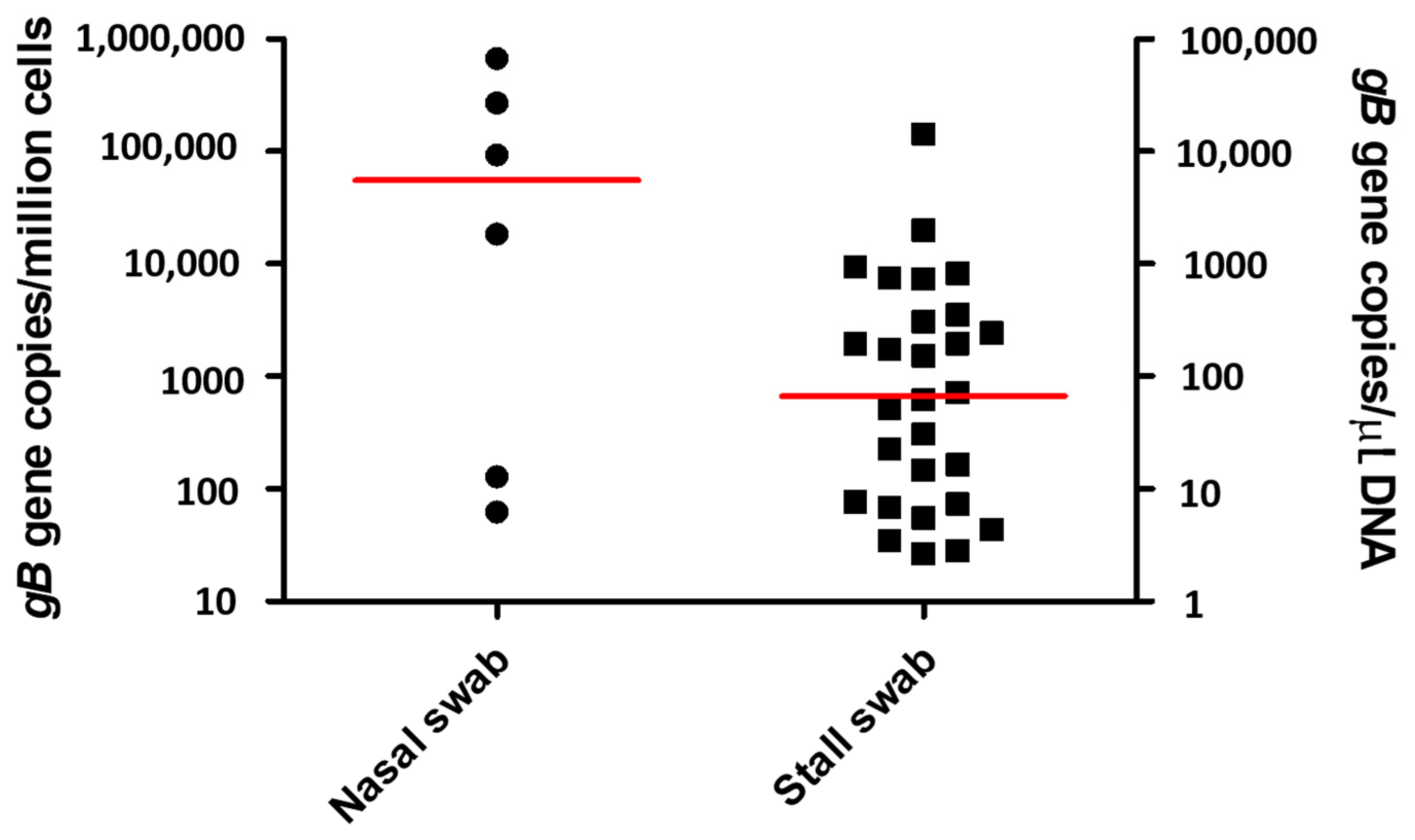
2. Results
3. Discussion
4. Materials and Methods
4.1. Outbreak and Study Population
4.2. Sample Collection and Analysis
4.3. Data Analysis
5. Conclusions
Author Contributions
Funding
Institutional Review Board Statement
Informed Consent Statement
Data Availability Statement
Acknowledgments
Conflicts of Interest
References
- Traub-Dargatz, J.L.; Pelzel-McCluskey, A.M.; Creekmore, L.H.; Geiser-Novotny, S.; Kasari, T.R.; Wiedenheft, A.M.; Bush, E.J.; Bjork, K.E. Case-control study of a multistate equine herpesvirus myeloencephalopathy outbreak. J. Vet. Intern. Med. 2013, 27, 339–346. [Google Scholar] [CrossRef] [PubMed]
- Lesté-Lasserre, C. Deadly viral outbreak ravages European horses. Science 2021, 371, 1297. [Google Scholar] [CrossRef] [PubMed]
- Pusterla, N.; Hussey, G.S. Equine herpesvirus 1 myeloencephalopathy. Vet. Clin. N. Am. Equine Pract. 2014, 30, 489–506. [Google Scholar] [CrossRef] [PubMed]
- Pusterla, N.; Wilson, W.D.; Mapes, S.; Finno, C.; Isbell, D.; Arthur, R.M.; Ferraro, G.L. Characterization of viral loads, strain and state of equine herpesvirus-1 using real-time PCR in horses following natural exposure at a racetrack in California. Vet. J. 2009, 179, 230–239. [Google Scholar] [CrossRef]
- Friday, P.A.; Scarratt, W.K.; Elvinger, F.; Timoney, P.J.; Bonda, A. Ataxia and paresis with equine herpesvirus type 1 infection in a herd of riding school horses. J. Vet. Intern. Med. 2000, 14, 197–201. [Google Scholar] [CrossRef]
- van Maanen, C.; Sloet van Oldruitenborgh-Oosterbaan, M.M.; Damen, E.A.; Derksen, A.G. Neurological disease associated with EHV-1-infection in a riding school: Clinical and virological characteristics. Equine Vet. J. 2001, 33, 191–196. [Google Scholar] [CrossRef]
- Henninger, R.W.; Reed, S.M.; Saville, W.J.; Allen, G.A.; Hass, G.F.; Kohn, C.W.; Sofaly, C. Outbreak of neurologic disease caused by equine herpesvirus-1 at a university equestrian center. J. Vet. Intern. Med. 2007, 21, 157–165. [Google Scholar] [CrossRef]
- Goehring, L.S.; Landolt, G.A.; Morley, P.S. Detection and management of an outbreak of equine herpesvirus type 1 infection and associated neurological disease in a veterinary teaching hospital. J. Vet. Intern. Med. 2010, 24, 1176–1183. [Google Scholar] [CrossRef]
- Burgess, B.A.; Tokateloff, N.; Manning, S.; Lohmann, K.; Lunn, D.P.; Hussey, S.B.; Morley, P.S. Nasal shedding of equine herpesvirus-1 from horses in an outbreak of equine herpes myeloencephalopathy in Western Canada. J. Vet. Intern. Med. 2012, 26, 384–392. [Google Scholar] [CrossRef]
- Vandenberghe, E.; Boshuizen, B.; Delesalle, C.J.G.; Goehring, L.S.; Groome, K.A.; van Maanen, K.; de Bruijn, C.M. New insights into the management of an EHV-1 (equine hospital) outbreak. Viruses 2021, 13, 1429. [Google Scholar] [CrossRef]
- Khusro, A.; Aarti, C.; Rivas-Caceres, R.R.; Barbabosa-Pliego, A. Equine herpesvirus-I infection in horses: Recent updates on its pathogenicity, vaccination, and preventive management strategies. J. Equine Vet. Sci. 2020, 87, 102923. [Google Scholar] [CrossRef] [PubMed]
- Pusterla, N.; Barnum, S.; Miller, J.; Varnell, S.; Dallap-Schaer, B.; Aceto, H.; Simeone, A. Investigation of an EHV-1 outbreak in the United States caused by a new H752 genotype. Pathogens 2021, 10, 747. [Google Scholar] [CrossRef] [PubMed]
- Pusterla, N.; Mapes, S.; Wademan, C.; White, A.; Estell, K.; Swain, E. Investigation of the role of mules as silent shedders of EHV-1 during an outbreak of EHV-1 myeloencephalopathy in California. Vet. Rec. 2012, 170, 465. [Google Scholar] [CrossRef] [PubMed]
- Yactor, J.; Lunn, K.F.; Traub-Dargatz, J.L.; Morley, P.S.; Barnett, C.D.; Kohler, A.K.; Kasper, K.S.; Kivi, A.J.; Lunn, D.P. Detection of nasal shedding of EHV-1 and 4 at equine show events and sales by multiplex real-time PCR. In Proceedings of the 52nd Annual Convention of the American Association of Equine Practitioners, San Antonio, TX, USA, 2–6 December 2006; p. 223. [Google Scholar]
- Pusterla, N.; Mapes, S.; Madigan, J.E.; Maclachlan, N.J.; Ferraro, G.L.; Watson, J.L.; Spier, S.L.; Wilson, W.D. Prevalence of EHV-1 in adult horses transported over long distances. Vet. Rec. 2009, 165, 473–475. [Google Scholar] [CrossRef]
- Doubli-Bounoua, N.; Richard, E.A.; Léon, A.; Pitel, P.H.; Pronost, S.; Fortier, G. Multiple molecular detection of respiratory viruses and associated signs of airway inflammation in racehorses. Virol. J. 2016, 13, 197. [Google Scholar] [CrossRef] [Green Version]
- Stasiak, K.; Dunowska, M.; Rola, J. Prevalence and sequence analysis of equid herpesviruses from the respiratory tract of Polish horses. Virol. J. 2018, 15, 106. [Google Scholar] [CrossRef]
- Smith, F.L.; Watson, J.L.; Spier, S.J.; Kilcoyne, I.; Mapes, S.; Sonder, C.; Pusterla, N. Frequency of shedding of respiratory pathogens in horses recently imported to the United States. J. Vet. Intern. Med. 2018, 32, 1436–1441. [Google Scholar] [CrossRef]
- Pusterla, N.; Rice, M.; Henry, T.; Barnum, S.; James, K. Investigation of the shedding of selected respiratory pathogens in healthy horses presented for routine dental care. J. Vet. Dent. 2020, 37, 88–93. [Google Scholar] [CrossRef]
- Estell, K.E.; Dawson, D.R.; Magdesian, K.G.; Swain, E.; Laing, S.T.; Siso, S.; Mapes, S.; Pusterla, N. Quantitative molecular viral loads in 7 horses with naturally occurring equine herpesvirus-1 infection. Equine Vet. J. 2015, 47, 689–693. [Google Scholar] [CrossRef]
- Pusterla, N.; Hussey, S.B.; Mapes, S.; Johnson, C.; Collier, J.R.; Hill, J.; Lunn, D.P.; Wilson, W.D. Molecular investigation of the viral kinetics of equine herpesvirus-1 in blood and nasal secretions of horses after corticosteroid-induced recrudescence of latent infection. J. Vet. Intern. Med. 2010, 24, 1153–1157. [Google Scholar] [CrossRef]
- Pusterla, N.; Mapes, S.; Wilson, W.D. Use of viral loads in blood and nasopharyngeal secretions for the diagnosis of EHV-1 infection in field cases. Vet. Rec. 2008, 162, 728–729. [Google Scholar] [CrossRef] [PubMed] [Green Version]
- Dayaram, A.; Franz, M.; Schattschneider, A.; Damiani, A.M.; Bischofberger, S.; Osterrieder, N.; Greenwood, A.D. Long term stability and infectivity of herpesviruses in water. Sci. Rep. 2017, 7, 46559. [Google Scholar] [CrossRef] [PubMed]
- Dayaram, A.; Seeber, P.A.; Greenwood, A.D. Environmental detection and potential transmission of equine herpesviruses. Pathogens 2021, 10, 423. [Google Scholar] [CrossRef]
- Saklou, N.T.; Burgess, B.A.; Ashton, L.V.; Morley, P.S.; Goehring, L.S. Environmental persistence of equid herpesvirus type-1. Equine Vet. J. 2021, 53, 349–355. [Google Scholar] [CrossRef] [PubMed]
- Pusterla, N.; Byrne, B.A.; Mapes, S.; Akana, N.; Wademan, C.; Fielding, L.C.; Slovis, N.; Elam, T.; Magdesian, K.G. Investigation of the use of pooled faecal and environmental samples following an enrichment step for the detection of Salmonella enterica by real-time PCR. Vet. Rec. 2014, 174, 252. [Google Scholar] [CrossRef]
- Pusterla, N.; Hussey, S.B.; Mapes, S.; Leutenegger, C.M.; Madigan, J.E.; Ferraro, G.L.; Wilson, W.D.; Lunn, D.P. Comparison of four methods to quantify Equid herpesvirus 1 load by real-time polymerase chain reaction in nasal secretions of experimentally and naturally infected horses. J. Vet. Diagn. Investig. 2009, 21, 836–840. [Google Scholar] [CrossRef] [PubMed] [Green Version]

| Barn Number | Horse/Stall ID | qPCR Results | |
|---|---|---|---|
| Horse Nasal Swab (gB Genes/Million Cells) | Stall Swab (gB Genes/µL of Purified DNA) | ||
| 1 | 305 | Positive (92,751) | Negative |
| 1 | 167 | Negative | Positive (5.5) |
| 1 | 169 | Negative | Positive (51.27) |
| 1 | 172 | Negative | Positive (820.6) |
| 1 | 174 | Negative | Positive (14.6) |
| 1 | 175 | Negative | Positive (71.5) |
| 1 | 178 | Negative | Positive (4.3) |
| 1 | 182 | Negative | Positive (1979.2) |
| 1 | 184 | Negative | Positive (936.1) |
| 1 | 185 | Negative | Positive (2.8) |
| 1 | 186 | Negative | Positive (30.5) |
| 1 | 188 | Negative | Positive (306.6) |
| 1 | 194 | Horse moved to isolation | Positive (195.4) |
| 2 | 24 | Negative | Positive (14,274.9) |
| 2 | 25 | Negative | Positive (352.2) |
| 2 | 27 | Negative | Positive (7.7) |
| 2 | 28 | Negative | Positive (61.8) |
| 2 | 30 | Negative | Positive (729.4) |
| 2 | 32 | Negative | Positive (243.9) |
| 2 | 34 | Negative | Positive (195.4) |
| 4 | 140 | Negative | Positive (744.7) |
| 4 | 148 | Horse moved to isolation | Positive (6.8) |
| 7 | 133 | Positive (670,939) | Negative |
| 8 | 200 | Negative | Positive (2.6) |
| 8 | 201 | Negative | Positive (151.2) |
| 32 | 153 | Positive (62) | Negative |
| 37 | 226 | Negative | Positive (173.7) |
| 39 | 314 | Positive (268,960) | Negative |
| 39 | 324 | Positive (128) | Negative |
| 45 | 105 | Positive (18,442) | Negative |
| 45 | 104 | Negative | Positive (3.5) |
| 45 | 106 | Negative | Positive (22.5) |
| FEI II | 240 | Negative | Positive (16.5) |
| FEI III | 257 | Negative | Positive (7.3) |
Publisher’s Note: MDPI stays neutral with regard to jurisdictional claims in published maps and institutional affiliations. |
© 2022 by the authors. Licensee MDPI, Basel, Switzerland. This article is an open access article distributed under the terms and conditions of the Creative Commons Attribution (CC BY) license (https://creativecommons.org/licenses/by/4.0/).
Share and Cite
Pusterla, N.; Barnum, S.; Young, A.; Mendonsa, E.; Lee, S.; Hankin, S.; Brittner, S.; Finno, C.J. Molecular Monitoring of EHV-1 in Silently Infected Performance Horses through Nasal and Environmental Sample Testing. Pathogens 2022, 11, 720. https://doi.org/10.3390/pathogens11070720
Pusterla N, Barnum S, Young A, Mendonsa E, Lee S, Hankin S, Brittner S, Finno CJ. Molecular Monitoring of EHV-1 in Silently Infected Performance Horses through Nasal and Environmental Sample Testing. Pathogens. 2022; 11(7):720. https://doi.org/10.3390/pathogens11070720
Chicago/Turabian StylePusterla, Nicola, Samantha Barnum, Amy Young, Eric Mendonsa, Steve Lee, Steve Hankin, Skyler Brittner, and Carrie J. Finno. 2022. "Molecular Monitoring of EHV-1 in Silently Infected Performance Horses through Nasal and Environmental Sample Testing" Pathogens 11, no. 7: 720. https://doi.org/10.3390/pathogens11070720