Review

Gurltia paralysans: A Neglected Angio-Neurotropic Parasite of Domestic Cats (Felis catus) and Free-Ranging Wild Felids (Leopardus spp.) in South America

1
Biomedical Research Center Seltersberg (BFS), Institute of Parasitology, Justus Liebig University Giessen, 35392 Giessen, Germany
2
Facultad de Ciencias Veterinarias, Instituto de Farmacología y Morfofisiología, Universidad Austral de Chile, Valdivia 5090000, Chile
3
Facultad de Ciencias Veterinarias, Instituto de Patología Animal, Universidad Austral de Chile, Valdivia 5090000, Chile
*
Author to whom correspondence should be addressed.
Pathogens 2022, 11(7), 792; https://doi.org/10.3390/pathogens11070792
Submission received: 13 June 2022 / Revised: 9 July 2022 / Accepted: 10 July 2022 / Published: 13 July 2022
(This article belongs to the Section Parasitic Pathogens)

Abstract

:
Gurltia paralysans is a neglected and re-emerging metastrongyloid angio-neurotropic nematode causing severe chronic meningomyelitis in domestic cats (Felis catus) as well as in free-ranging small wild felids such as kodkods (Leopardus guigna), margays (Leopardus wiedii) and the northern tiger cat (Leopardus triginus) in South America. Within these definitive hosts (DH), adult males and females of G. paralysans parasitize the leptomeningeal veins of the subarachnoid space and/or the meningeal veins of spinal cord parenchyma, inducing vascular alterations. Feline gurltiosis has been associated with progressive thrombophlebitis of the meningeal veins, resulting in ambulatory paraparesis, paraplegia, ataxia, hindlimb proprioceptive deficit, uni- or bilateral hyperactive patellar reflexes, faecal and urinary incontinence, and tail paralysis. The complete life cycle of G. paralysans has not been elucidated yet, but most probably involves gastropods as obligate intermediate hosts (IH). In terms of epidemiology, G. paralysans infections in domestic and wild felids are scattered around various South American countries, with hyperendemic areas in southern parts of Chile. Etiological diagnosis of G. paralysans still represents a challenge for clinicians due to a lack of evidence of the excretion of either eggs or larvae in faeces or in other body fluids. Diagnosis is based on clinical neurological signs, imaging findings through computed tomography (CT), myelography, magnetic resonance imaging (MRI), and post mortem examination. Nonetheless, novel diagnostic tools have been developed, including semi-nested PCR for detecting circulating G. paralysans DNA in the cerebrospinal fluid, serum and blood samples as well as in serological diagnostic kits detecting parasite-derived antigens, but these need validation for routine usage. The hypothetical life cycle of G. paralysans is addressed in this article, including the exogenous stages (i.e., eggs, and first- (L1), second- (L2) and third-stage (L3) larvae) and obligate gastropod IH and/or paratenic hosts (PH), and we propose possible anatomical migration routes of infective L3 that reach the leptomeningeal veins in vivo. Finally, the pro-inflammatory endothelium- and leukocyte-derived innate immune reactions of the host against G. paralysans, which most likely result in thrombophlebitis and meningomyelitis, are briefly touched on.

1. Introduction

Gurltia paralysans is a neglected metastrongyloid nematode (superfamily Metastrongyloidea, family Angiostrongylidae) causing severe chronic meningomyelitis in domestic cats (Felis catus) as well as in free-ranging wild felids of the genus Leopardus in South America. Historically, G. paralysans was first described by Kurt Wolfgang Wolffhügel (1933), a German scientist, naturalist, and parasitologist, who isolated adult nematodes from the vein system of the leptomeninges of 11 domestic cats suffering from chronic pelvic paraparesis within the provinces of Llanquihue and Puerto Varas in Southern Chile [1,2]. The etymology of the genus Gurltia is explained by Wolffhügel’s intention to honour Ernst Friedrich Gurlt (1794–1882), a German veterinary anatomist and teratologist. Wolffhügel (1933) initially described this neurological parasitosis associated with small felines as “paraplejia cruralis parasitaria felis” and placed the nematode species within the genus Hemostrongylus, later renamed Angiostrongylus [1]. One year later, Wolffhügel (1934) published an extended description of its geographic distribution, morphology, pathological findings, and clinical signs and speculated on its transmission and definitive host (DH) spectrum. This author showed the small wild felid species kodkod (Leopardus guigna; Figure 1) to be the main natural DH in Southern Chile and in the border regions of Argentina, locally known as “guiña” or “spotted tiger cat”. In addition, Wolffhügel (1993, 1934) proposed domestic cats as aberrant DH that were first introduced by European settlers into the South American continent and thereafter became exposed to this endemic nematode [3]. More recently, the spectrum of wild felid species acting as DH has increased, nowadays including the margay (Leopardus wiedii) and the northern tiger cat (Leopardus tigrinus) [4]. Additionally, within the genus Leopardus, other small wild felids have also been suggested as potential DH in South America [1,2,3,4,5,6,7]. Alongside the genus Leopardus, larger wild felids of South America, i.e., pumas (Puma concolor concolor), jaguars (Panthera onca), and jaguarondis (Herpailurus yagouaroundi), have also been suggested as potential DH, but this needs further investigation [1,5,7]. In a recent study on free-ranging guiñas in Chile, although no presence of G. paralysans was observed, the isolation of other closely related nematodes such as Angiostrongylus sp., Oslerus sp. and Troglostrongylus sp. was observed, indicating diversity, susceptibility, and the potential risk of lungworm infections in South America [8].
In vivo, G. paralysans has a marked angio-neurotropism invading the venous system of leptomeninges, specifically the thoracic, lumbar, and sacral spinal cord segments. The distribution of G. paralysans adults within the meningeal veins of the subarachnoid space implies activation of the highly immunoreactive endothelium, as seen for Angiostrongylus vasorum [9], probably resulting in thrombophlebitis with thrombus formation, venous congestion, and meningeal haemorrhages due to endothelium damage, as observed in severe feline gurltiosis [10]. Clinical manifestations can include chronic symmetrical or asymmetrical pelvic limb ataxia, ambulatory paraparesis, uni- or bi-lateral hyperactive patellar reflexes, proprioceptive deficit of pelvic limbs, pelvic limb muscular atrophy, diarrhoea, weight loss, coprostasis, urinary and faecal incontinence, and death [1,2,3,4,5,6] (Table 1). Neurological signs are typically associated with neuroanatomical lesions observed in post mortem examinations and histopathological specimens within the spinal cord [1,10,11,12]. Recently, histological and immunohistochemical characterization of vascular alterations in naturally G. paralysans-infected domestic cats of Chile unveiled suppurative vasculitis, haemorrhages, vascular congestion, and varicosis of not only spinal cord but also cerebrum-, cerebellum- and brain stem-associated veins [13], thereby supporting endothelium-derived pro-inflammatory innate immune reactions.

2. Hypothetical Life Cycle of Gurltia paralysans (Angiostrongylidae)

Small felids of the genera Felis and Leopardus are considered as DH of G. paralysans, as sexual replication occurs within these carnivorous mammals. As such, intravascular gravid G. paralysans females passing non-embryonated eggs (i.e., those containing 16 blastomeres) within the subarachnoid leptomeningeal veins of spinal cords from domestic cats (F. catus) [12], kodkods (L. guigna) [1,2,3], margays (L. wiedii) and northern tiger cats (L. tigrinus) [4] have been reported. Within Chilean territories, other small wild felids of the genus Leopardus have been discussed as potential DH [1,2,6], such as the Andean cat (Leopardus jacobita) and the Pampas cat (Leopardus colocolo). In semiarid, subtropical and tropical regions of South America, the southern tiger cat (Leopardus guttulus), Geoffroy’s cat (Leopardus geoffroyi) and the ocelot (Leopardus pardalis) might also act as DH [1,2,3,4,22]. Moreover, in South, Central and North America, larger wild felids such as the cougar (P. concolor concolor), the jaguar (P. onca) and the jaguarundi (H. yagouarundi) are proposed as DH, but this needs further clarification [1,7].
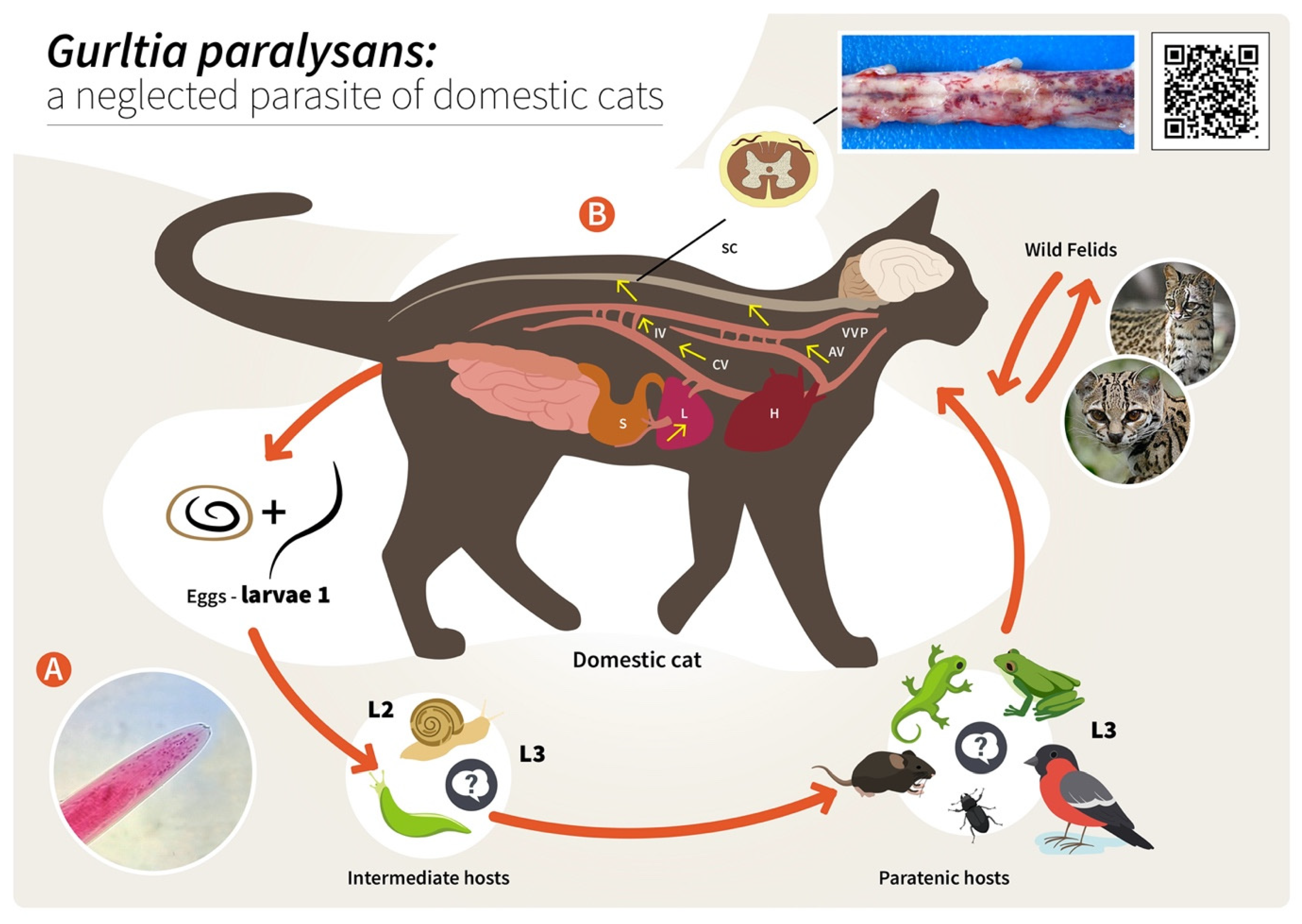
Unfortunately, nothing is known on other aspects of the life cycle, such as the mode of DH infection, exogenous larval development in obligate intermediate hosts (IH), endogenous in vivo migration of infective L3, pre-patency, patency and post-patency [5,6,12]. Neither eggs nor first-stage larvae (L1) have been detected in faeces, blood, bronchial lavage and/or other body fluids of naturally G. paralysans-infected domestic cats, northern tiger cats and margays [1,4,22,23].
As reported for other nematodes of the family Angiostrongylidae, terrestrial/aquatic gastropods (snails, semi-slugs and slugs) acting as obligate IH as well as paratenic hosts (PH) have been proposed in the biology of G. paralysans. Therefore, larval development occurring through moults from first-stage larvae (L1) to second-stage larvae (L2) and to the final infective third-stage larvae (L3) (Figure 2) has recently been proposed [1,5,6,7]. The suspected PH in this life cycle, such as crustaceans, amphibians, reptiles, rodents and birds, might become infected after ingesting L3-carrying gastropod IH, as reported for closely related Angiostronglylus species [24,25]. To elucidate the presence of G. paralysans larval stages in obligate gastropod IH, a large-scale epidemiological survey was recently conducted in the southern parts of Chile [26]. In this study, 835 terrestrial gastropods from a previously well-recognized endemic focus surrounding the city of Valdivia, Chile [1,26], were collected, demonstrating that neither PCR, enzymatic digestion nor histological examinations revealed the presence of larvae [1,26]. Collected gastropods included slugs of the families Arionidae, Limacidae and Milacidae as well as snails of the family Helicidae. Nonetheless, neither terrestrial semi-slugs (family Helicarionidae) nor aquatic snails were included in this survey, and thus this needs further investigation [1,7,26].
Proposed infection routes for felid DH are either after consumption of G. paralysans L3-infected gastropod IH or after consumption of L3-infected PH, as initially proposed by Wolffhügel (1934) [3]. As such, in his article, he referred to the colloquial name of feline gurltiosis used by locals, namely “lizard disease”, highlighting the pivotal role of PH in transmission. Alongside lizards, fish, frogs, toads, newts, snakes, turtles, birds, rodents, planarians, crustaceans, insects and myriapods have also been suspected in the life cycle of G. paralysans within South America [1,5,21,26]. Likewise, infective A. cantonensis-L3 larvae liberated from dead or living gastropods can survive outside IH for a short time, forming an important source of infection. The L3 larvae of A. cantonensis can enter new IH through the process known as intermediasis, which might occur in this life cycle as well, thereby extending the survival strategies of G. paralysans. These alternative transmission routes can occur with ease in neotropical South American rainforests, which have the highest biodiversity of protist, invertebrate and vertebrate species in the world [7].
Concerning the endogenous migration of G. paralysans L3 in felid DH in vivo, nothing is known so far. Hypothetically, L3 migration could be through the small intestinal mucosa in order to reach the mesenteric veins and/or lymphatic vessels of the abdominal viscera and thereafter via connections of either the azygos or the caval venous system (CVS) with the thoracic, lumbar or sacral intervertebral veins until they reach the vertebral venous plexus (VVP, Figure 2) [1]. The VVP is in direct communication with the cranial venous system, and because no valves exist in either of them, blood might flow cranially or caudally, depending on blood pressure [1,27]. G. paralysans could take advantage of the absence of valves in the VVP to reach either the spinal subarachnoid space or even the brain [1,12,28]. Similarly, in spinal schistosomiasis in humans, the dissemination of the parasite occurs via the intestinal veins to the VVP [29]. Spinal schistosomiasis usually involves the lower thoracic and lumbosacral spine, probably because the VVP connects the intra-abdominal veins with those of the lower spine [30]. The presence of fertile male and female nematodes, and gravid females passing eggs within the ventral VVP and basivertebral veins located in the vertebral bodies, were isolated during necropsies, confirming the marked angiotropism of G. paralysans in DH [1,12]. Moreover, parasitic localization within the VVP’s venous connections may explain the presence of eggs, L1, pre-adults and adults of G. paralysans in distant places, such as the cerebrum, cerebellum and anterior chamber of the eye of infected cats [1,20,27,31]. Nonetheless and in contrast to all Angiostrongylus species residing within arterial vessels, G. paralysans dwells within venous vessels. Thus, the adaptations of G. paralysans to the VVP’s venous connections might be associated not only with abiotic factors of the venous microenvironment, such as hypoxia and CO2 concentrations, but may also be linked to physical factors (e.g., temperature, blood flow velocity) and even nutrients, among others [1]. It seems indispensable for future investigations on the migratory pathways of G. paralysans to include not only vein tropism but also neuroanatomical localization within the subarachnoid VVP in felids [1]. During the patency period, gravid G. paralysans will then release un-embryonated eggs into the leptomeningeal vein system. Intravascular eggs will develop into L1, and hatching of the L1 will occur within the VVP, as demonstrated previously [1,12]. The free-released L1 will then breach the alveolar walls in order to access the bronchioles, bronchia and trachea, and are most likely expelled via faeces into the environment. Consistently, the life cycles of other closely related angio-neurotropic metastrongyloid genera of cervids (Elaphostrongylus and Parelaphostrongylus) might explain the final localizations of G. paralysans in the subarachnoid leptomeningeal veins of felid spinal cord. Likewise, adults of Elaphostrongylus alces occur in vessels of the epidural space of the vertebral canal and in the skeletal muscles of moose (Alces alces) [32,33]. Similar to G. paralysans, E. alces has obligate gastropod IH, causing neurological disorders in wild moose populations after ingestion of L3-carrying gastropods [32,33]. Earlier researchers suggested that E. alces L3 migrate directly from the gut into the epidural space of the caudal vertebral canal, where development to the adult stages takes place. During endogenous development, E. alces nematodes produce severe inflammation of the epidural tissue and spinal nerves [32]. In line with this, development of Elaphostrongylus rangiferi also takes place only within the brain and spinal cord vessels of reindeer (Rangifer tarandus), with subsequent migration of adult nematodes into the skeletal muscle [34]. In the case of Parelaphostrongylus tenuis, also known as “meningeal worm” or “brain worm”, which typically occurs in wild cervids (Cervidae), adult nematodes mate in the blood vessels of deer heads, and gravid females start releasing eggs into the circulatory system [35]. In fact, the release of eggs or larvae into the circulatory blood system might be linked with the appearance of P. tenuis larvae in the ventral portion of the anterior eye chamber [36], similar to a recent ophthalmic finding in a G. paralysans-infected domestic cat of Tenerife Island, Spain [20].
One of the most peculiar features of some Angiostrongylus species (Angiostrongylidae) is their strong neurotropism within warm-blooded DH or accidental/aberrant hosts (AH). As such, infectious A. cantonensis L3 must migrate through the central nervous system (CNS), where they develop further, reaching the L5 larval stage in the subarachnoidal space within two weeks post infection [37,38]. This part of the life cycle usually does not produce severe signs in DH (e.g., rats); however, infections in AH commonly result in eosinophilic meningitis, with several clinical scenarios [38], as was also the case for G. paralysans-infected domestic cats considered by Wolffhügel (1934) as AH. Clinical manifestations are common between G. paralysans and A. cantonensis due to the signs observed in infected animals resulting in increased intracranial pressure, neural tissue damage, the hosts’ pro-inflammatory response, congestion, thrombosis, thrombophlebitis, varices and thickening of the affected vessels [1,18,37].

3. Epizootiology and Environmental Factors Associated with the Presence of Feline Gurltiosis

Environmental factors have been known in the literature to significantly influence the occurrence and epizootiology of metastrongyloid parasites. Abiotic factors such as temperature, humidity and precipitation, as well as biotic factors such as the presence and physiology of DH, obligate IH and PH all play an important part in the continuation of the life cycle, and are involved in parasite mating, egg production, survival of the L1 stages in excreted felid faeces and the persistence of infective G. paralysans L3 in PH and/or AH [1,38].
The importance of climate in understanding the rates of transmission of meningeal metastrongyloid nematodes and the risk of infections is often underappreciated. Consequently, it has been reported that, firstly, winter length and severity can be important determinants of the geographic distribution area of wild felids, a situation that has also been observed in wild cervid populations infected with Elaphostrongylus and Parelaphostrongylus [32,33,34]. Secondly, the climate in summer, especially the amount of precipitation and the length of the summer, determines: (i) the survival of exogenous L1 stages, and (ii) the survival, abundance and mobility of suitable gastropods acting as obligate IH. Thus, climate determines the density of possible DH and gastropod IH, and, in turn, the rates at which each becomes infected, which means that the odds of an encounter between an infected gastropod and a DH depend on the density and degree of the spatial overlaps of both [1,7,38]. In the same way, the majority of terrestrial gastropod populations respond well to wet climatic conditions. In line with this, humidity (precipitation and dew) determines their reproductive success and survival, as well as their mobility in ground litter and on low vegetation, which could explain the epidemiological distribution and endemic foci for feline gurltiosis in the wet southern parts of Chile, not exclusively in rural areas [1,6,10,11,12,26] but also in urban areas, as recently observed [6]. While a lot of data are available on the geographic expansion of Angiostrongylus spp. infections in various continents, no data are currently accessible for G. paralysans infections within South American territories. The same holds true for the range of possible DH, IH, PH or even AH participating in this neurological parasitosis. Therefore, more epidemiological studies on the occurrence of feline gurltiosis not only in domestic cats, but also in free-ranging wild felids, and gastropod IH, PH and AH, are necessary to better understand the further geographic expansion, re-emergence and/or transmission routes involving other suitable DH, IH and PH in South America as well as in Europe.
The actual geographical distribution of G. paralysans corresponds mainly to South America, specifically to areas of Chile, Argentina, Colombia, Brazil and sporadically in Uruguay [1,2,3,4,5,6,7,10,11,12,14,15,16,17,18,19,20,21]. The presence of this nematode has been reported on the Island of Tenerife, Spain [25]. However, the exact prevalence of G. paralysans in these regions is still unknown, despite the fact that infections may be underdiagnosed and the prevalence underestimated. Previous reports have suggested that the focus of infection occurs mainly in rural, peri-urban and urban areas of Southern Chile, which has vast temperate forested areas [1,2,3,5,6,10,11,12,19]. The high endemicity of feline gurltiosis to the temperate southern parts of Chile was recently confirmed in a large-scale epidemiological survey of urban cats (n = 171) [6]. As such, Barrios et al. [6] reported the presence of G. paralysans in clinically unaffected urban feline populations of three southern Chilean cities, highlighting a considerable percentage of infection (54.4%) in the domestic cats analyzed, indicating, at the same time, that the infection levels seem to vary according to the geographical region [1,2,3,5,6,10,11,12,19]. Nevertheless, no other large-scale epidemiological study has been performed so far to address the occurrence of G. paralysans infections in domestic cat populations in any other country.

4. Diagnosis

The intra vitam diagnosis of feline gurltiosis is difficult and will depend on the clinical manifestations and diagnosis excluding other myelopathies. Currently, a presumptive diagnosis of G. paralysans infection is based on the medical history of affected cats with progressive clinical neurological features originating from potentially endemic areas. A definitive diagnosis can only be performed through a post mortem examination demonstrating and morphologically identifying nematodes in the spinal cord vasculature [1,12,17] (Figure 3). Male specimens of G. paralysans have a body length of 12–18 mm and are 0.026–0.032 mm wide in the cephalic region. Females have a body length of 20.5–36.06 mm and a width of 0.082–0.088 mm just anterior to the vulva [1,12,39].
Generally, there are five categories to explore from the appearance of the first clinical manifestations until progressive deterioration of the patients. The first involves physical evaluation of the patient, when it is possible to observe clinical neurological signs related with feline gurltiosis such as chronic symmetrical or asymmetrical pelvic limb ataxia, ambulatory paraparesis, uni- or bilateral hyperactive spinal reflexes, proprioceptive deficit of the pelvic limbs, pelvic limb muscular atrophy, diarrhoea, weight loss, and urinary and faecal incontinence [1,11,12]. Second, laboratory examinations are carried out in order to exclude some levels of anaemia, eosinophilia and thrombocytopenia [17], and even some alterations in the cerebrospinal fluid such as mononuclear pleocytosis [17]. Third, imaging studies are used to explore lesions in the thoracolumbar, lumbar or sacral regions and/or any other typical spinal pathology suggesting diffuse inflammatory spinal cord lesions resulting in severe myelitis or meningomyelitis [1,10,11,12]. Radiography, myelography, computed tomographic myelography (myelo-CT) and magnetic resonance imaging (MRI) are the tools used at present [1,10,17]. As the fourth category, more recently, molecular detection of G. paralysans DNA by semi-nested PCR in cerebrospinal fluid, serum and blood samples, using specific oligonucleotides to identify the parasite’s presence [1,39,40]. No other methods are available for a precise ante mortem detection of this nematode. An early diagnosis with conclusive results is required, since laboratory and imaging findings are not sufficient; for that reason, the implementation of molecular tools such as the abovementioned PCR technique may be considered as a complementary and routine diagnostic test for ante mortem detection of feline gurltiosis. Unfortunately, necropsies are conducted exclusively in cases of chronic paraplegia with the prior consent of the owners. The entire spinal cord is then removed from the vertebral canal and incisions are made in the dura mater to expose the affected veins in the subarachnoid space of the cervical, thoracic, lumbar and sacral regions, where the parasites are finally removed and identified (Figure 4A).
Recently, a commercial serological test for canine angiostrongylosis was applied in felines with chronic paraparesis or severe paraplegia [5]. Although the serological test was originally designed for detection of closely related A. vasorum, the detection of positive samples (n = 7) was achieved; four of the animals were necropsied and subsequently presented findings compatible with feline gurltiosis, both due to the presence of adult nematodes and the histological findings. Collectively, these results suggest cross-reactions between A. vasorum-specific antigens and G. paralysans when using the Angio Detect TM® Test (IDEXX), suggesting its use as a new diagnostic method for feline gurltiosis in live domestic felines; however, more data on the sensitivity, specificity and characterization of antigens are required for routine intra vitam immunodiagnosis [1,5].

5. Clinical Signs

The most frequent clinical manifestation of feline gurltiosis is chronic and progressive ambulatory paraplegia [11,14,15,16,17,18,19,20,21,39,40] (Table 1). The duration of clinical signs ranges from 2 weeks to 48 months [10,14,15,16,17,18,19,20,21,39,40]. Other clinical signs include pelvic limb ataxia, pelvic limb proprioceptive deficit, pelvic limb tremor, pelvic limb muscle atrophy, tail trembling, tail atony, and faecal and urinary incontinence [5,10,11,12,14,15,16,17,18,19,20,21] (Table 1). The neurological signs are associated with the neuroanatomical lesions observed during necropsy and in histopathological specimens [1,18].
The associated haematological abnormalities include non-regenerative anaemia and low mean corpuscular haemoglobin concentrations (hypochromia) [17], indicating chronic inflammatory disease or chronic blood loss. Although eosinophilia has commonly been associated with metazoan parasitism (helminths) in domestic animals and humans, it is not a common finding in cats suffering from feline gurltiosis [17], and has also been reported in dogs with neural angiostrongylosis [41]. Unlike canine angiostrongylosis [41], no signs of coagulopathies with bleedings have been observed in naturally infected cats. However, high levels of urea in the blood have been reported, probably arising from neurogenic urinary dysfunction [17]. A bronchial lavage analysis of five naturally G. paralysans-infected cats showed the absence of larval stages and eggs [23]. Ocular lesions (uveitis, chorioretinitis, posterior synechiae, corneal oedema) have recently been reported to be associated with the presence of a motile adult specimen of G. paralysans in the anterior chamber of the eye in a domestic cat [20].

6. Post Mortem Spinal Cord Examination

Post mortem macroscopic spinal cord findings in domestic cats with feline gurltiosis are characterized by diffuse submeningeal congestion (Figure 4A) [10,11,12]. The presence of nematode larvae and pre-adult stages can be identified histologically in the subarachnoid veins of the spinal cord, associated with congestion, thrombosis, and thickening of the meningeal vessels (Figure 4B) [10,11,12,17,18]. The presence of suppurative thrombophlebitis and phlebosclerosis with mild smooth-muscle hypertrophy, moderate adventitial fibroplasia and marked subintimal fibrosis of the spinal cord venules have been reported [13,18]. The presence of perivenous fibrosis is considered evidence of chronic inflammatory stages [13]. In some specimens, concentric thickening of the venule wall may produce vascular stenosis [10,18]. Intraluminal papillary projections, with an arboriform appearance, to the interior of dilated venules, have been interpreted as varicose lesions (venular varices) [12,18]. Dilated and tortuous varicose subarachnoid venules may contain thrombi with various levels of organization and G. paralysans eggs at different stages of development (Figure 4B,C) [18]. The spinal cord parenchyma may show multiple haemorrhages and extensive foci of malacia, with gitter cells and adjacent gliosis [10,18]. Lymphocytes intermingled with fewer macrophages primarily infiltrate the meninges, forming a perivascular pattern. Mature eosinophils scattered randomly within the leptomeninges have also been observed, which are consistent with extensive spinal leptomeningitis and thrombophlebitis [12,18]. Some animals may also show granulomatous leptomeningitis or suppurative leptomeningitis [18]. White-matter lesions in the spinal cord segments may have variable degrees of Wallerian degeneration, characterized by distension of the myelin sheath diameter, irregular axons, axonal swelling, bulbous axonal fragmentation (caused by the presence of axonal spheroids), microcavitation and focal areas of mineralization [10,12,42]. Varicose venules can also be observed in the white matter of the spinal cord but are associated with recesses in meninges [13,42]. The activation of glial and endothelial cells and immune cell infiltration, visualized with immunohistochemical markers (e.g., GFAP, CNPase, factor VIII, CD3 and CD45R) in affected spinal cord samples indicate gliosis and chronic inflammatory spinal cord lesions subsequent to the ischemia caused by parasitic vascular injury [42]. The predominant cellular infiltrate in the affected spinal cord is of the mononuclear type, indicating the chronic nature of these lesions [42]. A recent study analyzed the presence of histopathological lesions in the cerebrum, cerebellum, and brain stem in 13 feline patients with post mortem spinal lesions due to G. paralysans [43]. Congestion and hyperaemia were observed in the peripheral blood vessels of both the dorsal and ventral zones of the cerebrum (n = 13). In seven cases, mononuclear cell infiltrates were observed around the choroid plexus, the third and fourth ventricles, and the associated blood vein vessels. Six cats showed thickening of the meninges and two showed perivascular neutrophilic inflammatory infiltrate at the level of the cerebral subarachnoid space [31]. Similar findings of leptomeningeal vascular congestion, varices and perivascular cellular infiltrate were observed in the brains (frontal, temporal and occipital cortices) of 11 G. paralysans-infected cats in Brazil [21]. However, no clinical cases of feline gurltiosis have been observed with clinical cerebral, cerebellar, or brain stem syndromes.
Histological samples from 10 G. paralysans-infected cats were analyzed at the hepatic level. All samples showed signs of periportal hepatic degeneration and periportal inflammatory infiltration, comprising neutrophilic and mononuclear infiltration, indicating direct injury to the liver [44]. However, the possible mechanisms by which intravascular parasites could cause this type of hepatic injury pattern in the liver and other extraneural tissues remains unclear (Figure 5). A. vasorum can induce moderate liver parenchymal parasitic hepatitis and lesions such as interstitial haemorrhages with disseminated inflammatory cells in the portobiliar space or around the centrolobular veins, which was attributed to larval nematode migration [44,45]. In kidneys, feline gurltiosis has been associated with the presence of hyaline protein deposits inside Bowman’s capsule (in 8 out of 10 cases), the thickening of Bowman’s capsule in five cases and the presence of interstitial inflammatory infiltrate, consisting of polymorphonuclear neutrophils (PMN) and eosinophils (in 4 out of 10 cases) [44]. These findings are compatible with glomerulonephritis, which might result from immune-mediated reactions such as the deposition of immune complexes of the host [45].

7. Proposed Gurltia paralysans-Induced Endothelium and Polymorphonuclear Neutrophil (PMN) Activation, Resulting in Vascular Pathogenesis

Due to its localization in the subarachnoid leptomeningeal veins in vivo, G. paralysans is permanently exposed to the feline innate immune system, mainly composed of circulating PMN, other leukocytes (i.e., monocytes, eosinophils and basophils), the vascular endothelium, complement factors, antimicrobial peptides, cytokines and chemokines, among others. Feline PMN are the most abundant leukocyte population in the blood, representing the first line of defense. PMN are recruited immediately after parasite invasion, exhibiting diverse effector mechanisms including production of reactive oxygen species (ROS), degranulation of immunomodulatory molecules, phagocytosis and release of neutrophil extracellular traps (NETs) [46,47,48]. NETs are delicate extracellular fibers mainly composed of decondensed DNA through PAD4-mediated citrullination of global histones (i.e., H1, H2A/H2B, H3 and H4), decorated with antimicrobial/antiparasitic components such as lactoferrin, pentraxin (PTX), calprotein, neutrophil elastase (NE), LL37, myeloperoxidase (MPO), proteinase 3 and cathepsin G [31,41]. Different phenotypes of NETs against protozoan and metazoan parasites have been reported in the literature so far: spread NETs (sprNETs), diffuse NETs (diffNETs), aggregated NETs (aggNETs), and cell-free and anchored NETs [48,49,50,51,52,53,54,55,56,57]. In addition to PMN, endothelial cells are considered highly immunoreactive and, as such, rapidly produce a broad range of molecules, such as adhesion molecules (E-selectin, P-selectin, VCAM-1, ICAM-1, JAM-A), cytokines and chemokines upon activation, thereby inducing pro-inflammatory responses [48].
Interestingly, tight interactions between activated endothelium and parasite-induced NETs [54], and also adverse effects of parasite-triggered NETs on endothelial integrity have recently been described [57]. In line with this, within the family Angiostrongylidae, A. vasorum-induced activation of primary canine endothelial cells and PMN has recently been published [9]. As such, primary canine aortic endothelial cells (CAEC) as well as PMN responded rapidly to A. vasorum as well as soluble antigens (AvAg), showing the pivotal role of these early host innate immune reactions in the pathogenesis of canine angiostrongylosis [9]. Detailed scanning electron microscopy (SEM) analysis unveiled that canine PMN exposed to live A. vasorum L3 induced the formation of thick and fine NETs originating from dead PMN, resulting in suicidal NETosis [9]. Concerning A. vasorum-induced NET phenotypes, fine sprNETs as well as robust aggNETs were detected. CAEC-derived pro-inflammatory reactions, shown by the increased expression of various adhesion molecules (E- and P-selectin, VCAM1 and ICAM-1), were also confirmed after stimulation with soluble AvAg but with high individual donor variations [9]. The observed individual variations of endothelium-derived reactions might be linked to different clinical manifestations during canine angiostrongylosis [9]. Metastrongyloid-induced NETosis as well as endothelium reactions are highly conserved within the animal kingdom [40,51], and these immune reactions might be expected in feline gurltiosis as well. Subsequently, chronic vascular inflammation in the course of feline gurltiosis, e.g., thrombophlebitis, haemorrhages and suppurative vasculitis, might be associated with uncontrolled NET formation and endothelium activation in G. paralysans-parasitized leptomeningeal vasculature [12]. Likewise, host innate immune-mediated inflammation and vasculature damage have been reported in A. vasorum-infected dogs [58,59]. Zoonotically relevant A. cantonensis induced an increase in the blood–brain barrier via matrix metalloproteinase 9 (MMP9) upregulation, thereby suggesting parasite-mediated vascular endothelium activation [60]. Activated endothelial cells not only induce leukocyte recruitment and PMN adhesion but also secrete von Willebrand factor (vWF), a large multifunctional glycoprotein with adhesive properties mediating thrombocyte adhesion at the site of vascular damage and inducing thrombosis [59]. Consistently, as indirect evidence of vascular endothelium activation, increased vWF levels have been reported in the circulating blood of A. vasorum-infected dogs [61,62]. Interestingly, the A. vasorum-derived excretory/secretory and surface proteome revealed various putative modulators of the host’s coagulation system, such as vWF-type D domain protein orthologues, several serine type proteases, protease inhibitors and proteosome subunits that might be involved in coagulopathies [59]. Moreover, several studies on canine angiostrongylosis reported the pathohistological findings associated with long-term alteration of the endothelium physiology and integrity [63,64,65,66]. Consequently, in vivo evidence of endothelium activation demonstrated via immunohistochemical markers (e. g. GFAP, CNPase and Factor VIII) in G. paralysans-affected spinal cord vessels has recently been reported [1].
The complex composition of still unknown G. paralysans-derived excretory/secretory proteins (ESP), serine type proteases, antigens, inhibitors and/or modulators might lead to endothelium activation as well as intravascular NETosis, thereby causing thrombophlebitis and vasculitis [1,6,12,57]. Thus, it would be of interest to continue investigations into G. paralysans-triggered endothelial cell activation or damage of parasitized vessels [12,52] and further to verify the presence of G. paralysans-mediated NETosis, as reported for angiotropic A. vasorum and Dirofilaria immitis [52,54].

8. Conclusions

Gurltia paralysans is a metastrongyloid nematode that may affect the health status of domestic and wild felids of the genus Leopardus and possibly other felids, mainly due to the presence of adult, pre-adult, and larval stages in the subarachnoid space of spinal cord veins, triggering pro-inflammatory reactions resulting in thrombi and venous congestion that eventually become myelopathies with results that practically destroy the quality of life of the affected DH, with sometimes fatal outcomes. A definitive intra vitam diagnosis remains challenging, but the disease can be tentatively identified by clinical signs, blood or cerebrospinal fluid analysis, imaging studies, and molecular tools. Unfortunately, the definitive diagnosis is still post mortem examination based on the presence of G. paralysans within the spinal cord veins. As feline gurltiosis can also affect wild felids, biologists and ecologists involved in conservation programs of endangered species in South, Central, and North America should be aware of this neglected neuro-angiotropic nematode and avoid possible spillovers resulting from the presence of feral cats in national parks or protected biomes. Finally, veterinarians and cat owners should be warned and aware of the environmental control of obligate IH and/or PH as an efficient way to reduce the infection ratio and, therefore, the magnitude of the damage.

Author Contributions

Conceptualization, L.R.-B., M.G. and C.H.; methodology, L.R.-B., M.G., A.T., C.H., M.M. and P.M.; writing—original draft preparation, L.R.-B., M.G., A.T., C.H., M.M. and P.M.; writing—review and editing, L.R.-B., M.G., A.T., C.H., M.M. and P.M.; supervision, L.R.-B., M.G., A.T., C.H., M.M. and P.M.; project administration, M.G. and C.H. All authors have read and agreed to the published version of the manuscript.

Funding

This research received no external funding and was funded by the Institute of Parasitology, Justus Liebig University Giessen, Giessen, Germany, and the Instituto de Farmacología y Morfofisiología, Universidad Austral de Chile, Valdivia, Chile.

Conflicts of Interest

The authors declare no conflict of interest.

References

  1. Gómez, M.; Moroni, M.; Muñoz, P.; Taubert, A.; Hermosilla, C.; Hirzmann, J.; Rojas, L. Gurltia paralysans: A neglected parasite of domestic cats. Austral J. Vet. Sci. 2021, 52, 33–45. [Google Scholar] [CrossRef]
  2. Wolffhugel, K. Paraplegia cruralis felis causada por Gurltia paralysans nov. gen., n. sp. Rev. Chil. Hist. Nat. 1933, 37, 190–192. [Google Scholar]
  3. Wolffhugel, K. Paraplegia cruralis parasitaria felis durch Gurltia paralysans nov. gen., nov. sp. (Nematoda). 2. Infektkr. Haustiere 1934, 46, 28–47. [Google Scholar]
  4. Dazzi, C.; Santos, A.; Machado, T.; Ataíde, M.; Rodríguez, R.; Muller, A.; Sepúlveda, P.; Costa da Motta, A. First case report of nematode parasitic myelopathy in a wild feline in Brazil. Braz. J. Vet. Parasitol. 2020, 29, 1. [Google Scholar] [CrossRef] [PubMed] [Green Version]
  5. Gómez, M.; García, C.; Maldonado, I.; Pantchev, N.; Taubert, A.; Hermosilla, C.; Moroni, M.; Muñoz, P.; Durán, A.; Mieres, M.; et al. Intra vitam diagnosis of neglected Gurltia paralysans infections in domestic cats (Felis catus) by a commercial serology test for canine angiostrongylosis and insights into clinical and histopathological findings-Four case reports. Pathogens 2020, 9, 921. [Google Scholar] [CrossRef]
  6. Barrios, N.; Gómez, M.; Zanelli, M.; Rojas-Barón, L.; Sepúlveda-García, P.; Alabí, A.; Adasme, M.; Múller, A.; Rosenfeld, C.; González-Lagos, C.; et al. A Molecular Survey on Neglected Gurltia paralysans and Aelurostrongylus abstrusus Infections in Domestic Cats (Felis catus) from Southern Chile. Pathogens 2021, 10, 1195. [Google Scholar] [CrossRef]
  7. Uribe, M.; López-Osorio, S.; Chaparro-Gutiérrez, J.J. The Neglected Angio-Neurotrophic Parasite Gurltia paralysans (Nematoda: Angiostrongylidae): Northernmost South American Distribution, Current Knowledge, and Future Perspectives. Pathogens 2021, 10, 1601. [Google Scholar] [CrossRef]
  8. Acuña-Olea, F.; Sacristán, I.; Aguilar, E.; García, S.; López, M.J.; Oyarzún-Ruiz, P.; Brito, J.L.; Fredes, F.; Napolitano, C. Gastrointestinal and cardiorespiratory endoparasites in the wild felid guigna (Leopardus guigna) in Chile: Richness increases with latitude and first records for the host species. Int. J. Parasitol. Parasites Wildl. 2020, 13, 13–21. [Google Scholar] [CrossRef]
  9. Grob, D.; Conejeros, I.; López-Osorio, S.; Velásquez, Z.D.; Segeritz, L.; Gärtner, U.; Schaper, R.; Hermosilla, C.; Taubert, A. Canine Angiostrongylus vasorum-Induced Early Innate Immune Reactions Based on NETs Formation and Canine Vascular Endothelial Cell Activation In Vitro. Biology 2021, 10, 427. [Google Scholar] [CrossRef]
  10. Moroni, M.; Muñoz, P.; Mieres, M.; Gómez, M.; Vera, F. Severe spinal cord thrombophlebitis and meningoyelitis by Gurltia paralysans in a cat: A case report. Vet. Rec. Case Rep. 2016, 4, 1. [Google Scholar] [CrossRef]
  11. Gómez, M.; Mieres, M.; Moroni, M.; Mora, A.; Barrios, N.; Simeone, C.; Lindsey, D. Meningomyelitis due to nematode infection in four cats. Vet. Parasitol. 2010, 170, 327–330. [Google Scholar] [CrossRef]
  12. Moroni, M.; Muñoz, P.; Gómez, M.; Mieres, M.; Rojas, M.; Lillo, C.; Aguirre, F.; Acosta-Jamett, G.; Kaiser, M.; Lindsay, D. Gurltia paralysans (Wolffhugel, 1933): Description of adults and additional case reports of numerological diseases in three domestic cats from Southern Chile. Vet. Parasitol. 2012, 184, 377–380. [Google Scholar] [CrossRef]
  13. Hartung, S.; Weyrich, A.; Moroni, M.; Gómez, M.; Herden, C. Histological and Immunohistochemical Characterization of Vascular Alterations in Meninges of Cats Infected with Gurltia paralysans. Pathogens 2022, 11, 88. [Google Scholar] [CrossRef]
  14. Alzate, G.; Aranzazu, D.; Alzate, A.; Chaparro, J. Domestic cat paraplegia compatible with Gurltia paralysans nematode. First cases reported in Colombia. Rev. Colomb. Cienc. Pecu. 2011, 24, 663–669. [Google Scholar]
  15. Guerrero, I.; Paludi, A.; Saumell, L. Primera Descripción en Argentina de Gurltia paralysans en un Felino Doméstico. DVM Thesis, Universidad Del Centro De La Prov. Buenos Aires, Tandil, Argentina, 2011. [Google Scholar]
  16. Rivero, R.; Matto, C.; Adrien, M.; Nan, F.; Bell, T.; Gardiner, C. Parasite meningomyelitis in cats in Uruguay. Rev. Bras. Parasitol. Vet. 2011, 20, 259–261. [Google Scholar] [CrossRef] [PubMed] [Green Version]
  17. Mieres, M.; Gómez, M.; Lillo, C.; Rojas, M.; Moroni, M.; Muñoz, P.; Acosta-Jamett, G.; Wiegand, R. Clinical, imaging and pathological characteristics of Gurltia paralysans myelopathy in domestic cats from Chile. Vet. Radiol. Ultrasound. 2013, 53, 1–8. [Google Scholar]
  18. Togni, M.; Panziera, W.; Souza, T.; Oliveira, J.; Mazzanti, A.; Barros, C.; Fighera, R. Epidemiological, clinical and pathological aspects of Gurltia paralysans infections in cats. Pesqui. Veterinária Bras. 2013, 33, 363–371. [Google Scholar] [CrossRef] [Green Version]
  19. Bono, M.; Orcellet, V.; Marengo, R.; Bosio, A.; Junkers, E.; Plaza, D.; Marini, M.; Sánchez, A.; Rubio, M.; Candiotti, V. A description of three cases of parasitic meningomyelitis in felines of the province of Santa Fé, Argentina. Parasitaria 2016, 74, 1–4. [Google Scholar]
  20. Udiz-Rodríguez, R.; García-Livia, K.; Valladares-Salmerón, M.; Dorta-Almenar, M.; Martin-Carrillo, N.; Martin-Alonso, A.; Isquierdo-Rodríguez, E.; Feliu, C.; Valladares, B.; Foronda, P. First ocular report of Gurltia paralysans (Wolffhugel, 1933) in cat. Vet. Parasitol. 2018, 255, 74–77. [Google Scholar] [CrossRef]
  21. Melo Neto, G.; da Silva, L.; Alves, R.; Olinda, R.; Dantas, A.; Torres, M. Infeccao por Gurltia paralysans em gatos domésticos no Estado de Pernambuco, Brasil. Acta Scientiae Veterinariae 2019, 47, 418. [Google Scholar]
  22. Oliveira, B. Politraumatismo em Gato-Maracajá (Leopardus wiedii) com Infeccao por Gurltia paralysans; Semana do Conhecimiento UPF: Passo Fundo, Italy, 2015; pp. 1–3. [Google Scholar]
  23. Peña, G. Hallazgos Clínicos, Hematológicos, Bioquímicos y de Lavado Broncoalveolar en 8 Gatos Domésticos (Felis catus) con Paraparesis/Plejia Producida por Gurltia paralysans. DVM Thesis, Universidad Austral De Chile, Valdivia, Chile, 2014. [Google Scholar]
  24. Morgan, E.R.; Modry, D.; Paredes-Esquivel, C.; Foronda, P.; Traversa, D. Angiostrongylosis in Animals and Humans in Europe. Pathogens 2021, 10, 1236. [Google Scholar] [CrossRef] [PubMed]
  25. Segeritz, L.; Cardona, A.; Taubert, A.; Hermosilla, C.; Ruiz, A. Autochthonous Angiostrongylus cantonensis, Angiostrongylus vasorum and Aelurostrongylus abstrusus infections in native terrestrial gastropods from the Macaronesian Archipelago of Spain. Parasitol. Res. 2021, 120, 2671–2680. [Google Scholar] [CrossRef] [PubMed]
  26. Sepúlveda-García, P.; Gómez, M.; Moroni, M.; Muñoz, P.; Muller, A. Evaluation of terrestrial gastropods intermediate hosts of Gutlia paralysans in southern Chile. Braz. J. Vet. Parasitol. 2021, 30, 1. [Google Scholar]
  27. Gómez, M.; Freeman, L. Revisión del plexo venoso vertebral en el perro (Review of the vertebral venous plexus in the dog). Int. J. Morphol. 2003, 21, 237–244. [Google Scholar]
  28. Katchanov, J.; Nawa, Y. Helminthic invasion of the central nervous system: Many roads lead to Rome. Parasitol. Int. 2010, 59, 491–496. [Google Scholar] [CrossRef]
  29. Shahlaie, K.; Hawk, M.; Hu, B.; Theis, J.; Kim, K. Parasitic central nervous system infections: Echinococcus and Schistosoma. Rev. Neurol. Dis. 2005, 2, 76–85. [Google Scholar]
  30. Paz, J.; Valente, M.; Casella, E.B.; Marques-Dias, M.J. Spinal cord schistosomiasis in children: Analysis of seven cases. Arq Neuropisiquiatr. 2002, 60, 224–230. [Google Scholar] [CrossRef] [Green Version]
  31. Figueroa, N. Descripción Histopatológica de Lesiones Encefálicas en Gatos Domésticos Infectados con Gurltia paralysans. DVM Thesis, Universidad Austral De Chile, Valdivia, Chile, 2017. [Google Scholar]
  32. Handeland, K.; Gibbons, L.; Skorping, A. Aspects of the life cycle and pathogenesis of Elaphostrongylus cervi in red deer (Cervus elaphus). J. Parasitol. 2000, 86, 1061–1066. [Google Scholar] [CrossRef]
  33. Handeland, K. Cerebrospinal nematodiasis in a moose in Norway. J. Wildl. Dis. 2002, 38, 817–821. [Google Scholar] [CrossRef] [Green Version]
  34. Hemmingsen, W.; Halvorsen, O.; Skorping, A. Migration of adult Elaphostrongylus rangiferi (Nematoda: Protostrongylidae) from the spinal subdural space to the muscles of reindeer (Rangifer tarandus). J. Parasitol. 1993, 79, 728–732. [Google Scholar] [CrossRef]
  35. Lankester, M. Understanding the impact of meningeal worm, Parelaphostrongylus tenuis, on moose populations. Alces J. Devoted Biol. Manag. Moose 2010, 46, 53–70. [Google Scholar]
  36. Reinstein, S.; Lucio-Forster, A.; Bowman, D.; Eberhard, M.; Hoberg, E.; Pot, S.; Miller, P. Surgical extraction of an intraocular infection of Parelaphostrongylus tenuis in a horse. J. Am. Vet. Med. Assoc. 2010, 15, 196–199. [Google Scholar] [CrossRef] [Green Version]
  37. Modrý, D.; Fecková, B.; Putnová, B.; Manalo, S.; Otranto, D. Alternative pathway in Angiostrongylus cantonensis (Metastrongyloidea: Angiostrongyloidea) transmission. Parasitology 2021, 148, 167–173. [Google Scholar] [CrossRef] [PubMed]
  38. Ferdushy, T.; Hasan, M. Survival of first stage larvae (L1) of Angiostrongylus vasorum under various conditions of temperature and humidity. Parasitol Res. 2010, 107, 1323–1327. [Google Scholar] [CrossRef] [PubMed]
  39. Muñoz, P.; Hirzmann, J.; Rodríguez, E.; Moroni, M.; Taubert, A.; Gibbons, L.; Hermosilla, C.; Gómez, M. Redescription and first molecular characterization of the little-known feline neutropic nematode Gurtia paralysans (Nematoda: Metastrongyloidea). Vet. Parasitol. Reg. Stud. Rep. 2017, 10, 119–125. [Google Scholar]
  40. López-Contreras, F.; Rojas-Barón, L.; Gómez, M.; Morera, F.; Sepúlveda, P.; Moroni, M.; Muñoz, P.; Acosta-Jammett, G.; Mieres, M.; Hirzmann, J.; et al. Molecular Detection of Gurltia paralysans by Semi-Nested PCR in Cerebrospinal Fluid and Serum Samples from Domestic Cats (Felis catus). Animals 2020, 10, 1169. [Google Scholar] [CrossRef]
  41. Rinaldi, L.; Cortese, L.; Meomartino, L.; Pagano, T.B.; Pepe, P.; Cringoli, G.; Papparella, S. Angiostrongylus vasorum: Epidemiological, clinical and histopathological insights. BMC Vet. Res. 2014, 10, 236. [Google Scholar] [CrossRef] [Green Version]
  42. Vienenkötter, J.; Hermosilla, C.; Taubert, A.; Herden, C.; Gómez, M.; Muñoz, P.; Moroni, M. Spinal cord lesions in a cat infected with Gurltia paralysans. J. Comp. Pathol. 2015, 152, 80. [Google Scholar] [CrossRef]
  43. Jara, C. Caracterización de la Respuesta Leucocitaria y Glial en Gatos con Meningomielitis por Gurltia paralysans. DVM Thesis, Universidad Austral De Chile, Valdivia, Chile, 2018. [Google Scholar]
  44. Verscheure, A. Hallazgos Histológicos en Hígado, Pulmón y Riñón de Gatos Domésticos con Paraparesia o Paraplejia por Gurltia paralysans. DVM Thesis, Universidad Austral De Chile, Valdivia, Chile, 2014. [Google Scholar]
  45. Cook, S.; Priestnall, S.L.; Blake, D.; Meeson, R.L. Angiostrongylus vasorum Causing Severe Granulomatous Hepatitis with Concurrent Multiple Acquired PSS. J. Am. Anim. Hosp. Assoc. 2015, 51, 320–324. [Google Scholar] [CrossRef]
  46. Silva, L.M.; Muñoz-Caro, T.; Burgos, R.A.; Hidalgo, M.A.; Taubert, A.; Hermosilla, C. Far beyond Phagocytosis: Phagocyte-Derived Extracellular Traps Act Efficiently against Protozoan Parasites In Vitro and In Vivo. Mediators Inflamm. 2016, 2016, 5898074. [Google Scholar] [CrossRef] [Green Version]
  47. Villagra-Blanco, R.; Silva, L.M.R.; Conejeros, I.; Taubert, A.; Hermosilla, C. Pinniped- and Cetacean-Derived ETosis Contributes to Combating Emerging Apicomplexan Parasites (Toxoplasma gondii, Neospora caninum) Circulating in Marine Environments. Biology 2019, 8, 12. [Google Scholar] [CrossRef] [PubMed] [Green Version]
  48. Hermosilla, C.; Caro, T.M.; Silva, L.M.; Ruiz, A.; Taubert, A. The intriguing host innate immune response: Novel anti-parasitic defence by neutrophil extracellular traps. Parasitology 2014, 141, 1489–1498. [Google Scholar] [CrossRef] [PubMed]
  49. Grob, D.; Conejeros, I.; Velásquez, Z.D.; Preußer, C.; Gärtner, U.; Alarcón, P.; Burgos, R.A.; Hermosilla, C.; Taubert, A. Trypanosoma brucei brucei Induces Polymorphonuclear Neutrophil Activation and Neutrophil Extracellular Traps Release. Front. Immunol. 2020, 11, 559561. [Google Scholar] [CrossRef] [PubMed]
  50. Muñoz-Caro, T.; Machado-Ribeiro da Silva, L.; Rentería-Solis, Z.; Taubert, A.; Hermosilla, C. Neutrophil extracellular traps in the intestinal mucosa of Eimeria-infected animals. Asian Pac. J. Trop. Biomed. 2016, 6, 301–307. [Google Scholar] [CrossRef] [Green Version]
  51. Muñoz-Caro, T.; Conejeros, I.; Zhou, E.; Pikhovych, A.; Gartner, U.; Hermosilla, C.; Kulke, D.; Taubert, A. Dirofilaria immitis microfilariae and third-stage larvae induce canine NETosis resulting in different types of neutrophil extracellular traps. Front. Immunol. 2018, 9, 968. [Google Scholar] [CrossRef] [PubMed]
  52. Zhou, E.; Conejeros, I.; Velasquez, Z.D.; Muñoz-Caro, T.; Gartner, U.; Hermosilla, C.; Taubert, A. Simultaneous and positively correlated NET formation and autophagy in Besnoitia besnoiti tachyzoite-exposed bovine polymorphonuclear neutrophils. Front. Immunol. 2019, 10, 1131. [Google Scholar] [CrossRef]
  53. Lange, M.K.; Penagos-Tabares, F.; Muñoz-Caro, T.; Gartner, U.; Mejer, H.; Schaper, R.; Hermosilla, C.; Taubert, A. Gastropod-derived haemocyte extracellular traps entrap metastrongyloid larval stages of Angiostrongylus vasorum, Aelurostrongylus abstrusus and Troglostrongylus brevior. Parasit. Vectors. 2017, 10, 50. [Google Scholar] [CrossRef] [Green Version]
  54. Taubert, A.; Zahner, H.; Hermosilla, C. Dynamics of transcription of immunomodulatory genes in endothelial cells infected with different coccidian parasites. Vet. Parasitol. 2006, 142, 214–222. [Google Scholar] [CrossRef]
  55. Maksimov, P.; Hermosilla, C.; Kleinertz, S.; Hirzmann, J.; Taubert, A. Besnoitia besnoiti infections activate primary bovine endothelial cells and promote PMN adhesion and NET formation under physiological flow condition. Parasitol. Res. 2016, 115, 1991–2001. [Google Scholar] [CrossRef]
  56. Conejeros, I.; Velásquez, Z.D.; Grob, D.; Zhou, E.; Salecker, H.; Hermosilla, C.; Taubert, A. Histone H2A and Bovine Neutrophil Extracellular Traps Induce Damage of Besnoitia besnoiti-Infected Host Endothelial Cells but Fail to Affect Total Parasite Proliferation. Biology 2019, 8, 78. [Google Scholar] [CrossRef] [Green Version]
  57. Neumann, A.; Brogden, G.; von Köckritz-Blickwede, M. Extracellular Traps: An Ancient Weapon of Multiple Kingdoms. Biology 2020, 9, 34. [Google Scholar] [CrossRef] [PubMed] [Green Version]
  58. Schnyder, M.; Fahrion, A.; Riond, B.; Ossent, P.; Webster, P.; Kranjc, A.; Glaus, T.; Deplazes, P. Clinical, laboratory and pathological findings in dogs experimentally infected with Angiostrongylus vasorum. Parasitol. Res. 2010, 107, 1471–1480. [Google Scholar] [CrossRef] [PubMed] [Green Version]
  59. Glaus, T.; Schnyder, M.; Dennler, M.; Tschuor, F.; Wenger, M.; Sieber-Ruckstuhl, N. Natürliche Angiostrongylus vasorum Infektion: Charakterisierung des Krankheitsbildes bei drei Hunden mit pulmonärer Hypertonie [Natural infection with Angiostrongylus vasorum: Characterisation of 3 dogs with pulmonary hypertension]. Schweiz. Arch. Tierheilkd. 2010, 152, 331–338. [Google Scholar] [CrossRef] [PubMed] [Green Version]
  60. Chiu, P.S.; Lai, S.C. Matrix metalloproteinase-9 leads to blood-brain barrier leakage in mice with eosinophilic meningoencephalitis caused by Angiostrongylus cantonensis. Acta. Trop. 2014, 140, 141–150. [Google Scholar] [CrossRef]
  61. Löf, A.; Müller, J.P.; Brehm, M.A. A biophysical view on von Willebrand factor activation. J. Cell. Physiol. 2018, 233, 799–810. [Google Scholar] [CrossRef]
  62. Yang, J.; Wu, Z.; Long, Q.; Huang, J.; Hong, T.; Liu, W.; Lin, J. Insights Into Immunothrombosis: The Interplay Among Neutrophil Extracellular Trap, von Willebrand Factor, and ADAMTS13. Front. Immunol. 2020, 11, 610696. [Google Scholar] [CrossRef]
  63. Whitley, N.T.; Corzo-Menendez, N.; Carmichael, N.G.; McGarry, J.W. Cerebral and conjunctival haemorrhages associated with von Willebrand factor deficiency and canine angiostrongylosis. J. Small Anim. Pract. 2005, 46, 75–78. [Google Scholar] [CrossRef]
  64. Adamantos, S.; Waters, S.; Boag, A. Coagulation status in dogs with naturally occurring Angiostrongylus vasorum infection. J. Small Anim. Pract. 2015, 56, 485–490. [Google Scholar] [CrossRef] [Green Version]
  65. Gillis-Germitsch, N.; Kockmann, T.; Asmis, L.M.; Tritten, L.; Schnyder, M. The Angiostrongylus vasorum Excretory/Secretory and Surface Proteome Contains Putative Modulators of the Host Coagulation. Front. Cell Infect. Microbiol. 2021, 11, 753320. [Google Scholar] [CrossRef]
  66. Bourque, A.C.; Conboy, G.; Miller, L.M.; Whitney, H. Pathological findings in dogs naturally infected with Angiostrongylus vasorum in Newfoundland and Labrador, Canada. J. Vet. Diagn. Investig. 2008, 20, 11–20. [Google Scholar] [CrossRef] [Green Version]
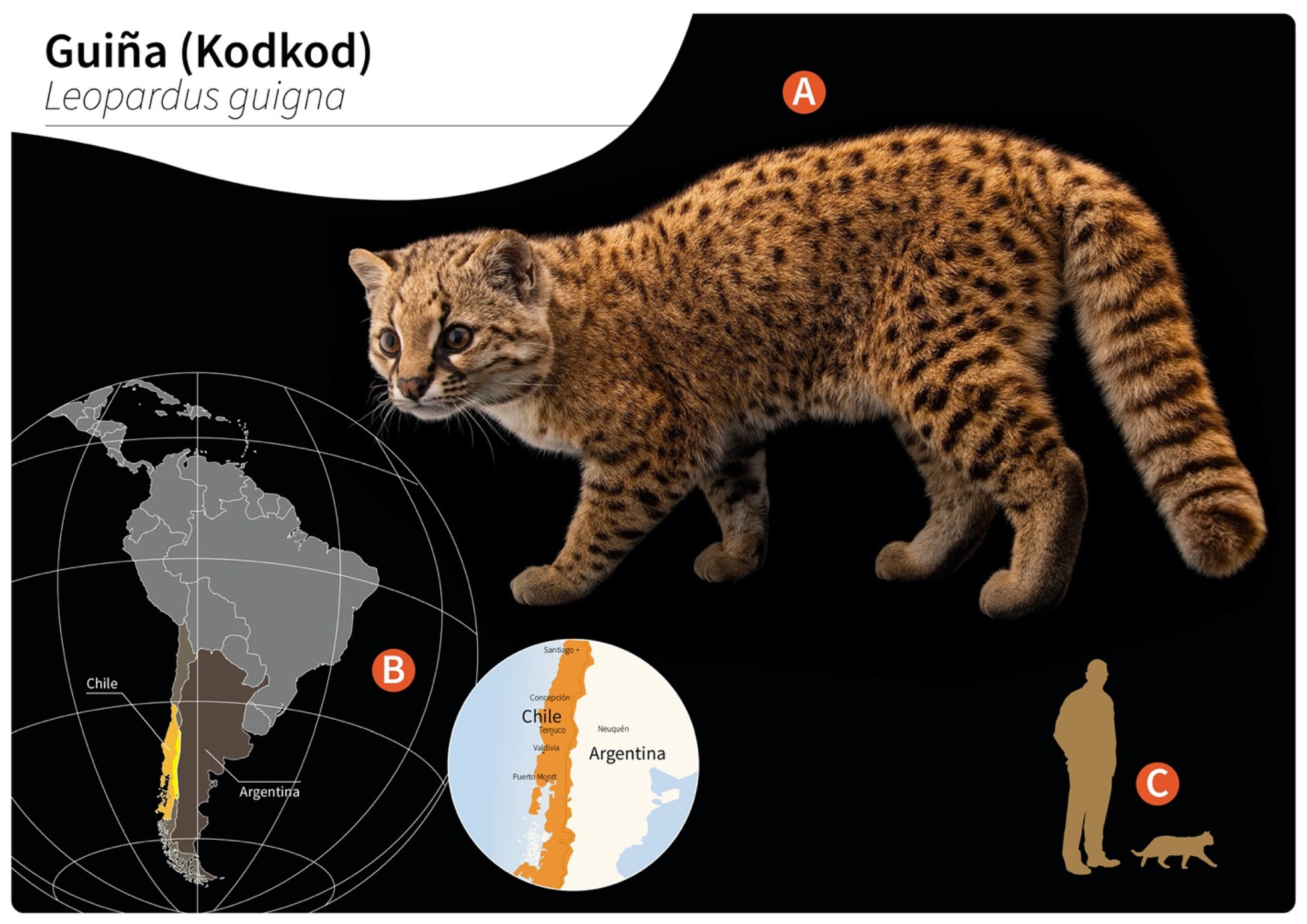
Figure 1. Distribution of wild guiñas (syn. huiñas, kodkods, spotted tiger cat) in South America. (A) Adult specimen of a guiña (Leopardus guigna) (image reprinted with permission from © Joel Sartore/Photo Ark, 2022). (B) Geographic distribution of guiña in Chile (orange) and Argentina (yellow). (C) Scale representation of an adult guiña.
Pathogens 11 00792 g001
Figure 2. Proposed life cycle and migration pathways of Gurltia paralysans. (A) Cranial end of an adult specimen of G. paralysans. (B) Domestic cats (Felis catus) or wild felids (Leopardus spp.) acquire the L3 larvae by ingesting an infected obligate intermediate host (gastropods) or paratenic hosts (lizards, rodents, amphibians, birds or insects). Infective larvae penetrate the stomach and enter the hepatic portal system, and then the caudal vena cava and/or the azygous venous system. From these vein systems, the larvae migrate to the spinal cord via the intervertebral veins and the vertebral venous plexus. The larvae invade the veins of the subarachnoid space of the spinal cord, where they mature and lay eggs. It is still unknown on how domestic cats eliminate the eggs or the first-stage larvae (L1) into the environment, their further development into the L2 and L3 larval stages, or how the obligate intermediate hosts become infected with L1. AV: azygos vein; CV: caudal vena cava; IV: intervertebral veins; H: heart; L: liver; S: stomach; SC: spinal cord; VVP: vertebral venous plexus; L1: first-stage larvae; L2: second-stage larvae; L3: third-stage larvae. The inserted QR code shows a video of a G. paralysans-infected cat with clinical signs of paraparesis.
Pathogens 11 00792 g002
Figure 3. Microscopic view of Gurltia paralysans. (A) Cephalic end of the specimen showing a tooth at the anterior margin (scale bar: 50 µm). (B) Caudal end of a male, showing the small copulatory bursa (scale bar: 50 µm). (C) Higher magnification of a male caudal extremity, showing the spicules (arrow) (scale bar: 50 µm).
Pathogens 11 00792 g003
Figure 4. Macroscopic and microscopic lesions of the spinal cord in a Gurltia paralysans-infected cat. (A) Lumbar, sacral and caudal segments of the spinal cord showing severe and diffuse submeningeal vascular congestion. (B) Histopathological view of transverse sections of an adult of G. paralysans (arrows) inside a subarachnoid vein and vascular congestion (asterisk) in the spinal subarachnoid space; HE, 4 × (scale bar: 500 µm). (C) Histopathological section of the spinal cord parenchyma showing developing eggs of G. paralysans (arrow); HE, 40 × (scale bar: 50 µm).
Pathogens 11 00792 g004
Figure 5. Macroscopic view of the right lung with multiple haemorrhagic foci in a cat with feline gurltiosis.
Pathogens 11 00792 g005
Table 1. Reported cases of feline gurltiosis including age, geographic location, clinical presentation and diagnosis.
AgeLocationNumber
of Cases
Clinical PresentationDiagnosisReference
1–3 y Chile
(Los Ríos/Los Lagos regions)
3Paraparesis (ambulatory)
PL ataxia
PL muscle atrophy
Anal/urinary incontinence
Post mortem
(histopathology)
[12]
6–8 mColombia
(Antioquia municipality)
6Paraparesis (ambulatory)
PL ataxia
Spinal pain
PL muscle atrophy
Anal/urinary incontinence
Decrease superficial/deep pain in PL
Post mortem
(histopathology, Myelo)
[14]
2 yArgentina
(Buenos Aires province)
1Paraparesis (non-ambulatory)
PL muscle atrophy
Increase spinal reflexes in PL
Decrease superficial/deep pain in PL
Post mortem
(histopathology)
[15]
NAUruguay
(Fray Bentos)
2Paraparesis (ambulatory)
Paraplegia
PL ataxia
Post mortem
(histopathology)
[16]
1–3 yChile3Paraparesis (ambulatory)
PL ataxia
PL muscle atrophy
Anal/urinary incontinence
Spinal hyperaesthesia
PL trembling
Increase spinal reflexes in PL
Paraplegia
Post mortem
(histopathology, specimens extracted from SSE)
[11]
8 m–10 yChile9Paraparesis (ambulatory)
Paraparesis (non-ambulatory)
Paraplegia
PL ataxia
Increase spinal reflexes in PL
Spinal hyperaesthesia
Anal/urinary incontinence
Post mortem
(histopathology, specimens extracted from SSE, Myelo, CT, MRI
[17]
NABrazil
(Río Grande do Sul)
4Paraparesis (ambulatory)
PL muscle atrophy
Vesical atony
Tail atony
Post mortem
(histopathology)
[18]
NAArgentina
(Santa Fé)
3Paraparesis (ambulatory)
Paraplegia
Decrease spinal reflexes in PL
Decrease superficial pain in PL
Skin lesions in the metatarsal region
Post mortem
(histopathology)
[19]
8 mChile
(Ancud, Los Lagos regions)
1Paraparesis (ambulatory)
Anal/urinary incontinence
Tail atony
Myelo-CT, CSF (mononuclear pleocytosis), post mortem
(histopathology)
[10]
2 ySpain
(Tenerife)
1Uveitis in left eyeSpecimen extracted from anterior chamber of the eye, PCR[20]
NABrazil
(Pernambuco)
11Paraparesis (ambulatory)
PL ataxia,
PL muscle atrophy
Skin lesions in metatarsal and phalangeal regions
Post mortem
(histopathology)
[21]
36 mChile10Paraparesis (ambulatory)
Paraplegia
PL ataxia
Anal/urinary incontinence
Post mortem
(histopathology), IDEXX (Angio Detect),
specimens extracted from SSE
[5]
NA: not available; SSE: spinal subarachnoid space; PL: pelvic limbs; Myelo: myelography, My—lo-CT: computed tomography myelography; MRI: magnetic resonance imaging; CSF: cerebrospinal fluid; y: years; m: months.
Publisher’s Note: MDPI stays neutral with regard to jurisdictional claims in published maps and institutional affiliations.