Article

Antibiotic Resistance and Biofilm Formation in Enterococcus spp. Isolated from Urinary Tract Infections

1
Biology Department, College of Science, Taif University, P.O. Box 11099, Taif 21944, Saudi Arabia
2
Botany and Microbiology Department, Faculty of Science, Tanta University, Tanta 31527, Egypt
3
Biofuels Institute, School of the Environment and Safety Engineering, Jiangsu University, Zhenjiang 212013, China
*
Authors to whom correspondence should be addressed.
Pathogens 2023, 12(1), 34; https://doi.org/10.3390/pathogens12010034
Submission received: 23 November 2022 / Revised: 12 December 2022 / Accepted: 22 December 2022 / Published: 25 December 2022

Abstract

:
Background: A urinary tract infection (UTI) resulting from multidrug-resistant (MDR) enterococci is a common disease with few therapeutic options. About 15% of urinary tract infections are caused by biofilm-producing Enterococcus spp. Therefore, the objective of this study was to identify the MDR enterococci associated with UTIs and assess their potential to produce biofilms. Methods: Thirty Enterococcus isolates were obtained from urine samples collected from UTI patients at King Abdulaziz Specialist Hospital in Taif, Saudi Arabia. The antimicrobial resistance profiles of the isolates were evaluated using disk diffusion techniques against 15 antimicrobial agents. Two techniques, Congo red agar (CRA) and a microtiter plate (MTP), were used to assess the potential of the isolates to produce biofilms. The enterococcal isolates were screened for biofilm-related genes, esp; ebpA; and ebpB, using the PCR method. Results: The molecular identification of the collected bacteria revealed the presence of 73.3% Enterococcus faecalis and 26.6% Enterococcus faecium. The antibiotic susceptibility test revealed that all the tested Enterococcus spp. were resistant to all antimicrobials except for linezolid and tigecycline. Additionally, by employing the CRA and MTP techniques, 76.6% and 100% of the Enterococcus isolates were able to generate biofilms, respectively. In terms of the association between the antibiotic resistance and biofilm’s formation, it was observed that isolates capable of creating strong biofilms were extremely resistant to most of the antibiotics tested. The obtained data showed that all the tested isolates had biofilm-encoding genes. Conclusions: Our research revealed that the biofilm-producing enterococci bacteria that causes urinary tract infections were resistant to antibiotics. Therefore, it is necessary to seek other pharmacological treatments if antibiotic medicine fails.

1. Introduction

The colonization of any urinary tract tissue, from the renal cortex to the urethral meatus, is described as a urinary tract infection (UTI) [1]. A UTI is one of the most prevalent nosocomial and community-acquired illnesses in humans [2]. Escherichia coli, Enterococcus sp., Klebsiella sp., Staphylococcus saprophyticus, Pseudomonas aeruginosa, Staphylococcus aureus, and Proteus sp. are the most common bacteria that cause UTIs [3]. Enterococci are an opportunistic pathogen that may cause severe and potentially serious infections in immunocompromised individuals, as well as those receiving broad-spectrum antibiotic treatment or those requiring extended hospitalization [4]. Enterococcus faecalis or Enterococcus faecium are the most common cause of UTIs and account for around 95% of human enterococcal infections, followed by E. coli [5]. In Saudi Arabia, the most often occurring bacteria that cause a UTI are E. coli, Enterococcus sp., and Klebsiella sp. [6].
Enterococci are attracting attention due to their multidrug resistance, which explains their preponderance in infections [7,8,9,10]. They are innately resistant to a broad spectrum of routinely used antibiotics, including cephalosporins, aminoglycosides, macrolides, and trimethoprim-sulfamethoxazole [11,12]. Bacteria that form biofilms cause more than 65% of nosocomial infections and 80% of bacterial infections, which may become a severe concern in the area of urology [13]. The ability of Enterococcus to produce biofilms is one of its most noticeable pathogenicity characteristics. This property enables the colonization of inert and biological surfaces as well as mediates the adherence to host cells. These biofilms are communities of cells that have been permanently attached to a variety of biotic and abiotic surfaces and are encased in a hydrated matrix of exopolymeric molecules, proteins, polysaccharides, and nucleic acids [14]. There is a noticeable variation in the behavior of bacteria in biofilms compared to their free-living counterparts. Biofilms protect their members from host immunological responses, phagocytosis, and antibiotics [15], thereby complicating the disease. While it is widely agreed that antibiotic therapy is the most important and successful method currently available for managing microbial infections, antibiotic therapy alone is sometimes insufficient to eradicate the bacterial biofilm and the associated infections [16,17].
Numerous enterococci virulence factors have been identified as being associated with the biofilm’s formation, including adhesions and generated virulence factors. Asa 1 (aggregation substance), Esp (extracellular surface protein), and Ebp (endocarditis and biofilm-associated pili) are the most important adhesion factors [18]. CylA (cytolysin), GelEA (gelatinase), and Hyl (hyaluronidase) are the produced pathogenic components of enterococci and play a role in pathogenesis [19]. Esp is a cell wall-associated protein that has been involved in the colonization, persistence, and production of biofilms of bacteria in the urinary system [20]. Asa1 is a pheromone-inducible protein that acts as a virulence factor by promoting a bacterial adhesion to renal tubular cells. Additionally, the Ebp operon contains the genes ebpA, ebpB, and ebpC which are involved in the creation of pills by enterococci and are required for bacteria to cause a UTI [21]. The recent focus has centered on the study of the genes implicated in the development of a biofilm and their significance in infections caused by enterococci [17].
Enterococcus faecalis and Enterococcus faecium have emerged as particularly important etiological pathogens of nosocomial infections, notably urinary tract infections. Therefore, the goal of this study was to investigate the MDR and biofilm formation among enterococci isolated from urinary tract infections and to assess the relationship between the two phenomena.

2. Materials and Methods

2.1. Isolates Selection

The clinicians of the King Abdulaziz Specialist Hospital in Taif, Saudi Arabia examined 224 patients with UTI symptoms for the isolation of enterococcal bacteria. The clinicians followed the guidelines and the standard protocols that are compatible with the requirements of the Declaration of Helsinki [22]. The study’s protocol was approved by the review board of the King Abdulaziz Specialist Hospital, Taif, KSA (SRU-KAASH-107) and the samples were collected between October 2018 and October 2019. There were 121 females (54.01%) and 103 males (45.98%), aged 6 to 112 years. The collected specimens were transferred to the Laboratory of Microbiology at the Department of Biology, the Faculty of Science, Taif University (the female section) for a further testing of the experiments.

2.2. Enterococcal Isolation

All urine samples were microscopically inspected after 10 min of centrifugation at 1000 rpm. Pus cells, RBCs, epithelial cells, casts, and crystals were found as signs of a urine infection. Using a sterile standard loop, all urine samples were cultured on conventional culture media, including blood agar (Oxoid, Basingstoke, UK), and Cystine–Lactose–Electrolyte Deficient (CLED) agar (Oxoid, Basingstoke, UK). The plates were incubated for 24–48 h at 37 °C. Gram’s stain; catalase, motility, and pigment tests; Enterococcus selective media (Bile-esculin-azide agar and Slanetzand Bartley agar); and a salt tolerance test (6.5% NaCl) was used to identify Enterococcus sp. Such brown-to-black colonies in the surrounding environment were suspected to be Enterococcus. Other testing, including growth in very alkaline conditions (pH = 9.6) and high temperatures, showed the isolates to be enterococci. An API 20 Strep (BioMerieux, St. Louis, MO, USA) biochemical test kit was also used to validate the enterococcal isolates biochemically.

2.3. Antibiotic Susceptibility Test

Kirby Bauer’s disc diffusion method was used to investigate the antibiotic sensitivity of the antimicrobial agents against clinical enterococcal isolates [23]. E. faecalis ATCC 2921 was used as a reference strain for the antibiotic susceptibility tests. Antimicrobials (MAST Diagnostics, Liverpool, UK) were categorized by classes and abbreviation, with concentrations, as shown in Supplementary Table S1. A 1 × 10⁵ CFU/mL, 20 μL overnight enterococci culture was added to 100 mL of Nutritional Broth (NB) medium and incubated for 24 h at 37 °C/120 rpm. A bacterial culture (100 μL) was streaked onto Mueller Hinton Agar (MHA; Oxoid, Ltd.) plates using cotton swabs and incubated aerobically at 37 °C for 18–24 h. The inhibitory zones (mm) were measured after the incubation, and the findings were classed as sensitive or resistant using the CLSI interpretation criteria [23]. The ‘Intermediate’ results were categorized as ‘resistant’. Multidrug-resistant (MDR) refers to bacteria that have developed a resistance to at least one agent in three or more antimicrobial classes, as described by Magiorakos et al. [24].

2.4. Enterococcal Biofilm Formation

The Congo red agar (CRA) and microtiter plate technique (MTP) methods were used to examine the biofilm’s formation by MDR enterococcal isolates [24].

2.4.1. Using Congo Red Agar (CRA)

The formation of the biofilm was assessed qualitatively by culturing enterococcal isolates on CRA plates [24,25]. To prepare CRA plates, 1 L of brain heart infusion agar (Merck, Darmstadt, Germany) was mixed with 0.8 g of Congo red dye (Merck, Darmstadt, Germany), and 36 g of saccharose. The plates were incubated for 24 h at 37 °C and followed overnight at room temperature. Black colonies were assessed to be strains with a high capacity to produce a biofilm (P; producer), whereas strains with red colonies were identified as the strains incapable of producing the biofilm (NP; non-producer).

2.4.2. Using Microtiter Plates (MTP)

The enterococci biofilm production was assessed using a semi-quantitative adhesion test on MTP. Briefly, the bacterial isolates were grown overnight at 37 °C in trypticase-soy broth (TSB, Merck, Germany). The enterococcal cultures were diluted 1:100 in fresh TSB containing 2% glucose. The diluted solution was then applied to the wells of a flat-bottomed polystyrene microtiter plate (Sigma Aldrich, Darmstadt, Germany) and incubated at 37 °C for 24 h. Wells with sterile TSB containing 2% glucose served as the negative controls. After incubation, each well was drained and cleaned three times with 300 μL of phosphate-buffered saline (PBS; pH 7.2). The adhesive bacteria were fixed in 95% ethanol for 5 min and stained with 100 μL of 1% crystal violet. This was followed by three thorough cleanings with 300 μL of sterile distilled water. The microplates were then dried in the air. The optical density (OD) of the stained adherent was measured at 570 nm using an ELISA reader (BioTek Instruments, Winooski, VT, USA). The strains were divided into groups based on the OD values produced by the bacterial biofilms, as described by Christensen et al. [26], with minor modifications, as indicated by Stepanovic et al. [27]. The bacterial strains were classified as follow: the OD value < 0.120 as weak/negative biofilm-producing (WP/NP) isolates, and those with OD > 0.120 and <0.240 were regarded as moderate biofilm producers (MP). An OD value > 0.240 was indicative of high biofilm-producing bacterial (HP) strains.

2.5. PCR Amplification and Phylogenetic Analysis

The genomes of the bacterial strains were isolated from pure bacterial cultures and sequenced based on the manufacturer’s instructions for the DNA extraction kit (Promega, USA). Two universal primers, 1492R (5′-TACGGYTACCTTGTTCGACTT-3′) and 27F (5′-AGAGTTTGATCTGGCTCAG-3′) were used in the 16S rRNA gene amplification. The Bio-Rad S1000™ Thermal Cycler (Bio-Rad, Hercules, CA, USA) was used to perform the PCR amplification [27]. In a TBE buffer 1×, an agarose gel at a concentration of 1.5% (w/v) was utilized. The gels were stained with ethidium bromide (EtBr) solution at a concentration of 0.5 mg/mL. A UV transilluminator (Biotium, Fremont, CA, USA) was used to visualize the DNA, and the results were photographed and reported. Purified PCR products were sequenced at Macrogen Co., South Korea. The initial sequence analysis was performed using BlastN (http://www.ncbi.nlm.nih.gov/BLAST/ accessed on 20 September 2020). A neighbor-joining phylogenetic tree was constructed based on 16S rRNA gene sequences of enterococci members and the related genera using molecular evolutionary genetics analysis version 7 (MEGA7) software [28].

2.6. Detection of Biofilm-Associated Genes

The presence of the biofilm-associated genes was also detected in this study using a PCR. Specific primers were used for the PCR testing of all the bacterial isolates to detect virulence genes encoding esp, ebpA, and ebpB genes. The primers used for the determination of these virulence genes were previously described [29,30,31]. The PCR procedure for all the genes was as follows: initial denaturation at 95 °C for 5 min, followed by 30 cycles of 30 s; denaturation at 94 °C, 30 s; primer annealing at 58 °C; and a 1 min extension at 72 °C. Following these cycles, a final 7 min extension step at 72 °C was performed. Gel electrophoresis (Cleaver Scientific, Warwickshire, UK), utilizing a 1.5% agarose gel (Cleaver Scientific Ltd., Warwickshire, UK), was used to analyze the amplification products.

2.7. Statistical Analysis

All the obtained data were analyzed by GraphPad Prism (GraphPad Software, Inc., San Diego, CA, USA) version (8.0.2) using a paired t-test to show the variations between the resistance and biofilm-forming ability of the investigated Enterococcus sp. The obtained results were significant at * p ≤ 0.05, ** p ≤ 0.01, *** p ≤ 0.001, and **** p ≤ 0.0001, while p > 0.05 is non-significant (ns).

3. Results and Discussion

Enterococci may be responsible for 15% of urinary tract infections in critical care units (ICU) [18]. In this study, only 30 (13%) enterococci bacterial isolates were found in 224 patient urine samples. Female culture-positive urine specimens were reported more frequently than male culture-positive urine specimens. Females have a greater incidence of urinary tract infections due to anatomical differences and a reliance on the male urinary system’s natural defensive systems [32]. The morphological and biochemical tests confirmed that all isolates (n = 30) were Enterococcus species and designated as E01–E30.
The susceptibility and resistance rates among enterococcal isolates were shown in Figure 1. According to the data shown in Figure 1A, the majority of E. faecalis strains were resistant to tetracycline (86.36%), erythromycin (81.81%), levofloxacin, and quinpristin–dalfopristin (77.27%). There were less resistant isolates, such as gentamicin (50%), vancomycin (31%), and streptomycin (27%). Nitrofurantoin showed the lowest degree of resistance (18.18%) regarding the E. faecium strains; the maximum resistance rate (100%) was seen for tetracycline and erythromycin, while the lowest resistance rate (12.5%) was observed for streptomycin (Figure 1B). All the Enterococcus isolate was completely susceptible to linezolid and tigecycline. According to a statistical analysis using the paired t-test, the resistance of E. faecium against the investigated antimicrobial agents was insignificantly (p = 0.8450) higher than E. faecalis, as shown in Figure 1C. A resistance to fluoroquinolones (levofloxacin) may be the result of alterations in the target enzymes related to the DNA replication, hence preventing the bacterial proliferation [33]. Chow [34] stated that all the enterococci tested exhibited a modest inherent resistance to aminoglycosides, including gentamicin, which was regarded as the most often recommended aminoglycoside for enterococci infections. Comparable observations by Maschieto et al. [35], Rajendiran et al. [36], and Wei et al. [37] revealed a moderate level of resistance to streptomycin.
MDR Enterococcus sp. strains displayed five resistance profiles for different classes of tested antimicrobials, as shown in Table 1, with 74% of isolates resistant to more than four classes. E18, E21, and E14 were the most resistant to seven antimicrobial classes, whereas E05 and E29 showed a resistance to three. These findings corroborated those of Fered et al. [38].
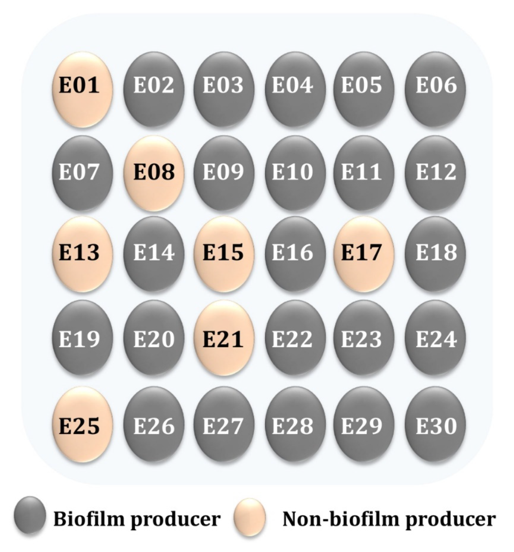
Additionally, biofilms played a crucial role in the high incidence, recurrence, and severity of UTIs, causing both acute and chronic infections [39]. As a result, the following tests subjectively recognized the formation of the biofilm in the tested enterococci isolates on a CRA media and quantified it using the MTP method. To summarize the CRA data, biofilm producers (P) appeared as black colonies, whereas non-biofilm producers (NP) appeared as pink colonies (Figure 2). The percentage of isolates forming biofilms was 17 (77.2%) for E. faecalis and 6 (75%) for E. faecium. Baldassarri et al. [40] observed that 80% of E. faecalis and 48% of E. faecium were significantly associated with the biofilm production in infected patients. Additionally, Dupre et al. [41] reported that E. faecalis produced biofilms to varying degrees in 86.7% of clinical specimens, whereas only 15.6% of E. faecium specimens formed biofilms. As illustrated in Figure 3, a biofilm formation was detected using the MTP methods. According to the obtained results, 45.45% of E. faecalis isolates were found to be high producers (HP), while 54.54% were found to be moderately adherent (MP) (Figure 3A). Where 37.5% of E. faecium isolates were found to be highly produced, 62.5% were found to be moderate producers (Figure 3B). As shown in Figure 4, a comparison of the two biofilm detection methods demonstrated that the biofilm’s development was subjectively seen in CRA, although only 23 (76.6%) of the isolates achieved the biofilm production criteria (Figure 4A). Using the MTP test, all 30 of the investigated isolates produced biofilms with varying degrees (Figure 4B). The findings of two techniques for determining the production of biofilms by the Enterococcus species were comparable, although it seems that MTP is the more precise and dependable methodology [24,41,42]. Lasa [43] obtained comparable findings; Mathur et al. [44] likewise encouraged the adoption of the MTP approach because of its excellent specificity, sensitivity, and positive predictive value. However, Hassan et al. [45] and Kafil et al. [18] advocated for the use of CRA to identify biofilms.
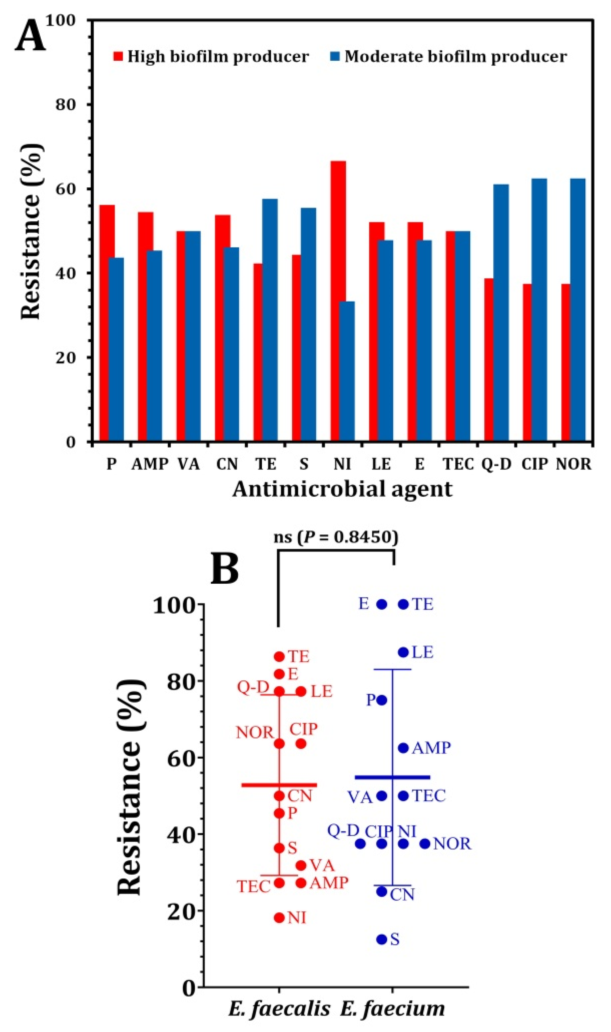
Statistical analysis indicated a significant correlation between the Enterococcus species that form biofilms and the antibiotic resistance to certain antimicrobials (Figure 5). A resistance to several antibiotics, including penicillin, ampicillin, gentamicin, nitrofurantoin, levofloxacin, and erythromycin, was significantly higher in high biofilm producers than in moderate biofilm producers (Figure 5A). According to the obtained data from the statistical analysis using t-test analysis, as shown in Figure 5B, a penicillin resistance (56.2% vs. 43.7%, p = 0.0002), ampicillin resistance (54.5% vs. 45.4%, p = 0.0008), gentamicin resistance (53.8% vs. 46.1%, p = 0.0006), nitrofurantoin resistance (66.6% vs. 33.3%, p < 0.0001), and levofloxacin and erythromycin resistance (52.1% vs. 47.8%, p = 0.0054). Similarly, various studies have shown a significant correlation between the formation of the biofilm and antibiotic resistance in the enterococci isolated from urinary tract infections [45,46,47], whereas a norfloxacin, ciprofloxacin, quinupristin/dalfopristin, and tetracycline resistance were rather uncommon among biofilm-forming bacteria. As a result, these antibiotics may be utilized effectively to treat UTIs caused by enterococcal isolates from biofilm producers. Akhter et al. [46] found a strong correlation between the biofilm’s formation in Enterococcus spp. isolated from urinary tract infections and the antibiotic resistance to amoxicillin, co-trimoxazole, ciprofloxacin, gentamycin, cefotaxime, and cefuroxime. Additionally, Fallah et al. [17] reported that the most effective drugs against the Enterococcus species were linezolid, chloramphenicol, and nitrofurantoin.
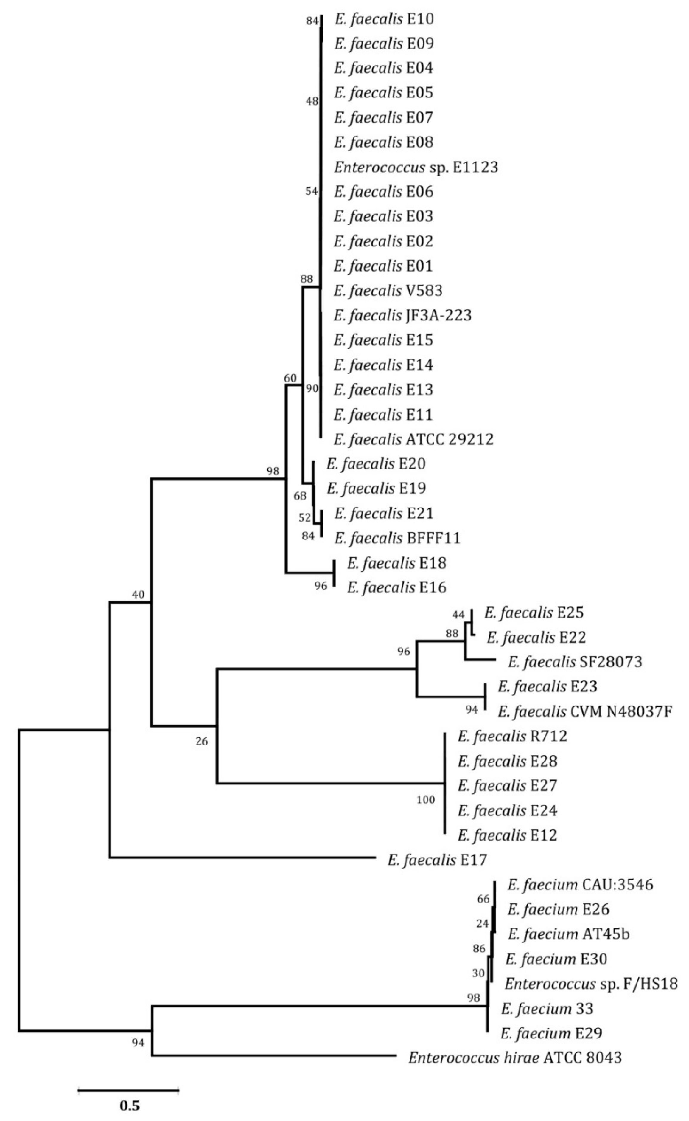
As depicted in Figure 6, 27 isolates were identified as E. faecalis and 3 as E. faecium. The 16S rRNA gene sequences may be used to place diagnoses into a phylogenetic tree and can be connected to hundreds of database sequences [48]. The comparison of these sequences enables the differentiation of organisms at the genus level across all of the major bacterial phyla, as well as the categorization of strains at numerous levels, including what is currently referred to as the species and subspecies levels [49].
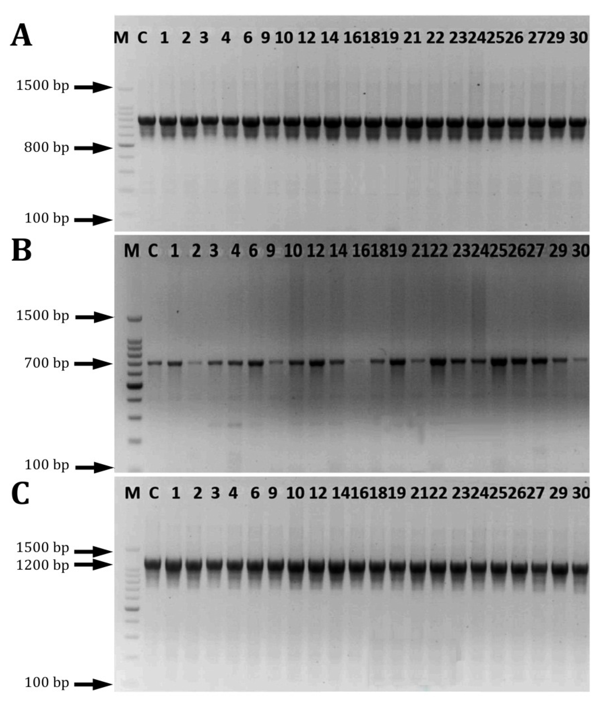
Numerous investigations have been conducted on the enterococci virulence genes involved in the biofilm’s formation. However, the pathogenic mechanisms and genes involved in the biofilm’s formation in enterococci were deemed to be insufficient [18]. In this study, the presence of biofilm-related genes was evaluated in E. facealis and E. faecium by PCR. The findings shown in Figure 7 indicated that the Esp, EbpA, and EbpB genes were detected in all of the tested strains at a 100% prevalence rate. Shankar et al. [50] detected Esp, a cell wall-related protein, in the enterococci strains. It has been shown that it promotes the adhesion, colonization, and evasion of the immune system, as well as having an influence on antibiotic resistance [51,52]. Soares et al. [53] established that Enterococcus faecalis harbored GelE, Esp, and Asa 1 genes. In clinical UTI isolates, Kafil and Mobarez [18] found a relationship between the virulence factors and the biofilm’s development. The findings of the statistical analysis indicate that there was a significant correlation between the frequency of esp, ebpA, and ebpB genes with a biofilm development in all of the tested isolates (p < 0.05). All of the investigated genes were found in high biofilm producers (42%) and intermediate biofilm producers (57%).

4. Conclusions

The results of this research reveal that the emergence of an antibiotic resistance in enterococci that cause urinary tract infections limits the therapeutic use of antimicrobials. In addition, the spread of MDR enterococci isolates poses a threat to hospitalized patients’ health. The research showed that all isolates had the potential to form a biofilm, which complicates their treatment with antibiotics, confirming the critical need to develop novel antimicrobial agents that control the infection associated with the development of a biofilm.

Supplementary Materials

The following supporting information can be downloaded at: https://www.mdpi.com/article/10.3390/pathogens12010034/s1, Table S1: Antibiotic susceptibility disks (Oxoid, England) along with codes and potencies.

Author Contributions

Conceptualization, M.A.K.; J.A.A. and S.E.E. methodology, M.A.K., J.A.A., L.M.A.-O. and S.E.E.; software, S.S.A.; validation, M.A.K. and J.A.A. and S.E.E.; formal analysis, S.E.E. and S.S.A.; investigation, M.A.K. and J.A.A.; resources, M.A.K., J.A.A., S.S.A. and S.E.E.,; data curation, S.E.E.; writing—original draft preparation, L.M.A.-O.; writing—review and editing, M.A.K., S.S.A. and S.E.E.; visualization, J.A.A. and L.M.A.-O.; supervision, M.A.K. and. J.A.A.; project administration, M.A.K. and J.A.A.; funding acquisition, M.A.K. All authors have read and agreed to the published version of the manuscript.

Funding

This study was funded by Taif University Researchers Supporting Project Number (TURSP-2020/95), Taif, Saudi Arabia.

Institutional Review Board Statement

All procedures performed in studies involving human participants followed the ethical standards of Tanta University and with the 1964 Helsinki declaration and its later amendments or comparable ethical standards. The study’s protocol was approved by the review board of the King Abdulaziz Specialist Hospital, Taif, KSA (SRU-KAASH-107).

Informed Consent Statement

Not applicable.

Data Availability Statement

All data generated or analyzed during this study are included in this published article (and its Supplementary Information Files).

Conflicts of Interest

All authors declare no competing interests.

References

  1. O’Brien, V.P.; Hannan, T.J.; Nielsen, H.V.; Hultgren, S.J. Drug and vaccine development for the treatment and prevention of urinary tract infections. Microbiol. Spectr. 2016, 4, 589–646. [Google Scholar] [CrossRef] [PubMed] [Green Version]
  2. Kumar, M.; Das, A. Molecular identification of multi drug resistant bacteria from urinary tract infected urine samples. Microb. Pathog. 2016, 98, 37–44. [Google Scholar] [CrossRef] [PubMed]
  3. Kot, B.; Grużewska, A.; Szweda, P.; Wicha, J.; Parulska, U. Antibiotic resistance of uropathogens isolated from patients hospitalized in district hospital in central Poland in 2020. Antibiotics 2021, 10, 447. [Google Scholar] [CrossRef] [PubMed]
  4. Krawczyk, B.; Wityk, P.; Gałęcka, M.; Michalik, M. The many faces of Enterococcus spp. commensal, probiotic and opportunistic pathogen. Microorganisms 2021, 9, 1900. [Google Scholar] [CrossRef]
  5. Krawczyk, B.; Wityk, P.; Gałęcka, M.; Michalik, M. Cranberry polyphenols and prevention against urinary tract infections: Relevant considerations. Molecules 2020, 25, 3523. [Google Scholar]
  6. Bazaid, A.S.; Saeed, A.; Alrashidi, A.; Alrashidi, A.; Alshaghdali, K.A.; Hammam, S.; Alreshidi, T.; Alshammary, M.; Alarfaj, A.; Thallab, R.; et al. Antimicrobial surveillance for bacterial uropathogens in Ha’il, Saudi Arabia: A Five-year multicenter retrospective study. Infect. Drug Resist. 2021, 16, 1455–1465. [Google Scholar] [CrossRef]
  7. Ali, S.S.; Sonbol, F.I.; Sun, J.; Hussein, M.A.; Hafez, A.-E.E.; Abdelkarim, E.A.; Kornaros, M.; Ali, A.; Azab, M. Molecular characterization of virulence and drug resistance genes-producing Escherichia coli isolated from chicken meat: Metal oxide nanoparticles as novel antibacterial agents. Microb. Pathog. 2020, 143, 104164. [Google Scholar] [CrossRef]
  8. Ali, S.S.; Moawad, M.S.; Hussein, M.A.; Azab, M.; Abdelkarim, E.A.; Badr, A.; Sun, J.; Khalil, M. Efficacy of metal oxide nanoparticles as novel antimicrobial agents against multi-drug and multi-virulent Staphylococcus aureus isolates from retail raw chicken meat and giblets. Int. J. Food Microbiol. 2021, 344, 109116. [Google Scholar] [CrossRef]
  9. Ali, S.S.; Shaaban, M.T.; Abomohra, A.E.-F.; El-Safity, K. Macroalgal activity against multiple drug resistant Aeromonas hydrophila: A novel treatment study towards enhancement of fish growth performance. Microb. Pathog. 2016, 101, 89–95. [Google Scholar] [CrossRef]
  10. Ali, S.S.; Morsy, R.; El-Zawawy, N.A.; Fareed, M.F.; Bedaiwy, M.Y. Synthesized zinc peroxide nanoparticles (ZnO2-NPs): A novel antimicrobial, anti-elastase, anti-keratinase, and anti-inflammatory approach toward polymicrobial burn wounds. Int. J. Nanomed. 2017, 12, 6059. [Google Scholar] [CrossRef] [Green Version]
  11. Samani, R.J.; Tajbakhsh, E.; Momtaz, H.; Samani, M.K. Prevalence of virulence genes and antibiotic resistance pattern in Enterococcus faecalis isolated from urinary tract infection in Shahrekord, Iran. Rep. Biochem. Mol. Biol. 2021, 10, 50–59. [Google Scholar] [CrossRef]
  12. Saraswathy, M.P. Multidrug resistant Enterococci isolated from urine samples at a tertiary care hospital. Indian J. Microbiol. Res. 2015, 2, 214–219. [Google Scholar] [CrossRef]
  13. Soto, S.M. Importance of biofilms in urinary tract infections: New therapeutic approaches. Adv. Biol. 2014, 2014, 543974. [Google Scholar] [CrossRef]
  14. Hashem, Y.A.; Abdelrahman, K.A.; Aziz, R.K. Phenotype-genotype correlations and distribution of key virulence factors in Enterococcus faecalis isolated from patients with urinary tract infections. Infect. Drug Resist. 2021, 14, 1713–1723. [Google Scholar] [CrossRef] [PubMed]
  15. Guzmán-Soto, I.; McTiernan, C.; Gonzalez-Gomez, M.; Ross, A.; Gupta, K.; Suuronen, E.J.; Alarcon, E.I. Mimicking biofilm formation and development: Recent progress in in vitro and in vivo biofilm models. Iscience 2021, 24, 102443. [Google Scholar] [CrossRef] [PubMed]
  16. Wu, H.; Moser, C.; Wang, H.Z.; Høiby, N.; Song, Z.J. Strategies for combating bacterial biofilm infections. Int. J. Oral Sci. 2015, 7, 1–7. [Google Scholar] [CrossRef] [Green Version]
  17. Fallah, F.; Yousefi, M.; Pourmand, M.R.; Hashemi, A.; Alam, A.N.; Afshar, D. Phenotypic and genotypic study of biofilm formation in Enterococci isolated from urinary tract infections. Microb. Pathog. 2017, 108, 85–90. [Google Scholar] [CrossRef]
  18. Kafil, H.S.; Mobarez, A.M. Assessment of biofilm formation by Enterococci isolates from urinary tract infections with different virulence profiles. J. King Saud Univ.-Sci. 2015, 27, 312–317. [Google Scholar] [CrossRef] [Green Version]
  19. Kayaoglu, G.; Ørstavik, D. Virulence factors of Enterococcus faecalis: Relationship to endodontic disease. Crit. Rev. Oral Biol. Med. 2004, 15, 308–320. [Google Scholar] [CrossRef] [Green Version]
  20. Moniri, R.; Ghasemi, A.; Moosavi, S.G.A.; Dastehgoli, K.; Rezaei, M. Virulence Gene’s relationship with biofilm formation and detection of aac (6′)/aph (2′′) in Enterococcus faecalis isolated from patients with urinary tract infection. Jundishapur J. Microbiol. 2013, 6, e94137. [Google Scholar] [CrossRef] [Green Version]
  21. Heidari, H.; Emaneini, M.; Dabiri, H.; Jabalameli, F. Virulence factors, antimicrobial resistance pattern and molecular analysis of Enterococcal strains isolated from burn patients. Microb. Pathog. 2016, 90, 93–97. [Google Scholar] [CrossRef] [PubMed]
  22. Declaration of Helsinki History Website. In Ethical Principles For Medical Research; Declaration of Helsinki; The JAMA Network: Helsinki, Finland, 2015.
  23. CLSI. Performance Standards for Antimicrobial Susceptibility Tests, 13th ed.; CLSI Standard M02; Clinical and Laboratory Standards Institute: Wayne, PA, USA, 2018. [Google Scholar]
  24. Magiorakos, A.P.; Srinivasan, A.; Carey, R.B.; Carmeli, Y.; Falagas, M.E.; Giske, C.G.; Harbarth, S.; Hindler, J.F.; Kahlmeter, G.; Olsson-Liljequist, B.; et al. Multidrug-resistant, extensively drug-resistant and pan drug-resistant bacteria: An international expert proposal for interim standard definitions for acquired resistance. Clin. Microbiol. Infect. 2012, 18, 268–281. [Google Scholar] [CrossRef] [PubMed] [Green Version]
  25. Khalil, M.A.; Sonbol, F.I. Investigation of biofilm formation on contact eye lenses caused by methicillin resistant Staphylococcus aureus. Niger. J. Clin. Pr. 2014, 17, 776–784. [Google Scholar] [CrossRef] [PubMed] [Green Version]
  26. Christensen, G.D.; Simpson, W.A.; Younger, J.J.; Baddour, L.M.; Barrett, F.F.; Melton, D.M.; Beachey, E.H. Adherence of coagulase-negative staphylococci to plastic tissue culture plates: A quantitative model for the adherence of staphylococci to medical devices. J. Clin. Microbiol. 1985, 22, 996–1006. [Google Scholar] [CrossRef] [PubMed] [Green Version]
  27. Stepanovic, S.; Vukovic, D.; Hola, V.; Bonaventura, G.; Djukic, S.; Irkovic, I.; Ruzicka, F. Quantification of biofilm in microtiter plates: Overview of testing conditions and practical recommendations for assessment of biofilm production by staphylococci. APMIS 2007, 115, 891–899. [Google Scholar] [CrossRef]
  28. Ali, S.S.; Al-Tohamy, R.; Sun, J.; Wu, J.; Huizi, L. Screening and construction of a novel microbial consortium SSA-6 enriched from the gut symbionts of wood-feeding termite, Coptotermes formosanus and its biomass-based biorefineries. Fuel 2019, 236, 1128–1145. [Google Scholar]
  29. Kumar, S.; Stecher, G.; Tamura, K. MEGA7 molecular evolutionary genetics analysis version 7.0 for bigger datasets. Mol. Biol. Evol. 2016, 33, 1870–1874. [Google Scholar] [CrossRef] [Green Version]
  30. Hashem, Y.A.; Amin, H.M.; Essam, T.M.; Yassin, A.S.; Aziz, R.K. Biofilm formation in enterococci: Genotype-phenotype correlations and inhibition by vancomycin. Sci. Rep. 2017, 7, 5733. [Google Scholar] [CrossRef] [Green Version]
  31. Vankerckhoven, V.; Van Autgaerden, T.; Vael, C.; Lammens, C.; Chapelle, S.; Rossi, R.; Jabes, D.; Goossens, H. Development of a multiplex PCR for the detection of asa1, gelE, cylA, esp, and hyl genes in enterococci and survey for virulence determinants among European hospital isolates of Enterococcus faecium. J. Clin. Microbiol. 2004, 42, 4473–4479. [Google Scholar] [CrossRef] [Green Version]
  32. Weiner, L.M.; Webb, A.K.; Limbago, B.; Dudeck, M.A.; Patel, J.; Kallen, A.J.; Sievert, D.M. Antimicrobial resistant pathogens associated with healthcare-associated infections: Summary of data reported to the National Healthcare Safety Network at the Centers for Disease Control and Prevention, 2011–2014. Infect. Control Hosp. Epidemiol. 2016, 37, 1288–1301. [Google Scholar] [CrossRef] [Green Version]
  33. Chen, H.; Wu, W.; Ni, M.; Liu, Y.; Zhang, J.; Xia, F.; Wang, H. Linezolid-resistant clinical isolates of Enterococci and Staphylococcus cohnii from a multicentre study in China: Molecular epidemiology and resistance mechanisms. Int. J. Antimicrob. Agents 2013, 42, 317–321. [Google Scholar] [CrossRef] [PubMed]
  34. Chow, J.W. Aminoglycoside resistance in enterococci. Clin. Infect. Dis. 2000, 31, 586–589. [Google Scholar] [CrossRef] [PubMed]
  35. Maschieto, A.; Martinez, R.; Palazzo, I.C.V.; Darini, A.L.D.C. Antimicrobial resistance of Enterococcus sp. isolated from the intestinal tract of patients from a university hospital in Brazil. Memórias do Instituto Oswaldo Cruz 2004, 99, 763–767. [Google Scholar] [CrossRef] [PubMed] [Green Version]
  36. Rajendiran, S.; Veloo, Y.; Thahir, S.S.A.; Shaharudin, R. Resistance towards critically important antimicrobials among Enterococcus faecalis and E. faecium in poultry farm environments in Selangor, Malaysia. Antibiotics 2022, 11, 1118. [Google Scholar] [CrossRef]
  37. Wei, L.; Wu, Q.; Zhang, J.; Guo, W.; Chen, M.; Xue, L.; Wang, J.; Ma, L. Prevalence and genetic diversity of Enterococcus faecalis isolates from mineral water and spring water in China. Front. Microbiol. 2017, 8, 1109. [Google Scholar] [CrossRef]
  38. Ferede, Z.T.; Tullu, K.D.; Derese, S.G.; Yeshanew, A.G. Prevalence and antimicrobial susceptibility pattern of Enterococcus species isolated from different clinical samples at Black Lion Specialized Teaching Hospital, Addis Ababa, Ethiopia. BMC Res. Notes 2018, 11, 793. [Google Scholar] [CrossRef]
  39. Delcaru, C.; Alexandru, I.; Podgoreanu, P.; Grosu, M.; Stavropoulos, E.; Chifiriuc, M.C.; Lazar, V. Microbial biofilms in urinary tract infections and prostatitis: Etiology, pathogenicity, and combating strategies. Pathogens 2016, 30, 65. [Google Scholar] [CrossRef] [Green Version]
  40. Baldassarri, L.; Cecchini, R.; Bertuccini, L.; Ammendolia, M.G.; Iosi, F.; Arciola, C.R.; Orefici, G. Enterococcus spp. produces slime and survives in rat peritoneal macrophages. Med. Microbiol. Immunol. 2001, 190, 113–120. [Google Scholar] [CrossRef]
  41. Duprè, I.; Zanetti, S.; Schito, A.M.; Fadda, G.; Sechi, L.A. Incidence of virulence determinants in clinical Enterococcus faecium and Enterococcus faecalis isolates collected in Sardinia (Italy). J. Med. Microbiol. 2003, 52, 491–498. [Google Scholar] [CrossRef] [Green Version]
  42. Mohamed, J.A.; Huang, D.B. Biofilm formation by enterococci. J. Med. Microbiol. 2007, 56, 15811588. [Google Scholar] [CrossRef] [Green Version]
  43. Lasa, I. Towards the identification of the common features of bacterial biofilm development. Int. Microbiol. 2006, 9, 21–28. [Google Scholar] [PubMed]
  44. Mathur, T.; Singhal, S.; Khan, S.; Upadhyay, D.J.; Fatma, T.; Rattan, A. Detection of biofilm formation among the clinical isolates of Staphylococci: An evaluation of three different screening methods. Indian J. Med. Microbiol. 2006, 24, 25–29. [Google Scholar] [CrossRef] [PubMed]
  45. Hassan, A.; Usman, J.; Kaleem, F.; Omair, M.; Khalid, A.; Iqbal, M. Detection and antibiotic susceptibility pattern of biofilm producing gram-positive and gram-negative bacteria isolated from a tertiary care hospital of Pakistan. Malays J. Microbiol. 2011, 7, 57–60. [Google Scholar] [CrossRef] [Green Version]
  46. Akhter, J.; Ahmed, S.; Anwar, S. Antimicrobial susceptibility patterns of Enterococcus species isolated from urinary tract infections. Bangladesh J. Med Microbiol. 2014, 8, 16–20. [Google Scholar] [CrossRef] [Green Version]
  47. Chakraborty, A.; Pal, N.K.; Sarkar, S.; Gupta, M.S. Antibiotic resistance pattern of Enterococci isolates from nosocomial infections in a tertiary care hospital in Eastern India. J. Nat. Sci. Biol. Med. 2015, 6, 394. [Google Scholar] [CrossRef] [Green Version]
  48. Sindhanai, V.; Avanthiga, S.S.; Chander, V.S. Antibiotic susceptibility pattern of biofilm forming and biofilm non forming Enterococci species. OSR J. Dent. Med. Sci. 2016, 15, 33–37. [Google Scholar]
  49. Janda, J.M.; Abbott, S.L. 16S rRNA gene sequencing for bacterial identification in the diagnostic laboratory: Pluses, perils, and pitfalls. J. Clin. Microbiol. 2007, 45, 27612764. [Google Scholar] [CrossRef] [Green Version]
  50. Clarridge, J.E. Impact of 16S rRNA gene sequence analysis for identification of bacteria on clinical microbiology and infectious diseases. Clin. Microbiol. Rev. 2004, 17, 840–862. [Google Scholar] [CrossRef] [Green Version]
  51. Shankar, V.; Baghdayan, A.S.; Huycke, M.M.; Lindahl, G.; Gilmore, M.S. Infection-derived Enterococcus faecalis strains are enriched in esp, a gene encoding a novel surface protein. Infect. Immun. 1999, 67, 193–200. [Google Scholar] [CrossRef] [Green Version]
  52. Zecconi, A.; Scali, F. Staphylococcus aureus virulence factors in evasion from innate immune defenses in human and animal diseases. Immunol. Lett. 2013, 150, 12–22. [Google Scholar] [CrossRef]
  53. Soares, R.O.; Fedi, A.C.; Reiter, K.C.; Caierão, J.; d’Azevedo, P.A. Correlation between biofilm formation and gelE, esp, and agg genes in Enterococcus spp. clinical isolates. Virulence 2014, 5, 634–637. [Google Scholar] [CrossRef] [PubMed]
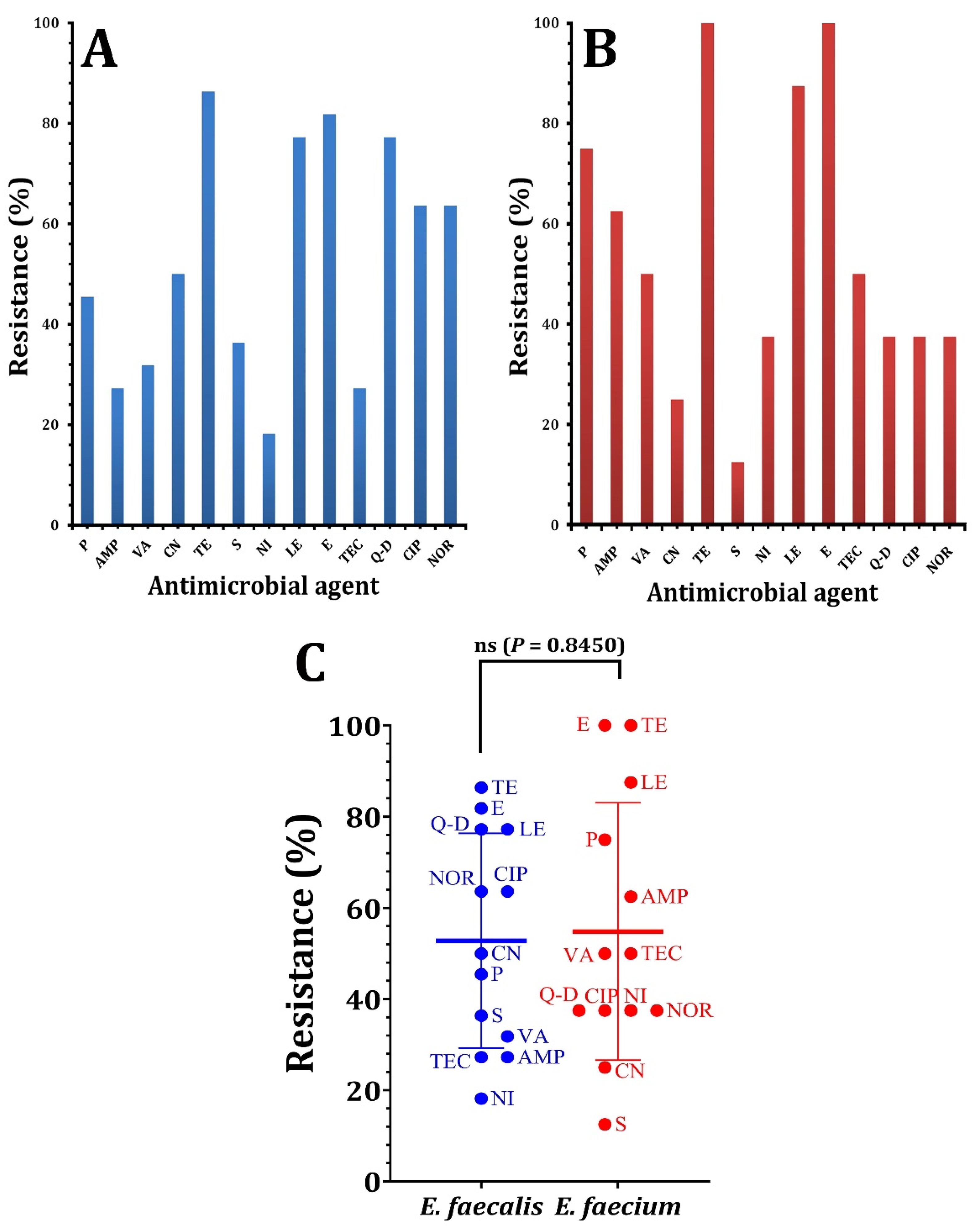
Figure 1. The percentages of antibiotic susceptibility of Enterococcus species against antimicrobial agents. (A) antibiotic susceptibility of E. feacalis, (B) antibiotic susceptibility of E. faecium. (C) the statistical analysis using paired t-test comparing the resistance of Enterococcus species against antimicrobial agents. p > 0.05 is non-significant (ns).
Pathogens 12 00034 g001
Figure 2. Biofilm production by tested Enterococcus species on CRA media.
Pathogens 12 00034 g002
Figure 3. Biofilm production by Enterococcus species using the MTP method. (A) E. faecium, (B) E. faecalis. Biofilm formation was determined using the crystal violet assay at optical density at 570 nm (OD570). High biofilm producer; OD value > 0.240, moderate biofilm producer; OD value > 0.120 and <0.240. p-values for significantly different mean values, p < 0.001 (***), p < 0.0001 (****).
Pathogens 12 00034 g003
Figure 4. Comparing biofilm forming ability of Enterococcus species in CRA and MTP techniques. (A) Biofilm formation using CRA method. (B) Biofilm formation using MTP assay.
Pathogens 12 00034 g004
Figure 5. Correlation between the production of biofilms and the pattern of antibiotic resistance in enterococci isolates (A) and the paired t-test analysis (B). p > 0.05 is non-significant (ns).
Pathogens 12 00034 g005
Figure 6. A neighbor-joining tree based on 16S rRNA gene sequences of enterococci strains and related genera. The scale bar indicates 0.05 substitutions per nucleotide position.
Pathogens 12 00034 g006
Figure 7. PCR detection of biofilm-related genes in Enterococcus isolates. (A) esp gene, (B) ebpA gene, (C) ebpB gene, M molecular weight markers (100 bp), and C control (reference strain).
Pathogens 12 00034 g007
Table 1. Resistance patterns of MDR Enterococcus sp. isolates against different antimicrobial agents.
Pattern CodeIsolate Code# Antimicrobial ResistanceNo. of Antimicrobial Classes
AE05TE V, CIP VII, NOR VII, Q-D IX3
E29P I, AMP I, TE V, LE VII
BE03VA II, TEC II, TE V, LE VII, CIP VII, NOR VII, E VIII4
E11P I, AMP I, TE V, LE VII, E VIII
E13CN III, LE VII, CIP VII, NOR VII, E VIII, Q-D IX
E19TE V, LE VII, CIP VII, NOR VII, E VIII, Q-D IX
E23P I, AMP I, S III, LE VII, E VIII
CE01P I, AMP I, VA II, TEC II, TE V, LE VII, CIP VII, NOR VII, E VIII5
E02CN III, S III, TE V, CIP VII, NOR VII, E VIII, Q-D IX
E04, E09P I, CN III, S III, TE V, CIP VII, NOR VII, LE VII, Q-D IX
E06CN III, S III, TE V, LE VII, CIP VII, NOR VII, E VIII, Q-D
E08AMP I, TE V, LE VII, E VIII, Q-D IX
E10P I, AMP I, VA II, TEC II, TE V, NIVI, LE VII
E12, E16CN III, TE V, LE VII, CIP VII, NOR VII, E VIII, Q-D IX
E15P I, AMP I, CN III, TE V, LE VII, E VIII
E17P I, AMP I, VA II,TEC II,TE V, CIP VII, NOR VII,E VIII
E20VA II, TEC II, TE V, CIP VII, NOR VII, E VIII, Q-D IX
E26VA II, TEC II, TE V, NI VI, LE VII, E VIII
E30P I, CN III, TE V, LE VII, Q-D IX
DE22P I, CN III, TE V, LE VII, E VIII, Q-D IX6
E27P I, AMP I, VA II, TEC II, CN III, TE V, LE VII, E VIII
E28P I, AMP I, VA II, TEC II, S III,TE V, LE VII, CIP VII, NOR VII, E VIII
EE18P I, CN III, S III, TE V, LE VII, CIP VII, NOR VII, E VIII, Q-D IX7
E21P I, VA II, TEC II, CN III, S III, TE V, LE VII, E VIII, Q-D IX
E14P I, AMP I, VA II, TEC II S III, TE V, NI VI, LE VII, CIP VII, NOR VII, E VIII
# P: penicillin, AMP: ampicillin, VA: vancomycin, TEC: teicoplanin, CN: gentamicin, S: streptomycin, TE: tetracycline, NI: nitrofurantoin, LE: levofloxacin, CIP: ciprofloxacin, NOR: norfloxacin, E: erythromycin, and Q-D: quinpristin–dalfopristin I β-Lactam antibiotics, II glycopeptides, III aminoglycosides, IV oxazolidinones, V tetracyclines, VI nitrofurantions, VII fluoroquinolones, VIII macrolides, IX streptogramins.
Disclaimer/Publisher’s Note: The statements, opinions and data contained in all publications are solely those of the individual author(s) and contributor(s) and not of MDPI and/or the editor(s). MDPI and/or the editor(s) disclaim responsibility for any injury to people or property resulting from any ideas, methods, instructions or products referred to in the content.