Article

New Insights into the Role of the Trypanosoma cruzi Aldo-Keto Reductase TcAKR

by
Florencia Díaz-Viraqué
1,
María Laura Chiribao
1,2,
Lisvane Paes-Vieira
1,
Matias R. Machado
3,
Paula Faral-Tello
1,
Ramiro Tomasina
4,
Andrea Trochine
5 and
Carlos Robello
1,2,*
1
Laboratorio de Interacciones Hospedero Patógeno, Unidad de Biología Molecular, Institut Pasteur de Montevideo, Montevideo 11400, Uruguay
2
Departamento de Bioquímica, Facultad de Medicina Universidad de la República, Montevideo 11400, Uruguay
3
Unidad de Proteínas Recombinantes, Institut Pasteur de Montevideo, Montevideo 11300, Uruguay
4
Laboratory of Apicomplexan Biology, Institut Pasteur de Montevideo and Departamento de Parasitología, Facultad de Medicina Universidad de la República, Montevideo 11300, Uruguay
5
Centro de Referencia en Levaduras y Tecnología Cervecera (CRELTEC), Instituto Andino Patagónico de Tecnologías Biológicas y Geoambientales (IPATEC), CONICET-Universidad Nacional del Comahue, Quintral 1250, San Carlos de Bariloche 8400, Argentina
*
Author to whom correspondence should be addressed.
Pathogens 2023, 12(1), 85; https://doi.org/10.3390/pathogens12010085
Submission received: 25 October 2022 / Revised: 30 December 2022 / Accepted: 1 January 2023 / Published: 5 January 2023
(This article belongs to the Special Issue Current Research on Trypanosoma cruzi Infection)

Abstract

:
Chagas disease is a zoonotic infectious disease caused by the protozoan parasite Trypanosoma cruzi. It is distributed worldwide, affecting around 7 million people; there is no effective treatment, and it constitutes a leading cause of disability and premature death in the Americas. Only two drugs are currently approved for the treatment, Benznidazole and Nifurtimox, and both have to be activated by reducing the nitro-group. The T. cruzi aldo-keto reductase (TcAKR) has been related to the metabolism of benznidazole. TcAKR has been extensively studied, being most efforts focused on characterizing its implication in trypanocidal drug metabolism; however, little is known regarding its biological role. Here, we found that TcAKR is confined, throughout the entire life cycle, into the parasite mitochondria providing new insights into its biological function. In particular, in epimastigotes, TcAKR is associated with the kinetoplast, which suggests additional roles of the protein. The upregulation of TcAKR, which does not affect TcOYE expression, was correlated with an increase in PGF2α, suggesting that this enzyme is related to PGF2α synthesis in T. cruzi. Structural analysis showed that TcAKR contains a catalytic tetrad conserved in the AKR superfamily. Finally, we found that TcAKR is also involved in Nfx metabolization.

1. Introduction

Chagas disease, or American Trypanosomiasis, is a chronic parasitosis caused by the protozoan parasite Trypanosoma cruzi. It is estimated that nearly 7 million people are infected worldwide [1]. However, this is probably an underestimation since no in-depth studies cover all of Latin America, the endemic area of this disease. In nature, it can be transmitted either through a triatomine vector (vector-dependent transmission that includes ingestion) or transplacentally (vector-independent transmission); in addition, human activities such as blood transfusion, organ transplantation, and laboratory accidents constitute additional sources of infection. It causes high morbidity and mortality rates in endemic and non-endemic areas, leading to the global spread of Chagas disease [2,3,4].
Benznidazole (Bzn) and Nifurtimox (Nfx) are currently the only drugs proven effective against Chagas disease, although they exhibit undesirable side effects that can lead to treatment interruption [5,6]. Both act as prodrugs and must be activated by reducing the nitro-group [7]. In T. cruzi, three enzymes have been related to Bzn and Nfx metabolism: the NADH-dependent Trypanosomal type I nitroreductase (TcNTRI) [8], the NADPH-dependent Old Yellow Enzyme (TcOYE) [9,10,11,12] and an aldo-keto reductase name TcAKR [13,14,15,16]. TcAKR belongs to a conserved superfamily present in all domains of life whose function is the reduction of aldehydes and ketones [17,18]. In Trypanosomatids, a member of this family exhibits PGF2α synthase activity both in Trypanosoma brucei and Leishmania spp. [19,20,21], but its putative ortholog in T. cruzi did not show such activity [13]. Instead, TcAKR has NADPH-dependent reductive activity on quinones, Bzn, methylglyoxal, and isoprostanes [13,14,16]. However, when the available information is analyzed, some contradictory results appear. On the one hand, this enzyme has been captured through an untargeted chemical proteomics approach using immobilized Bzn, which strongly suggests that it could be related to reductive drug activation and resistance mechanisms [15]. In line with this finding, it has been shown that both recombinant and native TcAKR enzymes reduce Bzn, and TcAKR-overexpressing epimastigotes showed higher NADPH-dependent Bzn reductase activity [14]. However, more recently, it was proposed that the enzyme does not use Bzn as a substrate, and this was studied kinetically and by mass spectrometry [16]. The second controversial point is that most characterized prostaglandin F2α synthases (PGFS) belong to the aldo-keto reductase protein family [22], including the PGF2α synthases identified in T. brucei and Leishmania spp. [19,20]. In this context, it is surprising that attempts to demonstrate this activity in TcAKR failed [13,16], taking into account their homologies and the ability of T. cruzi to convert exogenous arachidonic acid (AA) into PGF2α [23]. A possible explanation is that the presence of an “Old Yellow Enzyme” (TcOYE) in this parasite is entirely responsible for this activity. However, it does not seem like a plausible idea, especially considering that TcOYE was likely acquired more recently through horizontal transfer [11]. To provide clarity in this regard, we performed functional, structural, and phylogenetic studies to obtain new insights into the role of this protein.

2. Materials and Methods

2.1. Plasmids Construction, Recombinant Protein, and Antibody

TcAKR coding sequence was PCR-amplified from genomic DNA of T. cruzi Dm28c epimastigotes with Pfu DNA polymerase (Fermentas, Vilnius, Lithuania) using Fw_TcAKR_XbaI_Dm28c (AATCTAGAATGAAATGCAATTACAGCTGTGTG) and Rv_TcAKR_HindIII_cSTOP_Dm28c (AAAAGCTTTCACTCCTCTCCACCAGAGAAAAAATTATC) primers. PCR products were cloned in pGEM®-T Easy plasmid (Promega, WI, USA) using T4 DNA Ligase (Sigma-Aldrich, WI, USA) and sequenced. The coding sequence was subcloned into the pTREX-n vector [24] and the pQE30 vector (Qiagen, Venlo, The Neterlands). Recombinant TcAKR purification under non-native conditions was performed with nickel-charged affinity resin (Ni-NTA). The recombinant protein purity was analyzed by 12% SDS-PAGE stained with colloidal coomassie (Brilliant Blue G-250, Sigma-Aldrich, WI, USA), and protein concentration was determined by the Bradford method [25]. A polyclonal antiserum against TcAKR was obtained from New Zealand White rabbits after intraperitoneal injection of 100 μg of recombinant TcAKR in Freund’s Complete Adjuvant (Sigma-Aldrich, WI, USA), followed by two immunizations with 50 μg of recombinant protein in Freund’s Incomplete Adjuvant (Sigma-Aldrich, WI, USA). Serum was obtained after 15 days of the last boost.

2.2. Parasites and Cells

Vero cells [26] were cultivated in Dulbecco’s Modified Eagle’s Medium supplemented with 10% (v/v) fetal bovine serum (FBS), penicillin (100 U mL−1) and streptomycin (100 μg mL−1) at 37 °C in a humidified 5% CO2 atmosphere. T. cruzi Dm28c [27] epimastigotes were cultured axenically in liver infusion tryptose (LIT) medium supplemented with 10% (v v−1) inactivated FBS at 28 °C. Trypomastigotes were collected from the supernatant of infected monolayers of Vero cell lines and were maintained cyclically. For stable transfections, T. cruzi epimastigotes were transfected with pTREX-n (empty vector) or pTREX-n TcAKR construction. Parasites (8 × 10⁷) were washed three times with PBS, resuspended in HBS Buffer (21 mM HEPES, 137 mM NaCl, 5 mM KCl, 6 mM glucose, pH 7.4), and electroporated with 100 μg of plasmid DNA using two pulses at 450 V, 1300 μF and 13 Ω in 4 mm cuvettes. Transfected parasites were then selected with increasing G418 (Sigma-Aldrich, WI, USA) concentrations from 50 μg mL−1 to 300 μg mL−1. Overexpression of TcAKR was confirmed by Western blot analysis and indirect immunofluorescence.

2.3. PGF2α Synthase Activity

PGF2α determination was performed with the PGF2 alpha High Sensitivity ELISA Kit (Abcam, Boston, MA, USA). Briefly, TcAKR-overexpressing parasites were washed twice with PBS and incubated with 50 μM of arachidonic acid (Abcam, Boston, USA) for two hours. Cells were removed by pelleting at 1100× g for 10 min. Parasite-free supernatant was stored for PGF2α measurement, the parasites were resuspended in PBS, and an extract was performed by thermal shock (15 min at −80 °C and 15 min at 37 °C three times consecutively). Finally, the resulting homogenized was centrifuged at 20,000× g for 30 min at 4 °C, and the supernatant was used for PGF2α determination. The measurements were performed in biological replicates.

2.4. Structural Comparison of AKR Proteins

The structural-based phylogenetic analysis was performed with the Multiseq [28] plugin of VMD [29] considering AKRs from T. cruzi (TcAKR, PDB: 4GIE), T. brucei Homo sapiens (AKR5A2, PDB: 1VBJ), L. major (AKR5A, PDB: 4G5D), Bacillus subtilis (AKR5G1, PDB: 3D3F), Rattus norvegicus (AKR1B14, PDB: 3QKZ) Homo sapiens (AKR1C3, PDB: 2F38 and AKR1B1, PDB: 1US0). Three-dimensional structures were first aligned using the algorithm STAMP (Structural Alignment of Multiple Proteins) [30], which minimizes the Cα distance between residues of each molecule by applying globally optimal rigid-body rotations and translations. Then a QH [4] score was computed for each pair of aligned structures to infer the structural similarity (distance) from which trees were plotted:
Q H = N [ q a l n + q g a p ]
where qaln and qgap are score functions accounting for structurally aligned regions and structural deviations induced by insertions, respectively, and N is a normalization constant.
Electrostatic potentials were calculated with APBS [31] using a cubic grid of 120 Å per side with 10 points per Å2 setting a dielectric constant of 78.54 for water, a salt bath of 0.150 mM NaCl, and a temperature of 298 ºK. The potential was forced to converge to zero at boundaries. PQR file preparation was performed with the PDB2PQR server [32]. AMBER charges were used, and protonation states at pH 7.0 were set according to PROPKA [33]. The dimerization interface was explored with PDBePISA (Proteins, Interfaces, Structures, and Assemblies) [34].

2.5. Transcript Abundance Analysis

Transcriptomic analysis of both copies of TcAKR was performed using RNA-seq data from different stages of T. cruzi Dm28c [35]. Salmon [36] was used to estimate transcript levels. The normalized counts of TcAKR were plotted using R.

2.6. Epimastigotes Synchronization

Cell cycle synchronization was performed as described previously [37,38,39]. Wild-type Dm28c epimastigotes were washed twice with PBS and subsequently incubated with LIT supplemented with 20 mM of hydroxyurea (Sigma-Aldrich, WI, USA) for 24 h. Parasites were washed twice with PBS and cultured in LIT medium supplemented with 10% (v v−1) inactivated fetal bovine serum. At different times cells were washed with PBS and fixed for 30 min with 4% (w v−1) paraformaldehyde for immunolocalization studies.

2.7. Immunolocalization Studies

For indirect immunofluorescence (IIF) localization, parasites were fixed for 16 h at 4 °C with 4% (w v−1) paraformaldehyde and then incubated with 50 mM ammonium chloride (Sigma-Aldrich, WI, USA) for 10 min at room temperature. Parasites (1 × 10⁶) were settled in polylysine pre-treated slides and permeabilized for 5 min with 0.5% (v v−1) Triton™ X-100 (Sigma-Aldrich, WI, USA). Blocking was performed with 2% (w v−1) BSA, 0.1% (v v−1) Tween 20 in PBS for 1 h, and washing with 0.1% (v v−1) Tween 20 in PBS. Cells were incubated with polyclonal antibodies anti-TcAKR (1/30 dilution) and anti-TcmTXNPx (1/100 dilution) primary antibodies for 2 h. After three washes, Alexa Fluor® 488 goat anti-rabbit IgG) or Cy3® goat anti-mouse IgG (Invitrogen, MA, USA) secondary antibodies were added for 1 h at a 1/1000 dilution. After four washes, slides were mounted with FluoroshieldTM with DAPI (Sigma-Aldrich, WI, USA) and visualized under Zeiss confocal microscope. The whole procedure was performed at room temperature.
Ultrastructure expansion microscopy (UExM) was performed as described previously [40,41] without modifications. All images were acquired using a Zeiss confocal LSM880 microscope using a Plan-Apochromat 63×/1.40 oil objective. All images were acquired and processed using the Zeiss ZEN blue edition v2.0 software. All images were deconvolved using Huygens Professional v19.10.0p2 64b (https://svi.nl/, accessed on 30 December 2022). Alexa Fluor™ 488 NHS Ester (Succinimidyl Ester) (Thermo Fisher Scientific, MA, USA) was used to label the amines (R-NH2) of proteins.

2.8. Drug Susceptibility

Susceptibility experiments were performed as previously described [11]. Briefly, TcAKR-overexpressing epimastigotes (5 × 10⁶) were washed twice with 1% (w v−1) glucose in PBS and then incubated for 24 h with Nfx (20, 50 and 100 μM) in the same media. Parasites transfected with the empty vector were used as controls. The viability was evaluated with the resazurin reagent (Sigma-Aldrich, WI, USA), measuring absorbance at 490 and 595 nm. Results are referred to the condition of parasites without treatment.

2.9. IC50 Determination

For IC50 experiments, epimastigotes were seeded at 3 × 10⁶ cells per mL and incubated with serial dilutions of nifurtimox starting from 100 μM. Control conditions of parasites without the drug (100% survival) and culture medium without parasites were included. After 72 h at 28 °C, parasite survival was determined by the resazurin method as described [42]. Results are expressed as the mean of three different and independent experiments and refer to the condition of parasites without treatment.

2.10. Phylogenetic Analysis

A representative set of AKR sequences was obtained from NCBI [43]. Sequences with the characteristic domains of the protein family (“Aldo ket red” in Pfam y “Aldo ket red superfamily” in NCBI-CD search) were used to create an AKR database to search these proteins in complete nucleotide sequences of Trypanosomatids. To identify the sequences regardless of genome annotation, the AKR sequences were retrieved from protozoan genomes using the command-line Basic Local Alignment and Search Tool (BLAST)x [44] and several in-house scripts in R. The protein sequences were multiply aligned using the accurate mode of T-coffee [45]. This method considers the 3D structures available for improving the alignment quality. ProtTest v3.2.2 [46] was applied to find an optimal substitution model for each alignment. WAG (Whelan and Goldman) was the best-fit model. Phylogenetic analyses were performed with the maximum-likelihood method using the program PhyML [47]. For the visualization, FigTree v1.4.2 was used.

2.11. Statistical Analysis

GraphPad Prism® Version 5.0 was used to determine statistically significant differences. Data are presented as mean ± SE. Statistical significance was assumed with probability values less than or equal to 0.05 using the following convention: * p ≤ 0.05; ** p ≤ 0.01; *** p ≤ 0.001.

3. Results

3.1. Phylogenetic Analysis of Aldo-Keto Reductase Protein Family in Trypanosomatids

Given that most currently characterized PGFS belong to the AKR superfamily [22] and AKR proteins are taxonomically widely distributed [18], we performed the phylogenetic characterization of AKR proteins from Trypanosomatids. A representative set of well-known AKR (n = 70) sequences was obtained from NCBI (Supplementary Material S1). Sequences with the characteristic domains of the family (“Aldo ket red” PF00248 in Pfam and “Aldo ket red superfamily” cl00470 in NCBI-CD search) were used to create an AKR database to search these proteins in 37 genomes, and 161 recovered sequences (Supplementary Material S2) were used for phylogenetic reconstructions (Figure S1 and Table S1). The resulting tree shows that the proteins are grouped into four distinct phylogenetic groups (Figure 1). These clades consist of sequences with the same gene annotation. In particular, TcAKR clustered with AKRs annotated as PGFS. However, this clade is subdivided into two groups: one comprising proteins whose PGFS activities have been demonstrated: T. brucei (GenBank AB034727.1; seq73, seq75 and seq 79 in our analysis), L. major (GenBank J04483.1; seq 42, seq45, and seq52 in our analysis), L. donovani (GenBank BAC07250.1, seq106 and seq107 in our analysis), and L. tropica (GenBank ABI17868.1; seq110 in our analysis) [19,20]; and another, where TcAKR is found, which includes proteins annotated as PGFS but differ in sequence.

3.2. TcAKR Is Located in the Mitochondria and Is Expressed in Epimastigotes and Amastigotes

The Dm28c genome [48] contains two copies of tcakr (C4B63_207g13 and C4B63_207g15) encoded in the same chromosome and separated by a gene coding for the 40S ribosomal protein S2 (Figure 2a). TcAKR coding sequence was cloned and expressed in bacterial expression vectors, and the protein was purified from E. coli to generate polyclonal antibodies. To address the subcellular localization of TcAKR, we performed indirect immunofluorescence (IIF) assays in wild-type epimastigotes, intracellular amastigotes, and cell-derived trypomastigotes. We found that TcAKR is localized inside the single ramified mitochondria spread throughout the cell body in intracellular amastigotes and co-localizing with the mitochondrial tryparedoxin peroxidase (TcmTXNPx) [49]. Nevertheless, TcAKR was mainly localized at both ends of the kinetoplast in epimastigotes (Figure 2b). In trypomastigotes, the non-replicative stage, the levels of TcAKR were undetectable. The analysis of mRNA expression from RNA-seq data yielded the same differential expression pattern (Figure 2c). By performing ultrastructure expansion microscopy (UExM), we determined that TcAKR has a discrete location in the kinetoplast region proximal to the basal bodies (Figure 2d).
To better understand the particular subcellular localization of TcAKR in epimastigotes, we synchronized their cell cycle using hydroxyurea treatment (HU). To identify specific morphological changes that occur during the T. cruzi cell cycle, we used DAPI as a DNA marker for the nucleus (N) and kinetoplast (K) and phase contrast microscopy to assess the number and appearance of flagella (F). As previously described [37,38], G1/S stages are considered between 3 and 6 h after hydroxyurea removal. At these stages (1N1K1F), TcAKR was restricted to the kinetoplast ends (Figure 3). At the onset of the G2 phase, the stage defined when a second flagellum emerges from the cellular pocket (1N1K2F), the signal is more intense at one of the sites where TcAKR is localized. In addition, at this stage, TcAKR was observed in both extremes of the kinetoplast and more distributed in the kinetoplast. In late G2 and during the transition to M, TcAKR is localized in three positions on an elongated kinetoplast. After duplication of the kinetoplast (1N2K2F) and during mitosis (2N2K2F), TcAKR was again located at both extremes of the kinetoplast. These results indicate a dynamic and site-specific localization of TcAKR that changes concomitantly with the progress of the parasite cell cycle.

3.3. TcAKR Is Related to PGF2α Synthase Activity

To gain insight into the PGF2α synthase activity of TcAKR in parasites, we overexpressed the enzyme and measured PGF2α production. Western blot analysis showed an increased protein level in TcAKR-overexpressing parasites compared to control parasites transfected with an empty plasmid (Figure 4a). TcAKR overexpression did not interfere with TcOYE expression, as can be observed by cross overexpression controls performed by Western blot. As stated above, PGF2α synthase activity could not be previously demonstrated for TcAKR [13]. We first determined PGF2α synthesis in epimastigotes, finding an increase in the PGF2α production in TcAKR-overexpressing parasites (Figure 4b). To have adequate negative control, we used trypomastigotes, which under normal conditions do not express the enzyme, and TcAKR-overexpressing trypomastigotes. PGF2α was measured in the pellets and supernatants of parasites previously incubated with arachidonic acid as substrate, finding an increase in PGF2α synthase activity in the overexpressing trypomastigotes, which is statistically significant (Figure 4c).

3.4. Structural Analysis of TcAKR

We performed structural comparative analyses of TcAKR protein with AKR structures from T. brucei, L. major, Bacillus subtilis, Rattus norvegicus, and Homo sapiens to study whether TcAKR has structural similarities to other AKR with PGFS activity. Electrostatic potentials were calculated for every structure to perform a detailed comparative analysis at the active site, and three-dimensional structures were aligned. All catalytic and cofactor binding residues were conserved in all the compared AKR structures (Figure 5). Our results indicate that LmAKR, TbAKR, and TcAKR are homologous proteins with similar protein folding and active site architecture. The comparison with human PGFS highlights preserving the catalytic function through evolution. Our analysis concludes that no structural reason could prevent TcAKR from performing as a PGFS.
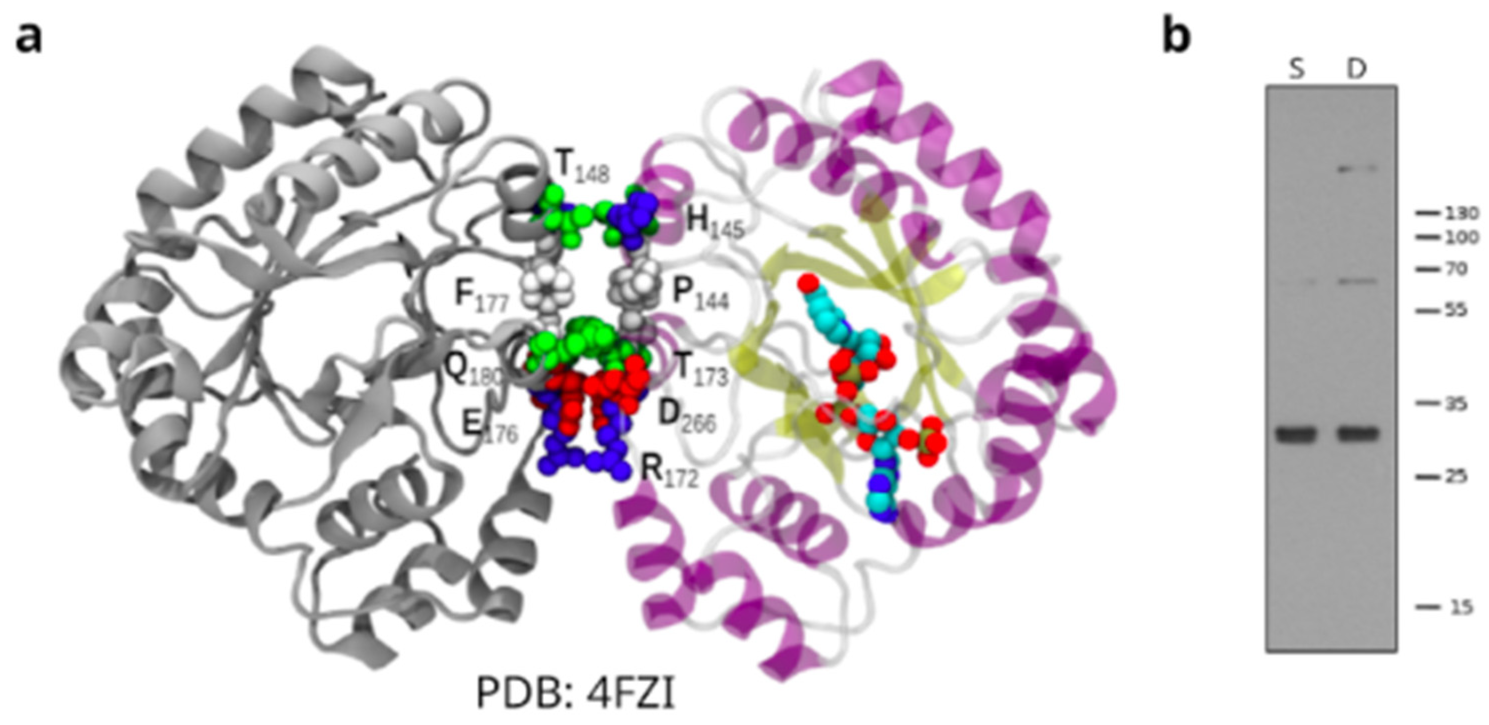
In addition, by modeling on the TcAKR crystal structure we cannot neither confirm nor discard a dimer conformation [16]. Analyzing the crystallographic structure of TcAKR NADP-bound (PDB ID: 4FZI) [50] representing the conformation of the enzyme, we show a contact surface where the monomers could interact (Figure 6a). On the other hand, when we increased the incubation time of the primary antibody (16 h), we observed, in addition to the expected band (32 kDa), bands of higher molecular weight (Figure 6b).

3.5. TcAKR-Overexpressing Parasites Increases Their Susceptibility to Nifurtimox

TcAKR has been studied in relation to the interaction, metabolization, and resistance to Bzn [14,15,16,51,52]. Nevertheless, there is no information regarding TcAKR and Nfx metabolism in the parasites. Here, we evaluated sensitivity to Nfx of TcAKR-overexpressing cells and calculated IC50. Transfected parasites overexpressing TcAKR were more susceptible to Nfx than the control (Figure 7a), showing a statistically significant decrease in all the concentration assays. Notably, Nfx IC50 in transfectant parasites decreases by nearly four times.

4. Discussion

We found that TcAKR is differentially regulated along the T. cruzi life cycle. It is expressed in the replicative parasite forms (epimastigotes and amastigotes) at the protein and mRNA levels but is undetectable in trypomastigotes. This expression pattern is the same as that of TcOYE, which turns off entirely in the trypomastigote stage [11]. TcAKR has a particular subcellular localization in the parasite. Through immunofluorescent and UExM assays, we observed that TcAKR is located in the mitochondria. This does not agree with previous reports that located the enzyme in the cytosol and reported its expression in trypomastigotes by Western blot [13]. The latter observation may be related to amastigotes contamination in the preparation, although strain differences cannot be ruled out. The enzyme exhibits a broad distribution in the mitochondria in amastigotes; nevertheless, in epimastigotes, it is located in proximity to the kinetoplast. TcAKR was observed to be located at both ends of the kinetoplast, close to the antipodal sites, two structure assemblies where ligases, topoisomerases, polymerases, and other proteins are located [53,54] and where the repair of gaps in the newly synthesized minicircles that are reattached to the kDNA network occurs [55,56]. Since the kDNA replication is closely related to the cell cycle, we synchronized epimastigotes and followed TcAKR along the process, finding that its location is linked to the changes in the kinetoplast during S, G1, G2, and mitosis and cytokinesis. It was previously reported that methylglyoxal is the most efficient substrate for TcAKR compared with other toxic ketoaldehydes [16]. The primary endogenous sources of methylglyoxal are glycolysis and the breakdown of threonine and lipid peroxidation [57]. These last reactions take place in mitochondria, in the exact subcellular location that TcAKR, indicating that this enzyme could have a relevant role in detoxification at the mitochondrial level, especially protecting kDNA replication, since methylglyoxal severely inhibits DNA replication [58]. Finally, high-resolution microscopy by UExM reveals that TcAKR is located in the kinetoplast region proximal to basal bodies. These localization changes along the life cycle deserve future studies using high-resolution microscopy methods.
By phylogenetic studies, we found that the AKR family in trypanosomatids are grouped in four main clades and TcAKR clusters with those with PGFS function in Leishmania and T. brucei [19,20,21]. However, in T. cruzi, this activity appears to be missing [13,16]. The absence of TcAKR expression in trypomastigotes constitutes an opportunity to revisit PGFS activity using TcAKR-overexpressing parasites. We observed that the level of PGF2α measured in transfectant parasites was significantly higher than in control parasites. Our experimental model allows us to conclude that TcAKR can catalyze the synthesis of PGF2α. In concordance with previous reports [11], high levels of PGF2α were found in measurements made from the culture supernatant of TcAKR-overexpressing parasites, suggesting that this molecule is released from the parasite once it is synthesized. In this sense, it was also reported that secretion of PGF2α synthesized by T. brucei and L. infantum [19,59]. Ashton and collaborators demonstrated, using TXA2 synthase-null mice, that most of the TXA2 in infected mice is parasite-derived [23]. All these studies point out that parasite-derived eicosanoids exert a paracrine or endocrine effect in Trypanosomatid infections. We determined that the catalytic tetrad, highly conserved throughout the AKR superfamily [60], is present in TcAKR, with a core folding of (α/β) TIM barrel that acts via classic ping-pong kinetics [17,61]. The phylogenetic tree shows that TcAKR groups with PFGS; however, this clade is subdivided into two groups: one comprising proteins whose PGFS activities have been demonstrated and another where TcAKR is found, which includes proteins annotated as PGFS but with some differences in sequences.
There is a discrepancy in the literature regarding the evidence of the oligomerization state of TcAKR [13,16]. Garavaglia et al. (2016) detected mixed species containing monomeric, dimeric, and tetrameric forms of the enzyme from the CL-Brener strain by gel chromatography with the recombinant soluble enzyme [13], while Roberts et al. (2018) found a single peak with a calculated MW close to that of a monomer [16]. When we increased the incubation time in western blots, bands compatible with TcAKR oligomers were detected, although mass spectrometry must confirmed their identity. Also, we found a contact surface where the monomers could interact, but we cannot discard that as a consequence of the crystallization process.
TcAKR has been widely studied in relation to drug metabolism, including Bzn. Its overexpression leads to increased resistance to Bzn [14]. However, recent studies have not found evidence for the reduction of Bzn by recombinant TcAKR and homologs in the related parasites T. brucei and L. donovani. Instead, these enzymes metabolize a variety of toxic ketoaldehydes, including glyoxal and methylglyoxal, suggesting a role in cellular defense against chemical stress [16]. Bzn is a prodrug and undergoes activation via two sequential two-electron reduction steps by a mitochondrial nitroreductase to form a toxic reactive hydroxylamine intermediate. This compound subsequently undergoes rearrangement to a dihydro-dihydroxy intermediate [62] or forms adducts with low molecular mass thiols [51]. This intermediate can then dissociate to form glyoxal and N-benzyl-2-guanidinoacetamide. UPLC-QToF/MS analysis of Bzn bioactivation by T. cruzi cell lysates confirmed previous reports identifying numerous drug metabolites, including a dihydro-dihydroxy intermediate that can separate into N-benzyl-2-guanidinoacetamide and glyoxal. However, metabolomic studies did not detect glyoxal adducts [51]. TcAKR may play a role in detoxifying some of the toxic metabolites generated by the reductive metabolism of Bzn by NTRI in the mitochondria, including glyoxal or others; also considering TcAKR was found in previous reports bound to immobilized Bzn [15]. In this work, we focused our study on Nfx, that, as most nitroheterocyclic agents, must undergo activation by nitroreduction, a process catalyzed by type I nitroreductases. It has been shown that these reductases generate cytotoxic nitrile metabolites responsible for trypanocidal activity [63]. We found that TcAKR-overexpressing parasites are more susceptible to Nfx, which constitutes a difference from TbAKR that does not affect sensibility to Nfx in T. brucei. Metabolomic and proteomic studies on Nfx metabolization are necessary to evaluate the direct or indirect (by favoring the overexpression of other proteins) role of this enzyme in the metabolism of this drug, where TcAKR overexpression parasites constitute a valuable tool that can provide clues on the unknown mode of action of Nfx.
In summary, the substrate-specificity of this aldo-keto reductases is broader than other prostaglandin F2α synthases, and TcAKR is likely to be involved in arachidonic metabolism, detoxification of ketoaldehydes and the activation of the prodrug nifurtimox. Its subcellular localization during the cell cycle is compatible with a significant role in kDNA metabolism by eliminating toxic metabolites that can inhibit replication. Together these results position TcAKR as a relevant target for the action of drugs.

Supplementary Materials

The following supporting information can be downloaded at: https://www.mdpi.com/article/10.3390/pathogens12010085/s1, Supplementary Material S1. Fasta file with the sequences used to create a blast database. Supplementary Material S2. Fasta file with the sequences retrieved from the genomes. The genome positions where the sequences were found are included. Figure S1. Maximum-likelihood phylogeny constructed with complete AKR sequences recovered from protozoa genomes. Table S1. Correlation between the names in the phylogenetic tree and the name of the sequences retrieved from the genomes

Author Contributions

Conceptualization, C.R., A.T. and M.L.C.; Methodology, C.R., F.D.-V., M.L.C., M.R.M., P.F.-T., A.T. and R.T.; Software, F.D.-V.; Validation, F.D.-V. and C.R.; Investigation, F.D.-V., M.L.C., A.T. and C.R.; Data Curation, F.D.-V.; Writing—Original Draft Preparation, F.D.-V. and C.R.; Writing—Review and Editing, C.R., F.D.-V., A.T., M.L.C. and L.P.-V.; Supervision, C.R.; Funding Acquisition, C.R. All authors have read and agreed to the published version of the manuscript.

Funding

This research was funded by Institut Pasteur de Montevideo (Uruguay) and FOCEM—Fondo para la Convergencia Estructural del Mercosur (COF 03/11). F.D.-V. has an ANII (Agencia Nacional de Investigación e Innovación, Uruguay) and CAP (Universidad de la República, Uruguay) doctoral fellowship (No. POS_NAC_2016_1_129916; BFPD_2021_1#45569540). F.D.V., M.L.C., M.R.M., P.F.T., and C.R. are members of the Sistema Nacional de Investigadores (SNI-ANII, Uruguay). C.R., M.L.C., and M.R.M. are PEDECIBA (Programa de Desarrollo de Ciencias Básicas, Uruguay) researchers.

Acknowledgments

We acknowledge Maria E. Francia (Institut Pasteur de Montevideo and Facultad de Medicina, UDELAR, Uruguay) for kindly giving us the expansion microscopy reagents and Gonzalo Ferreira and Mariana Di Doménico (Facultad de Medicina, UDELAR, Uruguay) for assistance with image processing.

Conflicts of Interest

The funders had no role in the design of the study; in the collection, analyses, or interpretation of data; in the writing of the manuscript; or in the decision to publish the results.

References

  1. World Health Organization (WHO). Available online: https://www.who.int/news-room/fact-sheets/detail/chagas-disease-(American-trypanosomiasis) (accessed on 6 October 2022).
  2. Conners, E.E.; Vinetz, J.M.; Weeks, J.R.; Brouwer, K.C. A global systematic review of Chagas disease prevalence among migrants. Acta Trop. 2016, 156, 68–78. [Google Scholar] [CrossRef] [Green Version]
  3. Lee, B.Y.; Bacon, K.M.; Bottazzi, M.E.; Hotez, P.J. Global economic burden of Chagas disease: A computational simulation model. Lancet Infect. Dis. 2013, 13, 342–348. [Google Scholar] [CrossRef] [Green Version]
  4. Nunes, M.C.P.; Dones, W.; Morillo, C.A.; Encina, J.J.; Ribeiro, A.L. Chagas disease: An overview of clinical and epidemiological aspects. J. Am. Coll. Cardiol. 2013, 62, 767–776. [Google Scholar] [CrossRef] [PubMed]
  5. Aldasoro, E.; Posada, E.; Requena-Méndez, A.; Calvo-Cano, A.; Serret, N.; Casellas, A.; Sanz, S.; Soy, D.; Pinazo, M.J.; Gascon, J. What to expect and when: Benznidazole toxicity in chronic Chagas’ disease treatment. J. Antimicrob. Chemother. 2018, 73, 1060–1067. [Google Scholar] [CrossRef] [Green Version]
  6. Jackson, Y.; Alirol, E.; Getaz, L.; Wolff, H.; Combescure, C.; Chappuis, F. Tolerance and safety of nifurtimox in patients with chronic Chagas disease. Clin. Infect. Dis. 2010, 51, e69–e75. [Google Scholar] [CrossRef] [Green Version]
  7. Wilkinson, S.R.; Bot, C.; Kelly, J.M.; Hall, B.S. Trypanocidal activity of nitroaromatic prodrugs: Current treatments and future perspectives. Curr. Top. Med. Chem. 2011, 11, 2072–2084. [Google Scholar] [CrossRef] [PubMed]
  8. Wilkinson, S.R.; Taylor, M.C.; Horn, D.; Kelly, J.M.; Cheeseman, I. A mechanism for cross-resistance to nifurtimox and benznidazole in trypanosomes. Proc. Natl. Acad. Sci. USA 2008, 105, 5022–5027. [Google Scholar] [CrossRef] [Green Version]
  9. Kubata, B.K.; Kabututu, Z.; Nozaki, T.; Munday, C.J.; Fukuzumi, S.; Ohkubo, K.; Lazarus, M.; Maruyama, T.; Martin, S.K.; Duszenko, M.; et al. A key role for old yellow enzyme in the metabolism of drugs by Trypanosoma cruzi. J. Exp. Med. 2022, 196, 1241–1252. [Google Scholar] [CrossRef] [Green Version]
  10. Murta, S.M.; Krieger, M.A.; Montenegro, L.R.; Campos, F.F.; Probst, C.M.; Avila, A.R.; Muto, N.H.; de Oliveira, R.C.; Nunes, L.R.; Nirdé, P.; et al. Deletion of copies of the gene encoding old yellow enzyme (TcOYE), a NAD(P)H flavin oxidoreductase, associates with in vitro-induced benznidazole resistance in Trypanosoma cruzi. Mol. Biochem. Parasitol. 2006, 146, 151–162. [Google Scholar] [CrossRef]
  11. Díaz-Viraqué, F.; Chiribao, M.L.; Trochine, A.; Gonzalez, F.; Castillo, C.; Liempi, A.; Kemmerling, U.; Maya, J.M.; Robello, C. Old Yellow Enzyme from Trypanosoma cruzi exhibits in vivo prostaglandin F2α synthase activity and has a key role in parasite infection and drug susceptibility. Front. Immunol. 2018, 9, 456. [Google Scholar] [CrossRef]
  12. Santi, A.M.M.; Ribeiro, J.M.; Reis-Cunha, J.L.; de Assis Burle-Caldas, G.; Santos, I.F.M.; Silva, P.A.; Resende, D.M.; Bartholomeu, D.C.; Teixeira, S.M.R. Disruption of multiple copies of the Prostaglandin F2alpha synthase gene affects oxidative stress response and infectivity in Trypanosoma cruzi. PLoS Negl. Trop. Dis. 2022, 16, e0010845. [Google Scholar] [CrossRef] [PubMed]
  13. Garavaglia, P.A.; Cannata, J.J.; Ruiz, A.M.; Maugeri, D.; Duran, R.; Galleano, M.; García, G.A. Identification, cloning and characterization of an aldo-keto reductase from Trypanosoma cruzi with quinone oxido-reductase activity. Mol. Biochem. Parasitol. 2010, 173, 132–141. [Google Scholar] [CrossRef] [PubMed]
  14. Garavaglia, P.A.; Laverrière, M.; Cannata, J.J.; García, G.A. Putative role of the aldo-keto reductase from Trypanosoma cruzi in benznidazole metabolism. Antimicrob. Agents Chemother. 2016, 60, 2664–2670. [Google Scholar] [CrossRef] [Green Version]
  15. Trochine, A.; Alvarez, G.; Corre, S.; Faral-Tello, P.; Durán, R.; Batthyany, C.; Cerecetto, H.; González, M.; Robello, C. Trypanosoma cruzi chemical proteomics using immobilized benznidazole. Exp. Parasitol. 2014, 140, 33–38. [Google Scholar] [CrossRef]
  16. Roberts, A.J.; Dunne, J.; Scullion, P.; Norval, S.; Fairlamb, A.H. A role for Trypanosomatid aldo-keto reductases in methylglyoxal, prostaglandin and isoprostane metabolism. Biochem. J. 2018, 475, 2593–2610. [Google Scholar] [CrossRef] [Green Version]
  17. Jez, J.M.; Bennett, M.J.; Schlegel, B.P.; Lewis, M.; Penning, T.M. Comparative anatomy of the aldo-keto reductase superfamily. Biochem. J. 1997, 326 Pt 3, 625. [Google Scholar] [CrossRef] [PubMed]
  18. Penning, T.M. The aldo-keto reductases (AKRs): Overview. Chem.-Biol. Interact. 2015, 234, 236–246. [Google Scholar] [CrossRef] [Green Version]
  19. Kubata, B.K.; Duszenko, M.; Kabututu, Z.; Rawer, M.; Szallies, A.; Fujimori, K.; Inui, T.; Nozaki, T.; Yamashita, K.; Horii, T.; et al. Identification of a novel prostaglandin F2α synthase in Trypanosoma brucei. J. Exp. Med. 2000, 192, 1327–1338. [Google Scholar] [CrossRef] [Green Version]
  20. Kabututu, Z.; Martin, S.K.; Nozaki, T.; Kawazu, S.I.; Okada, T.; Munday, C.J.; Duszenko, M.; Lazarus, M.; Thuita, L.W.; Urade, Y.; et al. Prostaglandin production from arachidonic acid and evidence for a 9, 11-endoperoxide prostaglandin H2 reductase in Leishmania. Int. J. Parasitol. 2002, 32, 1693–1700. [Google Scholar] [CrossRef]
  21. Alves-Ferreira, E.V.C.; Ferreira, T.R.; Walrad, P.; Kaye, P.M.; Cruz, A.K. Leishmania braziliensis prostaglandin F synthase impacts host infection. Parasit Vectors 2020, 13, 9. [Google Scholar] [CrossRef]
  22. Kabututu, Z.; Manin, M.; Pointud, J.C.; Maruyama, T.; Nagata, N.; Lambert, S.; Lefrançois-Martinez, A.M.; Martinez, A.; Urade, Y. Prostaglandin f2α synthase activities of aldo–keto reductase 1b1, 1b3 and 1b7. J. Biochem. 2008, 145, 161–168. [Google Scholar] [CrossRef] [PubMed]
  23. Ashton, A.W.; Mukherjee, S.; Nagajyothi, F.N.U.; Huang, H.; Braunstein, V.L.; Desruisseaux, M.S.; Factor, S.M.; Lopez, L.; Berman, J.W.; Wittner, M.; et al. Thromboxane A2 is a key regulator of pathogenesis during Trypanosoma cruzi infection. J. Exp. Med. 2007, 204, 929–940. [Google Scholar] [CrossRef] [PubMed]
  24. Vazquez, M.; Levin, M.J. Functional analysis of the intergenic regions of Tc 2β gene loci allowed the construction of an improved Trypanosoma cruzi expression vector. Gene 1999, 239, 217–225. [Google Scholar] [CrossRef]
  25. Bradford, M.M. A rapid and sensitive method for the quantitation of microgram quantities of protein utilizing the principle of protein-dye binding. Anal. Biochem. 1976, 72, 248–254. [Google Scholar] [CrossRef]
  26. Simizu, B.; Rhim, J.S.; Wiebenga, N.H. Characterization of the Tacaribe Group of Arboviruses. 1. Propagation and Plaque Assay of Tacaribe Virus in a Line of African Green Monkey Kidney Cells (Vero). Proc. Soc. Exp. Biol. Med. 1967, 125, 119–123. [Google Scholar] [CrossRef] [PubMed]
  27. Contreras, V.T.; Araújo-Jorge , T.C.D.; Bonaldo, M.C.; Thomaz, N.; Barbosa, H.S.; Roberts, E.; Meirelles, M.d.S.; Goldenberg, S. Biological aspects of the DM28C clone of Trypanosoma cruzi after metacyclogenesis in chemically defined media. Mem. Do Inst. Oswaldo Cruz 1988, 83, 123–133. [Google Scholar] [CrossRef] [Green Version]
  28. Roberts, E.; Eargle, J.; Wright, D.; Luthey-Schulten, Z. MultiSeq: Unifying sequence and structure data for evolutionary analysis. BMC Bioinform. 2006, 7, 382. [Google Scholar] [CrossRef] [Green Version]
  29. Humphrey, W.; Dalke, A.; Schulten, K. VMD: Visual molecular dynamics. J. Mol. Graph. 1996, 14, 33–38. [Google Scholar] [CrossRef]
  30. Russell, R.B.; Barton, G.J. Multiple protein sequence alignment from tertiary structure comparison: Assignment of global and residue confidence levels. Proteins Struct. Funct. Bioinform. 1992, 14, 309–323. [Google Scholar] [CrossRef]
  31. Baker, N.A.; Sept, D.; Joseph, S.; Holst, M.J.; McCammon, J.A. Electrostatics of nanosystems: Application to microtubules and the ribosome. Proc. Natl. Acad. Sci. USA 2001, 98, 10037–10041. [Google Scholar] [CrossRef]
  32. Dolinsky, T.J.; Czodrowski, P.; Li, H.; Nielsen, J.E.; Jensen, J.H.; Klebe, G.; Baker, N.A. PDB2PQR: Expanding and upgrading automated preparation of biomolecular structures for molecular simulations. Nucleic Acids Res. 2007, 35 (Suppl. S2), W522–W525. [Google Scholar] [CrossRef] [Green Version]
  33. Li, H.; Robertson, A.D.; Jensen, J.H. Very fast empirical prediction and rationalization of protein pKa values. Proteins Struct. Funct. Bioinform. 2005, 61, 704–721. [Google Scholar] [CrossRef] [PubMed]
  34. Krissinel, E.ñ.; Henrick, K. Inference of macromolecular assemblies from crystalline state. J. Mol. Biol. 2007, 372, 774–797. [Google Scholar] [CrossRef] [PubMed]
  35. Berná, L.; Chiribao, M.L.; Greif, G.; Rodriguez, M.; Alvarez-Valin, F.; Robello, C. Transcriptomic analysis reveals metabolic switches and surface remodeling as key processes for stage transition in Trypanosoma cruzi. PeerJ 2017, 8, e3017. [Google Scholar] [CrossRef] [Green Version]
  36. Patro, R.; Duggal, G.; Love, M.I.; Irizarry, R.A.; Kingsford, C. Salmon provides fast and bias-aware quantification of transcript expression. Nat. Methods 2017, 14, 417–419. [Google Scholar] [CrossRef] [PubMed] [Green Version]
  37. Galanti, N.; Dvorak, J.A.; Grenet, J.; McDaniel, J.P. Hydroxyurea-induced synchrony of DNA replication in the Kinetoplastida. Exp. Cell Res. 1994, 214, 225–230. [Google Scholar] [CrossRef] [PubMed]
  38. Elias, M.C.Q.; Faria, M.; Mortara, R.A.; Motta, M.C.M.; de Souza, W.; Thiry, M.; Schenkman, S. Chromosome localization changes in the Trypanosoma cruzi nucleus. Eukaryot. Cell 2002, 1, 944–953. [Google Scholar] [CrossRef] [Green Version]
  39. Elias, M.C.Q.; da Cunha, J.P.; de Faria, F.P.; Mortara, R.A.; Freymüller, E.; Schenkman, S. Morphological events during the Trypanosoma cruzi cell cycle. Protist 2007, 158, 147–157. [Google Scholar] [CrossRef]
  40. Alonso, V.L. Ultrastructure Expansion Microscopy (U-ExM) in Trypanosoma cruzi: Localization of tubulin isoforms and isotypes. Parasitol. Res. 2022, 121, 3019–3024. [Google Scholar] [CrossRef]
  41. Tomasina, R.; Gonzalez, F.C.; Martins-Duarte, É.S.; Bastin, P.; Gissot, M.; Francia, M.E. Separate to operate: The centriole-free inner core of the centrosome regulates the assembly of the intranuclear spindle in Toxoplasma gondii. Mbio 2022, 13, e0185922. [Google Scholar] [CrossRef]
  42. Rolón, M.; Vega, C.; Escario, J.A.; Gómez-Barrio, A. Development of resazurin microtiter assay for drug sensibility testing of Trypanosoma cruzi epimastigotes. Parasitol. Res. 2006, 99, 103–107. [Google Scholar] [CrossRef] [PubMed]
  43. Sayers, E.W.; Bolton, E.E.; Brister, J.R.; Canese, K.; Chan, J.; Comeau, D.C.; Connor, R.; Funk, K.; Kelly, C.; Kim, S.; et al. Database resources of the national center for biotechnology information. Nucleic Acids Res. 2022, 50, D20–D26, PMCID:PMC8728269. [Google Scholar] [CrossRef] [PubMed]
  44. Camacho, C.; Coulouris, G.; Avagyan, V.; Ma, N.; Papadopoulos, J.; Bealer, K.; Madden, T.L. BLAST+: Architecture and applications. BMC Bioinform. 2009, 10, 421. [Google Scholar] [CrossRef] [PubMed] [Green Version]
  45. Notredame, C.; Higgins, D.G.; Heringa, J. T-Coffee: A novel method for fast and accurate multiple sequence alignment. J. Mol. Biol. 2000, 302, 205–217. [Google Scholar] [CrossRef] [Green Version]
  46. Darriba, D.; Taboada, G.L.; Doallo, R.; Posada, D. ProtTest 3: Fast selection of best-fit models of protein evolution. Bioinformatics 2011, 27, 1164–1165. [Google Scholar] [CrossRef] [Green Version]
  47. Guindon, S.; Gascuel, O. A simple, fast, and accurate algorithm to estimate large phylogenies by maximum likelihood. Syst. Biol. 2003, 52, 696–704. [Google Scholar] [CrossRef] [Green Version]
  48. Berná, L.; Rodriguez, M.; Chiribao, M.L.; Parodi-Talice, A.; Pita, S.; Rijo, G.; Alvarez-Valin, F.; Robello, C. Expanding an expanded genome: Long-read sequencing of Trypanosoma cruzi. Microb. Genom. 2018, 4, e000177. [Google Scholar] [CrossRef]
  49. Piñeyro, M.D.; Parodi-Talice, A.; Arcari, T.; Robello, C. Peroxiredoxins from Trypanosoma cruzi: Virulence factors and drug targets for treatment of Chagas disease? Gene 2008, 408, 45–50. [Google Scholar] [CrossRef]
  50. Moen, S.O.; Fairman, J.W.; Barnes, S.R.; Sullivan, A.; Nakazawa-Hewitt, S.; Van Voorhis, W.C.; Staker, B.L.; Lorimer, D.D.; Myler, P.J.; Edwards, T.E. Structures of prostaglandin F synthase from the protozoa Leishmania major and Trypanosoma cruzi with NADP. Acta Cryst. F Struct. Biol. Commun. 2015, 71 Pt 5, 609–614. [Google Scholar] [CrossRef] [Green Version]
  51. Trochine, A.; Creek, D.J.; Faral-Tello, P.; Barrett, M.P.; Robello, C. Benznidazole biotransformation and multiple targets in Trypanosoma cruzi revealed by metabolomics. PLoS Negl. Trop. Dis. 2014b, 8, e2844. [Google Scholar] [CrossRef]
  52. González, L.; García-Huertas, P.; Triana-Chávez, O.; García, G.A.; Murta, S.M.F.; Mejía-Jaramillo, A.M. Aldo-keto reductase and alcohol dehydrogenase contribute to benznidazole natural resistance in Trypanosoma cruzi. Mol. Microbiol. 2017, 106, 704–718. [Google Scholar] [CrossRef] [PubMed] [Green Version]
  53. Jensen, R.E.; Englund, P.T. Network news: The replication of kinetoplast DNA. Annu. Rev. Microbiol. 2012, 66, 473–491. [Google Scholar] [CrossRef] [PubMed]
  54. Maldonado, E.; Morales-Pison, S.; Urbina, F.; Solari, A. Molecular and Functional Characteristics of DNA Polymerase Beta-Like Enzymes from Trypanosomatids. Front. Cell Infect. Microbiol. 2021, 11, 670564. [Google Scholar] [CrossRef]
  55. Klingbeil, M.M.; Englund, P.T. Closing the gaps in kinetoplast DNA network replication. Proc. Natl. Acad. Sci. USA 2004, 101, 4333–4334. [Google Scholar] [CrossRef] [PubMed] [Green Version]
  56. Liu, B.; Liu, Y.; Motyka, S.A.; Agbo, E.E.C.; Englund, P.T. Fellowship of the rings: The replication of kinetoplast DNA. Trends Paras. 2005, 21, 363–369. [Google Scholar] [CrossRef] [PubMed]
  57. Thornalley, P.J. Protein and nucleotide damage by glyoxal and methylglyoxal in physiological systems--role in ageing and disease. Drug Metab. Drug Interact 2008, 23, 125–150. [Google Scholar] [CrossRef] [PubMed]
  58. Murata-Kamiya, N.; Kamiya, H. Methylglyoxal, an endogenous aldehyde, crosslinks DNA polymerase and the substrate DNA. Nucleic Acids Res. 2001, 29, 3433–3438. [Google Scholar] [CrossRef] [Green Version]
  59. Araújo-Santos, T.; Rodríguez, N.E.; Moura-Pontes, S.; Dixt, U.G.; Abánades, D.R.; Bozza, P.T.; Wilson, M.E.; Borges, V.M. Role of prostaglandin F2α production in lipid bodies from Leishmania infantum chagasi: Insights on virulence. J. Infect. Dis. 2014, 210, 1951–1961. [Google Scholar] [CrossRef] [Green Version]
  60. Schlegel, B.P.; Jez, J.M.; Penning, T.M. Mutagenesis of 3α-Hydroxysteroid Dehydrogenase Reveals a “Push− Pull” Mechanism for Proton Transfer in Aldo-Keto Reductases. Biochemistry 1998, 37, 3538–3548. [Google Scholar] [CrossRef]
  61. Toogood, H.S.; Gardiner, J.M.; Scrutton, N.S. Biocatalytic reductions and chemical versatility of the old yellow enzyme family of flavoprotein oxidoreductases. ChemCatChem 2010, 2, 892–914. [Google Scholar] [CrossRef]
  62. Hall, B.S.; Wilkinson, S.R. Activation of benznidazole by Trypanosomal type I nitroreductases results in glyoxal formation. Antimicrob. Agents Chemother. 2012, 56, 115–123. [Google Scholar] [CrossRef] [PubMed] [Green Version]
  63. Hall, B.S.; Bot, C.; Wilkinson, S.R. Nifurtimox activation by Trypanosomal type I nitroreductases generates cytotoxic nitrile metabolites. J. Biol. Chem. 2011, 286, 13088–13095. [Google Scholar] [CrossRef] [PubMed]
Figure 1. Evolutionary relationship of Aldo-Keto Reductase proteins in Trypanosomatids. Evolutionary relationships of AKR sequences retrieved from protozoan genomes. The sequence alignment was performed using the accurate mode of T-Coffee software. The tree was built with the maximum-Likelihood method. The sequences are provided in Supplementary Material S2.
Pathogens 12 00085 g001
Figure 2. TcAKR subcellular localization. (a) Genomic localization of tcakr genes in PRFA01000207 scaffold. (b) Immunolocalization of TcAKR and TcmTXNPx in epimastigotes and amastigotes using rabbit α-TcAKR (1/30) (green) and α-TcmTXNPx (1/100) (red) polyclonal antibodies. DAPI was used as a nucleus and kinetoplast marker. Scale bar: 5 µm. (c) RNA-seq analysis to quantify the mRNA levels of the TcAKR during the life stages: A = amastigote, E = epimastigote, and T = trypomastigote. (d) Epimastigotes were subjected to ultrastructure expansion microscopy (UExM) and stained with rabbit α-TcAKR (1/30) (red), N-Hydroxysuccinimide (NHS) (gray), and DAPI. Note that NHS labels the pan-proteome, labeling different parasite structures as the nucleus, flagella, and kDNA, among others. (A) Z Maximum intensity projection showing TcAKR (red), NHS (gray), and kDNA and nucleus with DAPI (blue). (B) Z (23–26) Maximum intensity projection showing TcAKR (Red), NHS (gray) and kDNA and nucleus with DAPI (blue). (C) Z (23–26) Maximum intensity projection showing TcAKR (red) and kDNA and nucleus with DAPI (blue). Scale bar: 1 µm. BB: basal bodies.
Pathogens 12 00085 g002
Figure 3. TcAKR localization during the epimastigotes cell cycle. Localization of TcAKR by immunofluorescence in synchronized epimastigotes stained with α-TcAKR (1/30) (green) and α-TcmTXNPx (1/100) (red) rabbit polyclonal antibodies. Cell cycle stages (G1/S, G2, M, and C) are defined by the position of the DAPI-stained kinetoplast (K), nucleus (N) in blue, and the number of flagella (F) analyzed in DIC.
Pathogens 12 00085 g003
Figure 4. TcAKR overexpression and PGF2α detection. (A) Western blot analysis of total protein extracts of trypomastigotes transfected with pTREX-n (empty vector) and pTREX-n TcAKR (includes complete TcAKR coding sequence) using rabbit α-TcAKR polyclonal antiserum. The relative expression was estimated by image densitometry analysis normalized by tubulin expression. TcOYE expression was evaluated in TcAKR-overexpressing parasites using rabbit α-TcOYE polyclonal antiserum as a control. (B) Determination of PGF2α in cells and supernatant of TcAKR-overexpressing epimastigotes. (C) Determination of PGF2α in cells and supernatant of TcAKR-overexpressing cell-derived trypomastigotes. These results correspond to two biological replicates. Asterisks represent statistical significance (two-tailed Student’s t-Test) p-values: pellet p = 0.0001, supernatant p = 0.0623.
Pathogens 12 00085 g004
Figure 5. Structural comparatives analyses of TcAKR protein with AKR structures from T. brucei, L. major, B. subtilis, R. norvegicus, and H. sapiens. (A) Phylogenetic tree of AKRs based on the QH index. (B) Electrostatic potential mapped on solvent-accessible surfaces of AKR proteins. Values range from −130 (red) to 130 mV (blue). (C) Structural alignment and sequence conservation of AKR proteins. The first and second shells of residues around the active site of TcAKR. Residues that differ from other species are indicated. The structures used were: T. cruzi TcAKR PDB = 4GIE; T. brucei AKR5A2 PDB = 1VBJ; L. major AKR5A PDB = 4G5D; B. subtilis AKR5G1 PDB = 3D3F; R. norvegicus AKR1B14 PDB = 3QKZ; H. sapiens AKR1C3 PDB = 2F38.
Pathogens 12 00085 g005
Figure 6. TcAKR studies on oligomerization. (a) Interaction surface of the proposed monomers using TcAKR crystals. PDB 4FZI: interface A/B x, y, z. Area: 377.7 A^2. DG: −5.5 Kcal/mol. DG p-value = 0.21. (b) Western blot analysis of total protein extracts from Sylvio and Dm28c epimastigotes using rabbit α-TcAKR polyclonal antiserum overnight. Protein bands corresponding to molecular weights compatible with oligomerization states are observed. S: Sylvio, D: Dm28c.
Pathogens 12 00085 g006
Figure 7. Susceptibility to Nfx. (a) Viability percentages of transfected epimastigotes challenged with different concentrations of Nfx. The percentages of cell viability are normalized against parasites without treatment. Values are the means of three independent assays performed in quadruplicate. Nfx p-values: 20 μM p = 0.0868; 50 μM p = 0.0062; 100 μM p = 0.0054. (b) Percentage of epimastigotes survival. IC50 for NFX was determined in pTREX and pTREXn-TcAKR parasites. Values are the means of three independent assays performed in quadruplicate. (p = 0.0227; asterisks represent statistical significance; two-tailed Student’s t-test). (a) and (b) are two independent experiments (different datasets).
Pathogens 12 00085 g007
Disclaimer/Publisher’s Note: The statements, opinions and data contained in all publications are solely those of the individual author(s) and contributor(s) and not of MDPI and/or the editor(s). MDPI and/or the editor(s) disclaim responsibility for any injury to people or property resulting from any ideas, methods, instructions or products referred to in the content.