Article

Relationship between Biofilm Production and High Somatic Cell Count in Streptococcus agalactiae Isolated from Milk of Cows with Subclinical Mastitis

by
Erika Carolina Romão Bonsaglia
1,*,
Rodolfo S. Rossi
2,
Giulia Latosinski
2,
Bruna Fernanda Rossi
1,
Fernanda Cristina Campos
1,
Ary Fernandes Junior
1,
José Carlos F. Pantoja
2 and
Vera Lucia Mores Rall
1,*
1
Department of Chemical and Biological Sciences, Institute of Biosciences, Sao Paulo State University (UNESP), Botucatu 18618-970, SP, Brazil
2
Department of Veterinary Hygiene and Public Health, Sao Paulo State University (UNESP), Botucatu 18618-970, SP, Brazil
*
Authors to whom correspondence should be addressed.
Pathogens 2023, 12(2), 311; https://doi.org/10.3390/pathogens12020311
Submission received: 27 October 2022 / Revised: 30 January 2023 / Accepted: 8 February 2023 / Published: 14 February 2023
(This article belongs to the Special Issue Pathogens in Ruminant Mastitis)

Abstract

:
Streptococcus agalactiae (S. agalactiae) is one of the main agents that causes mastitis in dairy cows, mainly inducing the subclinical form, which is characterized by a high somatic cell count (SCC). The aim of this study was to correlate the increase in SCC caused by S. agalactiae in cows with subclinical mastitis to the presence of genes related to adhesion and invasion in bovine mammary epithelial cells (BMEC) and biofilm formation. Considering the 145 isolates tested, 57.2% presented the capsular type Ia and 42.8% presented type III. We identified the virulence genes among the isolates and determined nine genetic profiles. The most common profile was identified in 69 isolates (47.5%): Ia, fbsA+, fbsB-, pI1-, pI2a-, pI2b+, and hylb+. All isolates produced biofilm, with 58.6% classified as strong producers, 29% as moderate producers and 12.4% as weak producers. No statistical correlation was found between the presence of virulence genes and increased SCC or biofilm production. However, biological evidence was observed between increased SCC and biofilm production. One isolate from each profile was randomly subjected to adhesion and invasion assays, and all of them adhered to BEMC, but none were able to invade. Our results showed that different genetic profiles do not provide advantages for bacteria to invade BMEC in vitro. In addition, biofilm production appears to be related to high SCC.

1. Introduction

Streptococcus agalactiae has been considered one of the most common etiological agents of intramammary infection in dairy cows, causing both clinical and subclinical mastitis [1]. In the last decade, S. agalactiae has been isolated from herds in several countries around the world, including Italy, Colombia, China, Poland, and Denmark [2,3,4,5,6], and has been detected in 60% of herds in Brazil [7]. Over the years, S. agalactiae has been recognized as an obligate parasite of the mammary gland [8], but recent studies have demonstrated the ability of this pathogen to survive in extramammary environments, such as the bovine gastrointestinal tract and the milking environment (cow beds, floors, and water channels), suggesting a transmission cycle that includes infected mammary glands and contaminated environmental sources, such as the milking machine, bedding, and water [3,9]. This micro-organism primarily infects the cistern and lactiferous ducts of the mammary gland, where its associated metabolic products elicit an intense inflammatory response; this response results in tissue damage, reduced milk yield, and a geometric mean in the somatic cell count (SCC) of 857,000 cells/ml in infected cow’s milk [10]. It is the costliest disease of dairy cows, and the most significant economic losses occur from reduced milk production and costs associated with therapy [7]. Furthermore, S. agalactiae is a public health concern; studies have shown an association between raw milk consumption and an increased risk of S. agalactiae infection in humans. The authors demonstrated, by means of molecular characterization, that human samples associated with neonatal disease were of bovine origin [11,12]. The study of Carra et al. [2] also supported the hypothesis of S. agalactiae transmission between cattle and humans, and Botelho et al. [13] suggested that it can occur during milking, consumption of raw, contaminated milk, or by means of environmental contamination.
Capsular polysaccharide (CPS) search is the main method used to characterize the species and is an essential virulence factor of S. agalactiae that impairs the host’s recognition of the pathogen and is composed of sialic acid (commonly found in vertebrate glycans) [14]. CPSs are classified into 10 serotypes (Ia, Ib, and II-IX), with the Ia, Ib, III, IV, and V types being the most common in mastitis isolates. The pathogenicity of S. agalactiae varies with its serotypes, although its distribution seems to be related to the geographical region [7,14,15]. Even in the same country, a diverse prevalence rate can be found among regions, as shown by Carvalho-Castro et al. [7], who compared strains from the North, South, and Southeast regions of Brazil, demonstrating a difference between the capsular types of bovine isolates. This observation may indicate the occurrence of capsular switching in S. agalactiae, driven by the host immune response, an event that has been well documented in other streptococcal species [16]. Furthermore, studies have shown that CPS plays an important role in Group B streptococcus (GBS) adherence and invasion of cells, as well as contributing to biofilm formation [17].
Biofilm represents a bacterial mode of protection, and its presence has effects on many branches of the dairy environment. Damage is related to the contamination of raw milk pipelines, cooling milk tanks, and milking machines that affect udder health, and therefore, reduce milk quality [18]. Recent advances investigating the potential of Staphylococcus aureus to form biofilms in the mammary gland and induce distinct immune responses in mouse mastitis models may help to elucidate the complex relationship between biofilm and the host immune system [19,20]. These studies encourage further research on other pathogens that cause mastitis in cattle because it is already known that biofilms can activate the immune system in a unique way depending on the bacteria involved and the immune status of the host [21].
Biofilm formation by S. agalactiae has been studied in vitro and increasingly appears to be controlled by environmental conditions [22]. Studies have shown that host mammary cells´ viability is suddenly reduced after 2 h of infection with this pathogen and that bacterial adhesion is achieved after 6 h of infection [14]. Adhesion to host tissues is the initial event in bacterial pathogenesis, and S. agalactiae uses multiple adhesins to attach to the epithelium. The recognition of cell surface molecules and extracellular matrix components through specific domains and structures, such as pili, are considered precursors in the infection process [23]. In addition, the pili of S. agalactiae have been shown to promote the invasion of human endothelial cells and influence biofilm formation [22]. These structures are cell-wall appendages extending from the bacterial surface that were discovered during multigenome screening for surface proteins exposed to antigens as possible vaccine targets and are encoded by two loci designated as pilus islands 1 and 2 (PI-1, PI-2), the latter presenting two variants, PI-2a and PI-2b [24,25]. According to previous reports, the pilus may play a role in the host specificity of S. agalactiae, as PI-2a is highly correlated with human-derived isolates and PI-2b with animal-derived isolates [26,27].
Other virulence factors, such as the capacity to interact with extracellular matrix fibrinogen, are mediated by two proteins known as FbsA and FbsB, which are responsible for adhesion and invasion of host cells, and the hylB protein (hyaluronate lyase). This protein is responsible for cleaving hyaluronan, which facilitates the propagation of S. agalactiae during infection [28]. Based on current knowledge, the role of S. agalactiae biofilm in bovine mastitis is still unclear, and more in vitro and in vivo investigations are needed to uncover the relationship between S. agalactiae biofilm and the host immune system.
We, therefore, hypothesized that there is a relationship between the high SCC caused by S. agalactiae in cows with subclinical mastitis and biofilm formation and the presence of virulence genes related to adherence and invasion of bovine mammary epithelial cells (BMEC). Thus, the aim of this study was to correlate the increase in SCC caused by S. agalactiae in cows with subclinical mastitis to the presence of genes related to adhesion and invasion in bovine mammary epithelial cells (BMEC) and biofilm formation.

2. Material and Methods

2.1. Bacterial Isolates and Molecular Typing

This study was approved by the Sao Paulo State University (UNESP) Ethics Committee for Animal Use (protocol 07/2015). The one hundred and forty-five isolates used in this study were isolated from milk samples of cows diagnosed with subclinical S. agalactiae mastitis between February and October 2016; the cows were from nine farms located in the states of Sao Paulo and Minas Gerais, Brazil [29]. Milk samples were collected from all mammary quarters of all lactating cows as part of a herd screening plan. After the milking technicians performed the pre-milking hygiene procedures (visual examination of the first milk streams to detect clinical mastitis, pre-dipping with iodine or chlorine solutions, and drying of teats with a paper towel), the test end was disinfected with a cotton ball soaked in alcohol 70%. Subsequently, three milk streams were discarded, and 15 mL of milk were collected by manual milking into a sterile plastic vial. Samples were frozen after collection and transported to the laboratory.
Isolates were identified as S. agalactiae if they were gram-positive cocci, esculin- and bile-esculin negative, catalase-negative, and positive for the Christie, Atkins, Munch-Petersen (CAMP) test. All the isolates were further submitted to PCR to confirm the identification.
The DNA from bacterial cells was extracted using the MiniSpin Kit (GE Healthcare, Little Chafont, Buckinghamshire), according to the manufacturer’s instructions. GoTaq 2× Green Master Mix (Promega, Madison, WI, USA) was also used in the polymerase chain reaction (PCR) as recommended, and the assays were performed in a Gene Amp PCR System9700 (Applied Biosystems, Carlsbad, CA, USA). Molecular confirmation was performed using species-specific primers for S. agalactiae (A2 gene), according to Almeida et al. [30]. Following identification, PCR was performed to determine the capsular types for the Ia, Ib, and II-IX genes, as described by Poyart et al. [15]. The presence of pI1, pI2a, and pI2b (involved in host cell invasion and biofilm formation) [16,31], and fbsA, fbsB, and hylB (involved in host cell adhesion) was also investigated [32]. Sanger sequencing was performed on one strain that was positive for each gene and used as a positive control.
Somaticell (Idexx Laboratories Inc., Westbrook, ME, USA) was used to estimate SCC, and was performed using refrigerated milk upon arrival at the laboratory, following the manufacturer’s instructions. Two milliliters of reagent and milk were mixed in a plastic tube and homogenized for 30 s. The tube was then held upside down for 30 s to drain the solution, and the final reading (cells/mL) was performed using the tube scale. The range of SCC for S. agalactiae used in this study was previously described by Rossi et al. [29].

2.2. Biofilm Production

Biofilm production was performed according to Bonsaglia et al. [33]. An overnight growth of S. agalactiae, in tryptone soy broth (TSB, Oxoid, Basingstoke, United Kingdom) was adjusted to the 0.5 McFarland standard, and aliquots of 200 µL were distributed, in quadruplicate, into 96-well polystyrene microplates (TPP, Trasadingen, Switzerland) and incubated at 37 °C for 18 h in a 5% CO2 atmosphere.
After washing three times in sterile phosphate buffered saline (PBS, pH 7.2), the wells were stained with 0.1% crystal violet for 30 minutes. Subsequently, they were rinsed with PBS and left to dry at room temperature. Following, 200 μL of 33% acetic acid was added to each well for biofilm detachment and homogenization. An optical density (OD) of 595 nm was used to measure the wells using a microplate reader (Epoch 2 Microplate Reader, Biotek, Winooski, VT, USA). Uninoculated wells containing TSB were used as blanks to correct absorbance values. Biofilm quantification was performed according to Stépanovic et al. [34], classifying the isolates as: nonbiofilm producers (NP), strong biofilm producers (SBP), moderate biofilm producers (MBP), and weak biofilm producers (WBP). This was based upon the previously calculated OD values: (for this type of calculation, the average OD value of the strain should not be reduced by the optical density control value, ODc) OD ≤ ODc = no biofilm producer; ODc < OD ≤ 2× ODc = weak biofilm producer; 2× ODc < OD ≤ 4× ODc = moderate biofilm producer; 4× ODc < OD = strong biofilm producer.

2.3. Adhesion and Invasion Assays for BMEC Cells

Assays were performed according to Sharma et al. [35], with modifications. BMEC cells were cultured in 24-well microplates, and BMEC monolayers were infected, in duplicate, with S. agalactiae cultures diluted in Dulbecco modified eagle’s medium (DMEM) (1.5 × 10⁶ CFU/mL); and 1 ml of these suspensions were inoculated into each well and incubated at 37 °C in a 5% CO2 atmosphere. Following 3 h of incubation, infected monolayers were washed three times with PBS to remove non-adherent bacterial cells, and 500 μL of trypsin-EDTA (0.1%/0.04%) was added to detach the cells; this was followed by incubation for 15 minutes in 5% CO2. After that, a 500-μL aliquot of DMEM was mixed, and serial dilutions in saline solutions were created. Then, 10 mL were seeded onto tryptone soya agar (TSA, Oxoid) plates and incubated overnight at 37 °C; S. agalactiae CFU were then counted.
For the invasion test, every condition was kept the same, as mentioned above, until after the first three hours of incubation. At that time, the wells were washed again and incubated for 2 hours in DMEM with 100 μg/mL gentamicin + 5 μg/mL penicillin, to remove the extracellular bacteria. Next, 0.1% Triton X-100 was added to lyse the cells, and 10 μL of a serial dilution were plated onto TSA. A positive result was determined by recovering and counting the CFU in relation to the values obtained in the adhesion test (adhered bacteria/internalized bacteria × 100).

3. Results

3.1. Genetic Profile

S. agalactiae species were confirmed in all isolates carrying the A2 gene. The hylB gene was the most prevalent virulence gene in 97.2% (N = 141/145) of the isolates. Regarding capsular types, Ia was found in 57.2% (N = 83/145) of isolates, followed by type III in 42.7% (N = 62/145) of isolates. Nonetheless, the presence of fbsA and fbsB among the isolates was 59.3% (N = 86) and 40.6% (N = 59), respectively. PI2b was observed in 95.8% (N = 139) of isolates and pI2a (N = 29) in 20%. PI1 was not found.
According to molecular analysis, all isolates were classified into nine different genetic profiles (Table 1). Out of all isolates, 69 (47.5%) were found to share the same profile, making it the most common. This genetic profile consisted of: capsular type Ia, fbsA+, fbsB, pI1, pI2a, pI2b+, and hylb+.

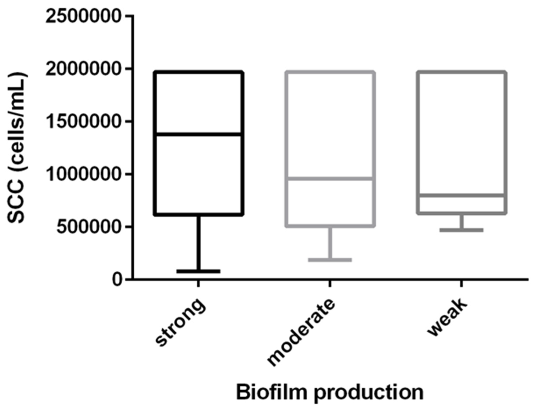
3.2. Biofilm Production and SCC the Relationship

We observed biofilm production in all isolates on polystyrene microplates. Eighty-five isolates (58.6%) were classified as SBP (51/85 serotype Ia and 34/85 serotype III), followed by 42 isolates (29%) as MBP (27/42 serotype Ia and 15/42 serotype III), and 18 isolates (12.4%) as WBP (9/18 serotype Ia and 9/18 serotype III).
Although no statistical difference was observed regarding biofilm production and SCC, the biological trend indicated that SBP triggered a higher SCC response than moderate or weak producers (Figure 1).
A comparative analysis regarding SCC and biofilm production between virulence genes was also performed, and no correlation was identified in either analysis regarding virulence genes. The data are shown separately in Table 2.

3.3. Cell Adhesion and Invasion

Considering the nine different profiles observed, only one isolate representing each profile was chosen randomly for the adhesion and invasion assays (Table 1).
A positive result was determined by recovering and counting the colonies in percentages related to the values obtained in the adhesion test. Results showed that all strains adhered to BMEC, but none were able to invade, regardless of the genotypic profile. The values for the adhesion assays are presented in Table 1. The Fisher exact test was used for analysis; however, a valid conclusion was difficult to obtain since some profiles presented too small a sample size.

3.4. Statistical Analysis

Initially, the distribution of the dependent variables, SCC, and biofilm OD was analyzed and transformed to a log10 scale to achieve normality. An analysis of variance (ANOVA, PROC GLM, SAS Institute, Cary, NC, USA) was used to: (1) compare the mean SCC (log10 cells/ml; dependent variable) among isolates that were WBP, MBP, or SBP (independent variable); and (2) compare the mean SCC and OD (dependent variables) among strains that were positive or not for the genes studied (independent variable). The Tukey´s test was used to adjust the P-values resulting from multiple comparisons. The analysis was performed at an alpha level of 0.05.

4. Discussion

Currently, S. agalactiae is a common cause of mastitis in dairy herds in many countries [19,20]. The correlation between intramammary infection and a high SCC response (> 600.000 cells/ml) has not yet been fully elucidated; however, our study provides evidence that biofilm production is related to this trait.
The capsular type is the most studied virulence factor, and the main types we identified were Ia (57%) and III (43%). However, Carvalho-Castro et al. [7] reported five cps types isolated from mastitis cases, with type III being the most prevalent (38.9%), followed by type II (20.3%), Ib (15%), Ia (8.4%), and IV (8.4%). In previous studies, the distribution of S. agalactiae capsular types depended on geographic location and diagnostic techniques. The fact that there is not a specific S. agalactiae strain associated with mastitis may be due to lack of host specificity, as observed by Sorensen et al. [36], who showed that herdspersons and cattle often share the same S. agalactiae clone, independent of capsular type. In any case, the capsular type allows bacteria to evade the host immune response [37], and this ability, associated with other virulence factors, contributes to the success of the infection. Another possible reason for the diversity of serotypes observed in bovine mastitis could be due to capsular switching events. Despite the scarce data reported in the literature, some studies suggest that this occurs in S. agalactiae [16]. Capsular exchange may confer a survival advantage for bacteria against host-acquired immune responses, although further research is required. According to Paveenkittiporn et al. [38], serotype III is the most associated with a broad spectrum of invasive human infections, as well as this serotype displaying much higher virulence than serotype Ia when infecting fish. In a previous investigation, we showed that some lineages were already adapted to specific host species, but on the other hand, and existed as “core population” of clones, capable of colonizing indistinct hosts. However, this could also be a variation within the same sequence-type (ST), triggering sub-lineages that are still unknown. A limitation of this study was that we were not able to perform sequence typing on our isolates, for further discussion. However, further studies with different serotypes, STs, and hosts may help determine how these host-pathogen interactions act to trigger the emergence of sub-lineages within the clone.
In our study, we also investigated the presence of virulence genes related to adhesion and invasion (fbsA, fbsB, PI1, PI2a, and PI2b), as well as tissue damage (hylB). Fibrinogen binding proteins known as FbsA and FbsB can mediate adherence and promote entry of S. agalactiae into host cells, respectively. The results show that 85 isolates carried the fbsA gene, followed by 59 isolates that were positive for the fbsB gene. Some differences were found by Carvalho-Castro et al. [7], where fbsA was present in 42.3% of the isolates and fbsB in all of them. Nagao [28] affirmed that even though both were binding proteins, the contributions of their interactions that influence virulence are not well-defined. Regardless of the presence of hylB, the prevalence was similar to that observed by Carvalho-Castro et al. [7] in dairy herds in other regions of Brazil. These authors also observed the same prevalence in their isolates as our results, in regards to pI2b, with pI2a being less prevalent in both studies.
Studies have shown the relationship between pilus loci and biofilm formation in S. agalactiae [26]. Here, all isolates were biofilm producers, but the distribution of the presence of pilus island genes (PI1 and PI2a - PI2b) was varied, and pI1 was not observed. Our results agree with those of Alvim et al. [39], who reported that PI2b represented 74% of the animal isolates. This may have occurred because each S. agalactiae genome encodes one or two distinct pilus islands, and pI1 has been observed among human isolates [27]. In our study, no statistical correlation was found between virulence genes and biofilm production, nor between virulence genes and SCC.
Pang et al. [27] related that S. agalactiae isolates from bovines grew much faster than strains isolated from other sources. This feature supports the fact that infection caused by this pathogen presents high SCC values, since the relationship between SCC and infection occurs because bacteria invade the udder and multiply. Consequently, high bacterial density promotes biofilm formation, causing a persistent and destructive inflammatory process [40]. In general, most knowledge of host immune defense comes from interactions with planktonic cell pathogens, and the response regarding biofilms has not been elucidated [41].
We observed a biological trend suggesting that SBP isolates were associated with higher SCC (Figure 1). The lack of statistical significance between biofilm production and increased SCC may be explained by the extreme SCC variation found among isolates (Figure 1) and the sample size used in the study. Larger studies are necessary to further investigate the SCC response among S. agalactiae strains with different levels of biofilm production. Gogoi-Tiwari et al. [19] noted an increase in TNF-α levels in mice infected with a strong biofilm-producing S. aureus, compared to a weak biofilm-producing S. aureus. In addition, in another study [20], these authors demonstrate that S. aureus in a biofilm induces stronger and more differentiated immune responses than its planktonic cells. Despite the lack of studies on S. agalactiae in biofilm and immune responses, we can suggest that the presence of S. agalactiae biofilm producers promotes the colonization of mammary tissue by the pathogen. This might result in higher bacterial loads and dispersion into the mammary glands, increasing the interaction between pathogens and the immune response. Since SCC is an indicator of the immunological condition of the mammary gland and TNF-α is capable of recruiting macrophages and monocytes [42], we can consider that higher immune system activation may depend on the pathogen’s ability to form a biofilm.
Bacterial pathogenicity engages many mechanisms to cause disease, and the first of them is related to host tissue adhesion. In this study, we also hypothesized that the different genetic profiles of the isolates could show distinct abilities in the establishment of infection. However, we observed that all tested isolates had the same behavior in in vitro adhesion and invasion assays. Curiously, neither of them could invade BMEC, even though the adhesion rate had been similar in all isolates, independent of their genetic profiles. Reduced invasion in S. agalactiae can occur due to the production of CPS [43]. In addition, according to Shabayek and Spellerberg [44], invasive S. agalactiae strains are more likely to carry a combination of pI and one of the pI-2 variants, and bovine isolates mostly lack PI1; corroborating our results, where pI1 was not observed in all isolates. Moreover, it should be noted that the ability to invade BMEC in vitro may not always reflect the in vivo situation.

5. Conclusions

In summary, our results showed that the different genetic profiles presented here do not provide advantages for bacteria to invade BMEC in vitro, and the capsular type showed no correlation with SCC. On the other hand, we demonstrated for the first time that biofilm production is closely related to SCC, considering that activation of the immune response may occur, according to the pathogen’s ability to form biofilm. Thus, the data obtained in our study constitutes a preliminary step for further research to better understand the host-pathogen interaction.

Author Contributions

Conceptualization, E.C.R.B., A.F.J., J.C.F.P. and V.L.M.R.; methodology, E.C.R.B., F.C.C., B.F.R., R.S.R. and G.L.; validation, E.C.R.B., F.C.C., B.F.R., R.S.R. and G.L.; formal analysis, E.C.R.B., A.F.J., J.C.F.P. and V.L.M.R.; investigation, F.C.C., B.F.R., R.S.R. and G.L.; resources, A.F.J. and V.L.M.R.; data curation, R.S.R., V.L.M.R. and J.C.F.P.; writing—original draft preparation, F.C.C., E.C.R.B., B.F.R. and V.L.M.R.; writing—review and editing, E.C.R.B., A.F.J. and V.L.M.R.; visualization, A.F.J., B.F.R., J.C.F.P.; G.L., R.S.R.; supervision, A.F.J., J.C.F.P. and V.L.M.R.; project administration, R.S.R., G.L. and J.C.F.P.; funding acquisition, A.F.J. and V.L.M.R. All authors have read and agreed to the published version of the manuscript.

Funding

This study was financed in part by the Coordenação de Aperfeiçoamento Pessoal de Nível Superior—Brasil (CAPES)—Finance Code 001 and Conselho Nacional de Desenvolvimento Científico e Tecnológico (CNPq, 308486/2021-8).

Institutional Review Board Statement

This study was approved by the Sao Paulo State University (UNESP) Ethics Commit-tee for Animal Use (protocol 07/2015).

Informed Consent Statement

Not applicable.

Data Availability Statement

The raw data of this study will be made available by the authors (corresponding author), without reservation, to any qualified researcher.

Conflicts of Interest

The authors certify that they have no affiliations with or involvement in any organization or entity with any financial or nonfinancial interest in the subject matter or materials discussed in this manuscript.

References

  1. Ruegg, P.L. 100-Year Review: Mastitis detection, management, and prevention. J. Dairy Sci. 2017, 12, 10381–10397. [Google Scholar] [CrossRef]
  2. Carra, E.; Russo, S.; Micheli, A.; Garbarino, C.; Ricchi, M.; Bergamini, F.; Bassi, P.; Prosperi, A.; Piva, S.; Cricca, M.; et al. Evidence of Common Isolates of Streptococcus agalactiae in Bovines and Humans in Emilia Romagna Region (Northern Italy). Front. Microbiol. 2021, 12, 673126. [Google Scholar] [CrossRef] [PubMed]
  3. Cobo-Ángel, C.; Jaramillo-Jaramillo, A.S.; Lasso-Rojas, L.M.; Aguilar-Marin, S.B.; Sanchez, J.; Rodriguez-Lecompte, J.C.; Ceballos-Márquez, A.; Zadoks, R.N. Streptococcus agalactiae is not always an obligate intramammary pathogen: Molecular epidemiology of GBS from milk, feces and environment in Colombian dairy herds. PLoS ONE 2018, 13, e0208990. [Google Scholar] [CrossRef] [PubMed]
  4. Han, G.; Zhang, B.; Luo, Z.; Lu, B.; Luo, Z.; Zhang, J.; Wang, Y.; Luo, Y.; Yang, Z.; Shen, L.; et al. Molecular typing and prevalence of antibiotic resistance and virulence genes in Streptococcus agalactiae isolated from Chinese dairy cows with clinical mastitis. PLoS ONE 2022, 17, e0268262. [Google Scholar] [CrossRef]
  5. Kaczorek, E.; Małaczewska, J.; Wójcik, R.; Rękawek, W.; Siwicki, A. Phenotypic and genotypic antimicrobial susceptibility pattern of Streptococcus spp. isolated from cases of clinical mastitis in dairy cattle in Poland. J. Dairy Sci. 2017, 100, 6442–6453. [Google Scholar] [CrossRef] [PubMed]
  6. Mahmmod, Y.; Klaas, I.; Katholm, J.; Lutton, M.; Zadoks, R. Molecular epidemiology and strain-specific characteristics of Streptococcus agalactiae at the herd and cow level. J. Dairy Sci. 2015, 98, 6913–6924. [Google Scholar] [CrossRef]
  7. Carvalho-Castro, G.A.; Silva, J.R.; Paiva, L.V.; Custódio, D.A.; Moreira, R.O.; Mian, G.F.; Prado, I.A.; Chalfun-Junior, A.; Costa, G.M. Molecular epidemiology of Streptococcus agalactiae isolated from mastitis in Brazilian dairy herds. Braz. J. Microbiol. 2017, 48, 551–559. [Google Scholar] [CrossRef]
  8. Keefe, G.P. Streptococcus agalactiae mastitis: A review. Am. Jew. Hist. 1997, 38, 429–437. [Google Scholar]
  9. Jørgensen, H.; Nordstoga, A.; Sviland, S.; Zadoks, R.; Sølverød, L.; Kvitle, B.; Mørk, T. Streptococcus agalactiae in the environment of bovine dairy herds—Rewriting the textbooks? Vet. Microbiol. 2016, 184, 64–72. [Google Scholar] [CrossRef]
  10. Amosun, E.A.; Ajuwape, A.T.P.; Adetosoye, A.I. Bovine streptococcal mastitis in Southwest and Northern states of Nigeria. Afr. J Biomed. Res. 2010, 13, 33–37. [Google Scholar]
  11. Becker, H. Streptococcus agalactiae (group B streptocci). In The Significance of Pathogenic Microorganisms in Raw Milk; International Dairy Federation: Brussels, Belgium, 1994; pp. 43–54. [Google Scholar]
  12. Bisharat, N.; Crook, D.W.; Leigh, J.; Harding, R.M.; Ward, P.N.; Coffey, T.; Maiden, M.C.; Peto, T.; Jones, N. Hyperinvasive Neonatal Group B Streptococcus Has Arisen from a Bovine Ancestor. J. Clin. Microbiol. 2004, 42, 2161–2167. [Google Scholar] [CrossRef] [PubMed]
  13. Botelho, A.C.N.; Ferreira, A.F.M.; Fracalanzza, S.E.L.; Teixeira, L.M.; Pinto, T.C.A. A Perspective on the Potential Zoonotic Role of Streptococcus agalactiae: Searching for a Missing Link in Alternative Transmission Routes. Front. Microbiol. 2018, 9, 608. [Google Scholar] [CrossRef] [PubMed] [Green Version]
  14. Rajagopal, L. Understanding the regulation of Group B Streptoccocal virulence factors. Future Microbiol. 2009, 4, 201–221. [Google Scholar] [CrossRef] [PubMed]
  15. Poyart, C.; Tazi, A.; Réglier-Poupet, H.; Billoet, A.; Tavares, N.; Raymond, J.; Trieu-Cuot, P. Multiplex PCR Assay for Rapid and Accurate Capsular Typing of Group B Streptococci. J. Clin. Microbiol. 2007, 45, 1985–1988. [Google Scholar] [CrossRef] [PubMed]
  16. Hedberg, C.; do Carmo, R.M. Translocal Ruralism: Mobility and Connectivity in European Rural Spaces; Springer: London, UK; New York, NY, USA, 2012. [Google Scholar]
  17. Di Xia, F.; Mallet, A.; Caliot, E.; Gao, C.; Trieu-Cuot, P.; Dramsi, S. Capsular polysaccharide of Group B Streptococcus mediates biofilm formation in the presence of human plasma. Microbes Infect. 2015, 17, 71–76. [Google Scholar] [CrossRef] [PubMed]
  18. Weber, M.; Liedtke, J.; Plattes, S.; Lipski, A. Bacterial community composition of biofilms in milking machines of two dairy farms assessed by a combination of culture-dependent and –independent methods. PLoS ONE 2019, 14, e0222238. [Google Scholar] [CrossRef]
  19. Gogoi-Tiwari, J.; Williams, V.; Waryah, C.B.; Costantino, P.; Al-Salami, H.; Mathavan, S.; Wells, K.; Tiwari, H.K.; Hegde, N.; Isloor, S.; et al. Mammary Gland Pathology Subsequent to Acute Infection with Strong versus Weak Biofilm Forming Staphylococcus aureus Bovine Mastitis Isolates: A Pilot Study Using Non-Invasive Mouse Mastitis Model. PLoS ONE 2017, 12, e0170668. [Google Scholar] [CrossRef]
  20. Gogoi-Tiwari, J.; Dorji, D.; Tiwari, H.K.; Shirolkar, G.; Aleri, J.W.; Mukkur, T. Phenotypic PIA-Dependent Biofilm Production by Clinical Non-Typeable Staphylococcus aureus Is Not Associated with the Intensity of Inflammation in Mammary Gland: A Pilot Study Using Mouse Mastitis Model. Animals 2021, 11, 3047. [Google Scholar] [CrossRef]
  21. Watters, C.; Fleming, D.; Bishop, D.; Rumbaugh, K. Host Responses to Biofilm. Prog. Mol. Biol. Transl. Sci. 2016, 142, 193–239. [Google Scholar] [CrossRef] [PubMed]
  22. Erosini, R.; Margarit, I. Biofilm formation by Streptococcus agalactiae: Influence of environmental conditions and implicated virulence factors. Front. Cell. Infect. Microbiol. 2015, 5, 6. [Google Scholar] [CrossRef]
  23. Moschioni, M.; Pansegrau, W.; Barocchi, M.A. Adhesion determinants of the Streptococcus species. Microb. Biotechnol. 2009, 3, 370–388. [Google Scholar] [CrossRef] [PubMed]
  24. Lazzarin, M.; Mu, R.; Fabbrini, M.; Ghezzo, C.; Rinaudo, C.D.; Doran, K.S.; Margarit, I. Contribution of pilus type 2b to invasive disease caused by a Streptococcus agalactiae ST-17 strain. BMC Microbiol. 2017, 17, 148. [Google Scholar] [CrossRef] [Green Version]
  25. Lauer, P.; Rinaudo, C.D.; Soriani, M.; Margarit, I.; Maione, D.; Rosini, R.; Taddei, A.R.; Mora, M.; Rappuoli, R.; Grandi, G.; et al. Genome Analysis Reveals Pili in Group B Streptococcus. Science 2005, 309, 105. [Google Scholar] [CrossRef] [PubMed]
  26. Parker, R.E.; Laut, C.; Gaddy, J.A.; Zadoks, R.N.; Dele Davies, H.; Manning, S.D. Association between genotypic diversity and biofilm production in group B Streptococcus. BMC Microbiol. 2016, 16, 86. [Google Scholar] [CrossRef]
  27. Pang, M.; Sun, L.; He, T.; Bao, H.; Zhang, L.; Zhou, Y.; Zhang, H.; Wei, R.; Liu, Y.; Wang, R. Molecular and virulence characterization of highly prevalent Streptococcus agalactiae circulated in bovine dairy herds. Vet. Res. 2017, 1, 65. [Google Scholar] [CrossRef]
  28. Nagao, P.E. Streptococcus agalactiae (Group B Streptococci). In Molecular Medical Microbiology; Academic Press: Cambridge, MA, USA, 2015; pp. 1751–1767. [Google Scholar] [CrossRef]
  29. Rossi, R.; Amarante, A.; Correia, L.; Guerra, S.; Nobrega, D.; Latosinski, G.; Rossi, B.; Rall, V.; Pantoja, J. Diagnostic accuracy of Somaticell, California Mastitis Test, and microbiological examination of composite milk to detect Streptococcus agalactiae intramammary infections. J. Dairy Sci. 2018, 101, 10220–10229. [Google Scholar] [CrossRef] [PubMed]
  30. Almeida, A.; Albuquerque, P.; Araujo, R.; Ribeiro, J.N.; Tavares, F. Detection and discrimination of common bovine mastitis-causing streptococci. Vet. Microbiol. 2013, 164, 370–377. [Google Scholar] [CrossRef]
  31. Arpini, C.M.; Cardoso, P.G. Virulence Genes of the Streptococcus agalactiae Associated with Bovine Mastitis in Minas Gerais Livestock Herds, Brazil. Appl. Microbiol. open Access 2016, 2, 1000119. [Google Scholar] [CrossRef]
  32. Godoy, D.T.; Carvalho-Castro, G.A.; Leal, C.A.; Pereira, U.P.; Leite, R.C.; Figueiredo, H.C. Genetic diversity and new genotyping scheme for fish pathogenic Streptococcus agalactiae. Lett. Appl. Microbiol. 2013, 57, 476–483. [Google Scholar] [CrossRef]
  33. Bonsaglia, E.C.R.; Latosinski, G.S.; Rossi, R.S.; Rossi, B.F.; Possebon, F.S.; Pantoja, J.C.F.; Júnior, A.F.; Rall, V.L.M. Biofilm production under different atmospheres and growth media by Streptococcus agalactiae isolated from milk of cows with subclinical mastitis. Arch. Microbiol. 2019, 202, 209–212. [Google Scholar] [CrossRef]
  34. Stepanović, S.; Vuković, D.; Hola, V.; Di Bonaventura, G.; Djukić, S.; Cirković, I.; Ruzicka, F. Quantification of biofilm in microtiter plates: Overview of testing conditions and practical recommendations for assessment of biofilm production by Staphylococci. APMIS 2007, 115, 891–899. [Google Scholar] [CrossRef]
  35. Sharma, P.; Lata, H.; Arya, D.K.; Kashyap, A.K.; Kumar, H.; Ali, M.D.A.; Johri, A.K. Role of pilus proteins in adherence and invasion of Streptococcus agalactiae to the lung and cervical epithelial cells. J. Biol. Chem. 2013, 6, 4023–4034. [Google Scholar] [CrossRef] [PubMed]
  36. Sørensen, U.B.S.; Klaas, I.C.; Boes, J.; Farre, M. The distribution of clones of Streptococcus agalactiae (group B streptococci) among herdspersons and dairy cows demonstrates lack of host specificity for some lineages. Vet. Microbiol. 2019, 235, 71–79. [Google Scholar] [CrossRef] [PubMed]
  37. Carlin, A.F.; Lewis, A.L.; Varki, A.; Nizet, V. Group B Streptococcal Capsular Sialic Acids Interact with Siglecs (Immunoglobulin-Like Lectins) on Human Leukocytes. J. Bacteriol. 2007, 189, 1231–1237. [Google Scholar] [CrossRef]
  38. Paveenkittiporn, W.; Ungcharoen, R.; Kerdsin, A. Streptococcus agalactiae infections and clinical relevance in adults, Thailand. Diagn. Microbiol. Infect. Dis. 2020, 97, 115005. [Google Scholar] [CrossRef]
  39. Alvim, D.C.S.S.; Ferreira, A.F.M.; Leal, M.A.; Oliveira, L.M.A.; Botelho, A.M.N.; Figueiredo, A.M.S.; Fracalanzza, S.E.L.; Teixeira, L.M.; Pinto, T.C.A. Biofilm production and distribution of pilus variants among Streptococcus agalactiae isolated from human and animal sources. Biofouling 2019, 35, 938–944. [Google Scholar] [CrossRef] [PubMed]
  40. Hänsch, G.M. Host Defence against Bacterial Biofilms: “Mission Impossible”? ISRN Immunol. 2012, 2012, 853123. [Google Scholar] [CrossRef]
  41. González, J.F.; Hahn, M.M.; Gunn, J.S. Chronic biofilm-based infections: Skewing of the immune response. Pathog. Dis. 2018, 76, fty023. [Google Scholar] [CrossRef] [PubMed]
  42. Shim, Y.-J.; Kang, B.-H.; Choi, B.-K.; Park, I.-S.; Min, B.-H. Clusterin induces the secretion of TNF-α and the chemotactic migration of macrophages. Biochem. Biophys. Res. Commun. 2012, 422, 200–205. [Google Scholar] [CrossRef]
  43. Gibson, R.L.; Lee, M.K.; Soderland, C.; Chi, E.Y.; E Rubens, C. Group B streptococci invade endothelial cells: Type III capsular polysaccharide attenuates invasion. Infect. Immun. 1993, 61, 478–485. [Google Scholar] [CrossRef]
  44. Shabayek, S.; Spellerberg, B. Group B Streptococcal colonization, molecular characteristics, and epidemiology. Front. Microbiol. 2018, 9, 437. [Google Scholar] [CrossRef] [PubMed]
Figure 1. Distribution of SCC in relation to biofilm production by S. agalactiae causing bovine mastitis.
Pathogens 12 00311 g001
Table 1. Distribution of S. agalactiae isolated from milk of cows with subclinical mastitis according to genotypic profile, and in adhesion assays with bovine mammary epithelial cells.
ProfileIsolates
(n = 145)
serotypefbsAfbsBpI1pI2apI2bhylbAdhesion
BMEC *
169Ia+---++1.5
211III+---++0.3
38Ia-+-+-+0.4
420III-+-+-+1.2
53III-+--+-1.6
61III-+---+0.3
727III-+--++0.5
81Ia+--+--1.3
95Ia+----+0.6
* Equivalent values to 106 CFU for one isolate in each profile; BMEC: bovine mammary epithelial cells
Table 2. Comparison of both SCC and biofilm among virulence genes of S. agalactiae isolated from cow’s milk with subclinical mastitis.
SCC * (log10cells/mL) Biofilm (log10 optical density)
genePositiveNegativep-value PositiveNegativep-value **
capIa5.98 ± 0.045.98 ± 0.050.92 −0.39 ± 0.03−0.38 ± 0.030.77
capIII5.98 ± 0.055.98 ± 0.040.92 −0.38 ± 0.03−0.39 ± 0.030.77
fbsA5.98 ± 0.045.98 ± 0.050.89 −0.38 ± 0.03−0.38 ± 0.030.99
fbsB5.98 ± 0.055.98 ± 0.040.89 −0.38 ± 0.03−0.38 ± 0.030.99
pI2a5.97 ± 0.075.98 ± 0.030.91 −0.39 ± 0.05−0.38 ± 0.030.84
pI2b5.98 ± 0.055.98 ± 0.040.89 −0.38 ± 0.02−0.51 ± 0.100.19
hlyB5.97 ± 0.036.30 ± 0.180.10 −0.39 ± 0.02−0.32 ± 0.130.63
* Somatic cell count. ** p-value derived from analysis of variance (Tukey test).
Disclaimer/Publisher’s Note: The statements, opinions and data contained in all publications are solely those of the individual author(s) and contributor(s) and not of MDPI and/or the editor(s). MDPI and/or the editor(s) disclaim responsibility for any injury to people or property resulting from any ideas, methods, instructions or products referred to in the content.

