Article

Molecular Detection of Anaplasma, Ehrlichia and Rickettsia Pathogens in Ticks Collected from Humans in the Republic of Korea, 2021

Division of Vectors and Parasitic Diseases, Korea Disease Control and Prevention Agency, 187 Osongsaengmyeong 2-ro, Osong-eup, Heungdeok-gu, Cheongju 28159, Republic of Korea
*
Author to whom correspondence should be addressed.
Pathogens 2023, 12(6), 802; https://doi.org/10.3390/pathogens12060802
Submission received: 12 May 2023 / Revised: 2 June 2023 / Accepted: 2 June 2023 / Published: 4 June 2023
(This article belongs to the Section Ticks)

Abstract

:
Tick-borne pathogens (TBPs), transmitted by the bites of ticks, are of great medical and veterinary importance. They include bacteria, viruses, and protozoan parasites. To provide fundamental data on the risk of tick contact and public health strategies, we aimed to perform a molecular investigation on four tick-borne bacterial pathogens in ticks collected from humans across the Republic of Korea (ROK) in 2021. In total, 117 ticks were collected, including Haemaphysalis longicornis (56.4%), Amblyomma testudinarium (26.5%), Ixodes nipponensis (8.5%), H. flava (5.1%), and I. persulcatus (0.9%). Among the ticks, 20.5% (24/117) contained tick-borne bacterial pathogens, with infection rates of 17.9% for Rickettsia (Candidatus Rickettsia jingxinensis, R. tamurae, R. monacensis, and Candidatus Rickettsia tarasevichiae), 2.5% for Anaplasma (A. phagocytophilum, A. capra, and A. bovis), and 0.9% for Ehrlichia (Ehrlichia sp.). Additionally, the co-detection rate for R. monacensis and A. phagocytophilum was 0.9%. To our knowledge, this is the first report of A. capra and A. bovis detection in ticks collected from humans in the ROK. This study contributes to the understanding of the potential risk of tick contact and provides fundamental data for establishing a public health strategy for tick-borne disease management in the ROK.

1. Introduction

Ticks are major blood-feeding arthropod vectors distributed worldwide, owing to their ability to adapt to various hosts, environments, and climates [1]. During blood feeding, ticks can transmit bacteria, viruses, and parasites that are of great concern for public health and veterinary medicine [2].
Tick populations have increased recently owing to rising temperatures associated with climate change. An increase in temperature has been observed to accelerate the developmental cycle, increase egg production, and lead to increased tick population densities [3,4]. Additionally, human activities such as leisure, travel and urban development have increased the opportunities for contact with ticks [5,6].
Several TBPs, including Anaplasma, Ehrlichia, Rickettsia, Borrelia, Babesia, Bartonella, and severe fever with thrombocytopenia syndrome virus, have been reported in the Republic of Korea (ROK) [7]. As tick-borne bacterial diseases present with non-specific symptoms, such as fever, headache, nausea, and vomiting, they are difficult to recognize without an accurate diagnosis [8]. Serological tests are commonly used for diagnosis; however, they require careful interpretation because of their reduced specificity for related pathogens or sensitivity to intrinsic factors, such as epitope selection [7,9].
Anaplasma species are zoonotic pathogens with tick vectors and mammalian reservoir hosts. Among them, A. phagocytophilum was the first species of tick reported to cause human granulocytic anaplasmosis (HGA) in the United States of America (USA) in 1994 [10,11]. Since then, the prevalence of HGA has been increasing, with more than 41,000 cases reported from 2000 to 2019 in the USA, while sporadic and clustered cases have been reported in Europe and Asia [12,13,14]. Vector ticks for this agent include Ixodes spp. ticks such as I. scapularis and I. pacificus in the USA, I. ricinus mostly in Europe, and I. persulcatus in Russia [15] and China [14]. In the ROK, A. phagocytophilum was the first species of tick reported to cause human disease in 2014 [16]. After the Korea Disease Control and Prevention Agency (KDCA) began investigating infectious diseases in 2016, 168 cases of HGA had been reported by 2020 using immunofluorescent antibodies [17]. Haemaphysalis longicornis, I. nipponensis, H. flava, and I. persulcatus are known as the primary vectors for Anaplasma in the ROK [7,18].
Ehrlichia is another zoonotic tick-borne pathogen transmitted by infected ticks. Its primary vectors are known to be Amblyomma americanum in the USA and Dermacentor variabils in Europe [19]. After the first identification of E. chaffeensis as a human pathogen, causing human monocytic ehrlichiosis (HME), in 1987, more than 18,000 cases were reported from 2000 to 2019 in the USA [20]. Moreover, the emergence of E. ewingii, which infects humans and animals (in particular dogs), has been noted. The first case of human infection caused by E. ewingii was reported in 1999 [21], and 262 cases were reported from 2008 to 2019 in the USA [20]. Since E. ewingii has similar clinical symptoms to A. phagocytophilum and E. chaffeensis, and cross-reaction with E. chaffeensis occurs using serological diagnostic methods, it is difficult to distinguish between them. For this reason, E. ewingii is likely to be misclassified as A. phagocytophilum or E. chaffeensis [22]. In the ROK, after the first case of HGE identified by means of positive serological diagnosis in 2000 [23], no further cases were reported. The primary vectors for Ehrlichia are same as Anaplasma in the ROK [7,18].
Rickettsia, which can cause diseases such as spotted fever rickettsiosis, epidemic typhus, and murine typhus, is also transmitted by arthropods such as ticks, lice, and fleas [24]. Rickettsia is categorized into the Spotted Fever Group (SFG), Typhus Group (TG), and Transitional Group (TRG). Among these groups, SFG is predominantly transmitted by ticks. It causes mild to severe disease in humans, with symptoms including high fever (39.5–40 °C), headache, eschar, and rash [25]. Spotted fever rickettsiosis was reported in between 3300 and 6200 cases per year in USA from 2012 to 2019 [26]. Ticks known to be vectors of SFG include Rhipicephalus, Ixodes, Amblyomma, Hyalomma, Haemaphysalis, and Dermacentor [27]. In the ROK, R. japonica and R. rickettsii were confirmed in H. longicornis by means of PCR in 2003 [28]. Jiang et al. [29] identified R. monacensis in H. longicornis and I. nipponensis and R. heilongjiangensis in H. longicornis and H. flava in 2019. Moreover, R. monacensis was isolated from a patient in the ROK in 2017 [30].
Borrelia burgdorferi sensu lato (s.l.) is the causative agent of Lyme disease, and its main vectors are known to be Ixodes genus ticks. Lyme disease causes a wide range of symptoms that include fever, fatigue, myalgia, arthralgia, and erythema migrans [31]. Lyme disease occurred in between 600,000 and 900,000 cases each year in the USA from 2000 to 2019 [32]. In the ROK, it was first reported as a human disease in 1993. Since it was designated as a national notifiable infectious disease in 2011, 160 cases have been reported, as of 2020 [33]. Borrelia burgdorferi s. l., including B. afzelii, B. garinii, B. valaisiana, B. yangtzensis, and B. tanukii, was detected in Ixodes ticks collected from wild rodents in 2017 from nine regions in the ROK [34].
In a previous survey in the ROK, studies on ticks collected from humans were performed in sporadic regions or with specific pathogens. Bang et al. [35] investigated Anaplasma, Rickettsia, and Babesia in ticks collected from humans in the southwestern region. Kim et al. [36] conducted a nationwide survey of ticks collected from humans to identify Anaplasma and Ehrlichia. We aimed to perform a molecular investigation on various tick-borne bacterial pathogens, including Anaplasma, Ehrlichia, Rickettsia, and Borrelia, in ticks collected from humans across the ROK in 2021.

2. Materials and Methods

2.1. Tick Collection and Identification

Ticks removed from human bodies were collected from hospitals and local public health centers across the ROK from January to October 2021. The tick specimens were transferred to the Korea Disease Control and Prevention Agency (KDCA).
The ticks were identified morphologically under a dissection microscope to the species level, while life stage (larva, nymph, and adult) and sex (female and male) were also identified, as per the study by Yamaguti et al. [37]. Ticks were recorded as engorged (swollen) or unengorged based on differences in external morphology. Individual ticks were placed in a 2 mL tube and frozen at −80 °C until DNA extraction was performed.

2.2. DNA Extraction

Genomic DNA was extracted from individual ticks using the Clear-S Quick DNA Extraction Kit (InVirus Tech, Gwangju, Republic of Korea) with 500 µL of lysis buffer. The ticks were homogenized using a Precelly Evolution homogenizer (Bertin Technologies, Bretonneux, France) twice for 30 s at a speed of 4.5 m/s. After homogenization, the tubes were centrifuged for 10 min at 12,000× g, and 150 µL of supernatant was used for DNA extraction, according to the manufacturer’s instructions. The extracted DNA was stored at −20 °C until use.

2.3. Molecular Detection of TBPs

Individual ticks were tested for the presence of the pathogens Anaplasma, Ehrlichia, Rickettsia, and Borrelia. The primer sets for the target gene fragments used to identify each pathogen are listed in Table 1. As positive controls, the genomic DNA of A. phagocytophilum and R. sibirica provided by the Division of Zoonotic and Vector Borne Disease Research and of E. chaffeensis and B. garinii provided by the Division of Bacterial Disease, KDCA, was used for each PCR.
The primary PCR was performed in 20 µL reaction volumes using the AccuPower PCR PreMix (Bioneer Corp., Daejeon, Republic of Korea). Each PCR mixture was composed of 1 µL of each oligonucleotide primer (10 pmol/µL), 5 µL genomic DNA as a template, and 13 µL distilled water. In the second round, nested PCR was performed using 1 µL of the primary PCR product as a template. Each reaction was performed using the ProFlex PCR System (Thermo Fisher Scientific, Waltham, MA, USA), and the amplified products were subjected to electrophoresis in the automated QIAxcel® system (QIAgen, Hilden, Germany). The DNA extraction, PCR amplification, and automated electrophoresis were performed in separate rooms to prevent cross-contamination.

2.4. Sequencing and Phylogenetic Analysis

PCR-positive samples were purified and sequenced using a commercial sequencing service (BIOFACT, Daejeon, Republic of Korea). The nucleotide sequences were compared with reference sequences obtained from GenBank using nucleotide BLAST (National Center for Biotechnology Information, NCBI) and aligned using CLUSTAL Omega (v.1.2.1). A phylogenetic tree was constructed using the MEGA 11.0 program based on the maximum likelihood method, and bootstrap analysis (1000 replicates) was performed according to the Kimura two-parameter method.

2.5. Statistical Analyses

Statistical analyses were performed using the GraphPad software (GraphPad Software, Inc.; https://www.graphpad.com/quickcalcs/chisquared1/ San Diego, CA, USA). A chi-square test was performed to analyze the significant differences among the pathogens for each tick species and life stage. p < 0.05 was considered statistically significant.

3. Results

3.1. Tick Collecction and Identification

In 2021, 117 ticks were collected from 114 humans in 12 regions. The greatest number of ticks were collected from the central area (n = 49, 41.9%; Chungcheongbuk-do, Chungcheongnam-do, Gyeongsangbuk-do, and Jeollabuk-do provinces), followed by the southern area (n = 42, 35.9%; Gyeongsangnam-do and Jeollanam-do province, Busan and Ulsan metropolitan cities) and the northern area (n = 26, 22.2%; Seoul special and Incheon metropolitan cities, Gyeonggi-do and Gangwon-do provinces). The collected ticks showed the highest prevalence in summer (n = 66, 56.4%; June–August), followed by the spring (n = 42, 35.9%; March–May), autumn (n = 7, 6.0%; September–November), and winter (n = 2, 1.7%; December–February).
Ticks were morphologically identified and taxonomically assigned to at least five species belonging to three genera. Some morphologically damaged ticks were identified as Haemaphysalis spp. and Ixodes spp. because the species or life stage could not be identified (Table 2). The most prevalent tick species was Haemaphysalis longicornis (n = 66, 56.4%), followed by Amblyomma testudinarium (n = 31, 26.5%), Ixodes nipponensis (n = 10, 8.5%), H. flava (n = 6, 5.1%), Ixodes spp. (n = 2, 1.7%), I. persulcatus (n = 1, 0.9%), and Haemaphysalis spp. (n = 1, 0.9%). Among the ticks collected, the highest number were females (n = 55, 47.0%), followed by nymphs (n = 50, 42.7%), males (n = 9, 7.7%), and larvae (n = 1, 0.9%). There were more engorged ticks (n = 70, 59.8%) observed than unengorged ticks (n = 47, 40.2%).

3.2. Molecular Detection of TBPs

In total, 20.5% (24/117) of the ticks were positive for TBPs (Table 2). Three ticks (2.6%) tested positive for the 16S rRNA genes of Anaplasma. Among them, the I. nipponensis specimen was positive for ankA and msp4, A. phagocytophilum-specific genes. One H. longicornis specimen was positive for groEL, an A. bovis-specific gene, and another H. longicornis specimen was negative for groEL and gltA, A. capra-specific genes.
When detecting Ehrlichia, one H. flava specimen was positive for 16S rRNA, groEL, and gltA.
For Rickettsia, 21 ticks (17.9%) were positive for the 17 kDa, ompA, and gltA genes. Candidatus R. jingxinensis was identified in 11 H. longicornis specimens. Rickettsia tamurae was detected in five A. testudinarium specimens, and R. monacensis was identified in three I. nipponensis specimens and one Ixodes spp. specimen. Ixodes persulcatus tested positive for Candidatus R. tarasevichiae. Co-detection with A. phagocytophilum and R. monacensis was identified in I. nipponensis. Borrelia was not observed in this study.
In the life stages, TBPs were detected more often in adults (26.5%, 17/64) than nymphs (12.0%, 6/50), although the difference was not statistically significant (p = 0.0545). Additionally, a higher rate of TBPs was detected in engorged ticks (22.8%, 16/70) than in unengorged ticks (17.0%, 8/47).

3.3. Sequencing and Phylogenetic Analysis

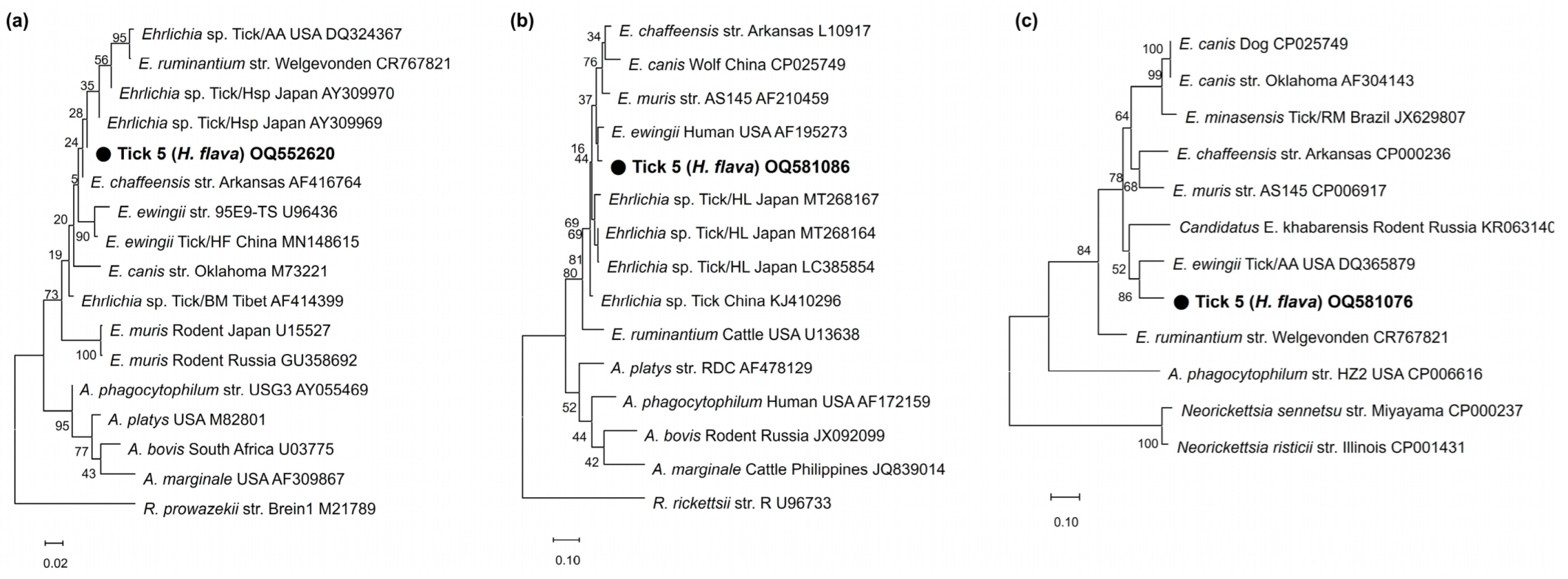
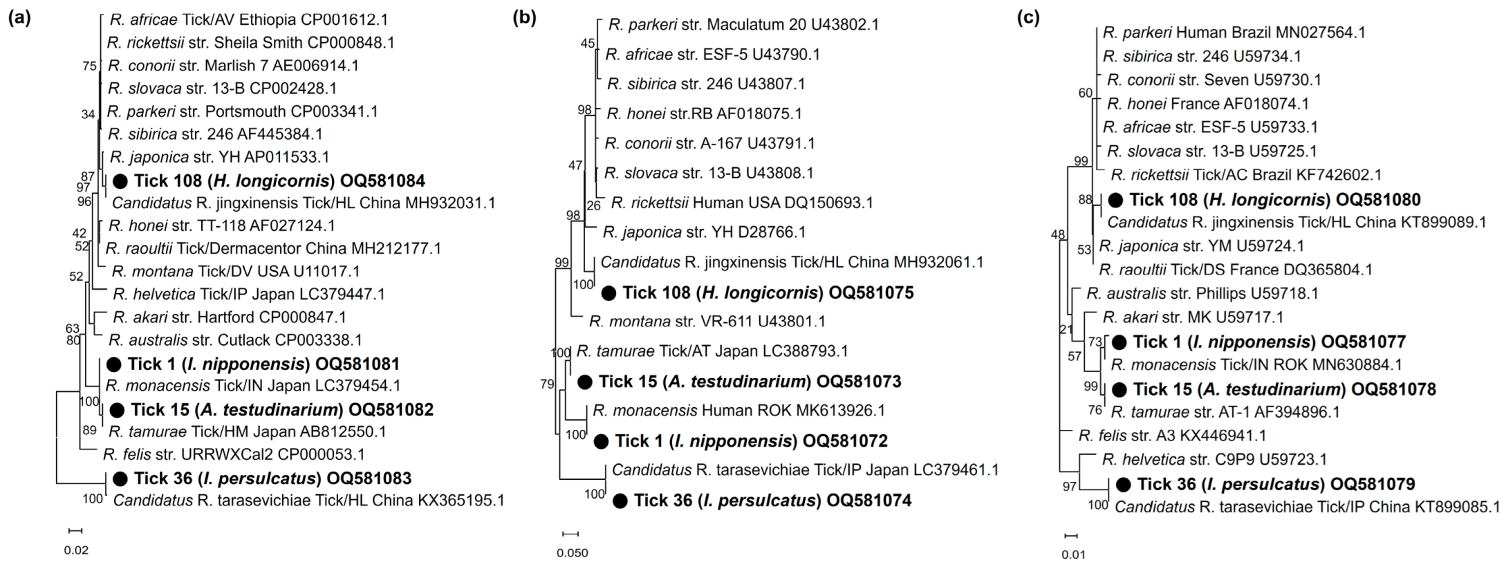
In phylogenetic analyses, the partial 16S rRNA sequences (OQ552617-19) of the three Anaplasma-positive specimens clustered with the previously reported A. phagocytophilum, A. bovis, and A. capra sequences (Figure 1). These sequences showed 100%, 99.6%, and 99.0% identity with 16S rRNA sequences previously reported from raccoon dogs (KY458570), ticks (H. longicornis, EU181143), and Korean water deer (LC432124) in the ROK, respectively. The A. phagocytophilum sequence showed 100% and 99.7% identity with ankA (OQ581070) and msp4 (OQ581071) sequences previously reported in humans in the ROK (MH492325) and sheep in China (GQ412346), respectively (Figure 2a,b). The A. bovis sequence (OQ581085) showed 95.5–97.1% identity with the groEL sequence in the reference sequences within the same clade (Figure 2c). In Ehrlichia, the 16S rRNA sequence (OQ552620) showed 98.0% identity with Ehrlichia sp. (AY309969) (Figure 3a). However, the groEL (OQ581086) and gltA sequences (OQ581076) aligned with those of E. ewingii isolated from humans (AF195273) (Figure 3b) and A. americanum (DQ365879) (Figure 3c) in the USA, with identities of 95.6% and 84.7%, respectively. Of the 21 positive sequences identified as Rickettsia species, four representative sequences were selected without duplicate sequences. They shared 99.8–100% identity with the 17 kDa sequence previously reported for R. monacensis (LC379454.1), R. tamurae (AB812550.1), Candidatus R. jingxinensis (MH932031.1), and Candidatus R. tarasevichiae (KX365195.1). Additionally, the four representative sequences shared 100% and 99.7–100% identity with the ompA and gltA sequences reported in previous studies, respectively (Figure 4).

4. Discussion

A total of 117 ticks were collected from humans across the ROK in 2021, and molecular detection of tick-borne bacterial pathogens, including Anaplasma, Ehrlichia, Rickettsia, and Borrelia, was performed in this study.
Among the 117 ticks collected from humans, H. longicornis (56.4%) was the most prevalent, followed by A. testudinarium (26.5%), I. nipponensis (8.5%), H. flava (5.1%), Ixodes spp. (1.7%), I. persulcatus (0.9%), and Haemaphysalis spp. (0.9%). This result was consistent with that of our previous study, in which ticks removed from the human body were identified as H. longicornis (70.0%), A. testudinarium (17.8%), I. nipponensis (6.1%), H. flava (4.4%), and I. persulcatus (1.7%) in 2020 [11]. Another study showed that H. longicornis (81.2%), A. testudinarium (6.5%), I. nipponensis (5.7%), and H. flava (5.4%) were found in ticks collected from humans in 2013 [48]. However, the species composition differed from that of the host species in the ROK. For example, Kim et al. reported that the Ixodes genus accounted for 98.7% of the ticks collected from wild rodents [34], and Suh et al. revealed that the majority of A. testudinarium (57.1%) and I. nipponensis (42.8%) were collected from reptiles [49].
In this study, ticks collected from humans had a high TBP infection rate of 20.5% (24/117). In particular, the infection rate in adult ticks (26.5%) was 2.2-times higher than that in nymphs (12.0%) (p > 0.05). This result is similar to that of a previous study that showed that the infection rate of tick-borne bacterial pathogens, including Anaplasma, Ehrlichia, and Rickettsia, was 1.8 times higher in adults (69.6%) than that in nymphs (39.2%) collected from water deer in the ROK [50]. Klitgaard et al. [51] reported that the infection rates of Anaplasma, Rickettsia, and Borrelia in adults (52.2%) were 2.7 times higher than those of nymphs (19.1%) collected by flagging in Denmark. Considering the transstadial transmission of various TBPs throughout the life stages of ticks, this is a reasonable result, as adults have one to two times more opportunities to acquire pathogens by means of blood feeding from different hosts infected with pathogens than nymphs [52,53].
The identified tick-borne bacterial pathogens were Anaplasma (2.5%, n = 3), Ehrlichia (0.9%, n = 1), and Rickettsia 17.9%, n = 21). Co-detection was observed in one tick (0.9%) carrying both Anaplasma and Rickettsia, whereas this was not the case for Lyme-disease-causing B. burgdorferi s.l.
The genus Rickettsia is an obligate intracellular bacterium responsible for tick-borne rickettsiosis. Rickettsia is among the oldest known vector-borne pathogens, and its public health importance has been re-evaluated [54,55]. It was initially identified in ticks from 1930 to 1960, and much later (after 1990) in human specimens [56]. Several species of tick-borne Rickettsiae previously considered non-pathogenic are now associated with human infections. Novel Rickettsia species with undetermined pathogenicity have been detected in ticks worldwide [54]. In this study, the genus Rickettsia was the most frequently detected pathogen, specifically Candidatus R. jingxinensis (9.4%, n = 11), R. tamurae (4.2%, n = 5), R. monacensis (3.4%, n = 4), and Candidatus R. tarasevichiae (0.9%, n = 1). These results are consistent with those of a previous study that found R. tamurae (24.2%), Candidatus R. jingxinensis (9.1%), and R. monacensis (3.0%) in the southwestern ROK [35]. In particular, Candidatus R. tarasevichiae is an emerging human pathogen [57] that widely infects I. persulcatus ticks in Russia and China [58,59]. In the ROK, Candidatus R. tarasevichiae was first detected in 2019 in a tick collected from a human [60]. It is necessary to conduct a monitoring survey of the spread and distribution of infections caused by Candidatus R. tarasevichiae in wild rodent and tick species.
Anaplasma and Ehrlichia are obligate intracellular bacteria belonging to the family Anaplasmataceae. The genus Anaplasma includes A. phagocytophilum, A. bovis, A. centrale, A. marginale, A. platy, A. ovis, and A. capra [61]. Of these, A. phagocytophilum is distributed worldwide because of its wide host range, which includes humans, carnivores, ruminants, rodents, insectivores, and birds [62]. Additionally, A. capra has recently been reported in small ruminants, as well as being a human pathogen [63], and A. bovis has been identified in ruminants in numerous countries [64]. In this study, A. phagocytophilum, A. capra, and A. bovis were identified in I. nipponensis and H. longicornis. In the ROK, A. phagocytophilum has been detected in ticks (2.9%, I. nipponensis and I. persulcatus) from humans [36], and another study confirmed its presence in I. nipponensis and patients bitten by it [65]. Additionally, A. bovis and A. capra have been reported in tick-infested cattle in the ROK [66]. In this study, A. phagocytophilum and A. bovis were confirmed with the 16S rRNA as well as specific genes of ankA, msp4, and groEL, whereas A. capra cannot be confirmed by other specific genes such as groEL and gltA. However, a previous study [66] reported the initial molecular detection of A. capra using a partial 16S rRNA sequence obtained from ticks infesting cattle and the 16S rRNA sequence of A. capra showed a higher identity of 99.0% compared to other Anaplasma species, A. ovis (97.6%) and A. phagocytophilum (95.9–96.2%) in this study. Based upon these results, it was considered as A. capra.
The genus Ehrlichia is a pathogenic agent of ehrlichiosis that affects both humans and animals, such as dogs and domestic ruminants [61]. Ehrlichia chaffeensis and E. ewingii are representative species that cause human monocytic and granulocytic ehrlichiosis, respectively. In the ROK, E. chaffeensis was detected in ticks (15.0%, 63/420 tick pools, H. longicornis and I. nipponensis) from grass vegetation and wild rodents in 2004–2005 [67]. E. ewingii was detected in ticks (0.1%, 2/1638 pools, H. longicornis) from grass vegetation in 2001–2003 [18] and in dogs in 2018 [68]. Based on the phylogenetic analysis of 16S rRNA for Ehrlichia in this study, one positive sequence was identified as Ehrlichia sp., whereas groEL and gltA sequences were 95.6% and 84.7% identical to E. ewingii, respectively. As it appears somewhat distinct from E. ewingii, it is suggested to be a new species of Ehrlichia. Therefore, further studies are required to identify and characterize this novel Ehrlichia species in relation to E. ewingii in ticks and their hosts.
The co-detection of R. monacensis and A. phagocytophilum in I. nipponensis was consistent with the results of previous studies on co-detection in ticks [69,70]. Co-detection can result from cofeeding with infected ticks on one host, blood meals on one host carrying several pathogens, or blood meals on different hosts [71,72].
To our knowledge, this is the first study to investigate various tick-borne bacterial pathogens, including Anaplasma, Ehrlichia, Rickettsia, and Borrelia, in ticks collected from humans across the ROK. However, there is no detection of Borrelia. Additionally, this is the first study to detect A. capra and A. bovis in ticks collected from humans in the ROK. Unfortunately, no pathogen analysis was conducted for tick-bitten humans. Because anaplasmosis, ehrlichiosis, and rickettsiosis are not designated as a national notifiable infectious disease, infected patients are not reported. For this reason, we cannot conduct analysis for tick-bitten humans. Therefore, further studies are needed to obtain direct evidence of pathogen transmission in both ticks and bitten humans. This study may be helpful in establishing a potential risk assessment for tick contact with TBPs and public health strategies for ticks in the ROK.

Author Contributions

Formal Analyses, J.-Y.S. and Y.-J.K.; Investigation, J.-Y.S. and Y.-J.K.; Resources, J.-Y.S.; Data Curation and Writing—Original Draft Preparation, J.-Y.S.; Conceptualization, Writing—Review and Editing, S.-Y.K. and H.-I.L.; and Funding Acquisition, H.-I.L. All authors have read and agreed to the published version of the manuscript.

Funding

This study was financially supported by the Korea Disease Control and Prevention Agency (grant no. 6300-6332-304).

Institutional Review Board Statement

Not applicable.

Informed Consent Statement

Not applicable.

Data Availability Statement

Data supporting the conclusions of this article are included within the article. The newly generated sequences were submitted to the GenBank database under the accession numbers OQ552617–OQ552620 and OQ581073–OQ581086. The datasets used and/or analyzed during the present study are available from the corresponding author upon reasonable request.

Acknowledgments

We are grateful to the members of the Division of Bacterial Diseases and the Division of Zoonotic and Vector Borne Disease Research, Korea Disease Control and Prevention Agency (KDCA) for providing the positive control samples used in this study.

Conflicts of Interest

The authors declare no conflict of interest.

References

  1. Dantas-Torres, F.; Chomel, B.B.; Otranto, D. Ticks and tick-borne diseases: A One Health perspective. Trends Parasitol. 2012, 28, 437–446. [Google Scholar] [CrossRef]
  2. Černý, J.; Lynn, G.; Hrnková, J.; Golovchenko, M.; Rudenko, N.; Grubhoffer, L. Management options for Ixodes ricinus-associated pathogens: A review of prevention strategies. Int. J. Environ. Res. Public Health 2020, 17, 1830. [Google Scholar] [CrossRef] [Green Version]
  3. Rossati, A. Global warming and its health impact. Int. J. Occup. Environ. Med. 2017, 8, 7–20. [Google Scholar] [CrossRef] [Green Version]
  4. Kim, S.-H.; Jang, J.-Y. Correlations between climate change-related infectious diseases and meteorological factors in Korea. J. Prev. Med. Public Health 2010, 43, 436–444. [Google Scholar] [CrossRef]
  5. Medlock, J.M.; Leach, S.A. Effect of climate change on vector-borne disease risk in the UK. Lancet Infect. Dis. 2015, 15, 721–730. [Google Scholar] [CrossRef]
  6. Bouchard, C.; Dibernardo, A.; Koffi, J.; Wood, H.; Leighton, P.A.; Lindsay, L.R. Increased risk of tick-borne diseases with climate and environmental changes. Can. Commun. Dis. Rep. 2019, 45, 83–89. [Google Scholar] [CrossRef]
  7. Im, J.H.; Baek, J.; Durey, A.; Kwon, H.Y.; Chung, M.-H.; Lee, J.-S. Current status of tick-borne diseases in South Korea. Vector-Borne Zoonotic Dis. 2019, 19, 225–233. [Google Scholar] [CrossRef]
  8. Sul, H.; Dong, M.K. Present state and future of tick-borne infectious diseases in Korea. J. Korean Med. Assoc. 2017, 60, 475–483. [Google Scholar] [CrossRef] [Green Version]
  9. Rogers, R.; O’Brien, T.; Aridi, J.; Beckwith, C.G. The COVID-19 diagnostic dilemma: A clinician’s perspective. J. Clin. Micobiol. 2020, 58, e01287-20. [Google Scholar] [CrossRef]
  10. Chen, S.M.; Dumler, J.S.; Bakken, J.S.; Walker, D.H. Identification of a granulocytotropic Ehrlichia species as the etiologic agent of human disease. J. Clin. Microbiol. 1994, 32, 589–595. [Google Scholar] [CrossRef] [Green Version]
  11. Mowla, S.J.; Drexler, N.A.; Cherry, C.C.; Annambholta, P.D.; Kracalik, I.T.; Basavaraju, S.V. Ehrlichiosis and Anaplasmosis among Transfusion and Transplant Recipients in the United States. Emerg. Infect. Dis. 2021, 27, 2768–2775. [Google Scholar] [CrossRef] [PubMed]
  12. Centers of Disease Control and Prevention. Epidemiology and Statistics. Number of Reported Cases of Anaplasmosis in US. 2000–2019. Available online: https://www.cdc.gov/anaplasmosis/ (accessed on 12 December 2022).
  13. Stuen, S. Anaplasma phagocytophilum—The most widespread tick-borne infection in animals in Europe. Vet. Res. Commun. 2007, 31, 79–84. [Google Scholar] [CrossRef] [PubMed]
  14. Cao, W.C.; Zhao, Q.M.; Zhang, P.H.; Dumler, J.S.; Zhang, X.T.; Fang, L.Q.; Yang, H. Granulocytic Ehrlichiae in Ixodes persulcatus ticks from an area in China where Lyme disease is endemic. J. Clin. Microbiol. 2000, 38, 4208–4210. [Google Scholar] [CrossRef] [PubMed] [Green Version]
  15. Alekseev, A.N.; Dubinina, H.V.; Van De Pol, I.; Schouls, L.M. Identification of Ehrlichia spp. and Borrelia burgdorferi in Ixodes ticks in the Baltic regions of Russia. J. Clin. Microbiol. 2001, 39, 2237–2242. [Google Scholar] [CrossRef] [Green Version]
  16. Kim, K.-H.; Yi, J.; Oh, W.S.; Kim, N.-H.; Choi, S.J.; Choe, P.G.; Kim, N.-J.; Lee, J.-K.; Oh, M.-d. Human granulocytic anaplasmosis, South Korea, 2013. Emerg. Infect. Dis. 2014, 20, 1708–1711. [Google Scholar] [CrossRef]
  17. Lee, S.J.; Kim, H.H.; Kim, J.Y.; Yoo, J.E.; Gill, B.C. Laboratory-based diagnostic test results for human granulocytic anaplasmosis in 2020. Public Health Wkly. Rep. PHWR 2021, 14, 2773–2780. [Google Scholar]
  18. Kim, C.-M.; Yi, Y.-H.; Yu, D.-H.; Lee, M.-J.; Cho, M.-R.; Desai, A.R.; Shringi, S.; Klein, T.A.; Kim, H.-C.; Song, J.-W.; et al. Tick-borne rickettsial pathogens in ticks and small mammals in Korea. Appl. Environ. Microbiol. 2006, 72, 5766–5776. [Google Scholar] [CrossRef] [Green Version]
  19. Heitman, K.N.; Dahlgren, F.S.; Drexler, N.A.; Massung, R.F.; Behravesh, C.B. Increasing incidence of ehrlichiosis in the United States: A summary of national surveillance of Ehrlichia chaffeensis and Ehrlichia ewingii infections in the United States, 2008–2012. Am. J. Trop. Med. Hyg. 2016, 94, 52–60. [Google Scholar] [CrossRef] [Green Version]
  20. Centers of Disease Control and Prevention. Epidemiology and Statistics. Number of Reported Cases of Ehrlichiosis in US. 2000–2019. Available online: https://www.cdc.gov/ehrlichiosis/ (accessed on 12 December 2022).
  21. Buller, R.S.; Arens, M.; Hmiel, S.P.; Paddock, C.D.; Sumner, J.W.; Rikihisa, Y.; Storch, G.A. Ehrlichia ewingii, a newly recognized agent of human ehrlichiosis. N. Engl. J. Med. 1999, 341, 148–155. [Google Scholar] [CrossRef]
  22. Madison-Antenucci, S.; Kramer, L.D.; Gebhardt, L.L.; Kauffman, E. Emerging tick-borne diseases. Clin. Microbiol. Rev. 2020, 33, e00083-18. [Google Scholar] [CrossRef]
  23. Sachar, D. Ehrlichia chaffeensis infection in an active duty soldier stationed in Korea. Med. Surveill. Mon. Rep. 2000, 6, 9–11. [Google Scholar]
  24. Cowan, G. Rickettsial diseases: The typhus group of fevers—A review. Postgrad. Med. J. 2000, 76, 269–272. [Google Scholar] [CrossRef] [Green Version]
  25. Parola, P.; Paddock, C.D.; Socolovschi, C.; Labruna, M.B.; Mediannikov, O.; Kernif, T.; Abdad, M.Y.; Stenos, J.; Bitam, I.; Fournier, P.-E.; et al. Update on tick-borne rickettsioses around the world: A geographic approach. Clin. Microbiol. Rev. 2013, 26, 657–702. [Google Scholar] [CrossRef] [PubMed] [Green Version]
  26. Centers of Disease Control and Prevention. Epidemiology and Statistics. Number of Reported Cases of Spotted Fever Group Rickettsioses in US. 2000–2019. Available online: https://www.cdc.gov/otherspottedfever/ (accessed on 12 December 2022).
  27. Ericsson, C.D.; Jensenius, M.; Fournier, P.E.; Raoult, D. Rickettsioses and the international traveler. Clin. Infect. Dis. 2004, 39, 1493–1499. [Google Scholar] [CrossRef] [Green Version]
  28. Lee, J.H.; Park, H.S.; Jung, K.D.; Jang, W.J.; Koh, S.E.; Kang, S.S.; Lee, I.Y.; Lee, W.J.; Kim, B.J.; Kook, Y.H.; et al. Identification of the spotted fever group rickettsiae detected from Haemaphysalis longicornis in Korea. Microbiol. Immunol. 2003, 47, 301–304. [Google Scholar] [CrossRef] [PubMed]
  29. Jiang, J.; Choi, Y.J.; Kim, J.; Kim, H.C.; Klein, T.A.; Chong, S.T.; Jang, W.J. Distribution of Rickettsia spp. in ticks from northwestern and southwestern provinces, Republic of Korea. Korean J. Parasitol. 2019, 57, 161–166. [Google Scholar] [CrossRef]
  30. Kim, Y.S.; Choi, Y.J.; Lee, K.M.; Ahn, K.J.; Kim, H.C.; Klein, T.; Jiang, J.; Richards, A.; Park, K.H.; Jang, W.J. First isolation of Rickettsia monacensis from a patient in South Korea. Microbiol. Immunol. 2017, 61, 258–263. [Google Scholar] [CrossRef] [Green Version]
  31. Nadelman, R.B.; Wormser, G.P. Erythema migrans and early Lyme disease. Am. J. Med. 1995, 98, 15S–24S. [Google Scholar] [CrossRef]
  32. Centers of Disease Control and Prevention. Epidemiology and Statistics. Number of Reported Cases of Lyme in US. 2000–2019. Available online: https://www.cdc.gov/lyme/ (accessed on 12 December 2022).
  33. Korea Disease Control and Prevention Agency. Number of Reported Cases of Lyme in the ROK. 2011–2020. Available online: https://www.kdca.go.kr (accessed on 12 December 2022).
  34. Kim, S.Y.; Kim, T.-K.; Kim, T.Y.; Lee, H.I. Geographical distribution of Borrelia burgdorferi sensu lato in ticks collected from wild rodents in the Republic of Korea. Pathogens 2020, 9, 866. [Google Scholar] [CrossRef]
  35. Bang, M.S.; Kim, C.-M.; Pyun, S.-H.; Kim, D.-M.; Yun, N.R. Molecular investigation of tick-borne pathogens in ticks removed from tick-bitten humans in the southwestern region of the Republic of Korea. PLoS ONE 2021, 16, e0252992. [Google Scholar] [CrossRef]
  36. Kim, Y.-J.; Seo, J.Y.; Kim, S.Y.; Lee, H.I. Molecular detection of Anaplasma phagocytophilum and Ehrlichia species in ticks removed from humans in the Republic of Korea. Microorganisms 2022, 10, 1224. [Google Scholar] [CrossRef] [PubMed]
  37. Yamaguti, N.; Tipton, V.J.; Keegan, H.L.; Toshioka, S. Ticks of Japan, Korea, and the Ryukyu islands. Brigham Young Univ. Sci. Bull. Biol. Ser. 1971, 15, 1. [Google Scholar]
  38. Barlough, J.E.; Madigan, J.E.; DeRock, E.; Bigornia, L. Nested polymerase chain reaction for detection of Ehrlichia equi genomic DNA in horses and ticks (Ixodes pacificus). Vet. Parasitol. 1996, 63, 319–329. [Google Scholar] [CrossRef] [PubMed]
  39. Massung, R.F.; Levin, M.L.; Munderloh, U.G.; Silverman, D.J.; Lynch, M.J.; Gaywee, J.K.; Kurtti, T.J. Isolation and propagation of the Ap-Variant 1 strain of Anaplasma phagocytophilum in a tick cell line. J. Clin. Microbiol. 2007, 45, 2138–2143. [Google Scholar] [CrossRef] [Green Version]
  40. Yang, J.; Liu, Z.; Niu, Q.; Liu, J.; Xie, J.; Chen, Q.; Chen, Z.; Guan, G.; Liu, G.; Luo, J.; et al. Evaluation of different nested PCRs for detection of Anaplasma phagocytophilum in ruminants and ticks. BMC Vet. Res. 2016, 12, 35. [Google Scholar] [CrossRef] [Green Version]
  41. Guo, W.-P.; Wang, X.; Li, Y.-N.; Xu, G.; Wang, Y.-H.; Zhou, E.-M. GroEL gene typing and genetic diversity of Anaplasma bovis in ticks in Shaanxi, China. Infect. Genet. Evol. 2019, 74, 103927. [Google Scholar] [CrossRef]
  42. Oh, J.Y.; Moon, B.-C.; Bae, B.K.; Shin, E.-H.; Ko, Y.H.; Kim, Y.-J.; Park, Y.H.; Chae, J.S. Genetic identification and phylogenetic analysis of Anaplasma and Ehrlichia species in Haemaphysalis longicornis collected from Jeju Island, Korea. J. Bacteriol. Virol. 2009, 39, 257–267. [Google Scholar] [CrossRef] [Green Version]
  43. Lee, S.O.; Na, D.K.; Kim, C.M.; Li, Y.H.; Cho, Y.H.; Park, J.H.; Lee, J.H.; Eo, S.K.; Klein, T.A.; Chae, J.S. Identification and prevalence of Ehrlichia chaffeensis infection in Haemaphysalis longicornis ticks from Korea by PCR, sequencing and phylogenetic analysis based on 16S rRNA gene. J. Vet. Sci. 2005, 6, 151–155. [Google Scholar] [CrossRef]
  44. Pritt, B.S.; Sloan, L.M.; Johnson, D.K.H.; Munderloh, U.G.; Paskewitz, S.M.; McElroy, K.M.; McFadden, J.D.; Binnicker, M.J.; Neitzel, D.F.; Liu, G.; et al. Emergence of a new pathogenic Ehrlichia species, Wisconsin and Minnesota, 2009. N. Engl. J. Med. 2011, 365, 422–429. [Google Scholar] [CrossRef] [Green Version]
  45. Su, H.; Onoda, E.; Tai, H.; Fujita, H.; Sakabe, S.; Azuma, K.; Akachi, S.; Oishi, S.; Abe, F.; Ando, S.; et al. Diversity unearthed by the estimated molecular phylogeny and ecologically quantitative characteristics of uncultured Ehrlichia bacteria in Haemaphysalis ticks, Japan. Sci. Rep. 2021, 11, 687. [Google Scholar] [CrossRef]
  46. Arai, R.; Sato, M.; Kato, M.; Aoki, J.; Nishida, A.; Watanabe, K.; Hirokawa, C.; Ikeda, S.; Watanabe, K.; Regilme, M.A.F.; et al. Spotted fever group rickettsiae (SFGR) detection in ticks following reported human case of Japanese spotted fever in Niigata Prefecture, Japan. Sci. Rep. 2021, 11, 2595. [Google Scholar] [CrossRef] [PubMed]
  47. Sato, Y.; Konishi, T.; Hashimoto, Y.; Takahashi, H.; Nakaya, K.; Fukunaga, M.; Nakao, M. Rapid diagnosis of lyme disease: Flagellin gene-based nested polymerase chain reaction for identification of causative Borrelia species. Int. J. Infect. Dis. 1997, 2, 64–73. [Google Scholar] [CrossRef] [Green Version]
  48. Yun, S.-M.; Lee, W.-G.; Ryou, J.; Yang, S.-C.; Park, S.-W.; Roh, J.Y.; Lee, Y.-J.; Park, C.; Han, M.G. Severe fever with thrombocytopenia syndrome virus in ticks collected from humans, South Korea, 2013. Emerg. Infect. Dis. 2014, 20, 1358–1361. [Google Scholar] [CrossRef]
  49. Suh, J.-H.; Kim, H.-C.; Yun, S.-M.; Lim, J.-W.; Kim, J.-H.; Chong, S.-T.; Kim, D.-H.; Kim, H.-T.; Kim, H.; Klein, T.A.; et al. Detection of SFTS virus in Ixodes nipponensis and Amblyomma testudinarium (Ixodida: Ixodidae) collected from reptiles in the Republic of Korea. J. Med. Entomol. 2016, 53, 584–590. [Google Scholar] [CrossRef] [PubMed]
  50. Seo, M.-G.; Kwon, O.-D.; Kwak, D. Molecular detection of Rickettsia raoultii, Rickettsia tamurae, and associated pathogens from ticks parasitizing water deer (Hydropotes inermis argyropus) in South Korea. Ticks Tick-Borne Dis. 2021, 12, 101712. [Google Scholar] [CrossRef] [PubMed]
  51. Klitgaard, K.; Kjær, L.J.; Isbrand, A.; Hansen, M.F.; Bødker, R. Multiple infections in questing nymphs and adult female Ixodes ricinus ticks collected in a recreational forest in Denmark. Ticks Tick-Borne Dis. 2019, 10, 1060–1065. [Google Scholar] [CrossRef] [PubMed]
  52. De la Fuente, J.; Antunes, S.; Bonnet, S.; Cabezas-Cruz, A.; Domingos, A.G.; Estrada-Peña, A.; Johnson, N.; Kocan, K.M.; Mansfield, K.L.; Nijhof, A.M.; et al. Tick-pathogen interactions and vector competence: Identification of molecular drivers for tick-borne diseases. Front. Cell. Infect. Microbiol. 2017, 7, 114. [Google Scholar] [CrossRef] [PubMed] [Green Version]
  53. Estrada-Peña, A.; de la Fuente, J. The ecology of ticks and epidemiology of tick-borne viral diseases. Antivir. Res. 2014, 108, 104–128. [Google Scholar] [CrossRef]
  54. Merhej, V.; Angelakis, E.; Socolovschi, C.; Raoult, D. Genotyping, evolution and epidemiological findings of Rickettsia species. Infect. Genet. Evol. 2014, 25, 122–137. [Google Scholar] [CrossRef]
  55. Raoult, D.; Roux, V. Rickettsioses as paradigms of new or emerging infectious diseases. Clin. Microbiol. Rev. 1997, 10, 694–719. [Google Scholar] [CrossRef]
  56. Paddock, C.D.; Sumner, J.W.; Comer, J.A.; Zaki, S.R.; Goldsmith, C.S.; Goddard, J.; McLellan, S.L.F.; Tamminga, C.L.; Ohl, C.A. Rickettsia parkeri: A newly recognized cause of spotted fever rickettsiosis in the United States. Clin. Infect. Dis. 2004, 38, 805–811. [Google Scholar] [CrossRef] [PubMed] [Green Version]
  57. Xue, J.; Ren, Q.; Jian, R.; Xie, G.-C.; Chen, Y.; Wang, J.; Du, L.; Guo, W.-P. Molecular detection of “Candidatus Rickettsia tarasevichiae” by Loop-mediated Isothermal Amplification (LAMP) of the ompA gene. J. Microbiol. Meth. 2022, 202, 106601. [Google Scholar] [CrossRef] [PubMed]
  58. Igolkina, Y.P.; Rar, V.A.; Yakimenko, V.V.; Malkova, M.G.; Tancev, A.K.; Tikunov, A.Y.; Epikhina, T.I.; Tikunova, N.V. Genetic variability of Rickettsia spp. in Ixodes persulcatus/Ixodes trianguliceps sympatric areas from Western Siberia, Russia: Identification of a new Candidatus Rickettsia species. Infect. Genet. Evol. 2015, 34, 88–93. [Google Scholar] [CrossRef] [PubMed]
  59. Liu, H.; Li, Q.; Zhang, X.; Li, Z.; Wang, Z.; Song, M.; Wei, F.; Wang, S.; Liu, Q. Characterization of rickettsiae in ticks in northeastern China. Parasites Vectors 2016, 9, 498. [Google Scholar] [CrossRef] [PubMed] [Green Version]
  60. Lee, M.-K.; Kim, H.-C.; Choi, Y.-J.; Kim, J.; Park, H.-J.; Song, D.; Park, K.-H.; Jang, W.-J. Detection of Candidatus rickettsia tarasevichiae from a tick collected from a human patient in South Korea. Syst. Appl. Acarol. 2019, 24, 193–197. [Google Scholar]
  61. Rar, V.; Golovljova, I. Anaplasma, Ehrlichia, and “Candidatus Neoehrlichia” bacteria: Pathogenicity, biodiversity, and molecular genetic characteristics, a review. Infect. Genet. Evol. 2011, 11, 1842–1861. [Google Scholar] [CrossRef]
  62. Stuen, S.; Granquist, E.G.; Silaghi, C. Anaplasma phagocytophilum—A widespread multi-host pathogen with highly adaptive strategies. Front. Cell. Infect. Microbiol. 2013, 3, 31. [Google Scholar] [CrossRef] [Green Version]
  63. Peng, Y.; Lu, C.; Yan, Y.; Song, J.; Pei, Z.; Gong, P.; Wang, R.; Zhang, L.; Jian, F.; Ning, C. The novel zoonotic pathogen, Anaplasma capra, infects human erythrocytes, HL-60, and TF-1 cells in vitro. Pathogens 2021, 10, 600. [Google Scholar] [CrossRef]
  64. Liu, Z.; Ma, M.; Wang, Z.; Wang, J.; Peng, Y.; Li, Y.; Guan, G.; Luo, J.; Yin, H. Molecular survey and genetic identification of Anaplasma species in goats from central and southern China. Appl. Environ. Microbiol. 2012, 78, 464–470. [Google Scholar] [CrossRef] [Green Version]
  65. Lee, S.H.; Shin, N.-R.; Kim, C.-M.; Park, S.; Yun, N.R.; Kim, D.-M.; Jung, D.S. First identification of Anaplasma phagocytophilum in both a biting tick Ixodes nipponensis and a patient in Korea: A case report. BMC Infect. Dis. 2020, 20, 826. [Google Scholar] [CrossRef]
  66. Seo, M.-G.; Kwon, O.-D.; Kwak, D. Genotypic analysis of piroplasms and associated pathogens from ticks infesting cattle in Korea. Microorganisms 2020, 8, 728. [Google Scholar] [CrossRef] [PubMed]
  67. Chae, J.-S.; Yu, D.-H.; Shringi, S.; Klein, T.A.; Kim, H.-C.; Chong, S.-T.; Lee, I.-Y.; Foley, J. Microbial pathogens in ticks, rodents and a shrew in northern Gyeonggi-do near the DMZ, Korea. J. Vet. Sci. 2008, 9, 285–293. [Google Scholar] [CrossRef] [PubMed] [Green Version]
  68. Han, J.-I.; Na, K.-J. Ehrlichia ewingii infection in a dog from South Korea-A case report. Korean J. Vet. Serv. 2018, 41, 277–280. [Google Scholar]
  69. Sun, J.; Liu, Q.; Lu, L.; Ding, G.; Guo, J.; Fu, G.; Zhang, J.; Meng, F.; Wu, H.; Song, X.; et al. Coinfection with four genera of bacteria (Borrelia, Bartonella, Anaplasma, and Ehrlichia) in Haemaphysalis longicornis and Ixodes sinensis ticks from China. Vector-Borne Zoonotic Dis. 2008, 8, 791–796. [Google Scholar] [CrossRef] [PubMed]
  70. Reye, A.L.; Stegniy, V.; Mishaeva, N.P.; Velhin, S.; Hübschen, J.M.; Ignatyev, G.; Muller, C.P. Prevalence of tick-borne pathogens in Ixodes ricinus and Dermacentor reticulatus ticks from different geographical locations in Belarus. PLoS ONE 2013, 8, e54476. [Google Scholar] [CrossRef] [Green Version]
  71. Karbowiak, G.; Biernat, B.; Szewczyk, T.; Sytykiewicz, H. The role of particular tick developmental stages in the circulation of tick-borne pathogens affecting humans in Central Europe. 1. The general pattern. Ann. Parasitol. 2015, 61, 221–228. [Google Scholar]
  72. Estrada-Peña, A.; Jongejan, F. Ticks feeding on humans: A review of records on human-biting Ixodoidea with special reference to pathogen transmission. Exp. Appl. Acarol. 1999, 23, 685–715. [Google Scholar] [CrossRef]
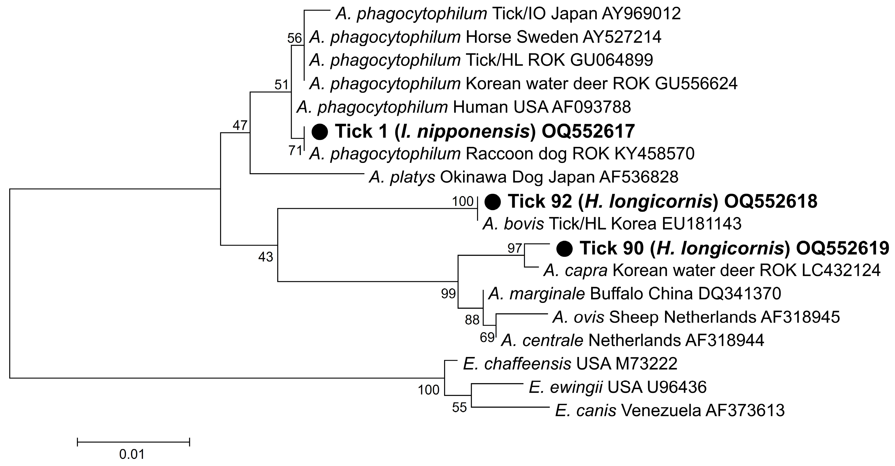
Figure 1. Phylogenetic analysis based on the 16S rRNA fragments of Anaplasma species. The phylogenetic tree was constructed using the maximum likelihood method based on the Kimura 2-parameter mode. The number on the branches indicates bootstrap percentages based on 1000 replications.
Pathogens 12 00802 g001
Figure 2. (a) Phylogenetic analysis of A. phagocytophilum based on the fragments of ankA and (b) msp4. (c) Phylogenetic analysis of A. bovis based on the fragments of groEL. The phylogenetic tree was constructed using the maximum likelihood method based on the Kimura 2-parameter mode. The number on the branches indicates bootstrap percentages based on 1000 replications.
Pathogens 12 00802 g002
Figure 3. Phylogenetic analysis of Ehrlichia based on the fragments of (a) 16S rRNA, (b) groEL, and (c) gltA. The phylogenetic tree was constructed using the maximum likelihood method based on the Kimura 2-parameter mode. The number on the branches indicates bootstrap percentages based on 1000 replications.
Pathogens 12 00802 g003
Figure 4. Phylogenetic analysis of Rickettsia based on the fragments of (a) 17 kDa, (b) ompA, and (c) gltA. The phylogenetic tree was constructed using the maximum likelihood method based on the Kimura 2-parameter mode. The number on the branches indicates bootstrap percentages based on 1000 replications.
Pathogens 12 00802 g004
Table 1. Oligonucleotide primer sequences used to detect tick-borne bacterial pathogens.
PathogensTarget GeneSequence 5′ to 3′Amplicon Size (bp)Reference
Anaplasma spp.16S rRNA1st5′-TCCTGGCTCAGAACGAACGCTGGCGGC-3′1433[38]
5′-AGTCACTGACCCAACCTTAAATGGCTG-3′
2nd5′-GTCGAACGGATTATTCTTTATAGCTTGC-3′926
5′-CCCTTCCGTTAAGAAGGATCTAATCTCC-3′
Anaplasma phagocytophilumankA1st5′-GAAGAAATTACAACTCCTGAAG-3′705[39]
5′-CAGCCAGATGCAGTAACGTG-3′
2nd5′-TTGACCGCTGAAGCACTAAC-3′664
5′-ACCATTTGCTTCTTGAGGAG-3′
msp41st5′-ATGAATTACAGAGAATTGCTTGTAGG-3′849[40]
5′-TTAATTGAAAGCAAATCTTGCTCCTATG-3′
2nd5′-CTATTGGYGGNGCYAGAGT-3′381
5′-GTTCATCGAAAATTCCGTGGTA-3′
Anaplasma bovisgroEL1st5′-GTTCGCAGTATTTTGCCAGT-3′845[41]
5′-CTGCRTTCAGAGTCATAAATAC-3′
2nd5′-ATCTGGAAGRCCACTATTGAT-3′
5′-CTGCRTTCAGAGTCATAAATAC-3′
Ehrlichia spp.16S rRNA1st5′-AAGCTTAACACATGCAAGTCGAA-3′1406[42]
5′-AGTCACTGACCCAACCTTAAATG-3′
2nd5′-CAATTGCTTATAACCTTTTGGTTATAAAT-3′390[43]
5′-TATAGGTACCGTCATTATCTTCCCTAT-3′
groEL1st5′-GAAGATGCWGTWGGWTGTACKGC-3′664[44]
5′-AGMGCTTCWCCTTCWACRTCYTC-3′
2nd5′-ATTACTCAGAGTGCTTCTCARTG-3′315
5′-TGCATACCRTCAGTYTTTTCAAC-3′
gltA1st5′-GGRRTRTTAACTTATGATCCAGG-3′575[45]
5′-GCATTYTGYTCATGATCAGCATG-3′
2nd5′-TTATGTCTACTGCTGCTTGTGA-3′478
5′-TARGAAGAAAYRTCAAACATCATATG-3′
Rickettsia spp.17 kDa1st5′-TTTACAAAATTCTAAAAACCAT-3′539[35]
5′-TCAATTCACAACTTGCCATT-3′
2nd5′-GCTCTTGCAACTTCTATGTT-3′450
5′-TCAATTCACAACTTGCCATT-3′
ompA1st5′-ATGGCGAATATTTCTCCAAAAA-3′634
5′-GTTCCGTTAATGGCAGCATCT-3′
2nd5′-ATGGCGAATATTTCTCCAAAAA-3′535
5′-AGTGCAGCATTCGCTCCCCCT-3′
gltA1st5′-GACCATGAGCAGAATGCTTCT-3′479[46]
5′-ATTGCAAAAAGTACAGTGAACA-3′
2nd5′-GGGGGCCTGCTCACGGCGG-3′382
5′-ATTGCAAAAAGTACAGTGAACA-3′
Borrelia spp.flagellin B1st5′-GATCARGCWCAAYATAACCAWATGCA-3′459[47]
5′-AGATTCAAGTCTGTTTTGGAAAGC-3′
2nd5′-GCTGAAGAGCTTGGAATGCAACC-3′351
5′-TGATCAGTTATCATTCTAATAGCA-3′
Table 2. Tick-borne bacterial pathogens identified using nested PCR performed with ticks collected from humans in the ROK.
SpeciesLife StageNo. of Collected TicksSub Total
(%)
Number of Bacterial Pathogens (%)
Anaplasma phagocyto-philumA. bovisA. capraEhrlichia sp.R. monacensisR. tamuraeCandidatus R. JingxinensisCandidatus R. Tarasevichiae
Haemaphysalis longicornisFemale3666 (56.4)01 (2.8)1 (2.8)0007 (19.4)0
Male10000001 (100.0)0
Nymph280000002 (7.1)0
Larva100000000
Haemaphysalis flavaFemale46 (5.1)0001 (25.0)0000
Nymph200000000
Haemaphysalis spp.Nymph11 (0.9)00000000
Amblyomma testudinariumFemale431 (26.5)00000000
Male8000001 (12.5)00
Nymph19000004 (21.0)00
Ixodes nipponensisFemale1010 (8.5)1 (10.0)0003 (30.0)01 (10.0)0
Ixodes persulcatusFemale11 (0.9)00000001 (100.0)
Ixodes spp.* n.d.22 (1.7)00001 (50.0)000
Total (%)Female55 (47.0)1 (1.8)1 (1.8)1 (1.8)1 (1.8)3 (5.4)08 (14.5)1 (1.8)
Male9 (7.7)000001 (11.1)1 (11.1)0
Nymph50 (42.7)000004 (8.0)2 (4.0)0
Larva1 (0.9)00000000
* n.d.2 (1.7)00001 (50.0)000
Total1171 (0.9)
(CI 0.0–4.8)
1 (0.9)
(CI 0.0–4.8)
1 (0.9)
(CI 0.0–4.8)
1 (0.9)
(CI 0.0–4.8)
4 (3.4)
(CI 0.9–8.7)
5 (4.2)
(CI 1.4–10.0)
11 (9.4)
(CI 4.7–16.8)
1 (0.9)
(CI 0.0–4.8)
* n.d.; not determined.
Disclaimer/Publisher’s Note: The statements, opinions and data contained in all publications are solely those of the individual author(s) and contributor(s) and not of MDPI and/or the editor(s). MDPI and/or the editor(s) disclaim responsibility for any injury to people or property resulting from any ideas, methods, instructions or products referred to in the content.