Article

Fasciola hepatica Excretory-Secretory Products (Fh-ES) Either Do Not Affect miRNA Expression Profile in THP-1 Macrophages or the Changes Are Undetectable by a Microarray Technique

1
Division of Pharmacology and Toxicology, Department of Preclinical Sciences, Institute of Veterinary Medicine, Warsaw University of Life Sciences, 02-786 Warsaw, Poland
2
Department of Physiological Sciences, Institute of Veterinary Medicine, Warsaw University of Life Sciences (SGGW), Nowoursynowska 159b, 02-776 Warsaw, Poland
3
Division of Parasitology and Parasitic Diseases, Department of Preclinical Sciences, Institute of Veterinary Medicine, Warsaw University of Life Sciences, 02-786 Warsaw, Poland
*
Author to whom correspondence should be addressed.
Pathogens 2024, 13(10), 854; https://doi.org/10.3390/pathogens13100854
Submission received: 31 August 2024 / Revised: 21 September 2024 / Accepted: 25 September 2024 / Published: 1 October 2024
(This article belongs to the Special Issue Immune Response of the Host and Vaccine Development—2nd Edition)

Abstract

:
Fasciola hepatica is a liver fluke that resides in the bile ducts of various mammals. The parasitosis leads to economic losses in animal production estimated at USD 3.2 billion annually. It is also considered a zoonosis of great significance and a problem for public health affecting 2.4 million people worldwide. Nevertheless, besides the negative aspects of infestation, the antigens released by the fluke, F. hepatica Excretory-Secretory Products (Fh-ES) contain several immunomodulatory molecules that may be beneficial during the course of type I diabetes, multiple sclerosis, ulcerative colitis, or septic shock. This phenomenon is based on the natural abilities of adult F. hepatica to suppress proinflammatory responses. To underline the molecular basis of these mechanisms and determine the role of microRNA (miRNA) in the process, lipopolysaccharide (LPS)-activated THP-1 macrophages were stimulated with Fh-ES, followed by miRNA microarray analyses. Surprisingly, no results indicating changes in the miRNA expression profile were noted (p < 0.05). We discuss potential reasons for these results, which may be due to insufficient sensitivity to detect slight changes in miRNA expression or the possibility that these changes are not regulated by miRNA. Despite the negative data, this work may contribute to the future planning of experiments by other researchers.

1. Introduction

Fasciola hepatica is a liver fluke found worldwide, infesting both wild and domestic animals, as well as humans. It is considered the most significant cause of cattle helminthiasis in Africa, with a prevalence of 30 to 90% [1], whereas in Australia, 46% of herds are likely to experience fluke-related production losses [2]. In Europe, it is becoming a re-emerging sheep disease, with infestation rates ranging from 6% to 61% [3]. Infested livestock suffer from severe anemia, liver failure, increased susceptibility to secondary infections, and, in cases of heavy infestations, death may occur [4]. This results in a reduction in milk yield, diminished weight gain, and decreased fertility, resulting in global economic losses estimated at USD 3.2 billion per year [5]. Humans can be accidentally infested by consuming water or plants contaminated with metacercariae, which is common for rural regions with low hygiene standards in the endemic areas. Infested individuals may suffer from right upper quadrant discomfort, anorexia, acute cholecystitis, and biliary obstruction; nevertheless, in most cases, the parasitosis is asymptomatic. Estimates indicate that 2.4 million people suffer from the disease, and a further 180 million are at risk, highlighting fascioliasis as a significant threat to public health. The scale of the problem led the World Health Organization (WHO) to classify human fascioliasis as a foodborne zoonosis belonging to the Neglected Tropical Diseases [6]. The Disability-Adjusted Life Years (DALY) value for the disease is estimated at 35,000, meaning that 35,000 years of human health are lost annually due to prevalent cases in the human population [7]. The presence of parasites in the bile ducts during the chronic phase is often asymptomatic, allowing the fluke to reproduce. Adult flukes release unembryonated eggs into the host’s bile ducts, which are then passed into the environment with stool during defecation. In water, the eggs become embryonated and hatch into free-swimming, ciliated miracidia that seek an intermediate host, typically a snail from the Lymnaeidae family [8]. Inside the snail’s tissues, the miracidia undergo several asexual metamorphoses, starting as sporocysts, which then transform into rediae, giving rise to another generation of rediae through clonal expansion [7,9]. Finally, the redia develops into a free-living cercaria, which exits the snail into the water [10], followed by encystation into metacercaria, which awaits ingestion by the definitive host. Once in the host’s duodenum, the metacercaria excysts into the newly excysted juvenile developmental stage (NEJ) and migrates through the intestinal wall and the peritoneum [11]—a process associated with the prehepatic (or the early) stage. Upon reaching the liver capsule, the immune system reacts vigorously, which is associated with the acute stage, manifesting as tissue destruction, inflammation, local or systemic toxic/allergic reactions, and internal damage [12]. The acute phase ceases once the parasite reaches the bile ducts, and the chronic phase begins, which may last 5 to 8 years [12]. The symptoms may resemble those of the acute stage but are usually more discrete [12]. Although fever, malaise, abdominal pain, eosinophilia, hepatomegaly, nausea, weight loss, liver failure [2,13], and biliary duct obstruction may occur [3,14], this stage is often asymptomatic.
During viral and bacterial infections or parasitoses, the type of immune response depends on a complicated interplay between the host and the intruder. It can be divided into four main subtypes: Th1, Th2, Th17, and Treg [15], although sometimes Th1 and Th17 responses are classified as Th1/Th17 due to overlapping effects [16]. These names refer to T helper (Th) and regulatory T (Treg) cell populations that orchestrate the particular response. Activated Th1 cells release significant amounts of interferon gamma (IFN-γ) and IL-12, activating cellular and inflammatory mechanisms, including proinflammatory M1 macrophages involved in phagocytosis and cell-mediated immunity, which are engaged in fighting intracellular pathogens and induce tissue damage. During a Th17 response, Th17 cells release substantial amounts of IL-17 and IL-22, which activate mechanisms against extracellular bacteria and fungi [17]. Th2 cells release IL-4, IL-5, and IL-13, leading to the alternative activation of macrophages, which counteract inflammation and promote tissue repair [18]. Treg (regulatory T cells) release IL-10 and transforming growth factor beta (TGF-β), which mitigate the effects of the other responses [19]. All of these types of immune responses need to be balanced and strictly regulated. Failure to control these processes can result in allergies, autoimmune diseases, or immunosuppression. Infectious agents elicit various immune responses depending on the pathogen, its isolate/strain [11,20], and the host’s genetics [21]. During the acute phase of fascioliasis, the predominant response is a Th2/Th17 response, which contributes to the liver fibrosis [11]. Once the parasite settles in the bile ducts, the immune response shifts towards Treg [11], which is beneficial for both the host and the parasite. The host avoids immunopathology, while the parasite can survive and reproduce. It is in the parasite’s interest to modulate the immune response, so it releases a number of bioactive molecules in Fasciola hepatica Excretory-Secretory Products (Fh-ES), which contain antioxidants, fatty acid binding proteins (FABPs), cysteine proteases, protease inhibitors, mucin-like peptides, TGF-β mimicking molecules, helminth defense molecules (HDMs) [22], as well as extracellular vesicles containing immunomodulatory proteins [23,24,25,26,27] and miRNAs [28]. These factors contribute in various ways to shifting the immune response and protecting the parasite. Fh-FABPs induce a tolerogenic phenotype in human dendritic cells [29] and suppress the expression of proinflammatory factors (IL-1β and tumor necrosis factor alpha (TNF-α)) in monocytes [30], similar to activin/TGF-like molecule (Fh-TLM—a member of the TGF superfamily), which induces a Th2/Treg-like phenotype in macrophages [31]. Proteases may activate the NLR family pyrin domain containing 3 inflammasome (NLRP3) [32] and protect the parasite by cleaving antibodies. Antioxidants exhibit pleiotropic effects by directly promoting a Th2 response, preventing the development of a tissue-damaging Th17 response, and detoxifying host reactive metabolites [22]. It seems that an overall effect of Fh-ES is a combination of both redundant and non-redundant actions of its components on the immune system, and further characterization of this impact is required.
Macrophages are an important part of both the innate and adaptive immune responses, although their interaction with Fh-ES is still not fully understood. While data show a common trend—Fh-ES’s ability to skew the immune response toward Th2/Treg—the mechanisms may be species-dependent. Previously, we demonstrated that Fh-ES reduces TNF-α release from bovine macrophages [20,33]. A similar anti-inflammatory effect of Fh-ES (or its fractions) has been shown for human monocytes and mouse macrophages; however, different mechanisms were observed in these models. In mouse macrophages, Fh-ES increased the release of TGF-β and IL-10, indicating a hallmark of Th2 activation [34]. On the other hand, Fh-EVs (Fasciola hepatica Extracellular Vesicles) affected neither TNF-α mRNA expression nor its release by human monocytes. However, prolonged stimulation with LPS and Fh-EVs increased the mRNA expression of TGF-β and IL-13, powerful modulators of Treg and Th2 responses, respectively [35]. The various mechanisms responsible for the upregulation of Th2/Treg mediators and the downregulation of Th1/Th17 mediators remain to be elucidated. Resolving this puzzle would enhance our understanding of the molecular mechanisms occurring not only during fascioliasis but also in other helminthiases. Transcriptome profiling and biological big data analyses are undoubtedly powerful tools to decipher those interactions. miRNAs are potent regulators of gene expression and are heavily involved in regulating the immune system [36], including macrophage polarization [37]. These small, ~22 nt nucleotide-long molecules downregulate gene expression by binding to target mRNAs, leading to mRNA cleavage, destabilization, or inhibition of translation. The miRNAs impact on gene expression is challenging to decipher since a single miRNA can bind to hundreds of target mRNAs, and a single mRNA may be downregulated by multiple miRNAs [38]. Moreover, parasites have evolved the ability to manipulate the host miRNAome by transporting their own miRNAs into host macrophages [39]. Given the significant role of miRNAs in regulating macrophage phenotypes, we undertook a study to determine changes in miRNA expression profile in human macrophages. We chose a well-established cell line model—human THP-1 cells—which has been used in numerous parasitological experiments [40,41].

2. Materials and Methods

2.1. Fh-ES Preparation

Fh-ES collected from adult flukes (Weybridge strain) were generously provided by Dr. Luke Norbury (Witold Stefański Institute of Parasitology, Polish Academy of Sciences, Twarda 51/55, 00-818, Warsaw, Poland). Upon recovery of flukes from the rats’ bile ducts, the Fh-ES was collected as described previously [42] with slight modifications. The worms were incubated (37 °C, 5% CO2) in 40 mL RPMI1640 (Merck, Germany, Darmstadt) supplemented with penicillin (100 U/mL) and streptomycin (100 µg/mL) for 30 min to eliminate host tissue contamination and residues of bile. The medium was discarded and the flukes were rinsed twice with the prewarmed medium before adding 30 mL of fresh prewarmed medium. The worms were incubated for 36 h, with medium change every 90–120 min. Each batch was immediately frozen at −80 °C upon collection and stored until use. All batches were thawed and pooled together, and the protein concentration was measured using the Bradford assay. The Fh-ES was then concentrated using a 5 kDa cutoff Amicon filter (Merck, Darmstadt, Germany) as follows: 10 mL of Fh-ES was added on the membrane and centrifuged (3000× g, 4 °C) until volume reached 0.5 mL. The flow-through was discarded, and 12 mL of a fresh RPMI1640 medium (4 °C) without antibiotics was added. The sample was centrifuged again (3000× g, 4 °C) until the volume reached 0.5 mL. The cycle was repeated three times, resulting in a media exchange of 390,000×. The concentrated fraction was filtered through a 0.22 µm syringe filter, followed by protein concentration measurement using the Bradford method [43], and stored at −80 °C until use.

2.2. Assessment of Endotoxin Level

The endotoxin level was evaluated using the Pierce LAL (Limulus amebocyte lysate) Chromogenic Endotoxin Quantitation Kit (Thermo Fisher Scientific, Waltham, MA, USA).

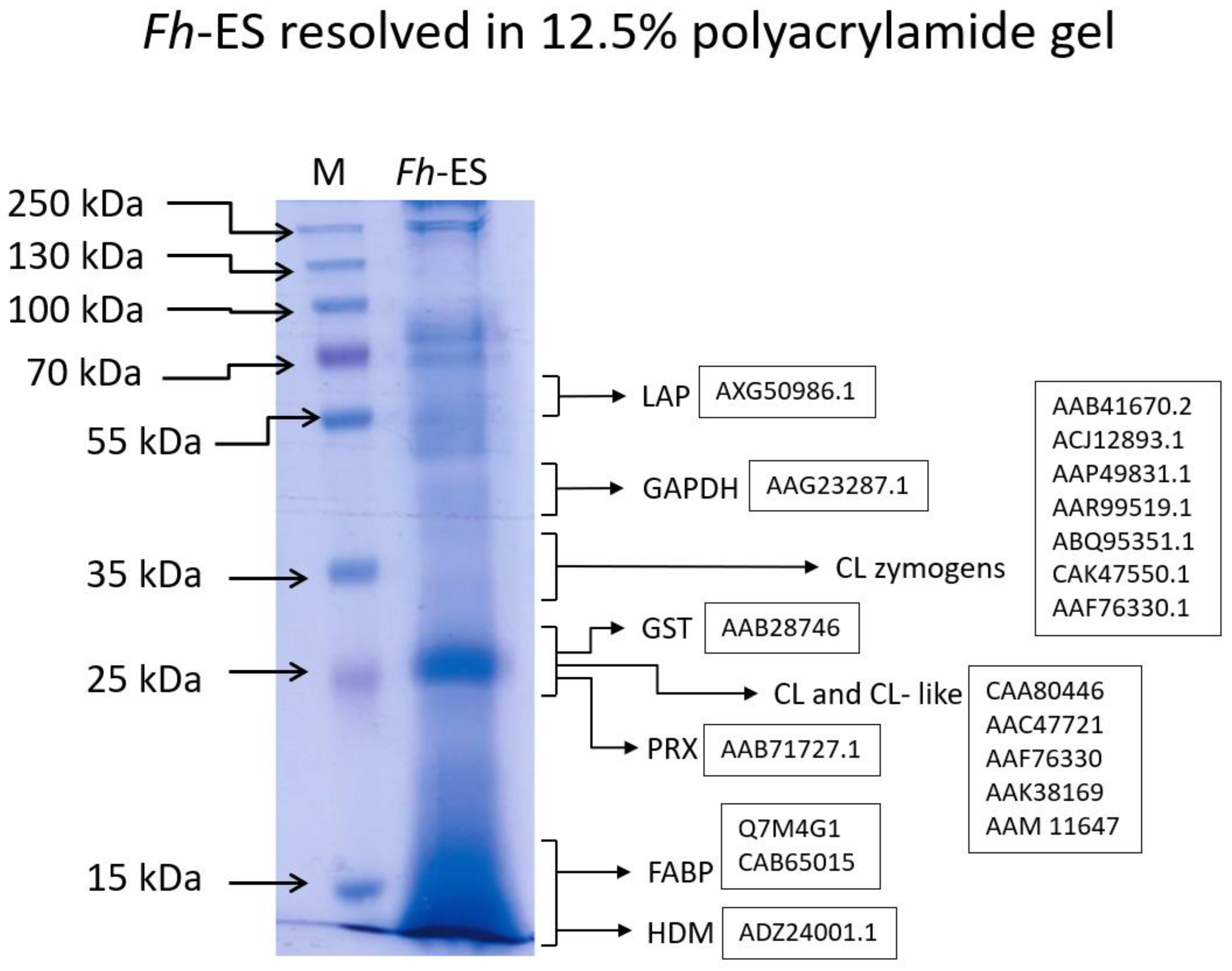
2.3. SDS PAGE Analysis

The concentrated fraction (10 µg of Fh-ES) was subjected to SDS PAGE analysis. Briefly, 10.6 µL of concentrated Fh-ES (0.93 ng/µL) were mixed with 42.4 µL of phosphate-buffered saline (PBS) and 13.25 µL of 5 × SDS PAGE Dye (10% SDS, 5% β-mercaptoethanol, 50% Glycerol, 500 mM Tris-HCl (pH 6.8) and 0.05% bromophenol blue dye) followed by incubation at 95 °C for 5 min. The sample and the molecular weight marker (Thermo Fisher Scientific Cat. No. 26619) were loaded onto a 4% stacking gel and resolved (40 V) until they reached a 12.5% polyacrylamide resolving gel containing 0.2% SDS and further resolved (at 80 V) until the bromophenol blue reached the bottom of the gel. The gel was stained overnight with a staining solution (5% v/v acetic acid, 30% v/v methanol, 0.1% w/v Coomassie Brilliant Blue R250) with gentle shaking following destaining using destaining solution (5% v/v acetic acid, 30% v/v methanol).

2.4. THP-1 Macrophages Stimulation

THP-1 monocytes were purchased from American Type Culture Collection (ATCC, Manassas, VA, USA). The monocytes were cultured at 37 °C with 5% CO2 in RPMI 1640 supplemented with 10% fetal bovine serum (FBS), 100 U/mL penicillin, and 100 µg/mL streptomycin. Once the cells reached the appropriate number, they were seeded at 5 × 10⁵ cells per well in a 24-well cell culture plate and differentiated into macrophages for 48 h using phorbol 12-myristate 13-acetate (PMA) (30 ng/mL). The cells were rinsed 3 times with fresh medium followed by treatment with either lipopolysaccharide (LPS) (100 ng/mL) for control cells or LPS (100 ng/mL) and Fh-ES (7 µg/mL) for experimental cells. Considering the final endotoxin level in the cell culture medium as an important parameter of the experiment, the concentration of Fh-ES used to stimulate THP-1 macrophages resulted in an endotoxin enrichment of the medium to a final concentration of 0.06 EU/mL, which complies with the strict FDA recommendations for endotoxin levels, even for solutions in contact with cerebrospinal fluid [44]. The cells were stimulated in octuplicates and incubated for 48 h (37 °C, 5% CO2). The medium was discarded followed by suspending the cells in QIAzol Lysis Reagent (Qiagen, Venlo, The Netherlands). The suspension of the lysed cells was stored until use at −80 °C.

2.5. Microarray Experiments

miRNA was isolated using the miRNeasy Mini Kit (Qiagen, Venlo, The Netherlands). The RNA concentration was assessed spectrophotometrically. The RNA quality and integrity were examined with the Agilent 2100 Bioanalyzer using Agilent RNA 6000 Nano Kit (Agilent Technologies, Santa Clara, CA, USA). The RNA integrity number (RIN) values for each sample were above 9. The RNA from each well (8 control wells and 8 experimental wells) was prepared for hybridization with arrays using the miRNA Complete Labeling and Hyb Kit (Agilent Technologies, Santa Clara, CA, USA) and hybridized to the Human miRNA Microarray, Release 21.0, 8 × 60 K (G4872A, Agilent Technologies) according to the manufacturer’s protocol. Microarrays were scanned using Agilent DNA Microarray Scanner G2505C and analysis of hybridization intensities was performed using Agilent Feature Extraction (FE) Software, version 10.7.3.1. The results were analyzed using GeneSpring 14 software (Agilent Technologies, Santa Clara, CA, USA).

2.6. Statistical Analyses

The analyses were performed on miRNA samples obtained from eight wells of control samples (LPS-activated) and eight wells of experimental samples (LPS-activated and Fh-ES). The probe sets were filtered by flags to remove poor-quality probes (absent flags) and were compared using a moderated t-test. However, as commonly observed, using a t-test to analyze multiple samples results in a number of false-positive results [45]. To address this issue, various multiple testing corrections were applied to adjust the p-values for multiple comparisons. These corrections included the Family-Wise Error Rate (FWER) methods: Bonferroni, Bonferroni-Holm, and Westfall-Young Permutation, and the False Discovery Rate (FDR) methods: Benjamini–Hochberg, Storey with Bootstrapping, and Storey with Curve Fitting. The corrections are listed in Table 1 according to their stringency [46], which defines their abilities to reduce the generation of false-positive data. The statistical analyses were performed using GenSpring 14 software (Agilent Technologies, Santa Clara, CA, USA).

3. Results

3.1. Fh-ES Assessment Using SDS-PAGE

The Fh-ES were resolved in a polyacrylamide gel prior to cell stimulation. A specific pattern was obtained (Figure 1), and the particular bands were identified according to the literature, with an emphasis on their immunomodulatory functions. The ~25 kDa band represents the most abundant protein released by the adult fluke: F. hepatica Cathepsins L (CL) and Cathepsin L-like proteins (CL-like) [24,42] which are released as 30–38 kDa zymogens [47] and processed into their mature forms. CLs have been shown to induce NLRP3 inflammasome in dendritic cells [32] and modulate CD4 expression on T Cells [48]. Other components of the ~25 kDa band are likely to include F. hepatica peroxiredoxins (PRXs) [49] and F. hepatica glutathione S-transferases (GSTs) [50], both of which act as antioxidants [51] and immunomodulators, influencing macrophages. PRX induces alternative activation [26], and GST stimulates prostaglandin production by macrophages [50]. The other abundant fraction (~12 kDa) likely represents F. hepatica fatty acid binding proteins (FABPs) [51], which induce alternative activation of macrophages [52] and F. hepatica helminth defense molecules (HDMs) [24,53], which are able to block Toll-like receptor (TLR) activation through LPS binding [27]. The composition of the proteins in bands with higher molecular masses is more challenging to decipher; however, Fh-ES is known to contain leucine aminopeptidases (LAP) (~70 kDa) [54] and somatic antigens like glyceraldehyde-3-phosphate dehydrogenase (GAPDH) and actin [42], which may be shed from the tegument during in vitro culture. The resolved fraction may also host proteins such as superoxide dismutase, serum albumin precursor, regucalcin or transferrin [42].

3.2. Statistical Analyses of Changes in miRNAnome Expression

The obtained microarray raw data were deposited in the Gene Expression Omnibus (GEO) database under the number GSE277108 and subjected to statistical analyses. A number of statistical methods were used to analyze the results. First, the moderated t-test was applied to identify miRNAs with changed expression fold (p < 0.05). The analyses without a correction identified 7 down-regulated and 11 upregulated miRNAs (Table 2). Nevertheless, multisample analyses are prone to I-type mistakes (generating false positive results). The classical p-value concept is suitable for testing differences between two samples, expecting one false positive discovery per twenty analyses when set as 0.05. During microarray analyses, multiple comparisons are made: mean expression difference of miRNA1 between the control group (LPS-activated macrophages) and the treated group (control stimulated with Fh-ES) is tested using a t-test. Next, the same comparison is made for miRNA2, miRNA3, … miRNAn. This approach results in an increase of false positive results (type 1 error), e.g., analyzing the expression of 3000 miRNAs with constant expression among samples will result in randomly identifying, with the highest probability, 150 (5%) as having changed expression (false positive). Assuming a further 20 miRNAs are expressed differently among samples and, if we identify all of them (rather unrealistic), we will have only a small fraction of true positives among all the positive hits: 20/(150 + 20). To overcome this issue, a statistical measure family-wise error rate (FWER) [55] was introduced for appropriate control of p-values [56]. It is defined as the probability of getting at least one false positive hit among the series of comparisons and can be calculated as FWER = 1 − (1 − α)n, where “α” is defined as a p-value threshold (0.05) and “n” as a number of comparisons. This formula shows that in analysis, e.g., 3000 miRNAs, we will get 1 - (1 − 0.05)3000 = 1, leading to conclusion of almost certainly false positive results. Controlling FWER is very efficient; nevertheless, it usually results in a decrease in true positive detection [57]. Another way to address multi-hypothesis analyses is the use of a parameter termed the false discovery rate (FDR) [56]. It is defined as a ratio of false positive to all positive hits, e.g., if there are 150 false positive hits and 20 true positive hits, FDR is calculated according to the formula: 150/170 = 88.2%. Both FWER and FDR may be controlled by the appropriate adjustment, but controlling FDR is more suitable for analyzing microarray data [58] due to lower stringency (an ability to reject false positive hits). During the microarray analysis, controlling both FWER and FDR was applied here. We used various corrections based on several modifications ranging from low to high stringency (Table 1) to increase the credibility of the results. Only the applied Westfall and Young permutation correction (p-value adjusted < 0.05) showed differences in expression of only one miRNA, miR-1537p (Table 2), while other corrections, including Benjamini–Hochberg, Bonferroni FWER, Bonferroni-Holm FWER, Storey with Bootstrapping, and Storey with Curve Fitting, showed no change in any miRNA upon stimulation of LPS-activated macrophages with Fh-ES (Table 2). There are no clear guidelines for FDR value since various experiments may require different approaches, e.g., clinical trials may require lower FDR than in vitro experiments [58], although it will increase false negative results. For that reason, we used a range of corrections, yet only one found a positive hit, which was considered an artifact.

4. Discussion

F. hepatica infestation is characterized by a dynamic immune response characterized by mixed Th2/Th17 type during the acute stage and Treg during established infestation [11]. The first stage is associated with NEJs‘ migration towards the liver through the intestine and peritoneum [11]. The latter stage is reached upon parasite settlement in the bile ducts and the release of immunomodulatory molecules [22]. The mounting Treg response during the adult stage is to prevent damage to host tissue induced by Th1 response and to preserve the comfort niche for the parasite suitable for survival and reproduction [59]. Fh-ES is a complicated mixture of antigens: antioxidants, FABPs, cysteine proteases, protease inhibitors, mucin-like peptides, TGF-β mimics, HDMs [22], and EVs carrying both proteins [24] and miRNAs [28] impacting host immune cells. A large body of research indicates the role of Fh-ES-derived molecules in suppressing inflammation in experimental models of autoimmune diseases: Fh-HDM-1 may ameliorate symptoms of type I diabetes or multiple sclerosis [60], GST attenuates septic shock in mice [41,61] whereas purified EVs are beneficial during ulcerative colitis [62]. These data encourage further investigation on immunomodulatory potential and molecular mechanisms induced in immune cells by Fh-ES. However, these experiments revealed the impact of Fh-ES-derived molecules, not the whole fraction of Fh-ES, which leaves a gap in the knowledge regarding the impact of the complete Fh-ES fraction on the immune response.
We chose to scrutinize Fh-ES’s impact on macrophages, which are a very plastic population that is involved not only in immune responses but also in wound healing, fibrosis, and neuroinflammation regulation [63]. Previous studies have shown significant effects of Fh-ES on these cells. For instance, they have been reported to affect LPS-activated human monocytes through TLR activation or deactivation [64] and to induce apoptosis in mouse peritoneal macrophages [65] or death of human hepatocytes [66]. Our previous research also demonstrated that Fh-ES ameliorates LPS-induced proinflammatory response in bovine macrophages [67] and indicated that this amelioration may depend on the isolate used for Fh-ES collection [33]. To further characterize the impact of Fh-ES on macrophages and their role in the modulation of immune response towards a Th2 phenotype, we used a widely employed laboratory model of LPS-activated THP-1 macrophages stimulated with the full Fh-ES fraction. Surprisingly, the experiments showed no change in the expression of miRNAs upon stimulation of LPS-activated THP-1 macrophages with Fh-ES. Despite employing various statistical corrections, only one out of six tests indicated a change. Moreover, according to the method, the expression of only one miRNA was changed, which seems to indicate a statistical error. On the other hand, Wang et al. (2021) recently indicated a change in the miRNA profile in goat PBMCs upon stimulation with Fg-ES (Fasciola gigantica ES) [68]. However, they used a significantly higher concentration of Fg-ES (80 µg) compared to our study (7 µg), which might explain the different cellular responses. Similarly, Guasconi et al. (2012) reported on an apoptotic impact of Fh-ESP on mouse macrophages (both ex vivo and in vitro) using Fh-ES in the concentration of 50 µg/mL [65]. In both the above-mentioned experiments, cells were not subjected to LPS activation. On the other hand, THP-1 cells stimulated with Toxocara canis ES at a concentration of 5 µg/mL showed significant changes in cytokine profile (both with and without LPS activation) [40]. THP-1 macrophages were also sensitive to 5 µg/mL of Hd-ES (Hymenolepis diminuta ES), showing changes in the gene expression profile [69].
Although specific data on the effective dose of Fh-ES required to change miRNA profile in THP-1 macrophages are lacking, there is evidence that recombinant antigens can alter cytokine expression. For example, recombinant Fg-Cyst (Fasciola gigantica type I cystatin) at a concentration of 5 µg/mL dampens IL-6, cyclooxygenase-2 (Cox-2), and inducible nitric oxide synthase iNOS [70] in LPS-activated THP-1 macrophages, which are controlled by miR-146a [71], miR29b [72] and miR-369-3p [73], respectively. However, this effect was observed only at the protein level, with no changes in the mRNA level encoding IL-6, Cox-2, and iNOS [70]. Similarly, Fasciola hepatica Fh-FABP-12 at 5 μg/mL decreased the mRNA expression of proinflammatory IL-12a, TNF-α, and IL-1β in LPS-activated human macrophages ex vivo [52]. Of course, direct comparison of concentrations of single recombinant antigens and the mixture of molecules such as Fh-ES is challenging. While a single antigen may be sufficient to bind to its receptor on a cell, concentrations in vivo are typically lower, and cells are not exposed to such high concentrations. Nevertheless, in vitro and ex vivo models may require a higher concentration of antigens due to a lack of interactions with other cells and extracellular matrix (ECM) rendering them less responsive to natural concentrations. Moreover, the use of recombinant proteins may require higher concentration than their in vivo counterparts due to differences in post-translational modifications, dampening their biological activity, especially when expressed in prokaryotic expression systems that lack a number of eukaryotic modifications. Extracellular vesicles (Fh-EV) are a significant component of Fh-ES. Sanchez-López demonstrated that a much lower concentration (10 µg/mL) of Fh-EV is sufficient to increase TGF-β and IL-13 expression and decrease IL-6, IL-1β, C-X-C motif ligand 8 (CXCL8), and nuclear factor kappa-light-chain-enhancer of activated B cells (NF-κB) expression in LPS-activated THP-1 cells. This effect was not observed in cells stimulated with Fh-EV depleted of various fractions [35]. This suggests that smaller concentrations may be effective in experiments since physiological outcomes result from complex interactions between Fh-ES and cell receptors, rather than the effects of single antigens alone. Another issue to consider is the sensitivity of the method to detect the changes of miRNA levels and the biological effects of their fluctuations. Recent research has shown that while COVID-19 patients exhibited altered miRNA expression profiles in peripheral blood, the most highly upregulated and downregulated miRNAs changed their levels by 1.6 and 2.3 times, respectively [74]. Such changes were detectable using a different technique, that is Next Generation Sequencing (NGS) performed on 14 samples. Moreover, although the LPS activation of macrophages is associated with higher fold change in miRNA levels [75], the vast majority of miRNAs remain similar across M1, M2a, M2b, and M2c macrophages [76], with only eight miRNAs showing distinct expression differences; however, at the same time some mRNAs may change expression up to 80 times [76]. This addresses the issue of statistical analyses of microarrays. It is more challenging to find differences when the change of miRNA expression is low. Moreover, multiple sample analyses of miRNA results [77] require correction, decreasing false positive rates [78]. We used a number of corrections with a workflow from the least to the most conservative. Of course, it cannot be excluded that false negative results did not occur here since none of the methods is 100% reliable [79]. Similar experiments were conducted with mouse RAW364-7 macrophages, where NGS analyses revealed that the cells upregulated and downregulated 16 miRNAs, with the greatest decrease and increase in expression being 3.01 and 10.18, respectively [80]. Our experiments used LPS-activated macrophages and employed microarray technology, which recent data suggest is less suitable for miRNAome analyses compared to NGS [81]. This might explain the difficulty in identifying differences with our approach, especially if the particular macrophage population shows stably expressed miRNAs [76].

5. Conclusions

Fh-ES is a powerful modulator of host immune response; however, no changes in miRNA expression in the macrophages have been noted in this study. The results may be due to subtle changes not detectable by the microarray technique and the statistical analysis workflow. A deeper investigation is still to be performed, possibly through more sensitive NGS techniques.

Author Contributions

Conceptualization, P.B. and M.W.; investigation, P.B., W.Z., E.D. and A.M.; data curation, P.B.; writing—original draft preparation, P.B.; writing—review and editing, M.W., E.D. and W.Z.; funding acquisition, P.B. All authors have read and agreed to the published version of the manuscript.

Funding

This study was supported by a research grant no 2017/01/X/NZ6/00475 (Miniatura) from the Polish National Science Centre.

Institutional Review Board Statement

Not applicable.

Informed Consent Statement

Not applicable.

Data Availability Statement

The raw data were deposited in GEO database (No.: GSE277108).

Acknowledgments

We thank Luke Norbury for providing the material: Fasciola hepatica Excretory-Secretory Products (Fh-ES).

Conflicts of Interest

The authors declare no conflicts of interest.

Abbreviations and Acronyms

CLCathepsins L
CL-likeCathepsin L-like proteins
Cox-2cyclooxygenase-2
CXCL8C-X-C motif ligand 8
DALYDisability-Adjusted Life Years
ECMextracellular matrix
FDRfalse discovery rate
FWERfamily wise error rate
FABPfatty acid binding protein
FBSFetal Bovine Serum
GAPDHglyceraldehyde-3-phosphate dehydrogenase
GEOGene Expression Omnibus
GSTglutathione S-transferases
HDMhelminth defense molecule
iNOSinducible nitric oxide synthase
LALLimulus amebocyte lysate
LAPleucine aminopeptidase
LPSlipopolysaccharide
miRNAmicro RNA
NEJnewly excysted juvenile
NF-κBnuclear factor kappa-light-chain-enhancer of activated B cells
NGSNext-Generation Sequencing
NLRP3family pyrin domain containing 3 inflammasome
PBSphosphate-buffered saline
PRXperoxiredoxin
TGF-βtransforming growth factor beta
TLRToll-like receptor

References

  1. Alemneh, T.; Ayelign, M. Fasciolosis—Public Health and Economic Impacts: A Review. Int. J. Zool. Investig. 2018, 4, 186–195. [Google Scholar]
  2. Kelley, J.M.; Rathinasamy, V.; Elliott, T.P.; Rawlin, G.; Beddoe, T.; Stevenson, M.A.; Spithill, T.W. Determination of the prevalence and intensity of Fasciola hepatica infection in dairy cattle from six irrigation regions of Victoria, South-eastern Australia, further identifying significant triclabendazole resistance on three properties. Vet. Parasitol. 2020, 277, 109019. [Google Scholar] [CrossRef] [PubMed]
  3. Rinaldi, L.; Biggeri, A.; Musella, V.; De Waal, T.; Hertzberg, H.; Mavrot, F.; Torgerson, P.R.; Selemetas, N.; Coll, T.; Bosco, A.; et al. Sheep and Fasciola hepatica in Europe: The GLOWORM experience. Geospat. Health 2015, 9, 309–317. [Google Scholar] [CrossRef] [PubMed]
  4. Alsulami, M.N.; Mohamed, K.; Wakid, M.H.; Abdel-Gaber, R.; Timsah, A.G.; Al-Megrin, W.A.I.; Khan, A.; Elkholy, W.A.; Abdelaal, K.A.A.; Elshabrawy, H.A.; et al. Molecular Characterization of Fasciola hepatica in Sheep Based on DNA Sequences of Ribosomal ITS-1. Infect. Drug Resist. 2023, 16, 6661–6671. [Google Scholar] [CrossRef] [PubMed]
  5. Mehmood, K.; Zhang, H.; Sabir, A.J.; Abbas, R.Z.; Ijaz, M.; Durrani, A.Z.; Saleem, M.H.; Rehman, M.U.; Iqbal, M.K.; Wang, Y.; et al. A review on epidemiology, global prevalence and economical losses of fasciolosis in ruminants. Microb. Pathog. 2017, 109, 253–262. [Google Scholar] [CrossRef]
  6. Available online: https://www.who.int/news-room/questions-and-answers/item/q-a-on-fascioliasis (accessed on 15 September 2024).
  7. Lalor, R.; Cwiklinski, K.; Calvani, N.E.D.; Dorey, A.; Hamon, S.; Corrales, J.L.; Dalton, J.P.; De Marco Verissimo, C. Pathogenicity and virulence of the liver flukes Fasciola hepatica and FasciolaGigantica that cause the zoonosis Fasciolosis. Virulence 2021, 12, 2839–2867. [Google Scholar] [CrossRef]
  8. Carnevale, S.; Malandrini, J.B.; Pantano, M.L.; Sawicki, M.; Soria, C.C.; Kuo, L.H.; Kamenetzky, L.; Astudillo, O.G.; Velásquez, J.N. Fasciola hepatica infection in humans: Overcoming problems for the diagnosis. Acta Parasitol. 2016, 61, 776–783. [Google Scholar] [CrossRef]
  9. Rondelaud, D.; Belfaiza, M.; Vignoles, P.; Moncef, M.; Dreyfuss, G. Redial generations of Fasciola hepatica: A review. J. Helminthol. 2009, 83, 245–254. [Google Scholar] [CrossRef]
  10. Available online: https://www.ncbi.nlm.nih.gov/books/NBK537032/ (accessed on 15 September 2024).
  11. Tanabe, M.B.; Caravedo, M.A.; Clinton White, A., Jr.; Cabada, M.M. An Update on the Pathogenesis of Fascioliasis: What Do We Know? Res. Rep. Trop. Med. 2024, 15, 13–24. [Google Scholar] [CrossRef]
  12. Available online: https://www.cdc.gov/liver-flukes/hcp/clinical-overview-fasciola/index.html (accessed on 15 September 2024).
  13. Kaya, M.; Beştaş, R.; Cetin, S. Clinical presentation and management of Fasciola hepatica infection: Single-center experience. World J. Gastroenterol. 2011, 17, 4899–4904. [Google Scholar] [CrossRef]
  14. Ezzat, R.F.; Karboli, T.A.; Kasnazani, K.A.; Hamawandi, A.M. Endoscopic management of biliary fascioliasis: A case report. J. Med. Case Rep. 2010, 4, 83. [Google Scholar] [CrossRef]
  15. Bailey, S.R.; Nelson, M.H.; Himes, R.A.; Li, Z.; Mehrotra, S.; Paulos, C.M. Th17 cells in cancer: The ultimate identity crisis. Front. Immunol. 2014, 5, 276. [Google Scholar] [CrossRef]
  16. Ferraro, A.; Buonocore, S.M.; Auquier, P.; Nicolas, I.; Wallemacq, H.; Boutriau, D.; van der Most, R.G. Role and plasticity of Th1 and Th17 responses in immunity to Staphylococcus aureus. Hum. Vaccines Immunother. 2019, 15, 2980–2992. [Google Scholar] [CrossRef] [PubMed]
  17. Corripio-Miyar, Y.; Hayward, A.; Lemon, H.; Sweeny, A.R.; Bal, X.; Kenyon, F.; Pilkington, J.G.; Pemberton, J.M.; Nussey, D.H.; McNeilly, T.N. Functionally distinct T-helper cell phenotypes predict resistance to different types of parasites in a wild mammal. Sci. Rep. 2022, 12, 3197. [Google Scholar] [CrossRef] [PubMed]
  18. Lee, J.; Lozano-Ruiz, B.; Yang, F.M.; Fan, D.D.; Shen, L.; González-Navajas, J.M. The Multifaceted Role of Th1, Th9, and Th17 Cells in Immune Checkpoint Inhibition Therapy. Front. Immunol. 2021, 12, 625667. [Google Scholar] [CrossRef] [PubMed]
  19. Asadi-Samani, M.; Bagheri, N.; Rafieian-Kopaei, M.; Shirzad, H. Inhibition of Th1 and Th17 Cells by Medicinal Plants and Their Derivatives: A Systematic Review. Phytother. Res. PTR 2017, 31, 1128–1139. [Google Scholar] [CrossRef] [PubMed]
  20. Bąska, P.; Norbury, L.J.; Zawistowska-Deniziak, A.; Wiśniewski, M.; Januszkiewicz, K. Excretory/secretory products from two Fasciola hepatica isolates induce different transcriptional changes and IL-10 release in LPS-activated bovine "BOMA" macrophages. Parasitol. Res. 2017, 116, 2775–2782. [Google Scholar] [CrossRef]
  21. Valanparambil, R.M.; Tam, M.; Jardim, A.; Geary, T.G.; Stevenson, M.M. Primary Heligmosomoides polygyrus bakeri infection induces myeloid-derived suppressor cells that suppress CD4+ Th2 responses and promote chronic infection. Mucosal Immunol. 2017, 10, 238–249. [Google Scholar] [CrossRef]
  22. Ryan, S.; Shiels, J.; Taggart, C.C.; Dalton, J.P.; Weldon, S. Fasciola hepatica-Derived Molecules as Regulators of the Host Immune Response. Front. Immunol. 2020, 11, 2182. [Google Scholar] [CrossRef]
  23. Murphy, A.; Cwiklinski, K.; Lalor, R.; O’Connell, B.; Robinson, M.W.; Gerlach, J.; Joshi, L.; Kilcoyne, M.; Dalton, J.P.; O’Neill, S.M. Fasciola hepatica Extracellular Vesicles isolated from excretory-secretory products using a gravity flow method modulate dendritic cell phenotype and activity. PLoS Negl. Trop. Dis. 2020, 14, e0008626. [Google Scholar] [CrossRef]
  24. Cwiklinski, K.; de la Torre-Escudero, E.; Trelis, M.; Bernal, D.; Dufresne, P.J.; Brennan, G.P.; O’Neill, S.; Tort, J.; Paterson, S.; Marcilla, A.; et al. The Extracellular Vesicles of the Helminth Pathogen, Fasciola hepatica: Biogenesis Pathways and Cargo Molecules Involved in Parasite Pathogenesis. Mol. Cell. Proteom. MCP 2015, 14, 3258–3273. [Google Scholar] [CrossRef] [PubMed]
  25. Donnelly, S.; O’Neill, S.M.; Stack, C.M.; Robinson, M.W.; Turnbull, L.; Whitchurch, C.; Dalton, J.P. Helminth cysteine proteases inhibit TRIF-dependent activation of macrophages via degradation of TLR3. J. Biol. Chem. 2010, 285, 3383–3392. [Google Scholar] [CrossRef] [PubMed]
  26. Donnelly, S.; O’Neill, S.M.; Sekiya, M.; Mulcahy, G.; Dalton, J.P. Thioredoxin peroxidase secreted by Fasciola hepatica induces the alternative activation of macrophages. Infect. Immun. 2005, 73, 166–173. [Google Scholar] [CrossRef] [PubMed]
  27. Robinson, M.W.; Donnelly, S.; Hutchinson, A.T.; To, J.; Taylor, N.L.; Norton, R.S.; Perugini, M.A.; Dalton, J.P. A family of helminth molecules that modulate innate cell responses via molecular mimicry of host antimicrobial peptides. PLoS Pathog. 2011, 7, e1002042. [Google Scholar] [CrossRef]
  28. Fromm, B.; Trelis, M.; Hackenberg, M.; Cantalapiedra, F.; Bernal, D.; Marcilla, A. The revised microRNA complement of Fasciola hepatica reveals a plethora of overlooked microRNAs and evidence for enrichment of immuno-regulatory microRNAs in extracellular vesicles. Int. J. Parasitol. 2015, 45, 697–702. [Google Scholar] [CrossRef]
  29. Zawistowska-Deniziak, A.; Lambooij, J.M.; Kalinowska, A.; Patente, T.A.; Łapiński, M.; van der Zande, H.J.; Basalaj, K.; de Korne, C.M.; Chaye, M.A.M.; Gasan, T.A.; et al. Fasciola hepatica Fatty Acid Binding Protein 1 Modulates T cell Polarization by Promoting Dendritic Cell Thrombospondin-1 Secretion Without Affecting Metabolic Homeostasis in Obese Mice. Front. Immunol. 2022, 13, 884663. [Google Scholar] [CrossRef]
  30. Ramos-Benítez, M.J.; Ruiz-Jiménez, C.; Aguayo, V.; Espino, A.M. Recombinant Fasciola hepatica fatty acid binding protein suppresses toll-like receptor stimulation in response to multiple bacterial ligands. Sci. Rep. 2017, 7, 5455. [Google Scholar] [CrossRef]
  31. Sulaiman, A.A.; Zolnierczyk, K.; Japa, O.; Owen, J.P.; Maddison, B.C.; Emes, R.D.; Hodgkinson, J.E.; Gough, K.C.; Flynn, R.J. A Trematode Parasite Derived Growth Factor Binds and Exerts Influences on Host Immune Functions via Host Cytokine Receptor Complexes. PLoS Pathog. 2016, 12, e1005991. [Google Scholar] [CrossRef]
  32. Celias, D.P.; Corvo, I.; Silvane, L.; Tort, J.F.; Chiapello, L.S.; Fresno, M.; Arranz, A.; Motran, C.C.; Cervi, L. Cathepsin L3 From Fasciola hepatica Induces NLRP3 Inflammasome Alternative Activation in Murine Dendritic Cells. Front. Immunol. 2019, 10, 552. [Google Scholar] [CrossRef]
  33. Bąska, P.; Zawistowska-Deniziak, A.; Norbury, L.J.; Wiśniewski, M.; Januszkiewicz, K. Fasciola Hepatica Isolates Induce Different Immune Responses in Unmaturated Bovine Macrophages. J. Vet. Res. 2019, 63, 63–70. [Google Scholar] [CrossRef]
  34. Guasconi, L.; Burstein, V.L.; Beccacece, I.; Mena, C.; Chiapello, L.S.; Masih, D.T. Dectin-1 on macrophages modulates the immune response to Fasciola hepatica products through the ERK signaling pathway. Immunobiology 2018, 223, 834–838. [Google Scholar] [CrossRef] [PubMed]
  35. Sánchez-López, C.M.; González-Arce, A.; Soler, C.; Ramírez-Toledo, V.; Trelis, M.; Bernal, D.; Marcilla, A. Extracellular vesicles from the trematodes Fasciola hepatica and Dicrocoelium dendriticum trigger different responses in human THP-1 macrophages. J. Extracell. Vesicles 2023, 12, e12317. [Google Scholar] [CrossRef] [PubMed]
  36. Curtale, G.; Rubino, M.; Locati, M. MicroRNAs as Molecular Switches in Macrophage Activation. Front. Immunol. 2019, 10, 799. [Google Scholar] [CrossRef] [PubMed]
  37. Khayati, S.; Dehnavi, S.; Sadeghi, M.; Tavakol Afshari, J.; Esmaeili, S.A.; Mohammadi, M. The potential role of miRNA in regulating macrophage polarization. Heliyon 2023, 9, e21615. [Google Scholar] [CrossRef]
  38. Ni, W.J.; Leng, X.M. Dynamic miRNA-mRNA paradigms: New faces of miRNAs. Biochem. Biophys. Rep. 2015, 4, 337–341. [Google Scholar] [CrossRef]
  39. Tran, N.; Ricafrente, A.; To, J.; Lund, M.; Marques, T.M.; Gama-Carvalho, M.; Cwiklinski, K.; Dalton, J.P.; Donnelly, S. Fasciola hepatica hijacks host macrophage miRNA machinery to modulate early innate immune responses. Sci. Rep. 2021, 11, 6712. [Google Scholar] [CrossRef]
  40. Długosz, E.; Basałaj, K.; Zawistowska-Deniziak, A. Cytokine production and signalling in human THP-1 macrophages is dependent on Toxocara canis glycans. Parasitol. Res. 2019, 118, 2925–2933. [Google Scholar] [CrossRef]
  41. Aguayo, V.; Valdés Fernandez, B.N.; Rodríguez-Valentín, M.; Ruiz-Jiménez, C.; Ramos-Benítez, M.J.; Méndez, L.B.; Espino, A.M. Fasciola hepatica GST downregulates NF-κB pathway effectors and inflammatory cytokines while promoting survival in a mouse septic shock model. Sci. Rep. 2019, 9, 2275. [Google Scholar] [CrossRef] [PubMed]
  42. Morphew, R.M.; Wright, H.A.; LaCourse, E.J.; Woods, D.J.; Brophy, P.M. Comparative proteomics of excretory-secretory proteins released by the liver fluke Fasciola hepatica in sheep host bile and during in vitro culture ex host. Mol. Cell. Proteom. MCP 2007, 6, 963–972. [Google Scholar] [CrossRef]
  43. Bradford, M.M. A rapid and sensitive method for the quantitation of microgram quantities of protein utilizing the principle of protein-dye binding. Anal. Biochem. 1976, 72, 248–254. [Google Scholar] [CrossRef]
  44. Available online: https://www.fda.gov/regulatory-information/search-fda-guidance-documents/guidance-industry-pyrogen-and-endotoxins-testing-questions-and-answers (accessed on 28 August 2024).
  45. Lesack, K.; Naugler, C. An open-source software program for performing Bonferroni and related corrections for multiple comparisons. J. Pathol. Inform. 2011, 2, 52. [Google Scholar] [CrossRef] [PubMed]
  46. Available online: https://genespring-support.com/files/gs_12_6/GeneSpring-manual.pdf (accessed on 15 September 2024).
  47. Collett, C.F.; Phillips, H.C.; Fisher, M.; Smith, S.; Fenn, C.; Goodwin, P.; Morphew, R.M.; Brophy, P.M. Fasciola hepatica Cathepsin L Zymogens: Immuno-Proteomic Evidence for Highly Immunogenic Zymogen-Specific Conformational Epitopes to Support Diagnostics Development. J. Proteome Res. 2022, 21, 1997–2010. [Google Scholar] [CrossRef] [PubMed]
  48. Prowse, R.K.; Chaplin, P.; Robinson, H.C.; Spithill, T.W. Fasciola hepatica cathepsin L suppresses sheep lymphocyte proliferation in vitro and modulates surface CD4 expression on human and ovine T cells. Parasite Immunol. 2002, 24, 57–66. [Google Scholar] [CrossRef] [PubMed]
  49. Sekiya, M.; Mulcahy, G.; Irwin, J.A.; Stack, C.M.; Donnelly, S.M.; Xu, W.; Collins, P.; Dalton, J.P. Biochemical characterisation of the recombinant peroxiredoxin (FhePrx) of the liver fluke, Fasciola hepatica. FEBS Lett. 2006, 580, 5016–5022. [Google Scholar] [CrossRef] [PubMed]
  50. LaCourse, E.J.; Perally, S.; Morphew, R.M.; Moxon, J.V.; Prescott, M.; Dowling, D.J.; O’Neill, S.M.; Kipar, A.; Hetzel, U.; Hoey, E.; et al. The Sigma class glutathione transferase from the liver fluke Fasciola hepatica. PLoS Neglected Trop. Dis. 2012, 6, e1666. [Google Scholar] [CrossRef]
  51. Martínez-Fernández, A.R.; Nogal-Ruiz, J.J.; López-Abán, J.; Ramajo, V.; Oleaga, A.; Manga-González, Y.; Hillyer, G.V.; Muro, A. Vaccination of mice and sheep with Fh12 FABP from Fasciola hepatica using the new adjuvant/immunomodulator system ADAD. Vet. Parasitol. 2004, 126, 287–298. [Google Scholar] [CrossRef]
  52. Figueroa-Santiago, O.; Espino, A.M. Fasciola hepatica fatty acid binding protein induces the alternative activation of human macrophages. Infect. Immun. 2014, 82, 5005–5012. [Google Scholar] [CrossRef]
  53. Martínez-Sernández, V.; Mezo, M.; González-Warleta, M.; Perteguer, M.J.; Muiño, L.; Guitián, E.; Gárate, T.; Ubeira, F.M. The MF6p/FhHDM-1 major antigen secreted by the trematode parasite Fasciola hepatica is a heme-binding protein. J. Biol. Chem. 2014, 289, 1441–1456. [Google Scholar] [CrossRef]
  54. Marcilla, A.; De la Rubia, J.E.; Sotillo, J.; Bernal, D.; Carmona, C.; Villavicencio, Z.; Acosta, D.; Tort, J.; Bornay, F.J.; Esteban, J.G.; et al. Leucine aminopeptidase is an immunodominant antigen of Fasciola hepatica excretory and secretory products in human infections. Clin. Vaccine Immunol. CVI 2008, 15, 95–100. [Google Scholar] [CrossRef]
  55. Nicholson, K.J.; Sherman, M.; Divi, S.N.; Bowles, D.R.; Vaccaro, A.R. The Role of Family-wise Error Rate in Determining Statistical Significance. Clin. Spine Surg. 2022, 35, 222–223. [Google Scholar] [CrossRef]
  56. Murray, M.H.; Blume, J.D. FDRestimation: Flexible False Discovery Rate Computation in R. F1000Research 2021, 10, 441. [Google Scholar] [CrossRef] [PubMed]
  57. Korthauer, K.; Kimes, P.K.; Duvallet, C.; Reyes, A.; Subramanian, A.; Teng, M.; Shukla, C.; Alm, E.J.; Hicks, S.C. A practical guide to methods controlling false discoveries in computational biology. Genome Biol. 2019, 20, 118. [Google Scholar] [CrossRef]
  58. Pawitan, Y.; Michiels, S.; Koscielny, S.; Gusnanto, A.; Ploner, A. False discovery rate, sensitivity and sample size for microarray studies. Bioinformatics 2005, 21, 3017–3024. [Google Scholar] [CrossRef] [PubMed]
  59. Bąska, P.; Norbury, L.J. The Role of the Intestinal Epithelium in the "Weep and Sweep" Response during Gastro-Intestinal Helminth Infections. Animals 2022, 12, 175. [Google Scholar] [CrossRef] [PubMed]
  60. Lund, M.E.; Greer, J.; Dixit, A.; Alvarado, R.; McCauley-Winter, P.; To, J.; Tanaka, A.; Hutchinson, A.T.; Robinson, M.W.; Simpson, A.M.; et al. A parasite-derived 68-mer peptide ameliorates autoimmune disease in murine models of Type 1 diabetes and multiple sclerosis. Sci. Rep. 2016, 6, 37789. [Google Scholar] [CrossRef]
  61. Valdes-Fernandez, B.N.; Ruiz-Jimenez, C.; Armina-Rodriguez, A.; Mendez, L.B.; Espino, A.M. Fasciola hepatica GST mu-class suppresses the cytokine storm induced by E. coli-lipopolysaccharide, whereas it modulates the dynamic of peritoneal macrophages in a mouse model and suppresses the classical activation of macrophages. Microbiol. Spectr. 2024, 12, e0347523. [Google Scholar] [CrossRef]
  62. Roig, J.; Saiz, M.L.; Galiano, A.; Trelis, M.; Cantalapiedra, F.; Monteagudo, C.; Giner, E.; Giner, R.M.; Recio, M.C.; Bernal, D.; et al. Extracellular Vesicles From the Helminth Fasciola hepatica Prevent DSS-Induced Acute Ulcerative Colitis in a T-Lymphocyte Independent Mode. Front. Microbiol. 2018, 9, 1036. [Google Scholar] [CrossRef]
  63. Mammana, S.; Fagone, P.; Cavalli, E.; Basile, M.S.; Petralia, M.C.; Nicoletti, F.; Bramanti, P.; Mazzon, E. The Role of Macrophages in Neuroinflammatory and Neurodegenerative Pathways of Alzheimer’s Disease, Amyotrophic Lateral Sclerosis, and Multiple Sclerosis: Pathogenetic Cellular Effectors and Potential Therapeutic Targets. Int. J. Mol. Sci. 2018, 19, 831. [Google Scholar] [CrossRef]
  64. Figueroa-Santiago, O.; Espino, A.M. Fasciola hepatica ESPs Could Indistinctly Activate or Block Multiple Toll-Like Receptors in a Human Monocyte Cell Line. Ann. Clin. Pathol. 2017, 5, 1112. [Google Scholar]
  65. Guasconi, L.; Serradell, M.C.; Masih, D.T. Fasciola hepatica products induce apoptosis of peritoneal macrophages. Vet. Immunol. Immunopathol. 2012, 148, 359–363. [Google Scholar] [CrossRef]
  66. Bąska, P.; Norbury, L.J.; Wiśniewski, M.; Januszkiewicz, K.; Wędrychowicz, H. Excretory/secretory products of Fasciola hepatica but not recombinant phosphoglycerate kinase induce death of human hepatocyte cells. Acta Parasitol. 2013, 58, 215–217. [Google Scholar] [CrossRef] [PubMed]
  67. Bąska, P.; Zawistowska-Deniziak, A.; Zdziarska, A.M.; Wasyl, K.; Wiśniewski, M.; Cywińska, A.; Klockiewicz, M.; Januszkiewicz, K.; Wkedrychowicz, H. Fasciola hepatica—The pilot study of in vitro assessing immune response against native and recombinant antigens of the fluke. Acta Parasitol. 2013, 58, 453–462. [Google Scholar] [CrossRef] [PubMed]
  68. Wang, S.S.; Chen, D.; He, J.J.; Zheng, W.B.; Tian, A.L.; Zhao, G.H.; Elsheikha, H.M.; Zhu, X.-Q. Fasciola gigantica-Derived Excretory-Secretory Products Alter the Expression of mRNAs, miRNAs, lncRNAs, and circRNAs Involved in the Immune Response and Metabolism in Goat Peripheral Blood Mononuclear Cells. Front. Immunol. 2021, 12, 653755. [Google Scholar] [CrossRef] [PubMed]
  69. Zawistowska-Deniziak, A.; Basałaj, K.; Strojny, B.; Młocicki, D. New Data on Human Macrophages Polarization by Hymenolepis diminuta Tapeworm-An In Vitro Study. Front. Immunol. 2017, 8, 148. [Google Scholar] [CrossRef] [PubMed]
  70. Chantree, P.; Tarasuk, M.; Prathaphan, P.; Ruangtong, J.; Jamklang, M.; Chumkiew, S.; Martviset, P. Type I Cystatin Derived from Fasciola gigantica Suppresses Macrophage-Mediated Inflammatory Responses. Pathogens 2023, 12, 395. [Google Scholar] [CrossRef]
  71. Luly, F.R.; Lévêque, M.; Licursi, V.; Cimino, G.; Martin-Chouly, C.; Théret, N.; Negri, R.; Cavinato, L.; Ascenzioni, F.; Del Porto, P. MiR-146a is over-expressed and controls IL-6 production in cystic fibrosis macrophages. Sci. Rep. 2019, 9, 16259. [Google Scholar] [CrossRef] [PubMed]
  72. Self-Fordham, J.B.; Naqvi, A.R.; Uttamani, J.R.; Kulkarni, V.; Nares, S. MicroRNA: Dynamic Regulators of Macrophage Polarization and Plasticity. Front. Immunol. 2017, 8, 1062. [Google Scholar] [CrossRef]
  73. Scalavino, V.; Liso, M.; Cavalcanti, E.; Gigante, I.; Lippolis, A.; Mastronardi, M.; Chieppa, M.; Serino, G. miR-369-3p modulates inducible nitric oxide synthase and is involved in regulation of chronic inflammatory response. Sci. Rep. 2020, 10, 15942. [Google Scholar] [CrossRef]
  74. Li, C.; Hu, X.; Li, L.; Li, J.H. Differential microRNA expression in the peripheral blood from human patients with COVID-19. J. Clin. Lab. Anal. 2020, 34, e23590. [Google Scholar] [CrossRef]
  75. Naqvi, A.R.; Zhong, S.; Dang, H.; Fordham, J.B.; Nares, S.; Khan, A. Expression Profiling of LPS Responsive miRNA in Primary Human Macrophages. J. Microb. Biochem. Technol. 2016, 8, 136–143. [Google Scholar] [CrossRef]
  76. Graff, J.W.; Dickson, A.M.; Clay, G.; McCaffrey, A.P.; Wilson, M.E. Identifying functional microRNAs in macrophages with polarized phenotypes. J. Biol. Chem. 2012, 287, 21816–21825. [Google Scholar] [CrossRef] [PubMed]
  77. Jung, S.H.; Bang, H.; Young, S. Sample size calculation for multiple testing in microarray data analysis. Biostatistics 2005, 6, 157–169. [Google Scholar] [CrossRef] [PubMed]
  78. Pounds, S.; Morris, S.W. Estimating the occurrence of false positives and false negatives in microarray studies by approximating and partitioning the empirical distribution of p-values. Bioinformatics 2003, 19, 1236–1242. [Google Scholar] [CrossRef] [PubMed]
  79. Ge, Y.; Sealfon, S.C.; Speed, T.P. Multiple testing and its applications to microarrays. Stat. Methods Med. Res. 2009, 18, 543–563. [Google Scholar] [CrossRef] [PubMed]
  80. Guo, X.; Zheng, Y. MicroRNA expression profile in RAW264.7 macrophage cells exposed to Echinococcus multilocularis metacestodes. Parasitology 2018, 145, 416–423. [Google Scholar] [CrossRef]
  81. Batan Pumeda, S. Comparison Between Next-Generation Sequencing and Microarrays for miRNA Expression in Cancer Samples. Natl. Acad. Sci. Lett. 2023. [Google Scholar] [CrossRef]
Figure 1. Fh-ES (10 µg) resolved in 12.5% polyacrylamide gel. M—molecular weight marker (ThermoFisher Scientific Cat. No. 26619), Fh-ES—Fasciola hepatica Excretory-Secretory Products. The protein identification in the bands is defined in the literature review. For the analyzed bands, the protein is given with Gen Bank numbers. CL—Cathepsin L, CL-like—Cathepsin L-like protein, FABP—fatty acid binding protein, GAPDH—glyceraldehyde-3-phosphate dehydrogenase, HDM—helminth defense molecule, LAP—leucine aminopeptidase, PRX—peroxiredoxin.
Pathogens 13 00854 g001
Table 1. Applied Corrections for Multiple Comparisons. The corrections are ordered according to their stringency (S) in comparison to the Benjamini–Hochberg correction. A higher number indicates a higher stringency of the test, corresponding to a lower probability of generating false-positive results.
SDescriptionCorrection
1Independence of p-values across genes is assumed.Benjamini–Hochberg (FDR)
Storey with Bootstrapping.
Storey with Curve Fitting.
Benjamini–Hochberg refinements
2Permutates all the genes at the same time, accounting for their dependence.Westfall and Young (FWER)
3A stepwise procedure. Tests each hypothesis in an ordered sequence, allowing one to accept or reject a hypothesis based on the previous step, with a focus on power and stringency.Bonferroni-Holm (FWER)
4A single step procedure where each p value is corrected independently.Bonferroni (FWER)
Table 2. The miRNAs considered as significant (p < 0.05) to change the expression upon stimulation of LPS-activated THP-1 macrophages with Fh-ES using a moderated t-test and several corrections. First, the analyses were performed using a moderated t-test and number of miRNAs were identified (p < 0.05). During the next step, various corrections were used to eliminate false positive results. ns—non significant (p > 0.05).
miRNAChangeFoldBenjamini–HochbergStorey with BootstrappingStorey with Curve FittingWestfall and YoungBonferroni-Holm FWERBonferroni FWER
p-Value
hsa-miR-4730down6.58nsnsnsnsnsns
hsa-miR-4728-3pdown5.72nsnsnsnsnsns
hsa-miR-1910-5pdown4.12nsnsnsnsnsns
hsa-miR-6824-3pdown3.31nsnsnsnsnsns
hsa-miR-4741down3.14nsnsnsnsnsns
hsa-miR-4462down3.1nsnsnsnsnsns
hsa-miR-26a-1-3pdown2.6nsnsnsnsnsns
hsa-miR-324-3pup1.4nsnsnsnsnsns
hsa-miR-19b-1-5pup3.31nsnsnsnsnsns
hsa-miR-7152-3pup3.51nsnsnsnsnsns
hsa-miR-6780a-5pup4nsnsnsnsnsns
hsa-miR-219a-5up4.09nsnsnsnsnsns
hsa-miR-6512-5pup4.32nsnsnsnsnsns
hsa-miR-378a-5pup4.52nsnsnsnsnsns
hsa-miR-34c-5pup4.57nsnsnsnsnsns
hsa-miR-487b-3pup5.15nsnsnsnsnsns
hsa-miR-4651up5.17nsnsnsnsnsns
hsa-miR-1537-3pup7.1nsnsns<0.05nsns
Disclaimer/Publisher’s Note: The statements, opinions and data contained in all publications are solely those of the individual author(s) and contributor(s) and not of MDPI and/or the editor(s). MDPI and/or the editor(s) disclaim responsibility for any injury to people or property resulting from any ideas, methods, instructions or products referred to in the content.

