Review

Advancements in Detection Methods for Salmonella in Food: A Comprehensive Review

by
Aayushi Patel
1,
Andrew Wolfram
1 and
Taseen S. Desin
2,*
1
Trinity School of Medicine, Trinity Medical Sciences University, Roswell, GA 30075, USA
2
Department of Medical Education, College of Medicine, University of Central Florida, Orlando, FL 32827, USA
*
Author to whom correspondence should be addressed.
Pathogens 2024, 13(12), 1075; https://doi.org/10.3390/pathogens13121075
Submission received: 29 August 2024 / Revised: 2 November 2024 / Accepted: 4 December 2024 / Published: 7 December 2024

Abstract

:
Non-typhoidal Salmonella species are one of the leading causes of gastrointestinal disease in North America, leading to a significant burden on the healthcare system resulting in a huge economic impact. Consequently, early detection of Salmonella species in the food supply, in accordance with food safety regulations, is crucial for protecting public health, preventing outbreaks, and avoiding serious economic losses. A variety of techniques have been employed to detect the presence of this pathogen in the food supply, including culture-based, immunological, and molecular methods. The present review summarizes these methods and highlights recent updates on promising emerging technologies, including aptasensors, Surface Plasmon Resonance (SPR), and Surface Enhanced Raman Spectroscopy (SERS).

1. Introduction

Non-typhoidal Salmonella (NTS) is a ubiquitous Gram-negative bacterial pathogen and among the leading causes of food and water-borne illnesses worldwide [1]. It causes an estimated 1.35 million infections in the United States annually, second only to Norovirus as the most prevalent causative agent for foodborne diseases [1]. NTS is the leading cause of foodborne infection-related hospitalizations and deaths in the United States, responsible for 26,500 hospitalizations and approximately 420 deaths each year [1]. The European Food Safety Authority (EFSA) has estimated that the overall economic burden of human salmonellosis could be as high as EUR 3 billion per year [2]. A member of the Enterobacteriaceae family, the bacterial genus Salmonella consists of two species, Salmonella enterica and Salmonella bongori, of which Salmonella enterica is further divided into six subspecies, with Salmonella enterica subspecies enterica consisting of at least 1531 serovars [3]. NTS harbors in the intestines and feces of animals and is primarily acquired through the consumption of contaminated foods, such as undercooked meat, raw eggs, dairy products, or fresh produce [4,5,6,7]. Of particular importance are poultry products, as chickens are a significant reservoir for NTS [8]. The most commonly implicated serovars in the case of human salmonellosis, according to the CDC, are S. enterica subspecies enterica serovar Typhimurium (S. Typhimurium), S. Enteritidis, and S. Newport [9,10].
Salmonellosis typically manifests as gastroenteritis characterized by diarrhea, abdominal cramps, nausea, vomiting, and fever [11,12]. Immunocompromised individuals, children under the age of five, and the elderly are particularly susceptible to infection [13,14]. The infection has an incubation period ranging from 6 to 96 h [15,16]. The disease is usually self-limited, resolving within 10 days in most healthy adults [11,12,13,14].
Upon ingestion, Salmonella survives its passage through the gastrointestinal tract, invades the M-cells of the intestinal epithelium, and replicates intracellularly, aided by Type 3 Secretion Systems (T3SS) encoded on Salmonella Pathogenicity Islands (SPI) [17,18,19,20]. In approximately 5% of Salmonella infections, the bacteria can spread from the intestine into the bloodstream, resulting in bacteremia. Additionally, in about 1.8% of cases of bacteremia, extraintestinal complications can manifest largely due to the persistence of Salmonella inside tissue macrophages [21]. According to the European Commission, the presence of Salmonella at 1 colony forming unit per milliliter (CFU/mL) in a portion of 25 g of ready-to-eat food is sufficient to cause disease in humans [1,9]. Early detection of Salmonella species in contaminated food is necessary to prevent human infections, allowing for at least 27 food recalls in 2024 by the U.S. Food and Drug Administration (FDA), as this pathogen is a public health concern and poses a significant threat to the safety of our food supply [1,22]. Additionally, timely detection of this pathogen results in the prevention of significant economic losses due to product recalls, medical expenses, and reduced consumer confidence in food safety [23]. Accurate detection of the Salmonella genus in food products also ensures compliance with various regulations and standards in different countries, which assists in maintaining the integrity of the food industry and avoids legal repercussions. In the USA, the validation of a Salmonella detection method involves a rigorous process that complies with guidelines set by regulatory agencies like the FDA, the U.S. Department of Agriculture (USDA), and sometimes the Environmental Protection Agency (EPA) [24]. Additionally, the requirements must also align with international standards, such as the International Organization for Standardization (ISO), particularly ISO 16140, which provides a framework for validating microbiological methods for food testing [25]. Similarly, in the European Union (EU), the validation of a Salmonella detection method must comply with standards set by the European Food Safety Authority (EFSA), Regulation (EC) No 2017/625 and ISO 16140 [26,27]. Moreover, with the increase in global food trade, it is essential to detect Salmonella in order to safeguard global food security and minimize the risk of spreading Salmonella across borders. A multitude of techniques are currently used to detect the bacterial pathogen Salmonella, and this review attempts to highlight new, promising technologies that can be used for this purpose.

2. Traditional Culture-Based Methods

Culture-based detection remains the ‘gold standard’ for microbiological analysis due to its high success rate and cost-effectiveness [1,9,28,29]. It can detect viable Salmonella in a 25 g sample with a detection limit of 1 CFU [29,30]. Since culture has been used to detect bacteria for so long, it is often required by law to detect foodborne pathogens via culture despite more modern detection methods being available [9]. Culture-based methods are sensitive, inexpensive, and can indicate the number and type of viable microorganisms in food samples. However, culture-based detection has several limitations, including slow growth rates (24–72 h) and labor-intensive steps [31].
The current procedure outlined by the International Organization for Standardization, ISO 6579-1:2017, involves non-selective enrichment in Buffered Peptone Water for approximately 18 h, followed by selective enrichment in Rappaport Vassiliadis broth or Muller-Kauffmann Tetrathionate Novobiocin broth for approximately 24 h [30]. Isolation of Salmonella colonies is done by plating on Xylose Lysine Deoxycholate (XLD) Agar and a second selective isolation agar complementary to XLD Agar as described in Annex E of ISO 6579-1:2017 [30]. This step requires another 24 h. Finally, biochemical identification and serological testing are performed, which can take an additional three days. However, with the use of rapid biochemical assay kits, multiple assays can be performed simultaneously, and results can be acquired within a day of inoculation [32]. More recently, an automated Biolog microbial identification system was developed, which uses redox indicators and imaging technology to identify multiple bacterial strains at the same time [33]. Additionally, serotyping is done for further typing of the Salmonella strains to the serovar level according to ISO/TR 6579-3:2014 [34]. It involves the use of standard agglutination methods using polyclonal antibodies against O antigens, flagellar H antigens, and capsular Vi antigens to identify the specific serogroup of Salmonella species. However, this poses a challenge for detection in fermented and heat-treated foods, as the surface antigens are often modified or missing, leading to false negatives [35]. Overall, agglutination tests are laborious, and often even acquiring the required antisera is time-consuming [32,35,36].

3. Immunological Techniques

Various immunological methods have been developed for the detection of Salmonella, such as enzyme-linked immunosorbent assays (ELISA) and lateral flow immunoassays, which utilize antigen–antibody interactions to offer rapid and specific identification suitable for both clinical and field applications [36]. Immunoassays use anti-Salmonella polyclonal antibodies fixed on a solid matrix that are bound to a chromogenic substrate [37]. Upon binding a specific Salmonella antigen to its corresponding antibody, an enzymatic reaction is triggered, producing a color change. ELISAs are one of the most commonly used immunoassays which have the potential of producing false positives due to the cross-reactivity of polyclonal antibodies. Additionally, ELISA’s poor sensitivity (106 CFU/mL) can be enhanced by using monoclonal antibodies, which are more specific, but costly and complex to produce [38]. ELISA also benefits from color changes visible to the naked eye, facilitated by tetramethylbenzidine (TMB) and p-nitrophenyl phosphate (pNPP). Sandwich ELISA and Duplex PCR-ELISA have higher sensitivity (1 CFU/mL) and specificity and are useful for the detection of Salmonella as well as other foodborne pathogens [39]. Novel methods, such as bispecific antibodies and DNAzymes (man-made DNA molecules), have also been developed for more efficient pathogen detection. Monoclonal IgY antibodies from chicken egg yolk are advantageous due to ease of purification and high yield. Other advanced methods include gold-labeled immunosorbent assays (GLISA), which offer lower detection limits and faster results through enrichment steps, and label-free immunoassays that enable simultaneous enrichment and detection [40].
Lateral Flow immunochromatographic devices allow for a sample to flow, via capillary action, through a solid substrate to a bioreceptor conjugated with a colorimetric label, which has been shown to be effective as a proof of principle for the detection of the lipopolysaccharides (LPS) of Salmonella without the need for pre-enrichment [41]. They are rapid, relatively inexpensive, and easy to use. The analyte is placed on the sample pad and filtered through. It can then bind nanoparticles in the Lateral Flow Assay (LFA) strip and flow through the membrane, reaching the anti-Salmonella antibodies. Upon binding of a Salmonella antigen, a color change is produced, forming the test line. Beyond the test line, there are additional bio-receptors used to validate the LFA device which detect nanoparticles regardless of bound analyte and form the control line. In 2021, using a proof-of-concept approach, Gao et al. developed LFA strips using aptamer-magnetic separation and nanoparticles for enhanced detection of S. Typhimurium in milk with a limit of detection of 4.1 × 10² CFU/mL without a pre-enrichment step [42].

4. Molecular Methods

A variety of rapid molecular methods have been developed for the detection of Salmonella, including PCR-based assays, nucleic acid amplification (NAA) tests, and next-generation sequencing (NGS) [40]. Polymerase chain reaction (PCR) is used to detect and amplify specific DNA or RNA sequences of pathogens using designed primers. PCR is widely used due to its speed, high sensitivity, specificity, reliability, and potential to be automated compared to most culture-based and immunoassay detection methods [40].
In 2020, Chirambo et al. conducted a prospective cohort study on 50 asymptomatic Malawian children to determine a reliable molecular diagnostic test for Salmonella in stool specimens using ttr and invA as target genes [43]. Many variants of this approach have been used to successfully detect Salmonella in cheese, milk, beef, salad, fish, pork, chicken, and egg products, including nested PCR, touchdown PCR, multiplex PCR, reverse transcriptase PCR (RT-PCR), real-time or quantitative PCR (qPCR), droplet digital PCR (ddPCR), and viable PCR (vPCR) [44]. Additionally, real-time or quantitative PCR allows for the detection and quantification of viable target organisms, which is not the case in conventional PCR [42]. Furthermore, methods such as RT-PCR are promising, yet challenging, as mRNA may be rapidly degraded in food matrices [45]. Salmonella screening via real-time PCR is typically completed within 24–48 h of sample collection due to a lengthy enrichment phase to grow the pathogen in a proper medium [40]. Reverse transcriptase PCR converts mRNA into complementary DNA used as a template for exponential amplification. RT-PCR is a typical first choice for viral pathogen detection. Despite mRNA being a better indicator of bacterial viability, its short half-life leads to false negative results of RT-PCR in bacterial detection [45]. In 2021, Zendrini and colleagues found qPCR to have a limit of detection of 10 CFU/g of Salmonella in ground chicken meat after just 4 h of enrichment [46]. One major drawback of most PCR variants, including qPCR, is how they amplify DNA from both dead and viable bacteria. Viable PCR improves the detection of cell viability and reduces false negative results by using intercalating dyes such as ethidium monoazide (EMA) and propidium monoazide (PMA). These dyes penetrate the membranes of damaged or dead bacterial cells, binding irreversibly to their DNA [46]. This binding prevents the DNA from being amplified by PCR primers, ensuring that only DNA from live bacterial cells is amplified [47].
In recent years, other amplification techniques have been developed to improve detection speed, sensitivity, and affordability compared to PCR [48]. Loop-mediated isothermal amplification (LAMP) is an isothermal reaction that requires ⅔ pairs of primers and a DNA polymerase with high strand displacement to amplify the target region through the elongation via a hairpin structure with stem-loops at each end [49]. Despite the complexities of setup and optimization of LAMP compared to PCR, LAMP has several advantages, including eliminating a thermal cycler, higher and faster amplification (~1 billion copies LAMP vs. ~1 million copies in normal PCR in 60 min), less sensitivity to inhibitors like detergents, salts, and lipids, and amplification that can be observed at a glance [46]. In 2021, Zendrini et al. evaluated the effect of short enrichment periods of 0, 2, 4, and 6 h combined with real-time PCR or colorimetric LAMP to establish a one-day workflow to detect Salmonella and Campylobacter in poultry meat at different concentration levels. They reported that both LAMP and PCR detected Salmonella in chicken meat down to 10 CFU/g and 103 CFU/g, respectively, without enriching their samples [46]. Moreover, several studies have reported that LAMP technology enabled the detection of Salmonella in chicken and pork that could be observed with the naked eye via turbidity anomaly or colorimetric changes during the amplification process, hinting at its on-site detection suitability [50,51,52].
Among the amplification techniques used for the detection of Salmonella species, isothermal amplification methods offer rapid detection without thermal cycles. However, issues of reagent stability and background signal interference still exist [51]. The advantages and disadvantages of many isothermal amplification methods, including Recombinase Polymerase Amplification (RPA), Recombinase Aided Amplification (RAA), Nucleic Acid Sequence-Based Amplification (NASBA), and Single Primer Isothermal Amplification (SPIA), are listed in Ndraha et al., 2023 [53]. Both RPA and RAA utilize recombinase enzymes to form primer complexes that scan DNA for homologous sequences and create D-loop structures. They are typically completed within 30 min and can operate at low temperatures (37–42 °C). The advantages of RPA and RAA are their rapid ability to detect various pathogens in food samples, often without the need for DNA enrichment [53]. Additionally, in 2021, Mu and colleagues found that RAA detected Salmonella Typhimurium in chicken meat at 10 CFU/mL without enrichment [54]. However, RPA and RAA can be labor-intensive, and the recombinase-primer complex can be expensive. Similarly, NASBA is sensitive and specific, without the need for denaturation steps, but requires complex equipment and poses challenges for those handling RNA. SPIA, likewise, is highly specific and sensitive but involves a complicated experimental procedure and utilizes expensive enzymes [53].
Rolling Circle Amplification (RCA) is another nucleic acid amplification (NAA) technique that utilizes a circular template to generate a lengthy single-strand product [55]. Studies have shown that RCA has high throughput detection, sensitivity, specificity, and application in pathogen detection. However, background interference and food matrices have proven to be a challenge during signal detection [55]. Researchers have reported that Saltatory Rolling Circle Amplification (SRCA) outperforms PCR in detecting pathogens in food samples [55]. SRCA has high specificity, sensitivity (4–40 CFU/g), a visual assessment of results, and is cheaper than LAMP, NASBA, and SPIA. However, the limited availability of SRCA in commercial kits, along with a complex primer selection process, are current drawbacks with this approach that require further refinement and research [53].
DNA microarrays form the basis of an important tool that can be used for the detection of Salmonella species in food samples. This involves the detection of the expression of thousands of genes simultaneously by extracting RNA from cells and converting it into cDNA [56]. The cDNA is then labeled with fluorescent dyes and hybridized to detect fluorescence, indicating the quantity of specific gene sequences to analyze gene expression. This technology is commercially available as PathogenDx [57] and has been validated by the Association of Analytical Chemists (AOAC) as AOAC-PTM #092001 for the detection of Salmonella in ground beef [58]. Recently, Delgado and colleagues used PathogenDx microarray to detect the invA gene of Salmonella in ground beef [56]. They established that their system could complete an enrichment step in 8 h, labeling, hybridization, and analysis within 12.5 h, and was able to correctly identify Salmonella in 93.33% and 100% of the samples when ground beef was inoculated with 1 and 5 CFU/g, respectively [56]. This study highlights the rapid and accurate detection of Salmonella using DNA microarray technology in ground beef. However, future studies on Salmonella detection using DNA microarrays with more rapid enrichment of other food media like poultry, dairy products, and fresh produce are needed.
Whole Genome Sequencing (WGS) is a process that involves sequencing the entire genome of an organism, which involves the use of technologies such as NGS, which have been effectively employed for Salmonella detection in food samples [59]. NGS technologies were launched in 2000 by Lynx Therapeutics to rapidly sequence large amounts of DNA or RNA [60]. Since its launch, numerous sequencing methods have been developed, including the 454 Roche method, sequencing by oligonucleotide ligation and detection (SOLiD), and the Illumina Solexa DNA sequencing system [60]. WGS is routinely used by the FDA for the detection of foodborne pathogens like Salmonella during routine surveillance and part of outbreak investigations [61]. Additionally, with the establishment of open access genomic reference databases such as GenomeTrackr, WGS can be effectively used for pathogen identification [62]. The aforementioned technologies provide reliable and accurate processing of many samples simultaneously, with predictable turn-around times for vast amounts of useful data in pathogen surveillance, tracing, and screening within the food chain [63]. However, NGS has a LoD of 5 × 10⁴ CFU/mL after 24 h enrichment, is expensive, requires trained personnel, and results are limited to the accuracy of the reference database used [63].
Matrix-Associated Laser Desorption Ionization-Time of Flight Mass Spectrometry (MALDI-TOF MS) is another promising technology that has been used for the identification and detection of Salmonella in food samples [64]. This method identifies microorganisms based on their unique protein profiles using a peptide mass fingerprint (PMF) database [65]. This allows for rapid identification and high accuracy but is culture-dependent and relies on the accuracy of reference databases [66]. The Bruker MALDI Biotyper method is a validated method for the detection of Salmonella in food samples that has been listed by the USDA Food Safety and Inspection Service (FSIS) [58,67]. Additionally, MALDI-TOF MS has also been used effectively in the poultry industry in Thailand as well as in an epidemiological study in Korea [68,69].

5. Emerging Technologies

Recent years have witnessed the emergence of novel detection technologies for Salmonella species in food, leveraging gene-editing, biosensors, and nanotechnology-based approaches [53]. Studies have combined conventional PCR amplification with a gene-editing system that utilizes clustered regularly interspaced short palindromic repeats (CRISPR) and associated proteins to enhance foodborne pathogen detection specificity and sensitivity [70]. In 2022, Wang and colleagues conducted one such study and suggested that their CRISPR/Cas9 integrated system could be used in on-site detection, as it provides accurate detection of Salmonella (log 2 CFU/mL) that can be visualized with the naked eye [70]. Furthermore, integrating NAA with technologies like CRISPR, microfluidic chips, biosensors, and nanotechnology may enhance the speed of detection as well as sensitivity and specificity [53,71,72].

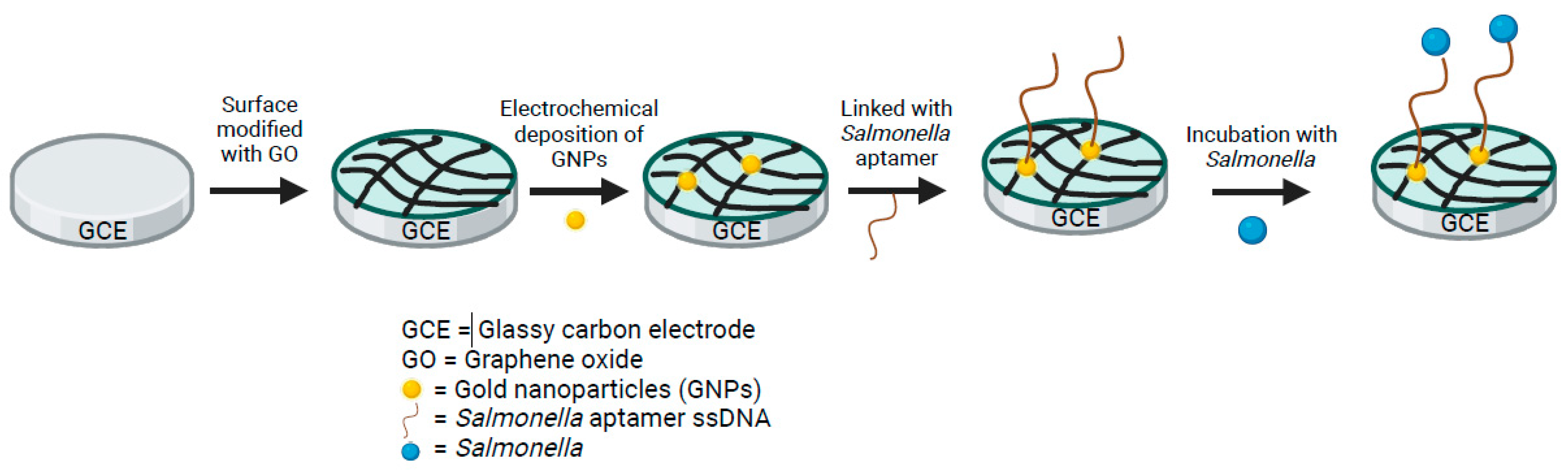
5.1. Electrochemical Aptasensors

Biosensor technology has gained popularity, as it can provide sensitive, reliable results and supply much higher speed with the additional benefit of bacteria inactivation directly on site [73]. Biosensors integrate biological recognition elements, such as antibodies or aptamers, with transducer platforms to enable real-time, label-free detection of Salmonella [74]. The specification of electrochemical biosensors determines the limit of detection (LoD) and detection time of the aptasensor platform. Aptasensors are frequently made with glassy carbon electrodes (GCE) and gold (Au) as the working electrode because of their high electrochemical stability, good biocompatibility with recognition molecules, and fast electron transfer for sensitive detection [73]. Adding nanomaterials onto the surface to make gold nanoparticles (AuNPs) enhances surface area, electron transfer kinetics, and electrical conductivity for more sensitive detection [73]. A 2022 study examined the performance of 15 published studies on electrochemical aptasensors for the detection of Salmonella species between 2014 and 2021 [74]. They found the developed sensors exhibited a LoD from 550 CFU/mL to as low as 1 CFU/mL (2 studies) within 5 to 240 min of detection time. Similarly, various sensor material combinations have been studied to enhance electron transfer properties and improve LoD and total detection time (Table 1). Aptasensors utilizing AuNPs decorated with graphite electrodes (GE) detected Salmonella in milk samples at 1 CFU/mL within 40 min [75] (Figure 1). In 2014, Ma and colleagues used GCE aptasensors modified with graphene oxide (GO) to detect Salmonella in pork samples with a LoD of 3 CFU/mL in 35 min [76]. In 2018, Ge and colleagues combined AuNPs with RCA to produce long DNA molecules and hybridize with the detection probes for enzyme-amplified readout on the surface of the aptasensor with a LoD of 16 CFU/mL in 1 h [77]. In 2019, Muniandy and colleagues utilized a reduced graphene oxide-titanium dioxide nanocomposite-based aptasensor to detect S. Typhimurium in just 5 min at a LoD of 10 CFU/mL [78]. Hasan and colleagues conducted a study in 2018 utilizing amino-modified ssDNA immobilized on multi-walled carbon nanotubes and found a LoD of 55 CFU/mL (for S. Enteritidis) and 67 CFU/mL (S. Typhimurium) in just 20 min. Taken together, electrochemical aptasensors’ portability, sensitivity, and rapid detection highlight their potential for widespread use for Salmonella detection in food safety and disease prevention. Future studies are needed on electrochemical aptasensor detection of other serovars of Salmonella like Typhi and Paratyphi to establish a quick, sensitive, cost-effective, and reliable technique for all Salmonella serovar detection [32,75,76,77,78,79].

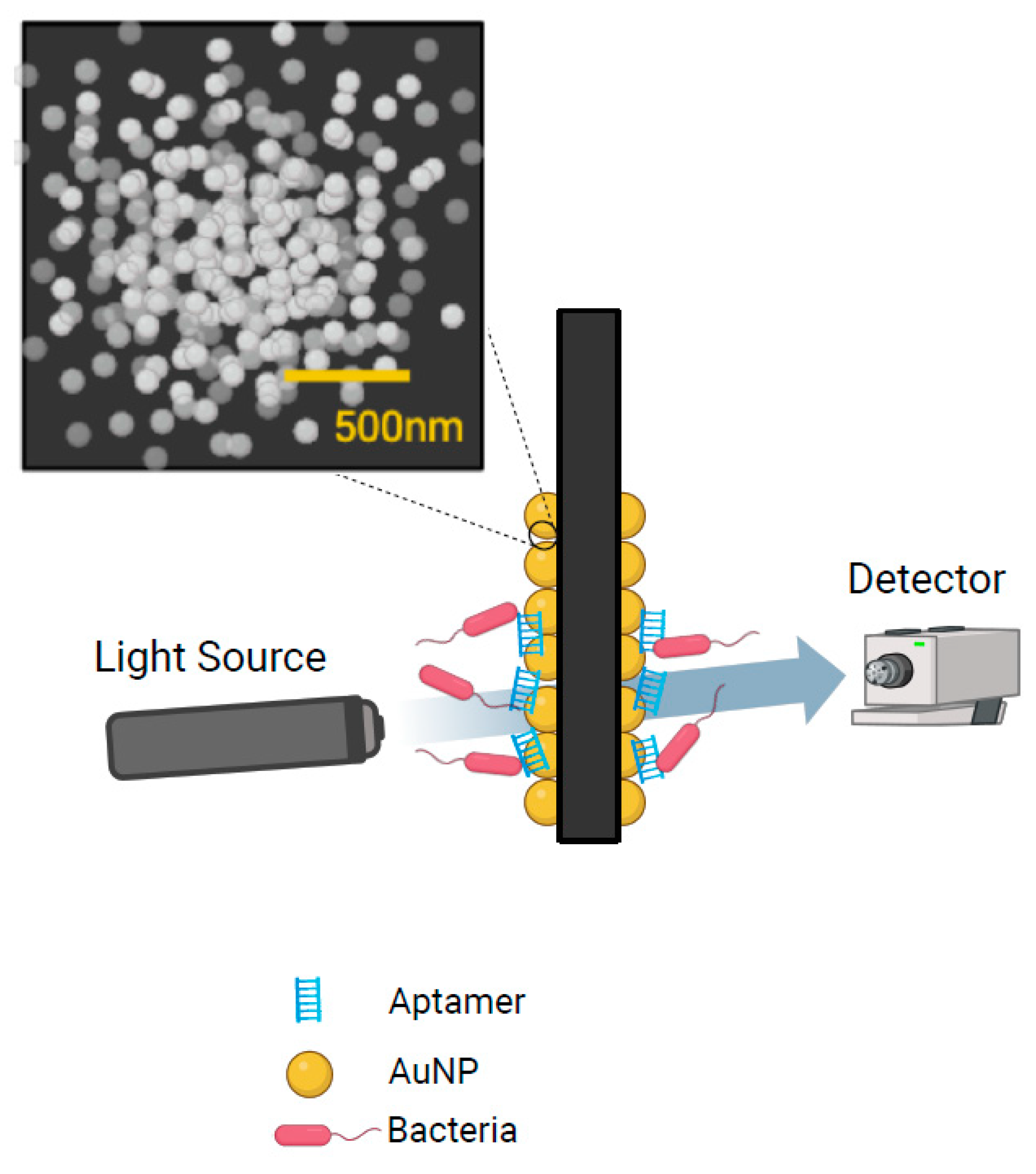
5.2. Surface Plasmon Resonance

Another promising emerging technology that has gained a significant amount of interest is Surface Plasmon Resonance (SPR) (Table 2). SPR uses polarized light directed at a metal film to create surface plasmons that cause specific energy transfers to cause a reduction in light intensity called the SPR angle [80]. When biomolecules bind to the sensor surface, the SPR angle changes proportional to the mass of the bound molecules to provide real-time, label-free detection with high sensitivity and specificity without the need for sample enrichment [81]. In 2019, Bhandari and colleagues evaluated direct SPR assay, sequential two-step sandwich assay, and pre-incubation one-step sandwich assays for the detection of Salmonella Typhimurium in samples of romaine lettuce at different levels. They found that all three formats had a LoD of 5.9 log CFU/g without enrichment in just 2 min and 0.9 log CFU/g with a 24 h enrichment step [80]. In 2022, Bhandari and colleagues studied the SPR method with magnetic nanoparticles to detect Salmonella Typhimurium in romaine lettuce [81]. The authors demonstrated that the preincubation one-step sandwich assay had the highest signal amplification by forming complexes with antibody-coupled magnetic nanoparticles to significantly increase analyte mass. This increased sensitivity allowed a LoD for S. Typhimurium of 4.7 log CFU/mL in buffer and 5.2 log CFU/mL in romaine lettuce samples with the overall detection process from preparation to SPR detection completed in 4 h [81]. Oh and colleagues developed a gold nanoparticle-aptamer-based localized SPR sensing chip for the detection of S. Typhimurium in pork meat with a LoD of 4 log CFU/mL in 35 min and found no effect of food matrix or background contaminant microflora [82] (Figure 2). In 2024, Liang and colleagues were able to reduce the LoD of SPR to 42 CFU/mL by utilizing a sandwich strategy with antibody-AuNPs with a total detection and separation time of less than 50 min. [83]. The rapid detection time and no food matrix effect on detection are advantageous aspects of SPR, however, sensitivity of 4–5 log CFU/mL is insufficient in future food detection as methods with a sensitivity of 1 CFU/mL have been demonstrated [46,53,81,82,83].

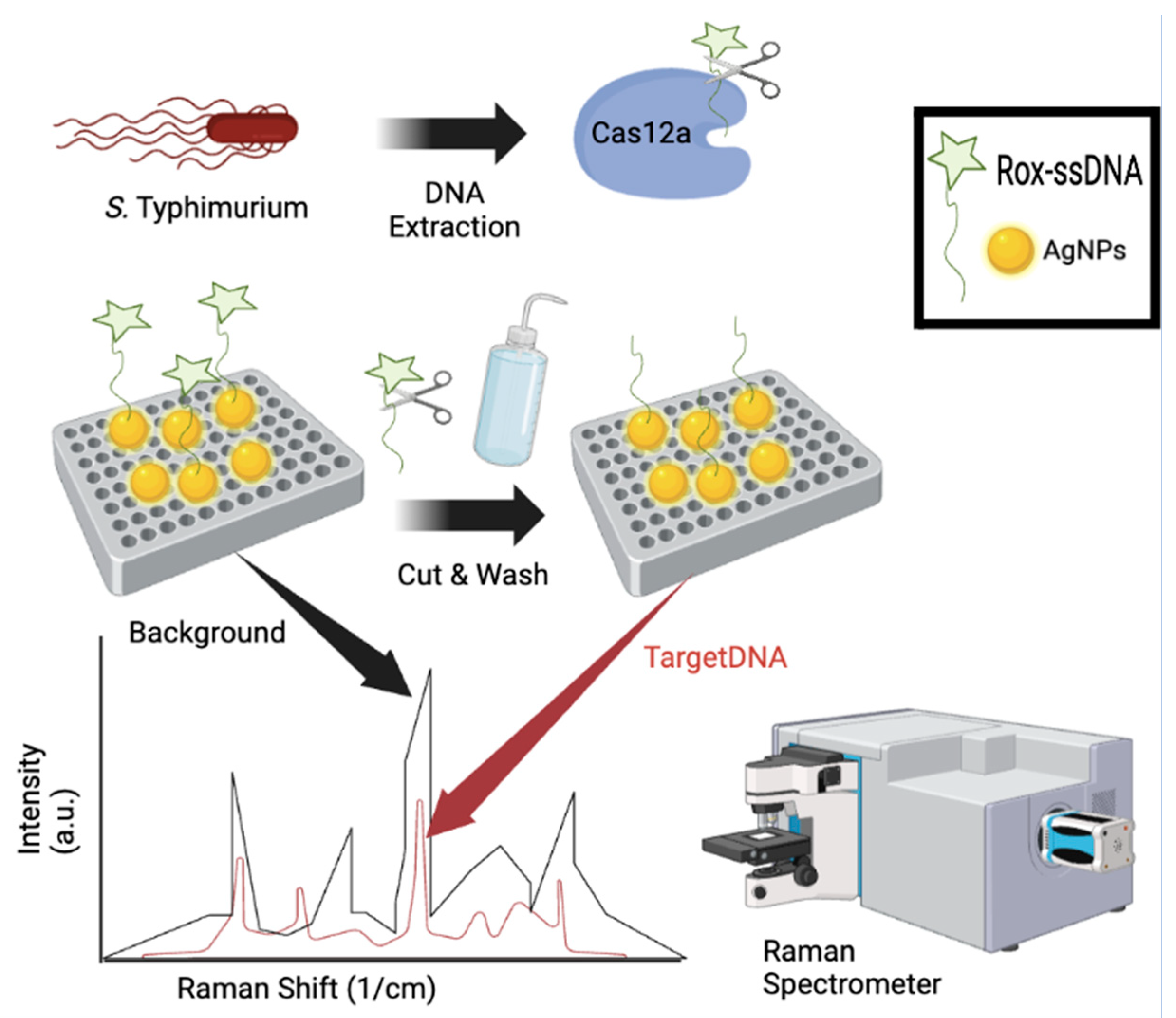
5.3. Surface Enhanced Raman Spectroscopy
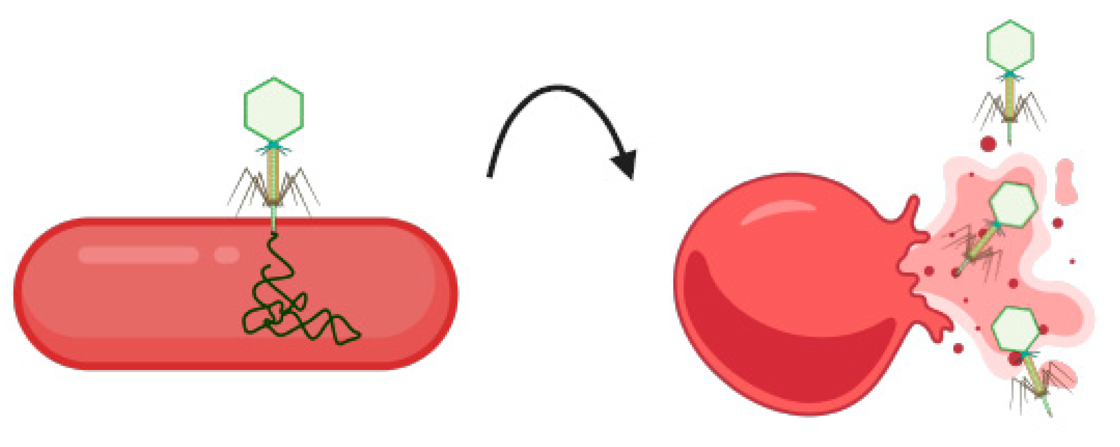
Raman spectroscopy is a third example of an upcoming Salmonella species detection technology that functions by monitoring the changes in vibrations within functional groups in target molecules [84]. Similar to electrochemical biosensors, Raman spectroscopy has recently been enhanced via noble metal and nanotechnology to create Surface-Enhanced Raman Spectroscopy (SERS) (Table 3). SERS offers enhanced sensitivity and multiplexing capabilities for Salmonella detection in complex food matrices [84,85,86]. In 2022, Asgari and colleagues utilized a SERS optofulidic sensor coupled with immunoassay to selectively separate and detect a wide range of E. coli and Salmonella species in lettuce and packed salad samples. After only a 15 min enrichment period, their proposed protocol displayed promising results with a LoD of 10 CFU/200 g sample with total analysis time of just 2 h [84]. Chuesiang and colleagues conducted a study using aptamer-based SERS for detection of Salmonella in ground beef. However, their LoD (4 log CFU/g) and total running time (4 h) were not as promising as the previous study [85]. Recently, Wang and colleagues conducted a groundbreaking study using bi-channel lateral flow immunoassay strips with three-dimensional membrane SERS nanostickers (GO@Au/Ag) for multiplex detection of four bacteria in real clinical samples. Their proposed sensor displayed high accuracy, stability, and efficiency by quantitatively detecting four different bacteria with a LoD of 9 cells/mL in just 20 min [86]. However, future studies are needed on this sensor using food matrices for foodborne pathogen detection. In 2022, Zhuang and colleagues utilized a recombinase polymerase amplification (RPA)-integrated microfluidic paper-based analytical device (RPA-Cas12a-µPAD) for SERS-based detection of S. Typhimurium in contaminated meat and milk samples. The RPA-Cas12a-µPAD displayed accurate tests of food samples with a LoD of 3–4 CFU/mL in just 45 min [87]. In 2023, Jia and colleagues developed the CRISPR-SERS biosensor by combining CRISPR technology with SERS analytical method, aimed at detecting invA of S. Typhimurium. Results from their study showed that CRISPR-SERS could detect S. Typhimurium in poultry samples with a LoD of 110 CFU/mL within 2 h [88] (Figure 3). Likewise, Zheng and colleagues studied a novel SERS chip using aptamer-modified multi-color SERS tags and a graphene-coated plasmonic gold substrate to detect and inactivate Salmonella in whole blood samples [89]. Using Python to analyze their Raman mapping, they found that the SERS chip provided sensitive (4 CFU/mL), specific, and reproducible multiplex detection and in-situ photothermal elimination of pathogens (nearly 100% eradication reported), making it suitable for onsite multiplex pathogen detection and inactivation [89].

5.4. Bacteriophages

Another emerging technology that has been used in foodborne Salmonella detection is bacteriophage technology [90]. Bacteriophages are natural viruses that infect bacteria and are a viable alternative to current antimicrobials in the control of these pathogens as numerous studies have shown their ability to lyse multi-drug-resistant Salmonella isolates [90,91,92] (Figure 4). In 2023, Lopez-Garcia et al., tested the identification and lytic activity of 22 previously characterized Salmonella bacteriophages against a panel of 143 Salmonella isolates (Table 4) [93]. Phage STW-77 was the most effective overall, lysing 37.76% of all Salmonella isolates; SPFM5 and SPFM13 were most effective against S. Enteritidis, lysing 85.71% of isolates. STW-77 was most effective against S. Typhimurium and its monophasic variants. The effectiveness of phages varied within serovars, suggesting that phages may detect and lyse specific subtypes within a serovar. Unfortunately, no phage in this study could completely lyse the S. Kentucky ST198 MDR clones [93]. Although future studies are needed, these emerging technologies hold promise for future enhanced food safety monitoring and surveillance.

6. Challenges Associated with Detection Technologies: Limits of Detection and Enrichment Time

According to the European Commission, the presence of Salmonella species at just one CFU/mL in a 25 g portion of ready-to-eat food is sufficient to cause disease in humans [1,9]. Emerging technologies such as LFA, PCR, NGS, ELISA, and SPR (Figure 5) have limits of detection (LoDs) significantly higher than the critical 1 CFU/mL level necessary to cause disease. While having a sensitive detection method is crucial, it is not the only factor to consider when selecting an on-site foodborne pathogen detection method (Figure 6). Traditional culture methods, though highly sensitive, are time-consuming [32,43,48,53,71,81,88]. Additionally, detection methods that require more than eight hours (480 min) are impractical for on-site use as they would necessitate multiple work shifts to complete, delaying the timely determination and control of pathogen contamination [48,81,88]. Thus, the ideal on-site foodborne pathogen detection method should be sensitive (detecting less than 10 CFU/mL), quick, cost-effective, and should not require complex equipment or highly trained personnel [70,81,88]. Furthermore, a comparison of detection methods by combining their LoD and total detection time to create a combined score (Figure 7) suggests that Electrochemical Aptasensors (AuNPs), SERS, and RAA with photonic crystals are among the top three emerging future technologies that may be employed for Salmonella species detection in food samples.

7. Future Directions

Significant advances have been made in food preservation methods that help eliminate food pathogens including storage at low temperatures, chemical preservatives, and vacuum packaging [48]. However, despite advances in food manufacturing and safety, regular outbreaks indicate that foodborne pathogens like Salmonella species remain a significant public health risk [1]. An exciting avenue has emerged with potential eradication techniques that can significantly change food safety measures. Using targeted approaches such as bacteriophage and photothermal effects, there is potential for eradicating Salmonella contamination from food supply chains [89,93]. The efficacy and practicality of these methods remain to be studied, but they appear promising. However, all proof-of-concept studies must be validated according to ISO 16140 standards and other requirements set out by regulatory agencies such as the FDA and EFSA. The aforementioned emerging technologies, including but not limited to, electrochemical aptasensors, SERS, and RAA, have the potential to significantly improve foodborne pathogen detection technology [48]. Additionally, further research on the implementation of these technologies at various stages of food preparation is required. By investing in rapid, sensitive, onsite emerging technologies early in the detection process, food companies can reduce the rate-limiting pretreatment step and prevent the need for other resource-intensive downstream confirmation tests [94]. Automated detection technologies would allow scalable detection to quickly screen large sample volumes, minimize labor costs, and potentially reduce the volume of goods that may need to be recalled or discarded later [95]. With the innovation of affordable and rapid testing methods, large-scale testing for Salmonella can be done for environmental monitoring as well as livestock and poultry testing.

8. Conclusions

Emerging technologies for Salmonella detection in food samples will ultimately enhance our ability to quickly and accurately identify contamination, thereby bolstering food safety and public health. Traditional culture methods, though the gold standard, are cost-effective but yet are time-consuming. In this review, we analyzed immunological methods, molecular methods, biosensors, and WGS on isolated strains to compare sensitivity and detection time. Of the technologies discussed, biosensors offer the highest sensitivity, capable of detecting as low as 1 CFU/mL within 40 min. Similarly, SERS can achieve similar sensitivity at 4 CFU/mL within 45 min [87], while RAA offers the quickest turnaround time of 30 min and has a LoD of 10 CFU/mL [54]. On the other hand, the Duplex PCR-ELISA method, although taking longer at 6 h, provides highly sensitive detection down to 1 CFU/mL, combining the specificity of PCR with the versatility of ELISA [39]. The aforementioned advanced methods combine speed, sensitivity, and accuracy, addressing critical requirements for effective on-site detection. Their integration into food safety protocols can significantly minimize health risks and economic impacts associated with Salmonella contamination. However, to implement these technologies for real-time in-field testing, the biggest challenge continues to be the need for highly trained staff and specialized equipment, which complicates transport and logistics. The integration of biosensors with cost-effective technologies such as LFAs can provide a cost-effective, highly sensitive, and rapid method of detection. Continued innovation, validation and adoption of these technologies will be pivotal in achieving practical, reliable, and comprehensive pathogen detection, ensuring safer food supply chains and better health outcomes against the threat of Salmonella contamination.

Author Contributions

A.P. and A.W. contributed significantly to the writing of the major portions of this review article. T.S.D. provided valuable guidance throughout the writing process and was responsible for the critical editing of this manuscript. All authors have read and agreed to the published version of the manuscript.

Funding

This article did not receive any specific grant from funding agencies in the public, commercial, or not-for-profit sectors.

Acknowledgments

We express our gratitude to the Deans, John Geisler, Kelly Manahan and Nagadharshan Devendra at the Trinity School of Medicine (Trinity Medical Sciences University) for their support.

Conflicts of Interest

The authors declare no conflict of interest.

References

  1. CDC. Salmonella Homepage. 2016. Available online: https://www.cdc.gov/salmonella/index.html (accessed on 15 January 2021).
  2. Galán-Relaño; Díaz, A.V.; Lorenzo, B.H.; Gómez-Gascón, L.; Rodríguez, M.M.; Jiménez, E.C.; Rodríguez, F.P.; Márquez, R.J.A. Salmonella and Salmonellosis: An Update on Public Health Implications and Control Strategies. Animals 2023, 13, 3666. [Google Scholar] [CrossRef] [PubMed]
  3. Grimont, P.A.; Weill, F.X. Antigenic Formulae of the Salmonella Serovars, 9th ed.; WHO Collaborating Centre for Reference and Research on Salmonella. Institute Pasteur: Paris, France, 2007; pp. 1–166. [Google Scholar]
  4. Sanchez, S.; Hofacre, C.L.; Lee, M.D.; Maurer, J.J.; Doyle, M.P. Animal sources of salmonellosis in humans. J. Am. Veter- Med Assoc. 2002, 221, 492–497. [Google Scholar] [CrossRef] [PubMed]
  5. Abulreesh, H.H. Salmonellae in the environment. In Salmonella—Distribution, Adaptation, Control Measures and Molecular Technologies; Bassam, A.A., Joshua, B.G., Eds.; IntechOpen: London, UK, 2012; pp. 22–31. [Google Scholar] [CrossRef]
  6. Rodriguez, A.; Pangloli, P.; Richards, H.A.; Mount, J.R.; Draughon, F.A. Prevalence of Salmonella in Diverse Environmental Farm Samples. J. Food Prot. 2006, 69, 2576–2580. [Google Scholar] [CrossRef] [PubMed]
  7. Pui, C.F.; Wong, W.C.; Chai, L.C.; Nillian, E.; Ghazali, F.M.; Cheah, Y.K.; Nakaguchi, Y.; Nishibuchi, M.; Radu, S. Simultaneous detection of Salmonella spp., Salmonella Typhi and Salmonella Typhimurium in sliced fruits using multiplex PCR. Food Control 2011, 22, 337–342. [Google Scholar] [CrossRef]
  8. Shen, X.; Yin, L.; Zhang, A.; Zhao, R.; Yin, D.; Wang, J.; Dai, Y.; Hou, H.; Pan, X.; Hu, X.; et al. Prevalence and Characterization of Salmonella Isolated from Chickens in Anhui, China. Pathogens 2023, 12, 465. [Google Scholar] [CrossRef]
  9. Institute of Medicine (US) and National Research Council (US) Committee on the Review of the Use of Scientific Criteria and Performance Standards for Safe Food. Scientific Criteria to Ensure Safe Food; National Academies Press (US): Washington, DC, USA, 2003. Available online: https://www.ncbi.nlm.nih.gov/books/NBK221565/ (accessed on 29 October 2024). [PubMed]
  10. He, Y.; Wang, J.; Zhang, R.; Chen, L.; Zhang, H.; Qi, X.; Chen, J. Epidemiology of foodborne diseases caused by Salmonella in Zhejiang Province, China, between 2010 and 2021. Front. Public Health 2023, 11, 1127925. [Google Scholar] [CrossRef]
  11. Huckstep, R.L. Typhoid Fever and Other Salmonella Infections. Am. J. Med Sci. 1963, 245, 154. [Google Scholar] [CrossRef]
  12. Acheson, D.; Hohmann, E.L. Nontyphoidal Salmonellosis. Clin. Infect. Dis. 2001, 32, 263–269. [Google Scholar] [CrossRef]
  13. Dhanoa, A.; Fatt, Q.K. Non-typhoidal Salmonella bacteraemia: Epidemiology, clinical characteristics and its’ association with severe immunosuppression. Ann. Clin. Microbiol. Antimicrob. 2009, 8, 15. [Google Scholar] [CrossRef]
  14. Scallan, E.; Hoekstra, R.M.; Angulo, F.J.; Tauxe, R.V.; Widdowson, M.-A.; Roy, S.L.; Jones, J.L.; Griffin, P.M. Foodborne Illness Acquired in the United States—Major Pathogens. Emerg. Infect. Dis. 2011, 17, 7–15. [Google Scholar] [CrossRef]
  15. Crump, J.A.; Kretsinger, K.; Gay, K.; Hoekstra, R.M.; Vugia, D.J.; Hurd, S.; Segler, S.D.; Megginson, M.; Luedeman, L.J.; Shiferaw, B.; et al. Clinical Response and Outcome of Infection with Salmonella enterica Serotype Typhi with Decreased Susceptibility to Fluoroquinolones: A United States FoodNet Multicenter Retrospective Cohort Study. Antimicrob. Agents Chemother. 2008, 52, 1278–1284. [Google Scholar] [CrossRef] [PubMed]
  16. Eikmeier, D.; Medus, C.; Smith, K. Incubation period for outbreak-associated, non-typhoidal salmonellosis cases, Minnesota, 2000–2015. Epidemiology Infect. 2018, 146, 423–429. [Google Scholar] [CrossRef] [PubMed]
  17. Jones, B.D.; Ghori, N.; Falkow, S. Salmonella typhimurium initiates murine infection by penetrating and destroying the specialized epithelial M cells of the Peyer’s patches. J. Exp. Med. 1994, 180, 15–23. [Google Scholar] [CrossRef] [PubMed]
  18. Lou, L.; Zhang, P.; Piao, R.; Wang, Y. Salmonella Pathogenicity Island 1 (SPI-1) and Its Complex Regulatory Network. Front. Cell. Infect. Microbiol. 2019, 9, 270. [Google Scholar] [CrossRef]
  19. Galán, J.E. Molecular and Cellular Bases of Salmonella Entry into Host Cells. Curr. Top. Microbiol. Immunol. 1996, 209, 43–60. [Google Scholar] [CrossRef] [PubMed]
  20. Chen, L.-M.; Hobbie, S.; Galán, J.E. Requirement of CDC42 for Salmonella -Induced Cytoskeletal and Nuclear Responses. Science 1996, 274, 2115–2118. [Google Scholar] [CrossRef]
  21. Vassiloyanakopoulos, A.P.; Okamoto, S.; Fierer, J. The crucial role of polymorphonuclear leukocytes in resistance to Salmonella dublin infections in genetically susceptible and resistant mice. Proc. Natl. Acad. Sci. USA 1998, 95, 7676–7681. [Google Scholar] [CrossRef]
  22. U.S. Food and Drug Administration. Recalls, Market Withdrawals, & Safety Alerts. U.S. Department of Health and Human Services. 2024. Available online: https://www.fda.gov/safety/recalls-market-withdrawals-safety-alerts (accessed on 30 October 2024).
  23. Wang, M.; Zhang, Y.; Tian, F.; Liu, X.; Du, S.; Ren, G. Overview of Rapid Detection Methods for Salmonella in Foods: Progress and Challenges. Foods 2021, 10, 2402. [Google Scholar] [CrossRef]
  24. U.S. Food and Drug Administration. Guidance for Industry: Testing for Salmonella Species in Human Foods and Di-rect-Human-Contact Animal Foods. 2012. Available online: https://www.fda.gov/regulatory-information/search-fda-guidance-documents/guidance-industry-testing-salmonella-species-human-foods-and-direct-human-contact-animal-foods (accessed on 30 October 2024).
  25. ISO 16140-2:2016; Microbiology of the Food Chain—Method Validation—Part 2: Protocol for the Validation of Alternative (Proprietary) Methods Against a Reference Method. International Organization for Standardization: Geneva, Switzerland, 2016. Available online: https://www.iso.org/obp/ui/en/#iso:std:iso:16140:-2:ed-1:v1:en (accessed on 29 October 2024).
  26. U.S. Food and Drug Administration Foods Program. Guidelines for the Validation of Analytical Methods for the Detection of Microbial Pathogens in Foods and Feeds, 3rd ed.; 2019. Available online: https://www.fda.gov/media/83812/download (accessed on 29 October 2024).
  27. European Union. Regulation (EU) 2017/625 of the European Parliament and of the Council of 15 March 2017 on Official Controls and Other Official Activities Carried Out to Ensure the Application of Food and Feed Law, Rules on Animal Health and Welfare, Plant Health and Plant Protection Products. EUR-Lex. 2017. Available online: http://data.europa.eu/eli/reg/2017/625/2022-01-28/eng (accessed on 29 October 2024).
  28. Neyaz, L.A.; Alghamdi, H.S.; Alghashmari, R.M.; Alswat, S.S.; Almaghrabi, R.O.; Bazaid, F.S.; Albarakaty, F.M.; Elbanna, K.; Abulreesh, H.H. A Comprehensive Review on the Current Status of Culture Media for Routine Standardized Isolation of Salmonella and Shigella Spp. from Contaminated Food. J. Umm Al-Qura Univ. Appll. Sci. 2024. [Google Scholar] [CrossRef]
  29. Mooijman, K.A. The new ISO 6579-1: A real horizontal standard for detection of Salmonella, at last! Food Microbiol. 2018, 71, 2–7. [Google Scholar] [CrossRef]
  30. ISO 6579-1:2017; Microbiology of the Food Chain—Horizontal Method for the Detection, Enumeration and Serotyping of Salmonella—Part 1: Detection of Salmonella spp. [WWW Document]. ISO: Geneva, Switzerland, 2017. Available online: https://www.iso.org/standard/56712.html (accessed on 29 October 2024).
  31. Zhuang, L.; Gong, J.; Shen, Q.; Yang, J.; Song, C.; Liu, Q.; Zhao, B.; Zhang, Y.; Zhu, M. Advances in detection methods for viable Salmonella spp.: Current applications and challenges. Anal. Sci. 2023, 39, 1643–1660. [Google Scholar] [CrossRef] [PubMed]
  32. Oslan, S.N.H.; Yusof, N.Y.; Lim, S.J.; Ahmad, N.H. Rapid and sensitive detection of Salmonella in agro-Food and environmental samples: A review of advances in rapid tests and biosensors. J. Microbiol. Methods 2024, 219, 106897. [Google Scholar] [CrossRef] [PubMed]
  33. Cruz, C.D.; Esteve, P.; Tammela, P. Evaluation and validation of Biolog OmniLog(R) system for antibacterial activity assays. Lett. Appl. Microbiol. 2021, 72, 589–595. [Google Scholar] [CrossRef] [PubMed]
  34. ISO/TR No. 6579-3:2014; Microbiology of the food chain-horizontal method for the detection, enumeration and serotyping of Salmonella-Part 3: Guidelines for serotyping of Salmonella spp. International Organization for Standardization: Geneva, Switzerland, 2014. Available online: https://www.iso.org/standard/56714.html (accessed on 29 October 2024).
  35. Wattiau, P.; Boland, C.; Bertrand, S. Methodologies for Salmonella enterica subsp. enterica Subtyping: Gold Standards and Alternatives. Appl. Environ. Microbiol. 2011, 77, 7877–7885. [Google Scholar] [CrossRef] [PubMed]
  36. Awang, M.S.; Bustami, Y.; Hamzah, H.H.; Zambry, N.S.; Najib, M.A.; Khalid, M.F.; Aziah, I.; Manaf, A.A. Advancement in Salmonella Detection Methods: From Conventional to Electrochemical-Based Sensing Detection. Biosensors 2021, 11, 346. [Google Scholar] [CrossRef]
  37. Tietjen, M.; Fung, D.Y.C. Salmonellae and Food Safety. Crit. Rev. Microbiol. 1995, 21, 53–83. [Google Scholar] [CrossRef]
  38. Mansfield, L.; Forsythe, S. The detection of Salmonella using a combined immunomagnetic separation and ELISA end-detection procedure. Lett. Appl. Microbiol. 2000, 31, 279–283. [Google Scholar] [CrossRef]
  39. Hu, J.; Huang, R.; Wang, Y.; Wei, X.; Wang, Z.; Geng, Y.; Jing, J.; Gao, H.; Sun, X.; Dong, C.; et al. Development of duplex PCR-ELISA for simultaneous detection of Salmonella spp. and Escherichia coli O157: H7 in food. J. Microbiol. Methods 2018, 154, 127–133. [Google Scholar] [CrossRef]
  40. Priyanka, B.; Patil, R.K.; Dwarakanath, S. A review on detection methods used for foodborne pathogens. Indian J. Med. Res. 2016, 144, 327–338. [Google Scholar] [CrossRef]
  41. Schenk, F.; Weber, P.; Vogler, J.; Hecht, L.; Dietzel, A.; Gauglitz, G. Development of a paper-based lateral flow immunoassay for simultaneous detection of lipopolysaccharides of Salmonella serovars. Anal. Bioanal. Chem. 2017, 410, 863–868. [Google Scholar] [CrossRef]
  42. Gao, P.; Wang, L.; He, Y.; Wang, Y.; Yang, X.; Fu, S.; Qin, X.; Chen, Q.; Man, C.; Jiang, Y. An Enhanced Lateral Flow Assay Based on Aptamer–Magnetic Separation and Multifold AuNPs for Ultrasensitive Detection of Salmonella Typhimurium in Milk. Foods 2021, 10, 1605. [Google Scholar] [CrossRef] [PubMed]
  43. Chirambo, A.C.; Nyirenda, T.S.; Jambo, N.; Msefula, C.; Kamng’Ona, A.; Molina, S.; Mandala, W.L.; Heyderman, R.S.; Iturizza-Gomara, M.; Henrion, M.Y.; et al. Performance of molecular methods for the detection of Salmonella in human stool specimens. Wellcome Open Res. 2021, 5, 237. [Google Scholar] [CrossRef] [PubMed]
  44. Aladhadh, M. A Review of Modern Methods for the Detection of Foodborne Pathogens. Microorganisms 2023, 11, 1111. [Google Scholar] [CrossRef] [PubMed]
  45. Xiao, L.; Zhang, L.; Wang, H.H. Critical Issues in Detecting Viable Listeria monocytogenes Cells by Real-Time Reverse Transcriptase PCR. J. Food Prot. 2012, 75, 512–517. [Google Scholar] [CrossRef] [PubMed]
  46. Zendrini, A.; Carta, V.; Filipello, V.; Ragni, L.; Cosciani-Cunico, E.; Arnaboldi, S.; Bertasi, B.; Franceschi, N.; Ajmone-Marsan, P.; De Medici, D.; et al. One-Day Molecular Detection of Salmonella and Campylobacter in Chicken Meat: A Pilot Study. Foods 2021, 10, 1132. [Google Scholar] [CrossRef]
  47. Chen, M.; Lan, X.; Zhu, L.; Ru, P.; Xu, W.; Liu, H. PCR Mediated Nucleic Acid Molecular Recognition Technology for Detection of Viable and Dead Foodborne Pathogens. Foods 2022, 11, 2675. [Google Scholar] [CrossRef]
  48. Foddai, A.C.G.; Grant, I.R. Methods for detection of viable foodborne pathogens: Current state-of-art and future prospects. Appl. Microbiol. Biotechnol. 2020, 104, 4281–4288. [Google Scholar] [CrossRef]
  49. Notomi, T.; Okayama, H.; Masubuchi, H.; Yonekawa, T.; Watanabe, K.; Amino, N.; Hase, T. Loop-mediated isothermal amplification of DNA. Nucleic Acids Res. 2000, 28, 63. [Google Scholar] [CrossRef] [PubMed]
  50. Wang, S.; Liu, N.; Zheng, L.; Cai, G.; Lin, J. A lab-on-chip device for the sample-in-result-out detection of viable Salmonella using loop-mediated isothermal amplification and real-time turbidity monitoring. Lab A Chip 2020, 20, 2296–2305. [Google Scholar] [CrossRef]
  51. Li, M.; Luan, Z.; Liu, Y.; Yang, C.; Wang, Y.; Ma, C.; Shi, C. Ultrafast bacterial cell lysis using a handheld corona treater and loop-mediated isothermal amplification for rapid detection of foodborne pathogens. Food Control. 2021, 128, 108178. [Google Scholar] [CrossRef]
  52. Williams, M.R.; Telli, A.E.; Telli, N.; Islam, D.T.; Hashsham, S.A. Direct or DNA Extraction-Free Amplification and Quantification of Foodborne Pathogens. Methods Mol. Biol. 2019, 2852, 3–17. [Google Scholar] [CrossRef] [PubMed]
  53. Ndraha, N.; Lin, H.-Y.; Wang, C.-Y.; Hsiao, H.-I.; Lin, H.-J. Rapid detection methods for foodborne pathogens based on nucleic acid amplification: Recent advances, remaining challenges, and possible opportunities. Food Chem. Mol. Sci. 2023, 7, 100183. [Google Scholar] [CrossRef] [PubMed]
  54. Mu, D.; Zhou, D.; Xie, G.; Liu, J.; Xiong, Q.; Feng, X.; Xu, H. The fluorescent probe-based recombinase-aided amplification for rapid detection of Escherichia coli O157:H7. Mol. Cell. Probes 2021, 60, 101777. [Google Scholar] [CrossRef] [PubMed]
  55. Milton, A.A.P.; Momin, K.M.; Priya, G.B.; Ghatak, S.; Das, S.; Gandhale, P.N.; Angappan, M.; Sen, A. Development of novel visual detection methodology for Salmonella in meat using saltatory rolling circle amplification. J. Appl. Microbiol. 2021, 131, 2361–2371. [Google Scholar] [CrossRef] [PubMed]
  56. Delgado, E.; Katchman, B.; Stice, S.; Calle, A. Independent evaluation of a DNA microarray system for Salmonella detection in ground beef. Food Microbiol. 2023, 118, 104406. [Google Scholar] [CrossRef]
  57. Katchman, B.A.; Sinton, M. DNA Microarrays: A Powerful Tool for Pathogen Detection; PathogenDx, 2019; Available online: https://pathogendx.com/wp-content/uploads/2019/12/What-is-a-DNA-Microarray_BAK-072419_1.pdf (accessed on 29 October 2024).
  58. U.S. Food and Safety Inspection Service. Foodborne Pathogen Test Kits Validated by Independent Organizations. 2024. Available online: https://www.fsis.usda.gov/sites/default/files/media_file/documents/Validated-Test-Kit.pdf (accessed on 29 October 2024).
  59. Den Bakker, H.C.; Allard, M.W.; Bopp, D.; Brown, E.W.; Fontana, J.; Iqbal, Z.; Kinney, A.; Limberger, R.; Musser, K.; Shudt, M.; et al. Rapid Whole-Genome Sequencing for Surveillance of Salmonella enterica Serovar Enteritidis. Emerg. Infect. Dis. 2014, 20, 1306–1314. [Google Scholar] [CrossRef]
  60. Nafea, A.M.; Wang, Y.; Wang, D.; Salama, A.M.; Aziz, M.A.; Xu, S.; Tong, Y. Application of next-generation sequencing to identify different pathogens. Front. Microbiol. 2024, 14, 1329330. [Google Scholar] [CrossRef]
  61. U.S. Food and Drug Administration. Whole Genome Sequencing (WGS) Program. U.S. Department of Health and Human Services. 2024. Available online: https://www.fda.gov/food/microbiology-research-food/whole-genome-sequencing-wgs-program (accessed on 29 October 2024).
  62. U.S. Food and Drug Administration. GenomeTrakr Fast Facts. U.S. Department of Health and Human Services. 2024. Available online: https://www.fda.gov/food/whole-genome-sequencing-wgs-program/genometrakr-fast-facts (accessed on 29 October 2024).
  63. Lin, A.; Singh, A.; Allred, A.; Allard, M.; Waltman, D.; Imanian, B.; Ng, J.H.; Sanahmadi, Y.; Khaksar, R. Targeted Next-Generation Sequencing Assay for Direct Detection and Serotyping of Salmonella from Enrichment. J. Food Prot. 2024, 87, 100256. [Google Scholar] [CrossRef]
  64. Persad, A.K.; Fahmy, H.A.; Anderson, N.; Cui, J.; Topalcengiz, Z.; Jeamsripong, S.; Spanninger, P.M.; Buchanan, R.L.; Kniel, K.E.; Jay-Russell, M.T.; et al. Identification and Subtyping of Salmonella Isolates Using Matrix-Assisted Laser Desorption–Ionization Time-of-Flight Mass Spectrometry (MALDI-TOF). Microorganisms 2022, 10, 688. [Google Scholar] [CrossRef]
  65. Kliem, M.; Sauer, S. The essence on mass spectrometry based microbial diagnostics. Curr. Opin. Microbiol. 2012, 15, 397–402. [Google Scholar] [CrossRef]
  66. Sandrin, T.R.; Goldstein, J.E.; Schumaker, S. MALDI TOF MS profiling of bacteria at the strain level: A review. Mass Spectrom. Rev. 2012, 32, 188–217. [Google Scholar] [CrossRef] [PubMed]
  67. Bruker. Microbial Analysis by MALDI-TOF. 2024. Available online: https://www.bruker.com/en/products-and-solutions/microbiology-and-diagnostics/microbial-identification.html (accessed on 30 October 2024).
  68. Mangmee, S.; Reamtong, O.; Kalambaheti, T.; Roytrakul, S.; Sonthayanon, P. MALDI-TOF mass spectrometry typing for predominant serovars of non-typhoidal Salmonella in a Thai broiler industry. Food Control. 2020, 113, 107188. [Google Scholar] [CrossRef]
  69. Yang, S.-M.; Kim, E.; Kim, D.; Baek, J.; Yoon, H.; Kim, H.-Y. Rapid Detection of Salmonella Enteritidis, Typhimurium, and Thompson by Specific Peak Analysis Using Matrix-Assisted Laser Desorption Ionization Time-of-Flight Mass Spectrometry. Foods 2021, 10, 933. [Google Scholar] [CrossRef] [PubMed]
  70. Wang, H.; Wu, Q.; Zhou, M.; Li, C.; Yan, C.; Huang, L.; Qin, P. Development of a CRISPR/Cas9-integrated lateral flow strip for rapid and accurate detection of Salmonella. Food Control. 2022, 142. [Google Scholar] [CrossRef]
  71. Guo, W.; Yang, Q.; Liu, J.; Chen, X.; Zhang, Y.; Zhang, W. Multiple fluorescent saltatory rolling circle amplification (SRCA) for simultaneous and sensitive detection of Salmonella spp. and Shigella spp. in food. LWT 2022, 168. [Google Scholar] [CrossRef]
  72. Huang, L.; Yuan, N.; Guo, W.; Zhang, Y.; Zhang, W. An electrochemical biosensor for the highly sensitive detection of Staphylococcus aureus based on SRCA-CRISPR/Cas12a. Talanta 2022, 252, 123821. [Google Scholar] [CrossRef]
  73. Cirocka, A.; Zarzeczańska, D.; Wcisło, A. Good Choice of Electrode Material as the Key to Creating Electrochemical Sensors—Characteristics of Carbon Materials and Transparent Conductive Oxides (TCO). Materials 2021, 14, 4743. [Google Scholar] [CrossRef]
  74. Zambry, N.S.; Najib, M.A.; Awang, M.S.; Selvam, K.; Khalid, M.F.; Bustami, Y.; Hamzah, H.H.; Ozsoz, M.; Manaf, A.A.; Aziah, I. Aptamer-Based Electrochemical Biosensors for the Detection of Salmonella: A Scoping Review. Diagnostics 2022, 12, 3186. [Google Scholar] [CrossRef]
  75. Fathi, S.; Saber, R.; Adabi, M.; Rasouli, R.; Douraghi, M.; Morshedi, M.; Farid-Majidi, R. Novel Competitive Voltammetric Aptasensor Based on Electrospun Carbon Nanofibers-Gold Nanoparticles Modified Graphite Electrode for Salmonella enterica serovar Detection. Biointerface Res. Appl. Chem. 2020, 11, 8702–8715. [Google Scholar] [CrossRef]
  76. Ma, X.; Jiang, Y.; Jia, F.; Yu, Y.; Chen, J.; Wang, Z. An aptamer-based electrochemical biosensor for the detection of Salmonella. J. Microbiol. Methods 2014, 98, 94–98. [Google Scholar] [CrossRef]
  77. Ge, C.; Yuan, R.; Yi, L.; Yang, J.; Zhang, H.; Li, L.; Nian, W.; Yi, G. Target-induced aptamer displacement on gold nanoparticles and rolling circle amplification for ultrasensitive live Salmonella typhimurium electrochemical biosensing. J. Electroanal. Chem. 2018, 826, 174–180. [Google Scholar] [CrossRef]
  78. Muniandy, S.; Teh, S.J.; Appaturi, J.N.; Thong, K.L.; Lai, C.W.; Ibrahim, F.; Leo, B.F. A reduced graphene oxide-titanium dioxide nanocomposite based electrochemical aptasensor for rapid and sensitive detection of Salmonella enterica. Bioelectrochemistry 2019, 127, 136–144. [Google Scholar] [CrossRef] [PubMed]
  79. Hasan, R.; Pulingam, T.; Appaturi, J.N.; Zifruddin, A.N.; Teh, S.J.; Lim, T.W.; Ibrahim, F.; Leo, B.F.; Thong, K.L. Carbon nanotube-based aptasensor for sensitive electrochemical detection of whole-cell Salmonella. Anal. Biochem. 2018, 554, 34–43. [Google Scholar] [CrossRef] [PubMed]
  80. Bhandari, D.; Chen, F.-C.; Bridgman, R.C. Detection of Salmonella Typhimurium in Romaine Lettuce Using a Surface Plasmon Resonance Biosensor. Biosensors 2019, 9, 94. [Google Scholar] [CrossRef] [PubMed]
  81. Bhandari, D.; Chen, F.-C.; Bridgman, R.C. Magnetic Nanoparticles Enhanced Surface Plasmon Resonance Biosensor for Rapid Detection of Salmonella Typhimurium in Romaine Lettuce. Sensors 2022, 22, 475. [Google Scholar] [CrossRef]
  82. Oh, S.Y.; Heo, N.S.; Shukla, S.; Cho, H.J.; Vilian, A.E.; Kim, J.; Lee, S.Y.; Han, Y.-K.; Yoo, S.M.; Huh, Y.S. Development of gold nanoparticle-aptamer-based LSPR sensing chips for the rapid detection of Salmonella typhimurium in pork meat. Sci. Rep. 2017, 7, 10130. [Google Scholar] [CrossRef]
  83. Liang, F.; Li, Y.; Hu, M.; Shi, C.; Cui, Y.; Young, G.M.; Yang, M.M.; Zhang, J. Nanoparticle-Enhanced SPR Immunobiosensor for Rapid Detection of Salmonella Typhimurium by Immunomagnetic Separation (SSRN Scholarly Paper 4680036). 2024. Available online: https://ssrn.com/abstract=4680036 (accessed on 3 December 2024).
  84. Asgari, S.; Dhital, R.; Mustapha, A.; Lin, M. Duplex detection of foodborne pathogens using a SERS optofluidic sensor coupled with immunoassay. Int. J. Food Microbiol. 2022, 383, 109947. [Google Scholar] [CrossRef]
  85. Chuesiang, P.; Ryu, V.; Siripatrawan, U.; He, L.; McLandsborough, L. Aptamer-based surface enhanced Raman spectroscopy (SERS) for the rapid detection of Salmonella Enteritidis contaminated in ground beef. LWT 2021, 150. [Google Scholar] [CrossRef]
  86. Wang, C.; Wang, C.; Li, J.; Tu, Z.; Gu, B.; Wang, S. Ultrasensitive and multiplex detection of four pathogenic bacteria on a bi-channel lateral flow immunoassay strip with three-dimensional membrane-like SERS nanostickers. Biosens. Bioelectron. 2022, 214, 114525. [Google Scholar] [CrossRef]
  87. Zhuang, J.; Zhao, Z.; Lian, K.; Yin, L.; Wang, J.; Man, S.; Liu, G.; Ma, L. SERS-based CRISPR/Cas assay on microfluidic paper analytical devices for supersensitive detection of pathogenic bacteria in foods. Biosens. Bioelectron. 2022, 207, 114167. [Google Scholar] [CrossRef]
  88. Jia, F.; Li, B.; He, Y.; Shen, Y.; Chen, J.; Li, X.; Li, Y. An amplification-free CRISPR-SERS biosensor for specific, sensitive and rapid detection of Salmonella Typhimurium in poultry. LWT 2023, 189. [Google Scholar] [CrossRef]
  89. Zheng, S.; Xiao, J.; Zhang, J.; Sun, Q.; Liu, D.; Liu, Y.; Gao, X. Python-assisted detection and photothermal inactivation of Salmonella typhimurium and Staphylococcus aureus on a background-free SERS chip. Biosens. Bioelectron. 2023, 247, 115913. [Google Scholar] [CrossRef] [PubMed]
  90. Jung, L.-S.; Ding, T.; Ahn, J. Evaluation of lytic bacteriophages for control of multidrug-resistant Salmonella Typhimurium. Ann. Clin. Microbiol. Antimicrob. 2017, 16, 1–9. [Google Scholar] [CrossRef] [PubMed]
  91. Thanki, A.M.; Brown, N.; Millard, A.D.; Clokie, M.R.J. Genomic Characterization of Jumbo Salmonella Phages That Effectively Target United Kingdom Pig-Associated Salmonella Serotypes. Front. Microbiol. 2019, 10, 1491. [Google Scholar] [CrossRef] [PubMed]
  92. Nale, J.Y.; Vinner, G.K.; Lopez, V.C.; Thanki, A.M.; Phothaworn, P.; Thiennimitr, P.; Garcia, A.; AbuOun, M.; Anjum, M.F.; Korbsrisate, S.; et al. An Optimized Bacteriophage Cocktail Can Effectively Control Salmonella in vitro and in Galleria mellonella. Front. Microbiol. 2021, 11. [Google Scholar] [CrossRef]
  93. Lopez-Garcia, A.V.; AbuOun, M.; Nunez-Garcia, J.; Nale, J.Y.; Gaylov, E.E.; Phothaworn, P.; Sukjoi, C.; Thiennimitr, P.; Malik, D.J.; Korbsrisate, S.; et al. Pathogen genomics and phage-based solutions for accurately identifying and controlling Salmonella pathogens. Front. Microbiol. 2023, 14. [Google Scholar] [CrossRef]
  94. Cho, I.-H.; Ku, S. Current Technical Approaches for the Early Detection of Foodborne Pathogens: Challenges and Opportunities. Int. J. Mol. Sci. 2017, 18, 2078. [Google Scholar] [CrossRef]
  95. Quintela, I.A.; Vasse, T.; Lin, C.-S.; Wu, V.C.H. Advances, applications, and limitations of portable and rapid detection technologies for routinely encountered foodborne pathogens. Front. Microbiol. 2022, 13, 1054782. [Google Scholar] [CrossRef]
Figure 1. Schematic illustration of aptamer-based electrochemical biosensor construction used for detection of Salmonella. GCE was modified with GO and GNPs for biocompatibility and high electron transfer properties. Then, thiolated aptamer ssDNA was attached to the surface, capable of capturing Salmonella. Created in BioRender. Wolfram, A. (2024) https://BioRender.com/r18r866 (accessed on 23 November 2024).
Pathogens 13 01075 g001
Figure 2. Schematic representation of the detection of bacteria using the gold nanoparticle-aptamer-based localized surface plasmon resonance (SPR) sensing chip. Created in BioRender. Wolfram, A. (2024) https://BioRender.com/c38m504 (accessed on 5 December 2024).
Pathogens 13 01075 g002
Figure 3. Schematic illustration of the CRISPR-SERS biosensor. DNA was extracted from S. Typhimurium and used to trigger the CRISPR system after binding with Cas12a-crRNA duplex for cleavage. The Raman signal reporter consists of ssDNA and Rox molecular which will be cleaved to decrease Raman intensity following wash out from SERS substrate. Without S. Typhimurium present, Cas12a/ccRNA would not initiate the cleavage activity of the probe, resulting in no detectable change in the Raman signal. Created in BioRender. Wolfram, A. (2024) https://BioRender.com/k99l567 (accessed on 23 November 2024).
Pathogens 13 01075 g003
Figure 4. Schematic illustration of a bacteriophage detecting, infecting, and lysing bacteria like Salmonella. Created in BioRender. Wolfram, A. (2024) https://BioRender.com/g33l156 (accessed on 23 November 2024).
Pathogens 13 01075 g004
Figure 5. A comparison of the Limits of Detection (LoD) across various technologies used for the Detection of Salmonella species. All LoDs were converted to CFU/mL from CFU/g using 1g to 1 mL conversion based on the density of water.
Pathogens 13 01075 g005
Figure 6. A comparison of the total time required across various technologies used for the detection of Salmonella species. The average reported detection time for each method (including any enrichment time) was used to create this chart.
Pathogens 13 01075 g006
Figure 7. Factoring both Limits of Detection (LoD) and total detection time (including enrichment) for comparison of the various detection methods of Salmonella species. Lower total values have lower LoDs and total detection times. All LoDs were converted to CFU/mL from CFU/g using 1 g to 1 mL conversion based on the density of water.
Pathogens 13 01075 g007
Table 1. Recent studies using Electrochemical Aptasensors for Salmonella species detection.
Emerging Technologies: Electrochemical Aptasensors
Sensor MaterialLimit of Detection (CFU/mL)Detection Time (Min)SourceAuthor
Gold nanoparticle (AuNPs)/Graphite electrode (GE)140S. enterica in milk[75]
Glassy Carbon Electrode (GCE)/Graphene oxide (GO)335Salmonella spp. in pork[76]
Gold (Au)/AuNPs1660S. Typhimurium in milk[77]
Reduced Graphene oxide-titanium dioxide (rGO-TiO2)105S. Typhimurium in
Chicken meat
[78]
Multi-walled carbon nanotubes (MWCNTs)55 (S. Enteritidis); 67 (S. Typhimurium)20S. Enteritidis + Typhimurium in chicken meat[79]
Table 2. Recent studies using Surface Plasmon Resonance with Magnetic Nanoparticles for Salmonella species Detection.
Emerging Technologies: Surface Plasmon Resonance (SPR)
SensorLimit of DetectionDetection TimeSourceAuthor
SPR Biosensor0.9 log CFU/g;
5.7 log CFU/g
24 h enrichment;
<2 min detection
S. Typhimurium in romaine lettuce[80]
Magnetic Nanoparticles Enhanced SPR5.2 log CFU/g4 h (3.9 h to prep, <2 min detection)S. Typhimurium in romaine lettuce[81]
Gold nanoparticle aptamer-based localized SPR 4 log CFU/mL35 minS. Typhimurium in pork meat [82]
Sandwich Antibody-AuNP
SPR
42 CFU/mL<50 minS. Typhimurium in milk[83]
Table 3. Recent studies using Surface Enhanced Raman Spectroscopy for Salmonella species Detection.
Emerging Technologies: Surface Enhanced Raman Spectroscopy (SERS)
SensorLimit of DetectionTotal time of DetectionSourceAuthor
SERS optofluidic sensor coupled with immunoassay10 CFU/200g2 h (w/15 min enrichment)S. enterica + E. coli strains in lettuce and packed salad samples[84]
SERS + short-chain adenine and fluorescein molecule4 log4 hS. Enteritidis and S. Gaminara in ground beef [85]
GO@Au/Ag-based SERS-LFA9 cells/mL20 minS. Typhimurium, E. coli, S. aureus, L. monocytogenes in human urine + blood[86]
RPA-Cas12a-μPAD SERS3–4 CFU/mL45 minS. Typhimurium in meat and milk[87]
CRISPR-SERS110 CFU/mL2 hS. Typhimurium in minced poultry [88]
Background-free SERS w/sandwich configuration (+ 100% photothermal inactivation of all bacteria)10 CFU/mL90 minS. Typhimurium & S. aureus in human blood[89]
Table 4. Using Bacteriophages for Salmonella species Detection.
Emerging Technologies: Bacteriophages [93]
PhageSensitivity for S. Enteritidis Sensitivity for S. TyphimuriumDetection Time (Hours)
SPFM176.19%33.33%18
SPFM276.19%40.00%18
SPFM376.19%26.67%18
SPFM466.67%33.33%18
SPFM585.71%33.33%18
SPFM676.19%33.33%18
SPFM766.67%33.33%18
SPFM880.95%33.33%18
SPFM980.95%20.00%18
SPFM1014.29%No effect18
SPFM1166.67%20.00%18
SPFM1214.29%No effect18
SPFM1385.71%40.00%18
SPFM1461.90%26.67%18
SPFM1571.43%26.67%18
SPFM1680.95%26.67%18
SPFM1779.19%26.67%18
SPFM1914.29%No effect18
SPFM2071.43%33.33%18
SPFM2171.43%26.67%18
STW-7780.95%66.67%18
SEW-10942.86%40.00%18
Disclaimer/Publisher’s Note: The statements, opinions and data contained in all publications are solely those of the individual author(s) and contributor(s) and not of MDPI and/or the editor(s). MDPI and/or the editor(s) disclaim responsibility for any injury to people or property resulting from any ideas, methods, instructions or products referred to in the content.

