Article

Functional Analysis of Type III Effectors in Xanthomonas campestris pv. campestris Reveals Distinct Roles in Modulating Arabidopsis Innate Immunity

1
State Key Laboratory for Conservation and Utilization of Subtropical Agro-Bioresources, College of Life Science and Technology, Guangxi University, Nanning 530005, China
2
Guangxi Key Laboratory for Polysaccharide Materials and Modifications, Guangxi Minzu University, Nanning 530006, China
*
Author to whom correspondence should be addressed.
These authors contributed equally to this work.
Pathogens 2024, 13(6), 448; https://doi.org/10.3390/pathogens13060448
Submission received: 10 April 2024 / Revised: 19 May 2024 / Accepted: 23 May 2024 / Published: 24 May 2024

Abstract

:
Xanthomonas campestris pv. campestris (Xcc) is a significant phytopathogen causing black rot disease in crucifers. Its virulence relies heavily on the type III secretion system (T3SS), facilitating effector translocation into plant cells. The type III effectors (T3Es) disrupt cellular processes, promoting pathogen proliferation. However, only a few T3Es from Xcc have been thoroughly characterized. In this study, we further investigated two effectors using the T3Es-deficient mutant and the Arabidopsis protoplast system. XopE2Xcc triggers Arabidopsis immune responses via an unidentified activator of the salicylic acid (SA) signaling pathway, whereas XopLXcc suppresses the expression of genes associated with patterns-triggered immunity (PTI) and the SA signaling pathway. These two effectors exert opposing effects on Arabidopsis immune responses. Additionally, we examined the relationship between the specific domains and functions of these two effector proteins. Our findings demonstrate that the N-myristoylation motif and N-terminal domain are essential for the subcellular localization and virulence of XopE2Xcc and XopLXcc, respectively. These novel insights enhance our understanding of the pathogenic mechanisms of T3Es and contribute to developing effective strategies for controlling bacterial disease.

1. Introduction

The Gram-negative bacterial genus Xanthomonas comprises 27 species that collectively infect approximately 400 different host plants [1]. Most Xanthomonas species utilize the type III secretion system (T3SS) to directly translocate type III effectors (T3Es) into host cells, where they interfere with key steps in plant immune responses. After long-term coevolution with host plants, the known T3Es exhibit diverse structures and functions, disrupting the plant immune system in various ways, including inhibiting patterns-triggered immunity (PTI) or effector-triggered immunity (ETI), ultimately leading to plant infection [2,3,4]. Despite the identification of numerous T3Es, the virulence targets and molecular mechanisms remain incompletely understood.
Xanthomonas campestris pv. campestris (Xcc), the causative agent of black rot disease in cruciferous crops, represents an economically significant pathogen [5]. Xcc infects various cruciferous plants, including the model plant Arabidopsis thaliana [6]. Previous studies have highlighted the pathogenic mechanisms of Xcc, identifying over 100 genes contributing to its pathogenicity [7,8]. In the Xcc 8004 genome, 34 putative genes encoding T3Es [7] were identified. However, only several of them, including AvrXccC, XopAC, XopD, XopL, XopN, XopJ, and XopAM, have been functionally investigated [9,10,11,12,13,14,15,16,17].
XopE2Xcc acts as an Avr protein, resulting in the avirulence of Xcc 8004 on the resistant host plant Chinese cabbage cv. Zhongbai-83 [8,18]. A previous study revealed that XopE2Xcc was regulated by hrpG and hrpX, and the product was translocated into plant cells in a T3SS-dependent manner [17]. Similarly, XopLXcc, a leucine-rich repeats protein encoded by the gene XC_4273, is crucial for Xcc 8004 pathogenicity. XopLXcc, a virulence factor, suppresses PTI signaling in Arabidopsis [15]. However, the underlying mechanisms of these T3Es remain incompletely understood. Further investigations are necessary to understand the pathogenicity of XopE2Xcc and XopLXcc. In this study, a T3Es-deficient mutant and a protoplast system were employed to investigate the function of the two effectors on the host plant Arabidopsis thaliana. These findings contribute to unveiling the molecular mechanisms of T3Es and aid in developing new strategies to control Xcc infections.

2. Material and Methods

2.1. Bacterial Strains and Growth Conditions

Xcc strains were cultured at 28 °C in either a nutrient broth-yeast extract (NYG) medium or minimal medium (MMX). Antibiotics were added as necessary at the following concentrations: rifampicin (50 μg/mL), ampicillin (50 μg/mL), and kanamycin (50 μg/mL for E. coli and 25 μg/mL for Xcc). For growth curve analysis, Xcc strains were pre-cultured in 5 mL of NYG until reaching OD600 = 1.0. Subsequently, for growth assessment in the MMX medium, pre-cultures were washed with MMX and inoculated in 300 μL of MMX at OD600 = 0.1. For growth assessment in the NYG medium, pre-cultures were inoculated in 300 μL of NYG at OD600 = 0.01. Optical density was measured every 12 h in MMX and every 4 h in NYG using an Automated Microbiology Growth Curve Analysis System.

2.2. Vector Constructions

Full-length DNA fragments of XopE2Xcc (XC_2602) and XopLXcc (XC_4273) were amplified from Xcc 8004. For protoplast transient expression, PCR products were cloned into the pXSN-HA vector [19] to generate hemagglutinin (HA)-tagged constructs. For subcellular localization, PCR products were fused to the yellow fluorescent protein (YFP) in the pA7-YFP vector [20] under the control of the Cauliflower mosaic virus (CaMV) 35S promoter to generate C-terminal EYFP-tagged constructs. Site-directed mutagenesis was employed to introduce the myristoylation mutation G2A in XopE2Xcc. Plasmids were verified through DNA sequencing. Primer details are provided in Table S1.

2.3. Arabidopsis Protoplast Transient Expression

Arabidopsis thaliana Columbia (Col-0) was cultivated at 22 °C and 70% relative humidity. Protoplasts were prepared and transfected as previously described [21]. Briefly, leaves from 5- to 6-week-old plants were used for protoplast isolation. Enzyme solutions containing Cellulase R10 and Macerozyme R10 (Yakult, Tokyo, Japan) were used for leaf digestion. Plasmid DNAs were purified using the HiSpeed Plasmid Mini Kit (QIAGEN, Dusseldorf, Germany) in accordance with the manufacturer’s instructions. Subsequently, isolated protoplasts were transfected with 20 μg plasmid DNA using the polyethylene glycol (PEG)-calcium method (20 µg per 1 × 10⁶ cells). After a 6-h incubation period, protoplasts were exposed to the flg22 peptide (a 22-amino acid sequence from the N-terminal region of flagellin) for 1 h. Protoplasts transfected with an empty vector served as controls.

2.4. Gene Expression Analyses

Total RNA was isolated from protoplasts using Trizol Reagent (Solarbio, Beijing, China). First-strand cDNA was synthesized from 500 ng of total RNA using a PrimeScript RT reagent kit (Takara, Tokyo, Japan) in accordance with the manufacturer’s instructions. For real-time RT-qPCR, 20 ng of cDNA was mixed with SYBR Premix Ex Taq (Takara, Tokyo, Japan) and analyzed in triplicate using a LightCycler® 480 Real-Time PCR System (Roche, Basel, Switzerland). Gene expression levels were normalized to the reference gene Atactin2. Primer sequences are listed in Table S1.

2.5. Virulence Assays

Virulence assays of Xcc strains on Col-0 were conducted using mesophyll infiltration as previously described [22]. Briefly, Xcc 8004 was cultured in NYG medium with appropriate antibiotics for 1 d. Bacterial cells were diluted to a concentration of 106 CFU/mL in 10 mM MgCl2, and 10 μL of the mixture was injected into the leaves of 5- to 6-week-old Arabidopsis plants. Plant samples were maintained under humid conditions post-inoculation. Bacterial growth in planta was monitored by sampling leaf discs at different time points (days 0, 1, 3, 5, and 7 post-inoculation) and quantifying bacterial densities in log CFU/leaf.

2.6. Subcellular Localization

Subcellular localization of EYFP fusion proteins in plant cells was determined by tagging effector proteins to the N-terminus of the EYFP protein. Arabidopsis mesophyll protoplasts were transformed with purified plasmid DNA (20 µg per 2 × 10⁵ cells). After 16 h of dark incubation at 23 °C, protoplasts were observed using a laser scanning confocal microscope (TCS SP8, Leica, Solms, Germany). YFP fluorescence was excited at 488 nm and detected in the 525–550 nm range. Autofluorescence of the mesophyll coat was visualized by excitation at 514 nm and detected in the 650–704 nm range.

3. Results

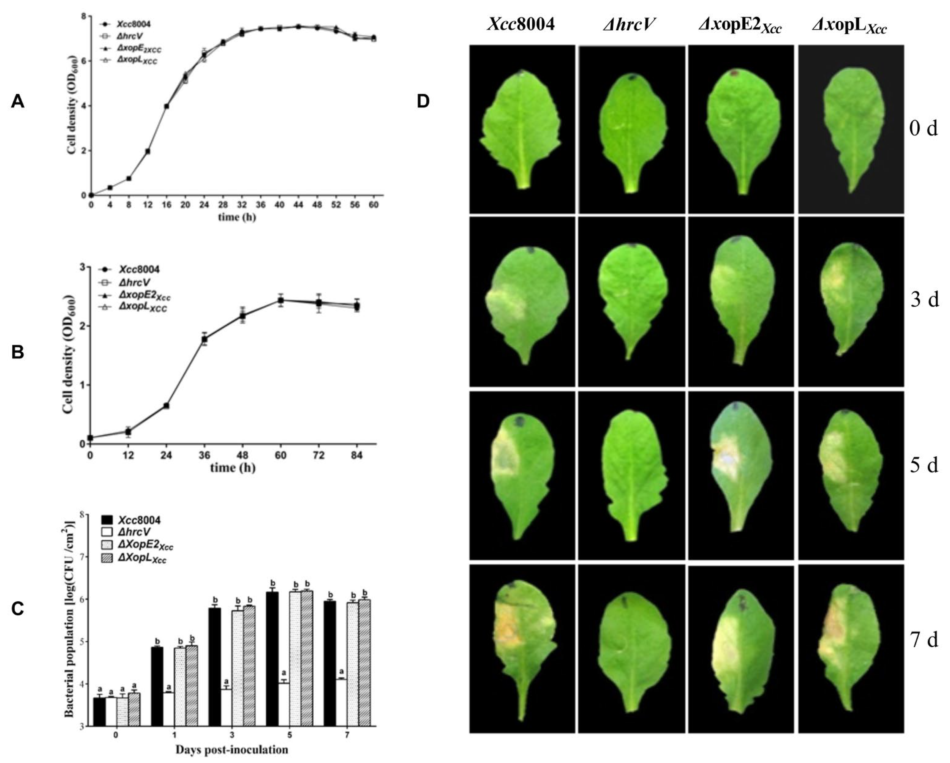
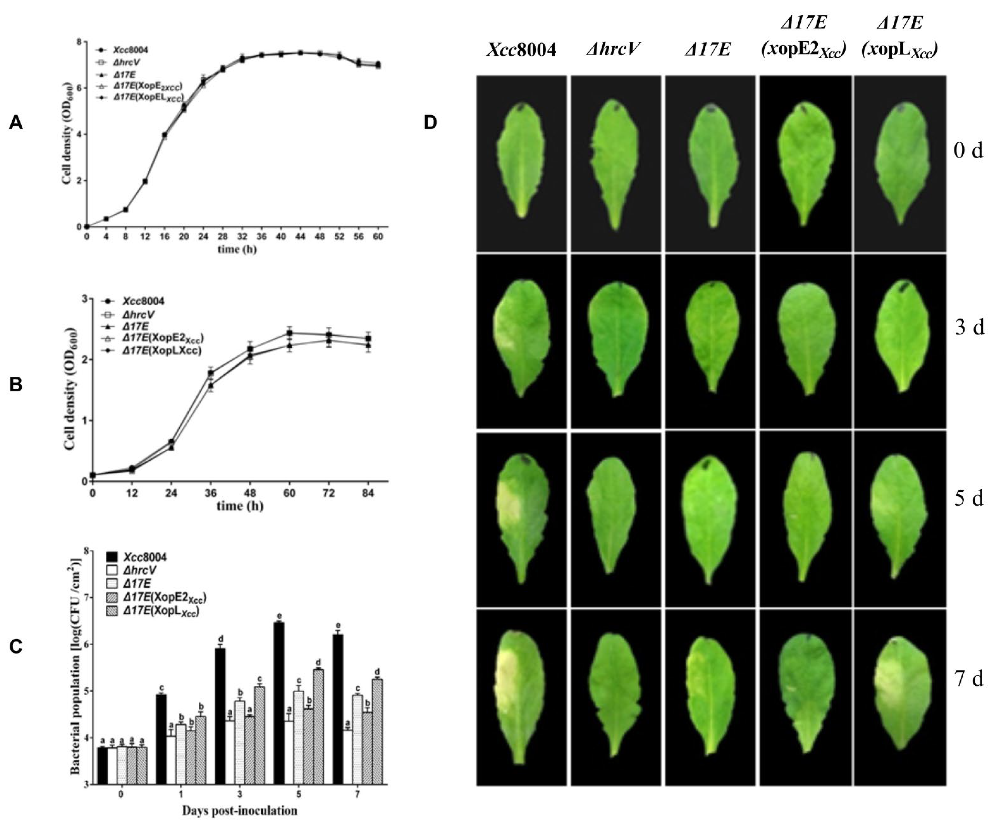
3.1. Distinct Contributions of XopE2Xcc and XopLXcc to Xcc 8004 Virulence on Arabidopsis

We investigated the impact of XopE2Xcc or XopLXcc on Xcc 8004 pathogenicity in Arabidopsis. Growth curves analysis (Figure 1A,B) revealed similar cell densities among mutants ΔxopE2Xcc, ΔxopLXcc, and Xcc 8004 in both NYG and MMX mediums. Moreover, no significant differences were observed in the bacterial population or disease symptom production among ΔxopE2Xcc, ΔxopLXcc, and Xcc 8004 (Figure 1C,D). These results indicate that the deletion of XopE2Xcc or XopLXcc does not significantly affect Xcc 8004 pathogenicity in Arabidopsis.
Furthermore, we generated a mutant lacking 17 known T3Es, including XopE2Xcc and XopLXcc, designated as Δ17E (Table S2). Subsequently, XopE2Xcc and XopLXcc were cloned into the pLAFR3 vector and introduced into the mutant Δ17E strain. The growth curves of Δ17E, Δ17E(xopE2Xcc), Δ17E(xopLXcc), and Xcc 8004 were similar in both NYG and MMX mediums (Figure 2A,B). Following the same experimental procedure, these strains infiltrated into Arabidopsis leaves. Δ17E caused slight yellowing around the inoculation site, whereas Δ17E(xopE2Xcc) exhibited significantly milder disease symptoms and low bacterial proliferation, indicating that XopE2Xcc expression conferred resistance to Xcc 8004 infection in Arabidopsis. Conversely, the bacterial populations of Δ17E(xopLXcc) were significantly higher than those of Δ17E and ΔhrcV in Arabidopsis plants but lower than those of Xcc 8004 (Figure 2C). Notably, Δ17E(xopLXcc) induced severe disease symptoms, which were attributed to high bacterial titers (Figure 2D). These findings demonstrated the critical role of XopLXcc in the virulence of Xcc 8004.

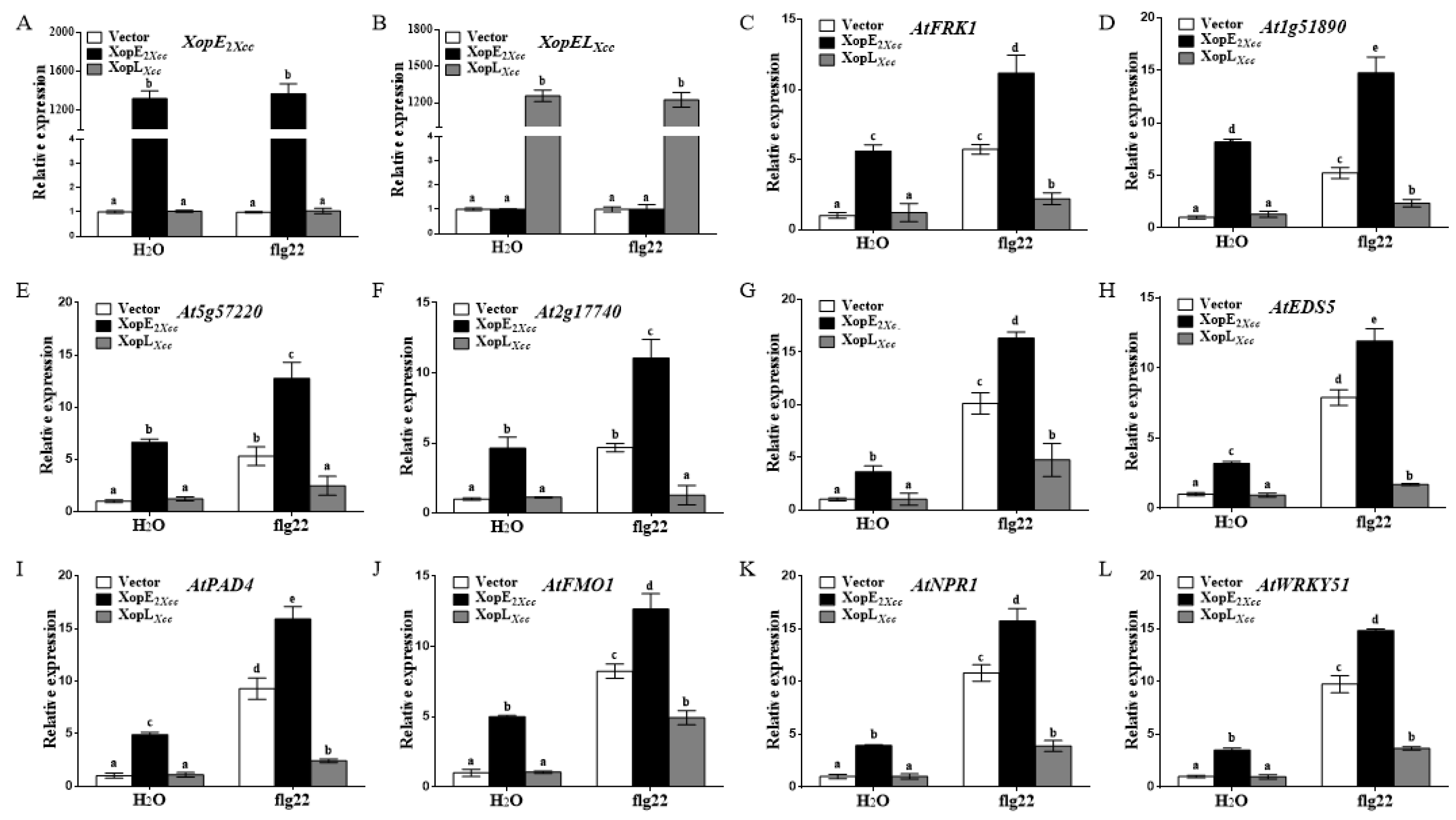
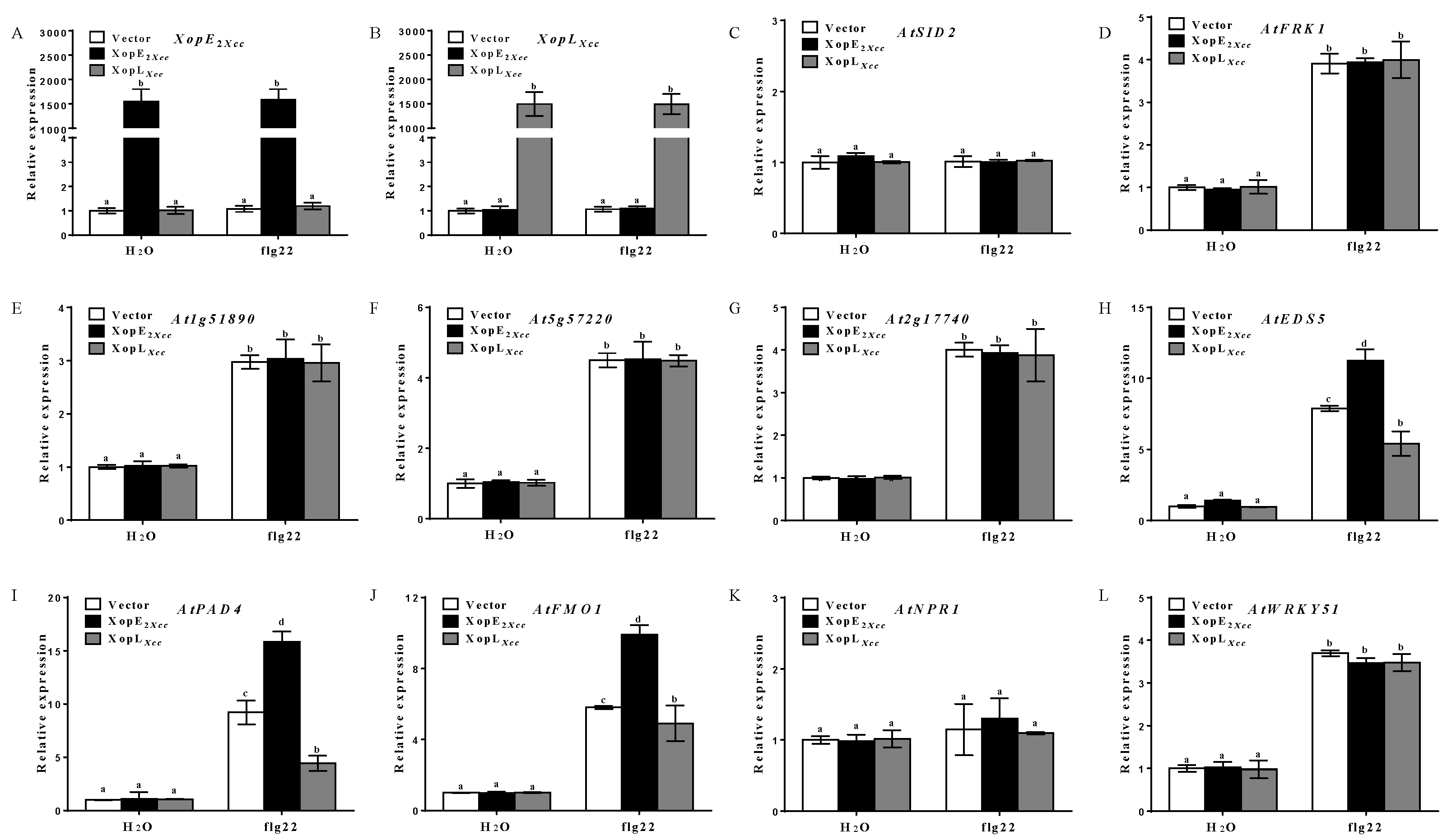
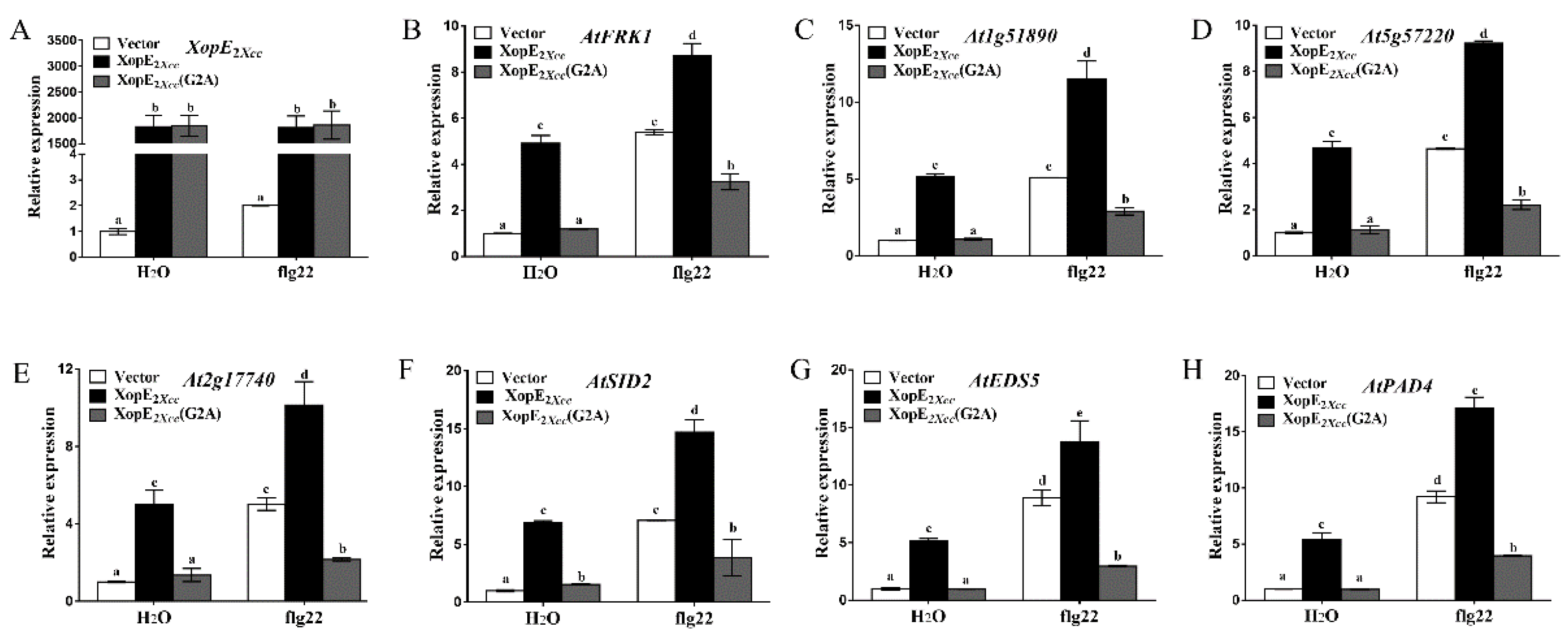
3.2. Effect of XopE2Xcc and XopLXcc on Defense Resistance in Arabidopsis

To investigate the impact of XopE2Xcc and XopLXcc on defense resistance in Arabidopsis, we analyzed the expression of four PTI-related genes and six salicylic acid (SA)-related genes using RT-qPCR. The expression levels of the PTI-related genes were significantly higher in XopE2Xcc-expressing protoplasts than in the control group (Figure 3A–F). In contrast, the expression levels of these genes were significantly weaker in XopLXcc-expressing protoplasts than those in the control. These results suggest that XopE2Xcc can activate the expression of PTI-related genes, whereas XopLXcc possesses the ability to inhibit their expression in Arabidopsis.
All SA-related genes were significantly upregulated in XopE2Xcc-expressing protoplasts (Figure 3G–L). Notably, AtSID2 expression was upregulated approximately 7.1-fold in control protoplasts, whereas it increased by approximately 18.3-fold in XopE2Xcc-expressing protoplasts. Conversely, XopLXcc downregulated the expression of these genes. These findings indicate that XopE2Xcc activates plant defense response-associated genes, including those involved in PTI and the SA signaling pathway. However, XopLXcc exerts the opposite effect, suggesting contrasting impacts on defense resistance in protoplasts.
To further explore whether XopE2Xcc directly induces the SA signaling pathway, we analyzed the expression level of defense response-associated genes in sid2 mutant protoplasts. All defense response-associated genes were induced in sid2 mutant protoplasts expressing XopE2Xcc, albeit to a significantly lower extent than that in wild-type protoplasts (Figure 4), indicating that XopE2Xcc expression in SA-deficient plants does not enhance the expression of these defense response-associated genes. Notably, the expression levels of three genes (AtEDS5, AtPAD4, and AtFMO1) in the SA signaling pathway were significantly increased in sid2 mutant protoplasts expressing XopE2Xcc (Figure 4H–J). These results suggest that XopE2Xcc boosts the expression of defense response-associated genes independently of SA accumulation, demonstrating a potential induction of upstream activators of the SA signaling pathway in Arabidopsis.

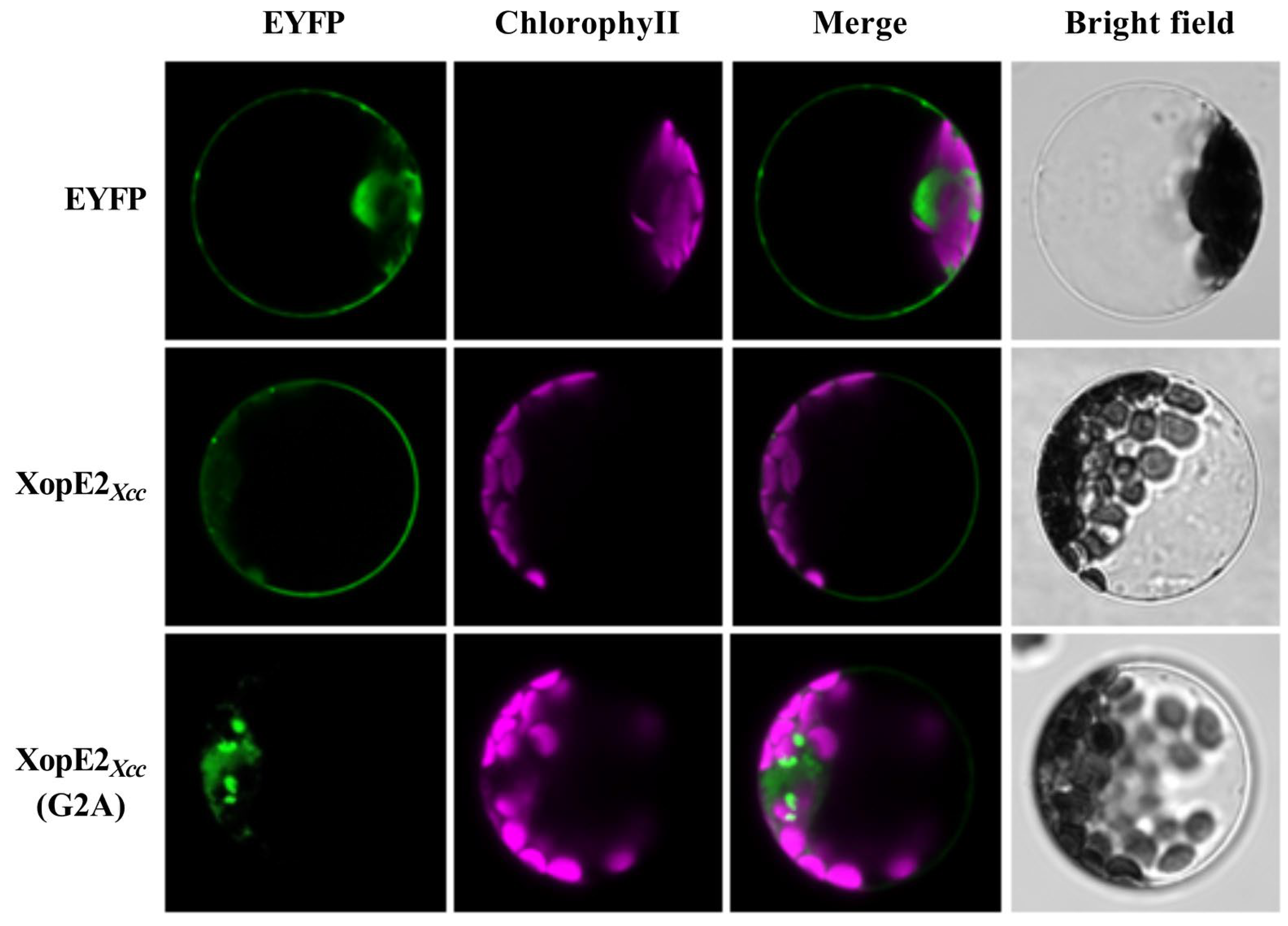
3.3. Subcellular Localization and Function Analysis of Mutant XopE2Xcc (G2A) Protein

Myristoylation modification is crucial for anchoring proteins to cell membranes [23,24]. XopE members possess a conserved putative myristoylation motif (Met-Gly-X-X-X-Ser/Thr-) at the N-terminus. It was reported that the covalent attachment of myristate to the Gly residue is an important mechanism for protein anchor [3,23,25]. To assess the significance of the putative myristoylation site in XopE2Xcc for plasma membrane localization, we substituted the glycine (Gly2) residue in XopE2Xcc with Alanine (Ala), resulting in the mutant XopE2Xcc (G2A) protein fused with EYFP. These constructs were transiently expressed in Arabidopsis protoplasts. XopE2Xcc was localized to the plasma membrane in protoplasts. Conversely, XopE2Xcc (G2A) was detected in the cytoplasm surrounding chloroplasts and some undefined organelles, rather than the plasma membrane (Figure 5). Furthermore, the mutant XopE2Xcc (G2A) protein failed to induce the expression of defense response-associated genes (Figure 6). These findings indicate that the G2A mutation in XopE2Xcc disrupts its plasma membrane localization and its ability to induce defense responses, highlighting the dependence of XopE2Xcc function on myristoylation modification.

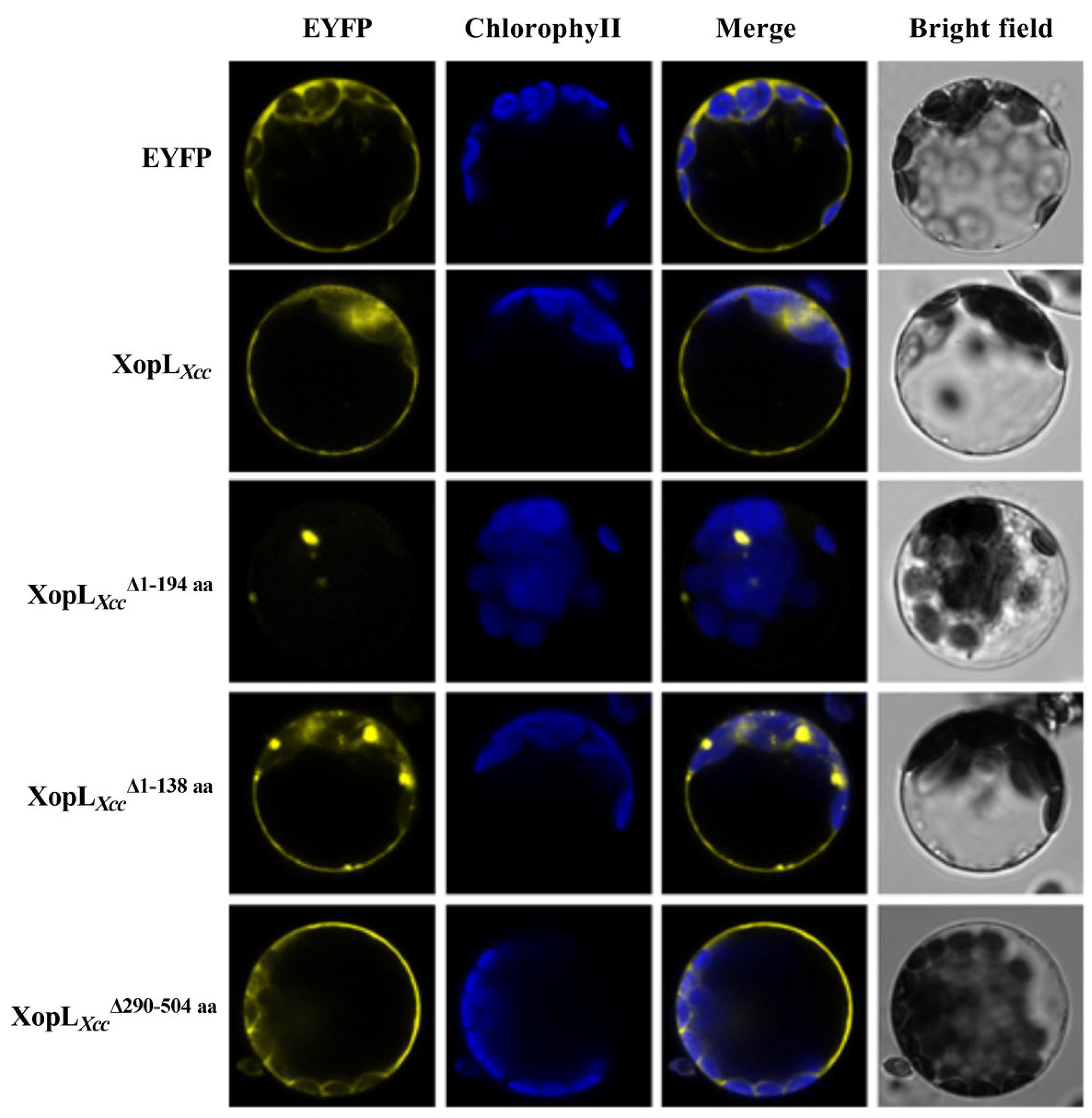
3.4. Subcellular Localization and Function Analysis of Various Mutant XopLXcc Proteins

Full-length XopLXcc cDNA and three mutants, XopLXccΔ1-194aa (lacking the N-terminal domain [NTD] and Leucine-rich repeat [LRR1 and LRR2]), XopLXccΔ1-138aa (lacking NTD and LRR1), and XopLXccΔ290-504aa (lacking the C-terminal domain), were constructed. The results indicated strong EYFP fluorescence at the plasma membrane and in the cytoplasm in XopLXcc-expressing protoplasts. The subcellular localization of XopLXccΔ290-504aa was similar to that of XopLXcc-expressing protoplasts, exhibiting membrane localization in protoplasts. XopLXccΔ1-138aa showed EYFP fluorescence in some undefined organelles and the plasma membrane. Interestingly, XopLXccΔ1-194aa exhibited subcellular localization surrounding chloroplasts, with no EYFP fluorescence observed in the protoplast membrane (Figure 7). These results demonstrate that the leucine-rich repeats (LRRs) of the N-terminal region of XopLXcc play a critical role in protein localization in protoplasts.
Furthermore, considering the varied localization of the N-terminal truncated versions and wild-type XopLXcc in Arabidopsis protoplasts, we evaluated the expression levels of defense-related genes. Notably, XopLXccΔ1-138aa and XopLXccΔ1-194aa significantly suppressed the expression of all four defense response-associated genes, while XopLXccΔ290-504aa only negatively affected two marker genes, At2g17740 and At5g57220 (Figure 8). The N-terminal deletion of XopLXcc altered the subcellular localization and function of this protein in plants, indicating that the N-terminal region of XopLXcc may play a critical role in suppressing plant immunity.

4. Discussion

Plant pathogenic bacteria typically secrete T3Es into host cells to modulate host responses, facilitating successful infection and proliferation within the host plant. Xcc 8004 causes economically significant block rot diseases in various crops [26], underscoring the importance of elucidating the pathogenesis mechanisms of T3Es. However, the functions and host targets of many T3Es remain largely elusive. Demonstrating the virulence functions of numerous T3Es in Arabidopsis-Xcc 8004 model systems can be challenging due to functional redundancy or similar pathogenicity-related genes [27]. Our previous research highlighted the Avr function of XopE2Xcc in the host plant Chinese cabbage cv. Zhongbai-83 [8], whereas XopLXcc was identified as critical for the full virulence of Xcc 8004 on the host plant Chinese radish [18]. Initial data from Arabidopsis infections did not reveal significant differences in disease symptoms or bacterial growth among ΔxopE2Xcc, ΔxopLXcc, and the wild-type Xcc 8004 strain, possibly due to masking effects of other T3Es in Xcc 8004. To address potential redundancy issues, we developed a T3E-deficient mutant, the Δ17E strain, with 17 known T3E-coding genes deleted. Using this mutant strain, we demonstrated that XopLXcc contributes to Xcc virulence, whereas XopE2Xcc functions as an Avr protein in Arabidopsis. These results confirm the presence of functional redundancy among certain effector proteins during infection of specific host plants. The Δ17E mutant strain serves as a valuable resource for the functional analysis of one or more T3Es.

4.1. XopE2Xcc Triggers Avirulence in Arabidopsis via Upstream Activation of the SA Signaling Pathway

The defense response-associated genes (At1g51890, At2g17740, AtFRK1, and At5g57220) are specifically induced by PTI from bacteria rather than other stress-related signals [28,29]. The expression levels of these genes were significantly elevated in Arabidopsis protoplasts transiently expressing XopE2Xcc, suggesting that XopE2Xcc activates their expression, thereby enhancing Arabidopsis PTI. Furthermore, SA-mediated immune responses play a critical role in Arabidopsis defense [4,30,31]. Our data revealed that expressing XopE2Xcc in Arabidopsis significantly activated the SA signaling pathway. Genes involved in SA biosynthesis (AtSID2), SA accumulation (AtEDS5, AtPAD4, and AtFMO1), and downstream signal transduction (AtNPR1 and AtWRKY51) exhibited significantly higher expression levels in XopE2Xcc-transgenic plants than in control plants. Notably, XopE2Xcc expression in the SA defective mutant (sid2 mutant) protoplasts failed to enhance the expression of four defense response-associated genes, confirming that XopE2Xcc elicits the Arabidopsis immune response possible through the SA signaling pathway. Moreover, the expression of genes involved in SA accumulation (AtEDS5, AtPAD4, and AtFMO1) in mutant protoplasts was significantly increased by XopE2Xcc, indicating that XopE2Xcc functions upstream of genes related to SA accumulation. These results suggest that XopE2Xcc triggers Arabidopsis immune responses via an unidentified activator upstream of the SA signaling pathway. Further identification and characterization of the target protein of XopE2Xcc in plants will enhance our understanding of how XopE2Xcc modulates plant defense responses.
N-myristoylation involves the covalent attachment of a hydrophobic 14-carbon saturated fatty acid to the glycine at residue two (Gly2), providing primary membrane targeting, signaling for several plant protein kinases and directing proteins to the plant cell plasma membrane [23]. Previous research has suggested that XopE2Xcc, a member of the XopE group, and possibly part of the transglutaminase superfamily, contains a putative N-myristoylation motif [3]. Consistent with findings on AvrXccC and XopE2Xe, our data indicate that the N-myristoylation motif is essential for the subcellular localization and avirulence function of XopE2Xcc in Arabidopsis [10,23].
Avr proteins are known to play a dual role in triggering ETI in plants with corresponding R proteins, while simultaneously suppressing plant defense in plants without the corresponding R proteins [32,33,34]. A previous study has demonstrated that XopE2Xcc (also known as avrXccE1) induces avirulence of Xcc 8004 in Chinese cabbage cv. Zhongbai-83, but not in Xcc strain 528T on Early Jersey Wakefield cabbage [35]. Our data indicate that the avirulence function of XopE2Xcc in Arabidopsis is dependent on plasma membrane anchoring for host recognition, suggesting the presence of its corresponding R protein on the plasma membrane of Arabidopsis. Further identification and characterization of the R protein of XopE2Xcc in Arabidopsis are necessary to enhance our understanding of how XopE2Xcc modulates plant defense responses.

4.2. XopLXcc Suppresses PTI-Related and SA-Related Genes in Arabidopsis

In this study, we observed significant suppression of four PTI-related genes in XopLXcc-expressing protoplasts, suggesting its ability to inhibit Arabidopsis PTI. These findings are consistent with previous reports indicating that XopLXcc enhances bacterial infection in Arabidopsis while suppressing the reactive oxygen species (ROS) burst, callose deposition, and other related responses [36]. Moreover, we observed significantly lower expression levels of SA-related genes in XopLXcc-expressing protoplasts than in controls, suggesting that XopLXcc promotes the synthesis and accumulation of SA, thereby suppressing Arabidopsis immune responses, possibly through the SA signaling pathway.
XopLXcc belongs to the XopL superfamily, characterized by three LRR domains and a putative XopL E3 ligase box domain (XL box). XopLXcv can suppress defense gene expression in plants and subvert plant immunity, with the XL box playing an essential role in E3 ubiquitin ligase activity and affecting plastid phenotypes [37,38]. However, our in vitro assessments did not reveal any E3 ligase activity for XopLXcc. Conversely, a truncated form of XopLXap (lacks the XL box) retained the ability to suppress plant immune responses [39]. Our research demonstrates that different N-terminal mutants exhibit varied localizations and effects on plant immunity, highlighting the essential role of the complete set of LRR domains of XopLXcc in modulating plant immunity.
The LRRs are 20–29 residue sequence motifs present in numerous proteins with diverse functions. All proteins containing these repeats are believed to facilitate protein–protein interactions [40]. Various invasive bacterial proteins were identified to contain multiple LRR motifs [41]. The structure of XopLXcc primarily consists of three LRRs, lacking known ligands and protein-anchoring sites in this region. The crystallographic study revealed that LRRs correspond to β-α structural units. These units are arranged so that the protein forms a curved structure [40]. It is hypothesized that the XopLXcc protein shares a similar three-dimensional structure, and the LRRs are potentially responsible for protein anchoring and membrane localization. Truncation of the C-terminal region of XopLXcc did not disturb protein conformation, as evidenced by its retained membrane localization ability. Further findings indicated that the membrane localization is likely mediated by the N-terminal region, possibly the LRRs, rather than the C-terminal part. We supposed that the parallel LRRs contribute to stabilizing the structure of β-sheets and α-helices through lateral interactions. Loss of the LRR region may lead to structural alterations that affect protein function.

5. Conclusions

Our study elucidates the divergent impacts of two T3E proteins on Arabidopsis immune responses. XopE2Xcc triggers Arabidopsis immune responses via an unidentified activator of the salicylic acid (SA) signaling pathway, whereas XopLXcc suppresses the expression of PTI- and SA-related genes. The presence of the N-myristoylation motif and N-terminal domain proved crucial for the subcellular localization and virulence function of XopE2Xcc and XopLXcc, respectively. These findings significantly enhance our understanding of the mechanisms employed by pathogenic bacteria T3Es and contribute to developing effective strategies for controlling bacterial diseases.

Supplementary Materials

The following supporting information can be downloaded at: https://www.mdpi.com/article/10.3390/pathogens13060448/s1, Table S1: Primers used in this work. Table S2: Bacterial strains and plasmids used in this work.

Author Contributions

Conceptualization, J.H. and Y.H.; methodology, J.H., H.Z., M.Z., N.L., Y.H. and B.J.; validation, J.H. and H.Z.; data curation, J.H., H.Z. and Y.H.; writing—original draft preparation, J.H.; writing—review and editing, Y.H.; project administration, Y.H.; funding acquisition, J.H. and Y.H. All authors have read and agreed to the published version of the manuscript.

Funding

This work is supported by the Natural Science Foundation of China (32060599 and 32160079).

Institutional Review Board Statement

Not applicable.

Informed Consent Statement

Not applicable.

Data Availability Statement

Data presented in this study will be available from the corresponding author upon request.

Acknowledgments

We thank the ABRC (Columbus, OH, USA) for providing the pXSN-HA plasmid, and Ji-Ming Gong (Shanghai Institute of Plant Physiology and Ecology) for providing the pA7-YFP plasmid. We thank Core Facilities Center of SKLCUSA for supporting microscopic observation.

Conflicts of Interest

The authors declare no conflicts of interest. The funders had no role in the design of the study; in the collection, analyses, or interpretation of data; in the writing of the manuscript, or in the decision to publish the results.

References

  1. Ryan, R.P.; Vorhölter, F.J.; Potnis, N.; Jones, J.B.; Van Sluys, M.A.; Bogdanove, A.J.; Dow, J.M. Pathogenomics of Xanthomonas: Understanding bacterium-plant interactions. Nat. Rev. Microbiol. 2011, 9, 344–355. [Google Scholar] [CrossRef] [PubMed]
  2. Gürlebeck, D.; Thieme, F.; Bonas, U. Type III effector proteins from the plant pathogen Xanthomonas and their role in the interaction with the host plant. J. Plant. Physiol. 2006, 163, 233–255. [Google Scholar] [CrossRef] [PubMed]
  3. White, F.F.; Potnis, N.; Jones, J.B.; Koebnik, R. The type III effectors of Xanthomonas. Mol. Plant. Pathol. 2009, 10, 749–766. [Google Scholar] [CrossRef] [PubMed]
  4. Jones, J.D.G.; Dangl, J.L. The plant immune system. Nature 2006, 444, 323–329. [Google Scholar] [CrossRef] [PubMed]
  5. Mansfield, J.; Genin, S.; Magori, S.; Citovsky, V.; Sriariyanum, M.; Ronald, P.; Dow, M.; Verdier, V.; Beer, S.V.; Machado, M.A.; et al. Top 10 plant pathogenic bacteria in molecular plant pathology. Mol. Plant. Pathol. 2012, 13, 614–629. [Google Scholar] [CrossRef] [PubMed]
  6. Vicente, J.; Cascón, T.; Vicedo, B.; García-Agustín, P.; Hamberg, M.; Castresana, C. Role of 9-lipoxygenase and α-dioxygenase oxylipin pathways as modulators of local and systemic defense. Mol. Plant. 2012, 5, 914–928. [Google Scholar] [CrossRef] [PubMed]
  7. Qian, W.; Jia, Y.T.; Ren, S.X.; He, Y.Q.; Feng, J.X.; Lu, L.F.; Sun, Q.H.; Ying, G.; Tang, D.J.; Tang, H.; et al. Comparative and functional genomic analyses of the pathogenicity of phytopathogen Xanthomonas campestris pv. campestris. Genome Res. 2005, 15, 757–767. [Google Scholar] [CrossRef] [PubMed]
  8. He, Y.Q.; Zhang, L.; Jiang, B.L.; Zhang, Z.C.; Xu, R.Q.; Tang, D.J.; Qin, J.; Jiang, W.; Zhang, X.; Liao, J.; et al. Comparative and functional genomics reveals genetic diversity and determinants of host specificity among reference strains and a large collection of Chinese isolates of the phytopathogen Xanthomonas campestris pv. campestris. Genome Biol. 2007, 8, R218. [Google Scholar] [CrossRef] [PubMed]
  9. Guy, E.; Lautier, M.; Chabannes, M.; Roux, B.; Lauber, E.; Arlat, M.; Noël, L.D. XopAC-triggered immunity against Xanthomonas depends on arabidopsis receptor-Like cytoplasmic kinase genes PBL2 and RIPK. PLoS ONE 2013, 8, e73469. [Google Scholar] [CrossRef]
  10. Wang, L.F.; Tang, X.Y.; He, C.Z. The bifunctional effector AvrXccC of Xanthomonas campestris pv. campestris requires plasma membrane-anchoring for host recognition. Mol. Plant. Pathol. 2007, 8, 491–501. [Google Scholar] [CrossRef]
  11. Tan, L.T.; Rong, W.; Luo, H.L.; Chen, Y.H.; He, C.Z. The Xanthomonas campestris effector protein XopDxcc8004 triggers plant disease tolerance by targeting DELLA proteins. New Phytol. 2014, 204, 595–608. [Google Scholar] [CrossRef] [PubMed]
  12. Sun, H.; Zhu, X.L.; Li, C.A.X.; Ma, Z.M.; Han, X.; Luo, Y.Y.; Yang, L.; Yu, J.; Miao, Y.S. Xanthomonas effector XopR hijacks host actin cytoskeleton via complex coacervation. Nat. Commun. 2021, 12, 4064. [Google Scholar] [CrossRef] [PubMed]
  13. Jiang, B.L.; He, Y.Q.; Cen, W.J.; Wei, H.Y.; Jiang, G.F.; Jiang, W.; Hang, X.H.; Feng, J.X.; Lu, G.T.; Tang, D.H.; et al. The type III secretion effector xopXccN of Xanthomonas campestris pv. campestris is required for full virulence. Res. Microbiol. 2008, 159, 216–220. [Google Scholar] [CrossRef] [PubMed]
  14. Feng, F.; Yang, F.; Rong, W.; Wu, X.G.; Zhang, J.; Chen, S.; He, C.Z.; Zhou, J.M. A Xanthomonas uridine 5′-monophosphate transferase inhibits plant immune kinases. Nature 2012, 485, 114–118. [Google Scholar] [CrossRef] [PubMed]
  15. Yan, X.; Tao, J.; Luo, H.L.; Tan, L.T.; Rong, W.; Li, H.P.; He, C.Z. A type III effector XopLxc8004 is vital for Xanthomonas campestris pathovar campestris to regulate plant immunity. Res. Microbiol. 2019, 170, 138–146. [Google Scholar] [CrossRef] [PubMed]
  16. Üstün, S.; Bartetzko, V.; Börnke, F. The Xanthomonas campestris type III effector XopJ targets the host cell proteasome to suppress salicylic-acid mediated plant defence. PLoS Pathog. 2013, 9, e1003427. [Google Scholar] [CrossRef] [PubMed]
  17. Xu, R.Q.; Li, X.Z.; Wei, H.Y.; Jiang, B.; Li, K.; He, Y.Q.; Feng, J.X.; Tang, J.L. Regulation of eight avr genes by hrpG and hrpX in Xanthomonas campestris pv. campestris and their role in pathogenicity. Prog. Nat. Sci. Mater. 2006, 16, 1288–1294. [Google Scholar]
  18. Jiang, W.; Jiang, B.L.; Xu, R.Q.; Huang, J.D.; Wei, H.Y.; Jiang, G.F.; Cen, W.J.; Liu, J.; Ge, Y.Y.; Li, G.H.; et al. Identification of six type III effector genes with the PIP box in Xanthomonas campestris pv. campestris and five of them contribute individually to full pathogenicity. Mol. Plant. Microbe Interact. 2009, 22, 1401–1411. [Google Scholar] [CrossRef]
  19. Chen, S.B.; Songkumarn, P.; Liu, J.L.; Wang, G.L. A versatile zero background T-vector system for gene cloning and functional genomics. Plant. Physiol. 2009, 150, 1111–1121. [Google Scholar] [CrossRef]
  20. Huang, J.; Zhang, Y.; Peng, J.S.; Zhong, C.; Yi, H.Y.; Ow, D.W.; Gong, J.M. Fission yeast HMT1 lowers seed cadmium through phytochelatin-dependent vacuolar sequestration in arabidopsis. Plant. Physiol. 2012, 158, 1779–1788. [Google Scholar] [CrossRef]
  21. Yoo, S.D.; Cho, Y.H.; Sheen, J. Mesophyll protoplasts: A versatile cell system for transient gene expression analysis. Nat. Protoc. 2007, 2, 1565–1572. [Google Scholar] [CrossRef] [PubMed]
  22. Meyer, D.; Lauber, E.; Roby, D.; Arlat, M.; Kroj, T. Optimization of pathogenicity assays to study the Arabidopsis thaliana-Xanthomonas campestris pv. campestris pathosystem. Mol. Plant. Pathol. 2005, 6, 327–333. [Google Scholar] [CrossRef] [PubMed]
  23. Thieme, F.; Szczesny, R.; Urban, A.; Kirchner, O.; Hause, G.; Bonas, U. New type III effectors from Xanthomonas campestris pv. vesicatoria trigger plant reactions dependent on a conserved N-myristoylation motif. Mol. Plant. Microbe Interact. 2007, 20, 1250–1261. [Google Scholar] [CrossRef] [PubMed]
  24. Resh, M.D. Fatty acylation of proteins: New insights into membrane targeting of myristoylated and palmitoylated proteins. Biochim. Biophys. Acta (BBA) Mol. Cell Res. 1999, 1451, 1–16. [Google Scholar] [CrossRef]
  25. Nimchuk, Z.L.; Fisher, E.J.; Desveaux, D.; Chang, J.H.; Dangl, J.L. The HopX (AvrPphE) family of Pseudomonas syringae type III effectors require a catalytic triad and a novel N-terminal domain for function. Mol. Plant. Microbe Interact. 2007, 20, 346–357. [Google Scholar] [CrossRef] [PubMed]
  26. Vicente, J.G.; Holub, E.B. Xanthomonas campestris pv. campestris (cause of black rot of crucifers) in the genomic era is still a worldwide threat to brassica crops. Mol. Plant. Pathol. 2013, 14, 2–18. [Google Scholar] [CrossRef] [PubMed]
  27. Kvitko, B.H.; Park, D.H.; Velásquez, A.C.; Wei, C.F.; Russell, A.B.; Martin, G.B.; Schneider, D.J.; Collmer, A. Deletions in the repertoire of Pseudomonas syringae pv. tomato DC3000 type III secretion effector genes reveal functional overlap among effectors. PLoS Pathog. 2009, 5, e1000388. [Google Scholar] [CrossRef]
  28. He, P.; Shan, L.; Lin, N.C.; Martin, G.B.; Kemmerling, B.; Nürnberger, T.; Sheen, J. Specific bacterial suppressors of MAMP signaling upstream of MAPKKK in Arabidopsis innate immunity. Cell 2006, 125, 563–575. [Google Scholar] [CrossRef] [PubMed]
  29. Akimoto-Tomiyama, C.; Furutani, A.; Tsuge, S.; Washington, E.J.; Nishizawa, Y.; Minami, E.; Ochiai, H. XopR, a type III effector secreted by Xanthomonas oryzae pv. oryzae, suppresses microbe-associated molecular pattern-triggered immunity in arabidopsis thaliana. Mol. Plant. Microbe Interact. 2012, 25, 505–514. [Google Scholar] [CrossRef]
  30. Vlot, A.C.; Dempsey, D.A.; Klessig, D.F. Salicylic acid, a multifaceted hormone to combat disease. Annu. Rev. Phytopathol. 2009, 47, 177–206. [Google Scholar] [CrossRef]
  31. Tsuda, K.; Sato, M.; Stoddard, T.; Glazebrook, J.; Katagiri, F. Network properties of robust immunity in plants. PLoS Genet. 2009, 5, e1000772. [Google Scholar] [CrossRef] [PubMed]
  32. Alfano, J.R.; Collmer, A. Type III secretion system effector proteins: Double agents in bacterial disease and plant defense. Annu. Rev. Phytopathol. 2004, 42, 385–414. [Google Scholar] [CrossRef] [PubMed]
  33. Xu, R.Q.; Blanvillain, S.; Feng, J.X.; Jiang, B.L.; Li, X.Z.; Wei, H.Y.; Kroj, T.; Lauber, E.; Roby, D.; Chen, B.; et al. AvrAC, a type III effector with a leucine-rich repeat domain from Xanthomonas campestris pathovar campestris confers avirulence in vascular tissues of Arabidopsis thaliana ecotype col-0. J. Bacteriol. 2008, 190, 343–355. [Google Scholar] [CrossRef] [PubMed]
  34. Stork, W.; Kim, J.G.; Mudgett, M.B. Functional analysis of plant defense suppression and activation by the Xanthomonas core type III effector XopX. Mol. Plant. Microbe Interact. 2015, 28, 180–194. [Google Scholar] [CrossRef] [PubMed]
  35. Castañeda, A.; Reddy, J.D.; El-Yacoubi, B.; Gabriel, D.W. Mutagenesis of all eight avr genes in Xanthomonas campestris pv. campestris had no detected effect on pathogenicity, but one gene affected race specificity. Mol. Plant. Microbe Interact. 2005, 18, 1306–1317. [Google Scholar] [CrossRef] [PubMed]
  36. Yan, S.P.; Dong, X.N. Perception of the plant immune signal salicylic acid. Curr. Opin. Plant. Biol. 2014, 20, 64–68. [Google Scholar] [CrossRef] [PubMed]
  37. Singer, A.U.; Schulze, S.; Skarina, T.; Xu, X.H.; Cui, H.; Eschen-Lippold, L.; Egler, M.; Srikumar, T.; Raught, B.; Lee, J.; et al. A pathogen type III effector with a novel E3 ubiquitin ligase architecture. PLoS Pathog. 2013, 9, e1003121. [Google Scholar] [CrossRef] [PubMed]
  38. Erickson, J.L.; Adlung, N.; Lampe, C.; Bonas, U.; Schattat, M.H. The Xanthomonas effector XopL uncovers the role of microtubules in stromule extension and dynamics in Nicotiana benthamiana. Plant J. 2018, 93, 856–870. [Google Scholar] [CrossRef] [PubMed]
  39. Soni, M.; Mondal, K.K. Xanthomonas axonopodis pv. punicae uses XopL effector to suppress pomegranate immunity. J. Integr. Plant. Biol. 2018, 60, 341–357. [Google Scholar] [CrossRef]
  40. Bengtsson, E.; Lindblom, K.; Tillgren, V.; Aspberg, A. The leucine-rich repeat protein PRELP binds fibroblast cell-surface proteoglycans and enhances focal adhesion formation. Biochem. J. 2016, 473, 1153–1164. [Google Scholar] [CrossRef]
  41. Nitipan, S.; Sritrakul, T.; Kunjantarachot, A.; Prapong, S. Identification of epitopes in Leptospira borgpetersenii leucine-rich repeat proteins. Infect. Genet. Evol. 2013, 14, 46–57. [Google Scholar] [CrossRef] [PubMed]
Figure 1. Pathogenicity analysis of Xcc 8004, ΔxopE2Xcc, and ΔxopLXcc in Arabidopsis. (A) Grow curves in rich medium (NYG). (B) Grow curves in minimal medium (MMX). (C) Bacterial populations. (D) Disease symptoms. ΔhrcV, T3SS-defective mutant strain; ΔxopE2Xcc, XopE2Xcc mutant strain; ΔxopLXcc, XopLXcc mutant strain. The a\b labels shown on panel C represent significant differences (n = 30, p < 0.05, two-way ANOVA with Tukey’s HSD test). The same letters mean no statistical difference.
Pathogens 13 00448 g001
Figure 2. Pathogenicity analysis of Xcc 8004, Δ17E, Δ17E(xopE2Xcc), and Δ17E(xopLXcc) in Arabidopsis under different conditions. (A) Grow curves in rich medium (NYG). (B) Grow curves in minimal medium (MMX). (C) Bacterial populations. (D) Disease symptoms. ΔhrcV, T3SS-defective mutant strain; Δ17E, 17 known T3Es-defficient mutant strain; Δ17E(xopE2Xcc), Δ17E strain containing XopE2Xcc; Δ17E(xopLXcc), Δ17E strain containing XopLXcc. The a\b\c\d\e labels shown on panel C represent significant differences (n = 30, p < 0.05, two-way ANOVA with Tukey’s HSD test). The same letters mean no statistical difference.
Pathogens 13 00448 g002
Figure 3. Influence of XopE2Xcc and XopLXcc on the expression of defense-related genes in Arabidopsis. (AL), The expression levels of defense-related genes in XopE2Xcc or XopLXcc-transfected protoplasts of Arabidopsis Col-0 were measured using real-time qRT-PCR, respectively. The mRNA levels of all genes were normalized with Atactin2, and the relative expression levels were determined in protoplasts transfected with the control vector. The a\b\c\d\e labels represent significant differences (n = 5, p < 0.05, two-way ANOVA with Tukey’s HSD test). The same letters mean no statistical difference.
Pathogens 13 00448 g003
Figure 4. Influence of XopE2Xcc on the expression of defense response-associated genes in sid2 mutant protoplasts. (AL), The expression levels of defense-associated genes in XopE2Xcc or XopLXcc-transfected protoplasts of Arabidopsis sid2 mutant were measured using real-time qRT-PCR, respectively. The mRNA levels of all genes were normalized with Atactin2, and the relative expression levels were determined in protoplasts transfected with the control vector. The a\b\c\d labels represent significant differences (n = 5, p < 0.05, two-way ANOVA with Tukey’s HSD test). The same letters mean no statistical difference.
Pathogens 13 00448 g004
Figure 5. Subcellular localization of mutant XopE2Xcc (G2A) protein in protoplasts.
Pathogens 13 00448 g005
Figure 6. Influence of mutant XopE2Xcc (G2A) protein on the expression of defense response-associated genes in Arabidopsis. (AH),The expression of response-associated genes in XopE2Xcc and XopE2Xcc (G2A) transfected protoplasts was measured using real-time qRT-PCR, respectively. The a\b\c\d\e labels represent significant differences (n = 5, p < 0.05, two-way ANOVA with Tukey’s HSD test). The same letters mean no statistical difference.
Pathogens 13 00448 g006
Figure 7. Subcellular localization of various mutant XopLXcc proteins in protoplasts.
Pathogens 13 00448 g007
Figure 8. Influence of various mutant XopLXcc proteins on the expression of defense response-associated genes in Arabidopsis.
Pathogens 13 00448 g008
Disclaimer/Publisher’s Note: The statements, opinions and data contained in all publications are solely those of the individual author(s) and contributor(s) and not of MDPI and/or the editor(s). MDPI and/or the editor(s) disclaim responsibility for any injury to people or property resulting from any ideas, methods, instructions or products referred to in the content.

