Article

Assessing the In Vitro Individual and Combined Effect of Arthrobotrys oligospora and A. musiformis (Orbiliales) Liquid Culture Filtrates against Infective Larvae of the Sheep Blood-Feeding Nematode Haemonchus contortus (Trichostrongylidae)

by
Antonio Colinas-Picazo
1,
Pedro Mendoza-de Gives
1,*,
Gustavo Pérez-Anzúrez
1,
Enrique Gutiérrez-Medina
1,
Génesis Andrea Bautista-García
1,
Edgar Jesús Delgado-Núñez
2 and
Agustín Olmedo-Juárez
1
1
Laboratory of Helminthology, National Centre for Disciplinary Research in Animal Health and Innocuity (CENID-SAI), National Institute for Research in Forestry, Agriculture and Livestock, INIFAP-SADER, Morelos, Jiutepec 62550, Mexico
2
Faculty of Agricultural, Livestock and Environmental Sciences, Autonomous University of the State of Guerrero, Iguala de la Independencia 40040, Mexico
*
Author to whom correspondence should be addressed.
Pathogens 2024, 13(6), 498; https://doi.org/10.3390/pathogens13060498
Submission received: 23 May 2024 / Revised: 5 June 2024 / Accepted: 8 June 2024 / Published: 11 June 2024

Abstract

:
Background: Arthrobotrys species are nematophagous fungi that secrete extracellular nematocidal products (ECP). The individual and combined effects of ECP from Arthrobotrys oligospora (Ao) and A. musiformis (Am) growth in liquid media against Haemonchus contortus L3 (HcL3) were assessed. Methods: The isolation, morphological (MI) and molecular identification (Mol-I), assessment of nematocidal activity (NA) of fungal liquid culture filtrates (LCF) in two liquid media alone and in combination and the myco-compound profile identification (MCP) were performed. Results: The MI suggested that the fungi corresponded to the species Ao and Am. This result was confirmed by PCR analysis followed by sequencing, alignment and a phylogenetic analysis. Likewise, the highest Hc mortalities were 91.4% with individual LCF of Am and 86.2% with those of Ao at the highest concentration (100 mg/mL) in Czapek-Dox Broth. The combination of both LCF resulted in a similarly high larval mortality with no statistical differences in relation to individual activity (p > 0.05). The MCP showed the presence of alkaloids in both fungi. Coumarins, sterols and saponins were found only in Ao. Main conclusions: Both fungi produced ECP with a high NA that could be identified and assessed in future studies as potential natural anthelmintic compounds.

1. Introduction

Haemonchus contortus is a blood-feeding parasitic nematode living in the abomasum (stomach) of small ruminants, that provokes gastritis, blood-loss in the abomasum, anorexia, anaemia, lethargy, weakness, weight loss, emaciation and, in severe cases, the death of young animals [1,2]. This and other genera/species of parasitic nematodes live in the gastrointestinal tract of small ruminants, where they cause severe deterioration of flock health and productivity. Gastrointestinal nematodiasis is controlled by the frequent and continuous administration of chemical anthelmintic drugs to the animals; however, the development of anthelmintic resistance in parasites has led to increasing inefficacy of these drugs, raising alarm among farmers worldwide [3,4]. During recent decades, strategies other than the use of chemical anthelmintic drugs have been explored, including the use of natural antagonists of nematodes, such as a group of micro-fungi called nematode-trapping fungi [5,6]. Nematode-trapping fungi are regular microorganisms from the soil mycobiota living as saprophyte organisms and taking their nitrogen and carbon sources from decaying wood and litter; however, they can transform their saprophyte lifestyle into a predatory or parasitic one in the presence of nematodes such that, once trapped, they are used as a food source by fungi [7]. The species Arthrobotrys oligospora and A. musiformis have been classified as Orbiliales fungi [8]. These species develop three-dimensional adhesive nets from their mycelia where nematodes are trapped and destroyed for eventual use by fungi as their main source of nutrients [9]. In addition to the mechanical capture exerted by trapping devices, nematode-trapping fungi possess other strategies to kill and penetrate nematodes using myco-chemical constituents, including enzymes and products derived from secondary metabolism [10]. The objectives of the present study were to isolate nematode-trapping fungi and perform a taxonomical identification via morphological and molecular procedures, as well as to assess the nematocidal activity of their liquid culture filtrates, either individually or combined, and produced in two liquid media (sweet potato dextrose broth [SPDB] and Czapek-Dox Broth [CzDB]) (Sigma, Aldrich, Merck, Darmstadt, Germany) against H. contortus infective larvae, and eventually to identify associated groups of myco-compounds.

2. Materials and Methods

2.1. Nematodes

2.1.1. Obtaining the Free-Living Nematode Panagrellus redivivus for Use as Bait to Isolate Nematophagous Fungi

A population of the free-living nematode P. redivivus was provided as fish food by a local pet store in Jiutepec Municipality, Morelos, Mexico. For en masse reproduction, nematodes were cultured in sterile plastic bowls containing sterile oat flakes (20 g), and 200 mL of sterile distilled water was added. Oat flakes and water were mixed and homogenized to finally obtain a humid mass that was used as a nutrient substrate for bacteria, and in turn, bacteria were used as the main source of food by the free-living nematodes. Bowls were covered with a cap of foil and gauze to prevent mosquitoes from entering. Cultures were incubated at room temperature (18–28 °C) for 7 days [11]. After the incubation period, some culture material was collected using a metal spoon and dissolved in a glass of water, producing many P. redivivus specimens swimming in an aqueous suspension. Nematodes were separated from the culture medium through a 74-µm sieve. This step was repeated several times until the nematodes were very clean. Nematodes were resuspended in sterile distilled water and sieved through a coffee filter. Nematodes were eventually recovered using the Baermann funnel technique [12].

2.1.2. Procurement of Haemonchus contortus Infective Larvae (L3)

A 4-month-old pelibuey lamb was used. The McMaster technique using the faecal matter of this lamb was negative for the presence of gastrointestinal nematode eggs. This lamb was orally inoculated with 350 H. contortus infecting larvae per kilogram of body weight. After a pre-patent period of 21 days, faecal samples taken directly from the rectum of this lamb tested positive for the presence of nematode eggs by the McMaster technique. All rules regarding the treatment of animals and the prevention of unnecessary animal suffering were carefully followed according to the Norma Oficial Mexicana (Official Mexican Standard) with official rule number NOM-052-ZOO-1995 (http://www.senasica.gob.mx, accessed on 8 August 2023). Additionally, the Ley Federal de Sanidad Animal (Federal Law for Animal Health) DOF 07-06-2012 was strictly followed in accordance with the ethical standards outlined by INIFAP. Fresh faeces were ground in a plastic bowl and mixed with small pieces of polyurethane foam to obtain a porous mass that retained the oxygen necessary for the optimum development of nematode eggs [13]. Faecal cultures were incubated at room temperature (18–28 °C) for 7 days. After 5–7 days of elaboration of faecal cultures, a rather large amount of H. contortus infective larvae was collected using the Baermann Funnel technique for 24 h [13,14]. Infective larvae of the parasite were cleaned using the differential centrifugation technique with 40% sucrose density gradients [15]. After centrifugation for 5 min at 3500 rpm, a white ring in the interphase between water and sucrose corresponding to clean larvae was visualized. Larvae were removed with a Pasteur pipette and deposited into assay tubes, which were filled with water. In order to discard sucrose residue from the aqueous suspension of larvae, the assay tubes containing larvae in suspension were centrifuged at the same speed and spin times, and larvae were sedimented to the base of the assay tubes. The supernatant was discarded, and the tubes were refilled with sterile water and centrifuged again. After three to four centrifugations, larvae in the sediment were free of sucrose residues.

2.2. Isolation of Nematophagous Fungi

Two 50-g individual samples of soil from a poultry farm in Cuernavaca Municipality, state of Morelos, Mexico were taken and transported in plastic bags to the Laboratory of Helminthology of CENID-SAI in Jiutepec, Morelos. A small soil sample (approximately 0.5 g) was sprinkled on three sterile water agar plates. Plates were incubated at room temperature (18–28 °C) for three days. After this period, some drops of an aqueous suspension containing an undetermined number of specimens of the free-living nematode P. redivivus were added to each plate to promote the growth of nematophagous fungi. After one week, the agar surface was observed under the microscope, and aerial structures typical of nematophagous fungi were seen, including trapping devices, conidiophores and trapped nematodes. These structures were transferred to fresh sterile water agar plates using a sterile metal needle. Plates were maintained at the same temperature, and aerial structures were again transferred to sterile water agar plates. This process was repeated until fungi were eventually obtained in pure culture [16].

2.3. Procurement of Fungal Liquid Culture Filtrates

Fungi were cultivated in two different liquid culture media: SPDB and CzDB. Briefly, SPDB was prepared using 200 g of organic sweet potato (obtained from an organic farm at the Autonomous University of the State of Morelos (UAEM) in Cuernavaca, city). Sweet potato was peeled and chopped into small, square pieces of approximately 1 cm2. Pieces of sweet potato were cooked in 1 L of distilled water and boiled for 25 min. Subsequently, cooked medium was sieved using a piece of gauze to separate the solid material, and 20 g of dextrose was added to the liquid phase and the volume adjusted to 1 L with distilled water. The liquid was transferred to 250 mL flasks, depositing 50 mL of media in each flask (n = 3). Flasks containing the medium were sterilized in an autoclave and allowed to cool. Fifty hundred microliters of penicillin (50 I.U./mL)/streptomycin (50 μg/mL) were added to each flask. This procedure was performed using a laminar flue cabinet. Later, three plugs (1 cm2) obtained from the surface of a water agar plate containing a 21-day-old fungal culture were deposited in each flask. This procedure was performed for each of the two fungi. The same number of flasks with only liquid medium and without any fungal inoculum was used as a negative control. After the incubation period, liquid culture filtrates were obtained as follows: mycelia were separated by filtration using different kinds of filters and pore diameters, including a Whatman® #4 25 µm filter paper (General Electric Company, Boston, MA, USA), and passed through three Millipore® filters (2 µm, 0.45 µm and 0.22 µm). Filtration was achieved using a Millipore filtration unit connected to a vacuum pump (Millipore High Performance Pump115 V/60 Hz, Darmstadt, Germany). Filtered media were concentrated using a rotatory evaporator (Büchi® R-300, Zürich, Switzerland) to eliminate the greatest volume of water without altering the sample characteristics. Finally, evaporated material was lyophilised using a LABCONCO® FreeZone 4.5 plus, U.S.A lyophiliser (Labconco, Kansas, MO, USA).

2.4. Traditional Taxonomic Identification of Fungal Isolates by Morphometry

The morphological identification of fungi was performed by both macroscopic and microscopic observations. In the case of macroscopic characteristics of both fungi growing in water-agar and potato dextrose agar were described. Regarding the microscopic characteristics of fungi, aerial structures such as conidia, conidiophores, trapping devices and the presence or absence of chlamydospores, among other characteristics, were observed and analysed under the microscope (20× and 40× objectives). Twenty-five conidia and conidiophores were randomly taken, and their dimensions, i.e., height and width, were measured and recorded. Taxonomic morphometric identification was achieved by comparing the characteristics of our isolates with those described in specialized taxonomic keys [17,18,19]. Additionally, a set of microphotographs of the aerial structures of taxonomical importance were taken in a Leica DM6 B optical microscope (Leica, Wetzlar, Germany).

2.5. Molecular Identification

Fungi were recovered from CzDB cultures and processed to obtain DNA using the Wizard® Genomic DNA Purification Kit (PROMEGA, Madison, WI, USA). Genomic material was quantified using an IMPLEN (NanoPhotometer NP80) spectrophotometer. The endpoint PCR technique was performed following the procedures standardized at the Laboratory of Helminthology of CENID-SAI, INIFAP according to the methodology described by Tigano-Milani et al. (1995) [20]. The Internal Transcribed Spacer (ITS)-1, ITS-2 and 5.8S complete regions and a partial sequence of small 18S and large 28S sub-unit regions were amplified. The selected primers were ITS5-forward (5′-TCC TCC GCT TAT TGA TAT GC-3′) and ITS4-reverse (5′-GGA AGT AAA AGT CGT AAC AAG G-3′) [21].
The PCR technique is briefly described next. The reaction was carried out in a 20-µL total volume, containing 100 ng of gDNA, 10 µL GoTaq® Green Master Mix 2X (PROMEGA, Madison, WI, USA), 1.5 µL of each primer at 20 µM and nuclease-free water to a total volume of 20 µL. The PCR method was carried out using a C1000 Touch® Thermal Cycler (BIORAD, Hercules, CA, USA). The PCR conditions were established as follows: initial denaturation at 94 °C for 3 min; an amplification stage, including 35 cycles of denaturation at 94 °C for 1 min, annealing at 42 ºC for 90 s, extension at 72 °C for 90 s; and a final extension stage at 72 °C for 5 min.
The Wizard® SV Gel and PCR Clean-Up System (Promega, Madison, WI, USA) was used to purify the PCR products. Genomic material was sequenced at the Institute of Biotechnology of the National Autonomous University (IBT-UNAM), Cuernavaca city, Morelos, Mexico, using an Applied Biosystem Sequencer (7700; Thermo Fisher Scientific, Waltham, MA, USA). Similarity and coverage of sequences were achieved using the BLAST tool from NCBI. The sequences were compared with the closest sequences previously reported in the NCBI database. Molecular analysis was used to confirm the morphological identification [22]. Multiple alignment with our sequences and 47 related species from Arthrobotrys, Dactylellina and Drechslerella genera was performed using MEGA software (v11.0.13). The best substitution model was estimated by JModelTest software (v2.1.10). Maximum likelihood (ML) analysis was made in IQTree (v1.6.12) to construct the best phylogenetic tree; bootstrap support values (BS) were obtained by ultrafast bootstrapping analysis employing 10,000 replicates. FigTree software (v1.3.1) was used to visualize and edit the generated tree.

2.6. Assessing the Predatory Activity of Fungi against Haemonchus contortus Infective Larvae

Thirty water agar plates (60 mm diameter) were used. Plates were divided into three groups (n = 10) and named as groups 1–3. Plates of groups 1 and 2 were added with a square piece of water agar (1 cm2) from 10-day-old culture plates of A. oligospora and A. musiformis; respectively. Plates of group 3 contained only water agar to be used as the control group. Plates containing fungi were incubated for 10 days at room temperature (18–28 °C). After the incubation period, 100 µL of PBS (7.2 pH) containing two hundred H. contortus infective larvae were added to the surface of each plate of the three experimental groups. All plates were incubated for 10 days at the same temperature mentioned above. After incubation, the agar from every plate was individually transferred to a Baermann funnel apparatus where it remained for 24 h. The face of the agar on which nematodes and fungi were located was placed toward the bottom of the assay tubes to allow non-trapped larvae to freely migrate and eventually remain sedimented at the bottom of the tubes. Non-trapped larvae from nematode/fungus interactions and whole larvae from control groups were recovered and quantified. The number of larvae in ten 5-µL aliquots from a 3-mL total volume recovered from experimental plates was quantified under a microscope (5×), and the mean numbers of larvae per group were estimated. The whole experiment was performed in triplicate.
The larval reduction percentage attributed to the predatory effect exerted by fungi was estimated using Abbott’s formula:
L a r v a l   R e d u c t i o n   % = ( R L C g r o u p R L i n t e r a c t i o n ) R L C g r o u p 100
where:
R L C g r o u p = Recovered larvae from the control group
R L i n t e r a c t i o n = Recovered larvae from the fungi/larvae interaction group

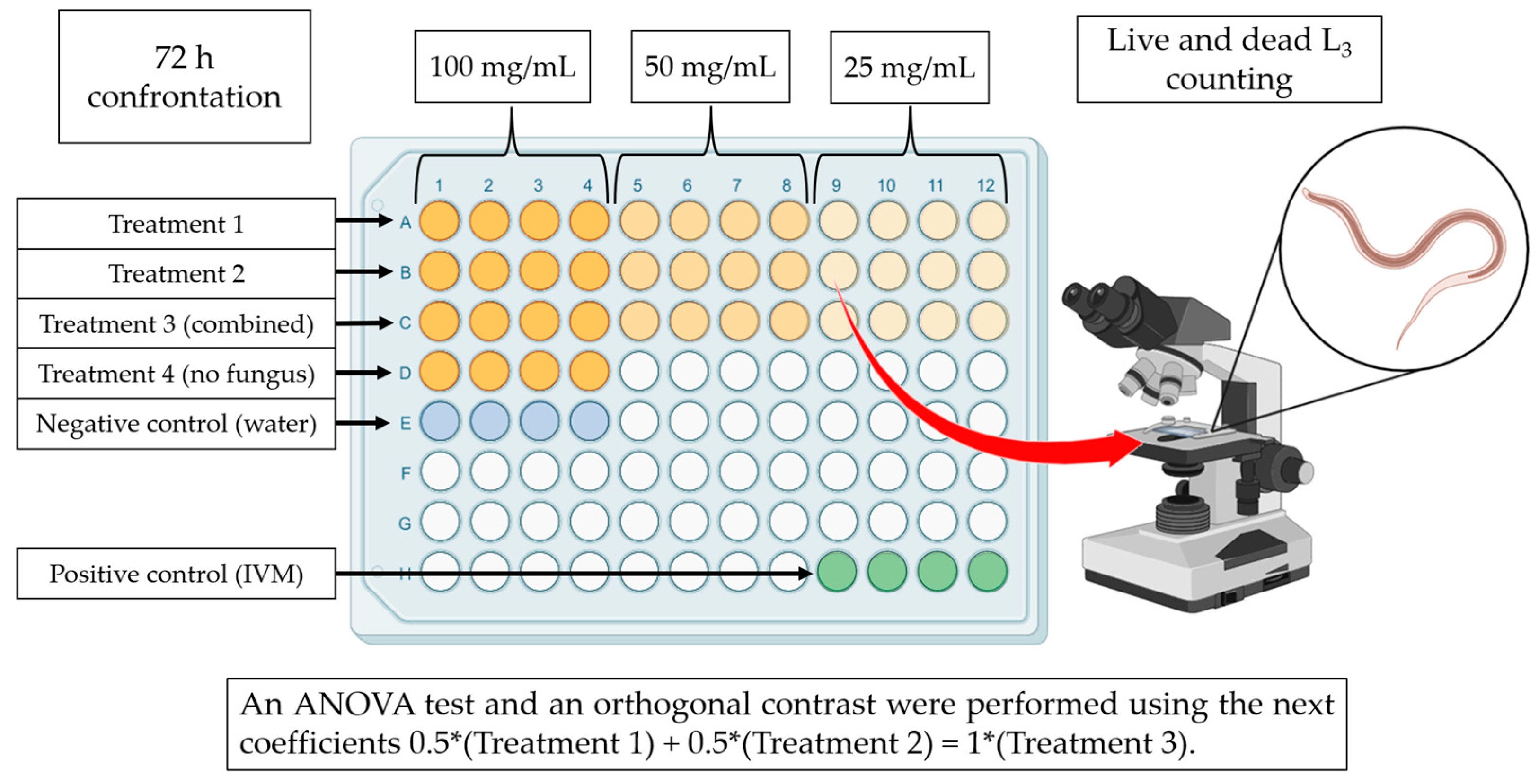
2.7. Assessing the Nematocidal Activity of Fungal Liquid Culture Filtrates against Haemonchus contortus Infective Larvae

The interaction between Liquid Culture Filtrates (LCF) and H. contortus infective larvae was carried out using 96-well microtiter plates (n = 4). The dried LCF were reconstituted using Phosphate Buffered Saline (PBS). Ten experimental groups were established to assess the lethal effect of LCF obtained from A. oligospora and A. musiformis growing in two media SPDB and CzDB. LCF of both fungi were assessed individually and combined (50:50) at three different concentrations (25, 50 and 100 mg/mL). Proper control with only the media was used to discard any possible lethal effect of these media on the nematodes. Additionally, PBS and ivermectin 0.5% were also used as negative and positive controls (Table 1). Fifty microliters of the corresponding treatment and 50 µL of an aqueous larval suspension (in PBS) containing approximately 100 H. contortus infective larvae were deposited in every one of the four wells per treatment. All plates were incubated at room temperature (18–28 °C) for 72 h (Figure 1). The whole experiment was performed in triplicate.
After incubation, the volume of each well was observed under the microscope, and dead and live larvae were counted following the previously described procedure. Motionless larvae were determined to be alive or dead by applying a physical stimulus by touching its cuticle using a sharp metallic needle. Live larvae normally start to move actively after this stimulus. Larvae that remain motionless after this stimulus are considered dead larvae [23]. The whole experiment was repeated three times. Larval mortality obtained for each treatment and control group was calculated. The larval mortality attributed to the effect of LCF of each fungus and each concentration was estimated using the following formula:
%   L a r v a l   M o r t a l i t y = ( M o r t a l i t y   L 3   T r e a t e d   g r o u p M o r t a l i t y   L 3   C o n t r o l   g r o u p ) ( 1 M o r t a l i t y   L 3   C o n t r o l   g r o u p ) 100
where:
M e a n   L 3   T r e a t e d   g r o u p = Mean of larval mortality from treated group.
M e a n   L 3   C o n t r o l   g r o u p = Mean of larval mortality from control group.

2.8. Microscopic Analysis of Haemonchus contortus Infective Larvae Exposed to Fungal Liquid Culture Filtrates

After exposure of H. contortus larvae to LCF, a random selection of larvae from the different treatments was carried out to photograph possible morphological changes attributed to the effect of compounds present in the fungal filtrates. Larvae were photographed using a LEICA DM6 compound microscope using the program LAS V4.9 to document our findings.

2.9. Myco-Chemical Profile

A myco-quantitative reagent analysis was carried out using standard procedures with the proper reagents and methods. Alkaloids were determined using Dragendorff Mayer and Wagner’s reagents, and the Bornträger test was performed to identify the presence of coumarins. Likewise, the presence of flavonoids was investigated using Mg2+ and HCl tests. On the other hand, the ferric chloride, gelatine and saline solution tests were carried out to identify tannins. Finally, the Lieberman–Burchard and Salkowski tests were used to identify the presence of Triterpenes [24].

2.10. Statistical Analysis

Data obtained from the predatory activity assay of both fungal isolates were individually analysed by the Student’s t-test, where the larval reduction percentage was obtained by comparing the mean number of recovered larvae from the nematode/fungi interaction plates and from the control group without fungus. For the results of the nematocidal activity of LCF, a completely random model was used, and ANOVA was performed where the means of larval mortality in the different treatments was considered as the dependent variable. An orthogonal contrast by the Bonferroni method was performed to compare the effect of the combination of both LCF from fungi grown in the same media using the following coefficients: 0.5(AspT, fungus 1) + 0.5(AspG, fungus 2) − 1(combined, 50:50). A p value of 0.05 was used for all tests.

3. Results and Discussion

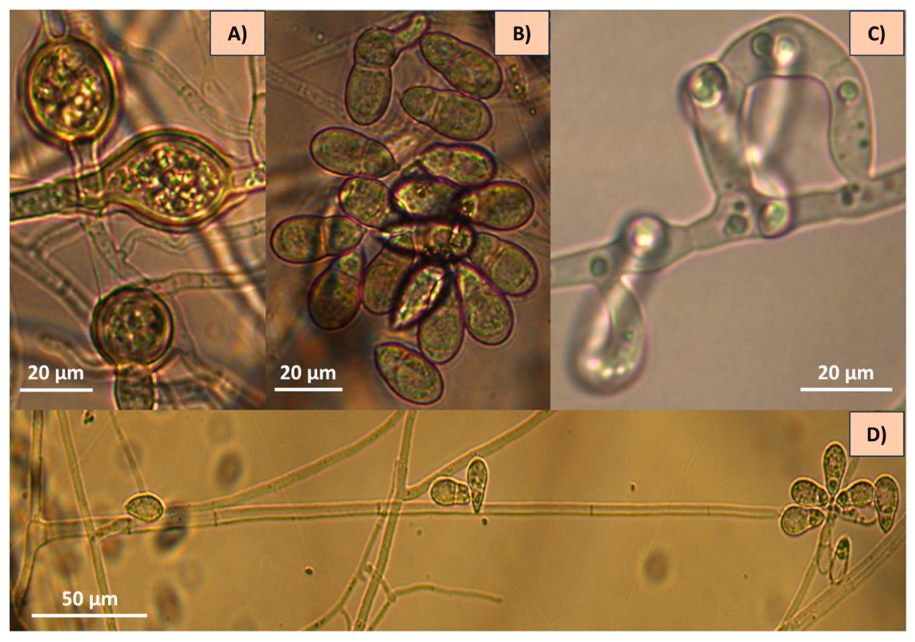
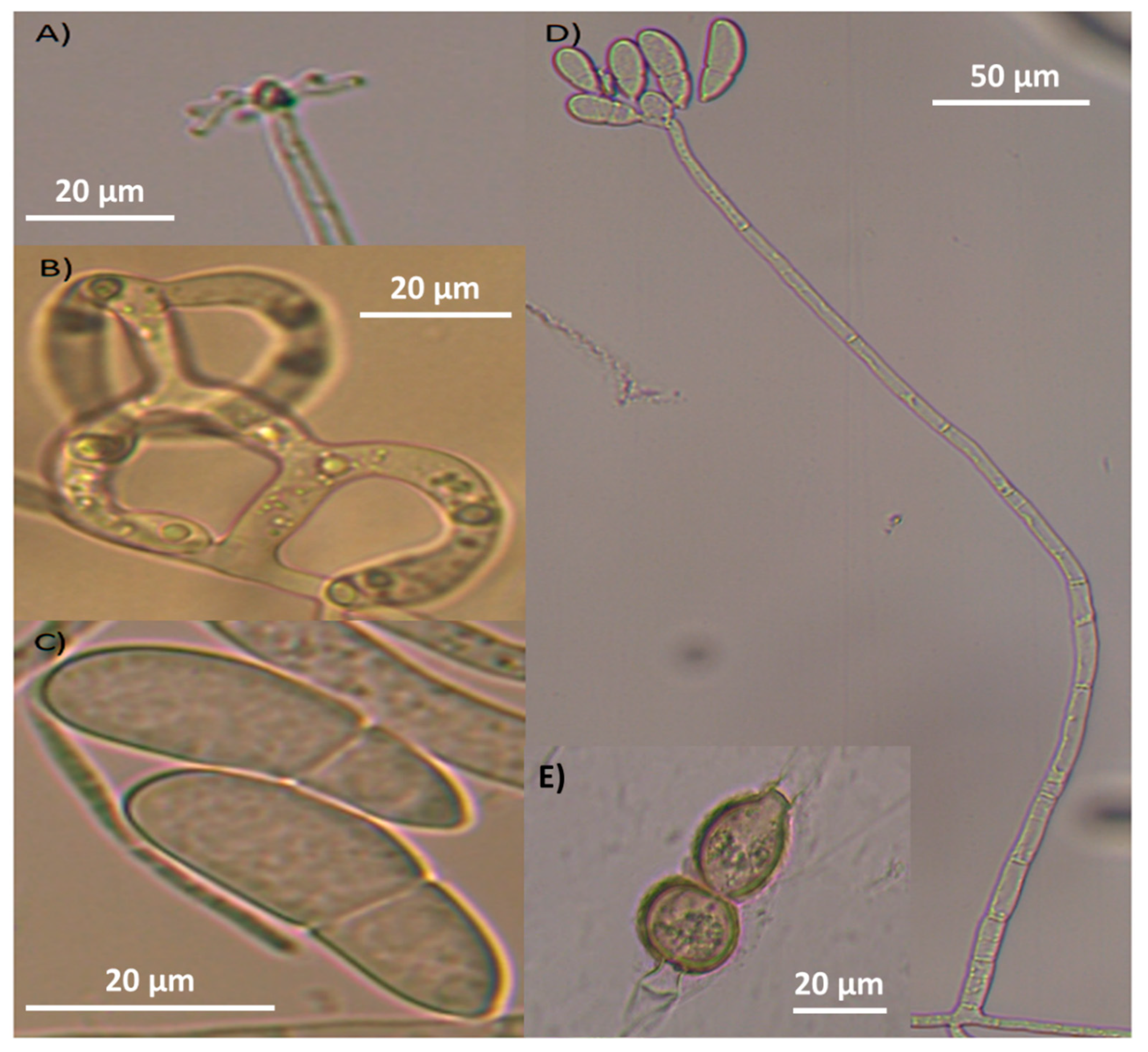
3.1. Traditional Taxonomy (Morphometrics)

Two isolates of nematode-trapping fungi were obtained, and an institutional collection code name was designed as AspT and AspG isolates.
After the main structures of taxonomic interest were observed and analysed under the microscope (including conidia and conidiophore measurements), the morphological characteristics were compared with those described in taxonomic keys, and the authors eventually decided to classify these fungi as A. oligospora and A. musiformis. The isolate AspT showed hyphae, conidia and hyaline conidiophores with repeated proliferation of conidia. Conidia showed a globose shape, lightly constricted in their septum. This fungus showed the presence of three-dimensional adhesive nets (Figure 2).
Likewise, the analysis of the isolate AspG showed hyaline hyphae, conidia and conidiophores. Nevertheless, conidiophores showed long and cylindrical denticles, produced by candelabra, where conidia were generated. Conidia were elongated and slightly curved, typical of A. musiformis. This isolate showed the same trapping devices corresponding to the three-dimensional adhesive nets described above (Figure 3).
The summarized information about measurements and observations of the main morphological characteristics of both isolates are shown in Table 2.
At first sight, the growth of erect and long-stem apical conidiophores crowned by conidia clusters of globose form, as well as the presence of one septum almost at the middle of the conidia in the AspT strain suggested to us the presence of a nematophagous fungus belonging to the genus Arthrobotrys [18]. However, there are several species sharing these characteristics, and this fact can confuse the taxonomic identification. Some of the species sharing these similar characteristics are A. oligospora, A. robusta, A. superba, A. conoides and A. arthrobotryoides, among others. After observing the characteristics in more detail, including the measurements of some structures, i.e., conidia length and width, the number of conidia in the clusters, the presence or absence of branched conidiophores, the presence or absence of chlamydospores and particularly, the type of trapping devices, we were able to differentiate our isolate from other species. Because our isolate showed only unbranched conidiophores, and the measurements of conidia and the length of conidiophores were similar to those of species described in the taxonomical identification keys, we were able to discard several species that do not share these characteristics.
Additionally, our isolate showed an important taxonomical characteristic: the conidiophore type at the top, precisely where the conidiophore produces denticles where conidia are generated (conidiogenesis). Such denticles are typical of Arthrobotryoid conidiophores (Figure 4A), and this characteristic differs from those of other species such as A. musiformis and A. javanica that, instead of these denticles, form a candelabrum where conidia are produced. The presence of conidiophores with denticle formation is typical of the species A. oligospora (Figure 4B). Other fungi produce conidiophores very differently, like the geniculate conidiophores shown in Figure 4C. It is important to mention that our isolate showed the presence of chlamydospores, as well as the formation of three-dimensional adhesive nets as trapping devices. These characteristics led us to classify our isolate as A. oligospora based on morphological analysis. Regarding the other fungal isolate recorded as AspG, this isolate showed a conidia cluster different to that of A. oligospora. Instead, this fungus showed the presence of 4–8 obovoidal, elongated, slightly curved and slightly constricted conidia. There was a septum slightly situated below the middle of the conidia. Elongated conidia were arranged laterally or slightly above the conidiophores. This isolate also showed the presence of the same trapping devices as A. oligospora, three-dimensional adhesive nets. This fungus also showed the presence of chlamydospores but possessed candelabrelloid conidiophores typical of A. musiformis. This structure resembles an elk horn where conidia are developed (Figure 4B).

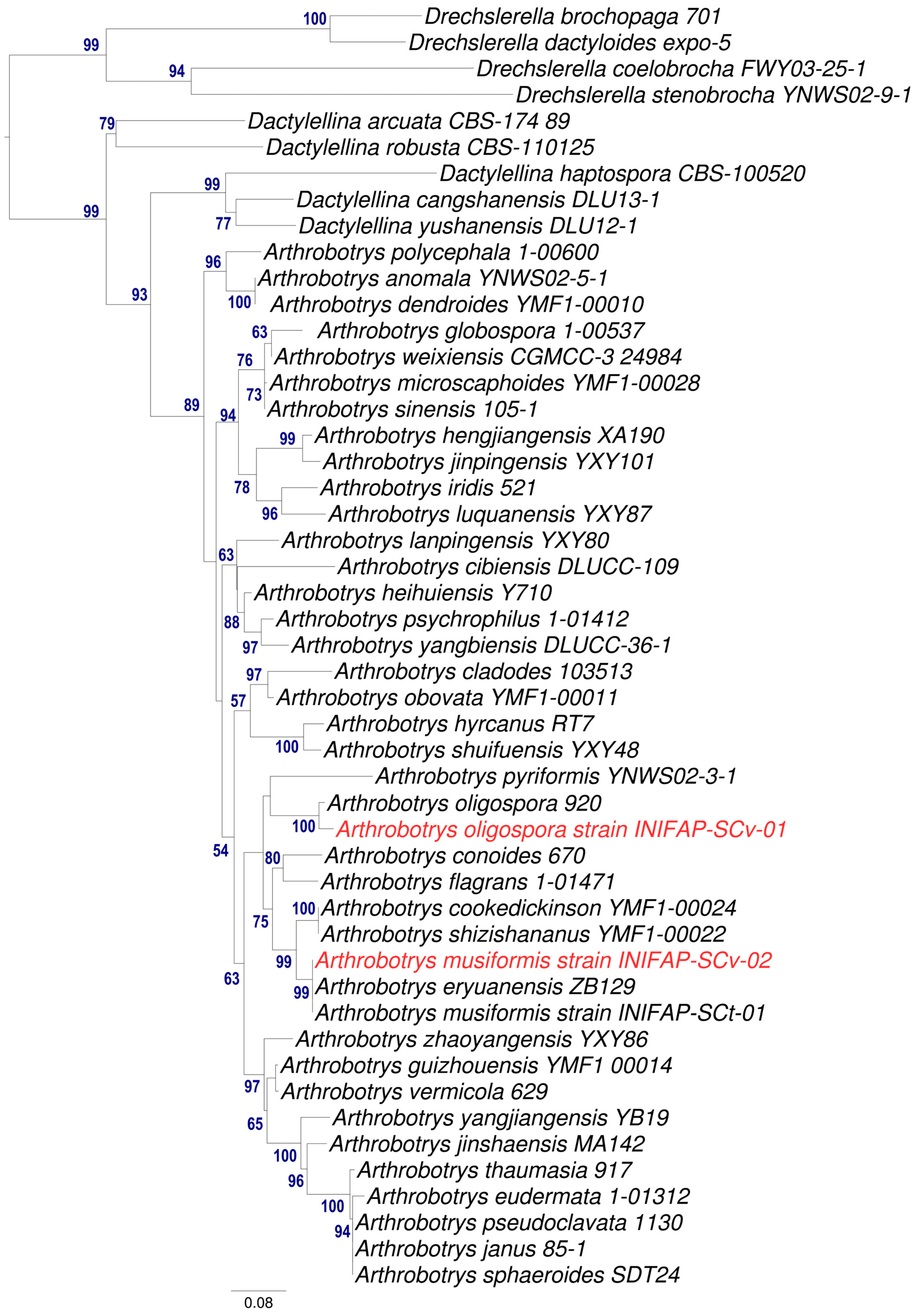
3.2. Molecular Identification

After analysing the DNA sequences of the AspT strain and comparing this information with the sequences of isolates previously reported in the NCBI database, high coverage (96–99%) and similarity (98.86–99.54%) values with respect to fungi from the species Orbilia oligospora and A. oligospora were found. On the one hand, both species seem to be synonyms according to recent nomenclature reported in the NCBI database [25] (Taxonomy ID: 2813651, NCBI:txis2813651). On the other hand, A. musiformis matched those sequences previously reported in the NCBI database with high levels of coverage (99.32%) corresponding to A. musiformis (MH855842.1., KP859624.1, OL454931.1) and A. eryuanensis (MT612105.1). However, there are some remarkable morphological differences between A. musiformis and A. eryuanensis. For example, the length of erect conidiophores in A. musiformis can reach up to 900 μM [18]; meanwhile, A. eryuanensis reaches a length range of 110–308 µm, and this species also produces macro- and microconidia [19]. It is important to mention that both isolates were recorded at the NCBI as Arthrobotrys oligospora strain INIFAP-SCv-01 (PP567275) and Arthrobotrys musiformis strain INIFAP-SCv-02 (PP567276).
To obtain a more robust molecular taxonomic identification, a Maximum Likelyhood phylogenetic tree using ITS1,2 and 5.8S regions was created (Figure 5). This analysis revealed that our isolate AspT is closely related to A. oligospora (strain 920) with high BS in this node. Meanwhile, the AspG strain is closely related with two reported species A. musiformis and A. eryuanensis and the analysis did not provide sufficient evidence to differentiate our isolate from both species. To elucidate this doubt, we re-revised the morphological descriptions for both species and found important morphological differences between them. For example, A. musiformis forms only macroconidia, while A. eryuanensis produces macro and microconidia; additionally, A. musiformis produces erect and not branched conidiophores, opposite to A. eryuanensis, which produces branched conidiophores [19].

3.3. Predatory Activity Assessment of the Two Nematophagous Fungal Isolates against Haemonchus contortus Infective Larvae

The mean numbers of H. contortus infective larvae recovered from agar plate groups with and without fungi, as well as the larval reduction attributed to the predatory effect of both fungi, are shown in Table 3. Arthrobotrys oligospora (AspT) showed a moderate larval reduction (around 45%) after 10 days of interaction; meanwhile, A. musiformis (AspG) reached a higher larval reduction (>70%) (p < 0.05).
In the literature consulted, we found several reports where different strains of both species obtained from different sources showed variable results in terms of their predatory activity against ruminant parasitic nematodes, i.e., in a recent study, an A. oligospora isolate obtained from a fig plantation soil sample from Tepalcingo, Morelos, Mexico showed 73.6% predatory activity against the H. contortus infective larvae [26]. Likewise, Cao et al. (2018) [27], using Chinese isolates of A. musiformis, reported a range of 89.02% to 94.80% reduction of infective larvae of the nematode Trichostrongylus colubriformis [27]. In another study, an A. musiformis strain isolated from a soil sample from Cuautla municipality, Morelos state, Mexico showed 71.54% larval mortality using H. contortus (L3) as a target [22].
Other isolates of A. oligospora and A. musiformis showing variable results regarding their predatory capability against infective larvae of ruminant parasitic nematodes and other nematode targets are shown in Table 4. In this regard, we can build the following reflection: the biological behaviour of nematophagous fungi is a dynamic process and it depends on the adaptation to environmental circumstances of each microorganism from the soil microbiota; such variation in the ability to form traps and capture nematodes of different taxonomic groups can be attributed to the influence of diverse abiotic and biotic factors in fungal habitats [28].

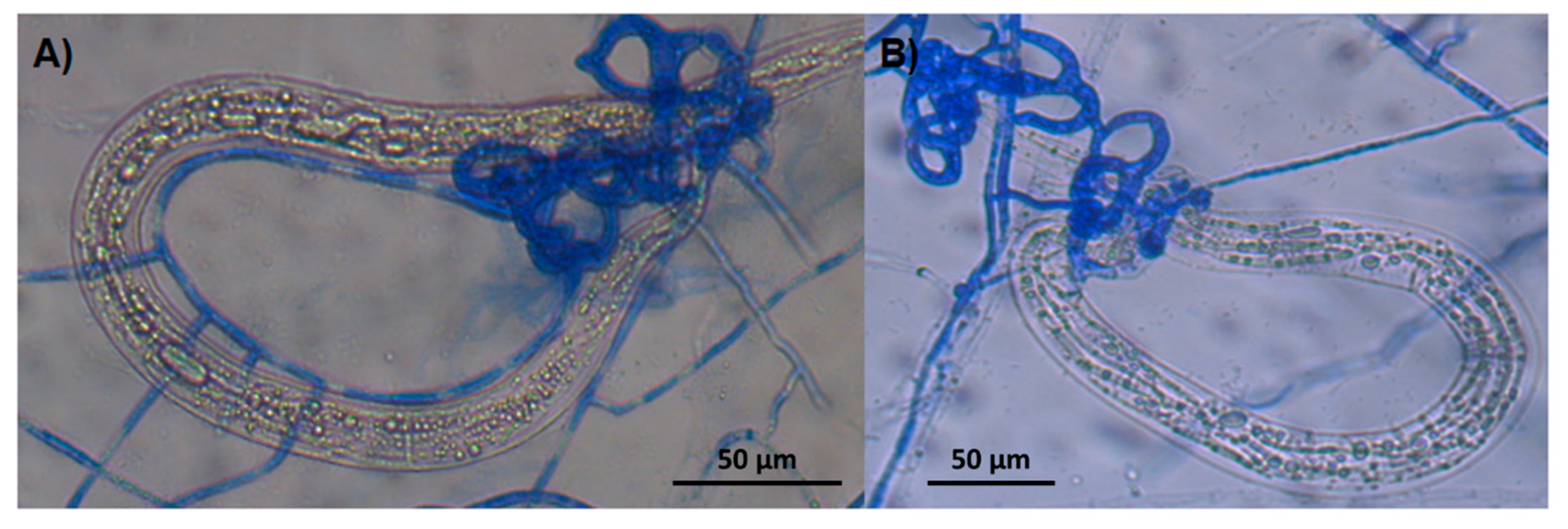
3.4. Microscopic Findings about Predatory Activity of Fungi

The microscopical analysis of the agar plate surfaces of both fungi interacting with H. contortus larvae revealed the formation of three-dimensional adhesive nets where nematodes appeared trapped (Figure 6A, A. oligospora and Figure 6B A. musiformis).
Both species of nematode-trapping fungi are saprophytic organisms living in soil, decaying wood, plant roots or litter and they take their requirements of nitrogen and carbon from these organic materials; however, when nematodes are in their proximity some protein products peeled from the nematode cuticle can stimulate genes responsible of regulating the morphogenesis process. This process is a physiological mechanism of fungi where they modify their mycelia and transform them into trapping devices [33]. The interaction fungi/nematodes process includes various steps, i.e., the formation of an infecting bulb (or appressoria) that exerts a physical pressure [7], and together with the production of cuticle-degrading enzymes, can penetrate the nematode body to eventually invade and take their nutritional requirements from the internal nematode tissues [34]. In both images (Figure 4), in addition to the trap formations, invasive hyphae can be seen filling the inside of the larval corps.

3.5. Assessment of the In Vitro Nematocidal Activity of Liquid Culture Filtrates of Arthrobotrys oligospora and Arthrobotrys musiformis Produced in SPDB and CzDB against Haemonchus contortus Infective Larvae

The results regarding the larval mortality of H. contortus attributed to the effect of LCF of both nematophagous fungi either individually or in combination (50:50) grown in the two evaluated media are shown in Table 5. The larval mortality caused by LCF of each fungus in individual use in both media was observed under a concentration-dependent effect; where the highest larval mortality was observed at the highest concentration used (100 mg/mL). It is important to remark that relevant differences between the individual and combined use of both fungi were not recorded. In both cases, the two LCF of both fungi showed a high lethal effect against the H. contortus infective larvae at the highest concentration. However, a higher larval mortality was observed in LCF obtained from CzDB in comparison with LCF obtained from SPDB. In this regard, we expected to observe a synergistic effect of the combined use of LCF; however, this effect was only found in the LCF concentration of 25 mg/mL in CzDB. No antagonistic effect in the combined use of LCF in both media was observed. In a study, the effect of LCF on different nematode-trapping fungi including Arthrobotrys conoides, A. cookedickinson, A. eudermata, A. microscaphoides, A. oligospora, A. thaumasia and Clonostachys rosea in combination with Trichoderma harzianum and the bacterium Pseudomonas fluorescence were assessed against eggs and juveniles of the phytoparasitic nematode Meloidogyne spp. and authors reported a range of juvenile mortality (J2) between 47.22–59.72% (used individually); meanwhile, the LCF combinations ranged between 65.26–73.61%; the highest juvenile mortality was achieved with P. fluorescence and T. harzianum [35]. In another study, the combined effect of the bacterium Pseudomonas fluorescence and the entomopathogenic fungus Metharhizium anisopliae was assessed against eggs and juveniles (J2) of M. incognita; authors reported an improvement in the nematocidal effect of the combined use of both filtrates of bacteria + fungus that reached 96.98% juvenile mortality and 98.22% egg-hatching inhibition [36].
In an extensive revision of the available literature, the authors did not find more “ad hoc” studies to compare with our results.

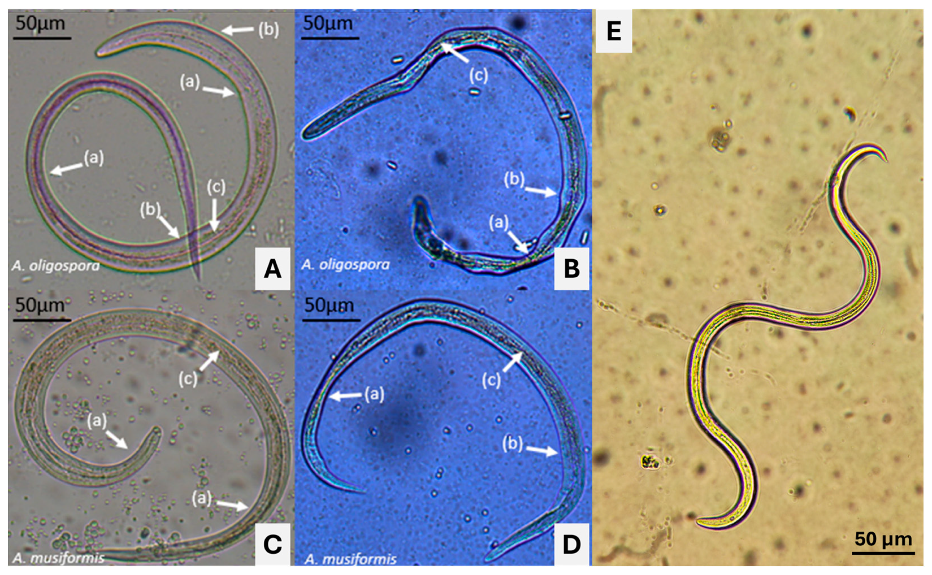
3.6. Microscopic Findings Regarding Haemonchus contortus Infective Larvae Exposed to Liquid Culture Filtrates of Two Nematophagous Fungi Grown in Sweet Potato Dextrose Broth and Czapek-Dox Broth

A set of microphotographs showing morphological changes identified in H. contortus infective larvae after exposure to LCF of A. oligospora and A. musiformis are shown in Figure 7. Images A and C show the aspects of H. contortus infective larvae after 72 h exposure to LCF of A. oligospora and A. musiformis, respectively (grown in CzDB), assessed at 100 mg/mL. In these images, a thickening of the larval bodies was observed in some areas; meanwhile, in other areas, a diminishing of the body thickness was visualized. In both cases, a clear loss of the intestinal cellular structural architecture was observed (c). Likewise, larvae exposed to LCF of both fungi grown in SPDB (B) and (D) showed similar damage, but was more severe in SPDB, and a general deformation of larval bodies with a slimming in some areas of the nematode corps (a) and loss of turgor body in other areas (b) were observed. After analysing the images captured by microscopy, we observed malformations in different sites of the larval bodies exposed to both LCFs. The larvae exposed to LCF from both fungi obtained from CzDB showed some morphological changes; however, the damage to those larvae exposed to LCF obtained from both fungi in SPBD showed more severe morphological alterations. These findings suggest that the culture media can influence the production of bioactive mycocompounds derived from the secondary metabolism of fungi. In future studies, we plan to use confocal laser scanning microscopy to perform a co-localization analysis to determine the site of action and the damage caused by metabolites responsible of the nematocidal activity [37].

3.7. Myco-Chemical Compound Profile

The results of the myco-quantitative reagent analysis obtained with LCF of both fungi in both culture media are shown in Table 6. The groups of secondary metabolites found after mycochemical compound analysis of LCF obtained in both liquid media (CzDB and SPDB) of the two nematophagous fungi (A. musiformis and A. oligospora) were alkaloids with a light reaction in the four treatments: coumarins with a light reaction in A. musiformis in SPDB and A. oligospora in both media. Regarding the presence of sterols, this group of compounds was found with a light reaction in LCF of A. musiformis in SPDB and A. oligospora in CzDB; however, in A. oligospora, a positive reaction was found in SPDB and a strong reaction in CzDB. These findings show that A. oligospora, despite not showing high predatory activity of H. contortus infective larvae using physical mechanisms (three-dimensional adhesive nets), in certain respects, compensates for this lack of a strong predatory activity by producing many secondary metabolites with nematocidal activity, which could contribute to the immobilization and killing of nematodes. Some groups of compounds identified in the present study in both species of Arthrobotrys have been found in other isolates of this genus and are associated with several biological activities. On the one hand, coumarins obtained from other sources i.e., plants and marine plants have been reported to possess important therapeutic activities, i.e., antifungal, anti-inflammatory and antimicrobial [38] and nematocidal activity [39,40]. On the other hand, some compounds belonging to the group of Alkaloids are also demonstrated to possess potent nematocidal activity against gastrointestinal parasitic nematodes [41] and against phytoparasitic nematodes [42]. Other compounds obtained in the present study including sterols and saponins have also been reported to have important nematocidal activity against nematodes of importance in agriculture [43,44].

4. Conclusions

In the present study, no enhancing effect in the nematocidal activity was identified after using the LCF of A. oligospora and A. musiformis in combination against H. contortus infective larvae. Liquid culture filtrates of both fungi exerted an important nematocidal effect at the highest concentration (100 mg/mL) when used either individually or in a combined manner. The results of the present study contribute important information about the predatory activity of these species of nematophagous fungi and additionally about the nematocidal activity of LCF produced by these fungi that could have implications for future studies exploring the use of this LCF or the metabolites responsible for nematocidal activity as potential tools of control against sheep haemonchosis.

Author Contributions

Conceptualization, P.M.-d.G. and G.P.-A.; methodology, A.C.-P.; software, G.A.B.-G.; validation, A.O.-J. and E.G.-M.; formal analysis, E.J.D.-N.; investigation, A.C.-P.; resources, P.M.-d.G.; data curation, A.O.-J. and G.P.-A.; writing—original draft preparation, P.M.-d.G.; writing—review and editing, P.M.-d.G. and G.P.-A.; supervision, P.M.-d.G. and G.P.-A.; project administration, P.M.-d.G.; funding acquisition, P.M.-d.G. All authors have read and agreed to the published version of the manuscript.

Funding

This research received no external funding.

Institutional Review Board Statement

The animal that acted as an egg donor of the parasite was maintained under indoor conditions, following the rules of care, respect and animal welfare and special attention was put to avoiding any unnecessary animal suffering. These conditions were strictly performed according to the Good Management Practices policies established at INIFAP. The Norma Oficial Mexicana (Official Mexican Standard) with official rule number NOM-052-ZOO-1995 (http://www.senasica.gob.mx, accessed on 6 February 2024) and the Ley Federal de Sanidad Animal (Federal Law for Animal Health) DOF 07-06-2012 were strictly applied (https://www.gob.mx/cms/uploads/attachment/file/1187 61/LFSA.pdf, accessed on 6 February 2024) in accordance with the ethical standards outlined by INIFAP.

Data Availability Statement

All the data available is included in the manuscript.

Acknowledgments

This study did not receive any specific budget. The reagents, consumables, and other materials as well as the equipment were supplied by the Helminthology Laboratory of CENID-SAI, INIFAP. This study formed part of the thesis work of Antonio Colinas Picazo to obtain the grade of DVM at the Universidad Mesoamericana, Cuernavaca, Morelos, Mexico, under the direction of Pedro Mendoza-de Gives and tutored by Gustavo Pérez-Anzúrez.

Conflicts of Interest

The authors declare no conflicts of interest.

References

  1. Besier, R.B.; Kahn, L.P.; Sargison, N.D.; Van Wyk, J.A. Diagnosis, treatment and management of Haemonchus contortus in small ruminants. Adv. Parasitol. 2016, 93, 181–238. [Google Scholar] [CrossRef] [PubMed]
  2. Flay, K.J.; Hill, F.I.; Muguiro, D.H. A Review: Haemonchus contortus Infection in Pasture-Based Sheep Production Systems, with a Focus on the Pathogenesis of Anaemia and Changes in Haematological Parameters. Animals 2022, 12, 1238. [Google Scholar] [CrossRef] [PubMed]
  3. Höglund, J.; Gustafsson, K.; Ljungström, B.L.; Skarin, M.; Varady, M.; Engström, F. Failure of ivermectin treatment in Haemonchus contortus infected-Swedish sheep flocks. Veter.-Parasitol. Reg. Stud. Rep. 2015, 1–2, 10–15. [Google Scholar] [CrossRef] [PubMed]
  4. Martindah, E.; Sawitri, D.H.; Wardhana, A.H.; Ekawasti, F.; Dewi, D.A. Anthelmintic Resistance Status in Gastrointestinal Nematodes of Seven Different Breeds of Sheep in intensive management. IOP Conf. Ser. Earth Environ. 2023, 1174, 012030. [Google Scholar] [CrossRef]
  5. Jiang, X.; Xiang, M.; Liu, X. Nematode-trapping fungi. Microbiol. Spectr. 2017, 5. [Google Scholar] [CrossRef] [PubMed]
  6. Reyes-Guerrero, D.E.; Olmedo-Juárez, A.; Mendoza-de Gives, P. Control and prevention of nematodiasis in small ruminants: Background, challenges and outlook in Mexico. Rev. Mex. Cienc. Pecu. 2021, 12, 186–204. [Google Scholar] [CrossRef]
  7. Nordbring-Hertz, B. Morphogenesis in the nematode-trapping fungus Arthrobotrys oligospora - an extensive plasticity of infection structures. Mycologist 1999, 18, 125–133. [Google Scholar] [CrossRef]
  8. Index Fungorum. Available online: https://www.indexfungorum.org/names/namesrecord.asp?RecordID=149365 (accessed on 10 March 2024).
  9. Hyde, K.; Swe, A.; Zhang, K.Q. Nematode-Trapping Fungi; Fungal Diversity Research Series; Zhang, K.Q., Hyde, K., Eds.; Springer: Dordrecht, The Netherlands, 2014; Volume 23, pp. 1–12. [Google Scholar]
  10. Rahman, M.U.; Chen, P.; Zhang, X.; Fan, B. Predacious Strategies of Nematophagous Fungi as Bio-Control Agents. Agronomy 2023, 13, 2685. [Google Scholar] [CrossRef]
  11. de Lara, R.; Castro, T.; Castro, J.; Castro, G. Cultivo del nematodo Panagrellus redivivus (Goodey, 1945) en un medio de avena enriquecida con Spirulina sp. Rev. Biol. Mar. Oceanogr. 2007, 42, 29–36. [Google Scholar] [CrossRef]
  12. Valcárcel-Sancho, F.; Rojo-Vázquez, F.A.; Olmeda-García, A.S.; Arribas-Novillo, B.; Márquez-Sopeña, L.; Fernando-Pat, N. Atlas de Parasitología Ovina; Editorial Servet: Zaragoza, Spain, 2009; 137p. [Google Scholar]
  13. Iliev, P.; Ivanov, A.; Prelezov, P. Effects of temperature and desiccation on survival rate of Haemonchus contortus infective larval stage. Trakia J. Sci. 2018, 16, 17–21. [Google Scholar] [CrossRef]
  14. Cedillo-Borda, M.; López-Arellano, M.; Reyes-Guerrero, D.E. In vitro assessment of ivermectin resistance and gene expression profiles of P-glycoprotein genes from Haemonchus contortus (L3). Bio-Protocol 2020, 10, e3851. [Google Scholar] [CrossRef]
  15. Reyes-Guerrero, D.E.; Cedillo-Borda, M.; Alonso-Morales, R.A.; Alonso-Díaz, M.A.; Olmedo-Juárez, A.; Mendoza-de-Gives, P.; López-Arellano, M.E. Comparative study of transcription profiles of the P-glycoprotein transporters of two Haemonchus contortus isolates: Susceptible and resistant to ivermectin. Mol. Biochem. Parasitol. 2020, 238, 111281. [Google Scholar] [CrossRef] [PubMed]
  16. Sánchez-Martínez, E.; Aguilar-Marcelino, L.; Hernández-Romano, J.; Castañeda-Ramírez, G.S.; Mendoza-de-Gives, P. Taxonomic and biological characterization and predatory activity of four nematophagous fungi isolates of Arthrobotrys species from Tapachula, Chiapas, Mexico. Arch. Agron. Soil Sci. 2021, 69, 327–343. [Google Scholar] [CrossRef]
  17. Drechsler, C. Some hyphomycetes that prey on free-living terricolous nematodes. Mycologia 1937, 2, 447–552. [Google Scholar] [CrossRef]
  18. de Hoog, G.S.; Van Oorschot, C.A.N. Taxonomy of the Dactylaria complex, V. A review of Arthrobotrys and allied genera. Stud. Mycol. 1985, 26, 61–96. [Google Scholar]
  19. Zhang, F.; Boonmee, S.; Bhat, J.D.; Xiao, W.; Yang, X.-Y. New Arthrobotrys Nematode-Trapping Species (Orbiliaceae) from Terrestrial Soils and Freshwater Sediments in China. J. Fungi 2022, 8, 671. [Google Scholar] [CrossRef] [PubMed]
  20. Tigano-Milani, M.S.; Honeycutt, R.J.; Lacey, L.A.; Assis, R.; McClelland, M.; Sobral, B.W. Genetic Variability of Paecilomyces fumosoroseus Isolates Revealed by Molecular Markers. J. Invertebr. Pathol. 1995, 65, 274–282. [Google Scholar] [CrossRef] [PubMed]
  21. White, T.J.; Bruns, T.; Lee, S.; Taylor, J. Amplification and direct sequencing of fungal ribosomal RNA genes for phylogenetics. In PCR Protocols: A Guide to Methods and Applications; Innis, M.A., Gelfand, D.H., Sninsky, J.J., White, T.J., Eds.; Academic Press: New York, NY, USA, 1990; Volume 18, pp. 315–322. [Google Scholar]
  22. Pérez-Anzúrez, G.; Olmedo-Juárez, A.; de Fernex, E.V.-S.; Alonso-Díaz, M.; Delgado-Núñez, E.J.; López-Arellano, M.E.; González-Cortázar, M.; Zamilpa, A.; Ocampo-Gutierrez, A.Y.; Paz-Silva, A.; et al. Arthrobotrys musiformis (Orbiliales) Kills Haemonchus contortus Infective Larvae (Trichostronylidae) through Its Predatory Activity and Its Fungal Culture Filtrates. Pathogens 2022, 11, 1068. [Google Scholar] [CrossRef] [PubMed]
  23. Olmedo-Juárez, A.; Rojo-Rubio, R.; Zamilpa, A.; Mendoza-de Gives, P.; Arece-García, J.; López-Arellano, M.E.; Fernex, E.v.S.d. In vitro larvicidal effect of a hydroalcoholic extract from Acacia cochliacantha leaf against ruminant parasitic nematodes. Veter Res. Commun. 2017, 41, 227–232. [Google Scholar] [CrossRef]
  24. Delgado-Núñez, E.J.; Zamilpa, A.; González-Cortazar, M.; Olmedo-Juárez, A.; Cardoso-Taketa, A.; Sánchez-Mendoza, E.; Tapia-Maruri, D.; Salinas-Sánchez, D.O.; Mendoza-de Gives, P. Isorhamnetin: A Nematocidal Flavonoid from Prosopis laevigata Leaves Against Haemonchus contortus Eggs and Larvae. Biomolecules 2020, 10, 773. [Google Scholar] [CrossRef]
  25. Baral, H.O.; Weber, E.; Marson, G. Monograph of Orbiliomycetes (Ascomycota) Based on Vital Taxonomy. Part II, 1st ed.; National Museum of Natural History: Luxembourg, 2020; 1752p. [Google Scholar]
  26. Bahena-Nuñez, D.S.; Ocampo-Gutiérrez, A.Y.; Mendoza-de Gives, P.; González-Cortázar, M.; Zamilpa, A.; Higuera-Piedrahita, R.I.; Pérez-Anzúrez, G.; Olmedo-Juárez, A.; López-Arellano, M.E.; Delgado-Núñez, E.J.; et al. Arthrobotrys oligospora (Fungi: Orbiliales) and its liquid culture filtrate myco-constituents kill Haemonchus contortus infective larvae (Nematoda: Trichostrongylidae). Biocont. Sci. Technol. 2024; in press. [Google Scholar]
  27. Cao, X.; Xu, Q.; Wan, X.M.; Yang, X.C.; Jia, X.Y.; Xue, X.J.; Du, J.L.; Cai, K.Z. Isolation, identification, and in vitro predatory activity of nematophagous fungus Arthrobotrys musiformis and its interaction with nematodes using scanning electron microscopy. Biocontrol Sci. Techn. 2018, 28, 94–104. [Google Scholar] [CrossRef]
  28. Dhawan, S.C.; Narayana, R.; and Babn, N.P. Influence of abiotic and biotic factors on growth of Paecilomyces lilacinus, Arthrobotrys oligospora and Pochonia chlamydosporia and parasitization of eggs/trapping of Meloidogyne incognita juveniles. Ann. Plant Prot. Sci. 2004, 12, 369–372. [Google Scholar]
  29. Alfaro-Gutiérrez, I.C.; Mendoza-de Gives, P.; Liébano-Hernández, E.; López-Arellano, M.E.; Valero-Coss, R.O.; Hernández-Velázquez, V.M. Nematophagous fungi (Orbiliales) capturing, destroying and feeding on the histotrophic larvae of Haemonchus contortus (Nematoda: Trichostrongylidae). Rev. Mex. Micol. 2011, 33, 29–35. [Google Scholar]
  30. Ojeda-Robertos, N.F.; Aguilar-Marcelino, L.; Olmedo-Juárez, A.; Luna-Palomera, C.; Peralta-Torres, J.A.; López-Arellano, M.E.; Mendoza-de-Gives, P. In vitro predatory activity of nematophagous fungi isolated from water buffalo feces and from soil in the Mexican southeastern. Rev. Bras. Parasitol. Veter 2019, 28, 314–319. [Google Scholar] [CrossRef]
  31. Jaramillo-Tlalapango, J.; Mendoza-de Gives, P.; Higuera-Piedrahita, R.I.; Ocampo-Gutiérrez, A.Y.; López-Arellano, M.E.; Pérez-Anzúrez, G.; Olmedo-Juárez, A.; Hernández-Romano, J.; Maza-López, J.; Delgado-Núñez, E.J.; et al. Study of a Mexican isolate of Arthrobotrys musiformis (Orbiliales): Predatory behavior and nematocidal activity of liquid culture filtrates against Haemonchus contortus (Trichostrongylidae), protein profile and myco-constituent groups. Fungal Biol. 2023, 127, 1345–1361. [Google Scholar] [CrossRef] [PubMed]
  32. Wu, Y.; Yang, Z.; Jiang, Z.; Nizamani, M.M.; Zhang, H.; Liu, M.; Shan, W.; Wang, Y.; Li, K. Isolation, Identification, and Evaluation of the Predatory Activity of Chinese Arthrobotrys Species towards Economically Important Plant-Parasitic Nematodes. J. Fungi 2023, 9, 1125. [Google Scholar] [CrossRef] [PubMed]
  33. Zhu, M.C.; Li, X.M.; Zhao, N.; Yang, L.; Zhang, K.Q.; Yang, J.K. Regulatory Mechanism of Trap Formation in the Nematode-Trapping Fungi. J. Fungi 2022, 8, 406. [Google Scholar] [CrossRef] [PubMed]
  34. Tzean, Y.; Chou, T.-H.; Hsiao, C.-C.; Shu, P.-Y.; Walton, J.D.; Tzean, S.-S. Cloning and characterization of cuticle-degrading serine protease from nematode-trapping fungus Arthrobotrys musiformis. Mycoscience 2016, 57, 136–143. [Google Scholar] [CrossRef]
  35. Lafta, A.A.; Kasim, A.A. Effect of nematode-trapping fungi, Trichoderma harzianum and Pseudomonas fluorescens in controlling Meloidogyne spp. Plant Arch. 2019, 19, 1163–1168. [Google Scholar]
  36. Jahanbazian, L.; Abdollahi, M.; Rezaie, R. Combined effect of Metarhizium anisopliae and Pseudomonas fluorescens CHA0 on root-knot nematode, Meloidogyne incognita in tomato. Iran. J. Plant Pathol. 2015, 51, 339–355. [Google Scholar]
  37. Cortes-Morales, J.A.; Zamilpa, A.; Salinas-Sánchez, D.O.; González-Cortazar, M.; Tapia-Maruri, D.; Mendoza-de Gives, P.; Rivas-González, J.M.; Olmedo-Juárez, A. In vitro ovicidal effect of p-coumaric acid from Acacia bilimekii aerial parts against Haemonchus contortus. Veter. Parasitol. 2023, 320, 109971. [Google Scholar] [CrossRef] [PubMed]
  38. Pal, D.; Saha, S. Coumarins: An Important Phytochemical with Therapeutic Potential. In Plant-Derived Bioactives, 2nd ed.; Swamy, M., Ed.; Springer: Singapore, 2020; Volume 1, pp. 205–222. [Google Scholar]
  39. Jasso-Díaz, G.; Torres-Hernández, G.; Zamilpa, A.; Becerril-Pérez, C.; Ramírez-Bribiesca, J.E.; Hernández-Mendo, O.; Sánchez-Arroyo, H.; Olmedo-Juárez, A.; González-Cortazar, M.; Mendoza-de Gives, P. Ruta chalepensis full extract and organic phases exhibit nematocidal activity against Haemonchus contortus eggs and infective larvae (L3). Helminthologia 2022, 59, 46–54. [Google Scholar] [CrossRef] [PubMed]
  40. Feng, J.; Qin, C.; Liu, X.; Li, R.; Wang, C.; Li, C.; Du, G.; Guo, Q. Nematicidal Coumarins from Cnidium monnieri Fruits and Angelica dahurica Roots and Their Physiological Effect on Pine Wood Nematode (Bursaphelenchus xylophilus). Molecules 2023, 28, 4109. [Google Scholar] [CrossRef] [PubMed]
  41. da Silva, G.D.; de Lima, H.G.; de Sousa, N.B.; Genipapeiro, I.L.d.J.; Uzêda, R.S.; Branco, A.; Costa, S.L.; Batatinha, M.J.M.; Botura, M.B. In vitro anthelmintic evaluation of three alkaloids against gastrointestinal nematodes of goats. Veter. Parasitol. 2021, 296, 109505. [Google Scholar] [CrossRef] [PubMed]
  42. Kusano, M.; Koshino, H.; Uzawa, J.; Fujiok, S.; Kawano, T.; Kimura, Y. Nematicidal alkaloids and related compounds produced by the fungus Penicillium cf. simplicissimum. Biosci. Biotechnol. Biochem. 2000, 64, 2559–2568. [Google Scholar] [CrossRef] [PubMed]
  43. D’addabbo, T.; Argentieri, M.P.; Żuchowski, J.; Biazzi, E.; Tava, A.; Oleszek, W.; Avato, P. Activity of Saponins from Medicago Species against Phytoparasitic Nematodes. Plants 2020, 9, 443. [Google Scholar] [CrossRef]
  44. Velasco-Azorsa, R.; Cruz-Santiago, H.; del Prado-Vera, I.C.; Ramirez-Mares, M.V.; Gutiérrez-Ortiz, M.d.R.; Santos-Sánchez, N.F.; Salas-Coronado, R.; Villanueva-Cañongo, C.; Lira-de León, K.I.; Hernández-Carlos, B. Chemical Characterization of Plant Extracts and Evaluation of their Nematicidal and Phytotoxic Potential. Molecules 2021, 26, 2216. [Google Scholar] [CrossRef]
Figure 1. Scheme representing the steps of the experimental process to evaluate the lethal effect of liquid culture filtrates obtained with two nematophagous fungi Arthrobotrys oligospora and A. musiformis growth in two culture media, Sweet Potato Dextrose Broth and Czapek-Dox Broth, against Haemonchus contortus infective larvae.
Pathogens 13 00498 g001
Figure 2. Microphotographs of Arthrobotrys oligospora showing structures of taxonomic importance: (A) chlamydospores, (B) conidia, (C) three-dimensional adhesive nets and (D) conidiophore.
Pathogens 13 00498 g002
Figure 3. Arthrobotrys musiformis structures of taxonomic importance: (A) Candelabrum; (B) Three-Dimensional adhesive net; (C) Conidia; (D) Conidiophore and (E) Chlamydospores.
Pathogens 13 00498 g003
Figure 4. Aspects of different types of apical extremes of conidiophores of different nematophagous fungi. (A) The conidiophore apex bifurcates and it shows an elk horn appearance, where conidiogenesis is produced; (B) The conidiophore keeps its erect position and groups of denticles are produced along the conidiophore apex; (C) The conidiophore shows repeated bents where conidiogenesis occurs.
Pathogens 13 00498 g004
Figure 5. Phylogenetic analysis from the AspG and AspT fungal isolates and related species using 50 sequences from the ITS1, 2 and 5.8S region. Support values > 50% for Maximum Likelihood, are shown in nodes (in blue).
Pathogens 13 00498 g005
Figure 6. Aspect of Haemonchus contortus infective larvae captured in three-dimensional adhesive nets of two nematophagous fungi, Arthrobotrys oligospora (A) and A. musiformis (B).
Pathogens 13 00498 g006
Figure 7. Microphotographs showing the aspect of Haemonchus contortus infective larvae after 72 h exposure to liquid culture filtrates obtained with two nematophagous fungi Arthrobotrys musiformis and A. oligospora grown in Czapek-Dox Broth (A) and (C) and Sweet Potato Dextrose Broth (B) and (D), and a live larva from the control group, without fungus culture (E). (a) deformation of the larval bodies; (b), loss of turgor body; (c), loss of the intestinal cellular structural architecture.
Pathogens 13 00498 g007
Table 1. Experimental design to assess the lethal effect of liquid culture filtrates of Arthrobotrys oligospora and A. musiformis produced in two liquid media against Haemonchus contortus infective larvae (L3).
TreatmentMediumFungal Filtrate
1Sweet Potato Dextrose BrothArthrobotrys oligospora (*)
2A. musiformis (*)
3A. oligospora + A. musiformis (*)
4Control (without fungus)
5Czapek-Dox BrothArthrobotrys oligospora (*)
6A. musiformis (*)
7A. oligospora + A. musiformis (*)
8Control (without fungus)
Controls
9Phosphate Buffered SalineNegative control
10Ivermectin (0.5%)Positive control
n = 4; (*) =The nematocidal effect was assessed at three different concentrations (25, 50 and 100 mg/mL).
Table 2. Main morphological characteristics observed in the two isolates of nematophagous fungi belonging to the genus Arthrobotrys and means of measurement of taxonomically important structures.
CharacteristicStrain 1 (AspT)Strain 2 (AspG)
Conidium shapeGlobose, slightly constricted in the septum (on occasion).Ellipsoidal to obovoidal, slightly curved
SeptumOne septum situated slightly below the half conidiumOne septum situated slightly below the half conidium
Number of conidia per conidiophore6 (4–8)7 (3–11)
Conidia length (µm)21.1 (19–23)32.7 (27–40)
Conidia width (µm)11.4 (10–12)13 (11–17)
Conidiophores length (µm)344 (193–429)291 (141–403)
Identified speciesArthrobotrys oligosporaArthrobotrys musiformis
Table 3. Results of the predatory activity of two nematophagous fungi Arthrobotrys oligospora (AspT) and A. musiformis (AspG) against Haemonchus contortus infective larvae on water agar plates.
Isolate
(Species)
Recovered Larvae Control Group
(Mean ± SE)
Recovered Larvae
Treated Group
(Mean ± SE)
Larval Reduction
%
Arthrobotrys oligospora192 ± 38.21105 ± 26.4245.14 a
Arthrobotrys musiformis197 ± 60.5357 ± 30.3570.95 b
a, b Different letters show statistical differences between groups.
Table 4. Results of the in vitro predatory activity of Arthrobotrys musiformis and A. oligospora isolates obtained from different sources and using different nematode targets.
IsolateSource of IsolationNematode TargetPredatory Activity %Author (s)
A. oligosporaNot availableMeloidogyne incognita (**)79.6–87.5[28]
A. musiformisMossy soil, decaying plant material (a rotten trunk) and soil containing Brahea palm rootsH. contortus (L3)>97[29]
A. oligosporaFaeces of water buffaloH. contortus (L3)>89[30]
A. oligosporaSoil and animal faecesPanagrellus redivivus (*)57.2[16]
A. musiformisSoil sampleH. contortus (L3)>74[31]
A. oligosporaSoil samplesAphelenchoides besseyi, Bursaphelenchus xylophilus and Ditylenchus destructor (**)54.6–97.3[32]
(*) =A Free-living nematode; (**) = Plant parasitic nematodes.
Table 5. Mean of Haemonchus contortus dead and total recovered larvae and mean larval mortality percentages after 72 h exposure to individual and combined liquid culture filtrates obtained from Arthrobotrys musiformis and A. oligospora produced in two liquid media.
Concentration
(mg/mL)
Fungal FiltrateDL/TLMortality %SESignificance *
Sweet potato dextrose broth
0A. oligospora6/1195.030.871
A. musiformis6/1195.030.87
Combination6/1195.030.87
25A. oligospora12/9612.142.740.072
A. musiformis15/10713.703.24
Combination21/10120.543.95
50A. oligospora31/10429.836.030.456
A. musiformis25/10424.505.08
Combination19/8622.185.06
100A. oligospora82/10478.292.650.543
A. musiformis83/11075.555.37
Combination79/9979.843.04
Czapek-Dox Broth
0A. oligospora2/802.120.380.588
A. musiformis2/862.650.40
Combination2/802.120.38
25A. oligospora23/8627.345.750.011
A. musiformis22/8127.083.42
Combination37/8444.075.75
50A. oligospora38/8943.038.840.916
A. musiformis44/8651.595.54
Combination36/7548.297.96
100A. oligospora75/8786.173.390.308
A. musiformis83/9191.353.05
Combination81/8792.552.47
* Values < 0.05 shows that the contrast was significant; Coefficients: 0.5*A. musiformis + 0.5*Treatment 2 = 1*Treatment 3.
Table 6. Mycochemical profile obtained from liquid culture filtrates of two nematophagous fungi Arthrobotrys musiformis and A. oligospora grown in two culture media, Czapek-Dox Broth and Sweet Potato Dextrose Broth, by myco-quantitative reagent analysis.
Metabolites and ReagentsArthrobotrys musiformisArthrobotrys oligospora
CzDBSPDBCCzDoxSPDB
Alkaloids++++
Coumarins-+++
Flavonoids----
Tannins----
Triterpenes----
Sterols-+++
Saponins-++++++
-: undetected reaction; +: slightly positive reaction; ++: positive reaction; +++; strong positive reaction.
Disclaimer/Publisher’s Note: The statements, opinions and data contained in all publications are solely those of the individual author(s) and contributor(s) and not of MDPI and/or the editor(s). MDPI and/or the editor(s) disclaim responsibility for any injury to people or property resulting from any ideas, methods, instructions or products referred to in the content.

