Review

Insights into Peptidyl-Prolyl cis-trans Isomerases from Clinically Important Protozoans: From Structure to Potential Biotechnological Applications

by
Verónica Aranda-Chan
1,
Rosa Elena Cárdenas-Guerra
1,
Alejandro Otero-Pedraza
1,
Esdras Enoc Pacindo-Cabrales
1,
Claudia Ivonne Flores-Pucheta
1,
Octavio Montes-Flores
1,
Rossana Arroyo
2 and
Jaime Ortega-López
1,*
1
Departamento de Biotecnología y Bioingeniería, Centro de Investigación y de Estudios Avanzados del Instituto Politécnico Nacional (CINVESTAV-IPN), Av. IPN # 2508, Col. San Pedro Zacatenco, Gustavo A. Madero, Mexico City 07360, Mexico
2
Departamento de Infectómica y Patogénesis Molecular, Centro de Investigación y de Estudios Avanzados del Instituto Politécnico Nacional (CINVESTAV-IPN), Av. IPN # 2508, Col. San Pedro Zacatenco, Gustavo A. Madero, Mexico City 07360, Mexico
*
Author to whom correspondence should be addressed.
Pathogens 2024, 13(8), 644; https://doi.org/10.3390/pathogens13080644
Submission received: 10 June 2024 / Revised: 26 July 2024 / Accepted: 28 July 2024 / Published: 31 July 2024

Abstract

:
Peptidyl-prolyl cis/trans isomerases (PPIases) are present in a wide variety of microorganisms, including protozoan parasites such as Trypanosoma cruzi, Trypanosoma brucei, Trichomonas vaginalis, Leishmania major, Leishmania donovani, Plasmodium falciparum, Plasmodium vivax, Entamoeba histolytica, Giardia intestinalis, Cryptosporidium parvum, and Cryptosporidium hominis, all of which cause important neglected diseases. PPIases are classified as cyclophilins, FKBPs, or parvulins and play crucial roles in catalyzing the cis-trans isomerization of the peptide bond preceding a proline residue. This activity assists in correct protein folding. However, experimentally, the biological structure–function characterization of PPIases from these protozoan parasites has been poorly addressed. The recombinant production of these enzymes is highly relevant for this ongoing research. Thus, this review explores the structural diversity, functions, recombinant production, activity, and inhibition of protozoan PPIases. We also highlight their potential as biotechnological tools for the in vitro refolding of other recombinant proteins from these parasites. These applications are invaluable for the development of diagnostic and therapeutic tools.

Graphical Abstract

1. Introduction

Parasitic protozoa are responsible for a significant number of diseases worldwide, leading to an estimated of 500 million people affected by morbidity and dead annually [1]. Owing to their relevance in public health, numerous parasite genomes have been partially or entirely described to better understand the genes and proteins associated with these organisms [2].
Peptidyl-prolyl cis/trans isomerases (PPIases) are specialized enzymes that catalyze conformational changes in proteins and are highly conserved in all living organisms, including archaea, bacteria, protists, and eukaryotes. They participate in various biological processes, such as protein folding, protein trafficking, cell signaling, and immune response, and they can act as virulence factors [3,4,5].
The overall domain architecture of human PPIases (hPPIases) shows remarkable similarity to that in other organisms. hPPIase orthologs have been found in clinically important protozoan parasites such as Trichomonas, Entamoeba, Giardia, Trypanosoma, Leishmania, Plasmodium, Toxoplasma, and Cryptosporidium species, emphasizing their importance in the survival, development, and pathogenicity of these organisms [6]. However, the role of PPIases in parasites remains unknown.
PPIases facilitate the interconversion of the cis- and trans-isomers of the N-terminal bond preceding a proline residue (X-Pro) in nascent proteins (Figure 1A). The trans conformation is generally more favorable for most amino acids (aa), except for proline, where the difference in free energy between the cis and trans conformations is small. Furthermore, most cis prolines are exposed on the protein surface [7]. Although the exact mechanism of action of PPIases has not been determined, several theories, including substrate desolvation, substrate autocatalysis, preferential transition-state binding, and nucleophilic catalysis, have been proposed. The proper conformation of these bonds in a protein is crucial, since different functions can rely on the distinction between the cis and trans states [5].
The superfamily of PPIases includes four families of nonhomologous proteins, namely cyclophilins (CyPs), FK506-binding proteins (FKBPs), parvulins (Pars), and protein phosphatase 2A phosphatase activators (PTPAs). Each family can be distinguished by specific inhibitors (Figure 1C). For instance, CyPs and FKBPs are inhibited by immunosuppressive drugs such as cyclosporin A (CsA) and FK506 (tacrolimus), respectively. FKBPs can also be inhibited by rapamycin (sirolimus). Pars, on the other hand, are inhibited by the natural compound juglone (5-hydroxy-1,4-naphthalenedione). However, specific PTPA inhibitors have not yet been identified [4,8,9,10]. PPIases are characterized by a catalytic domain located in the protein’s central region responsible for the PPIase activity. Additionally, these enzymes have isoforms with sequence extensions in either the N- or C-terminal region or in both regions, and these isoforms are associated with their specific cellular functions.
The discovery that mammalian tissues contain CyPs with a high affinity for CsA marked the beginning of PPIase research [3,11]. Human CyPs (hCyPs) have been extensively studied. The structure of CyPs is a closed, eight-stranded, antiparallel β-barrel structure with two α helixes enclosing the barrel from either side (Figure 1B). These proteins share a common domain of ~109 aa known as the cyclophilin-like domain (CLD) [12]. Four hCyPs have been identified based on their localization to specific organelles; hCyP19 is linked to the nuclear spliceosome, CyPB is found in the endoplasmic reticulum (ER), CyPC is present in the membrane and has a signal peptide (SP) for the ER, and CyPD is associated with the mitochondria because an SP directs it to this organelle [12,13]. Furthermore, hCyPs share high sequence homology within their own family, and their active region contains highly conserved aa residues, such as R55, F60, M61, Q63, A101, F113, W121, L122, and H126 [14].
FKBPs contain an FKBP domain that is responsible for PPIase catalytic activity. hFKBP12 is a well-studied reference protein in this family. Structurally, FKBPs comprise a five-stranded β-sheet wrapped around one short α-helix, forming a β-barrel similar to that of CyPs (Figure 1B) [15,16,17]. These proteins are regarded as divergent due to their lack of universally conserved aa residues, which underlies their unique and varied nature [17].
Pars, the third type of enzymes with PPIase activity, are structurally unrelated to CyPs and FKBPs. The term “parvulins” is derived from the Latin word “parvulus”, meaning “very small”, and refers to the low molecular weight of these proteins [18,19,20]. hPar (Pin1) is a well-characterized nuclear protein, and its tertiary structure contains four β-strands, one α-helix, and a loop. Pin1 consists of the following two domains: (1) the N-terminal WW domain, which is a protein–protein interaction motif related to its cellular localization through recognition of proline-rich peptide motifs (PRMs) and phosphorylated phosphate Ser/Thr-Pro sites [21], and (2) the C-terminal catalytic domain (PpiC), which promotes the isomerization of the Ser-Pro or Thr-Pro bonds [22,23] (Figure 1B).
In addition to the extensive investigations of hPPIases, additional research is needed to understand the role of PPIases in parasites. This work provides an overview of the current information on PPIases reported in databases (VEuPathDB and UniProt) of clinically relevant protozoan parasites, such as Trichomonas vaginalis, Entamoeba histolytica, Giardia intestinalis, Trypanosoma cruzi, Trypanosoma brucei, Leishmania major, Leishmania donovani, Plasmodium falciparum, Plasmodium vivax, Toxoplasma gondii, Cryptosporidium parvum, and Cryptosporidium hominis. We focus on the structural characteristics, localization, and functions of these proteins. Similarly, we analyze several biotechnological aspects. We collected important information about the expression processes, purification, and activity of recombinantly produced protozoan PPIases, and we discuss the possible biotechnological applications of these proteins in assisted protein refolding.

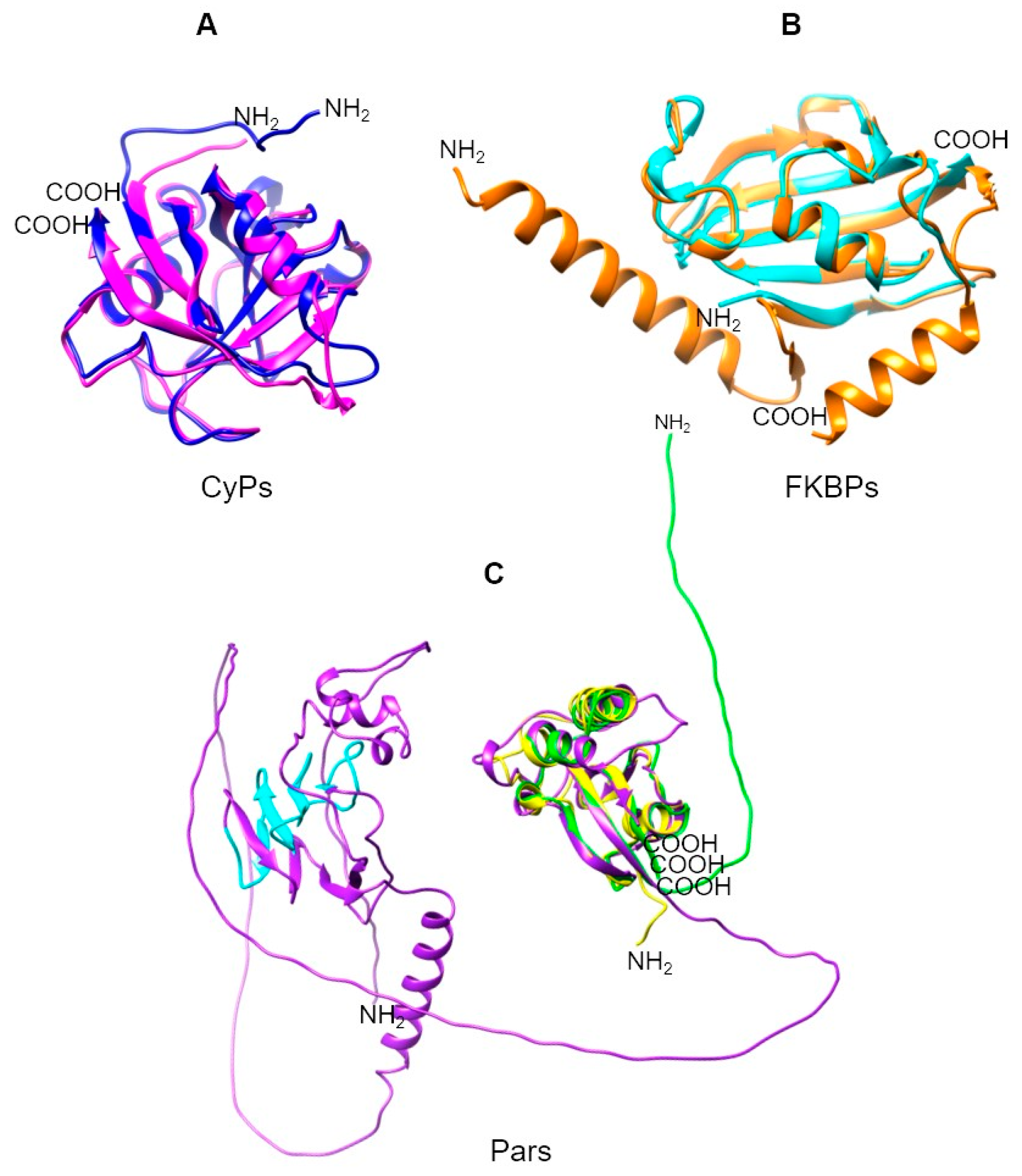
2. Parasite PPIases: Disease, Genome Database, and Structural Characteristics

2.1. Anaerobic or Microaerophilic Protozoan Parasites

2.1.1. Trichomonas vaginalis

Trichomoniasis is caused by protist parasite T. vaginalis, considered the most common non-viral sexually transmitted disease worldwide. In the USA, an estimated 2.6 million infected people, with a prevalence of 2.1% in women and 0.5% in men, have been recently reported. Infected people can present a variety of symptoms, but most are asymptomatic. Trichomonal infection has been linked to other sexually transmitted diseases, such as HIV, and ~53% of women who have HIV also have T. vaginalis. Infection with this parasite is also considered a risk factor for vertical transmission of HIV [24].
The genome of T. vaginalis strain G3 encodes 13 CyPs, 9 FKBPs, and 3 Pars, as reported in the TrichDB database (https://trichdb.org/trichdb/app/, accessed on 1 June 2023) (Release 63 3 May 2023) [25] (Table 1). In this review, we use the nomenclature for PPIases reported by Galat (2003) [13], in which PPIases are named with a prefix of two letters indicating the species, followed by a number for the calculated molecular weight in kDa.
The PPIases from T. vaginalis present sequence variations within each family, with few conserved regions. Phylogenetic analysis revealed that the CyPs were grouped into clades with low bootstrap values, suggesting that they do not share a common origin. In contrast, the FKBPs and Pars formed distinct clades with higher bootstrap values, indicating a common evolutionary origin for these proteins. However, TvCyP44 was found to share a clade with the FKBPs, which might indicate that it is more closely related to FKBPs than to CyPs, despite containing a CLD (Supplementary Figure S1, [26]).
Most CyPs from T. vaginalis retain the following aa residues essential for catalytic activity within the highly conserved CLD identified in TvCyP19 (referred to as TvCyP1 by Hsu et al., 2014) [27]: H62, R63, F68, M69, Q71, G80, A109, N110, A111, Q119, F121, W129, L130, and H134. Exceptions to this pattern were observed in TvCyP21, TvCyP37, and TvCyP44 (Supplementary Figure S2, [25,28]).
Table 1. Peptidyl-prolyl cis-trans isomerase repertoire from Trichomonas vaginalis 1.
UniProtTrichDB 2TrichDB 3NCBIPDBPPIase NameLocalization 4Function 4References
A2FJP1TVAG_370440TVAGG3_0054050XP_001307803 TvCyP14Nucleus [29]
A2EC21TVAG_137880TVAGG3_0269460XP_001322019.1 TvCyP18Cytoplasm [29]
A2DT06TVAG_004440TVAGG3_0649370XP_001328636.15YB9TvCyP19 (TvCyP1) Cytoplasm
hydrogenosomes, cytoplasm, and
membrane
Protein trafficking[30,31]
A2F1H0TVAG_027250TVAGG3_0947870XP_001314072 TvCyP19.2Cytoplasm [29]
A2FAA8TVAG_047830TVAGG3_0485720XP_001311112 TvCyP19.8Cytoplasm [29]
A2DLL4TVAG_062520TVAGG3_0580400XP_001579633.16LXOTvCyP19.9
TvCyP2
Cytoplasm
ER, cytoplasm, and membrane
Protein trafficking[32,33]
A2E5J4TVAG_038810TVAGG3_0240350XP_001324258 TvCyP20Cytoplasm [29]
A2FIV3TVAG_078570TVAGG3_0462310XP_001308098 TvCyP21Cytoplasm [29]
A2DKZ9TVAG_146960TVAGG3_0362200XP_001579938 TvCyP22Cytoplasm [29]
A2FTU8TVAG_27739TVAGG3_0951420XP_001304599 TvCyP23Cytoplasm [29]
A2E6H3TVAG_106810TVAGG3_0040330XP_001324009.1 TvCyP37Nucleus [29]
A2GDG2TVAG_583670TVAGG3_0820230XP_001297735.1 TvCyP44Cytoplasm and nucleus [29]
A2DEW6TVAG_172150TVAGG3_0530670XP_001581944.1 TvCyP63Nucleus [29]
A2DA37TVAG_476140TVAGG3_0266130XP_001583671.1 TvFKBP12Cytoplasm [29]
A2DYS7TVAG_426610TVAGG3_0538360XP_001326690.1 TvFKBP15.1ER [29]
A2G763TVAG_062070TVAGG3_0922950XP_001299933.1 TvFKBP15.2ER [29]
A2FYT1TVAG_435000TVAGG3_0194750XP_001302863.1 TvFKBP19Cytoplasm [29]
A2F0D0TVAG_292580TVAGG3_0216440XP_001330357.1 TvFKBP20Cytoplasm [29]
A2EV02TVAG_368970TVAGG3_0441630XP_001315748.1 TvFKBP30Cytoplasm [29]
A2EC50TVAG_413760TVAGG3_0204900XP_001321950.1 TvFKBP32Cytoplasm [29]
A2G9L9TVAG_428320TVAGG3_0107870XP_001299079.1 TvFKBP33Cytoplasm [29]
A2FER9TVAG_140950TVAGG3_0603860XP_001309536.1 TvFKBP63 [29]
A2ECU0TVAG_102340TVAGG3_0563910XP_001321708.1 TvPar17.84Cytoplasm [29]
A2ED59TVAG_420360TVAGG3_0425040XP_001321637.1 TvPar17.87Cytoplasm and nucleus [29]
A2EWG2TVAG_325610TVAGG3_0877000XP_001315212.1 TvPar102Cytoplasm and nucleus [29]
1 Previously reported PPIase names are presented in parentheses. 2 The TrichDB database corresponds to TVAG of the G3 non-reference strain, the first classification. 3 The TrichDB database corresponds to TVAG and is associated with the genome update of the G3 2022 reference strain and a new classification. 4 The localization and function of PPIases were taken from the cited references or from the UniProt database and were predicted by the Gene Ontology Consortium [29]. ER: endoplasmic reticulum. Blank spaces: data not reported.
The structures of only two of the CyPs of T. vaginalis have been solved, namely those of TvCyP19 and TvCyP19.9 (referred to as TvCyP1 and TvCyP2, respectively, by Hsu et al., 2014; 2020) [27,30]. Both proteins present the typical structure, characterized by a β-barrel composed of eight antiparallel β-strands and two α-helixes. TvCyP19 mainly consists of the CLD domain, whereas TvC19.9, in addition to the catalytic domain, has a longer N-terminal segment. An important difference between the two CyPs is that TvCyP19 is a dimer, whereas TvCyP19.9 is a monomer [31,32].
We analyzed the sequence identities of CyPs from T. vaginalis, taking the sequence of TvCyP19 as a reference (Supplementary Table S1, [28,34]). Low-molecular-weight CyPs (TvCyP14, TvCyP18, TvCyP19.2, TvCyP19.8, TvCyP20, TvCyP21, TvCyP22, and TvCyP23) have sequence identities of approximately 40–70% (Supplementary Table S1) with respect to TvCyP19. The eight small TvCyPs are characterized by having only the CLD in their sequence, similar to TvCyP19 and TvCyP19.9 (Figure 2A,B). Moreover, the high-molecular-weight CyPs (TvCyP37, TvCyP44, and TvCyP63) have low sequence identity to TvCyP19 (Supplementary Table S1). These TvCyPs exhibit a unique domain in addition to the conserved CLD. For example, TvCyP37 contains the SF-CC1 domain and the RRM motif at its C terminus (Figure 2C). The SF-CC1 domain is characteristic of RNA splicing factors and is marked by Arg- and Ser-rich sequences, typically followed by RNA recognition domains; this feature is also found in TvCyP37 [33]. Similarly, the presence of an RNA splicing factor has been noted in CyPs from other organisms, such as humans and Arabidopsis thaliana [35], along with the RNA recognition motif (RRM), which is involved in nucleic acid and/or protein recognition. The structural versatility of RRM interactions contributes to the diverse biological functions of RRM-containing proteins [36].
TvCyP44 has an N-terminal U-box domain, a specialized type of RING finger that differs from other RING fingers in terms of the lack of metal binding sites [37]. This domain has an estimated length of 67 aa (Figure 2D) and has been identified in ubiquitin ligase-like proteins in Saccharomyces cerevisiae that serve as scaffolds for proteins during ubiquitination and is associated with protein degradation pathways [38,39]. TvCyP63 contains a WD40 domain near the N terminus (aa 32–349; Figure 2E). This WD40 domain is also present in other large CyPs, such as PfCyP87, PvCyP83, and TgCyP86 from P. falciparum, P. vivax, and T. gondii, respectively. The WD40 domains function in anchoring to other proteins or in DNA binding. These domains are present in a wide variety of proteins with diverse functions, including chaperone proteins, but have no catalytic activity [40].
Compared to CyPs, T. vaginalis FKBPs presented lower sequence identities (less than 24%) when TvFKBP-12 was used as a reference (Supplementary Table S1). This low similarity is attributed to the absence of consensus sequences in FKBPs. Each of the nine TvFKBPs has an FKBP domain with a sequence length in the range of ~84 to 100 aa near the N terminus (Figure 2F–H). Six of the FKBPs (TvFKBP-12, TvFKBP-15.1, TvFKBP-15.2, TvFKBP-19, TvFKBP-20, and TvFKBP-33) range from 12 to 33 kDa and consist of a single FKBP domain that spans most of the protein sequence (Figure 2F). In contrast, three of the FKBPs (TvFKBP-30, TvFKBP-32, and TvFKBP-63) include another domain in addition to the catalytic domain. TvFKBP-30 and TvFKBP-32 contain three tetratricopeptide repeat (TPR) motifs near the C terminus (Figure 2G). TPRs are structural motifs that usually comprise approximately 34 aa and mediate protein–protein interactions and the assembly of multiprotein complexes [41]. TvFKBP-63 contains an MukB domain (Figure 2H), which is found in MukB proteins and is associated with chromatin remodeling [42]. No other reports of FKBPs containing MukB domains were found.
Two of the three T. vaginalis Par proteins, namely TvPar17.8, and TvPar17.9, have similar molecular weights and 41.6% identity (Supplementary Table S1). Both proteins have a PpiC domain close to the C terminus, which covers most of the protein (~100 aa). In addition to this domain, the three TvPar proteins possess a WW domain of ~30 aa near the N terminus (Figure 2I,J).
The third TvPar, with a molecular weight of ~102 kDa (TvPar102), contains the PpiC domain, which spans ~100 aa near the N terminus. In addition, the WW domain near the N terminus contains a suppressor of forked (Suf) domain (Figure 2J), which is commonly associated with mRNA formation and polyadenylation in organisms such as Drosophila melanogaster [43]. Understanding the role of the Suf domain in TvPars could provide valuable insights.
TvPar102 is the largest Par identified among these clinically important parasites and is significantly larger than the typical Pars (~13 kDa). Its distinct C-terminal end, which consists of a series of α-helixes, sets it apart from other proteins in this group. However, its function remains unknown.

2.1.2. Entamoeba histolytica

Amoebiasis is a parasitic disease caused by protozoan parasites belonging to the Entamoeba genus, among which E. histolytica is the most pathogenic species. This infection is transmitted through oral–fecal contamination, often via the consumption of food or water contaminated with parasite cysts, which migrate from the small intestine to the large intestine, where they divide by binary fission and are eliminated in the feces. Amoeboid forms may migrate out of the intestine and invade other body organs. This disease causes dysentery and various intestinal problems, affecting approximately 500 million people worldwide and causing more than one hundred thousand deaths per year [44].
The E. histolytica HM1-IMSS strain reference genome in the AmoebaDB database [25] (https://amoebadb.org/amoeba/app/, accessed on 1 June 2023) (Release 63 3 May 2023) contains six genes encoding CyPs, five genes encoding FKBPs, and two genes encoding Pars (Table 2).
Alignment of the EhCyP18 CLD with CLDs of CyPs from other organisms revealed high sequence conservation of this domain, in addition to the conservation of important aa residues in the active site (Supplementary Figure S3, [46]). Unlike the EhCyPs, the five FKBPs in E. histolytica are poorly conserved, which is unsurprising, since FKBPs are known to be highly divergent. The lowest sequence identities among the E. histolytica PPIases were found between its two Pars, EhPar13 and EhPar13.25, which share 20% identity, indicating significant divergence between these proteins.
The CyPs found in E. histolytica have molecular weights of between 10 and 40 kDa, and the CLD domain is consistently located at the N terminus. Interestingly, EhCyP10 is the smallest known CyP among parasites and contains a CLD. EhCyP21 and EhCyP22 contain an N-terminal sequence that allows them to localize to the ER. Notably, in addition to the CLD domain, CyPs EhCyP10 and EhCyP40 also contain an RRM domain of ~77–78 aa at the C terminus.
The five FKBPs of E. histolytica are globular proteins with molecular weights ranging from 18 to 46 kDa. Interestingly, in addition to the PPIase-FKBP domain, four of the FKBPs (EhFKBP-29, EhFKBP-35, EhFKBP-43, and EhFKBP-46) possess the TPR domain, which is related to protein–protein interactions. In contrast, EhFKBP-18 contains only the FKBP-like PPIase domain and a signal sequence at the N terminus with an unknown function. E. histolytica Pars contain only the PpiC-like PPIase domain; both Pars are small in size, which is common in the Pars identified to date. [47]

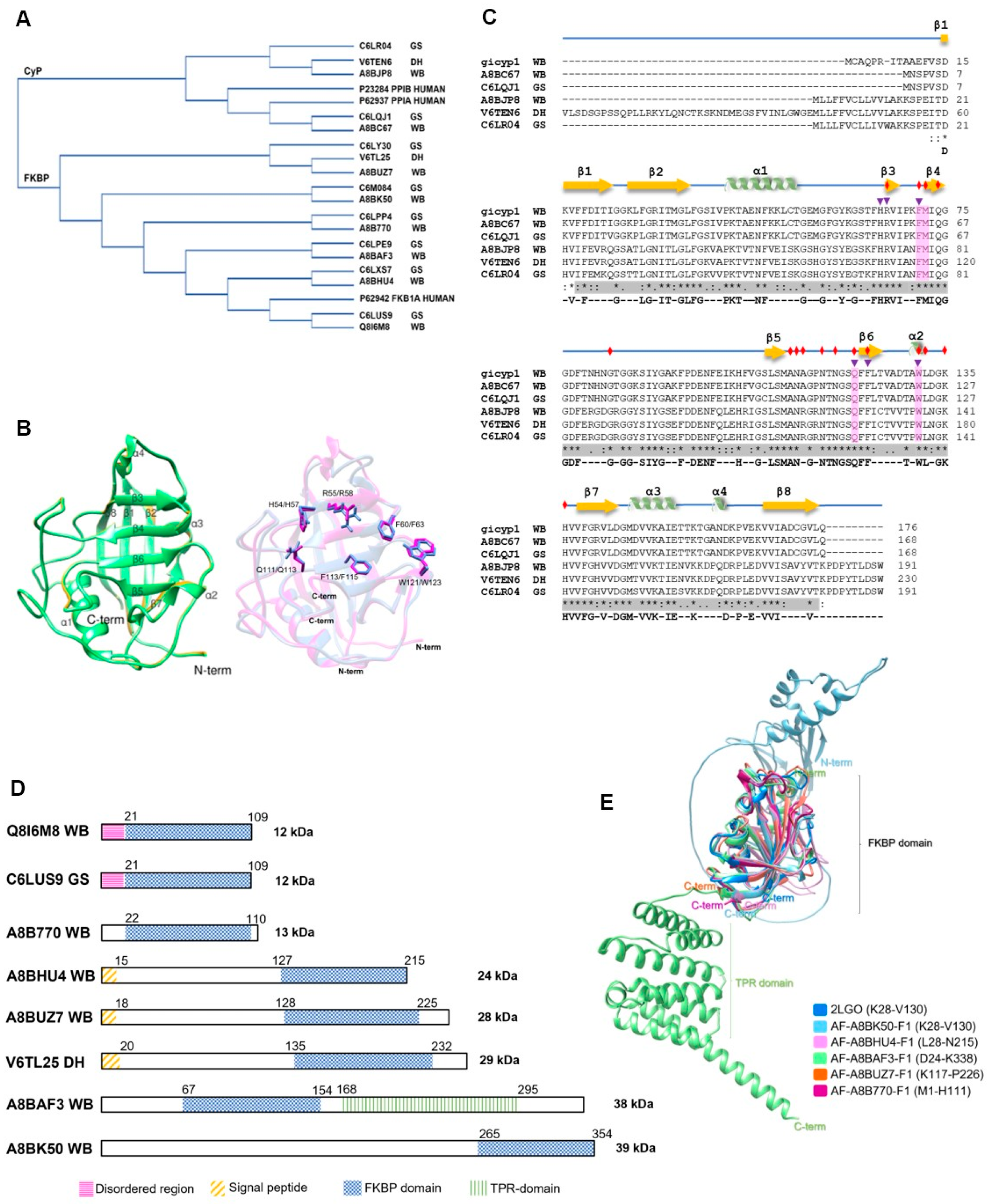
2.1.3. Giardia intestinalis

Giardiasis, caused by protozoan parasite Giardia intestinalis (also known as G. duodenalis or G. lamblia), is a disease of global concern, affecting both developed and undeveloped nations. While asymptomatic cases are common, various intestinal and extraintestinal symptoms, as well as postinfection problems, have been recorded [48]. Giardiasis was designated as a neglected disease by the WHO in 2004, highlighting its public health significance [49]. It was estimated ~200 million cases of giardiasis worldwide in 2010 [50]. More recent reports in the European Union revealed that 18,000 cases were reported in 2019, with the highest incidence occurring in children between 0 and 4 years of age [51]. Likewise, in the United States alone, ~7 cases per 100,000 habitants were reported in 2019 [52].
G. intestinalis is a complex species comprising eight genetically related groups (assemblages A to H). Assemblages A and B, which are responsible for infecting humans, are subject to debate regarding their potential classification as different Giardia species due to significant genetic differences. Further research is required to resolve this question [53,54,55].
In this review, we focus on PPIases from the G. intestinalis genome reported in the GiardiaDB database (https://giardiadb.org/giardiadb/app, accessed on 1 June 2023) (Release 63 3 May 2023) [25], specifically from reference isolates WB (assemblage A), DH (sub-assemblage AII), and GS (assemblage B), which are all responsible for human infections [53,56,57]. These G. intestinalis isolates contain two CyPs (molecular weight < 25 kDa) and six FKBPs (molecular weight < 39 kDa), and no Pars were reported, similar in number to Cryptosporidium spp. and in contrast to parasites such as T. vaginalis, Trypanosoma, Leishmania, and Toxoplasma, which have more than 20 PPIases with molecular weights < 100 kDa.
The numbers of CyPs and FKBPs in the genomes of Giardia assemblages A and B are relatively consistent between the isolates. Some PPIases are the same in the two assemblages (Table 3), indicating potential similarities and a common ancestor (Figure 3A). In the DH isolate (sub-assemblage AII), the two GiCyPs and five GiFKBPs showed high sequence identity (>94.24%) compared to that of the WB isolate. However, differences in molecular weight were found for only one GiCyP and one GiFKBP (Table 3).
Table 3. Peptidyl-prolyl cis-trans isomerase repertoire from Giardia intestinalis 1,2.
IsolateUniProtGiardiaDBNCBIPDBPPIase NameLocalization 3 Function 3References
G. intestinalis WB GiCyP19
(GiCyP1)
[58]
A8BC67GL50803_0017163XP_001707838.1 GiCyP18Cytoplasm
Secreted
Virulence factor[29,59,60]
A8BJP8GL50803_0017000XP_001706629.1 GiCyP21Cytoplasm
Secreted
[29,59]
G. intestinalis DHV6TEN6DHA2_17000 GiCyP25Membrane [29]
G. intestinalis GSC6LQJ1GL50581_1019 GiCyP18Secreted [29]
C6LR04GL50581_1186 GiCyP21Secreted [43]
G. intestinalis WBQ8I6M8GL50803_10450XP_001709141.12LGOGiFKBP12Secreted [59,61]
A8B770GL50803_7246XP_001709155.1 GiFKBP13Cytoplasm [29]
A8BHU4GL50803_101339XP_001706925.1 GiFKBP24Cytoplasm
Secreted
[29,59]
A8BUZ7GL50803_42780XP_001704692.1 GiFKBP28
A8BAF3GL50803_3643XP_001708385.1 GiFKBP38Cytoplasm
Secreted
[29,59]
A8BK50GL50803_10570XP_001706462.1 GiFKBP39
G. intestinalis DHV6TL25DHA2_151252 GiFKBP29
G. intestinalis GSC6LUS9GL50581_2531 GiFKBP12Secreted [59]
C6LPP4GL50581_711 GiFKBP13
C6LXS7GL50581_3593 GiFKBP24Secreted [59]
C6LY30GL50581_3701 GiFKBP28
C6LPE9GL50581_614 GiFKBP38Secreted [59]
C6M084GL50581_4472 GiFKBP39
1 Giardia assemblage A, isolate WB C6 (WB). Giardia sub-assemblage A2, isolate DH (DH). Giardia assemblage B, isolate GS/M, clone H7 (GS). 2 Previously reported names are presented in parentheses. 3 The localization and functions of PPIases were taken from the cited references or predicted by the Gene Ontology Consortium [29] in the UniProt database. Blank spaces: no reported data.
Figure 3. PPIases of the WB, GS, and DH G. intestinalis isolates. (A) Phylogram of PPIases from G. intestinalis and three human PPIases (PPIA/CyPA, PPIB/CyPB, and FKB1A/FKBP-12). (B) Overlapping structures of the GiCyP18 3D model of the WB (UniProt ID A8BC67 in green) and GS (UniProt ID C6LQJ1 in yellow) G. intestinalis isolates. Overlapping structures of hPPIA (1Ak4_A in magenta) and GiCyp18 (WB isolate in blue) are shown on the right. Active site residues taken from the Protein Domain Family of Conserved Domain Database (CDD) are represented by ball-and-stick illustration. (C) CyPs multiple sequence alignment. The secondary structure is depicted by yellow arrows for β-sheets and green ribbons for α-helixes. In gray is the cyclophilin-like domain (CLD). Red diamonds represent the putative CsA binding sites for GiCyP19. Purple triangles correspond to putative active sites. In pink are the binding pockets suggested for homology with hPPIA (UniProt ID P62937). * Represent conserved aa residues. (D) Domain architecture for FKBPs from G. intestinalis. The numbers above the bars indicate the amino acid position of each region. (E) Overlapping structures of FKBP domains from G. intestinalis WB isolate. The FKBP domains that were conserved in the alignment are indicated in parentheses. (A,B) were developed using the Clustal Omega 1.2.4, Clustal W and Clustal X tools [62,63]. The PDB and AlphaFold models were visualized with the UCSF Chimera 1.16 program [64]. All proteins have UniProt accession numbers (Supplementary Table S3, [34,62]).
Pathogens 13 00644 g003
All G. intestinalis CyPs share a common CLD domain of ~89 aa, spanning 159 residues (Supplementary Table S2) (Figure 3C). The GiCyP18 proteins in the WB and GS isolates share 99.4% identity (Supplementary Table S3) and contain only a CLD domain (Supplementary Table S2) (Figure 3C). The GiCyP21 proteins in the WB and GS isolates share 94.2% identity (Supplementary Table S3) and have some characteristics in common with hCyPB [12]. These proteins contain an SP of 13 residues, 2 nonstructural regions at both ends of the CLD domain, and an additional 9 residues at the C terminus but lack the C-terminal ER sequence found in human hCyPB (Supplementary Table S2). In the DH isolate, GiCyP25 contains regions resembling those in hCyPB and a transmembrane helix (TMH) segment (Supplementary Table S2) (Figure 3C).
We also explored 3D GiCyP models using the AlphaFold database in UniProt (Release 2023_01). Despite the lack of crystallized GiCyPs to date, our analysis revealed that all the GiCyPs share a secondary and tertiary structure similar to that of hCyPA, as previously described [4,14]. Specifically, the secondary structure comprises eight β-strands, two α-helixes, two small helixes, and twelve regions of random-coil turns, which are also widely reported in other species (Figure 3C).
In the 3D structures, we observed that the central domain consists of eight β-strands flanked by an α-helix at each end of the barrel. These regions are connected by random-coil turn structures that are exposed on the protein surface. To determine the active site residues, we referenced the protein domain families in the Conserved Domains Database (CDD) [58] (Figure 3B). Notably, these active site residues coincide with those involved in CsA binding, including W123 in GiCyP1 (referred to as GiCyP19 in this review) (Table 3). GiCyP19 was the first CyP characterized from the G. intestinalis WB strain by Yu et al. in 2002 [65]. It should be noted that although the sequence of this gene had not yet been deposited in a database at the time of its publication, through BLAST analysis, we found an identity of around 98% of the sequence of the original publication with that of GiCyP19 found in the Giardia spp. genome.
Giardia FKBPs (Table 3) share similarities in terms of size and aa sequence among isolates (Supplementary Tables S2 and S3 [34,62]) (Figure 3D). The molecular weights of the proteins ranged from ~12 to 39 kDa, and their isoelectric points varied significantly from highly acidic at 4.6 to highly alkaline at 9.5 (Supplementary Table S2 [34]). Similar variations in size and charge have been observed in other organisms [13,66]. Among the Giardia FKBPs, only GiFKBP-12 has been crystallized (PDB: 2LGO) [61]. Its FKBP domain is located at the C-terminus and consists of 88 to 97 aa, forming six antiparallel β-strands and one α-helix. It contains conserved residues responsible for inhibitor binding (Figure 3D) [67]. Other Giardia FKBPs possess additional regions along with the FKBP domain (Table 3). Some have a small, disordered region of approximately 21 aa at the N terminus (GiFKBP-12), while others have an SP of 15 or 18 aa (GiFKBP-24, GiFKBP-28, and GiFKBP-29). Only one FKBP (GiFKBP-38) contains a TPR region (Supplementary Table S2) formed by six antiparallel α-helixes (Figure 3E).

2.2. Trypanosomatid Parasites

2.2.1. Trypanosoma cruzi and Trypanosoma brucei gambiense

T. cruzi and T. brucei gambiense are protozoan parasites that cause significant health impacts through trypanosomiasis. T. cruzi causes Chagas disease (also known as American trypanosomiasis), which is transmitted to humans and other mammals by Triatominae insects. T. brucei gambiense and T. brucei rhodesiense cause sleeping sickness (also known as African trypanosomiasis), which is transmitted by the tsetse fly [68]. According to the WHO, between 6 and 7 million people worldwide, the majority in Latin America, are infected by T. cruzi, including around 30,000 and 40,000 new infections and between 10,000 and 20,000 deaths every year. At the same time, it is estimated that ~75 million people are at risk of infection [69]. For example, in 2016 to 2018 in Brazil the average chronic disease occurrence rate is 3.2 per 100,000 people per year. Furthermore, a total of 350 deaths are recorded annually (male predominance, age ≥60 years, and chronic disease with cardiac involvement as the main mortality cause), with an annual average mortality proportion of 1.6 per 100,000 people [70]. Moreover, African trypanosomiasis is endemic in numerous African regions, putting 70 million people at risk throughout sub-Saharan Africa. T. brucei gambiense accounts for more than 95% of the reported cases of African trypanosomiasis [69]. This infection is mainly present in Western and Central Africa. It has a chronic progressive course lasting over three years, mimicking hematological conditions [71].
The CyPs in the T. cruzi CL Brener Esmeraldo-like genome have been reported in the TriTrypDB database (https://tritrypdb.org/tritrypdb/app/, accessed on 1 April 2023) (Release 62 9 Mar 2023) [25] and include 15 paralogs [72]. We analyzed the 3D structure of these CyPs using the predicted structures generated by AlphaFold in the UniProt database (Release 2023_01). All these CyPs share the highly conserved CLD domain, an eight-stranded, antiparallel ꞵ-barrel structure accompanied by two α-helixes, and show some differences in regions outside the CLD domain. TcCyP19 consists predominantly of the CLD domain, whereas the other fourteen are isoforms (Table 4). For instance, TcCyP21 and TcCyP24 have SPs of 26 and 25 aa residues in length, respectively. Notably, TcCyP21 is the only T. cruzi CyP with a reported crystal structure (PDB:1XO7) [61]. Its PDB ID is linked to a different UniProt ID (Q4DPB9) due to its origin in the T. cruzi CL Brener non-Esmeraldo-like genome. However, the alignment of the two protein sequences revealed 97.9% identity.
Furthermore, TcCyP22, TcCyP26, TcCyP30, and TcCyP42 contain an elongated region in the N terminus, while TcCyP20 has a small, elongated region in the C terminus. Additionally, TcCyP25, TcCyP28, TcCyP29, TcCyP35.3, and TcCyP35 (previously named TcCyP34 by Potenza et al., 2006) [73] exhibit elongated regions in both the N- and C-terminal segments. Moreover, TcCyP38 (previously named TcCyP40) [73] contains the TPR motif. Finally, TcCyP103 (previously named TcCyP110) [73] is the largest CyP (103 kDa) and contains an array of structures adjacent to the CLD domain, which were identified as disordered regions with both basic and acidic residues.
Table 4. Peptidyl-prolyl cis-trans isomerase repertoire from Trypanosoma cruzi 1.
ParasiteUniProtTriTrypDBNCBIPDBPPIase NameLocalization 2Function 2References
T. cruzi CL BrenerQ4E4L9TcCLB.506925.300
(CYPA)
XP_821578.1 TcCyP19 Extracellular spacePromotes ROS production in host cells[72,74,75,76,77]
Q4DC03TcCLB.507009.100XP_811912.1 TcCyP20
Q4DNC9TcCLB.507521.70XP_815879.11XO7TcCyP21 [72,76]
Q4DI85TcCLB.504035.70XP_814080.1 TcCyP22 MitochondriaCell death regulation[72,78]
Q4CXV1TcCLB.506413.80XP_806960.1 TcCyP24
Q4DFL3TcCLB.508323.94XP_813175.1 TcCyP25 [72,76]
Q4D4K3TcCLB.503885.40XP_809302.1 TcCyP26
Q4CX88TcCLB.509499.10XP_806737.1 TcCyP28 [72,76]
Q4DQI8TcCLB.505807.10XP_816616.1 TcCyP29
Q4DNS3TcCLB.511589.50XP_816007.1 TcCyP30Membrane [29,72]
Q4DM35TcCLB.511577.40
(CYP35)
XP_815421.1 TcCyP35
(TcCyP34)
[72,76]
Q4DVC9TcCLB.511217.120XP_818332.1 TcCyP35.3
(TcCyP35)
[72]
Q4E4G0TcCLB.506885.400
(CYP40)
XP_821542.1 TcCyP38
(TcCyP40)
[29,72]
Q4DG41TcCLB.510761.44XP_813344.1 TcCyP42Membrane [29,72]
Q4D1M5TcCLB.504215.10XP_808273.1 TcCyP103
TcCyP110
[72]
T. cruzi YQ09734TcYC6_0113560CAA49346.11JVWTcFKBP22
(TcMIP)
Extracellular spaceHost cell entry/invasion
[73,79]
T. cruzi CL BrenerQ4D5W5TcCLB.508169.69XP_809772.1 TcFKBP12
Q4DFL5TcCLB.508323.84XP_813174.1 TcFKBP12.2
Q4D7F5TcCLB.511731.89XP_810317.1 TcFKBP35
Q4CZN2TcCLB.511353.10XP_807578.1 TcFKBP52
Q4CYE6TcCLB.507629.39XP_807152.1 TcFKBP93
T. cruzi CL BrenerQ4D8F7TcCLB.508567.70
(Pin1)
XP_810661.1 TcPar12.6
(TcPin1)
Cytosol [80,81]
Q4D394TcCLB.506697.50XP_808848.1 TcPar13
(TcPar14)
[82]
Q4D9J4TcCLB.506857.60
(Par45)
XP_811046.1 TcPar45 Nucleus [82]
1 Previously reported PPIasa names are presented in parentheses. 2 The localization and functions of PPIases were taken from the cited references or from the UniProt database and were predicted by the Gene Ontology Consortium [29]. Blank spaces: data not reported.
The TriTrypDB database includes six T. cruzi FKBP genes [72] (Table 4). Structural analysis revealed that these FKBPs range in size from 12 to 93 kDa and share similarities in the catalytic domain. Two FKBPs, namely TcFKBP-12 and TcFKBP-12.2, consist mostly of the FKBP domain. The four remaining proteins contain an FKBP domain plus other motifs. TcFKBP-22 (also referred to as TcMIP, the Microphage Infectivity Potentiator, by Moro et al., 1995) [79] contains a 29 aa SP. TcFKBP-22 is from T. cruzi strain Y and is the only T. cruzi FKBP that has been characterized and crystallized (PDB: 1JVW) [61]. TcFKBP-35 has extensive elongation in its C terminus, and TcFKBP-52 and TcFKBP-93 have elongated regions in their N termini that have been identified as coiled-coils. These coiled-coils are involved in various biological functions as molecular spacers within proteins, influencing the architecture of organelles such as centrioles and the Golgi apparatus and facilitating the binding of transport vesicles to the Golgi apparatus [80].
Finally, T. cruzi contains three Pars [72] (Table 4) ranging from 12 to 45 kDa, all possessing the PpiC domain. TcPar12.6 (previously named TcPin1 by Erben et al., 2007 [82] and a homolog of hPin1) consists entirely of the PpiC domain and lacks the protein–protein interaction-related WW domain at the N terminus. In addition, TcPar13 (previously named TcPin14 by Erben et al., 2010 [83] and a homolog of hPar14) has an elongated region in its N terminus, which is described as disordered and lacks the WW domain. Unlike TcPar13, hParv14 lacks the N-terminal WW domain but has an unstructured N-terminal extension that is essential for its nuclear localization and DNA binding. The third Par, TcPar45, has an elongated N-terminal segment and contains a fork-head-associated domain (FHA) instead of a WW domain; FHA domains play a role in recognizing phosphopeptides related to biological processes [84].
The genome of T. brucei gambiense strain DAL972 is closely related to the T. brucei genome, which suggests that the DAL972 genome is an effective scaffold for any T. brucei genome sequence [74]. The genome of T. brucei gambiense strain DAL972 in TriTrypDB (Release 62 9 Mar 2023) [25] encodes 19 members of the CyP family (four more than T. cruzi), 6 members of the FKBP family, and 3 members of the Pars family [75] (Table 5).
We analyzed the 3D models of these PPIases and observed similarities with those in T. cruzi. The TbgCyPs exhibit elongated N- or C-terminal segments or both (TbgCyP21.1, TbgCyP21.4, TbgCyP25.55, TbgCyP27.1, TbgCyP27.4, TbgCyP29, TbgCyP30, TbgCyP33, TbgCyP43, TbgCyP46, and TbgCyP100). TbgCyP38, like TcCyP38, contains a TPR motif. Furthermore, four CyPs have SP domains (TbgCyP21.2, TbgCyP24, TbgCyP25.56, and TbgCyP58). T. cruzi does not contain a 58 kDa CyP, unlike T. brucei gambiense. TbgCyP58 also contains an RRM motif involved in nucleic acid and/or protein recognition. T. brucei gambiense has three CyPs (TbgCyP19, TbgCyP20.3, and TbgCyP20.5) that consist predominantly of the CLD domain, whereas T. cruzi has only one CyP with this domain.
We found that the T. brucei gambiense FKBPs also contain elongated N- and C-terminal regions (TbgFKBP12.3 and TbgFKBP36), disordered regions and a coiled coil (TbgFKBP92), and an SP (TbgFKBP21). Additionally, TbgFKBP48 contains a disordered region paired with coiled-coils and a TPR motif. Only TbgFKBP12 consists almost exclusively of the FKBP domain.
The T. brucei gambiense Pars are also homologous to T. cruzi Pars. TbgPar12 and TbgPar13 share more than 68% sequence identity with TcPar12.6 and TcPar13, respectively. TbgPar12 consists mainly of the PpiC domain, while TbgPar13 contains an elongated N-terminal segment that is reported to be disordered. Moreover, TbgPar42 shares 60% sequence identity with TcPar45 and contains both a PpiC domain and an FHA domain.

2.2.2. Leishmania major and Leishmania donovani

Leishmaniasis is caused by protozoan parasites in the Leishmania genus, which are transmitted to humans and other animals via the bites of infected sandfly mosquitos. Leishmaniasis is present in 88 countries around the world, most of them being developing nations, with a prevalence of at least 12 million infected people and a population at risk of almost 350 million people [89]. It is also estimated that there is an annual incidence of 600,000 to 1 million cases of cutaneous leishmaniasis and ~50,000–90,000 cases of visceral leishmaniasis [90]. L. major and L. donovani are linked to cutaneous and visceral leishmaniasis, respectively, in Asia, Africa, and parts of Europe [91].
The TriTrypDB database (https://tritrypdb.org/tritrypdb/app/, accessed on 1 June 2023) (Release 63 3 May 2023) [25] contains at least 24 PPIase genes from the L. major isolate Friendlin reference genome [92] and the same number from the L. donovani BPK282A1 reference genome [93]. Both species contain 17 CyPs, 5 FKBPs, and 2 Pars (Table 6 and Table 7). Despite their high overall sequence identity and similar molecular weights, LmPar47 and LdPar17 exhibit only 34% sequence identity, mainly due to differences in molecular weight (Supplementary Table S4). Interestingly, the gene encoding LdCyP108, categorized as a conserved hypothetical protein in the Leishmania database (TriTrypDB), was confirmed to be a CyP through further verification via the UniProt and NCBI databases. The number of PPIases in Leishmania is comparable to that in other trypanosomatids, such as T. cruzi and T. brucei (Table 4 and Table 5).
The first PPIases discovered in L. major and L. donovani were CyPs identified during studies involving Cyclosporin A (CsA) [96,99]. Leishmania CyPs vary in size from 19 to 108 kDa and have additional N-terminal or C-terminal extensions or both alongside the CLD domain [90]. Three Leishmania CyPs stand out in particular, namely LmCyP24.6 (also known as LmaCyP5), which features a PLD (prokaryotic lipid attachment domain); LmCyP38 (also known as LmaCyP40), which is distinguished by two additional TPR domains; and LdCyP38.4 (also known as LdCyP40), which contains a TPR domain at its C terminus.
The crystal structure of LdCyP20 (PDB: 2HAQ) from L. donovani closely resembles that of other CyPs, with an eight-stranded β-barrel and two α-helixes, albeit with minor differences from hCyPA [97]. LmCyP32 from L. major (PDB: 2HQJ) shares the same secondary structure, with slight variations in the loops. Additionally, L. major has two more crystallized CyPs, namely LmCyP25 (PDB: 7AIH) and LmCyP29 (PDB: 7AM2), both of which are part of a large subunit of the L. major mitoribosome [94].
The FKBPs identified in both L. major and L. donovani display notable similarities. These proteins share the FKBP PPIase domain and fall within the 11.8 to 48 kDa range. Interestingly, LmFKBP48 and LdFKBP47 contain an additional TPR domain at the C-terminus. Among Leishmania Pars, LmPar13, LdPar12, and LdPar17 are characterized by a PpiCPPIase domain and similar molecular weights. In the case of LmPar47, an FHA domain is present alongside the PpiC domain.

2.3. Apicomplexan Parasites

2.3.1. Plasmodium falciparum and Plasmodium vivax

The Plasmodium genus causes malaria, which is transmitted by infected female Anopheles mosquitoes. P. falciparum and P. vivax are the most prominent species that cause malaria in humans due to their characteristics and impact on public health. P. falciparum is the most prevalent pathogen in Africa, and P. vivax is the dominant parasite in most countries outside of sub-Saharan Africa. According to the latest report, there were 247 million cases of malaria in 2022 and 608,000 malaria deaths in 85 countries in 2022, with 80% of deaths occurring in children under 5 years of age [104].
The reference genome of the P. falciparum 3D7 isolate is widely used in malaria research. According to the PlasmoDB database (https://plasmodb.org/plasmo/app, accessed on 1 June 2023) (Release 63 3 May 2023) [25], this genome contains the 13 genes encoding PPIases, namely 11 CyPs and 2 FKBPs. To date, no Pars have been reported (Table 8). The new reference genome for P. vivax was obtained from the P01 isolate [105]. According to PlasmoDB (Release 63 03 May 2023), this genome contains the same number of PPIases as P. falciparum. Thus, this family is conserved between species even though the P. vivax genome presents almost twice the genetic diversity of P. falciparum [106,107].
The P. falciparum CyPs have molecular weights ranging from 19 to 87 kDa. They contain a conserved CLD domain with a typical eight-strand β-barrel and two α-helixes. Most of the PfCyPs contain a CLD region of ~142–165 aa, but the largest CLD region is 209 aa (PfCyP81) (Table 8). PfCyP19 and PfCyP18.6 (also known as PfCyP19C) consist only of the CLD domain, and the rest are CyP isoforms. For example, PfCyP22 contains an SP, and PfCyP23 has a coiled coil near the N terminus. PfCyP25 has N-terminal extensions, whereas PfCyP26 and PfCyP53 have C-terminal extensions, and PfCyP32 and PfCyP72 have extensions in both the N- and C-terminal regions. PfCyP87 contains a WD40 repeat, and PfCyP81 contains a region related to the SYF2 family (Figure 4A,B). This analysis is similar to that reported by Marín-Menéndez and Bell (2011) [108], with some differences in the length of regions or domains. Additionally, two crystallized structures of the CyP catalytic domain have been reported for P. falciparum, namely PfCyP19 (PDB: 1QNG) and PfCyP87 (PDB: 2FU0) [109] (Table 8).
Table 8. Peptidyl-prolyl cis-trans isomerase repertoire from Plasmodium falciparum 1,2.
UniProtPlasmoDBNCBIPDBPPIase NameLocalization 3References
Q8IIK3PF3D7_1116300 (CYP19C)XP_001347841.1 PfCyP18.6 (PfCyP19C) Nucleus (Spliceosome)[29,107,110]
Q76NN7PF3D7_0322000 (CYP19A)XP_001351290.11QNGPfCyP19 (PfCyP19A) Cytoplasm[29,107,110,111,112,113]
Q8IIK8PF3D7_1115600 (CYP19B)XP_001347835.1 PfCyP22 (PfCyP19B) Cytoplasm
Membrane
[29,107,113,114,115]
Q8I3I0PF3D7_0528700 (CYP23)XP_001351841.1 PfCyP23Nucleus (Spliceosome)[29,102,107,109,110]
Q8I6S4PF3D7_0804800 (CYP24)XP_001349469.1 PfCyP25(PfCyP24)Membrane[29,107,110,116,117]
Q8I621PF3D7_1202400 (CYP26)XP_001350433.1 PfCyP263Cytoplasm
Q8I5Q4PF3D7_1215200 (CYP32)XP_001350556.1 PfCyP32Cytoplasm and Mitochondria[29,107,110]
Q8ILM0PF3D7_1423200 (CYP52)XP_001348397.2 PfCyP53 (PfCyP52)Nucleus (Spliceosome)[29,107,110]
Q8I2K8PF3D7_0930600 (CYP72)XP_001352173.1 PfCyP72Nucleus[11,29]
Q8IAN0PF3D7_0803000 (CYP81)XP_001349484.1 PfCyP81Nucleus[11,29]
Q8I402PF3D7_0510200 (CYP87)XP_001351660.12FU0PfCyP87Nucleus (Spliceosome)[29,107,108]
C0H5B2PF3D7_1313300XP_002809009.1 PfFKBP25.6
Q8I4V8PF3D7_1247400XP_001350859.12OFNPfFKBP35Cytoplasm and Nucleus[29,109,118,119,120]
1 Isolate: Plasmodium falciparum 3D7. 2 Previously reported PPIAse names are presented in parentheses. 3 The localization and functions of PPIases were taken from the cited references or predicted by the Gene Ontology Consortium [29] in the UniProt database. ER: endoplasmic reticulum. Blank spaces: no reported data.
To date, no P. vivax CyPs have been characterized (Table 9). Approximately half the CyP sequences of the two Plasmodium species exhibited ≥80% identity. However, PvCyP29, PvCyP52, PvCyP65, PvCyP71, and PvCyP87 have below-average identity (Supplementary Table S5). PvCyP19 consists predominantly of the CLD domain. Outside this domain, the P. vivax homolog CyPs show similarities to PfCyPs. For example, PvCyP21 contains an SP, and PvCyP83 contains a WD40 repeat. The CyPs with elongated segments in the N terminus, C terminus, or both are PvCyP18, PvCyP23, PvCyP26, PvCyP29, PvCyP32, and PvCyP52. Intriguingly, P. vivax has two CyPs with distinct molecular weights, namely PvCyP29 and PvCyP65, instead of PfCyP25 and PfCyP81. PvCyP65 also contains a region belonging to the SYF2 family, as suggested for PfCyP81.
P. falciparum and P. vivax possess only two FKBPs, in contrast to most of the other mentioned parasites, which typically have at least five FKBPs. Some of the Plasmodium FKBPs have 60% identity (PfFKBP25.6 and PvFKBP25) and 80% identity (PfFKBP35 and PvFKBP34) (Supplementary Table S5). The conserved FK506 binding domains of three Plasmodium FKBPs have been crystallized, namely PfFKBP35 (PDB: 2OFN) [116,121], PvFKBP25 (PDB: 4JYS) [122], and PvFKBP34 (PDB: 2KI3) [123] (Table 8 and Table 9). This domain shares many of its secondary structures, comprising a six-stranded β-sheet and short α-helixes, with an additional β-strand at the N terminus. In addition to this domain, PfFKBP25 and PvFKBP25 contain extensions at the N terminus. PvFKBP25 is considered an atypical FKBP that lacks catalytic activity and does not have the conserved active site in typical FKBPs [118,122]. PfFKBP35 and PvFKBP34 have three and one TPR domains, respectively. These domains control the dimeric form of PfFKBP35, while the FKBP domain remains a monomer in solution [111].
Table 9. Peptidyl-prolyl cis-trans isomerase repertoire from Plasmodium vivax 1,2.
UniProtPlasmoDBNCBIPDBPPIase NameLocalization 3References
A0A1G4HCW7PVP01_0916900 (CYP19C)XP_001615280.1 PvCyP18.5
A0A1G4HBM6PVP01_0818200 (CYP19A)XP_001614493.1 PvCyP19
A0A1G4HCM3PVP01_0916400XP_001615276.1 PvCyP21
A0A1G4HDR7PVP01_1005100 (CYP23)XP_001613671.1 PvCyP23
A0A1G4H2Q1PVP01_1301700 (CYP26)XP_001616500.1 PvCyP26
A0A1G4GR33PVP01_0115700 (CYP24)XP_001608574.1 PvCyP29
A0A1G4H4X8PVP01_1434000 (CYP32)XP_001617250.1 PvCyP32
A0A1G4HIV6PVP01_1325800CAG9475874.1 PvCyP52
A0A1G4HAY2PVP01_0729200XP_001614845.1 PvCyP71
A0A1G4GR20PVP01_0117200 (CYP81)CAG9485095.1 PvCyP65Nucleus[29]
A0A1G4HEA6PVP01_1023800 (CYP87)XP_001613274.1 PvCyP83
A0A1G4H4D0PVP01_1414200XP_001617060.14JYSPvFKBP25 3 [116]
A0A565A3M9PVP01_1464500XP_001613999.12KI3PvFKBP34 3
(PvFKBP35)
[120,122]
1 Isolate: Plasmodium vivax P01. 2 Previously reported PPIAse names are presented in parentheses. 3 The localization and functions of PPIases were taken the cited references or predicted by the Gene Ontology Consortium [29] in the UniProt database. Blank spaces: no reported data.

2.3.2. Toxoplasma gondii

Toxoplasmosis is caused by the parasite Toxoplasma gondii in warm-blooded animals, including humans. Most infected individuals with strong immune systems do not show symptoms and do not require treatment. However, pregnant women and immunocompromised individuals need to be cautious, as toxoplasmosis can cause severe health problems [124]. According to the latest U.S. CDC report, more than 40 million people in the United States are infected by this parasite [125]. The worldwide incidence of congenital infection is estimated to be 1.5 cases/1000 live births, with a higher burden in South America and some Middle Eastern and low-income countries and a lower burden in European countries [126]. In 2021, the ECDC reported that the incidence of congenital toxoplasmosis in the European Union/EEA was 5.51 cases per 100,000 live births [127].
The T. gondii ME49 isolate is type II and considered the priority type due to having the closest association with human disease [128]. According to the ToxoDB database (https://toxodb.org/toxo/app, accessed on 1 June 2023) (Release 63 3 May 2023) [25], the T. gondii ME49 genome contains 20 genes encoding PPIases, 13 of which are CyPs, 4 of which are FKBPs, 2 of which are Pars, and 1 of which is a dual PPIase (FKBP-CyP) (Table 10).
The size of CyPs in T. gondii ranges from 18 to 86 kDa. Two crystallized CyP proteins, namely TgCyP64 (PDB: 3BKP) [61] and TgCyP69 (PDB: 3BO7) [61], exhibit conserved secondary structures within the CLD domain, featuring eight β-strands and two α-helixes. TgCyP18 mainly consists of the CLD domain, and the rest of the CyPs are isoforms. TgCyP20 contains an SP, and TgCyP21, TgCyP23, TgCyP26, TgCyP32, TgCyP35, and TgCyP38 have a TMH domain and extra extensions at the N or C terminus or both, in addition to the CLD domain. TgCyP38 initially appeared to be CyP20 due to its molecular weight, as previously reported [129]. However, a closer analysis revealed that the ~20 kDa region corresponds to the CLD. This finding suggests that this protein undergoes a specific post-translational modification process distinct from glycosylation to remove its N-terminal extension. TgCyP38 also has a transmembrane domain that appears to play an important role in its folding, assembly, and function [128].
Table 10. Peptidyl-prolyl cis-trans isomerase repertoire from Toxoplasma gondii 1.
UniProtToxoDBNCBIPDBPPIase NameLocalization 3Function 3References
A0A125YZ79TGME49_289250XP_018636397.1 TgCyP18 Manipulates host cell responses[130,131,132]
S8F7V1TGME49_221210XP_002369951.1 TgCyP20SecretedManipulates host cell responses[129,133]
A0A125YV51TGME49_270560XP_002365722.1 TgCyP21
A0A125YL73TGME49_285760XP_002369214.1 TgCyP23 [132]
A0A125YLU4TGME49_230520XP_002367963.2 TgCyP26
S8FB56TGME49_238000XP_018637703.1 TgCyP32
A0A125YQ35TGME49_262520XP_002365354.1 TgCyP35
S8F5I7TGME49_205700XP_002367801.1 TgCyP38Membrane [29,129]
A0A125YVH7TGME49_241830XP_002366733.13BKPTgCyP64 [61]
A0A125YII8TGME49_229940XP_002367918.1 TgCyP66.21Nucleus
S8GFQ1TGME49_227850XP_002366408.1 TgCyP66.25Nucleus
A0A125YUW2TGME49_305940XP_002370366.13BO7TgCyP69 [61]
S8FD30TGME49_320640XP_002369921.1 TgCyP86
S8GFX3TGME49_228360
FKBP-12
XP_002366458.1 TgFKBP38Membrane [29]
S8F5H8TGME49_285850XP_002369223.1 TgFKBP46
Q4VKI5TGME49_283850XP_018637740.1 TgFCBP57 2 [134]
A0A125YIR1TGME49_318275XP_018637996.1 TgFKBP64
S8F128TGME49_258625XP_018637023.1 TgFKBP66
A0A125YRG0TGME49_258930XP_002365107.1 TgPar13
S8EUZ2TGME49_228040XP_002366427.1 TgPar96
1 Isolate: Toxoplasma gondii ME49. 2 TgFCBP57 exhibited two activities: an FK506 and cyclosporin-binding protein. 3 The localization and functions of PPIases were taken from the cited references or predicted by the Gene Ontology Consortium [29] in the UniProt database. Blank spaces: no reported data.
Moreover, TgCyP66.2 has a coiled-coil structure associated with the 2YF2 family due to sequence similarities, while TgCyP66.25 contains an RRM. Additionally, TgCyP69 contains a U-box domain. TgCyP86, the largest CyP in this organism (Table 10), contains a WD40 repeat, similar to the largest CyPs in other parasites, such as P. falciparum and P. vivax.
T. gondii FKBPs range in size from 38 to 67 kDa and share similarities at the structural level, primarily due to their FKBP domain. Notably, none of these FKBPs has TPRs, unlike those in other parasites. An intriguing PPIase in T. gondii is TgFCBP57, which is classified as a dual-family PPIase because it possesses both FKBP and CyP domains at the N and C terminus, respectively, linked by TPRs [134] (Table 10). Figure 5 shows that the PPIase domains of other Apicomplexa parasites, such as P. falciparum and P. vivax, are highly conserved, even for this dual PPIase, showing very similar three-dimensional structures. Although an RNAi study showed that PPIases are essential for T. gondii growth [134], the specific function(s) of the FKBPs has/have yet to be determined.
Two members of the Parv family are present, namely TgPar13 and TgPar96 (Table 10). TgPar13 comprises an entire PpiC domain. TgPar96 is the second largest Parv after TvPar102. This Par is 912 aa long and contains a PpiC domain within the last 146 aa, along with an FHA domain. However, the sequences outside the catalytic domain have not been well characterized. These structural and sequence resemblances among parasite PPIases demonstrate their shared evolutionary history.

2.3.3. Cryptosporidium parvum and Cryptosporidium hominis

Cryptosporidium spp. is the causal agent of the infection known as cryptosporidiosis, a diarrheal-type infection that can include symptoms such as vomiting or nausea, with self-limiting symptoms in immunocompetent patients and more severe symptoms in immunocompromised patients. The main Cryptosporidium species known to affect humans are C. parvum and C. hominis, which cause more than 90% of infections [135].
Cryptosporidiosis is considered one of the most lethal diseases for infants under two years of age, who are usually also affected by malnutrition [136]. In Africa and Asia alone, there are ~7.5 million cryptosporidiosis cases each year, with more than 200,000 deaths caused by Cryptosporidium spp. [137].
Analysis of the Cryptosporidium genome (https://cryptodb.org/cryptodb/app, accessed on 9 July 2024), particularly C. parvum (Release 52, 20 May 2021) and C. hominis (Release 52, 20 May 2021), showed a total of nine PPIases in both cases. Seven are cyclophilin-type PPIases, and two are FKBP-type PPIases, with no parvulins found. The number of PPIases is much lower than that found in the other Apicomplexa species analyzed in this work. However, the range of their molecular weights is consistent with those from other parasite PPIases, ranging between 18 and 89 kDa (Table 11 and Table 12).
Furthermore, it was observed that the number and size of PPIases were very similar between the two Cryptosporidium spp. Therefore, sequence analyses were carried out by pairwise alignment. It was found that these PPIases shared very high sequence identities (over 88%; Supplementary Table S6). This homology in the protein sequences indicates that these proteins could come from orthologous genes. This is also corroborated by the structural comparison analyses, which showed 97% identity between the crystal structure of CpCyP18 (PDB:2PLU) [61] and the structure predicted by AlphaFold [139] for ChCyP18, with minor differences observed in the lengths of the N and C termini. In both proteins, seven β-strands and three α-helixes were observed (Figure 6A). The same type of structures were also observed between the 19 kDa CyPs of both parasites, with an identity of ~90% in structural alignment and the greatest difference observed in the length of the C-terminal end (Figure 6B). These structural and sequence similarities between the PPIases of the two Cryptosporidium species could indicate that they fulfill the same functions in both parasites.

3. Localization and Functions of PPIases in Parasites

The localization and functions of PPIases in clinical protozoan parasites are not yet well understood. In this section, we provide a broad outline of the importance of these PPIase isoform-specific extensions, which can provide valuable insights into the precise mechanisms by which PPIases regulate essential cellular processes, making them attractive targets for further research and potential therapeutic interventions.
T. vaginalis contains only two characterized CyPs, namely TvCyP19 and TvCyP19.9 (known as TvCyP1 and TvCyP2, respectively). TvCyP19 localizes to the cytoplasm and to hydrogenosomes. It interacts with transcription factor Myb1, participating in the translocation of the transcription factor to the nucleus [27]. TvCyP19.9 is present in ER membranes and can associate with TvCyP19. Thus, both CyPs could be involved in a putative trafficking pathway [30]. Almost all the PPIases of T. vaginalis (except TvFKBP-63) have UniProt annotations indicating their subcellular locations (Table 1). Most of the PPIases are predicted to be present in the cytoplasm and a few in the ER (TvFKBP-15.1 and TvFKBP-15.2) or nucleus (TvCyP14, TvCyP37, TvCyP44, TvCyP63, and TvPar102) (Table 1).
Limited information is available regarding the functions and subcellular locations of PPIases in E. histolytica. However, predictions made by UniProt suggest subcellular locations for these proteins. EhCyP18, EhCyP20, EhCyP21, and EhCyP22 may be located in the cytoplasm; EhCyP40 in the ER; EhPar13 in the nucleus; and EhPar13.2 in the cytosol and nucleus. The locations of the remaining E. histolytica PPIases could not be predicted (Table 2).
There is little information on the subcellular localization of G. intestinalis PPIases. Only certain Giardia PPIases, such as GiCyP21 (DH isolate), exhibit membrane localization, and according to UniProt annotations, GiCyP18, GiCyP21, GiFKBP-13, GiFKBP-24, and GiFKBP-38 of the WB isolate have a cytoplasmic presence [29] (Table 3). Only three reports are available on Giardia PPIase localization and functions. In 2017, Ma’ayeh et al. [62] identified PPIases in the Giardia secretome from isolates WB and GS; GiCyP18 and GiFKBP-38 were found in both GiCyP21 in WB and GiFKBP-12 in GS. Interaction with the host cells resulted in the secretion of the following five PPIases from both isolates: GiCyP18, GiCyP21, GiFKBP-12, GiFKBP-24, and GiFKBP-38 (Table 3). Moreover, the authors proposed that non-SP secreted proteins, such as GiCyP18 (in WB and GS isolates), which lacks SP, might be released via vesicles. Additionally, GiCyP18 (in the WB isolate), a highly expressed secreted protein [140], has been suggested to play a role in triggering macrophage pyroptosis via TLR4 signaling [60]. However, the detailed role of PPIases in giardiasis remains to be explored.
Among trypanosomatids, T. cruzi has been the focus of most PPIase research. TcCyP19 is the most studied CyP and shares 71.9% identity with hCyPA [75]. TcCyP19 is the main CyP expressed and secreted by T. cruzi [73,81]. This protein is expressed in all stages of T. cruzi [76]. Furthermore, TcCyP19, released by the epimastigote form, inhibits insect antimicrobial peptides, increasing parasite survival [141]. Furthermore, this resistance is related to the mechanism of benznidazole resistance [142]. TcCyP19 is also involved in the modulation of ROS production during infection, promoting T. cruzi proliferation [76]. Recently, TcCyP19 has been identified as a promising target of treatment for this disease [143] and seems to be a promising biomarker for the evaluation of trypanocidal therapies and disease diagnosis [144].
Moreover, TcCyP21, a low-abundance protein with an SP (Figure 7A), was identified in a membrane-enriched fraction [145]. TcCyP22, a homolog of mammalian CyPD, localizes to the mitochondria in all three stages of the T. cruzi life cycle and is involved in parasite cell death under oxidative stress. In addition, TcCyP21, TcCyP22, TcCyP24, and TcCyP25 are predicted to localize to the mitochondria [77].
Furthermore, the UniProt annotations for TcCyP30 and TcCyP42, two of the four CyPs with elongated regions in their N terminus, indicate that these CyPs exhibit cell membrane localization due to their transmembrane regions. Mitochondrial TcCyP22 is another CyP with this extended region. Thus, these elongations are not random or disordered regions; rather, they could represent specific localization signals that have not been previously reported.
TcFKBP-22 (TcMIP) is the only T. cruzi FKBP whose function and localization have been previously described. TcFKBP-22 possesses an SP (Figure 7B) and is secreted by trypomastigotes, playing an important role in the process of host cell entry and invasion [79,146]. Additionally, information on two of the three Pars have been reported (Figure 7C). TcPar12.6 is cytosolic and nonessential for cell proliferation, although the protein is present in all parasite stages [85]. TcPar45 is more nuclear than cytosolic [83] due to its phosphopeptide recognition module (FHA domain). This Par might be involved in various cellular processes, such as signal transduction, protein transport, transcription, protein degradation, and DNA repair [84].
In this review, we infer that the functional PPIases in T. brucei brucei are closely related to those in the T. brucei gambiense strain (>98.7% identity) and likely present the same location and function in T. brucei gambiense strain DAL972. T. brucei gambiense CyP19 (TbgCyP19), a hCyPA homolog, is secreted and localized to the cytosol and flagellum [85,87]. Due to this location diversity, this gene product is suggested to have a role in the survival strategy of Trypanosoma. TbgCyP21.2 and TbgCyP38 are other secreted CyPs that might be part of protein complexes, considering that TbgCyP38 possesses a predicted TPR motif. Additionally, CyPs lacking an SP could be secreted through microvesicles as an alternative secretory pathway [87]. However, only two of these CyPs have UniProt annotations indicating their subcellular localization. TbgCyP43, which has an elongation in its N terminus, is predicted to localize to the parasite membrane, and TbgCyP58 is predicted to localize to the nucleus due to the presence of an SP and a structural motif for RNA recognition (RRM).
Among the T. brucei FKBP proteins, only TbgFKBP-12.3 has been characterized. It is associated with the cytoskeleton and is located in the flagellar pocket, mainly in the bloodstream form of the parasite. TbgFKBP-12.3 contributes to cytokinesis in the bloodstream form and to motility in the procyclic form [88]. Interestingly, only TbgFKBP-48 has been identified as a secreted protein [87]. TbgFKBP-48 contains a predicted TPR, which, like that of TbgCyP38 mentioned above, suggests a potential role in the assembly of protein complexes. Among Pars, only the localization of TbgPar12 (TbgPin1) and TbgPar42 has been previously described [147]. TbgPar12 is localized to the cytosol, and TbPar42 is localized to the nucleus, similarly to their T. cruzi homologs, TcPar12.6 and TcPar45, respectively. Furthermore, the association of TbPar42 with cell growth suggests that its function might resemble that of its counterpart Parv in T. cruzi.
Experimental evidence regarding the localization and function of PPIases in other trypanosomatids, such as Leishmania, remains limited. In the case of L. major, the UniProt database predicts that the majority of the PPIases are localized to the cytoplasm (Table 6). Moreover, the discovery that the two crystallized CyPs (LmCyP25 and LmCyP29) of L. major are components of the mitoribosome [94] suggests that these CyPs are involved in the cis-trans isomerization of newly synthesized peptides. This mechanism is similar to that of E. coli trigger factor PPIase, which catalyzes the cis-trans isomerization of RNAse T1 at the 50S ribosomal subunit [146]. Moreover, the Vibrio cholerae trigger factor has a similar interaction with the 50S ribosomal subunit, suggesting its involvement in the cis-trans isomerization of novel peptides [148].
Among L. donovani CyPs, LdCyP20.4 (also referred to as LdCyP) is a noncytolytic CyP [96] that is released into the cytoplasm and is localized in the ER of the parasite under stress conditions. This translocation pattern suggests a regulatory role during transformation in L. donovani [149]. In addition, LdCyP20.4 has a chaperone function that contributes to the disaggregation of adenosine kinase (AK) aggregates in vitro and prevents AK aggregation in vivo [101,150]. Moreover, according to Yau et al., 2010 [100], LdCyP19 (CyP2 identified by MS) and LdCyP38 (also known as LmCyP40) might be implicated in Leishmania growth or differentiation. Interestingly, LdCyP38 was identified as a phosphoprotein in amastigotes, and LdFKBP-47 was identified as a phosphoprotein in both stages [150,151,152]. This finding suggests that this post-translational modification can regulate protein activity, location, and interactions in a stage-specific manner. Nonetheless, further research is needed to validate the precise roles of these PPIases.
Among P. falciparum CyPs, PfCyP22 and PfCyP19 may be cytosolic CyPs [114]. Additionally, PfCyP22 is localized to the membrane [117], consistent with the UniProt annotation. P. falciparum is the parasite with the second most UniProt annotations for subcellular CyP location, following T. vaginalis. CyPs are found in the cytoplasm (PfCyP26 and PfCyP32), mitochondria (PfCyP32), and nucleus (PfCyP18.6, PfCyP23, PfCyP53, PfCyP72, PfCyP81, and PfCyP87). Among the P. vivax CyPs, only PvCyP65 is suggested to localize to the nucleus (Table 8 and Table 9). The prevalence of the prediction of nuclear localization for these CyPs is intriguing. Notably, PfKBP-35 is the only FKBP with a nuclear prediction in UniProt, which is supported by the data of Kumar et al. (2005) [112], suggesting a role in parasite–nucleosome interactions [153].
The localization of T. gondii PPIases is similar. However, studies on the localization and functions of these CyPs are rare. For example, TgCyp18-induced nitric oxide production plays a critical role in inhibiting parasite replication and triggering bradyzoite development [133]. TgCyP20 is a secreted protein that interacts with cysteine–cysteine chemokine receptor 5 (CCR5) and triggers IL-12 production [131]. Interestingly, its PPIase activity is not necessary for the CCR5 interaction but is required for IL-12 induction [154]. T. gondii employs a sophisticated strategy of manipulating pro- and anti-inflammatory host cell signaling to promote parasite growth and dissemination while preserving host survival. Furthermore, the UniProt database suggests that TgCyP66.21 and TgCyP66.25 are localized to the nucleus, while TgCyP38 and TgFKBP-38 are found in the membrane. Nonetheless, additional research is required to explore other functions of T. gondii PPIases.
There are no experimental studies regarding the localization and function of Cryptopoisdium spp. PPIases in parasites. However, in silico predictions show that the PPIases of this parasite are located mainly in the cytosol, plasma membrane, and spliceosomes (Table 11 and Table 12) [29]. PPIases located in the nucleus (CpCyP34 and ChCyP19), spliceosome (CpCyP19, CpCyP89, ChCyP19, and ChCyP89), or nucleolus (CpFKBP-34, CpFKBP-37, ChFKBP-34, and ChFKBP-37) showed similar localization of PPIases found in other parasites (PfCyP87 and TgCyP86) or in humans (PPWD1). It can also be predicted that these proteins may help in the proper folding or formation of spliceosomes, since this process is also inhibited in the presence of CsA [35,40,155].
Although some advances have been made in understanding the localization and functions of PPIases in parasites, much remains unknown. Furthermore, understanding the significance of isoform-specific extensions of PPIases might provide valuable insights into the precise mechanisms of these proteins in parasite biology and pathogenesis, making them attractive targets for further investigation and potential therapeutic interventions.

4. Recombinant Expression and Purification of PPIases from Clinically Important Protists

Producing recombinant proteins from protist parasites is often difficult because of the challenges of both the protein expression in the model organism itself and the heterologous expression of recombinant proteins with enzymatic activity [156]. This difficulty arises from the uniqueness of protist protein sequences and the intrinsic complexity of certain proteins [157].
The lack of information about PPIase proteins in many protists has underscored the importance of their recombinant production for molecular and biochemical characterization. Researchers have successfully generated recombinant PPIases with full enzymatic activity through heterologous expression. Various commercial and modified expression vectors can include His or GST tags or solubility tag SUMO (Table 13). E. coli is the preferred expression platform; multiple strains have been used, including Rosetta, JM109, XL-Blue, and BL21 (DE3), the latter of which is the most widely used. Most utilized purification methods involve affinity and ion-exchange chromatography, e.g., IMAC, IEX, and GST/GSH Sepharose (Table 13).
The expression of six of the T. cruzi CyPs (TcCyP19, TcCyP21, TcCyP25, TcCyP28, TcCyP34, and TcCyP40) was investigated. The TcCyP19 coding sequence was cloned into a pQE30 expression vector and expressed in the M15 E. coli strain with 1 mM of IPTG [75]. TcCyP19 and TcCyP40 coding sequences were cloned into a pQE30 plasmid and expressed in XL1 Blue and M15 E. coli strains, respectively. Additionally, TcCyP25 and TcCyP34, were cloned into a pRSETA plasmid and expressed in Origami and BL21 DE3 E. coli strains, respectively. Furthermore, TcCyP28 gene was cloned into a pET41b plasmid and expressed in the BL21RIL E. coli strain. The TcCyP21 coding sequence was cloned into a pET14 vector and expressed in the BL21 PLys E. coli strain. The cyclophilins were purified via Immobilized Metal Affinity Chromatography (IMAC) on a Ni-NTA agarose column [78]. Moreover, only one T. cruzi FKBP with recombinant expression was reported. The TcMIP gene was cloned into a pGEX-2T expression vector, expressed in the XL1 Blue E. coli strain with 0.1 mM IPTG for 5 h, and purified on a Sepharose-glutathione affinity column [79]. T. cruzi is the only parasite in which all of the Par proteins were recombinantly produced. The TcPin1 coding sequence was cloned into a pQE30 vector, expressed in the JM109 E. coli strain with 0.4 mM IPTG for 6 h at 37 °C, and purified by IMAC. Coding sequences for TcPar14 and TcPar45 were cloned into the pET22b(+) and pET28a vectors, respectively, and expressed in the BL21-CodonPlus (DE3)-RIL E. coli strain. Bacterial cultures were grown similarly to TcPin1 and purified by IMAC and Size Exclusion Chromatography (SEC) with a Superdex 75 column [83].
T. brucei TbgCyP19 was also cloned into a pQE30 expression vector, expressed in the M15 E. coli strain, and purified by IMAC [86]. The TbgPar12 and TbgPar42 coding sequences were cloned into the pET28b vector and expressed in the BL21 (DE3) E. coli strain with 0.4 mM IPTG. The proteins were purified by IMAC and SEC using a Superdex 75 column [147].
In Trichomonas spp. and Leishmania spp., not many PPIase proteins have been recombinantly produced. However, those that have been produced are successfully purified from the soluble fraction, such as TvCyP19 and TvCyP19.9 from T. vaginalis. These proteins were the first two recombinantly generated PPIases in T. vaginalis. Their coding sequences were cloned into pET vectors modified by Dr. Tai’s group for expression without and with His and GST tags [27,30]. It should be noted that for most of the recombinant proteins of Leishmania and Trichomonas, short expression times at temperatures of 30–37 °C are sufficient to achieve adequate overexpression of the protein. Methods to purify T. vaginalis and Leishmania spp. proteins include nickel or GST affinity chromatography (Table 13). Most of the recombinant proteins from these two organisms are cyclophilins, with a range size of 17–20 kDa. No FKBP proteins have been recombinantly produced yet. Only one parvulin-like protein has been recombinantly produced in Leishmania spp., namely LmPar13, which was expressed in E. coli for 20 h at 18 °C and purified by nickel affinity chromatography [101].
Most of the cyclophilins of P. falciparum PPIases have been recombinantly expressed and purified. PfCyP19 and PfCyP22 have been extensively characterized. Their coding sequences were cloned into pET-3a and pET22b+ vectors, respectively, and purified using IMAC. However, some difficulties in expression of the other P. falciparum cyclophilins have been reported. One of the alternatives to express them is to amplify and express only the CyP domain (PfCyP32 and PfCyP53). However, PfCyP32 was only obtained from the insoluble fraction as inclusion bodies [108,113,119]. In contrast, PfKBP35 was successfully cloned in pMAL-c2X and pSUMO vectors; expressed in the BL21 E. coli strain; and purified by IMAC, GFC, and AC. However, expression and purification of FKBP25.6 are still lacking [111,112,123,153,160]. The opposite case is true for P. vivax, since only its two of its FKBP PPIases have been recombinantly expressed [122,159].
TgCyP18 and TgCyP23 of T. gondii cyclophilins were successfully expressed using the pET28a vector in the BL21(DE3) E. coli strain with 0.5 mM IPTG for 16 h at 22 °C, followed by purification by IMAC [132]. The dual PPIase TgFCBP57 and its individual domains have also been successfully produced; they were cloned into the pET15b vector, expressed in the BL21(DE3) E. coli strain, and purified by IMAC [130].
In G. intestinalis, three PPIases, namely GiCyP1, GiCyP18, and GiCyP19, and one FKBP, namely GiFKBP12, have been successfully expressed as recombinant proteins. All of them were expressed in E. coli and recovered as soluble proteins [60,65,67].
To date, there are no reports of expression or purification of C. parvum or C. hominis PPIases. However, two crystallized Cyps, namely CpCyP18 and CpCyP19, are currently available in the PDB. Each has two structures, namely CpCyP18 alone or in complex with cyclosporin A and CpCyP19 with or without the ala-pro dipeptide. Both CpCys were expressed in E. coli [58]. However, no further information on their purification was provided.
As observed, PPIases from E. histolytica, G. intestinalis, and P. vivax have undergone the least heterologous production and study among the clinically important parasites. In contrast, more than 50% of recombinant PPIases are from T. cruzi and P. falciparum (Table 13). These recombinant PPIases (rPPIases) from parasites have been produced for structural analysis, biological characterization, antibody production for further studies, and research on their potential as therapeutic targets.
The primary focus of rPPIase production has been on CyPs rather than on FKBPs and Pars. Only five FKBPs have been produced, namely G. intestinalis (GiFKBP-12), T. cruzi (TcFKBP-22), P. falciparum (PfFKBP-35), P. vivax (PvFKBP34), and T. gondii (TgFCBP-57). Only six Pars have been recombinantly produced from trypanosomatids, namely T. cruzi (TcPar12.6, TcPar14, and TcPar45), T. brucei (TbgPar12 and TbPar42), and L. major (LmPar13). The scant production of recombinant FKBPs can be attributed to the sequence complexity of some of these proteins. Furthermore, FKBPs and Pars are relatively new discoveries compared to CyPs, which is another reason they are only beginning to be studied in protist parasites (Table 13).
Notably, most of the protozoan rPPIases have been obtained in the soluble fraction, except for TgCyP18, which was purified from inclusion bodies but not used in activity assays [131]. These soluble proteins have molecular weights of between 18 and 30 kDa (Table 13). Difficulties in obtaining large recombinant proteins were evident with certain CyPs from P. falciparum, such as PfCyP32, PfCyP72, PfCyP81, and PfCyP87, which cannot be cloned or expressed in E. coli. Consequently, only the CLD was expressed, except for PfCyP81, which could not be produced.
Interestingly, many rPPIases from parasites have been expressed in the soluble fraction, in contrast to the general challenges faced in obtaining recombinant proteins from protist parasites. Typically, it is estimated that only 30–50% of parasite proteins are heterologously expressed, and an even smaller fraction of those proteins is successfully purified [108].

5. Assays on the Activity of PPIases from Clinically Important Protists

Most of the activity assays conducted for the recombinant PPIases discussed in this review are based on a spectrophotometric assay proposed by Fischer (1984) [161] and modified by Kofron et al. (1991) [162]. The Kofron assay is commonly used to evaluate the cis-trans isomerization of chromogenic peptide N-suc-APPF-pNA by PPIases via a chymotrypsin-coupled method. Additionally, modifications have been made to the chromogenic substrate sequence to assess the enzymatic affinity of PPIases. For example, succinyl-Ala-Leu-Pro-Phe-p-nitroanilide has been widely used for analysis of FKBP PPIase activity [163]. In the case of Pars from T. brucei, activity was evaluated using phosphorylated peptide SSYFSG[p]TPLEDDSD, as Pars are known to exhibit activity on phosphorylated peptides [147]. A protease-free variant of the Kofron assay has also been used to evaluate PPIase activity. For instance, in the case of T. cruzi Pars, a succinyl-Ala-Glu-Pro-Phe-p-nitroanilide substrate was used that included a negatively charged glutamyl instead of a positively charged alanine, modifying the classical substrate of the assay [83,85].
Several CyPs, such as PfCyP19 and PfCyP22 from P. falciparum, LmCyP19 from L. major, and TgCyP23 from T. gondii, have demonstrated high levels of activity comparable to those of hCyPA (Kcat/Km = 4.9 × 10⁶ M−1s−1) [113,132]. Moreover, among the recombinant CyPs from protist parasites, TvCyP19 and TvCyP19.9 from T. vaginalis exhibited the lowest PPIase activity. Their activity is lower than that hCyPA [27,30,132], which could be attributed to differing substrate affinities. These data suggest that these TvCyPs might exhibit different activities on other substrates (Table 13). Moreover, it is important to note that some CyPs showed no PPIase activity, such as several P. falciparum recombinant CyPs (PfCyP18.6, PfCyP23, PfCyP25, PfCyP26, PfCyP32 CLD, and PfCyP25 CLD). However, the activities of these proteins were evaluated using two different methods, namely the classical Kofron assay and RNAse T refolding. One possible reason for the lack of enzymatic activity might be the absence of H126 in the catalytic site, an aa residue considered crucial for binding to CsA in hCyP18. Notably, some hCyPs lack PPIase activity while still retaining their chaperone role. Hence, it is plausible that both functions are not universally associated with all PPIases [119].
Regarding FKBP activity, only three out of the six recombinant protist FKBPs produced thus far have been evaluated. Among these, the P. falciparum and P. vivax FKBPs, PfFKBP-35 and PvFKBP-34, exhibited similar activities, both of which were greater than the activity of T. cruzi TcFKBP-22 (Table 13). In contrast, PvFKBP-25 showed no PPIase activity, which could be attributed to mutations in the active site similar to those in T. gondii CyPs. These data suggest that this FKBP in P. falciparum might differ from the others [122]. Notably, protist FKBPs generally exhibit lower activity than CyPs. This difference was also observed for hFKBP, which has 25 times lower activity than hCyP. This significant difference in activity could be due to the varying affinities of FKBPs for the substrate used in the Kofron assay [164].
Only Pars from T. cruzi have been analyzed using two different substrates. Specifically, rTcPar14 and rTcPar45 demonstrated very high affinities for the succinyl-Ala-Arg-Pro-Phe-NH-Np substrate, while their affinities were minimal or negligible for substrates lacking arginine immediately preceding proline [83]. Conversely, TcPar12.6 exhibited a greater affinity for the Ala-Glu-Pro-Phe-p-nitroanilide substrate than for the other tested substrates [82].
These data are significant because they highlight the importance of considering cases where recombinant PPIases exhibit little or no activity. These proteins might possess distinct functions or higher activity levels with different substrates. Therefore, identifying specific substrates for parasite PPIases represents a vital area of research that deserves further development.

6. Inhibition Assays of rPPIases from Protozoan Parasites

The importance of PPIase inhibitors has been highlighted since the discovery of CyPs that bind CsA, a molecule with immunosuppressive activity [11]. The antiparasitic activity of CsA is more strongly associated with calcineurin inhibition than PPIase inhibition [165]. To fully understand the mechanism by which CsA inhibits infection, it is necessary to identify the parasite CsA receptor [132]. An important part of parasite rPPIase studies is identifying whether known inhibitors (CsA, FK506, and rapamycin) or new inhibitors inhibit multiple biological processes in which PPIases are involved.
For example, the recombinant CyPs that showed high sensitivity to CsA were TvCyP19 and TvCyP19.9 from T. vaginalis, EhCyP18 from E. histolytica, PfCyP19 and PfCyP22 from P. falciparum, and TgCyP23 from T. gondii in the nanomolar range (IC50= 0.6–10 nM), comparable to the inhibition of hCyPA (6.6 nM) [113]. Intermediate sensitivity to CsA (IC50 13–31 nM) was observed for GiCyP19 from G. intestinalis and TcCyP19, TcCyP21, TcCyP25, and TcCyP28 from T. cruzi. However, the lowest sensitivity to CsA (IC50 of160 nM) was determined for TcCyP35.1 and TcCyP38 from T. cruzi and LmCyP38 and LdCyP20.4 from Leishmania (Table 13). These values are comparable to those of hCyP40 [166]. In contrast, the dual PPIase TgFCBP-57 from T gondii required high concentrations of CsA for inhibition (Table 13).
Moreover, the competitive inhibition constant (Ki) of PPIases has been reported only for LmCyP19 (Ki =5.2 nM) from L. major and PfCyP19 from P. falciparum (Ki =3.3–14.4 nM), which showed similar affinities for CsA [98,113]. These values are also comparable to the Ki value of a mammalian CyP (Ki = 3 nM) [150]. Interestingly, not all recombinant CyPs have detectable enzymatic activity. The P. falciparum CyPs, which have molecular weights of between 18.6 and 53 kDa, do not (Table 13). Moreover, despite their strong in vitro inhibitory effects on P. falciparum CyPs (Table 13), the CsA derivatives did not exhibit significant antimalarial activity in in vivo tests, unlike CsA [113]. Among the nine recombinant CyPs, only one exhibited high sensitivity to CsA derivatives (Table 13). In addition, no immunosuppressive CsA derivatives demonstrated IC50 values comparable to those of CsA in inhibition assays using recombinant T. cruzi CyPs, except for TcCyP35.1 and TcCyP38, which required high concentrations of inhibitors [78]. Variations in affinities between PPIases and their inhibitors, such as TgCyP18.4 and TgCyP23, have been attributed to changes in crucial binding-site residues. These alterations influence the affinities of these PPIases for CsA [132].
Inhibition assays with FK506 and the four FKBs showed IC50 values in the nanomolar range (70–410 nM) (Table 13). Other inhibitors tested on FKBPs, such as L-685-818, rapamycin, and D44, also had IC50 values in the nanomolar range (Table 13). Notably, the double inhibition of TgFCBP-57 from T. gondii by CsA and FK506 identified this protein as an FCBP, a protein with both CLD and FKBP domains. It is the only FCBP from protozoa that has been recombinantly produced thus far [130]. These inhibitors also reduced parasitic infection and growth [73,167].
Interestingly, inhibitor D44 selectively targeted PfFKBP-35 through its PPIase activity and inhibited P. falciparum growth [160]. Notably, this PPIase can inhibit calcineurin independently of the presence of the inhibitor [130]. There are no reports on inhibition assays of protozoan Pars, although the juglone inhibitor inhibits this type of PPIase [8]. Since a unique feature of Par is its binding to phosphopeptides using a positively charged surface, inhibitors of this protein may require negatively charged substituents [168].
Finding specific and relevant inhibitors for PPIases is challenging due to several factors, including the superficiality of their binding sites. This characteristic makes it difficult to create small-molecule inhibitors that can bind to enzymes with high affinity. In general, PPIases have structurally conserved binding sites across different families, further complicating the search for inhibitors [168]. Finally, inhibiting PPIase activity may not always affect parasite infection, as evidenced in some studies.

7. Biotechnological Applications of Protozoan PPIases

The important roles of protozoan parasite PPIases in protein folding, sexual differentiation, virulence, and immunomodulation make these proteins potential drug targets. Many parasite PPIases play important roles as virulence factors or are important in parasite life cycles. Therefore, these PPIases have potential biomedical and biotechnological applications (Figure 8).
These PPIases are also considered potential inhibitors of viral infections. For example, TgCyP18 from T. gondii was found to be an inhibitor of HIV-1 cell fusion and cell-free viral infection. This protein binds to human immunodeficiency virus (HIV) coreceptor CCR5 and inhibits viral fusion and infection of T cells and macrophages. Importantly, such findings may lead to new anti-HIV drugs [169,170]. Moreover, TgCyP18 has the potential for use as a vaccine antigen. It has been tested in combination with vehicle and BCG adjuvant in a vaccine against T. gondii. This vaccine antigen was found to be highly immunogenic and showed good protection against T. gondii infection in BALB/c mice [171,172].
No reports on the use of PPIases in any study related to C. parvum or C. hominis were found in the literature. However, it should be noted that PPIases from Cryptosporidium spp., as well as their inhibitors, are an important case study as molecular markers, vaccine antigens, or therapeutic molecules and in the search for new inhibitors of PPIase activity, as they are closely related to cellular and infectivity processes, as observed in other parasites. The only study of Cryptosporidium related to PPIase inhibitors was reported by Perkins et al. (1998) [173]. They found that both cyclosporin and some analogs (SDZ 033-243 and SDZ PSC-833) inhibited the growth of C. parvum. However, further studies are needed to relate this effect to PPIases.
PPIases have also assisted in the in vitro refolding of denatured proteins. One example is the refolding of human creatine kinase, a protein with many prolines in its sequence. This enzyme was denatured in 6 M urea and refolded in the absence or presence of human PPIase. The results showed that PPIase accelerated the slow phase of refolding, and the enzyme became active at the end of the refolding process. This highlighted the cis-trans isomerization of its prolines as the critical step in the refolding of human creatine kinase [174]. However, no research has been conducted on assisted refolding by parasite PPIases. Specifically, it would be interesting to analyze them in the refolding of recombinant proteins from the same parasite expressed as inclusion bodies that contain many prolines, such as TSA-1 of T. cruzi, a promising antigen for the development of a therapeutic vaccine against Chagas disease [175].

8. Conclusions

PPIases are found in large numbers in most clinically important protozoans. However, these enzymes have not been fully studied, possibly because many of them have complex structures. Notably, PPIases play important roles as chaperones, participating in various parasite functions. Thus, several PPIases are considered virulence factors, suggesting that they are potential targets for therapeutic inhibition and candidate vaccine antigens against parasitic infections. Therefore, the recombinant production of protozoan PPIases is an important and necessary tool to expand the biological and biotechnological information on these enzymes and determine their potential as therapeutic targets. Interestingly, PPIases from different parasites are often recombinantly produced in a soluble form and with catalytic activity. These characteristics endow these proteins with great potential for use in different biotechnological applications.

Supplementary Materials

The following supporting information can be downloaded at: https://www.mdpi.com/article/10.3390/pathogens13080644/s1, Figure S1: Phylogenetic tree of T. vaginalis PPIases compared to examples of PPIases from humans and E. coli; Figure S2: Sequence alignments of the CLD of T. vaginalis cyclophilins; Figure S3: Sequence alignment of the most similar members of the PPIAse CyP type and the 18 kDa CyP from Entamoeba histolytica; Table S1: Sequence identities of T. vaginalis PPIases; Table S2: Comparison of PPIases among G. intestinalis isolates and human orthologs; Table S3: Percent identity matrix for G. intestinalis PPIases; Table S4: Percentage identity among PPIase sequences of L. major and L. donovani; Table S5: Comparison of PPIase sequences between P. falciparum and P. vivax. Table S6. Identity percentages among PPIases of C. parvum and C. hominis.

Author Contributions

Investigation, V.A.-C., R.E.C.-G., A.O.-P., E.E.P.-C., C.I.F.-P., O.M.-F., R.A. and J.O.-L.; original draft preparation, V.A.-C. and R.E.C.-G.; writing—review and editing, V.A.-C., R.E.C.-G., R.A. and J.O.-L. All authors have read and agreed to the published version of the manuscript.

Funding

This research was partially funded by Centro de Investigación y de Estudios Avanzados del Instituto Politécnico Nacional (CINVESTAV-IPN) and by Consejo Nacional de Humanidades Ciencias y Tecnologías (CONAHCYT) (grant numbers A1-S-34224 and IFR-2016-01-269657 to J.O.-L.) and Fundación Carlos Slim de la Salud (Chagas Vaccine Initiative) (grant numbers WO3 and WO4 to J.O.-L.).

Acknowledgments

The authors are grateful to Maria Eugenia Zuñiga-Trejo for her technical support and Silvia Zuñiga-Trejo for her secretarial assistance. Students were supported by CONAHCYT scholarship numbers 762544 (to V.A.-C.), 800089 (to A.O.-P.), 800043 (to E.E.P.-C.).

Conflicts of Interest

The authors declare no conflicts of interest.

References

  1. Lee, S.M.; Kim, M.S.; Hayat, F.; Shin, D. Recent advances in the discovery of novel antiprotozoal agents. Molecules 2019, 24, 3886. [Google Scholar] [CrossRef]
  2. Forrester, S.J.; Hall, N. The revolution of whole genome sequencing to study parasites. Mol. Biochem. Parasitol. 2014, 195, 77–81. [Google Scholar] [CrossRef] [PubMed]
  3. Fischer, G. Peptidyl-Prolyl cis/trans Isomerases and Their Effectors. Angew. Chem. Int. Ed. Engl. 1994, 33, 1415–1436. [Google Scholar] [CrossRef]
  4. Fanghanel, J.; Fischer, G. Insights into the catalytic mechanism of peptidyl prolyl cis/trans isomerases. Front. Biosci. 2004, 9, 78. [Google Scholar] [CrossRef] [PubMed]
  5. Lu, K.P.; Finn, G.; Lee, T.H.; Nicholson, L.K. Prolyl cis-trans isomerization as a molecular timer. Nat. Chem. Biol. 2007, 3, 619–629. [Google Scholar] [CrossRef]
  6. Bell, A.; Monaghan, P.; Page, A.P. Peptidyl-prolyl cis-trans isomerases (immunophilins) and their roles in parasite biochemistry, host-parasite interaction and antiparasitic drug action. Int. J. Parasitol. 2006, 36, 261–276. [Google Scholar] [CrossRef] [PubMed]
  7. Pahlke, D.; Freund, C.; Leitner, D.; Labudde, D. Statistically significant dependence of the Xaa-Pro peptide bond conformation on secondary structure and amino acid sequence. BMC Struct. Biol. 2005, 5, 8. [Google Scholar] [CrossRef]
  8. Hennig, L.; Christner, C.; Kipping, M.; Schelbert, B.; Rücknagel, K.P.; Grabley, S.; Küllertz, G.; Fischer, G. Selective inactivation of parvulin-like peptidyl-prolyl cis/trans isomerases by juglone. Biochemistry 1998, 37, 5953–5960. [Google Scholar] [CrossRef] [PubMed]
  9. Jordens, J.; Janssens, V.; Longin, S.; Stevens, I.; Martens, E.; Bultynck, G.; Engelborghs, Y.; Lescrinier, E.; Waelkens, E.; Goris, J.; et al. The protein phosphatase 2A phosphatase activator is a novel peptidyl-prolyl cis/trans-isomerase. J. Biol. Chem. 2006, 281, 6349–6357. [Google Scholar] [CrossRef]
  10. Magnusdottir, A.; Stenmark, P.; Flodin, S.; Nyman, T.; Hammarström, M.; Ehn, M.; Bakali, H.M.A.; Berglund, H.; Nordlund, P. The crystal structure of a human PP2A phosphatase activator reveals a novel fold and highly conserved cleft implicated in protein-protein interactions. J. Biol. Chem. 2006, 281, 22434–22438. [Google Scholar] [CrossRef]
  11. Handschumacher, R.; Harding, M.; Rice, J.; Drugge, R.; Speicher, D. Cyclophilin: A specific cytosolic binding protein for Cyclosporin A. Science 1984, 226, 544–547. [Google Scholar] [CrossRef] [PubMed]
  12. Wang, P.; Heitman, J. The cyclophilins. Genome Biol. 2005, 6, 226. [Google Scholar] [CrossRef]
  13. Galat, A. Peptidyl prolyl cis/trans isomerases (Immunophilins): Biological diversity-targets-functions. Curr. Top. Med. Chem. 2003, 3, 1315–1347. [Google Scholar] [CrossRef] [PubMed]
  14. Davis, T.L.; Walker, J.R.; Campagna-Slater, V.; Finerty, P.J.; Finerty, P.J.; Paramanathan, R.; Bernstein, G.; Mackenzie, F.; Tempel, W.; Ouyang, H.; et al. Structural and biochemical characterization of the human cyclophilin family of peptidyl-prolyl isomerases. PLoS Biol. 2010, 8, e1000439. [Google Scholar] [CrossRef] [PubMed]
  15. Michnick, S.W.; Rosen, M.K.; Wandless, T.L.; Karplus, M.; Schreiber, S.L. Solution structure of FKBP, a rotamase enzyme and receptor for FK506 and rapamycin. Science 1991, 252, 836–839. [Google Scholar] [CrossRef] [PubMed]
  16. Van Duyne, A.D.; Standaert, R.F.; Karplus, P.A.; Schreiber, S.L.; Clardy, J. Atomic structure of FKBP-FK506, an immunophilin-immunosuppressant complex. Science 1991, 252, 839–842. [Google Scholar] [CrossRef]
  17. Galat, A. Sequence diversification of the FK506-binding proteins in several different genomes. Eur. J. Biochem. 2000, 267, 4945–4959. [Google Scholar] [CrossRef] [PubMed]
  18. Rahfeld, J.-U.; Riicknagel, K.P.; Schelbert, B.; Ludwigb, B.; Hackerb, J.; Mannc, K.; Fischer, G. Confirmation of the existence of a third family among peptidyl-prolylcisltrans isomerases Amino acid sequence and recombinant production of parvulin. FEBS Lett. 1994, 352, 180–184. [Google Scholar] [CrossRef] [PubMed]
  19. Rahfeld, J.-U.; Schierhornb, A.; Mannc, K.; Fischer, G. A novel peptidyl-prolylcisltrans isomerase from Escherichia coli. FEBS Lett. 1994, 343, 6569. [Google Scholar] [CrossRef]
  20. Rudd, K.E.; Sofia, H.J.; Koonin, E.V.; Plunkett, G.; Lazar, S.; Rouviere, P.E. A new family of peptidyl-prolyl isomerases. Trends Biochem. Sci. 1995, 20, 12–14. [Google Scholar] [CrossRef]
  21. Salah, Z.; Alian, A.; Aqeilan, R.I. WW domain-containing proteins: Retrospectives and the future. Front. Biosci.-Landmark 2012, 17, 331–348. [Google Scholar] [CrossRef] [PubMed]
  22. Ranganathan, R.; Lu, K.P.; Hunter, T.; Noel, J.P. Structural and functional analysis of the mitotic rotamase Pin1 suggests substrate recognition is phosphorylation dependent. Cell 1997, 89, 875–886. [Google Scholar] [CrossRef] [PubMed]
  23. Flaherty, P.T.; Jain, P. Peptidyl prolyl isomerase inhibitors. Annu. Rep. Med. Chem. 2011, 46, 337–349. [Google Scholar] [CrossRef]
  24. Trichomoniasis. STI treatment Guidelines. Available online: https://www.cdc.gov/std/treatment-guidelines/trichomoniasis.htm (accessed on 9 July 2024).
  25. Alvarez-Jarreta, J.; Amos, B.; Aurrecoechea, C.; Bah, S.; Barba, M.; Barreto, A.; Basenko, E.Y.; Belnap, R.; Blevins, A.; Böhme, U.; et al. VEuPathDB: The eukaryotic pathogen, vector and host bioinformatics resource center in 2023. Nucleic Acids Res. 2024, 52, D808–D816. [Google Scholar] [CrossRef] [PubMed]
  26. Tamura, K.; Stecher, G.; Kumar, S. MEGA11: Molecular Evolutionary Genetics Analysis version 11. Mol. Biol. Evol. 2021, 38, 3022–3027. [Google Scholar] [CrossRef]
  27. Hsu, H.M.; Chu, C.H.; Wang, Y.T.; Lee, Y.; Wei, S.Y.; Liu, H.W.; Ong, S.J.; Chen, C.; Tai, J.H. Regulation of nuclear translocation of the Myb1 transcription factor by TvCyclophilin 1 in the protozoan parasite Trichomonas vaginalis. J. Biol. Chem. 2014, 289, 19120–19136. [Google Scholar] [CrossRef] [PubMed]
  28. Madeira, F.; Pearce, M.; Tivey, A.R.; Basutkar, P.; Lee, J.; Edbali, O.; Madhusoodanan, N.; Kolesnikov, A.; Lopez, R. Search and sequence analysis tools services from EMBL-EBI in 2022. Nucleic Acids Res. 2022, 50, W276–W279. [Google Scholar] [CrossRef] [PubMed]
  29. Gaudet, P.; Livstone, M.S.; Lewis, S.E.; Thomas, P.D. Phylogenetic-based propagation of functional annotations within the Gene Ontology consortium. Brief. Bioinform. 2011, 12, 449–462. [Google Scholar] [CrossRef]
  30. Hsu, H.M.; Huang, Y.H.; Aryal, S.; Liu, H.W.; Chen, C.; Chen, S.H.; Chu, C.H.; Tai, J.H. Endomembrane Protein Trafficking Regulated by a TvCyP2 Cyclophilin in the Protozoan Parasite, Trichomonas vaginalis. Sci. Rep. 2020, 10, 1275. [Google Scholar] [CrossRef]
  31. Aryal, S.; Hsu, H.M.; Lou, Y.C.; Chu, C.H.; Tai, J.H.; Hsu, C.H.; Chen, C. N-terminal segment of tvcyp2 cyclophilin from trichomonas vaginalis is involved in self-association, membrane interaction, and subcellular localization. Biomolecules 2020, 10, 1239. [Google Scholar] [CrossRef]
  32. Martin, T.; Lou, Y.C.; Chou, C.C.; Wei, S.Y.; Sadotra, S.; Cho, C.C.; Lin, M.H.; Tai, J.H.; Hsu, C.H.; Chen, C. Structural basis of interaction between dimeric cyclophilin 1 and Myb1 transcription factor in Trichomonas vaginalis. Sci. Rep. 2018, 8, 5410. [Google Scholar] [CrossRef] [PubMed]
  33. Graveley, B.R. A protein interaction domain contacts RNA in the prespliceosome. Mol. Cell 2004, 13, 302–304. [Google Scholar] [CrossRef] [PubMed]
  34. The UniProt Consortium. UniProt: The universal protein knowledge base in 2023. Nucleic Acids Res. 2023, 51, D523–D531. [Google Scholar] [CrossRef] [PubMed]
  35. Davis, T.L.; Walker, J.R.; Ouyang, H.; MacKenzie, F.; Butler-Cole, C.; Newman, E.M.; Eisenmesser, E.Z.; Dhe-Paganon, S. The crystal structure of human WD40 repeat-containing peptidylprolyl isomerase (PPWD1). FEBS J. 2008, 275, 2283–2295. [Google Scholar] [CrossRef]
  36. Maris, C.; Dominguez, C.; Allain, F.H.T. The RNA recognition motif, a plastic RNA-binding platform to regulate post-transcriptional gene expression. FEBS J. 2005, 272, 2118–2131. [Google Scholar] [CrossRef]
  37. Hatakeyama, S.; Kei-ichi, I.N. U-box proteins as a new family of ubiquitin ligases. Biochem. Biophys. 2003, 302, 635–645. [Google Scholar] [CrossRef]
  38. Ohi, M.D.; vander Kooi, C.W.; Rosenberg, J.A.; Chazin, W.J.; Gould, K.L. Structural insights into the U-box, a domain associated with multi-ubiquitination. Nat. Struct. Biol. 2003, 10, 250–255. [Google Scholar] [CrossRef] [PubMed]
  39. Richly, H.; Rape, M.; Braun, S.; Rumpf, S.; Hoege, C.; Jentsch, S. A series of ubiquitin binding factors connects CDC48/p97 to substrate multiubiquitylation and proteasomal targeting. Cell 2005, 120, 73–84. [Google Scholar] [CrossRef]
  40. Xu, C.; Min, J. Structure and function of WD40 domain proteins. In Protein and Cell; Higher Education Press Limited Company: Beijing, China, 2011; Volume 2, pp. 202–214. [Google Scholar] [CrossRef]
  41. D’Andrea, L.D.; Regan, L. TPR proteins: The versatile helix. In Trends in Biochemical Sciences; Elsevier Ltd.: Amsterdam, The Netherlands, 2003; Volume 28, pp. 655–662. [Google Scholar] [CrossRef]
  42. Zawadzka, K.; Zawadzki, P.; Baker, R.; Rajasekar, K.V.; Wagner, F.; Sherratt, D.J.; Arciszewska, L.K. MukB ATPases are regulated independently by the N-and C-terminal domains of MukFkleisin. eLife 2018, 7, e31522. [Google Scholar] [CrossRef]
  43. Audibert, A.A.; Simonelig, M. Autoregulation at the level of mRNA 3 end formation of the suppressor of forked gene of Drosophila melanogaster is conserved in Drosophila virilis. Proc. Natl. Acad. Sci. USA 1998, 95, 14302–14307. [Google Scholar] [CrossRef]
  44. Morán, P.; Serrano-Vázquez, A.; Rojas-Velázquez, L.; González, E.; Pérez-Juárez, H.; Hernández, E.G.; Padilla, M.d.L.A.; Zaragoza, M.E.; Portillo-Bobadilla, T.; Ramiro, M.; et al. Amoebiasis: Advances in Diagnosis, Treatment, Immunology Features and the Interaction with the Intestinal Ecosystem. Int. J. Mol. Sci. 2023, 24, 11755. [Google Scholar] [CrossRef] [PubMed]
  45. Ostoa-Saloma, P.; Carrero, J.C.; Petrossian, P.; Hérió, P.; Landa, A.; Laclette, J.P. Cloning, characterization and functional expression of a cyclophilin of Entamoeba histolytica. Mol. Biochem. Parasitol. 2000, 107, 219–225. [Google Scholar] [CrossRef] [PubMed]
  46. Lu, S.; Wang, J.; Chitsaz, F.; Derbyshire, M.K.; Geer, R.C.; Gonzales, N.R.; Gwadz, M.; I Hurwitz, D.; Marchler, G.H.; Song, J.S.; et al. CDD/SPARCLE: The conserved domain database in 2020. Nucleic Acids Res. 2020, 48, D265–D268. [Google Scholar] [CrossRef] [PubMed] [PubMed Central]
  47. Matena, A.; Rehic, E.; Hönig, D.; Kamba, B.; Bayer, P. Structure and function of the human parvulins Pin1 and Par14/17. Biol. Chem. 2018, 399, 101–125. [Google Scholar] [CrossRef] [PubMed]
  48. Adam, R.D. Giardia duodenalis: Biology and pathogenesis. Clin. Microbiol. Rev. 2021, 34, e0002419. [Google Scholar] [CrossRef] [PubMed]
  49. Savioli, L.; Smith, H.; Thompson, A. Giardia and Cryptosporidium join the “Neglected Diseases Initiative”. Trends Parasitol. 2006, 22, 203–208. [Google Scholar] [CrossRef]
  50. Torgerson, P.R.; Devleesschauwer, B.; Praet, N.; Speybroeck, N.; Willingham, A.L.; Kasuga, F.; Rokni, M.B.; Zhou, X.N.; Fevre, E.M.; Sripa, B.; et al. World Health Organization estimates of the global and regional disease burden of 11 foodborne parasitic diseases, 2010: A data synthesis. PLoS Med. 2015, 12, e1001920. [Google Scholar] [CrossRef]
  51. European Centre for Disease Prevention and Control. Giardiasis (lambliasis). 2022. In Annual Epidemiological. Report for 2019; ECDC: Stockholm, Sweden, 2019. [Google Scholar]
  52. Giardiasis NNDSS Summary Report for 2019. Waterborne Disease and Outbreak Surveillance Reporting. Available online: https://www.cdc.gov/healthy-water-data/documentation/giardiasis-nndss-summary-report-for-2019.html#:~:text=In%202019%2C%20there%20were%2014%2C887,confirmed%20and%203.4%25%20nonconfirmed (accessed on 22 May 2024).
  53. Franzén, O.; Jerlström-Hultqvist, J.; Castro, E.; Sherwood, E.; Ankarklev, J.; Reiner, D.S.; Palm, D.; Andersson, J.O.; Andersson, B.; Svärd, S.G. Draft genome sequencing of Giardia intestinalis assemblage B isolate GS: Is human giardiasis caused by two different species? PLoS Pathog. 2009, 5, e1000560. [Google Scholar] [CrossRef]
  54. Ankarklev, J.; Franzén, O.; Peirasmaki, D.; Jerlström-Hultqvist, J.; Lebbad, M.; Andersson, J.; Andersson, B.; Svärd, S.G. Comparative genomic analyses of freshly isolated Giardia intestinalis assemblage A isolates. BMC Genom. 2015, 16, 697. [Google Scholar] [CrossRef]
  55. Zajaczkowski, P.; Lee, R.; Fletcher-Lartey, S.M.; Alexander, K.; Mahimbo, A.; Stark, D.; Ellis, J.T. The controversies surrounding Giardia intestinalis assemblages A and B. Curr. Res. Parasitol. Vector-Borne Dis. 2021, 1, 100055. [Google Scholar] [CrossRef]
  56. Morrison, H.G.; McArthur, A.G.; Gillin, F.D.; Aley, S.B.; Adam, R.D.; Olsen, G.J.; Best, A.A.; Cande, W.Z.; Chen, F.; Cipriano, M.J.; et al. Genomic minimalism in the early diverging intestinal parasite Giardia lamblia. Science 2007, 317, 1921–1926. [Google Scholar] [CrossRef]
  57. Adam, R.D.; Dahlstrom, E.W.; Martens, C.A.; Bruno, D.P.; Barbian, K.D.; Ricklefs, S.M.; Hernandez, M.M.; Narla, N.P.; Patel, R.B.; Porcella, S.F.; et al. Genome sequencing of Giardia lamblia genotypes A2 and B isolates (DH and GS) and comparative analysis with the genomes of Genotypes A1 and E (WB and pig). Genome Biol. Evol. 2013, 5, 2498–2511. [Google Scholar] [CrossRef]
  58. Wang, J.; Chitsaz, F.; Derbyshire, M.K.; Gonzales, N.R.; Gwadz, M.; Lu, S.; Marchler, G.H.; Song, J.S.; Thanki, N.; Yamashita, R.A.; et al. The conserved domain database in 2023. Nucleic Acids Res. 2023, 51, D384–D388. [Google Scholar] [CrossRef] [PubMed]
  59. Ma’ayeh, S.Y.; Liu, J.; Peirasmaki, D.; Hörnaeus, K.; Bergström Lind, S.; Grabherr, M.; Bergquist, J.; Svärd, S.G. Characterization of the Giardia intestinalis secretome during interaction with human intestinal epithelial cells: The impact on host cells. PLoS Neglected Trop. Dis. 2017, 11, e0006120. [Google Scholar] [CrossRef]
  60. Liu, L.; Yang, Y.; Fang, R.; Zhu, W.; Wu, J.; Li, X.; Patankar, J.V.; Li, W. Giardia duodenalis and its secreted ppib trigger inflammasome activation and pyroptosis in macrophages through TLR4-induced ROS signaling and A20-mediated NLRP3 deubiquitination. Cells 2021, 10, 3425. [Google Scholar] [CrossRef] [PubMed]
  61. Berman, H.M.; Westbrook, J.; Feng, Z.; Gilliland, G.; Bhat, T.N.; Weissig, H.; Shindyalov, I.N.; Bourne, P.E. The Protein Data Bank. Nucleic Acids Res. 2000, 28, 235–242. [Google Scholar] [CrossRef]
  62. Sievers, F.; Wilm, A.; Dineen, D.G.; Gibson, T.J.; Karplus, K.; Li, W.; Lopez, R.; McWilliam, H.; Remmert, M.; Söding, J.; et al. Fast, scalable generation of high-quality protein multiple sequence alignments using Clustal Omega. Mol. Syst. Biol. 2011, 7, 539. [Google Scholar] [CrossRef]
  63. Larkin, M.A.; Blackshields, G.; Brown, N.P.; Chenna, R.; McGettigan, P.A.; McWilliam, H.; Valentin, F.; Wallace, I.M.; Wilm, A.; Lopez, R.; et al. Clustal W and Clustal X version 2.0. Bioinformatics 2007, 23, 2947–2948. [Google Scholar] [CrossRef]
  64. Pettersen, E.F.; Goddard, T.D.; Huang, C.C.; Couch, G.S.; Greenblatt, D.M.; Meng, E.C.; Ferrin, T.E. UCSF Chimera—A visualization system for exploratory research and analysis. J. Comput. Chem. 2004, 25, 1605–1612. [Google Scholar] [CrossRef] [PubMed]
  65. Yu, H.-S.; Kong, H.-H.; Chung, D.-I. Cloning and characterization of Giardia intestinalis cyclophilin. Korean J. Parasitol. 2002, 40, 131–138. [Google Scholar] [CrossRef]
  66. Galat, A. Peptidylprolyl isomerases as in vivo carriers for drugs that target various intracellular entities. Biomolecules 2017, 7, 72. [Google Scholar] [CrossRef] [PubMed]
  67. Buchko, G.W.; Hewitt, S.N.; Van Voorhis, W.C.; Myler, P.J. Solution structure of a putative FKBP-type peptidyl-propyl cis-trans isomerase from Giardia lamblia. J. Biomol. NMR 2013, 57, 369–374. [Google Scholar] [CrossRef] [PubMed]
  68. World Health Organization. Trypanosomiasis, Human African (Sleeping Sickness). 2023. Available online: https://www.who.int/news-room/fact-sheets/detail/trypanosomiasis-human-african-(sleeping-sickness) (accessed on 24 August 2023).
  69. World Health Organization. Chagas Disease (Also Known as American Trypanosomiasis). 2023. Available online: https://www.who.int/news-room/fact-sheets/detail/chagas-disease-(american-trypanosomiasis) (accessed on 24 August 2023).
  70. Medeiros, C.A.; Silva, M.B.A.; Oliveira, A.L.S.; Alves, S.M.M.; Barros, M.D.N.D.D.S.; Cavalcanti, M.D.G.A.M.; Oliveira, G.M.A.; Carrazzone, C.F.V.; Oliveira, W.A., Jr.; Medeiros, Z.M. Mapping the morbidity and mortality of Chagas disease in an endemic area in Brazil. Rev. Inst. Med. Trop. Sao Paulo 2022, 64, e5. [Google Scholar] [CrossRef] [PubMed]
  71. Papagni, R.; Novara, R.; Minardi, M.L.; Frallonardo, L.; Panico, G.G.; Pallara, E.; Cotugno, S.; Ascoli Bartoli, T.; Guido, G.; De Vita, E.; et al. Human African Trypanosomiasis (sleeping sickness): Current knowledge and future challenges. Front. Trop. Dis. 2023, 4, 1087003. [Google Scholar] [CrossRef]
  72. El-Sayed, N.M.; Myler, P.J.; Blandin, G.; Berriman, M.; Crabtree, J.; Aggarwal, G.; Caler, E.; Renauld, H.; Worthey, E.A.; Hertz-Fowler, C.; et al. Comparative genomics of trypanosomatid parasitic protozoa. Science 2005, 309, 404–409. [Google Scholar] [CrossRef] [PubMed]
  73. Potenza, M.; Galat, A.; Minning, T.A.; Ruiz, A.M.; Duran, R.; Tarleton, R.L.; Marín, M.; Fichera, L.E.; Búa, J. Analysis of the Trypanosoma cruzi cyclophilin gene family and identification of Cyclosporin A binding proteins. Parasitology 2006, 132, 867–882. [Google Scholar] [CrossRef] [PubMed]
  74. Jackson, A.P.; Sanders, M.; Berry, A.; McQuillan, J.; Aslett, M.A.; Quail, M.A.; Chukualim, B.; Capewell, P.; Macleod, A.; Melville, S.E.; et al. The genome sequence of Trypanosoma brucei gambiense, causative agent of chronic human African Trypanosomiasis. PLoS Negl. Trop. Dis. 2010, 4, e658. [Google Scholar] [CrossRef]
  75. Búa, J.; Aslund, L.; Pereyra, N.; García, G.A.; Bontempi, E.J.; Ruiz, A.M. Characterisation of a cyclophilin isoform in Trypanosoma cruzi. FEMS Microbiol. Lett. 2001, 200, 43–47. [Google Scholar] [CrossRef] [PubMed]
  76. Santos, G.P.; Abukawa, F.M.; Souza-Melo, N.; Alcântara, L.M.; Bittencourt-Cunha, P.; Moraes, C.B.; Jha, B.K.; McGwire, B.S.; Moretti, N.S.; Schenkman, S. Cyclophilin 19 secreted in the host cell cytosol by Trypanosoma cruzi promotes ROS production required for parasite growth. Cell. Microbiol. 2020, 23, e13295. [Google Scholar] [CrossRef]
  77. Bustos, P.L.; Volta, B.J.; Perrone, A.E.; Milduberger, N.; Búa, J. A homolog of cyclophilin D is expressed in Trypanosoma cruzi and is involved in the oxidative stress–damage response. Cell Death Discov. 2017, 3, 16092. [Google Scholar] [CrossRef]
  78. Búa, J.; Fichera, L.E.; Fuchs, A.G.; Potenza, M.; Dubin, M.; Wenger, R.O.; Moretti, G.; Scabone, C.M.; Ruiz, A.M. Anti-Trypanosoma cruzi effects of cyclosporin A derivatives: Possible role of a P-glycoprotein and parasite cyclophilins. Parasitology 2008, 135, 217–228. [Google Scholar] [CrossRef] [PubMed]
  79. Moro, A.; Ruiz-Cabello, F.; Fernández-Cano, A.; Stock, R.P.; Gonzalez, A. Secretion by Trypanosoma cruzi of a peptidyl-prolyl cis-trans isomerase involved in cell infection. EMBO J. 1995, 14, 2483–2490. [Google Scholar] [CrossRef] [PubMed]
  80. Truebestein, L.; Leonard, T.A. Coiled-coils: The long and short of it. BioEssays 2016, 38, 903–916. [Google Scholar] [CrossRef] [PubMed]
  81. Bayer-Santos, E.; Aguilar-Bonavides, C.; Rodrigues, S.P.; Cordero, E.M.; Marques, A.F.; Varela-Ramirez, A.; Choi, H.; Yoshida, N.; Da Silveira, J.F.; Almeida, I.C. Proteomic analysis of Trypanosoma cruzi secretome: Characterization of two populations of extracellular vesicles and soluble proteins. J. Proteome Res. 2013, 12, 883–897. [Google Scholar] [CrossRef] [PubMed]
  82. Erben, E.D.; Daum, S.; Téllez-Iñón, M.T. The Trypanosoma cruzi PIN1 gene encodes a parvulin peptidyl-prolyl cis/trans isomerase able to replace the essential ESS1 in Saccharomyces cerevisiae. Mol. Biochem. Parasitol. 2007, 153, 186–193. [Google Scholar] [CrossRef] [PubMed]
  83. Erben, E.D.; Valguarnera, E.; Nardelli, S.; Chung, J.; Daum, S.; Potenza, M.; Schenkman, S.; Téllez-Iñón, M.T. Identification of an atypical peptidyl-prolyl cis/trans isomerase from trypanosomatids. Biochim. Biophys. Acta Mol. Cell Res. 2010, 1803, 1028–1037. [Google Scholar] [CrossRef] [PubMed]
  84. Durocher, D.; Jackson, S.P. The FHA domain. FEBS Lett. 2002, 513, 58–66. [Google Scholar] [CrossRef] [PubMed]
  85. Erben, E.D.; Nardelli, S.C.; De Jesus, T.C.L.; Schenkman, S.; Tellez-Iñon, M.T. Trypanosomatid pin1-type peptidyl-prolyl isomerase is cytosolic and not essential for cell proliferation. J. Eukaryot. Microbiol. 2013, 60, 101–105. [Google Scholar] [CrossRef] [PubMed]
  86. Pellé, R.; McOdimba, F.; Chuma, F.; Wasawo, D.; Pearson, T.W.; Murphy, N.B. The African trypanosome cyclophilin A homologue contains unusual conserved central and N-terminal domains and is developmentally regulated. Gene 2002, 290, 181–191. [Google Scholar] [CrossRef]
  87. Geiger, A.; Hirtz, C.; Bécue, T.; Bellard, E.; Centeno, D.; Gargani, D.; Rossignol, M.; Cuny, G.; Peltier, J.-B. Exocytosis and protein secretion in Trypanosoma. BMC Microbiol. 2010, 10, 20. [Google Scholar] [CrossRef]
  88. Brasseur, A.; Rotureau, B.; Vermeersch, M.; Blisnick, T.; Salmon, D.; Bastin, P.; Pays, E.; Vanhamme, L.; Pérez-Morgaa, D. Trypanosoma brucei FKBP12 differentially controls motility and cytokinesis in procyclic and bloodstream forms. Eukaryot. Cell 2013, 12, 168–181. [Google Scholar] [CrossRef] [PubMed]
  89. Desjeux, P. Leishmaniasis: Current situation and new perspectives. Comp. Immunol. Microbiol. Infect. Dis. 2004, 27, 305–318. [Google Scholar] [CrossRef] [PubMed]
  90. World Health Organization. Leishmaniasis. 2023. Available online: https://www.who.int/news-room/fact-sheets/detail/leishmaniasis (accessed on 1 May 2023).
  91. Burza, S.; Croft, S.L.; Boelaert, M. Leishmaniasis. Lancet 2018, 392, 951–970. [Google Scholar] [CrossRef]
  92. Ivens, A.C.; Peacock, C.S.; Worthey, E.A.; Murphy, L.; Aggarwal, G.; Berriman, M.; Sisk, E.; Rajandream, M.-A.; Adlem, E.; Aert, R.; et al. The genome of the kinetoplastid parasite, Leishmania major. Science 2005, 309, 436–442. [Google Scholar] [CrossRef] [PubMed]
  93. Downing, T.; Imamura, H.; Decuypere, S.; Clark, T.G.; Coombs, G.H.; Cotton, J.A.; Hilley, J.D.; De Doncker, S.; Maes, I.; Mottram, J.C.; et al. Whole genome sequencing of multiple Leishmania donovani clinical isolates provides insights into population structure and mechanisms of drug resistance. Genome Res. 2011, 21, 2143–2156. [Google Scholar] [CrossRef] [PubMed]
  94. Soufari, H.; Waltz, F.; Parrot, C.; Durrieu-Gaillard, S.; Bochler, A.; Kuhn, L.; Sissler, M.; Hashem, Y.; Protéomique, P. Structure of the mature kinetoplastids mitoribosome and insights into its large subunit biogenesis. Proc. Natl. Acad. Sci. USA 2020, 117, 29851–29861. [Google Scholar] [CrossRef] [PubMed]
  95. Yurchenko, V.; Xue, Z.; Sherry, B.; Bukrinsky, M. Functional analysis of Leishmania major cyclophilin. Int. J. Parasitol. 2008, 38, 633–639. [Google Scholar] [CrossRef] [PubMed]
  96. Dutta, M.; Delhi, P.; Sinha, K.M.; Banerjee, R.; Datta, A.K. Lack of abundance of cytoplasmic cyclosporin A-binding protein renders free-living Leishmania donovani resistant to Cyclosporin A. J. Biol. Chem. 2001, 276, 19294–19300. [Google Scholar] [CrossRef]
  97. Venugopal, V.; Sen, B.; Datta, A.K.; Banerjee, R. Structure of cyclophilin from Leishmania donovani at 1.97 Å resolution. Acta Crystallogr. Sect. F Struct. Biol. Cryst. Commun. 2007, 63, 60–64. [Google Scholar] [CrossRef]
  98. Rascher, C.; Pahl, A.; Pecht, A.; Brune, K.; Solbach, W.; Bang, H. Leishmania major parasites express cyclophilin isoforms with an unusual interaction with calcineurin. Biochem. J 1998, 334, 659–667. [Google Scholar] [CrossRef]
  99. Hoerauf, A.; Rascher, C.; Bang, R.; Pahl, A.; Solbach, W.; Brune, K.; Röllinghoff, M.; Bang, H. Host-cell cyclophilin is important for the intracellular replication of Leishmania major. Mol. Microbiol. 1997, 24, 421–429. [Google Scholar] [CrossRef] [PubMed]
  100. Yau, W.L.; Blisnick, T.; Taly, J.F.; Helmer-Citterich, M.; Schiene-Fischer, C.; Leclercq, O.; Li, J.; Schmidt-Arras, D.; Morales, M.A.; Notredame, C.; et al. Cyclosporin A treatment of Leishmania donovani reveals stage-specific functions of cyclophilins in parasite proliferation and viability. PLoS Neglected Trop. Dis. 2010, 4, e729. [Google Scholar] [CrossRef] [PubMed]
  101. Biswas, G.; Ghosh, S.; Raghuraman, H.; Banerjee, R. Probing conformational transitions of PIN1 from L. major during chemical and thermal denaturation. Int. J. Biol. Macromol. 2020, 154, 904–915. [Google Scholar] [CrossRef] [PubMed]
  102. Chakraborty, A.; Das, I.; Datta, R.; Sen, B.; Bhattacharyya, D.; Mandal, C.; Datta, A.K. A single-domain cyclophilin from Leishmania donovani reactivates soluble aggregates of adenosine kinase by isomerase-independent chaperone function. J. Biol. Chem. 2002, 277, 47451–47460. [Google Scholar] [CrossRef] [PubMed]
  103. Roy, S.; Basu, S.; Datta, A.K.; Bhattacharyya, D.; Banerjee, R.; Dasgupta, D. Equilibrium unfolding of cyclophilin from Leishmania donovani: Characterization of intermediate states. Int. J. Biol. Macromol. 2014, 69, 353–360. [Google Scholar] [CrossRef] [PubMed]
  104. World Health Organization. Malaria. 2023. Available online: https://www.who.int/news-room/fact-sheets/detail/malaria (accessed on 24 August 2023).
  105. Auburn, S.; Böhme, U.; Steinbiss, S.; Trimarsanto, H.; Hostetler, J.; Sanders, M.; Gao, Q.; Nosten, F.; Newbold, C.I.; Berriman, M.; et al. A new Plasmodium vivax reference sequence with improved assembly of the subtelomeres reveals an abundance of pir genes. Wellcome Open Res. 2016, 1, 4. [Google Scholar] [CrossRef]
  106. Neafsey, D.E.; Galinsky, K.; Jiang, R.H.Y.; Young, L.; Sykes, S.M.; Saif, S.; Gujja, S.; Goldberg, J.M.; Young, S.; Zeng, Q.; et al. The malaria parasite Plasmodium vivax exhibits greater genetic diversity than Plasmodium falciparum. Nat. Genet. 2012, 44, 1046–1050. [Google Scholar] [CrossRef] [PubMed]
  107. Carlton, J.M.; Das, A.; Escalante, A.A. Genomics, population genetics and evolutionary history of Plasmodium vivax. Adv. Parasitol. 2013, 81, 203–222. [Google Scholar] [CrossRef] [PubMed]
  108. Marín-Menéndez, A.; Bell, A. Overexpression, purification and assessment of cyclosporin binding of a family of cyclophilins and cyclophilin-like proteins of the human malarial parasite Plasmodium falciparum. Protein Expr. Purif. 2011, 78, 225–234. [Google Scholar] [CrossRef]
  109. Vedadi, M.; Lew, J.; Artz, J.; Amani, M.; Zhao, Y.; Dong, A.; Wasney, G.A.; Gao, M.; Hills, T.; Brokx, S.; et al. Genome-scale protein expression and structural biology of Plasmodium falciparum and related Apicomplexan organisms. Mol. Biochem. Parasitol. 2007, 151, 100–110. [Google Scholar] [CrossRef] [PubMed]
  110. Peterson, M.R.; Hall, D.R.; Berriman, M.; Nunes, J.A.; Leonard, G.A.; Fairlamb, A.H.; Hunter, W.N. The three-dimensional structure of a Plasmodium falciparum cyclophilin in complex with the potent anti-malarial cyclosporin A. J. Mol. Biol. 2000, 298, 123–133. [Google Scholar] [CrossRef] [PubMed]
  111. Yoon, H.R.; Kang, C.B.; Chia, J.; Tang, K.; Yoon, H.S. Expression, purification, and molecular characterization of Plasmodium falciparum FK506-binding protein 35 (PfFKBP35). Protein Expr. Purif. 2007, 53, 179–185. [Google Scholar] [CrossRef] [PubMed]
  112. Kumar, R.; Adams, B.; Musiyenko, A.; Shulyayeva, O.; Barik, S. The FK506-binding protein of the malaria parasite, Plasmodium falciparum, is a FK506-sensitive chaperone with FK506-independent calcineurin-inhibitory activity. Mol. Biochem. Parasitol. 2005, 141, 163–173. [Google Scholar] [CrossRef] [PubMed]
  113. Berriman, M.; Fairlamb, A.H. Detailed characterization of a cyclophilin from the human malaria parasite Plasmodium falciparum. Biochem. J. 1998, 334, 437–445. [Google Scholar] [CrossRef] [PubMed]
  114. Gavigan, C.S.; Kiely, S.P.; Hirtzlin, J.; Bell, A. Cyclosporin-binding proteins of Plasmodium falciparum. Int. J. Parasitol. 2003, 33, 987–996. [Google Scholar] [CrossRef] [PubMed]
  115. Hirtzlin, J.; Farber, P.M.; Franklin, R.M.; Bell, A. Molecular and biochemical characterization of a Plasmodium falciparum cyclophilin containing a cleavable signal sequence. Eur. J. Biochem. 1995, 232, 765–772. [Google Scholar] [CrossRef] [PubMed]
  116. Cong, B.K.; Ye, H.; Hye, R.Y.; Ho, S.Y. Solution structure of FK506 binding domain (FKBD) of Plasmodium falciparum FK506 binding protein 35 (PfFKBP35). Proteins Struct. Funct. Genet. 2008, 70, 300–302. [Google Scholar] [CrossRef]
  117. Wu, Y.; Craig, A. Comparative proteomic analysis of metabolically labelled proteins from Plasmodium falciparum isolates with different adhesion properties. Malar. J. 2006, 5, 67. [Google Scholar] [CrossRef]
  118. Rajan, S.; Yoon, H.S. Structural insights into Plasmodium PPIases. Front. Cell. Infect. Microbiol. 2022, 12, 931635. [Google Scholar] [CrossRef]
  119. Marín-Menéndez, A.; Monaghan, P.; Bell, A. A family of cyclophilin-like molecular chaperones in Plasmodium falciparum. Mol. Biochem. Parasitol. 2012, 184, 44–47. [Google Scholar] [CrossRef]
  120. Reddy, G.R. Cloning and characterization of a Plasmodium falciparum cyclophilin gene that is stage-specifically expressed. Mol. Biochem. Parasitol. 1995, 73, 111–121. [Google Scholar] [CrossRef] [PubMed]
  121. Alag, R.; Shin, J.; Yoon, H.S. NMR assignments of the FK506-binding domain of FK506-binding protein 35 from Plasmodium vivax. Biomol. NMR Assign. 2009, 3, 243–245. [Google Scholar] [CrossRef] [PubMed]
  122. Rajan, S.; Austin, D.; Harikishore, A.; Nguyen, Q.T.; Baek, K.; Yoon, H.S. Crystal structure of Plasmodium vivax FK506-binding protein 25 reveals conformational changes responsible for its noncanonical activity. Proteins Struct. Funct. Bioinform. 2014, 82, 1235–1244. [Google Scholar] [CrossRef] [PubMed]
  123. Alag, R.; Qureshi, I.A.; Bharatham, N.; Shin, J.; Lescar, J.; Yoon, H.S. NMR and crystallographic structures of the FK506 binding domain of human malarial parasite Plasmodium vivax FKBP35. Protein Sci. 2010, 19, 1577–1586. [Google Scholar] [CrossRef] [PubMed]
  124. Centers for Disease Control and Prevention. Toxoplasmosis. 2023. Available online: https://www.cdc.gov/dpdx/toxoplasmosis/index.html (accessed on 23 August 2023).
  125. About Toxoplasmosis. Toxoplasmosis. Available online: https://www.cdc.gov/toxoplasmosis/about/index.html (accessed on 22 April 2024).
  126. Torgerson, P.R.; Mastroiacovo, P. The global burden of congenital toxoplasmosis: A systematic review. Bull. World Health Organ. 2013, 91, 501–508. [Google Scholar] [CrossRef] [PubMed]
  127. Congenital Toxoplasmosis—Annual Epidemiological Report for 2020. European Centre for Disease Prevention and Control. Available online: https://www.ecdc.europa.eu/en/publications-data/congenital-toxoplasmosis-annual-epidemiological-report-2020 (accessed on 29 August 2023).
  128. Kim, K.; Weiss, L.M. Toxoplasma: The next 100 years. Microbes Infect. 2008, 10, 978–984. [Google Scholar] [CrossRef] [PubMed]
  129. Langosch, D.; Arkin, I.T. Interaction and conformational dynamics of membrane-spanning protein helices. Protein Sci. 2009, 18, 1343–1358. [Google Scholar] [CrossRef] [PubMed]
  130. Adams, B.; Musiyenko, A.; Kumar, R.; Barik, S. A novel class of dual-family immunophilins. J. Biol. Chem. 2005, 280, 24308–24314. [Google Scholar] [CrossRef] [PubMed]
  131. Aliberti, J.; Valenzuela, J.G.; Carruthers, V.B.; Hieny, S.; Andersen, J.; Charest, H.; Sousa, C.R.; Fairlamb, A.; Ribeiro, J.M.; Sher, A. Molecular mimicry of a CCR5 binding-domain in the microbial activation of dendritic cells. NaturenImmunology 2003, 4, 485–490. [Google Scholar] [CrossRef] [PubMed]
  132. Favretto, F.; Jiménez-Faraco, E.; Conter, C.; Dominici, P.; Hermoso, J.A.; Astegno, A. Structural basis for cyclosporin isoform-specific inhibition of cyclophilins from Toxoplasma gondii. ACS Infect. Dis. 2023, 9, 365–377. [Google Scholar] [CrossRef]
  133. Ibrahim, H.M.; Bannai, H.; Xuan, X.; Nishikawa, Y. Toxoplasma gondii cyclophilin 18-mediated production of nitric oxide induces bradyzoite conversion in a CCR5-dependent manner. Infect. Immun. 2009, 77, 3686–3695. [Google Scholar] [CrossRef] [PubMed]
  134. High, K.P.; Joiner, K.A.; Handschumacher, R.E. Isolation, cDNA sequences, and biochemical characterization of the major cyclosporin-binding proteins of Toxoplasma gondii. J. Biochem. Chem. 1994, 269, 9105–9112. [Google Scholar] [CrossRef]
  135. Cryptosporidiosis|CDC Yellow Book 2024. Available online: https://wwwnc.cdc.gov/travel/yellowbook/2024/infections-diseases/cryptosporidiosis (accessed on 9 July 2024).
  136. Gilbert, I.H.; Vinayak, S.; Striepen, B.; Manjunatha, U.H.; Khalil, I.A.; Van Voorhis, W.C.; Cryptosporidiosis Therapeutics Advocacy Group CTAG; Cryptosporidiosis Therapeutics Advocacy CTAG. Safe and effective treatments are needed for cryptosporidiosis, a truly neglected tropical disease. BMJ Glob. Health 2023, 8, e012540. [Google Scholar] [CrossRef] [PubMed]
  137. Sow, S.O.; Muhsen, K.; Nasrin, D.; Blackwelder, W.C.; Wu, Y.; Farag, T.H.; Panchalingam, S.; Sur, D.; Zaidi, A.K.M.; Faruque, A.S.G. The burden of Cryptosporidium Diarrheal disease among children < 24 months of age in moderate/high mortality regions of sub-Saharan Africa and South Asia, utilizing data from the global Enteric multicenter study (GEMS). PLoS Negl. Trop. Dis. 2016, 10, e0004729. [Google Scholar] [CrossRef]
  138. Krücken, J.; Greif, G.; von Samson-Himmelstjerna, G. In silico analysis of the cyclophilin repertoire of apicomplexan parasites. Parasites Vectors 2009, 2, 27. [Google Scholar] [CrossRef] [PubMed]
  139. Jumper, J.; Evans, R.; Pritzel, A.; Green, T.; Figurnov, M.; Ronneberger, O.; Tunyasuvunakool, K.; Bates, R.; Žídek, A.; Potapenko, A.; et al. Highly accurate protein structure prediction with AlphaFold. Nature 2021, 596, 583–589. [Google Scholar] [CrossRef]
  140. Dubourg, A.; Xia, D.; Winpenny, J.P.; Al Naimi, S.; Bouzid, M.; Sexton, D.W.; Wastling, J.M.; Hunter, P.R.; Tyler, K.M. Giardia secretome highlights secreted tenascins as a key component of pathogenesis. GigaScience 2018, 7, giy003. [Google Scholar] [CrossRef] [PubMed]
  141. Kulkarni, M.M.; Karafova, A.; Kamysz, W.; Schenkman, S.; Pelle, R.; McGwire, B.S. Secreted trypanosome cyclophilin inactivates lytic insect defense peptides and induces parasite calcineurin activation and infectivity. J. Biol. Chem. 2013, 288, 8772–8784. [Google Scholar] [CrossRef] [PubMed]
  142. Rêgo, J.V.; Duarte, A.P.; Liarte, D.B.; de Carvalho Sousa, F.; Barreto, H.M.; Búa, J.; Romanha, A.J.; Rádis-Baptista, G.; Murta, S.M.F. Molecular characterization of cyclophilin (TcCyP19) in Trypanosoma cruzi populations susceptible and resistant to benznidazole. Exp. Parasitol. 2015, 148, 73–80. [Google Scholar] [CrossRef]
  143. Jha, B.K.; Varikuti, S.; Verma, C.; Shivahare, R.; Bishop, N.; Dos Santos, G.P.; McDonald, J.; Sur, A.; Myler, P.J.; Schenkman, S.; et al. Immunization with a Trypanosoma cruzi cyclophilin-19 deletion mutant protects against acute Chagas disease in mice. Npj Vaccines 2023, 8, 63. [Google Scholar] [CrossRef]
  144. Perrone, A.E.; Pinillo, M.; Rial, M.S.; Fernández, M.; Milduberger, N.; González, C.; Bustos, P.L.; Fichera, L.E.; Laucella, S.A.; Albareda, M.C.; et al. Trypanosoma cruzi secreted cyclophilin TcCyP19 as an early marker for trypanocidal treatment efficiency. Int. J. Mol. Sci. 2023, 24, 11875. [Google Scholar] [CrossRef]
  145. Bustos, P.L.; Perrone, A.E.; Milduberger, N.A.; Búa, J. Improved immuno-detection of a low-abundance cyclophilin allows the confirmation of its expression in a protozoan parasite. Immunochem. Immunopathol. 2015, 1, 103. [Google Scholar] [CrossRef]
  146. Stoller, G.; Rücknagel, K.P.; Nierhaus, K.H.; Schmid, F.X.; Fischer, G.; Rahfeld, J.U. A ribosome-associated peptidyl-prolyl cis/trans isomerase identified as the trigger factor. EMBO J. 1995, 14, 4939–4948. [Google Scholar] [CrossRef] [PubMed]
  147. Goh, J.Y.; Lai, C.Y.; Tan, L.C.; Yang, D.; He, C.Y.; Liou, Y.C. Functional characterization of two novel parvulins in Trypanosoma brucei. FEBS Lett. 2010, 584, 2901–2908. [Google Scholar] [CrossRef] [PubMed]
  148. Ludlam, A.V.; Moore, B.A.; Xu, Z. The crystal structure of ribosomal chaperone trigger factor from Vibrio cholerae. Proc. Natl. Acad. Sci. USA 2004, 101, 13436–13441. [Google Scholar] [CrossRef]
  149. Sen, B.; Venugopal, V.; Chakraborty, A.; Datta, R.; Dolai, S.; Banerjee, R.; Datta, A.K. Amino acid residues of Leishmania donovani cyclophilin key to interaction with its adenosine kinase: Biological implications. Biochemistry 2007, 46, 7832–7843. [Google Scholar] [CrossRef]
  150. Chakraborty, A.; Sen, B.; Datta, R.; Datta, A.K. Isomerase-independent chaperone function of cyclophilin ensures aggregation prevention of adenosine kinase both in vitro and under in vivo conditions. Biochemistry 2004, 43, 11862–11872. [Google Scholar] [CrossRef]
  151. Morales, M.A.; Watanabe, R.; Laurent, C.; Lenormand, P.; Rousselle, J.C.; Namane, A.; Späth, G.F. Phosphoproteomic analysis of Leishmania donovani pro- and amastigote stages. Proteomics 2008, 8, 350–363. [Google Scholar] [CrossRef]
  152. Morales, M.A.; Watanabe, R.; Dacher, M.; Chafey, P.; Osorio, Y.; Fortéa, J.; Scott, D.A.; Beverley, S.M.; Ommen, G.; Clos, J.; et al. Phosphoproteome dynamics reveal heat-shock protein complexes specific to the Leishmania donovani infectious stage. Proc. Natl. Acad. Sci. USA 2010, 107, 8381–8386. [Google Scholar] [CrossRef]
  153. Leneghan, D.; Bell, A. Immunophilin-protein interactions in Plasmodium falciparum. Parasitology 2015, 142, 1404–1414. [Google Scholar] [CrossRef]
  154. Yarovinsky, F.; Andersen, J.F.; King, L.R.; Caspar, P.; Aliberti, J.; Golding, H.; Sher, A. Structural determinants of the anti-HIV activity of a CCR5 antagonist derived from Toxoplasma gondii. J. Biol. Chem. 2004, 279, 53635–53642. [Google Scholar] [CrossRef] [PubMed]
  155. Horowitz, D.S.; Lee, E.J.; Mabon, S.A.; Misteli, T. A cyclophilin functions in pre-mRNA splicing. EMBO J. 2002, 21, 470–480. [Google Scholar] [CrossRef] [PubMed] [PubMed Central]
  156. Fernández-Robledo, J.A.; Vasta, G.R. Production of recombinant proteins from protozoan parasites. Trends Parasitol. 2010, 26, 244–254. [Google Scholar] [CrossRef] [PubMed]
  157. Guerra, Á.P.; Calvo, E.P.; Wasserman, M.; Chaparro-Olaya, J. Production of recombinant proteins from Plasmodium falciparum in Escherichia coli. Biomedica 2016, 36, 97–108. [Google Scholar] [CrossRef] [PubMed]
  158. Pereira, P.J.; Vega, M.C.; González-Rey, E.; Fernández-Carazo, R.; Macedo-Ribeiro, S.; Gomis-Rüth, F.X.; González, A.; Coll, M. Trypanosoma cruzi macrophage infectivity potentiator has a rotamase core and a highly exposed alpha-helix. EMBO Rep. 2002, 3, 88–94. [Google Scholar] [CrossRef] [PubMed] [PubMed Central]
  159. Monaghan, P.; Bell, A. A Plasmodium falciparum FK506-binding protein (FKBP) with peptidyl-prolyl cis-trans isomerase and chaperone activities. Mol. Biochem. Parasitol. 2005, 139, 185–195. [Google Scholar] [CrossRef] [PubMed]
  160. Harikishore, A.; Niang, M.; Rajan, S.; Preiser, P.R.; Yoon, H.S. Small molecule plasmodium FKBP35 inhibitor as a potential antimalaria agent. Sci. Rep. 2013, 3, srep02501. [Google Scholar] [CrossRef]
  161. Fischer, G.; Bang, H.; Mech, C. NachweiseinerEnzymkatalyse für die cis-trans-Isomerisierung der Peptidbindung in prolinhaltigenPeptiden. Biomed Biochim Acta 1984, 43, 1101–1111. [Google Scholar] [PubMed]
  162. Kofron, J.L.; Kuzmic, P.; Kishore, V.; Colón-Bonilla, E.; Rich, D.H. Determination of kinetic constants for peptidyl prolyl cis-trans isomerase by an improved spectrophotometric assay. Biochemistry 1991, 30, 6127–6134. [Google Scholar] [CrossRef]
  163. Harrison, R.K.; Stein, R.L. Substrate specificities of the peptidyl prolyl cis-trans isomerase activities of distinct enzymes cyclophilin and FK-506 binding protein: Evidence for the existence of a family of distinct enzymes. Biochemistry 1990, 29, 3813–3816. [Google Scholar] [CrossRef]
  164. Siekierka, J.; Hung, S.; Poe, M.; Lin, C.; Sigal, N. A cytosolic binding protein for the immunosuppressant FK506 has peptidyl-prolyl isomerase activity but is distinct from cyclophilin. Lett. Nat. 1989, 341, 755–757. [Google Scholar] [CrossRef]
  165. Ünal, C.M.; Steinert, M. Microbial peptidyl-prolyl cis/trans isomerases (PPIases): Virulence factors and potential alternative drug targets. Microbiol. Mol. Biol. Rev. 2014, 78, 544–571. [Google Scholar] [CrossRef] [PubMed]
  166. Hoffmann, K.; Kakalis, L.T.; Anderson, K.S.; Armitage, I.M.; Kandschumacher, R.E.; Handschumacher, R.E. Expression of human cyclophilin-40 and the effect of the His141+Trp mutation on catalysis and cyclosporin A binding. Eur. J. Biochem. 1995, 229, 188–193. [Google Scholar] [CrossRef]
  167. Connern, C.P.; Halestrap, A.P. Purification and N-terminal sequencing of peptidyl-prolyl cis-trans-isomerase from rat liver mitochondrial matrix reveals the existence of a distinct mitochondrial cyclophilin. Biochem. J. 1992, 284, 381–385. [Google Scholar] [CrossRef] [PubMed]
  168. Dunyak, B.M.; Gestwicki, J.E. Peptidyl-proline isomerases (PPIases): Targets for natural products and natural product-inspired compounds. J. Med. Chem. 2016, 59, 9622–9644. [Google Scholar] [CrossRef] [PubMed]
  169. Golding, H.; Aliberti, J.; King, L.R.; Manischewitz, J.; Andersen, J.; Valenzuela, J.; Landau, N.R.; Sher, A. Inhibition of HIV-1 infection by a CCR5-binding cyclophilin from Toxoplasma gondii. Blood 2003, 102, 3280–3286. [Google Scholar] [CrossRef] [PubMed]
  170. Golding, H.; Khurana, S.; Yarovinsky, F.; King, L.R.; Abdoulaeva, G.; Antonsson, L.; Owman, C.; Platt, E.J.; Kabat, D.; Andersen, J.F.; et al. CCR5 N-terminal region plays a critical role in HIV-1 inhibition by Toxoplasma gondii-derived cyclophilin-18. J. Biol. Chem. 2005, 280, 29570–29577. [Google Scholar] [CrossRef] [PubMed]
  171. Gong, P.; Huang, X.; Yu, Q.; Li, Y.; Huang, J.; Li, J.; Yang, J.; Li, H.; Zhang, G.; Ren, W.; et al. The protective effect of a DNA vaccine encoding the Toxoplasma gondii cyclophilin gene in BALB/c mice. Parasite Immunol. 2013, 35, 140–146. [Google Scholar] [CrossRef] [PubMed]
  172. Yu, Q.; Huang, X.; Gong, P.; Zhang, Q.; Li, J.; Zhang, G.; Yang, J.; Li, H.; Wang, N.; Zhang, X. Protective immunity induced by a recombinant BCG vaccine encoding the cyclophilin gene of Toxoplasma gondii. Vaccine 2013, 31, 6065–6071. [Google Scholar] [CrossRef] [PubMed]
  173. Perkins, M.E.; Wu, T.W.; Le Blancq, S.M. Cyclosporin analogs inhibit in vitro growth of Cryptosporidium parvum. Antimicrob. Agents Chemother. 1998, 42, 843–848. [Google Scholar] [CrossRef]
  174. Yang, H.-P.; Zhong, H.-N.; Zhou, H.-M. Catalysis of the refolding of urea denatured creatine kinase by peptidyl-prolyl cis-trans isomerase. Biochim. Et Biophys. Acta 1997, 1338, 147–150. [Google Scholar] [CrossRef] [PubMed]
  175. de la Cruz, J.J.; Villanueva-Lizama, L.; Dzul-Huchim, V.; Ramírez-Sierra, M.-J.; Martinez-Vega, P.; Rosado-Vallado, M.; Ortega-Lopez, J.; Flores-Pucheta, C.I.; Gillespie, P.; Zhan, B.; et al. Production of recombinant TSA-1 and evaluation of its potential for the immuno-therapeutic control of Trypanosoma cruzi infection in mice. Hum. Vaccines Immunother. 2018, 15, 210–219. [Google Scholar] [CrossRef] [PubMed]
Figure 1. Families of PPIase proteins. (A) PPIase activity: cis-trans isomerization of X-Pro bonds. (B) Crystal structures of human PPIases from the four different families: cyclophilins (CyP), FK506-binding proteins (FKBP), parvulins (Par), and protein phosphatase two A phosphatase activator (PTPA). Cyclophilin A is indicated in dark blue (PDB: 3K0M). FKBP12 (PDB: 2PPN). PIN1 is indicated in green (PDB 1PIN). PTPA (PDB 2IXM) is indicted in gray. (C) Inhibitors of different PPIase families.
Pathogens 13 00644 g001
Figure 2. Domains present in PPIases of T. vaginalis. Examples of domains found in each family of T. vaginalis PPIases. (AE), Cyclophilins (Cyp); (FH); FKBP (I,J), parvulin (PpiC)-type PPIases. SF-CC1: splicing factor, CC1-like family; RRM: RNA recognition motif; WD-40: Trp-Asp dipeptide repeats; U-box: modified RING finger domain; TPR: Tetratricopeptide repeat; MukB: domain of chromosome partition protein MukB; WW: WWP repeating motif; Suf: Suppressor of forked domain. Numbers in bold indicate the length of the aa sequence. Sequences were retrieved from the UniProt database (https://www.uniprot.org/, accessed on 1 July 2023, Release 2023_02).
Pathogens 13 00644 g002
Figure 4. The 3D structures of P. falciparum PPIases. (A) Cyclophilins. The CLD domain is shown in red, the coiled coil in yellow, and the WD40 region in magenta. (B) FKBPs. The FKBP domain is shown in cyan, and the TPR region is shown in green. P. vivax 3D models are very similar to P. falciparum PPIases. The PDB 1QNG (PfCyP19A) and AlphaFold models were visualized using the UCSF Chimera 1.16 program [64].
Pathogens 13 00644 g004
Figure 5. Comparison of the 3D structures of P. falciparum and P. vivax PPIases FKBP-35 and CyP19A with the Toxoplasma gondii dual PPIase (TgFCBP-57). (A) Front view. (B) Back view. The 3D structure of TgFCBP57 is shown in violet, and the 3D structures of PfFKBP-35 (PDB: 2OFN) and PvFKBP-35 (PDB: 2KI3) are shown in cyan and green, respectively. The 3D structures of PfCyP19 (PDB: 1QNG) and PvCyP19 are shown in yellow and blue, respectively. The PDB and AlphaFold models were visualized with the UCSF Chimera 1.16 program [64].
Pathogens 13 00644 g005
Figure 6. Structural alignment between cyclophilins of C. parvum and C. hominis. (A) Comparison of the crystal structure of CpCyP18 (PDB: 2PLU, blue) and the predicted structure of ChCyP18 (cyan). (B) Comparison of the crystal structure of CpCyP19 (PDB: 2POE, green) and the predicted structure of ChCyP19 (light green). The PDB and AlphaFold models were visualized with the UCSF Chimera 1.16 program [64].
Pathogens 13 00644 g006
Figure 7. The 3D structures of T. cruzi PPIases. (A) Overlapping 3D structures of TcCyP21 (PDB: 1XO7) in blue and the extracellular TcCyP19 in pink. (B) Overlapping 3D structure of TcMIP (PDB: 1JVW) in orange and a 12 kDa FKBP domain (TcFKBP-12) in cyan. (C) Overlapping 3D structure of the following three T. cruzi Parvs: TcPar12.6 in yellow, TcPar13 in green, and TcPar45 in purple. The PDB and AlphaFold models were visualized with UCSF Chimera 1.17.1 [64].
Pathogens 13 00644 g007
Figure 8. Biotechnological applications of protozoan PPIases.
Pathogens 13 00644 g008
Table 2. Peptidyl-prolyl cis-trans isomerase repertoire from Entamoeba histolytica 1,2.
UniProtAmoebaDBNCBIPPIase NameLocalization 3References
C4LYX1EHI_117870XP_656069.1EhCyP10
O15729EHI_125840XP_656494.1EhCyP18
(EhCyP)
Cytoplasm[29,45]
C4M7U6EHI_020340XP_654585.1EhCyP20Cytoplasm[29]
C4M525EHI_128100XP_648283.1EhCyP21Cytoplasm[29]
C4M942EHI_083580XP_654418.1EhCyP22Cytoplasm[29]
C4M2J5EHI_054760XP_654797.2EhCyP40Nucleus[29]
C4LTN0EHI_012390XP_655852.2EhFKBP18ER[29]
C4M276EHI_180160XP_653822.1EhFKBP29
C4LTA4EHI_044850XP_657211.1EhFKBP35
B1N302EHI_051870XP_001913568.1EhFKBP43
C4LUU9EHI_178850XP_656239.1EhFKBP46
C4M181EHI_188070XP_653673.2EhPar13Cytoplasm and nucleus[29]
C4LT92EHI_044730XP_657226.1EhPar13.25Nucleus[29]
1 Isolate E. histolytica HM1-IMSS. 2 Previously reported names are presented in parentheses. 3 The localization and functions of PPIases were taken from the cited references or from the UniProt database and were predicted by the Gene Ontology Consortium [29]. ER: endoplasmic reticulum. Blank spaces: data not reported.
Table 5. Peptidyl-prolyl cis-trans isomerase repertoire from Trypanosoma brucei gambiense 1.
UniProtTriTrypDBNCBIPPIase Name 2Localization 3Function 3References
D0A5M6Tbg972.11.920(CYPA)XP_011779241.1TbgCyP19
(TbgCyPA)
Cytoplasm, flagellum, and extracellular space [85,86]
C9ZYX4Tbg972.9.6990XP_011776889.1TbgCyP20.3
D0A8E1Tbg972.11.10610XP_011780206.1TbgCyP20.5
C9ZIV0Tbg.972.2.170XP_011771617.1TbgCyP21.1
C9ZT99Tbg972.7.5450XP_011774914.1TbgCyP21.2Extracellular space [86]
C9ZWH7Tbg972.8.7100XP_011776042.1TbgCyP21.4
C9ZRQ0Tbg972.7.160XP_011774319.1TbgCyP24
C9ZSQ5Tbg972.7.3760XP_011774720.1TbgCyP25.55
C9ZNS2Tbg972.5.1880XP_011773337.1TbgCyP25.56
C9ZWA7Tbg972.8.6340XP_011775972.1TbgCyP27.1
C9ZXF5Tbg972.9.1740XP_011776370.1TbgCyP27.4
C9ZQE6Tbg972.6.1040XP_011773911.1TbgCyP29
C9ZVY5Tbg972.8.5140XP_011775850.1TbgCyP30
C9ZUX8Tbg972.8.1650XP_011775493.1TbgCyP33
C9ZYI8Tbg972.9.5630XP_011776753.1TbgCyP38Extracellular space [86]
C9ZZI1Tbg972.9.9060XP_011777096.1TbgCyP43Membrane [29]
C9ZIB2Tbg972.1.930XP_011771345.1TbgCyP46
C9ZPQ4Tbg972.5.5220XP_011773669.1TbgCyP58Nucleus [29]
C9ZZU0Tbg972.10.15980XP_011778762.1TbgCyP100
D0A2I5Tbg972.10.5640XP_011777743.1TbgFKBP12
C9ZSQ4Tbg972.7.3750XP_011774719.1TbgFKBP12.3
(TbgFKBP12)
Flagellar pocketMotility and cytokinesis[87]
D0A0P0Tbg972.10.19020(MIP)XP_011779062.1TbgFKBP21
D0A0P1Tbg972.10.19030XP_011779063.1TbgFKBP36
D0A0V5Tbg972.10.19710XP_011779127.1TbgFKBP48Extracellular space [86]
D0A6H9Tbg972.11.3980XP_011779544.1TbgFKBP92
C9ZUI9Tbg972.8.300(Pin1)XP_011775354.1TbgPar12
(TbgPin1)
Cytoplasm [88]
C9ZKX9Tbg972.3.3260XP_011772278.1TbgPar13
(TbgPar14)
[88]
C9ZRL7Tbg972.7.2770(Par45)XP_011774600.1TbgPar42 NucleusCell growth[88]
1 Isolate: Trypanosoma brucei gambiense DAL972. 2 Previously reported PPIase names are presented in parentheses. 3 The localization and functions of PPIases were taken from the cited references or from the UniProt database and were predicted by the Gene Ontology Consortium [29]. Blank spaces: data not reported.
Table 6. Peptidyl-prolyl cis-trans isomerase repertoire from Leishmania major 1,2.
UniProtTriTrypDBNCBIPDBPPIase NameLocalization 3References
O02614LmjF.25.0910 (CYPA)XP_001683845.1 LmCyP19 (LmaCyP1)Cilium, Cytoplasm, and Nucleus[29,94,95]
Q4QJ67LmjF.06.0120 (CYP2)XP_001680781.1 LmCyP20.3 (LmaCyP2)Cytoplasm[29,96]
Q4QBG3LmjF.23.0125 (CyP3)XP_001683335.1 LmCyP20.4 (LmaCyP3)Nucleus[29,96]
Q4Q424LmjF.33.1630 (CYP4)XP_001685924.1 LmCyP24 (LmaCyP4)Cytoplasm[29,96]
Q4Q6Q9LmjF.31.0050 (CYP5)XP_001684989.1 LmCyP24.6 (LmaCyP5)Cytoplasm[29,96]
Q4QBK2LmjF.22.1450 (CYP6)XP_001683296.17AIHLmCyP25 (LmaCyP6)Cilium, cytoplasm, and nucleus[29,94,96]
E9AFI5LmjF.35.3610 (CYP7)XP_003722755.1 LmCyP26 (LmaCyP7)Cilium, cytoplasm, and nucleus[29,96]
Q4QAK0LmjF.24.1315 (CYP8)XP_001683648.1 LmCyP26.5 (LmaCyP8)Cytoplasm[29,96]
Q4Q7V7LmjF.30.0020 (CYP9)XP_001684591.1 LmCyP27 (LmaCyP9)Axoneme and cytoplasm[29,96]
Q4Q1A6LmjF.36.3130 (CYP10)XP_001686892.17AM2LmCyP29 (LmaCyP10)Cytoplasm[29,94,96,97]
Q4QBH1LmjF.23.0050 (CYP11)XP_001683327.12HQJLmCyP32 (LmaCyP11)Cytoplasm and nucleolus[29,61,96]
E9AC11LmjF.01.0220 (CYP12)XP_003721542.1 LmCyP36 (LmaCyP12)Axoneme and cytoplasm[29,96]
E9AFV2LmjF.35.4770 (CYP40)XP_003722872.1 LmCyP38 (LmaCyp40) Cytoplasm[29,96]
Q4QEP7LmjF.16.1200 (CYP13)XP_001682201.1 LmCyP39 (LmaCyP13)Axoneme and cytoplasm[29,96]
E9AEZ3LmjF.35.1720 (CYP14)XP_003722563.1 LmCyP48 (LmaCyP14)Cytoplasm and membranes[29,96]
Q4QCV2LmjF.20.0940 (CYP15)XP_001682846.1 LmCyP49 LmaCyP15 [96]
Q4QDV4LmjF.18.0880 (CYP16)XP_001682494.1 LmCyP108 LmaCyP16Nucleoplasm[29,96]
Q4QBK4LmjF.22.1430XP_001683294.1 LmFKBP-11.8 (maFKBPL1)Axoneme and cytoplasm[29,96]
Q4Q255LmjF.36.0230XP_001686593.1 LmFKBP-11.9 (LmaFKBPL2) [96]
Q4QHC5LmjF.10.0890XP_001681423.1 LmFKBP-17.3 (LmaFKBPL3) [96]
Q4QDB9LmjF.19.0970XP_001682679.1 LmFKBP-23 (LmaFKBPL4) [96]
Q4QD56LmjF.19.1530XP_001682742.1 LmFKBP-48 (LmaFKBPL5) [96]
Q4QII4LmjF.07.1030 (PIN1)XP_001681014.1 LmPar13 (LmaPPICL1) (LmPIN1)Cytosol and nucleus[29,96,98]
Q4QBU3LmjF.22.0530 (PAR45)XP_001683205.1 LmPar47 (LmaPPICL2)Nucleus[29,96]
1 Isolate: Leishmania major Friedlin. 2 Previously reported PPIAse names are presented in parentheses. 3 The localization and functions of PPIases are taken from the cited references cited or predicted by the Gene Ontology Consortium [29] in the UniProt database. Blank spaces: no reported data.
Table 7. Peptidyl-prolyl cis-trans isomerase repertoire from Leishmania donovani 1,2.
UniProtTriTrypDBNCBIPDBPPIase NameLocalization 3Function 3References
E9BHJ8LdBPK_250940.1 (CYPA)XP_003861424.1 LdCyP19
A0A3S7WXE3LdBPK_230140.1XP_003860915.1 LdCyP20.3
Q9U9R3LdBPK_060120.1XP_003858320.12HAQLdCyP20.4
(LdCyP)
Cytoplasm and ERDisaggregation and aggregation prevention[99,100,101,102,103]
A0A3S7X410LdBPK_310060.1XP_003863096.1 LdCyP24
A0A3Q8ICB3LdBPK_221300.1XP_003860876.1 LdCyP25
E9BSN7LdBPK_353660.1XP_003864946.1 LdCyP26
E9BGZ8LdBPK_241350.1XP_003861226.1 LdCyP27
A0A3S7X325LdBPK_300020.1XP_003862718.1 LdCyP28
E9BQA4LdBPK_331730.1XP_003863999.1 LdCyP28.6Membrane [100]
E9BU37LdBPK_363280.1XP_003865443.1 LdCyP29
E9BG26
A0A504XWA0
LdBPK_230060.1XP_003860907.1 LdCyP32
A0A451EJ79LdBPK_010220.1XP_003857835.1 LdCyP36
A0A3Q8IIG9LdBPK_354830.1XP_003865060.1 LdCyP38.4
(LdCyP40)
[96]
A0A504WZ51LdBPK_161250.1XP_003859812.1 LdCyP39
E9BS46LdBPK_351710.1 (CYP14)XP_003864755.1 LdCyP48.5Membrane [29]
E9BEP2LdBPK_200950.1XP_003860435.1 LdCyP49
E9BDR8LdBPK_180880.1XP_003860101.1 LdCyP108
E9BFZ3LdBPK_221280.1 XP_003860874.1 LdFKBP11.8
E9BT84LdBPK_360250.1XP_003865142.1 LdFKBP11.9
E9BAD9LdBPK_100940.1XP_003858930.1 LdFKBP17
E9BE85LdBPK_190920.1XP_003860268.1 LdFKBP22
E9BEE5LdBPK_191560.1XP_003860328.1 LdFKBP47
E9B9B2LdBPK_071180.1XP_003858557.1 LdPar12
E9BFR0LdBPK_220410.1XP_003860791.1 LdPar17
1 Isolate: Leishmania donovani BPK282A1. 2 Previously reported PPIAse names are presented in parentheses. 3 The localization and functions of PPIases are taken from the cited references or predicted by the Gene Ontology Consortium [29] in the UniProt database. ER: endoplasmic reticulum. Blank spaces: no reported data.
Table 11. Peptidyl-prolyl cis-trans isomerase repertoire from Cryptosporidium parvum 1.
UniProtCryptoDBNCBIPDBPPIase NameLocalization 2Reference
A3FQA7cgd2_4120XP_001388243.12PLU
2POY
CpCyP18Cytosol and plasma membrane[29,61]
Q9Y0F5cgd2_1660XP_001388204.12POE
2QER
CpCyP19Spliceosome[29,61]
F0X4J7cgd5_3350XP_001388285.1 CpCyP21.1Cytosol and plasma membrane[29]
Q5CW56cgd8_1560XP_627063.1 CpCyP21.2Cytosol and plasma membrane[29]
Q5CSY2cgd1_870XP_627935.1 CpCyP23Cytosol and plasma membrane[29]
K9ME21cgd8_2350XP_001388428.1 CpCyP34Nucleus[29]
Q5CYY7cgd7_520XP_628243.1 CpCyP89Spliceosome[29]
Q5CZ15cgd7_210XP_628215.1 CpFKBP-34Nucleolus[29]
Q5CX33cgd6_2690XP_627621.1 CpFKBP-37Nucleolus[29]
1 Isolate: Cryptosporidium parvum IOWA II. 2 The localization and functions of PPIases were taken from the cited references or from the UniProt database and were predicted by the Gene Ontology Consortium [29]. Blank spaces: data not reported.
Table 12. Peptidyl-prolyl cis-trans isomerase repertoire from Cryptosporidium hominis 1.
UniProtCryptoDBNCBIPPIase NameLocalization 2Reference
A0A0S4TC12Chro.20441XP_667665.1ChCyp18.4
ChCyP18
Cytosol and plasma membrane[29,138]
A0A0S4TBF4Chro.20180XP_667461.1ChCyp18.9
ChCyP19
Spliceosome[29,138]
A0A0S4TFY9Chro.50038XP_665956.1ChCyp17.9
ChCyP21
Cytosol and plasma membrane[29,138]
A0A0S4TJX7Chro.80184XP_665525.1ChCyP21.2Cytosol and plasma membrane[29,138]
A0A0S4TA19Chro.10107XP_667336.1ChCyp22.9
ChCyP23
Cytosol and plasma membrane[29,138]
A0A0S4TLJ2Chro.80276XP_666493.1ChCyp34.5
ChCyP40
Nucleus[29,138]
A0A0S4THZ0Chro.70067XP_666650.1ChCyp88.9
ChCyP89
Spliceosome[29,138]
A0A0S4TJS1Chro.70034XP_668351.1ChFKBP-34Nucleolus[29]
A0A0S4TGW0Chro.60310XP_667889.1ChFKBP-37Nucleolus[29]
1 Isolate: Cryptosporidium hominis TU502. 2 The localization and functions of PPIases were taken from the cited references or from the UniProt database and were predicted by the Gene Ontology Consortium [29]. Blank spaces: data not reported.
Table 13. Recombinant PPIases from protozoan parasites expressed in E. coli 1,2.
ParasitePPIaseUniProtkDapIExpression SystemPurification Catalytic
Efficiency 3
InhibitionReferences
StrainVectorkcat/KmInhibitorIC50 nM 4
T. cruziTcCyP19Q9U66418.98.4M15, XL1BluepQE30IMAC CsA
H-7-94
F-7-62
MeVal-4
14.4-18.4
12.5
13.3
15.3
[75,78,81]
TcCyP21Q4DPB921.19.1BL21 pLysSpET14IMAC CsA
H-7-94
F-7-62
MeVal-4
28.7
23.6
25.2
30.0
[73,78]
TcCyP25Q9NAT525.68.5OrigamipRSETAIMAC CsA
H-7-94
F-7-62
MeVal-4
31.7
17.2
17.8
30.0
[73,78]
TcCyP28O7699028.49.7BL21 RILpET41bIMAC CsA
H-7-94
F-7-62
MeVal-4
13.1
9.2
10.1
13.5
[73,78]
TcCyP34K2NAL433.49.0BL21(DE3)pRSETAIMAC CsA
H-7-94
F-7-62
MeVal-4
>200 5[73,78]
TcCyP38
(TcCyP40)
Q6V7K638.45.7M15pQE30IMAC CsA
H-7-94
F-7-62
MeVal-4
>200 5[73]
T. bruceiTbgCyP19
(TbCypA)
D0A5M618.78.3M15pQE30IMAC [86]
T. vaginalisTvCyP19
(TvCyP1)
A2DT06197.7BL21pET32aIMAC, IEX, AC7.1 μM−1s−1
4.0 μM−1s−1
CsA7.5[27,32]
TvCyP19.9
(TvCyP2)
A2DLL420.09.1BL21pET, pGEX2t
pET29b
IMAC, IEX, AC4.5 μM−1s−1 [30,31]
L. majorLmaCyP19O0261419.07.7M15pQE30, pREP4
pET14b, pTYB1
pGEX4T-3
IMAC, HIC, AC1.5 × 106 M−1s−1
2.6 × 10⁶ M−1s−1
CsAKi = 0.5 3[95,98]
LmaCyP38 (LmaCyp40)E9AFV238.45.6BL21pGEX-5X-StrepAC [100]
L. donovaniLdCyP20.4
(LdCyP)
Q9U9R317.76.9BL21 pLysSpET3a, pQE32IMAC [96,97,102,103]
T. gondiiTgCyP18A0A125YZ7918.36.9BL21pET28aIMAC, AC, SEC, RPC1.0 × 104 M−1s−1 [132]
TgCyP20S8F7V119.66.0 AC [131,134]
TgCyP23A0A125YL7322.97.0BL21pET28aIMAC, SEC3.8 × 106 M−1s−1 [132]
P. falciparumPfCyP19
(PfCyP19A)
Q76NN719.08.2BL21pET-3a, pET22b+IMAC6.3 × 106 M−1s−1
1.2 × 10⁷ M−1s−1
CsA
CsC
CsD
Rapamycin
FK506
10
581
238
>5000
>10,000
[108,110,113,114,119]
PfCyP22
(PfCyP19B)
Q8IIK822.07.1BL21pET22b+IMAC2.3 × 106 M−1s−1
5.7 × 10⁶ M−1s−1
CsA10[108,113,114,115,117]
PfCyP18.6
(PfCyP19C)
Q8IIK318.65.9BL21pET22b+IMAC [108,119]
PfCyP23Q8I3I023.25.3BL21pET22b+ IMAC [108,119]
PfCyP25
(PfCyP24)
Q8I6S424.96.7BL21pET22b+ IMAC [108,119,120,122]
PfCyP26Q8I62126.48.5BL21pET22b+ IMAC [108,119]
PfCyP32Q8I5Q432.39.8RosettapET22b+ IMAC [108,119]
PfCyP53
(PfCyP52)
Q8ILM052.77.0BL21pET22b+ IMAC [108,119]
E. histolyticaEhCyP18
(EhCyP)
O1572918.1 XL1BluepTrcHis AIMAC CsA10[45]
G. intestinalisGiCyP19
(GiCyP1)
19.0 BL21pGEX 4T-1AC CsA500[65]
GiCyP18A8BC6718.08.4BL21pColdIIMAC [60]
T. cruziTcFKBP22
(TcMIP)
Q0973422.16.8XL1 BluepGEX-2TAC0.745 M−1s−1FK506410[79,158]
T. gondiiTgFCBP57Q4VKI557.25.5BL21(DE3)pET15bIMAC FK506
CsA
70
750
[130]
P. falciparum
PfFKBP35Q8I4V834.85.4BL21, TB1pMALc2X, pSUMOIMAC, SEC, AC1.7 × 104 M−1s−1
1.0 × 10⁵ M−1s−1
FK506
Rapamycin
D44
320, 260
480
125
[111,112,116,159]
P. vivaxPvFKBP25A0A1G4H4D025.29.5BL21(DE3)pNIC28-Bsa4IMAC, SEC [122]
PvFKBP34
(PvFKBP35)
A0A565A3M934.06.1BL21(DE3)pSUMOIMAC, SEC1.0 × 105 M−1s−1FK506
D44
160
125
[122,159]
G. intestinalisGiFKBP12Q8I6M812.09.2BL21(DE3)-R3-RAREAVA0421IMAC, SEC [67]
T. cruziTcPar12.6
(TcPin1)
Q4D8F7/
Q4DKA4
12.67.7JM109pQE30IMAC3.97 × 105 M−1s−1
1.54 × 10⁴ M−1s−1
[82,85]
TcPar13
(TcPar14)
Q4D394/
Q4E641
13.39.4BL21(DE3)-CodonPlus RILpET-22b+IMAC, SEC0.194 M−1s−1 [83]
TcPar45Q4D9J4/
Q4DH56
45.58.7BL21(DE3)-CodonPlus RILPET28aIMAC, SEC7.1 × 103 M−1s−1 [83]
T. bruceiTbgPar12
(TbPin1)
C9ZUI912.56 pET28bSEC [147]
TbPar42C9ZRL741.77.1 pET28bSEC [147]
L. majorLmaPar13
(LmPIN1)
Q4QII412.67.2BL21 IMAC, SEC [100,101]
1 Experimental data reported in the cited references. 2 Previously reported PPIase names are presented in parentheses. 3 Reported catalytic efficiency values determined by Kofron assay. 4 Values for IC50. Ki only for LmaCyP19. 5 >200 nm for the four inhibitors. Blank spaces: no reported data. AC, affinity chromatography; IMAC, immobilized metal affinity chromatography; SEC, size exclusion chromatography; HIC, hydrophobic interaction chromatography; RPC, reversed-phase chromatography; IEX, ion-exchange chromatography; CsA, cyclosporin A; CsC, cyclosporin C; CsD, cyclosporin D (CsD); pI, isoelectric point; IC50, inhibitory concentration at 50%; Ki, inhibitory constant.
Disclaimer/Publisher’s Note: The statements, opinions and data contained in all publications are solely those of the individual author(s) and contributor(s) and not of MDPI and/or the editor(s). MDPI and/or the editor(s) disclaim responsibility for any injury to people or property resulting from any ideas, methods, instructions or products referred to in the content.

