Article

Investigation of a Serine Protease Inhibitor Active in the Infectious Stage of the Human Liver Fluke Opisthorchis viverrini

Graduate Program in Biomedical Sciences, Faculty of Allied Health Sciences, Thammasat University, Khlong Luang, Pathum Thani 12120, Thailand
*
Author to whom correspondence should be addressed.
Pathogens 2024, 13(8), 678; https://doi.org/10.3390/pathogens13080678 (registering DOI)
Submission received: 3 July 2024 / Revised: 7 August 2024 / Accepted: 7 August 2024 / Published: 10 August 2024
(This article belongs to the Section Parasitic Pathogens)

Abstract

:
Serine protease inhibitors (serpins) participate in the regulation of inflammation, blood coagulation, and complement activation in humans. This research aimed to identify and characterize such inhibitors of the human liver fluke Opisthorchis viverrini. Parasite proteins that might contribute to the modulation of host physiology are of particular interest, especially as chronic opisthorchiasis increases the risk of developing biliary cancer. BLAST was used to find hypothetical serpins predicted from the parasite genome data. RNA extraction and reverse transcriptase PCR were used to isolate a serpin cDNA and to determine developmental transcript abundance. The evolutionary relation to other trematode serpins was revealed by phylogenetic analysis. Recombinant serpin was expressed in Escherichia coli and used to test the immunoreactivity of human opisthorchiasis sera and the inhibition of human serine proteases. A substantial serpin family with high sequence divergence among the members was found in the genus Opisthorchis. A serpin, different from previously analyzed trematode serpins, was cloned. The transcript was only detected in metacercariae and newly excysted juveniles. Human opisthorchiasis sera showed statistically significant reactivity to recombinant serpin. The serpin caused moderate inhibition of thrombin and low inhibition of kallikrein and chymotrypsin. This parasite serpin could be further evaluated as a diagnostic tool for early infection. Kallikrein and thrombin are involved in fibrinolysis; therefore, further research should explore the effects of the parasite serpin on this process.

Graphical Abstract

1. Introduction

The liver fluke Opisthorchis viverrini is an important human foodborne parasite in the northeastern regions of mainland Southeast Asia. The WHO stated that “foodborne trematode infections cause 2 million life years lost to disability and death worldwide every year” in a fact sheet from 2021 (https://www.who.int/news-room/fact-sheets/detail/foodborne-trematode-infections (accessed on 23 July 2024)). The flukes reside in the host biliary system, and parasite-released embryonated eggs are flushed with the bile into the feces. Following defecation into freshwater resources, the eggs are eventually ingested by freshwater snails in the genus Bithynia. Within the snail, the hatched miracidia develop through sporocysts and rediae into cercariae, which, upon release, infest cyprinoid fish and encyst as metacercariae. The consumption of raw/undercooked infested fish then leads to infection when the juveniles excyst in the stomach, reach the duodenum, and migrate into the biliary system (see the life cycle at https://www.cdc.gov/dpdx/opisthorchiasis/ (accessed on 23 July 2024)). Inflammatory responses in the biliary system during heavy infections and the development of cholangiocarcinoma (CCA) in chronic infections have been connected to released and surface-exposed parasite antigens [1,2]. In mammals, serine protease inhibitors are involved in the regulation of biological processes that are also of interest for helminth infection [3]. Inflammation, coagulation, and complement activation are some of the important pathways in which serpins participate. They were found to inhibit a variety of proteases, most often serine proteases, including trypsin, kallikrein, chymase, and plasmin, but some serpins also affect cysteine proteases such as cathepsin. With respect to trematode serpins, the largest body of work is available for blood flukes, including Schistosoma mansoni, S. japonicum, and S. haematobium, dating back several decades. It includes data on molecular characterization, biochemical function, spatial/temporal expression, and biological function in host/parasite interactions. In schistosomes, some serpins have been found to be present in the epithelium of the digestive tract and on the surface of the tegument, while other isoforms had intracellular functions in different tissues, for example, in eggs (reviewed in [4]). Likewise, different serpin genes showed different developmental expression patterns, including high expression in cercariae, indicative of a role in the infection process [5]. They were found to be able to inhibit important host serine proteases, e.g., high-molecular-weight kininogen and elastase.
Recently, several serpins have been analyzed in the cattle/sheep liver fluke Fasciola hepatica [6,7], and their possible roles were reviewed in [7]. The importance of distinct isoforms during infection was, comparable to schistosomes, revealed by the upregulation of a serpin prior to the penetration of the host’s intestinal epithelium [8].
In the human liver fluke Clonorchis sinensis, which is closely related to O. viverrini, three serpins have been described; to avoid confusion, they are referred to here by their UniProt accession numbers E2DHH4, A6YID8, and A0A059VDA6 [9,10,11,12]. The study of transcript abundance showed that E2DHH4 and, in particular, A0A059VDA6 were much more abundant in metacercariae than in mature flukes, while A6YID8 was more abundant in eggs/mature flukes. A6YID8 was localized to eggs and vitelline glands in immunohistochemical analysis and was found in soluble worm extract but not in the excreted/secreted products (ESPs). Recombinant A6YID8 inhibited chymotrypsin but not trypsin, thrombin, elastases, or cathepsin G. In metacercariae, E2DHH4 was localized to the subtegument and oral sucker and A0A059VDA6 was localized to the tegument. Recombinant E2DHH4 inhibited trypsin, chymotrypsin, and thrombin, and recombinant A0A059VDA6 inhibited chymotrypsin. Recombinant E2DHH4 and A0A059VDA6 were detected in immunoblots by sera of (1) rats experimentally infected with C. sinensis and (2) rats immunized with ESPs (the developmental stage of ESPs is not mentioned).
In this research, we have isolated and functionally analyzed the first serpin of O. viverrini and named it OvSIS (Serpin Infectious Stage) for its stage-specific expression pattern. Based on its sequence, the selected serpin is different from previously described trematode serpins with a unique RCL sequence. It is evolutionarily close to schistosome SIP but not a direct ortholog of it. This research includes cloning by RT-PCR and sequence analysis, as well as the expression as a recombinant protein in Escherichia coli and investigation of human opisthorchiasis sera for reactivity against the O. viverrini serpin. Refolded soluble serpin was used in inhibition assays with human serine proteases to determine the inhibition profile. Future research should focus on two major aspects: application as a diagnostic tool for acute infection and the identification of the host and/or parasite serine protease(s) that are the target of this inhibitor.

2. Materials and Methods

2.1. Parasites

Opisthorchis viverrini metacercariae, newly excysted juveniles (NEJs), 2-week-old juveniles, and 6-week-old mature flukes were collected, as previously described [13]. Briefly, metacercariae were obtained from naturally infected cyprinoid fish and either used directly for RNA extraction or to obtain newly excysted juveniles. In addition, the metacercariae were used to experimentally infect Golden Syrian hamsters to obtain 2-week-old juveniles and 6-week-old mature flukes. The ethics approval is found in the Institutional Review Board Statement.

2.2. Molecular Cloning of Opisthorchis viverrini SIS cDNA

Metacercariae and juvenile and mature O. viverrini were the sources of RNA. RNA isolation was performed using TRIzol reagent (Invitrogen, Carlsbad, CA, USA). Following DNase I (Promega, Madison, WI, USA) treatment, reverse transcription was performed with 100 ng RNA of each sample. RevertAid reverse transcriptase (Thermo Scientific, Vilnius, Lithuania) and an oligo(dT) 18 primer were used. The coding sequence of O. viverrini SIS was then amplified from the obtained cDNA by PCR at 50 °C annealing and 72 °C extension temperature. The used primer pair of 5′-gct agc ATG TGT CTC CGC AGT GAG-3′ and 5′-ctc gag GAA AAC TGG TTT GAC CAC-3′ introduced terminal NheI and XhoI restriction endonuclease sites. The primers were designed from a putative serpin-encoding gene and its transcript (Gene: 20320087, GenBank: XM_009171040.1), derived from a whole genome shotgun project of the parasite [14]. The sequence was found through its hypothetical protein (GenPept: XP_009169304.1) by NCBI BLASTP, with the described serpins of Clonorchis sinensis [9,10,12] used as query sequences.
Only cDNA produced from metacercarial/NEJ RNA generated a PCR product. It was inserted into the pGEM®-T Easy vector (Promega, Madison, WI, USA) using the introduced NheI and XhoI restriction endonuclease sites, and the DNA sequence of the insert was verified by conventional Sanger DNA sequencing (Macrogen, Seoul, Republic of Korea). The analyses, retrieval, and editing of sequences were performed using EMBOSS 6.6 [15], Muscle 5 [16], SignalP 6 [17], BLAST services (https://blast.ncbi.nlm.nih.gov/Blast.cgi (accessed on 18 June 2024)), the UniProt database (https://www.uniprot.org (accessed on 25 June 2024)), and the NCBI nucleotide database (https://www.ncbi.nlm.nih.gov/nucleotide (accessed on 18 June 2024)).

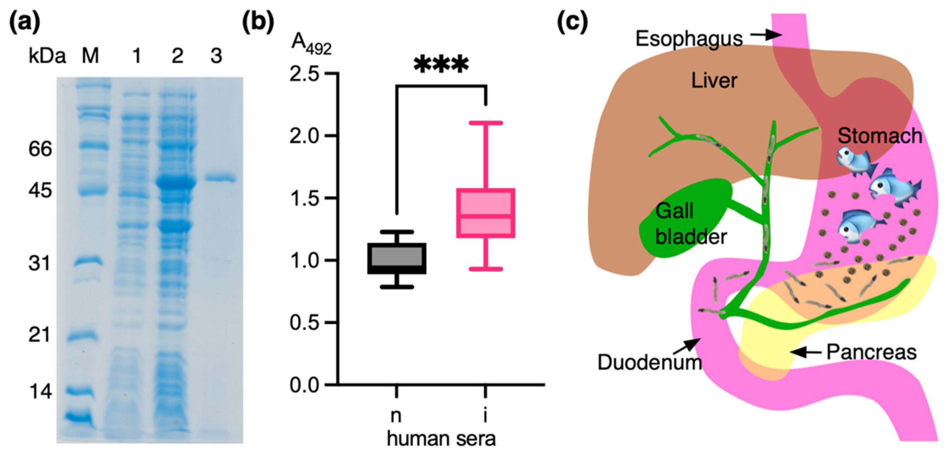
2.3. Expression and Purification of Recombinant OvSIS

The cDNA insert encoding OvSIS was released from the pGEM®-T Easy vector using the introduced NheI and XhoI restriction endonuclease sites and inserted into the expression vector pET21b (Novagen, Darmstadt, Germany) by ligation. The cloning procedure led to the addition of amino acids LEHHHHHH at the C-terminus of recombinant OvSIS, and the inserted sequence was verified again by sequencing, as detailed above. Escherichia coli BL-21(DE3) pLysS was used as the expression host. Following chemical transformation, single positive colonies were identified by using colony PCR and restriction endonuclease digestion. Single positive colonies were inoculated in 5 mL of Luria-Bertani (LB) medium supplemented with ampicillin (100 µg/mL), chloramphenicol (34 µg/mL), kanamycin (12.5 µg/mL), and tetracycline (3.125 µg/mL). The cultures were incubated by shaking at 37 °C overnight. Briefly, 5 mL of starter culture was added to 100 mL of fresh medium with antibiotics and incubated at 37 °C and 250 rpm until the OD600 reached ~0.5–0.6. Then, 1 mM isopropyl-β-D-1-thiogalactopyranoside (Fermentas, Vilnius, Lithuania) was used to induce the expression of recombinant OvSIS. Four hours after induction, the bacterial cells were harvested by centrifugation at 6000× g and 4 °C for 30 min. The recombinant protein was purified by Ni-NTA affinity chromatography (QIAGEN, Hilden, Germany) under denaturing conditions following the manufacturer’s instructions. The purified protein was refolded by stepwise dialysis in urea to obtain soluble and active serpin. In detail, 1 mL of eluate was transferred into a dialysis bag (Spectra/Por 3 RC Dialysis Tubing, MWCO 3.5 kD, Spectrum Laboratories, Repligen, Rancho Dominguez, CA, USA), and the closed bag was placed in 1000 mL of 6 M urea, 20 mM Tris-HCl (pH 8.0), and 0.1 mM DTT and incubated with slow stirring at 4 °C for 3 h. This step was repeated and then followed by two incubation steps each at 4 and 2 M urea at 4 °C for 3 h. Afterwards, the protein solution was incubated in 20 mM Tris-HCl (pH 8.0) at 4 °C for 3 h. Following a final change in the buffer, the incubation was continued overnight at 4°C. The protein solution was centrifuged at 5000× g and 4 °C for 10 min, and the supernatant was stored at −20 °C. No precipitates were observed during dialysis and the subsequent freeze/thaw steps when the refolded protein was used in further experiments. The protein concentration was determined by a Bradford assay (Bio-Rad Laboratories, Hercules, CA, USA) and showed that 2.24 mg rOvSIS was obtained. Standard 12.5% SDS-PAGE in 0.025 M Tris-HCl (pH 8.9), 0.2 M glycine, and 0.1% (w/v) SDS running buffer was used to analyze the purity of the refolded protein. The samples (pre- and 4-h-induced bacterial protein equivalent to ~80 µL culture volume, 1 µg refolded rOvSIS) were mixed at a ratio of 1:1 with 0.125 M Tris-HCl (pH 6.8), 0.2 M DTT, 0.02% (w/v) bromophenol blue, 4% (w/v) and SDS, 20% (v/v) sterile glycerol, heated at 95 °C for 5 min, cooled on ice for 1 min, and then added to the wells. The Broad Range Molecular Weight Standards (Bio-Rad Laboratories, Hercules, CA, USA) were run in parallel at an amount of ~10 µg and used to calculate the molecular weight of rOvSIS. The proteins were separated at 20 mA constant current until the front dye had moved to the bottom of the gel. The stacking gel was washed in distilled water, submerged in staining solution (0.05% Coomassie Blue G-250, 35 mM HCl), heated in a microwave oven for 20 s, and incubated with gentle shaking overnight. The gel was destained in distilled water, and an image was captured on a flatbed scanner (CanoScan LiDE 700F, Canon, Bangkok, Thailand).

2.4. Reverse Transcriptase PCR Analysis

Total RNA was extracted from NEJs, 2-week juveniles, and 6-week mature O. viverrini and treated with DNase. Briefly, 200 ng of RNA from each stage were used for reverse transcription with RevertAid reverse transcriptase (Thermo Scientific, Vilnius, Lithuania) and the gene-specific reverse primer 5′-CATGCATCATTGGGACATTC-3′. Conventional PCR was then performed under the conditions described in Section 2.2 with the gene-specific forward primer 5′-AGTCGCACTCCCTTAGATTG-3′ and the above-mentioned reverse primer to produce a 640 bp PCR product. O. viverrini actin (OvActin) was used as an internal control and amplified with primers 5′-TCGAGAGAGATGACACAGA-3′ and 5′-GATATCACGCACGATTTCTC-3′ to produce a 292 bp product. The RT-PCR products were resolved on a 1% agarose gel.

2.5. Phylogenetic Analysis of Serpins in the Genera Opisthorchis/Clonorchis and Schistosoma

Forty-six full-length trematode serpin sequences were fetched from UniProt (https://www.uniprot.org (accessed on 8 June 2024)). They included ten sequences from O. felineus, seven sequences from O. viverrini, nine sequences from C. sinensis, six from S. haematobium, five from S. japonicum, and nine sequences from S. mansoni. The sequences were aligned in Muscle v5 [16]. Trimal v1.4 [18] was used with the automated1 option to prepare the aligned sequences for phylogenetic analysis. PhyML v3.3.20220408 [19] was used for analysis under the LG model, with a starting BioNJ tree and Bayes support.

2.6. Indirect ELISA of Human Opisthorchiasis Sera with Recombinant OvSIS

Indirect ELISA was used to determine the reactivity of human opisthorchiasis sera against recombinant OvSIS (rOvSIS). The human sera used in this study were collected in Thailand and were classified as normal or infected based on the microscopic investigation of stool samples for parasite eggs [20]. First, 100 ng of rOvSIS I2 in 100 µL coating buffer (30 mM Na2CO3 and 75 mM NaHCO3) were transferred to the wells of a 96-well microtiter plate (NUNC, Roskilde, Denmark) and incubated at 37 °C overnight. The next day, the wells were washed with 10 mM PBS (pH 7.2) and 0.05% Tween 20. Non-specific binding was blocked with a 0.25% BSA blocking solution. Thirty human opisthorchiasis sera were randomly assigned to three groups of 10 samples each, and a group of ten normal human sera was used as a negative control. At first, the sera (diluted 1:100) were tested as pooled sera (n = 10), and when found to be reactive with rOvSIS, they were tested individually. Each of the forty serum samples was diluted to 1:100 and added to a well. The plate was incubated at 37 °C for 1 h. Polyclonal rabbit anti-human IgG HRP conjugated antibody (Dako, Glostrup, Denmark) at 1:6000 dilution was added as a secondary antibody and incubated at 37 °C for 1 h. The substrate o-phenylenediamine dihydrochloride (OPD, Sigma-Aldrich, St. Louis, MO, USA) was used for colorimetric detection. The reaction was stopped with 3 M H2SO4 solution, and absorption was measured at 492 nm on a plate reader (VarioSkan Flash, Thermo Scientific, Waltham, MA, USA). The assay was performed in duplicate. Statistical analysis of the data by an unpaired t-test was performed in Prism version 10 (GraphPad Software, LLC, Boston, MA, USA). The ethics approval is found in the Institutional Review Board Statement.

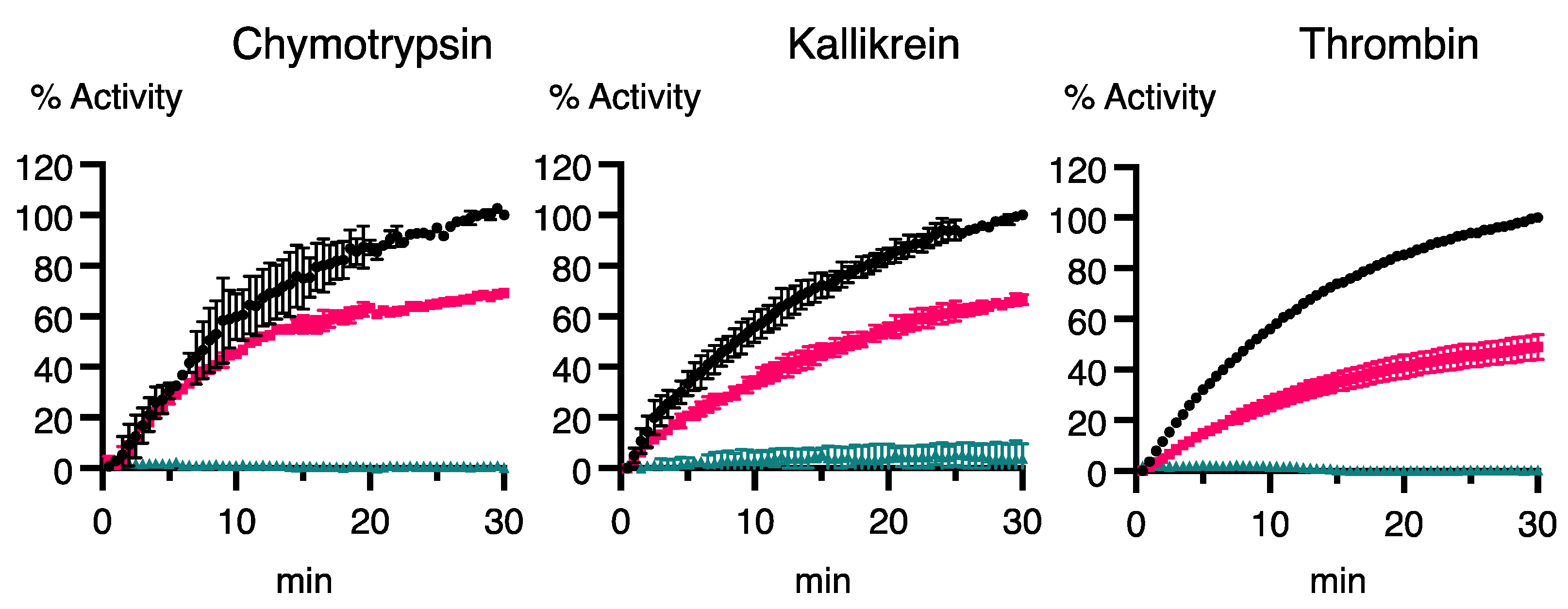
2.7. Inhibition of Human Serine Proteases by rOvSIS

The inhibition properties of rOvSIS were tested with native human serine proteases. The residual proteolytic activity was determined after incubating the serine proteases with rOvSIS in the presence of protease-specific fluorogenic substrates in black flat-bottom microtiter plates (Thermo Scientific, Roskilde, Denmark). Details of the used protease and substrate concentrations and buffer composition are shown in Table 1. Sources of proteases and substrates: chymotrypsin from the human pancreas (C8946, Sigma-Aldrich, St. Louis, MO, USA) with substrate Suc-Ala-Ala-Pro-Phe-MCA (3114-v; Peptide Institute Inc, Ibaraki-Shi, Japan), thrombin from human plasma (T6884, Sigma-Aldrich, St. Louis, MO, USA) with substrate Boc-Val-Pro-Arg MCA (B9385, Sigma-Aldrich, St. Louis, MO, USA), and kallikrein from human plasma (K2638, Sigma-Aldrich, St. Louis, MO, USA) with substrate Z-Phe-Arg-MCA (C9521, Sigma-Aldrich, St. Louis, MO, USA). Each serine protease was incubated with 0.5 µM rOvSIS in 100 µL of buffer at 37 °C for 30 min. Then, 100 µL substrate solution was added, and enzyme activity was recorded for 30 min. PMSF at a concentration of 20 mM was used as a positive control inhibitor. Measurements were carried out in duplicate, and the experiment was repeated twice. The residual enzyme activity was measured by monitoring the release of fluorescence with excitation at 355 nm and emission at 460 nm on a Varioskan Flash spectral scanning multimode reader (Thermo Scientific, Waltham, MA, USA). Statistical analysis and graphical representation of the data were performed in Prism version 10 (GraphPad Software, LLC, Boston, MA, USA).

3. Results

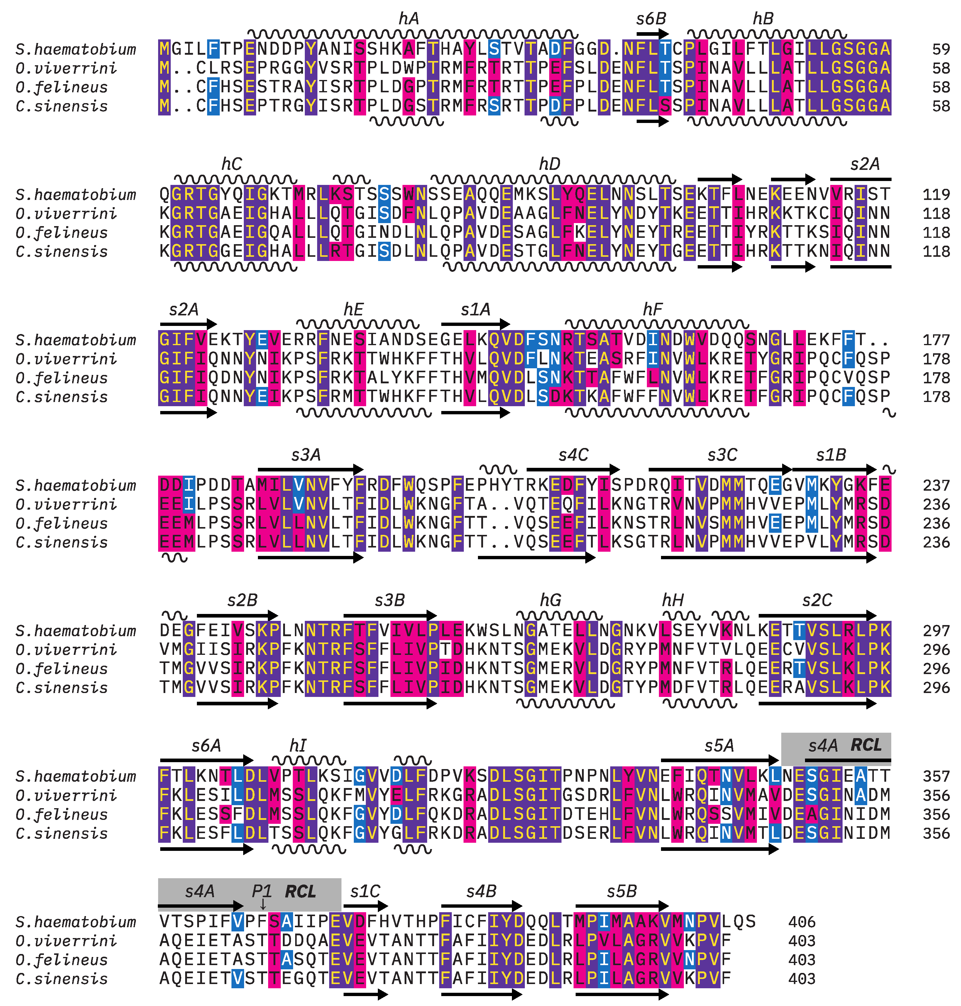
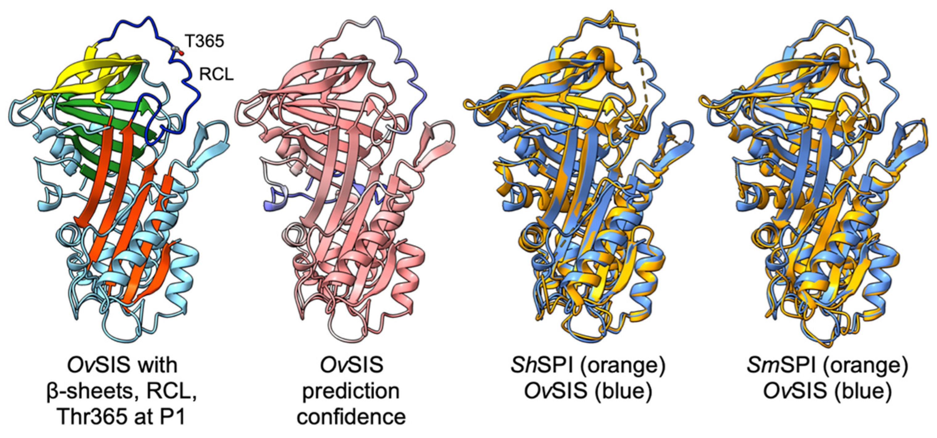
3.1. Molecular Cloning of OvSIS and Analysis of Sequence and Structure

This is the first analyzed serine protease inhibitor (serpin) of Opisthorchis viverrini. The sequences of hypothetical serpins of O. viverrini were identified by using NCBI BLASTP to search the relevant databases at NCBI GenBank. A total of 48 sequences predicted from the genome data of O. viverrini (BioProject: PRJNA259756 [14]; BioProject: PRJNA230518, Mitreva et al., unpublished) were found. From this set of hypothetical serpins, XP_009169304 (UniProt: A0A075AEP2) was selected for analysis because (1) the sequence was complete, and (2) it was highly different in its sequence to previously analyzed serpins in the closely related trematode Clonorchis sinensis [9,10,12]. In support of this predicted protein, highly conserved orthologous serpins were found by BLAST searches in C. sinensis (UniProt: A0A419PW56) and O. felineus (UniProt: A0A4S2MGE0), as shown in Table 2. The O. felineus sequence originates from a transcriptome project and, thereby, supports the conclusion that this protein is indeed produced. No developmental stage was specified for the O. felineus transcript.
Total RNA was isolated from various developmental stages (metacercariae, newly excysted juveniles, juveniles, and mature parasites) of O. viverrini and used for reverse transcriptase PCR with a gene-specific primer pair to obtain the coding sequence of O. viverrini A0A075AEP2. A PCR product was obtained from metacercarial and NEJ-stage RNA, and the sequencing demonstrated that the correct 1212 bp sequence from the start codon to the stop codon had been amplified. The deduced 403-residue amino acid sequence had a calculated molecular weight of 46 kDa and did not contain a signal peptide. Due to the finding that the transcript was present in the human infectious stage but not in later stages, the protein was termed O. viverrini Serpin Infectious Stage (SIS, OvSIS). The nucleotide sequence of OvSIS is available in the EMBL, GenBank, and DDJB databases under the accession number PP968418.
Further BLAST searches for homologous proteins in more distantly related trematode species resulted in matches to a hypothetical Fasciola hepatica serpin (UniProt: A0A4E0RBU2, 35% identity, 56.7% similarity), schistosome SPI (serine protease inhibitor), S. mansoni SPI (G4LYU6, 32.8% identity, 56.0% similarity), S. japonicum SPI (C1LNS7, 34.0% identity, 54% similarity), and S. haematobium SPI (UniProt: Q26502, 31.8% identity, 53.3% similarity). Using BLAST to compare OvSIS to human serpins, it was found that those clustered into sub-family B showed higher sequence similarity than those of sub-family A, e.g., the human leukocyte elastase inhibitor, i.e., LEI (UniProt: P30740, 21.5% identity, 37.6% similarity, 10.6% gaps). This does not imply that these serpins have equivalent biological function(s) in humans and trematodes. Human serpin B members as well as all discussed trematode serpins lack a signal peptide. Schistosome SPI was selected for comparison to OvSIS because it is, by sequence, the closest described trematode serpin and its structure has already been analyzed [21,22]. A multiple alignment of ShSPI and Opisthorchis/Clonorchis SIS is shown in Figure 1.
The multiple alignment (Figure 1) shows that the overall secondary structure, with its α-helical and β-strand content, is conserved between the parasite serpins. This is also evident in the tertiary structure model of OvSIS compared to the experimentally resolved structures of S. haematobium and S. mansoni SPI [21,22] (Figure 2). The extended helix D of schistosome SPI [20] is also found in SIS. The AlphaFold prediction confidence values of OvSIS were low at the N-terminal end up to F32 and in the reactive center loop (RCL), as shown in Figure 2.

3.2. Phylogenetic Analysis of Serpins in the Genera Opisthorchis/Clonorchis and Schistosoma

As mentioned before, OvSIS was selected based on its high sequence divergence from the three previously reported C. sinensis serpins. These serpins show similar low sequence conservation with OvSIS, as does human LEI at around 19–22% identity and 37–40% similarity. Indeed, the phylogenetic tree calculation placed all analyzed C. sinensis serpins on a different branch from OvSIS (Figure 3a). OvSIS and the orthologous C. sinensis and O. felineus SIS are more closely related but not orthologous to schistosome SPI. The SPI orthologs are potentially O. viverrini A0A074ZHZ0, C. sinensis G7Y5W4, and O. felineus A0A4S2MGT0, as can be seen in the constructed tree and the sequence alignment of their reactive center loop in Figure 3a,b.

3.3. Developmental Expression of OvSIS RNA

Transcripts of the OvSIS gene (NCBI GenBank Gene: 20320087) were detected by RT-PCR in newly excysted juveniles but not in later juveniles or mature flukes (Figure 4a). Originally, we had cloned the OvSIS cDNA from metacercarial RNA. This suggests that the protein is already produced in metacercariae, and the metacercarial presence of RNA has also been reported in previous analyses of serpins in C. sinensis. All three analyzed C. sinensis serpins showed a wider expression range at the RNA level than OvSIS up to the mature stage but, interestingly, the amounts of RNA of A0A059VDA6 and E2DHH4 were much higher in metacercariae than in the mature fluke [10,12]. It can be assumed that these serpins are required in large amounts during the infection process. Whether they have a redundant function or if they prefer unique serine proteases as targets remains to be determined. Metacercariae and newly excysted juveniles represent the infectious stage of the parasite for its human host. As shown in Figure 4b, these developmental stages are of microscopic size. The excretory bladder is filled with dark pigment granules. In the lung fluke Paragonimus westermani, cysteine proteases were located on these granules and speculated to be released for excystment [28].

3.4. Analysis of Human Opisthorchiasis Sera Against Recombinant OvSIS

Recombinant OvSIS was expressed as an insoluble protein in E. coli. After purification of the urea-solubilized protein by Ni-NTA affinity chromatography (Figure 5a), it was refolded by stepwise dialysis and tested with the immune sera of thirty individuals naturally infected with the parasite; a positive reaction was observed (Figure 5b). An unpaired t-test between normal and infected sera showed a statistically significant difference with a p-value of 0.0003. Mean ± standard deviation absorbance values of normal and infected sera were 0.9900 ± 0.1474 and 1.386 ± 0.2990. As schematically shown in Figure 5c, the metacercariae are released when infested fish is digested in the stomach. Gastric juices then stimulate excystation, and once released from the stomach, the newly excysted juveniles (NEJs) must migrate to the sphincter of Oddi/ampulla of Vater to reach the biliary system. Therefore, OvSIS must be released from the start of excystation and stimulate an immune response in the contacted areas of the gastrointestinal tract and biliary system for several days before gene expression is stopped.

3.5. Inhibition of Human Serine Proteases by Recombinant OvSIS

Native human plasmas kallikrein, thrombin, and chymotrypsin were tested for inhibition by rOvSIS (Figure 6). Thrombin was the best inhibited protease at about 50% reduced activity followed by plasmas kallikrein (33%) and chymotrypsin (30%). As only a small number of proteases could be tested, it should just be concluded that OvSIS is an active serpin. Based on the OvSIS sequence TASTTDDQ around the P1 site, low inhibition against these three proteases would be expected, but that would also be the case for trypsin and plasmin. The analysis of this sequence with PeptideCutter [29] (https://web.expasy.org/peptide_cutter/ (accessed on 8 June 2024)) did not result in a hit with any serine protease.

4. Discussion

In this investigation, a serpin of the human liver fluke Opisthorchis viverrini was characterized with respect to its sequence, structure, phylogeny, and antigenicity. Due to its presence in the infectious stage, it was termed OvSIS, an acronym for ‘Serpin Infectious Stage’. It represents a new member of the serpin family in the trematodes because it is not orthologous to any of the previously described serpins in this lineage. Based on the phylogenetic analysis shown in Figure 3a, it is in a clade that also contains schistosome SPI, from which it seems to be separated by a few gene duplication events, i.e., it is not an ortholog of SPI. SPI was first isolated and described in S. haematobium as an antigenic serpin that was recognized by the sera of schistosome-infected individuals and located on the surface of mature flukes [30]. The antigenicity of ShSPI was confirmed with the finding of specific IgG4 and IgE responses against the protein in infected individuals [31]. Later, the orthologs of S. japonicum [32] and S. mansoni [33] were described, with comparable findings of tegumental localization and antigenic properties. In this study, OvSIS was found to act as an antigen and to cause a humoral immune response in infected individuals (Figure 5).
In addition to the close phylogenetic relationship and higher sequence conservation, OvSIS shares structural features with SPI, especially the extended helix D [21]. On the other hand, the sequence of the reactive center loop (RCL) around and including the residues that form the reactive bond, which is important for the recognition and interaction with serine proteases, was found to be very different (Figure 3b). The presence of threonine, together with serine at P1 and P2 positions, is unusual, and the observed limited inhibition of chymotrypsin, kallikrein, and thrombin seems to reflect this (Figure 6). Other human serine proteases should be tested in further studies. In addition, the analysis results of schistosome cercarial elastases suggest that the OvSIS sequence from P4 to P4′ TASTTDDQ would not be a preferred substrate of those either [34]. Thus, a specific metacercarial/juvenile serine protease might exist in the genus Opisthorchis/Clonorchis, which is the native target of OvSIS, and it is possible that the inhibitor acts on certain cysteine proteases as they are also abundant in these flukes in the infectious stage. Serine proteases have not yet been characterized in the genus Opisthorchis/Clonorchis, and GenBank contains only a small number of predicted membrane-bound and soluble serine protease sequences.
Transcripts of OvSIS were detected in the metacercarial/newly excysted juvenile stages of the parasite but not in 2-week-old juveniles or mature flukes. Therefore, OvSIS must have a time-limited function during the short developmental period, important for the infection process in the human host. Interestingly, SmSPI showed a significant upregulation in 2-h-old schistosomules compared to cercariae and mature flukes [33]. In S. japonicum, schistosomules were not tested, but a 1500:1 ratio of SjSPI RNA in mature flukes compared to cercariae was reported [32]. As mentioned in the Introduction Section, unrelated serpins characterized in C. sinensis showed varying transcript amounts in different developmental stages too [9,10,11,12]. Differential gene expression during development can occur due to various reasons; it could occur simply because the same target (serine protease) is present at different amounts or that the inhibitor has different/additional functions, for example, as reported in the eggs of mature worms.
Overall, OvSIS is interesting because of its specific presence during the early infection stage, and further investigation of its role during this period could help to better understand the steps of infection and, possibly, how to interfere with this process. It is also important to determine how it stimulates an immune response, i.e., whether it is secreted even without a classical signal peptide and whether it is circulating or attached to the tegument, as observed for schistosome SPI and other trematode serpins. Due to travel restrictions during the recent COVID-19 epidemic, it was not possible to collect sufficient early-stage parasites in this study to perform these analyses. With respect to the biochemical properties of OvSIS, the inhibition profile shows that it could interfere with fibrinolysis as it was found to moderately inhibit thrombin and kallikrein (with low efficiency). However, is that important considering that the juvenile parasite moves quickly and directly into the biliary system and does not feed on blood, even as a mature fluke? Further research is required to identify the protease that OvSIS inhibits in order to better understand its function.

Author Contributions

Conceptualization, R.G.; methodology, R.S., W.P. and A.G.-K.; software, R.G.; validation, R.G. and W.P.; formal analysis, R.S.; investigation, R.S.; resources, A.G.-K. and W.P.; data curation, R.S.; writing—original draft preparation, R.G.; writing—review and editing, R.G.; visualization, R.G.; supervision, R.G.; project administration, R.G., A.G.-K. and W.P.; and funding acquisition, R.G. All authors have read and agreed to the published version of the manuscript.

Funding

This research was supported by the Thailand Science Research and Innovation Fundamental Fund (TUFF12/2564 and TUFF37/2565) and the Thammasat University Research Unit in Parasitic Diseases. The APC was funded by the Thailand Science Research and Innovation Fundamental Fund and the Thammasat University Research Unit of Parasitic Diseases.

Institutional Review Board Statement

The use of human sera in this study was conducted in accordance with the Declaration of Helsinki and approved by the Institutional Ethics Committee of Thammasat University (protocol code 189/2563 from 30 December 2020), with an exemption for the reason of “existing human serum samples used in secondary research”. The original informed consent and ethics statements obtained for these sera can be found in Geadkaew-Krenc et al., 2020 [20]. The animal study protocol was approved by the Institutional Animal Care and Use Committee of the Thammasat University (protocol code 001/2020 from 8 April 2020) in accordance with the Ethics of Animal Experimentation of the National Research Council of Thailand.

Informed Consent Statement

Patient consent was not applicable in this study. It was obtained in a previous study [20], and the present research was approved with exemption, as detailed in the Institutional Review Board Statement.

Data Availability Statement

The nucleotide sequence of OvSIS is available under GenBank accession PP968418. Other data supporting the findings of this study are available from the corresponding author upon request.

Acknowledgments

We would like to thank Nanthawat Kosa and Kornkanok Nakudom (Faculty of Allied Health Sciences, Thammasat University) and Smarn Tesana (Faculty of Medicine, Khon Kaen University) for their help with collecting O. viverrini metacercariae and other materials for this study.

Conflicts of Interest

The authors declare no conflicts of interest. The funders had no role in the design of this study, the collection, analyses, or interpretation of the data, the writing of the manuscript, or the decision to publish the results.

References

  1. Sripa, B.; Brindley, P.J.; Mulvenna, J.; Laha, T.; Smout, M.J.; Mairiang, E.; Bethony, J.M.; Loukas, A. The tumorigenic liver fluke Opisthorchis viverrini—Multiple pathways to cancer. Trends Parasitol. 2012, 28, 395–407. [Google Scholar] [CrossRef]
  2. Brindley, P.J.; da Costa, J.M.; Sripa, B. Why does infection with some helminths cause cancer. Trends Cancer 2015, 1, 174–182. [Google Scholar] [CrossRef] [PubMed]
  3. Heit, C.; Jackson, B.C.; McAndrews, M.; Wright, M.W.; Thompson, D.C.; Silverman, G.A.; Nebert, D.W.; Vasiliou, V. Update of the human and mouse SERPIN gene superfamily. Hum. Genom. 2013, 7, 22. [Google Scholar] [CrossRef]
  4. Molehin, A.J.; Gobert, G.N.; McManus, D.P. Serine protease inhibitors of parasitic helminths. Parasitology 2012, 139, 681–695. [Google Scholar] [CrossRef]
  5. Molehin, A.J.; Gobert, G.N.; Driguez, P.; McManus, D.P. Functional characterization of SjB10, an intracellular serpin from Schistosoma japonicum. Parasitology 2014, 141, 1746–1760. [Google Scholar] [CrossRef] [PubMed]
  6. De Marco Verissimo, C.; Jewhurst, H.L.; Tikhonova, I.G.; Urbanus, R.T.; Maule, A.G.; Dalton, J.P.; Cwiklinski, K. Fasciola hepatica serine protease inhibitor family (serpins): Purposely crafted for regulating host proteases. PLoS Negl. Trop. Dis. 2020, 14, e0008510. [Google Scholar] [CrossRef]
  7. Sánchez Di Maggio, L.; Tirloni, L.; Uhl, M.; Carmona, C.; Logullo, C.; Mulenga, A.; da Silva Vaz, I.; Berasain, P. Serpins in Fasciola hepatica: Insights into host-parasite interactions. Int. J. Parasitol. 2020, 50, 931–943. [Google Scholar] [CrossRef] [PubMed]
  8. Becerro-Recio, D.; Serrat, J.; López-García, M.; Molina-Hernández, V.; Pérez-Arévalo, J.; Martínez-Moreno, Á.; Sotillo, J.; Simón, F.; González-Miguel, J.; Siles-Lucas, M. Study of the migration of Fasciola hepatica juveniles across the intestinal barrier of the host by quantitative proteomics in an ex vivo model. PLoS Negl. Trop. Dis. 2022, 16, e0010766. [Google Scholar] [CrossRef]
  9. Yang, Y.; Hu, D.; Wang, L.; Liang, C.; Hu, X.; Wang, X.; Chen, J.; Xu, J.; Yu, X. Molecular cloning and characterization of a novel serpin gene of Clonorchis sinensis, highly expressed in the stage of metacercaria. Parasitol. Res. 2009, 106, 221–225. [Google Scholar] [CrossRef]
  10. Kang, J.M.; Sohn, W.M.; Ju, J.W.; Kim, T.S.; Na, B.K. Identification and characterization of a serine protease inhibitor of Clonorchis sinensis. Acta Trop. 2010, 116, 134–140. [Google Scholar] [CrossRef]
  11. Lei, H.; Tian, Y.; Chen, W.; Wang, X.; Li, X.; Mao, Q.; Sun, J.; Li, R.; Xu, Y.; Liang, C.; et al. The biochemical and immunological characterization of two serpins from Clonorchis sinensis. Mol. Biol. Rep. 2013, 40, 3977–3985. [Google Scholar] [CrossRef] [PubMed]
  12. Yang, Y.; Hu, D.; Wang, L.; Liang, C.; Hu, X.; Xu, J.; Huang, Y.; Yu, X. Comparison of two serpins of Clonorchis sinensis by bioinformatics, expression, and localization in metacercaria. Pathog. Glob. Health 2014, 108, 179–185. [Google Scholar] [CrossRef] [PubMed]
  13. Geadkaew, A.; von Bülow, J.; Beitz, E.; Tesana, S.; Vichasri Grams, S.; Grams, R. Bi-functionality of Opisthorchis viverrini aquaporins. Biochimie 2015, 108, 149–159. [Google Scholar] [CrossRef] [PubMed]
  14. Young, N.D.; Nagarajan, N.; Lin, S.J.; Korhonen, P.K.; Jex, A.R.; Hall, R.S.; Safavi-Hemami, H.; Kaewkong, W.; Bertrand, D.; Gao, S.; et al. The Opisthorchis viverrini genome provides insights into life in the bile duct. Nat. Commun. 2014, 5, 4378. [Google Scholar] [CrossRef] [PubMed]
  15. Rice, P.; Longden, I.; Bleasby, A. EMBOSS: The European Molecular Biology Open Software Suite. Trends Genet. 2000, 16, 276–277. [Google Scholar] [CrossRef]
  16. Edgar, R.C. MUSCLE: Multiple sequence alignment with high accuracy and high throughput. Nucleic Acids Res. 2004, 32, 1792–1797. [Google Scholar] [CrossRef] [PubMed]
  17. Teufel, F.; Almagro Armenteros, J.J.; Johansen, A.R.; Gíslason, M.H.; Pihl, S.I.; Tsirigos, K.D.; Winther, O.; Brunak, S.; von Heijne, G.; Nielsen, H. SignalP 6.0 predicts all five types of signal peptides using protein language models. Nat. Biotechnol. 2022, 40, 1023–1025. [Google Scholar] [CrossRef] [PubMed]
  18. Capella-Gutiérrez, S.; Silla-Martínez, J.M.; Gabaldón, T. trimAl: A tool for automated alignment trimming in large-scale phylogenetic analyses. Bioinformatics 2009, 25, 1972–1973. [Google Scholar] [CrossRef] [PubMed]
  19. Guindon, S.; Dufayard, J.F.; Lefort, V.; Anisimova, M.; Hordijk, W.; Gascuel, O. New algorithms and methods to estimate maximum-likelihood phylogenies: Assessing the performance of PhyML 3.0. Syst. Biol. 2010, 59, 307–321. [Google Scholar] [CrossRef]
  20. Geadkaew-Krenc, A.; Grams, R.; Phadungsil, W.; Chaibangyang, W.; Kosa, N.; Adisakwattana, P.; Dekumyoy, P. Evaluation of Rhophilin Associated Tail Protein (ROPN1L) in the human liver fluke Opisthorchis viverrini for diagnostic approach. Korean J. Parasitol. 2020, 58, 475–479. [Google Scholar] [CrossRef]
  21. Granzin, J.; Huang, Y.; Topbas, C.; Huang, W.; Wu, Z.; Misra, S.; Hazen, S.L.; Blanton, R.E.; Lee, X.; Weiergraber, O.H. Three-dimensional structure of a schistosome serpin revealing an unusual configuration of the helical subdomain. Acta Crystallogr. D. Biol. Crystallogr. 2012, 68, 686–694. [Google Scholar] [CrossRef] [PubMed]
  22. De Benedetti, S.; Di Pisa, F.; Fassi, E.M.A.; Cretich, M.; Musicò, A.; Frigerio, R.; Mussida, A.; Bombaci, M.; Grifantini, R.; Colombo, G.; et al. Structure, Immunoreactivity, and In Silico Epitope Determination of SmSPI S. mansoni Serpin for Immunodiagnostic Application. Vaccines 2021, 9, 322. [Google Scholar] [CrossRef]
  23. Wu, R.; Ding, F.; Wang, R.; Shen, R.; Zhang, X.; Luo, S.; Su, C.; Wu, Z.; Xie, Q.; Berger, B.; et al. High-resolution de novo structure prediction from primary sequence. bioRxiv 2022. [Google Scholar] [CrossRef]
  24. Huber, R.; Carrell, R.W. Implications of the three-dimensional structure of alpha 1-antitrypsin for structure and function of serpins. Biochemistry 1989, 28, 8951–8966. [Google Scholar] [CrossRef]
  25. Beitz, E. TEXshade: Shading and labeling of multiple sequence alignments using LATEX2 epsilon. Bioinformatics 2000, 16, 135–139. [Google Scholar] [CrossRef] [PubMed]
  26. Varadi, M.; Anyango, S.; Deshpande, M.; Nair, S.; Natassia, C.; Yordanova, G.; Yuan, D.; Stroe, O.; Wood, G.; Laydon, A.; et al. AlphaFold Protein Structure Database: Massively expanding the structural coverage of protein-sequence space with high-accuracy models. Nucleic Acids Res. 2022, 50, D439–D444. [Google Scholar] [CrossRef]
  27. Pettersen, E.F.; Goddard, T.D.; Huang, C.C.; Meng, E.C.; Couch, G.S.; Croll, T.I.; Morris, J.H.; Ferrin, T.E. UCSF ChimeraX: Structure visualization for researchers, educators, and developers. Protein Sci. 2021, 30, 70–82. [Google Scholar] [CrossRef] [PubMed]
  28. Chung, Y.B.; Kim, T.S.; Yang, H.J. Early cysteine protease activity in excretory bladder triggers metacercaria excystment of Paragonimus westermani. J. Parasitol. 2005, 91, 953–954. [Google Scholar] [CrossRef]
  29. Gasteiger, E.; Hoogland, C.; Gattiker, A.; Duvaud, S.; Wilkins, M.R.; Appel, R.D.; Bairoch, A. Protein Identification and Analysis Tools on the ExPASy Server. In The Proteomics Protocols Handbook; Walker, J.M., Ed.; Humana Press: Totowa, NJ, USA, 2005; pp. 571–607. [Google Scholar]
  30. Blanton, R.E.; Licate, L.S.; Aman, R.A. Characterization of a native and recombinant Schistosoma haematobium serine protease inhibitor gene product. Mol. Biochem. Parasitol. 1994, 63, 1–11. [Google Scholar] [CrossRef]
  31. Li, Z.; King, C.L.; Ogundipe, J.O.; Licate, L.S.; Blanton, R.E. Preferential recognition by human IgE and IgG4 of a species-specific Schistosoma haematobium serine protease inhibitor. J. Infect. Dis. 1995, 171, 416–422. [Google Scholar] [CrossRef]
  32. Yan, Y.; Liu, S.; Song, G.; Xu, Y.; Dissous, C. Characterization of a novel vaccine candidate and serine proteinase inhibitor from Schistosoma japonicum (Sj serpin). Vet. Parasitol. 2005, 131, 53–60. [Google Scholar] [CrossRef] [PubMed]
  33. Pakchotanon, P.; Molee, P.; Nuamtanong, S.; Limpanont, Y.; Chusongsang, P.; Limsomboon, J.; Chusongsang, Y.; Maneewatchararangsri, S.; Chaisri, U.; Adisakwattana, P. Molecular characterization of serine protease inhibitor isoform 3, SmSPI, from Schistosoma mansoni. Parasitol. Res. 2016, 115, 2981–2994. [Google Scholar] [CrossRef] [PubMed]
  34. Salter, J.P.; Choe, Y.; Albrecht, H.; Franklin, C.; Lim, K.C.; Craik, C.S.; McKerrow, J.H. Cercarial elastase is encoded by a functionally conserved gene family across multiple species of schistosomes. J. Biol. Chem. 2002, 277, 24618–24624. [Google Scholar] [CrossRef] [PubMed]
Figure 1. Multiple sequence alignment of S. haematobium SPI (UniProt: Q26502), O. viverrini SIS (UniProt: A0A075AEP2), C. sinensis SIS (UniProt: A0A419PW56), and O. felineus SIS (UniProt: A0A4S2MGE0). The α-helical and β-strand regions of ShSPI and OvSIS based on their AlphaFold native state models [23] are indicated at the top and bottom, respectively. The numbering of the secondary structures follows α1-antitrypsin [24]. The region of the reactive center loop (RCL) with the P1 residue is indicated by a gray bar. Strand 4A in the RCL was manually added as it forms and integrates into β-sheet A after cleavage at P1. Sequence conservation using the BLOSUM 62 similarity matrix and ShSPI as the reference sequence is indicated by color shading (fully conserved: purple background, ≥50% conserved: blue background, and similar: red background). The graphical representation of the aligned sequences was created using TEXshade v1.28 [25].
Pathogens 13 00678 g001
Figure 2. OvSIS structure model predicted in AlphaFold [23] and obtained from the AlphaFold Protein Structure Database [26]. The OvSIS model with the three β-sheets A (red), B (green), and C (yellow), RCL (dark blue), and threonine 365 in the P1 position is shown on the left. The OvSIS model prediction confidence from high to low is indicated by red to blue shading, respectively. Overlays with ShSPI (PDB: 3STO) and SmSPI (PDB: 6SSV) demonstrate structural conservation between the three serpins. Graphical representations were created using ChimeraX [27].
Pathogens 13 00678 g002
Figure 3. (a) Phylogenetic tree of trematode serpins specified by their UniProt accession numbers. The three previously described serpins of C. sinensis are indicated with a trailing •. Schistosome serpins are shown in blue, with the SPI in bold lettering. OvSIS and its orthologs are shown in red. Opisthorchis/Clonorchis SIS is not orthologous to schistosome SIP. (b) Sequence conservation of the reactive center loop in evolutionarily close serpins (specified by their UniProt accession numbers) in the species O. viverrini (Ov), O. felineus (Of), C. sinensis (Cs), S. haematobium (Sh), S. japonicum (Sj), and S. mansoni (Sm). The P1 position in the protease interaction is indicated. The color shading indicates fully conserved: purple background, ≥50% conserved: blue background, and similar: red background.
Pathogens 13 00678 g003
Figure 4. (a) RT-PCR products resolved in 0.8% agarose gels. M: marker, 1: newly excysted juveniles, 2: 2-week-old juveniles, and 3: mature parasites. O. viverrini β-actin (292 bp) was used as a positive control. A 640 bp OvSIS cDNA was only produced with RNA of newly excysted juveniles. (b) Micrograph of a newly excysted O. viverrini juvenile (top) and a metacercaria (bottom), the developmental stages in which OvSIS is present. The dark granule-filled excretory bladder, which can also be seen through the cyst wall, is the most prominent. Other outstanding morphological features are the oral and ventral suckers. It is thought that parasite-secreted serine proteases participate in the release of the encysted juveniles and that cognate serpins regulate their excess activity.
Pathogens 13 00678 g004
Figure 5. (a) SDS-PAGE showing pre-induced (1) and IPTG-induced bacterial proteins (2) and affinity-purified recombinant OvSIS (3). The protein migrates at the expected 46 kDa molecular weight. (b) Absorption values of two groups of human sera against recombinant OvSIS obtained by indirect ELISA. n: normal sera (n = 10). i: sera of O. viverrini-infected individuals (n = 30). The minimum, 25th percentile, median, 75th percentile, and maximum are indicated in the box plots. The star symbols indicate that all infected sera were statistically significantly different (*** p < 0.05) from the normal sera. (c) Human infection with O. viverrini. The metacercariae are released from infested undercooked fish in the stomach. The acidic environment activates the excystment of the juveniles. The newly excysted juveniles are released with the stomach content into the duodenum and migrate via the sphincter of Oddi into the biliary system (green).
Pathogens 13 00678 g005
Figure 6. Inhibition kinetics of OvSIS against human serine proteases. Proteases without inhibitors are indicated in black, proteases incubated with recombinant OvSIS are shown in red, and proteases incubated with 20 mM PMSF are shown in teal. After 30 min of preincubation with 0.5 µM recombinant OvSIS and 30 min of incubation with the substrate, the activities of 1 nM chymotrypsin, 1 nM kallikrein, and 0.1 U thrombin were lowered to 69.21, 66.76, and 48.84%, respectively. The enzyme substrates are listed in Table 1.
Pathogens 13 00678 g006
Table 1. Final concentrations of enzymes, substrates, and reaction buffers.
EnzymeSubstrateBuffer
1 nM Chymotrypsin15 μM Suc-Ala-Ala-Pro-Phe-MCA100 mM Tris-HCl pH 7.4, 10 mM CaCl2
1 nM Kallikrein30 μM Z-Phe-Arg-MCA100 mM Tris-HCl pH 7.4, 10 mM CaCl2, 0.3 M NaCl
0.1 U Thrombin30 μM Boc-Val-Pro-Arg-MCA100 mM Tris-HCl pH 7.4, 10 mM CaCl2, 0.3 M NaCl
Table 2. Sequence conservation, shown as the percent identity (bold) and similarity (italics) between orthologous serpins of O. viverrini (UniProt: A0A075AEP2), C. sinensis (UniProt: A0A419PW56), and O. felineus (UniProt: A0A4S2MGE0).
SpeciesO. viverriniC. sinensisO. felineus
O. viverrini-----83.982.1
C. sinensis90.1-----87.1
O. felineus88.892.1-----
Disclaimer/Publisher’s Note: The statements, opinions and data contained in all publications are solely those of the individual author(s) and contributor(s) and not of MDPI and/or the editor(s). MDPI and/or the editor(s) disclaim responsibility for any injury to people or property resulting from any ideas, methods, instructions or products referred to in the content.

