Article

Chemical Composition, Antifungal Activity, and Plant-Protective Potential of Rosa damascena Mill. Essential Oil Against Fusarium graminearum

1
Department of Molecular Biology and Genetics, Faculty of Sciences and Literature, Istanbul Yeni Yuzyil University, Cevizlibag, Istanbul 34010, Türkiye
2
Department of Pharmacognosy, Faculty of Pharmacy, Acıbadem University, Ataşehir, Istanbul 34758, Türkiye
3
Department of Pharmacognosy, Faculty of Pharmacy, Istanbul Yeni Yuzyil University, Cevizlibag, Istanbul 34010, Türkiye
4
Department of Life Technologies/Molecular Plant Biology, University of Turku, FI-20520 Turku, Finland
*
Author to whom correspondence should be addressed.
Pathogens 2025, 14(4), 383; https://doi.org/10.3390/pathogens14040383
Submission received: 7 March 2025 / Revised: 10 April 2025 / Accepted: 11 April 2025 / Published: 15 April 2025
(This article belongs to the Special Issue Current Research in the Control of Plant Pathogenic Fusarium Species)

Abstract

:
Fusarium graminearum is a common plant pathogen among cereals worldwide. The application of chemical antifungal compounds is the most frequently used method in controlling F. graminearum. However, its excessive use and the genomic plasticity of the fungal genome lead to increased resistance levels to these chemical antifungal compounds. In this context, plant-derived compounds might play a role in protecting against Fusarium head blight (FHB) and crown rot (CR) as an alternative. In this study, we aimed to examine the antifungal effects of an essential oil obtained from Rosa damascena Mill. on the plant pathogen F. graminearum using molecular and analytical methods. The chemical composition of the essential oil was determined by GC-MS. The half effective concentration (EC50) value of R. damascena essential oil (REO) for F. graminearum was determined as 604.25 µg mL−1. Water-soluble tetrazolium 1 (WST-1) analyses revealed that REO caused cytotoxicity in F. graminearum. The potential oxidative stress and autophagic cell death capacity of REO towards F. graminearum was revealed via gene expression analysis and fluorescence microscopy. It was also revealed that, due to the plant-protective effect of REO, the disease severity of treated plants decreased by up to 27.78% in juvenile wheat seedlings infected by F. graminearum. Our data show that R. damascena essential oil might be used as an alternative natural ingredient in the field of plant protection.

1. Introduction

Fusarium species cause various diseases, including fusarium head blight (FHB) and crown rot (CR) in cereals, especially wheat and barley. F. graminearum and F. culmorum have been reported as the most common causal agents for FHB and CR worldwide [1,2,3,4]. F. graminearum, regarded as a species complex with more than 10 members, has been shown to be the predominant causal agent of FHB in many regions throughout the world. F. graminearum isolates lead to devastating effects, including yield losses, significant economic impacts, and mycotoxin contamination [5]. F. graminearum isolates produce various types of mycotoxins, such as zearalenone, nivalenol (NIV), deoxynivalenol (DON), and their derivatives, and these mycotoxins may lead to serious health problems in humans and animals [6,7,8,9,10]. Due to the relatively high incidence of F. graminearum and its mycotoxins in stored wheat and barley samples, it is very important to combat Fusarium species.
For many years, fungicides have been used against fungal pathogens as the main approach and disease management strategy. However, these chemical compounds have detrimental effects on the environment, animals, and human health [11,12]. In addition, due to the presence of increasingly aggressive fungal pathogens that are resistant to fungicides, there is a rising demand to limit the use of chemicals as antifungal agents in the field of disease management. Therefore, it is crucial to identify alternative antifungal compounds or strategies to combat FHB and CR diseases. These include biocontrol agents such as Trichoderma spp. [13,14,15] and the use of organic extracts from plants [16,17,18,19]. In particular, the potential for the application of antagonistic fungal treatment (such as Clonostachys spp., Cladosporium spp., and Trichoderma spp.) and essential oil mixtures such as Combretum spp. and Ocimum spp. in plant management appears promising for the near future.
According to previous research [17,20,21], essential oils, as plant-based materials, can play an important role in plant disease management. Numerous studies have demonstrated in recent years that essential oils demonstrate antifungal properties and can reduce the growth of fungal mycelia. Methanol extracts of Combretum caffrum, Salix capensis, and Schotia latifolia plantlets originating from South Africa have shown potential as antibacterial and antifungal agents for more than five pathogenic microorganisms. Similarly, Cymbopogon citratus, Ocimum basilicum, and Ocimum gratissimum essential oil treatment resulted in a reduction in both fungal growth and mycotoxin production in Fusarium spp. [21,22,23,24]. In addition, in comparison to fungicides, essential oils, as natural products, have various advantages in terms of sustainability, their modes of action, and environmental toxicity [25].
Rosa spp. are known for their beauty and healing properties. Rosaceae (rose family) is a rich plant family that includes at least 100 genera and more than 2000 species. Studies related to Rosaceae have mainly focused on its morphological classification, hybridization, anatomical analysis, and ecotype characterization [26,27]. However, the number of studies related to the processing, metabolite extraction, antioxidant characteristics, and antimicrobial potential of Rosa spp. has increased recently [28,29,30]. In this study, we aimed to determine the composition of Rosa damascena Mill. essential oil from Turkey via GC-MS and to show and characterize the potential antifungal effects against a worldwide phytopathogenic fungus, the F. graminearum species complex, in different physiological and molecular tests.

2. Materials and Methods

2.1. Plant Material

R. damascena buds (1 kg) were purchased from a local market in Türkiye. The plant materials were identified by Assoc. Prof. Huseyin Servi. The essential oil of R. damascena buds was obtained using a previously described method [31]. Briefly, 100 g R. damascena buds were soaked in 1000 mL distilled water and then extracted by hydrodistillation for 3 h using a Clevenger apparatus. Here, 1 kg R. damascena buds produced 1 mL essential oil.

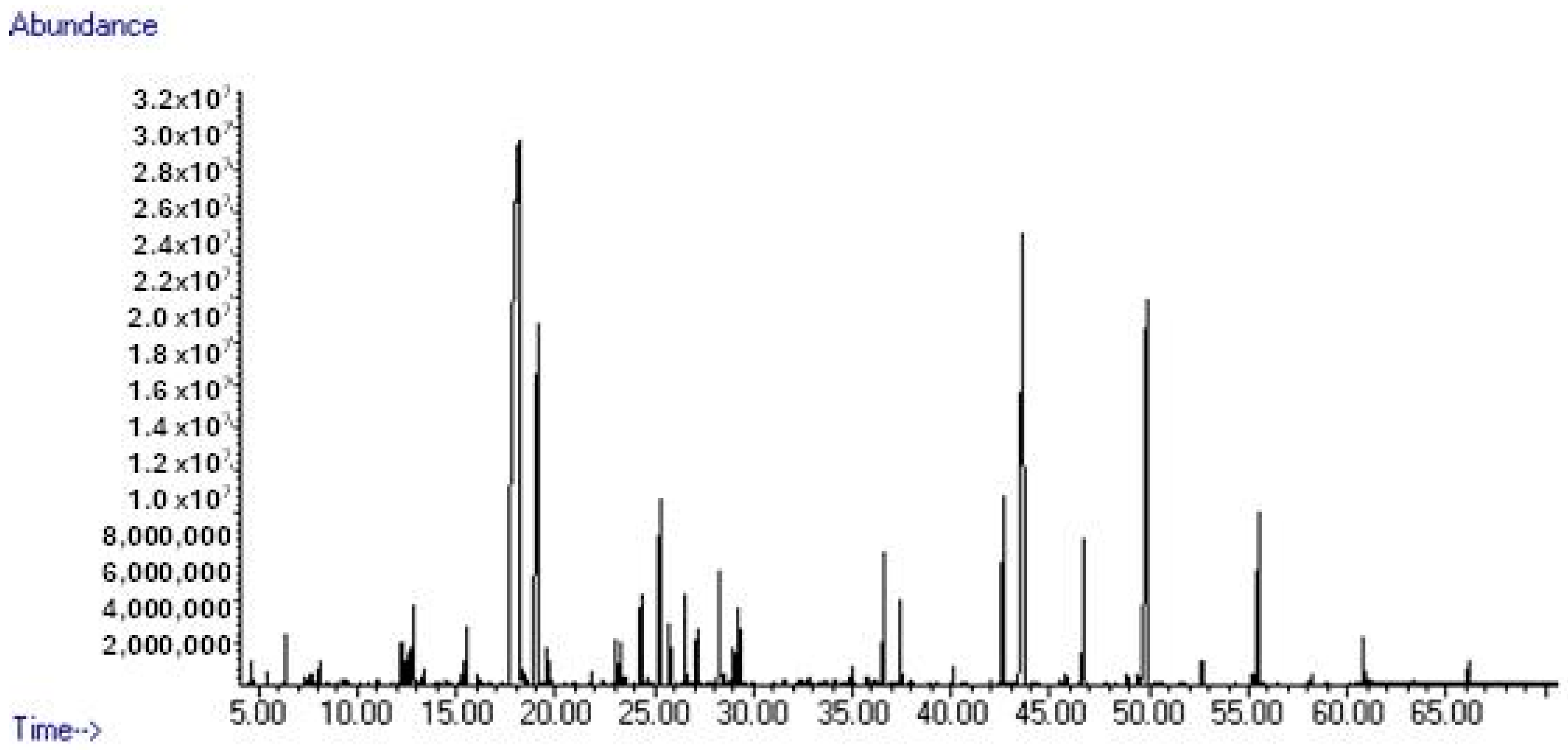
2.2. Gas Chromatography/Mass Spectrometry Analysis

The essential oil was analyzed by GC-MS using a non-polar column, HP-5MS (5% phenyl, 95% methyl polysiloxane; 30 m × 0.25 mm, 0.25 m film thickness). The oven temperature was programmed as follows: isothermal at 60 °C for 1 min and then increased to 246 °C at a rate of 3 °C min−1 and subsequently kept isothermal for 30 min. The carrier gas was helium, with a flow rate of 0.9 mL min−1. The essential oil was injected (1 μL) in split mode. The identification of the compounds was performed by comparing the relative retention indices of the n-alkane series to the literature and with a mass spectrum comparison (NIST17 Mass Spectra Library). The relative percentage quantities of the separated compounds were calculated from the integration of the peaks in MS chromatograms [32].

2.3. Evaluation of Antifungal Activity

Dr. Tapani Yli-Mattila of Turku University provided the F. graminearum PH-1 reference strain. The F. graminearum PH-1 strain was incubated at 26 ± 2 °C in 9-cm-diameter Petri dishes on potato dextrose agar (PDA, Sigma Aldrich, St. Louis, MO, USA) and carboxymethyl cellulose (CMC) for 7 days. The experimental sets were grown on PDA medium amended with different concentrations of REO (0, 250, 500, 750, and 1000 µg mL−1; Sigma-Aldrich, USA). Meanwhile, 0.25 cm2 mycelial sample obtained from fresh PDA medium were used for cultivation. The MIC and EC50 values were calculated as described before [33]. For this purpose, the linear growth rate (mm/day) was calculated by measuring the radial growth length (mm) values at the 4th and 7th days of incubation. The half effective and maximum effective suppressor concentrations were determined as half effective (EC50) and minimum inhibitory concentration (MIC) sets, respectively. The final formula for EC50 calculation was as follows: EC50 = x + ((50 − y1)/(y2 − y1))*(x + y)/2, where x indicates a low inhibitor concentration (inhibition lower than 50%), y indicates a high inhibitor concentration (inhibition higher than 50%), y1 indicates the inhibition value for x concentration, and y2 indicates the inhibition value for y concentration.

2.4. Proliferation Analysis

The cytotoxic action of REO at the cellular level was determined by water-soluble tetrazolium 1 (WST-1; Roche, Switzerland) analysis. A 1 cm2 disc from the control and experimental groups was dissolved in 1X phosphate-buffered saline (PBS). Then, 10 µL (1:10 V:V) WST-1 (ScienCell, Carlsbad, CA, USA) was added to microplates containing 90 µL of the cells, and then the fungal cells were incubated for 3 h at 28 °C in the dark. Fold changes in cell proliferation were calculated by spectrophotometric measurement at 450 and 620 nm wavelengths [34].

2.5. Fluorescence Microscopy Assay

To determine the presence of oxidative stress and autophagic cell death responses in the tested strain treated with REO, 2′,7′-dichlorofluorescein (DCF-DA; Thermo, Waltham, MA, USA) and monodansylcadaverine (MDC; Sigma Aldrich, USA) staining tests were carried out via fluorescence microscopy (Carl-Zeiss, Baden-Württemberg, Germany). Mycelial plugs obtained from 7-day-old PDA cultures were transferred to CMC medium. Then, liquid cultures were incubated on a rotary shaker for 120 rpm at 26 ± 2 °C. The 7-day-old liquid cultures were filtrated with 2X sterile gauze to remove the mycelium. The obtained 1 × 10⁵ macroconidium cultures were used in fluorescence microscopy analysis. Fixation, stain treatment, washing, and cell collection steps were carried out following previous procedures [20,35,36]. Quantitative assays were carried out using 100 cells per replicate (n = 3).

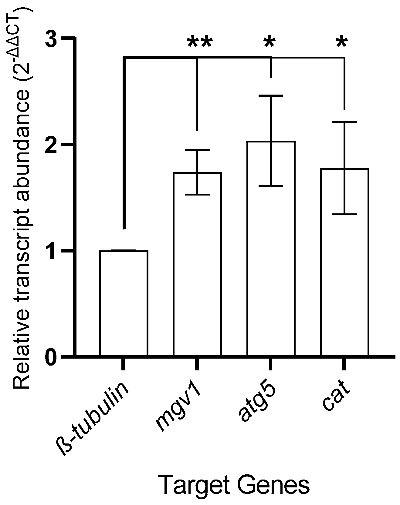
2.6. Real-Time Polymerase Chain Reaction (qRT-PCR) Assays

To analyze the changes in gene expression, qRT-PCR assays were conducted. For this purpose, total RNA was isolated from fungal samples via Triagent (Macherey-Nagel, Düren, Valencienner Str., Germany). The manufacturer’s recommendations were followed for the binding, washing, and eluting processes. After DNase I treatment (Zymo, Irvine, CA, USA), spectrophotometric (260/280 nm absorbance) measurements and agarose gel (0.8%) assays were used in order to reveal the quantity and quality of the total RNA. Then, the total RNA was converted to cDNA molecules via a commercial kit (Nepenthe, TürkiyeTurkey-Kocaeli), and the cDNA molecules were used in gene expression analysis. In the qRT-PCR analysis, β-tubulin (AY303689) was used as an endogenous gene. The autophagy protein 5 (atg5, FGRA07_10212) and catalase_3 (cat, FGSG_06733) genes were used as target genes, and they were associated with different physiological processes, i.e., the autophagy response and oxidative stress. Mitogen-activated protein kinase (mgv1, AF492766.1), related to sexual–asexual growth, was used as a positive calibration gene in order to identify potential alterations in gene expression. The primers used in this study are given in Table 1. The qRT-PCR assays were conducted with mixture including a volume of cDNA corresponding to 50 ng total RNA, 3 pmol of each primer, and 1X SYBR Green I Master Mix (Episozyme, Turkey), with the remaining volume consisting of nuclease-free water. The cycling conditions were as follows: 95 °C for 2 min (pre-denaturation); a loop with 40 repeats consisting of 95 °C for 10 s, 58 °C for 15 s, and 72 °C for 20 s; 40 °C for 30 s (cooling); and a melting step (Applied Biosystems, England). Fold changes in the gene expression values were calculated using the 2-ΔΔCT formula, which is presented as follows: 2−ΔΔCT = 2−[ΔCT sample − ΔCT control], where ΔCT is CTtarget − CTreference gene [37]. Each experiment was replicated with at least three biological replicates and two technical replicates.

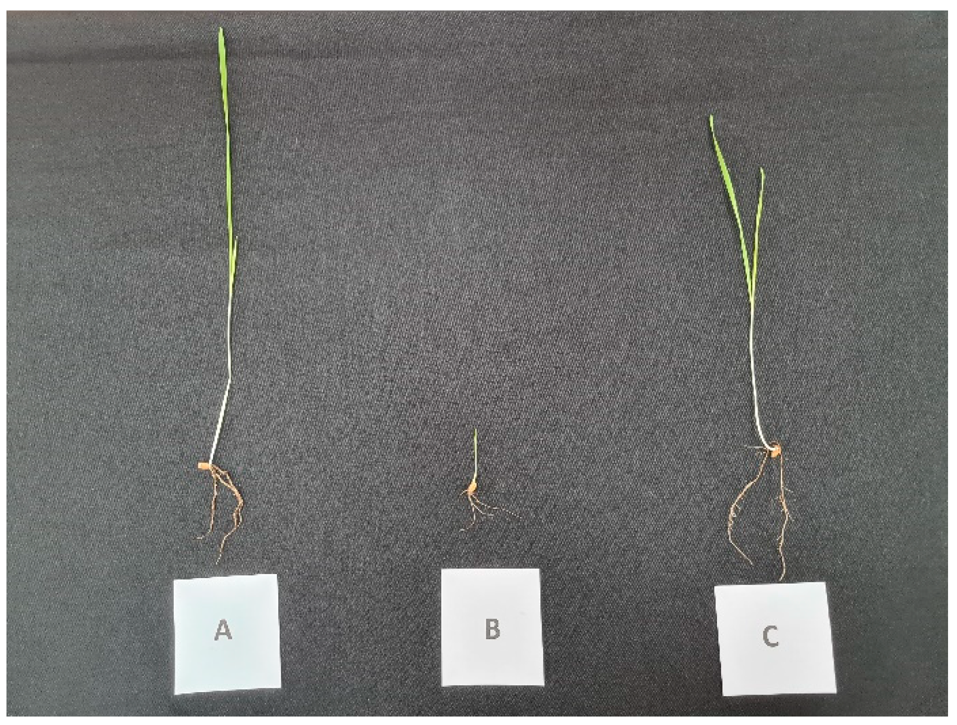
2.7. Infection Tests for Fusarium Crown Rot (FCR) Disease

Triticum aestivum L. cv. Lucilla seeds (kindly provided by the Seed Progen Company, Turkey) were used in plant protection tests for juvenile plantlets that were infected with the F. graminearum PH-1 strain. The seeds were firstly surface-sterilized with 0.64% NaOCl for 5 min, 10% ethanol for 5 min, and sterile dH2O at least three times. The seeds were transferred to filter papers inside Petri dishes; they were incubated at 26 °C (in the dark) for 3 days. CMC cultures were used as the inoculum source. Then, 1 × 10⁶ macroconidia were filtered through 2X gauze, and then they were added to autoclaved soil at a ratio of 5:95/W:V macroconidia–soil. The wheat seeds [38,39,40] were planted in 13-cm-diameter plastic pots containing soil with F. graminearum and incubated at 26 °C in a 16/8 h light/dark cycle with 1500 lux for two weeks. Plantlets were watered with distilled water (control set) or an REO solution. Juvenile plantlets were evaluated regarding the FCR disease scale, ranging from “0” to “4”, with three biological replicates each, including three technical replicates [41,42]. The disease severity as a percentage of infection was calculated according to the formula disease severity (percentage) = 100 × (Σ (n × V))/(Z × N), where “n” is the number of samples with different scores, “V” is the value for the scale, “Z” is the highest value of the sample scale, and “N” is the sample number.

2.8. Statistical Analysis

The column statistics, including the mean and standard error values, as well as normality tests and t-tests, were obtained using the software GraphPad Prism, version 9.0 (Dotmatics, Boston, MA, USA). The correlation matrix, using Pearson’s coefficients and principal component analysis (PCA), was constructed using the R programming language. The “Hmisc”, “corrplot”, “ggcorrplot”, “PerformanceAnalytics”, and “ggbiplot” packages were used in the correlation matrix and PCA graphics formation processes. The R terminal commands have been deposited in the GitHub repository (please see https://www.github.com/eyoruk, (accessed on 8 September 2024)). The confidence interval was 0.05. Each experiment was carried out with three biological replicates and two technical repeats.

3. Results

3.1. Phytochemical Evaluation

Rosa damascena is highly prominent due to its essential oil content, which has significant economic and biological value. Thus, numerous studies have been conducted to reveal its phytochemical profile. Gas chromatography with tandem mass spectrometry (GC-MS) is usually considered as the standard and most convenient method for the examination of phytochemical ingredients in essential oils. For these reasons, GC-MS was used to reveal the phytochemistry of REO in this study. Forty-five compounds were identified, constituting 99.6% of the REO; see Table 2 and Figure 1. Citronellol (37.1%), nonadecane (13.8%), geraniol (10.6%), and heneicosane (8.9%) were observed as the main compounds of the essential oil, and monoterpenoids (52.3%) and n-alkane derivatives (35.3%) were identified as the dominant groups.

3.2. Antifungal Treatment Analysis

The radial growth rate based on EC50 determination was determined using a common agar dilution technique. F. graminearum PH-1 cultures were grown on PDA medium amended with REO, including 0, 250, 500, 750, and 1000 µg mL−1 REO. While the 1000 µg mL−1 REO-amended PDA medium suppressed fungal growth entirely, the remaining PDA media with different concentrations of REO showed only the partial suppression of fungal growth. According to the radial growth formula, the EC50 value was calculated as 604.25 µg mL−1 REO treatment. Further studies, including transcriptional, epigenetics, physiological, and phytoprotection tests, were carried out with the control and EC50 sets.
The WST-1 assay was used in order to reveal the potential cytotoxic effects of REO on the F. graminearum PH-1 strain. Similarly, the relative inhibition of in vitro growth was detected in the WST-1 analysis. The relative absorbance values (Δ450–620) for the control and experimental sets were calculated as 0.18 ± 0.0 and 0.1 ± 0.0, respectively. The fold change in cell proliferation was recorded as 58.88 ± 3.53%, which corresponded to a significant change with p < 0.001.

3.3. Fluorescence Microscopy Analysis

The potential oxidative stress and autophagic cell death induction effects of REO on the F. graminearum PH-1 strain were evaluated via DCF-DA and MDC staining, respectively. The staining profile for 100 cells per biological replicate was determined. The DCF-DA staining percentages for the control and experiment sets were recorded as 20.33 ± 6.06 and 31.67 ± 5.23, respectively (p < 0.05). A similar increase with a significant difference (p < 0.05) in the stained cell percentage was observed in the control (25.67 ± 3.180) and experiment (39.00 ± 4.04) cells in the MDC staining tests. Figure 2 shows cells stained with oxidative stress and autophagy fluorescence agents.

3.4. Gene Expression Analysis

The β-tubulin normalized gene expression analysis included fold changes in genes related to autophagy (atg5: autophagy protein 5), oxidative stress (cat: catalase_3), and sexual–asexual growth kinetics (mgv1: mitogen-activated protein kinase). The 2−ΔΔCT value for the atg5 gene was calculated as 2.03 ± 0.42 (p < 0.05). Similarly, upregulation in the REO-treated F. graminearum PH-1 strain was detected for the cat gene. The fold change in the cat gene was recorded as 1.77 ± 0.43 (p < 0.05). Sexual and asexual growth-related mgv1 expression was used to validate the potential abiotic and/or biotic stress factor presence via qRT-PCR analysis. This revealed similarly upregulated expression for the mgv1 gene with 2−ΔΔCT values of 1.73 ± 0.21 (p < 0.01) in the REO-treated F. graminearum PH-1 strain. Figure 3 shows the alterations in gene expression in the REO-treated F. graminearum PH-1 strain.

3.5. Disease Severity Analysis

F. graminearum PH-1 infection led to a significant level of FCR disease severity in T. aestivum L. cv. Lucilla, a wheat cultivar with high yield potential. The disease scores ranged from one to three. The FCR disease severity was calculated as 80.56 ± 5.31%. REO-treated juvenile plants showed similar shoot and root development profiles in wheat. The disease severity was calculated as 52.78 ± 11.45. Significant changes were found in the regression of the disease symptoms (p < 0.05). Figure 4 shows plantlets belonging to the control, infected, and REO-treated infected sets.

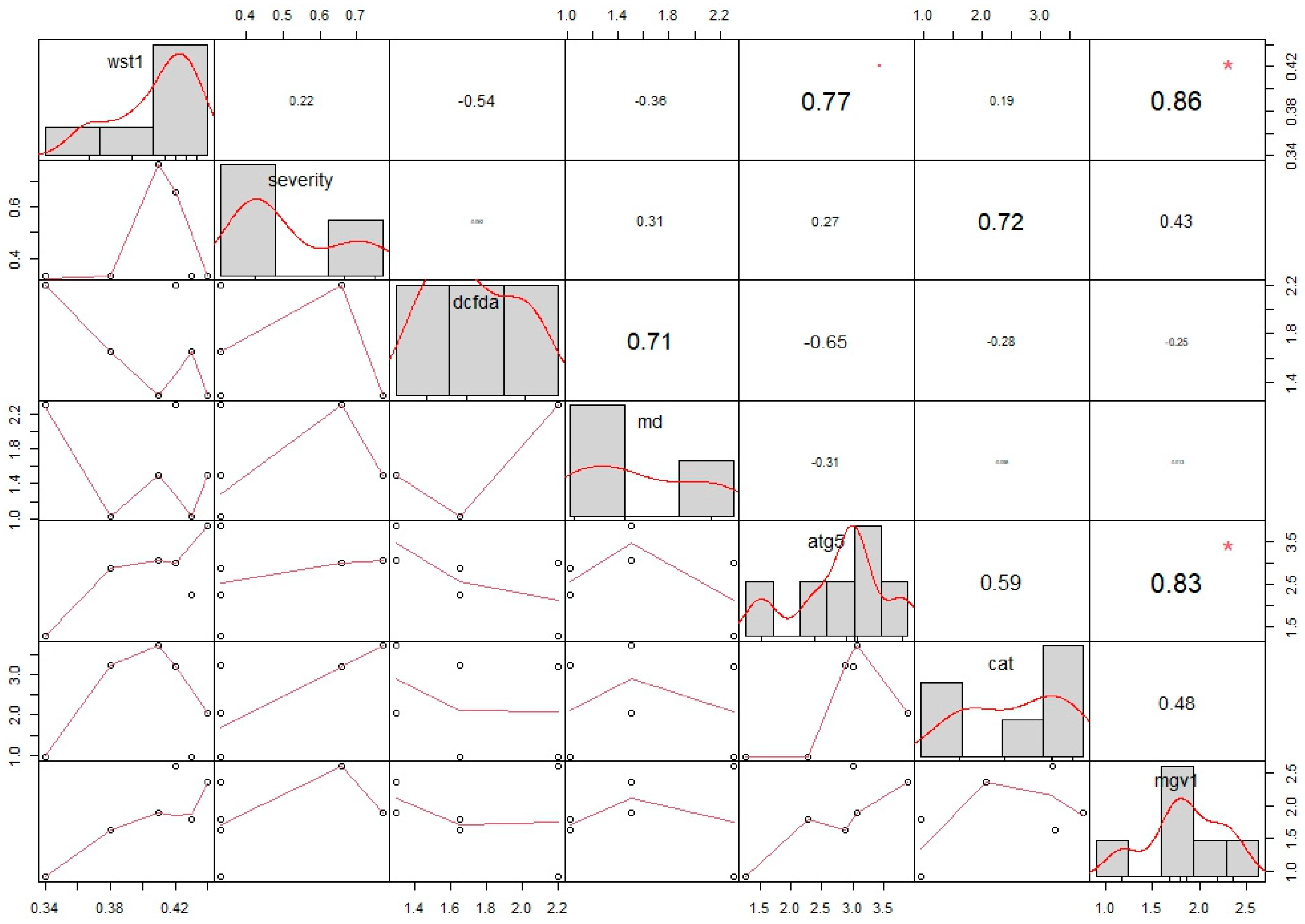
3.6. Statistical Analysis

The correlation matrix (CM) was supported by Pearson’s correlation coefficients and principal component analysis (PCA). Each value was normalized to a fold change of “1”. The CM revealed that only a positive correlation was present in the limited data sets. No negative correlation was present in the data sets with significant differences (p > 0.05). The WST-1 assay and mgv1 expression showed similar patterns of upregulation (Table 3). These two data sets showed a positive correlation, with a p value = 0.02 and r value = 0.86. Similarly, a positive correlation was found between mgv1 and atg5 expression. The p and r values were calculated as 0.04 and 0.83, respectively (Figure 5). A highly heterogenous distribution among the different data sets was observed through the correlation matrix (Figure 5). Similarly heterogeneous profiling was indicated by the PCA tests. The percentage for the first and largest component was calculated as 51.1%. The second-largest percentage was 26.2% (Figure 6).

4. Discussion

It is apparent that fungicide treatment still dominates fungal disease management in crops worldwide. The list provided by the Fungicide Resistance Action Committee (FRAC) provides comprehensive data regarding the characteristics of fungicides used for disease management worldwide, being updated yearly [43,44], and the latest version of the list is provided on the FRAC Code List website (please see https://www.frac.info/ (accessed on 15 January 2025)). The fungicides belonging to the classes C2, C3, and G have been adapted to be used on crop fields. However, resistance development, the usage of fungicides with unknown modes of action (class U), and some other ecological issues could cause problems in the near future [45,46,47,48]. Therefore, it is necessary to incorporate alternative practices into the control strategies for plant diseases.
It can be observed that plant-derived secondary metabolite application is a major topic, offering an alternative to the use of fungicides [49]. In particular, mixtures including essential oils or specific plant metabolites such as camphor and thymol have been shown to be in vitro growth suppressors for Fusarium spp. However, the majority of existing studies include only the analytical compositions of these plant-derived secondary metabolites and MIC and sub-MIC determination assays [50,51,52,53]. The mechanistic routes behind the potential antifungal effects of these mixtures or specific compounds are still missing from the literature. Here, we aimed to reveal the chemical composition of R. damascena essential oil, to present its potential antifungal effects on the worldwide phytopathogenic fungus F. graminearum PH-1, to determine the phytoprotective effects of REO on wheat, and to show the destructive effects of REO on F. graminearum via different methods.
It is known that the ingredients of essential oils can be highly diverse due to various parameters, including the climate, geography, processing, etc. [54]. In a study, Rosa damascena samples were collected from four different regions of Iran, and a GC-MS analysis was conducted on the essential oils. The results showed that citronellol was the major ingredient for all samples; however, the amount varied between 22.4 and 35.2% [55]. In another study, the effect of the harvesting time on the chemical profile of REO was investigated. The results demonstrated that geraniol and citronellol were the dominant ingredients; however, most major ingredients were altered due to the harvesting time [56]. Similar results were observed in a study conducted in Turkey. The harvesting time of Rosa damascena samples from Mardin province significantly altered the chemical composition [57]. In another study, researchers obtained essential oils from samples harvested at the same province in Turkey. The phytochemical profile obtained was highly comparable to that observed in our work. Citronellol was detected as the major ingredient; in addition, geraniol, nonadecane, and heneicosane were observed in significant amounts [58]. These results show that the phytochemical profile of the REO sample used in this study is analogous to those described in the literature.
Fortunately, knowledge regarding the potential antimicrobial effects of REO has been increasing. However, there are few studies concerning the compound composition and the antifungal effects of REO on F. graminearum, as well as its plant-protective effects. In contrast, studies investigating the antifungal effects of REO are increasing. In a previous study, REO from Iran was investigated against various bacterial and fungal strains in waste and well water [59]. The results showed that REO significantly inhibited Candida albicans strains, with an MIC = 62.50 μg mL−1. In another study, five different samples from Saudi Arabia were investigated for their inhibitory effects against various bacterial strains and a fungal strain, Aspergillus flavus. The results showed that all samples significantly inhibited fungal development [60]. Another study investigated REO against Aspergillus flavus along with 74 other essential oil samples. The results demonstrated that REO was one of the most active essential oils among 75 samples [61]. The volatile organic compounds from Rosa damascena were studied in a prior report against fungal strains of Fusarium oxysporum and Fusarium graminearum. The results demonstrated that VOCs from R. damascena showed the greater inhibition of F. graminearum [62]. Although there are a few reports of REO’s activity against fungal strains, there is a scarcity of studies investigating the antifungal properties of REO in detail from a mechanistic point of view. For these reasons, in this study, the antifungal and plant-protective properties of REO were investigated comprehensively.
In a previous study, 3-(4,5-dimethylthiazol-2-yl)-2,5-diphenyltetrazolium bromide (MTT) assays on Candida albicans showed that REO significantly suppressed fungal growth, similarly to the data from the WST-1 assay in this study. A relatively lower MIC value was found in the determination of the phytoprotective effects of REO on F. graminearum-infected wheat plantlets. Our results showed that REO treatment positively reduced the adverse effects of F. graminearum infections in wheat. The findings obtained from this study reveal that REO required a relatively low concentration for treatment in in vitro and semi-in vivo antifungal tests.
The potential oxidative stress- and autophagy-mediated antifungal effects of REO on F. graminearum PH-1 were tested by gene expression analysis and fluorescence microscopy tests. There are only limited investigations of the potential oxidative stress- and autophagy-related effects of REO on fungal species. The cell membrane integrity, mode of action, and antioxidant assays using C. albicans and Fusarium spp. have been reported previously. The results showed that the SYTOX-green and DCF-DA staining protocols are effective tools in conducting oxidative stress and mode of action tests for REO [63,64]. Gene expression studies on the oxidative stress and autophagic cell death effects of REO on F. graminearum are still missing from the literature. Our results showed that REO led to increased oxidative stress levels in REO-treated fungi, and autophagic cell death was present in the REO-treated F. graminearum PH-1 strain. While there was no positive correlation between the gene expression and fluorescence microscopy results, both test types revealed significant alterations due to REO treatment in F. graminearum. Interestingly, PCA profiling showed that the fluorescence microscopy and gene expression data were clustered separately. While the DCF-DA and MDC tests were closely related on the second axis, the gene expression tests were distributed on the first axis. However, both the correlation matrix and PCA profiling assays revealed that mgv1 expression, which is directly related to asexual and sexual development, showed a positive correlation with and a similar distribution pattern to atg5 expression. The significant level of correlation between vegetative reproduction and autophagy processes could indicate the destructive presence of stress within a fungal cell. However, further studies aimed at providing detailed information about the antifungal effects of REO are needed. In particular, studies including the investigation of the crosstalk between autophagy and other programmed cell death types, including apoptosis and necroptosis, would provide data about the interconnected and complementary pathways behind the potential antifungal effects of REO.

Author Contributions

Conceptualization, E.Y., T.Y.-M., E.Ö. and T.H.B.; methodology, E.Ö., H.S., E.Y. and T.H.B.; software, E.Y., E.Ö. and H.S.; validation, E.Y., T.H.B. and T.Y.-M.; formal analysis, E.Y. and T.Y.-M.; investigation, E.Y. and H.S.; resources, E.Y., H.S. and T.H.B.; data curation, E.Y., T.Y.-M. and T.H.B.; writing—original draft preparation, E.Y., E.Ö. and T.H.B.; writing—review and editing, E.Y., E.Ö., H.S., T.H.B. and T.Y.-M.; visualization, E.Y. and T.H.B.; supervision, H.S. and T.Y.-M.; project administration, H.S.; funding acquisition, H.S. All authors have read and agreed to the published version of the manuscript.

Funding

This research was funded by the Board Regent of Istanbul Yeni Yuzyil University, grant number İYYÜ-BAP-AP-2023-19.

Institutional Review Board Statement

Not applicable.

Informed Consent Statement

Not applicable.

Data Availability Statement

The original contributions presented in this study are included in the article. Further inquiries can be directed to the corresponding author.

Acknowledgments

The authors are grateful to the Seed Progen Company, Turkey, for providing the wheat (Triticum aestivum L. cv. Lucilla) seeds.

Conflicts of Interest

The authors declare no conflicts of interest.

References

  1. Alisaac, E.; Mahlein, A.K. Fusarium head blight on wheat: Biology, modern detection and diagnosis and integrated disease management. Toxins 2023, 15, 192. [Google Scholar] [CrossRef] [PubMed]
  2. Mielniczuk, E.; Skwaryło-Bednarz, B. Fusarium head blight, mycotoxins and strategies for their reduction. Agronomy 2020, 10, 509. [Google Scholar] [CrossRef]
  3. Parry, D.W.; Jenkinson, P.; McLeod, L. Fusarium ear blight (scab) in small grain cereals—A review. Plant Pathol. 1995, 44, 207–238. [Google Scholar] [CrossRef]
  4. Shude, S.; Yobo, K.S.; Mbili, N.C. Progress in the management of Fusarium head blight of wheat: An overview. S. Afr. J. Sci. 2020, 116, 1–7. [Google Scholar] [CrossRef] [PubMed]
  5. Salgado, J.D.; Madden, L.V.; Paul, P.A. Efficacy and Economics of Integrating In-Field and Harvesting Strategies to Manage Fusarium Head Blight of Wheat. Plant Dis. 2014, 98, 1407–1421. [Google Scholar] [CrossRef]
  6. Desjardins, A.E.; Proctor, R.H. Molecular biology of Fusarium mycotoxins. Int. J. Food Microbiol. 2007, 119, 47–50. [Google Scholar] [CrossRef]
  7. Foroud, N.A.; Eudes, F. Trichothecenes in cereal grains. Int. J. Mol. Sci. 2009, 10, 147–173. [Google Scholar] [CrossRef] [PubMed]
  8. Lavkor, I. Fungal Species Causing Ear Rot in Corn and Occurred Mycotoxins in Corn. CPN-2001. 2019, pp. 1197–1209. Available online: https://cropprotectionnetwork.org/publications/an-overview-of-ear-rots (accessed on 11 December 2024).
  9. Lu, Q.; Luo, J.-Y.; Ruan, H.-N.; Wang, C.-J.; Yang, M.-H. Structure-toxicity relationships, toxicity mechanisms and health risk assessment of food-borne modified deoxynivalenol and zearalenone: A comprehensive review. Sci. Total Environ. 2022, 806, 151192. [Google Scholar] [CrossRef]
  10. Reddy, K.E.; Song, J.; Lee, H.-J.; Kim, M.; Kim, D.-W.; Jung, H.J.; Kim, B.; Lee, Y.; Yu, D.; Kim, D.-W. Effects of high levels of deoxynivalenol and zearalenone on growth performance, and hematological and immunological parameters in pigs. Toxins 2018, 10, 114. [Google Scholar] [CrossRef]
  11. Gao, S.; Hanson, B.D.; Qin, R.; Wang, D.; Yates, S.R. Comparisons of Soil Surface Sealing Methods to Reduce Fumigant Emission Loss. J. Environ. Qual. 2011, 40, 1480–1487. [Google Scholar] [CrossRef]
  12. Qin, R.; Gao, S.; Ajwa, H.; Sullivan, D.; Wang, D.; Hanson, B.D. Field Evaluation of a New Plastic Film (Vapor Safe) to Reduce Fumigant Emissions and Improve Distribution in Soil. J. Environ. Qual. 2011, 40, 1195–1203. [Google Scholar] [CrossRef] [PubMed]
  13. Schöneberg, A.; Musa, T.; Voegele, R.T.; Vogelgsang, S. The potential of antagonistic fungi for control of Fusarium graminearum and Fusarium crookwellense varies depending on the experimental approach. J. Appl. Microbiol. 2015, 118, 1165–1179. [Google Scholar] [CrossRef] [PubMed]
  14. Xue, A.G.; Chen, Y.; Voldeng, H.D.; Fedak, G.; Savard, M.E.; Längle, T.; Zhang, J.; Harman, G.E. Concentration and cultivar effects on efficacy of clo-1 biofungicide in controlling Fusarium head blight of wheat. Biol. Control 2014, 73, 2–7. [Google Scholar] [CrossRef]
  15. Masika, P.J.; Afolayan, A.J. Antimicrobial activity of some plants used for the treatment of livestock disease in the Eastern Cape, South Africa. J. Ethnopharmacoogy 2002, 83, 129–134. [Google Scholar] [CrossRef]
  16. Fandohan, P.; Gbenou, J.D.; Gnonlonfin, B.; Hell, K.; Marasas, W.F.; Wingfield, M.J. Effect of essential oils on the growth of Fusarium verticillioides and fumonisin contamination in corn. J. Agric. Food Chem. 2004, 52, 6824–6829. [Google Scholar] [CrossRef]
  17. Abbas, A.; Wright, C.W.; El-Sawi, N.; Yli-Mattila, T.; Malinen, A.M. A methanolic extract of Zanthoxylum bungeanum modulates secondary metabolism regulator genes in Aspergillus flavus and shuts down aflatoxin production. Sci. Rep. 2022, 12, 5995. [Google Scholar] [CrossRef]
  18. Abbas, A.; Yli-Mattila, T. Biocontrol of Fusarium graminearum, a causal agent of Fusarium head blight of wheat, and deoxynivalenol accumulation: From in vitro to in planta. Toxins 2022, 14, 299. [Google Scholar] [CrossRef] [PubMed]
  19. Hu, Y.; Zhang, J.; Kong, W.; Zhao, G.; Yang, M. Mechanisms of antifungal and anti-aflatoxigenic properties of essential oil derived from turmeric (Curcuma longa L.) on Aspergillus flavus. Food Chem. 2017, 220, 1–8. [Google Scholar] [CrossRef]
  20. Kalemba, D.; Kunicka, A. Antibacterial and antifungal properties of essential oils. Curr. Med. Chem. 2003, 10, 813–829. [Google Scholar] [CrossRef]
  21. Aristimuño Ficoseco, M.E.; Sequin, C.J.; Aceñolaza, P.G.; Vattuone, M.A.; Catalán, C.A.N.; Sampietro, D.A. Antifungal metabolites from Schinopsis balansae Engl (Anacardiaceae): Isolation, identification and evidences of their mode of action on Fusarium graminearum Schwabe. Nat. Prod. Res. 2017, 31, 1450–1453. [Google Scholar] [CrossRef]
  22. Christova, P.K.; Dobreva, A.M.; Dzhurmanski, A.G.; Dincheva, I.N.; Dimkova, S.D.; Zapryanova, N.G. The impact of plant essential oils on the growth of the pathogens Botrytis cinerea, Fusarium solani, and Phytophthora pseudocryptogea. Life 2024, 14, 817. [Google Scholar] [CrossRef]
  23. Natu, K.N.; Tatke, P.A. Essential oils—Prospective candidates for antifungal treatment? J. Essent. Oil Res. 2019, 31, 347–360. [Google Scholar] [CrossRef]
  24. Barak, T.H.; Eryilmaz, M.; Karaca, B.; Servi, H.; Kara Ertekin, S.; Dinc, M.; Ustuner, H. Antimicrobial, Anti-Biofilm, Anti-Quorum Sensing and Cytotoxic Activities of Thymbra spicata L. subsp. spicata Essential Oils. Antibiotics 2025, 14, 181. [Google Scholar]
  25. Barak, T.H.; Bölükbaş, E.; Bardakci, H. Evaluation of marketed rosemary essential oils (Rosmarinus officinalis L.) in terms of European pharmacopoeia 10.0 criteria. Turk. J. Pharm. Sci. 2023, 20, 253. [Google Scholar] [CrossRef] [PubMed]
  26. El-Banhawy, A.; Acedo, C.; Qari, S.; Elkordy, A. Molecular identification and phylogenetic placement of Rosa arabica Crép.(Rosaceae), a critically endangered plant species. Life 2020, 10, 335. [Google Scholar] [CrossRef] [PubMed]
  27. Tomljenovic, N.; Pejić, I. Taxonomic review of the genus Rosa. Agric. Conspec. Sci. 2018, 83, 139–147. [Google Scholar]
  28. Franco, D.; Pinelo, M.; Sineiro, J.; Núñez, M.J. Processing of Rosa rubiginosa: Extraction of oil and antioxidant substances. Bioresour. Technol. 2007, 98, 3506–3512. [Google Scholar] [CrossRef]
  29. Jiménez-López, J.; Ruiz-Medina, A.; Ortega-Barrales, P.; Llorent-Martínez, E.J. Rosa rubiginosa and Fraxinus ox-ycarpa herbal teas: Characterization of phytochemical profiles by liquid chromatography-mass spectrometry, and evaluation of the antioxidant activity. New J. Chem. 2017, 41, 7681–7688. [Google Scholar] [CrossRef]
  30. Peña, F.; Valencia, S.; Tereucán, G.; Nahuelcura, J.; Jiménez-Aspee, F.; Cornejo, P.; Ruiz, A. Bioactive compounds and antioxidant activity in the fruit of rosehip (Rosa canina L. and Rosa Rubiginosa L.). Molecules 2023, 28, 3544. [Google Scholar] [CrossRef]
  31. Üstüner, H.; Barak, T.H.; Servi, H.; Ertekin, S.K.; Sen, A.; Dinç, M.; Özkan, V.K. Anticancer, antidiabetic, and anti-oxidant activities of endemic Stachys buttleri Mill. and Stachys pinardii Boiss. essential oils. J. Essent. Oil Bear. Plants 2023, 26, 1502–1514. [Google Scholar] [CrossRef]
  32. Servi, H.; Demir, U.; Servi, E.Y.; Gundogdu, B.; Barak, T.H. Antiproliferative and Antibacterial Activities of Four Commer-cial Essential Oil Samples from Boswellia carteri, B. serrata, and two chemotypes of Canarium luzonicum. J. Essent. Oil Bear. Plants 2023, 26, 79–94. [Google Scholar] [CrossRef]
  33. Alexander, B.; Browse, D.J.; Reading, S.J.; Benjamin, I.S. A simple and accurate mathematical method for calculation of the EC50. J. Pharmacol. Toxicol. Methods 1999, 41, 55–58. [Google Scholar] [CrossRef] [PubMed]
  34. Albayrak, G.; Yörük, E.; Teker, T.; Sefer, Ö. Investigation of antifungal activities of myrcene on Fusarium reference strains. Arch. Microbiol. 2023, 205, 82. [Google Scholar] [CrossRef] [PubMed]
  35. Savi, G.D.; Scussel, V.M. Effects of Ozone Gas Exposure on Toxigenic Fungi Species from Fusarium, Aspergillus, and Penicillium Genera. Ozone Sci. Eng. 2014, 36, 144–152. [Google Scholar] [CrossRef]
  36. Yörük, E. Rutin hydrate induces autophagic cell death and oxidative stress response in phytopathogenic fungus Fusarium graminearum. Zemdirbyste-Agriculture 2023, 110, 375–382. [Google Scholar] [CrossRef]
  37. Livak, K.J.; Schmittgen, T.D. Analysis of relative gene expression data using real-time quantitative PCR and the 2−ΔΔCT method. Methods 2001, 25, 402–408. [Google Scholar] [CrossRef]
  38. Gargouri-Kammoun, L.; Gargouri, S.; Rezgui, S.; Trifi, M.; Bahri, N.; Hajlaoui, M.R. Pathogenicity and aggressiveness of Fusarium and Microdochium on wheat seedlings under controlled conditions. Tunis. J. Plant Prot. 2009, 4, 135–144. [Google Scholar]
  39. Moradi, M.; Dehne, H.-W.; Steiner, U.; Oerke, E.-C. Improved procedure for mass inoculum production of Fusarium species in a short period of time. Appl. Entomol. Phytopathol. 2017, 84, 21–31. [Google Scholar]
  40. Moya-Elizondo, E.A.; Jacobsen, B.J. Integrated management of Fusarium crown rot of wheat using fungicide seed treatment, cultivar resistance, and induction of systemic acquired resistance (SAR). Biol. Control 2016, 92, 153–163. [Google Scholar] [CrossRef]
  41. Beccari, G.; Covarelli, L.; Nicholson, P. Infection processes and soft wheat response to root rot and crown rot caused by Fusarium culmorum. Plant Pathol. 2011, 60, 671–684. [Google Scholar] [CrossRef]
  42. Covarelli, L.; Gardiner, D.; Beccari, G.; Nicholson, P. Fusarium virulence assay on wheat and barley seedlings. Bio-Protocol 2013, 3, e446. [Google Scholar] [CrossRef]
  43. Hermann, D.; Stenzel, K. FRAC Mode-of-action Classification and Resistance Risk of Fungicides. In Modern Crop Protection Compounds, 1st ed.; Jeschke, P., Witschel, M., Krämer, W., Schirmer, U., Eds.; Wiley: Hoboken, NJ, USA, 2019; pp. 589–608. [Google Scholar] [CrossRef]
  44. Russell, P.E. Fungicide resistance action committee (FRAC): A resistance activity update. Outlooks Pest Man-Agement 2009, 20, 122–125. [Google Scholar] [CrossRef]
  45. Wightwick, A.; Walters, R.; Allinson, G.; Reichman, S.; Menzies, N. Environmental risks of fungicides used in hor-ticultural production systems. Fungicides 2010, 1, 273–304. [Google Scholar]
  46. Wiederhold, N. Antifungal resistance: Current trends and future strategies to combat. Infect. Drug Resist. 2017, 10, 249–259. [Google Scholar] [CrossRef]
  47. Lucas, J.A.; Hawkins, N.J.; Fraaije, B.A. The evolution of fungicide resistance. Adv. Appl. Microbiol. 2015, 90, 29–92. [Google Scholar]
  48. Umaru, I.J.; Badruddin, F.A.; Umaru, H.A.; Umaru, K.I. Antifungal potential of some medicinal plants on selected pathogenic fungi. MOJ Proteom. Bioinform. 2018, 7, 271–276. [Google Scholar] [CrossRef]
  49. Zubrod, J.P.; Bundschuh, M.; Arts, G.; Brühl, C.A.; Imfeld, G.; Knäbel, A.; Payraudeau, S.; Rasmussen, J.J.; Rohr, J.; Scharmüller, A.; et al. Fungicides: An Overlooked Pesticide Class? Environ. Sci. Technol. 2019, 53, 3347–3365. [Google Scholar] [CrossRef] [PubMed]
  50. Fraternale, D.; Bertoli, A.; Giamperi, L.; Bucchini, A.; Ricci, D.; Menichini, F.; Trinciarelli, E.; Pistelli, L. Antifungal Evaluation of Hypericum triquetrifolium Polar Extracts against Fusarium spp. Nat. Prod. Commun. 2006, 1, 1934578X0600101209. [Google Scholar] [CrossRef]
  51. Harcarova, M.; Conkova, E.; Proskovcova, M.; Váczi, P.; Marcincakova, D.; Bujnak, L. Comparison of antifungal activity of selected essential oils against Fusarium graminearum in vitro. Ann. Agric. Environ. Med. 2021, 28, 414–418. [Google Scholar] [CrossRef]
  52. Kong, W.; Huo, H.; Gu, Y.; Cao, Y.; Wang, J.; Liang, J.; Niu, S. Antifungal activity of camphor against four phyto-pathogens of Fusarium. S. Afr. J. Bot. 2022, 148, 437–445. [Google Scholar] [CrossRef]
  53. Liu, Y.; Liu, S.; Luo, X.; Wu, X.; Ren, J.; Huang, X.; Feng, S.; Lin, X.; Ren, M.; Dong, P. Antifungal activity and mechanism of thymol against Fusarium oxysporum, a pathogen of potato dry rot, and its potential application. Postharvest Biol. Technol. 2022, 192, 112025. [Google Scholar] [CrossRef]
  54. Barak, T.H.; Dogenci, İ.; Bardakcı, H. Evaluation of the essential oil samples that are sold as “Eucalyptus oil” on the market in Türkiye in terms of European Pharmacopoeia 10.0 criteria. J. Res. Pharm. 2023, 27, 1463–1473. [Google Scholar]
  55. Yaghoobi, M.; Farimani, M.M.; Sadeghi, Z.; Asghari, S.; Rezadoost, H. Chemical analysis of Iranian Rosa damascena essential oil, concrete, and absolute oil under different bio-climatic conditions. Ind. Crops Prod. 2022, 187, 115266. [Google Scholar] [CrossRef]
  56. Rathore, S.; Kundlas, K.; Kumar, R. Variability in essential oil content and constituent profile of damask rose (Rosa damascena Mill.) at altered intervals of harvest in the Indian Western Himalaya. J. Appl. Res. Med. Aromat. Plants 2024, 39, 100537. [Google Scholar] [CrossRef]
  57. Izgi, M.N. Effect of different harvest dates to essential oil components of oil-bearing rose (Rosa damascena Mill.) in Mardin. J. Essent. Oil Bear. Plants 2022, 25, 250–261. [Google Scholar] [CrossRef]
  58. Aslan, E.G.; Ulusoy, S.; Kınaytürk, N.K.; Sarıçam, T.; Periz, Ç.D. Bioinsecticidal potential of rose essential oil against the cowpea beetle, Callosobruchus maculatus (Fabricius) (Coleoptera: Chrysomelidae). J. Appl. Entomol. 2024, 148, 1144–1156. [Google Scholar] [CrossRef]
  59. Ghavam, M.; Afzali, A. Evaluation of the effect of treated wastewater and well water irrigation on the quality, quantity and antibacterial/antifungal activities of Rosa× damascena Herrm. essential oil. Agric. Water Manag. 2022, 269, 107644. [Google Scholar] [CrossRef]
  60. Kurkcuoglu, M.; Baser, K.H.C.; Akterian, S.G.; Fidan, H.N.; Stoyanova, A.S. Chemical composition, sensory evaluation and antimicrobial activity of Taif rose (Rosa damascena Mill.) essential oils. Bulg. Chem. Commun 2020, 52, 460–466. [Google Scholar]
  61. Pawar, V.C.; Thaker, V.S. In vitro efficacy of 75 essential oils against Aspergillus niger. Mycoses 2006, 49, 316–323. [Google Scholar] [CrossRef]
  62. Sharma, R.; Sharma, S.K.; Chander, R.; Kumar, R.; Agnihotri, V.K. In-vitro antidiabetic and antimicrobial studies of volatile organic compounds (VOCs) from the roots of Rosa damascena Mill. of North-western Himalaya. S. Afr. J. Bot. 2024, 168, 518–528. [Google Scholar] [CrossRef]
  63. Barakat, H.; Ghazal, G.A. Physicochemical properties of Moringa oleifera seeds and their edible oil cultivated at different regions in Egypt. Food Nutr. Sci. 2016, 7, 472–484. [Google Scholar]
  64. Shahina, Z.; Al Homsi, R.; Price, J.D.; Whiteway, M.; Sultana, T.; Dahms, T.E. Rosemary essential oil and its com-ponents 1, 8-cineole and α-pinene induce ROS-dependent lethality and ROS-independent virulence inhibition in Candida albicans. PLoS ONE 2022, 17, e0277097. [Google Scholar] [CrossRef] [PubMed]
Figure 1. GC-MS chromatogram of R. damascena essential oil.
Pathogens 14 00383 g001
Figure 2. Fluorescence profiling of REO-treated F. graminearum PH-1 strain for DCF-DA (A,B) and MDC (C,D) tests. (A,C) illustrates control sets for DFC-DA and MDC treatment, respectively. Similarly, (B,D) illustrate REO sets for DCF-DA and MDC treatment.
Pathogens 14 00383 g002
Figure 3. Fold changes in expression of genes related to autophagy, oxidative stress, and sexual–asexual reproduction in F. graminearum PH-1 strain. Significant changes were yielded with different levels of alteration, where “*” means p < 0.05, ** means p < 0.01.
Pathogens 14 00383 g003
Figure 4. (A) Control, (B) F. graminearum PH-1-infected, and (C) REO-treated infection sets of juvenile plantlets of T. aestivum L. cv. Lucilla.
Pathogens 14 00383 g004
Figure 5. The correlation matrix obtained with Pearson’s coefficients, showing a highly heterogeneous distribution and fold changes in different data sets. “*” means p < 0.05.
Pathogens 14 00383 g005
Figure 6. PCA profiling of different data sets obtained in this study. “atg5”, “mgv1”, and “cat” denote gene expression data in F. graminearum PH-1, while “dcf-da” and “md” denote fluorescence microscopy data (oxidative stress and autophagy test) and “wst1” refers to the cell proliferation test. Finally, “severity” refers to the FCR disease assessment in T. aestivum L. cv. Lucilla.
Pathogens 14 00383 g006
Table 1. qPCR primers used in this study.
GenePrimer SetForward Sequence/Reverse Sequence (5′–3′)Band Size (bp)
β-tubulinbetaF/betaRAGGGTCATTACACCGAGGGT/GTACCACCACCAAGAGAGTGG121
FgMgv1MgvRTF/MgvRTRAGGTTCAACGATTCCGACAG/ GACCATTACCCTGAGGCAGA100
catalase_3CatrtF/CatrtRAATTCCACGTTCGTTTCGTC/ CCATACTAGGCTCGCTTTGC130
atg5Atg5rtF/Atg5rtRATGTCTTCTCCCATCCCGC/ GCTGAAGCGTGGAATACTGG108
Table 2. Chemical composition of R. damascena essential oil.
RT 1RRI Exp. 2RRI Lit. 3Compound(%)
5.438903899Heptanal0.1
6.351934939α-Pinene0.4
7.3749699701-Heptanol0.1
7.647978980β-Pinene0.1
8.070992991Myrcene0.2
12.22311031098Linalool0.5
12.35111061102Nonanal0.3
12.63611131111Cis-Rose oxide0.4
12.81011171117Phenylethyl alcohol1.1
13.31811291126Trans-Rose oxide0.2
15.22711741172Nonanol0.2
15.48311801177Terpinen 4-ol0.8
16.09011951189α-Terpineol0.1
18.14912431228Citronellol37.1
18.21212441237Isogeraniol0.1
18.33212471240β-Citral0.3
19.11812651255Geraniol10.6
19.58512761270α-Citral0.5
21.80113281323(E)-Methylgeranate0.1
23.03613571354Citronellol acetate0.5
23.26813631356Eugenol0.6
24.32813881365Neryl acetate1.1
25.27314111401Methyl eugenol2.7
25.76114231418(E)-Caryophyllene0.8
26.54414421443α-Guaiene1.2
26.67214451449β-Phenylethyl butyrate0.1
27.15714571455α-Caryophyllene0.8
28.28514851480Germacrene D1.6
28.48214901485β-Selinene0.2
28.99515021500Pentadecane0.5
29.26415091495δ-Guaiene1.0
36.59017031700Heptadecane1.8
37.42917271717Trans-Farnesol1.2
40.08418031800Octadecane0.2
42.6151878 9-Nonadecene2.9
43.63519091900Nonadecane13.8
45.8011977 1-Eicosene0.1
46.68520052000Eicosane2.1
49.3982093 Henicos-1-ene0.2
49.85221082100Heneicosane8.9
52.66222042200Docosane0.3
55.2222295 1-Tricosene0.1
55.52023062300Tricosane2.6
60.80725052500Pentacosane0.7
66.12727052700Heptacosane0.4
Monoterpenes0.7
Monoterpenoids52.3
Sesquiterpenes5.6
Sesquiterpenoids1.2
n-Alkane derivatives35.3
Others4.5
Total99.6
1 RT: retention time; 2 RRI Exp.: relative retention index calculated against n-alkanes (C5-C30); 3 RRI Lit: relative retention index given in the literature for the compound in similar columns and analysis conditions.
Table 3. Pearson’s r correlation matrix obtained with separate data sets, including those for the F. graminearum PH-1 strain (WST-1 toxicity test (wst1), DCF-DA oxidative stress test (dcfda), MDC autophagy test (md), gene expression assays (atg5, cat, and mgv1)), and T. aestivum L. cv. Lucilla (FCR severity test (severity)).
r correlation values obtained via Pearson’s correlation matrix
wst1severitydcfdamdatg5catmgv1
wst11.000.22−0.54−0.360.770.190.86
severity0.221.00−0.060.310.270.720.43
dcfda−0.54−0.061.000.71−0.65−0.28−0.25
md−0.360.310.711.00−0.31−0.06−0.01
atg50.770.27−0.65−0.311.000.590.83
cat0.190.72−0.28−0.060.591.000.48
mgv10.860.43−0.25−0.010.830.481.00
p values obtained via Pearson’s correlation matrix
wst1severitydcfdamdatg5catmgv1
wst1 0.660.260.470.070.710.02
severity0.66 0.900.550.600.100.39
dcfda0.260.90 0.110.150.590.63
md0.470.550.11 0.540.910.98
atg50.070.600.150.54 0.220.04
cat0.710.100.590.910.22 0.33
mgv10.020.390.630.980.040.33
Disclaimer/Publisher’s Note: The statements, opinions and data contained in all publications are solely those of the individual author(s) and contributor(s) and not of MDPI and/or the editor(s). MDPI and/or the editor(s) disclaim responsibility for any injury to people or property resulting from any ideas, methods, instructions or products referred to in the content.

