Brief Report

Gut Microbiome Profiles in Colonizations with the Enteric Protozoa Blastocystis in Korean Populations

1
Department of Parasitology and Tropical Medicine, Chonnam National University Medical School, Hwasun 58128, Korea
2
Department of Rheumatology, Chonnam National University Medical School and Hospital, Gwangju 61469, Korea
3
Department of Laboratory Medicine, Chonnam National University Hwasun Hospital, Hwasun 58128, Korea
*
Authors to whom correspondence should be addressed.
Microorganisms 2022, 10(1), 34; https://doi.org/10.3390/microorganisms10010034
Submission received: 15 October 2021 / Revised: 7 December 2021 / Accepted: 20 December 2021 / Published: 24 December 2021
(This article belongs to the Special Issue Evolution and Genetic Diversity of Gut Protozoan Parasites)

Abstract

:
The influence of unicellular eukaryotic microorganisms on human gut health and disease is largely unexplored. Blastocystis species commonly colonize the gut, but their clinical significance and ecological role are unclear. We evaluated the effect of Blastocystis colonization on the fecal microbiota of Koreans. In total, 39 Blastocystis-positive and -negative fecal samples were analyzed. The fecal microbiome was assessed by targeting the V3–V4 region of the bacterial 16S ribosomal gene. Bacterial diversity was greater in the Blastocystis-positive than in the Blastocystis-negative group. The bacterial community structure and phylogenetic diversity differed according to the presence of Blastocystis. The mean proportions of Faecalibacterium species and Ruminococcaceae were larger in the Blastocystis-positive group, and that of Enterococcus species was larger in the Blastocystis-negative group. Linear discriminant analysis showed that Faecalibacterium, Prevotella 9, Ruminococcaceae UCG-002, Muribaculaceae, Rikenellaceae, Acidaminococcaceae, Phascolarctobacterium, and Ruminococcaceae UCG-005 were highly enriched in the Blastocystis-positive group, whereas Enterococcus hirae, Enterococcus faecalis, Enterococcus durans, Enterococcaceae, Lactobacillales, and Bacilli were highly abundant in the Blastocystis-negative group. Overall, our results enlighten the notion that Blastocystis colonization is associated with a healthy gut microbiota.

1. Introduction

The mammalian gut contains a microbial community, defined as the microbiome, which includes bacteria, archaea, viruses, phages, yeast, and fungi [1]. The microbiome components have complex roles in gut homeostasis and host health, such as immune system modulation and protection against disease [2,3]. The disruption of the gut ecosystem can lead to gastrointestinal and systemic diseases, such as obesity, diabetes, inflammatory bowel disease, and autoimmune disease [2,3]. Based on this notion, the modulation of gut homeostasis has been recognized as a novel therapeutic target. The plethora of studies documenting the role of the microbiome in human health and disease has directed considerable attention to the factors that influence microbiome diversity and composition [4]. Exploration of the relationship between human-associated gut protists and the resident gut bacterial community has only recently begun.
Advances in DNA-based technologies for the detection and differentiation of intestinal parasites have facilitated exploration of how parasites affect our lives [5]. Luminal intestinal parasitic genera, such as Blastocystis, Dientamoeba, and Entamoeba, are more common in healthy individuals than in patients with functional gastrointestinal disorders [6], indicating that some parasites are beneficial, rather than detrimental, to the host [7]. Of them, Blastocystis has been emerging to alter the composition and diversity of the gut bacterial microbiota [8,9,10]. A recent Korean study showed that Blastocystis was present in diarrheal and non-diarrheal groups, but that the subtype prevalence differed between the groups, suggesting a role as a modulator of the gut microbiota [11].
The pathogenicity of Blastocystis is still unclear and debate over whether Blastocystis is a pathogen or mutualist should consider environmental, dietary, and geographic factors. To clarify their implication, clinical studies of diverse populations are required. In this study, we investigated the effect of Blastocystis colonization on the gut bacterial microbiome in a Korean population. We identified the taxonomic and functional bacterial features of Blastocystis colonization and highlight the role of Blastocystis as a common eukaryotic commensal affecting the gut ecosystem.

2. Materials and Methods

2.1. Collection of Fecal Samples and Blastocystis Screening

Fecal samples were collected from subjects who underwent general checkups or whose gastrointestinal cultures were submitted to the laboratory in accordance with the guidelines, and with the approval, of the Institutional Review Board of Chonnam National University Hospital and Chonnam National University Hwasun Hospital (IRB CNUH-2015-052, CNUHH-2020-045). The samples were transferred immediately to our clinic on ice and stored at −80 °C until processing. Fecal genomic DNA was extracted as described previously using the Cica Geneus® DNA Prep Kit (Kanto Chemical, Tokyo, Japan) according to the manufacturer’s instructions. Firstly, Blastocystis screening was conducted by PCR using the Blast-505–532 (5′-GGA GGT AGT GAC AAT AAA TC-3′) and Blast-998–1017 (5′-TGC TTT CGC ACT TGT TCA TC-3′) primers [11], and then amplified PCR products were confirmed by sequencing with the same PCR primers. In addition, Blastocystis-negative samples were verified by Real-time PCR using AllplexTM Gastrointestinal Panel-Parasite Assay (GIPPA) (Seegene, Seoul, Korea) [12]. In total, 39 Blastocystis-positive (n = 23) and Blastocystis-negative (n = 16) samples were analyzed.

2.2. Metagenomic Analysis

PCR amplification was performed using 16S universal primers targeting the V3–V4 region of the bacterial 16S ribosomal gene. The sequencing fragments were detected using MiSeq technology by Macrogen (Seoul, Korea). The targeted metagenomic sequences from the microbiota were analyzed using the bioinformatics pipeline established by Vaiomer (Labège, France) according to the FROGS guidelines. Low-depth samples (with <9000 sequences) were removed from the analysis. The sequences were trimmed, merged, and clustered into operational taxonomic units (OTUs) using CLC Genomics Workbench v.10.1.1 and CLC Microbial Genomics Module v.2.5 (Qiagen, Hilden, Germany). A taxonomic assignment of these sequences was performed based on the NCBI taxonomy database, with an OTU cutoff of 3%. The most abundant sequences were considered to be representative of each cluster and were assigned taxonomy level–based CLC Microbial Genomics default values. To create an OTU table, we first performed multiple sequence alignment with MUSCLE v.3.8.31 with the pre-aligned 16S and 18S data from the SILVA v.132 database. Chimeric sequences were identified and removed, and clustering was performed using the UPARSE protocol. Alpha diversity metrics (richness and Shannon’s index) were calculated using the phyloseq R package based on rarefied OTU counts. The beta diversity index was defined as the difference between the total number of species in the two groups and the number of species common to both groups. The exploratory principal coordinate analysis (PCoA) of beta (between-sample) diversity was performed based on the Bray-Curtis measure of dissimilarity. For the hierarchical cluster analysis, Bray-Curtis metrics and complete linkage clustering were implemented. At deeper taxonomic levels (from the phylum to the genus level), we performed linear discriminant effect-size (LEfSe) analysis based on the non-parametric factorial Kruskal–Wallis rank sum test to detect bacterial taxa with significantly different abundance between responders and non-responders. Next, LEfSe was conducted to estimate the effect size of each differential taxon abundance based on the following criteria: linear discriminant analysis (LDA) value > 4.0; p < 0.05. Volcano plots were created to show estimated log2-fold differences in OTU abundance between responders and non-responders.

2.3. Statistical Analysis

Alpha diversity metrics were compared by Mann–Whitney tests; for comparisons involving more than two groups, Bonferroni’s correction was applied. The LDA was performed, and the cladogram were generated at http://huttenhower.sph.harvard.edu/galaxy/ (accessd date: 3 December 2021). The two-tailed Student’s t-test and χ2 test were used to assess differences in the phenotypic characteristics of the microbiome using Prism software (GraphPad Software Inc., San Diego, CA, USA).

3. Results

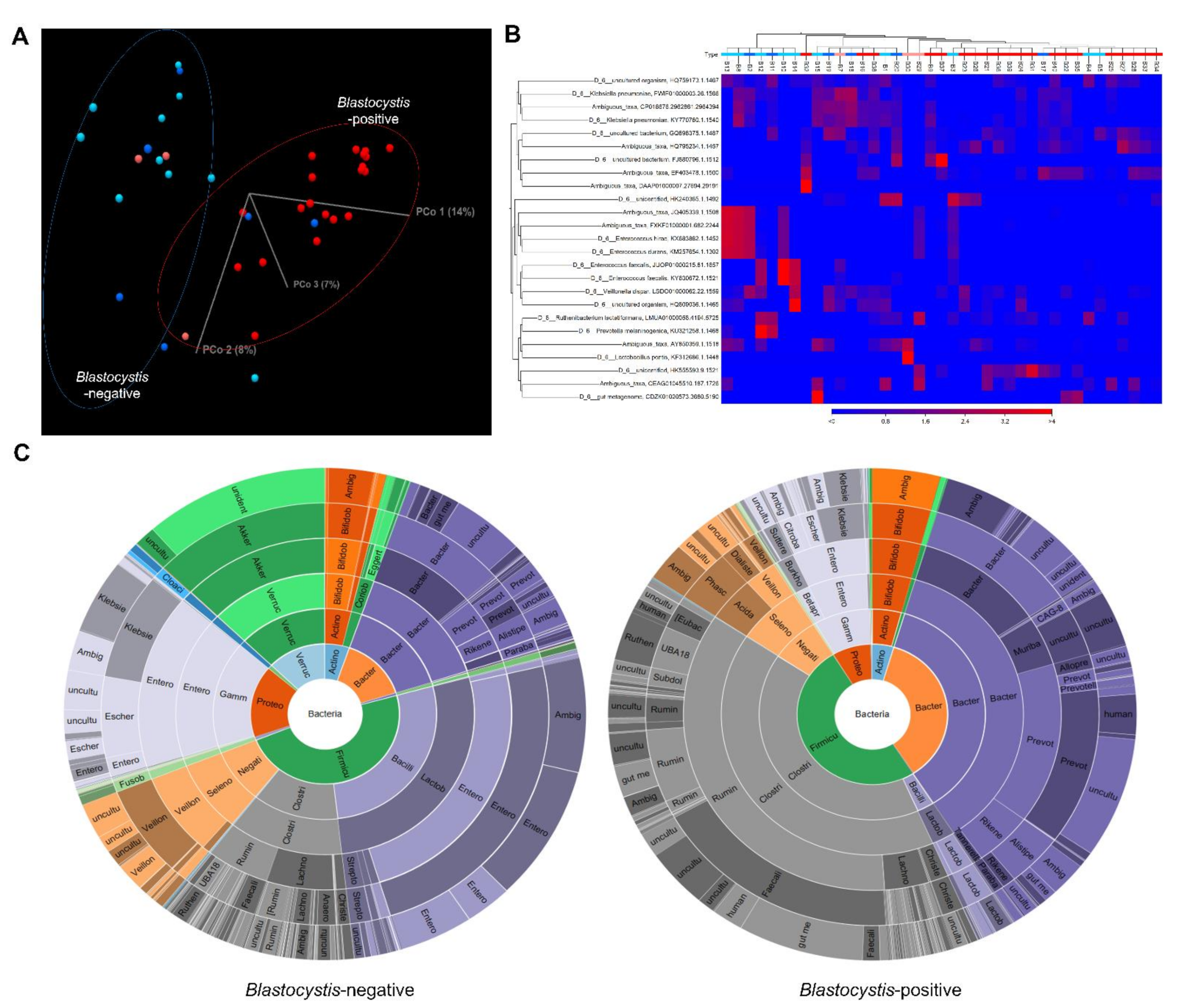
3.1. Blastocystis Colonization Is Associated with Increased Diversity of Gut Bacterial Microbiota

A total of 4,183,214 good-quality reads with a mean length of 301 base pairs was generated. The alpha diversity (Shannon diversity index) is shown in a boxplot in Figure 1. The Shannon diversity index was significantly higher in the Blastocystis-positive group than in the Blastocystis-negative group; however, it did not significantly differ in accordance with the presence or absence of diarrhea. To explore the differences in the overall microbial community composition across the two main groups of patients, the phylogenetic taxonomic Bray–Curtis dissimilarities were calculated. The unweighted beta diversity analysis also showed that the overall bacterial community structure and phylogenetic diversity differed between the Blastocystis-negative and -positive groups (Figure 2A,B). Statistical values obtained by the PERMANOVA test are significant (p < 0.001).

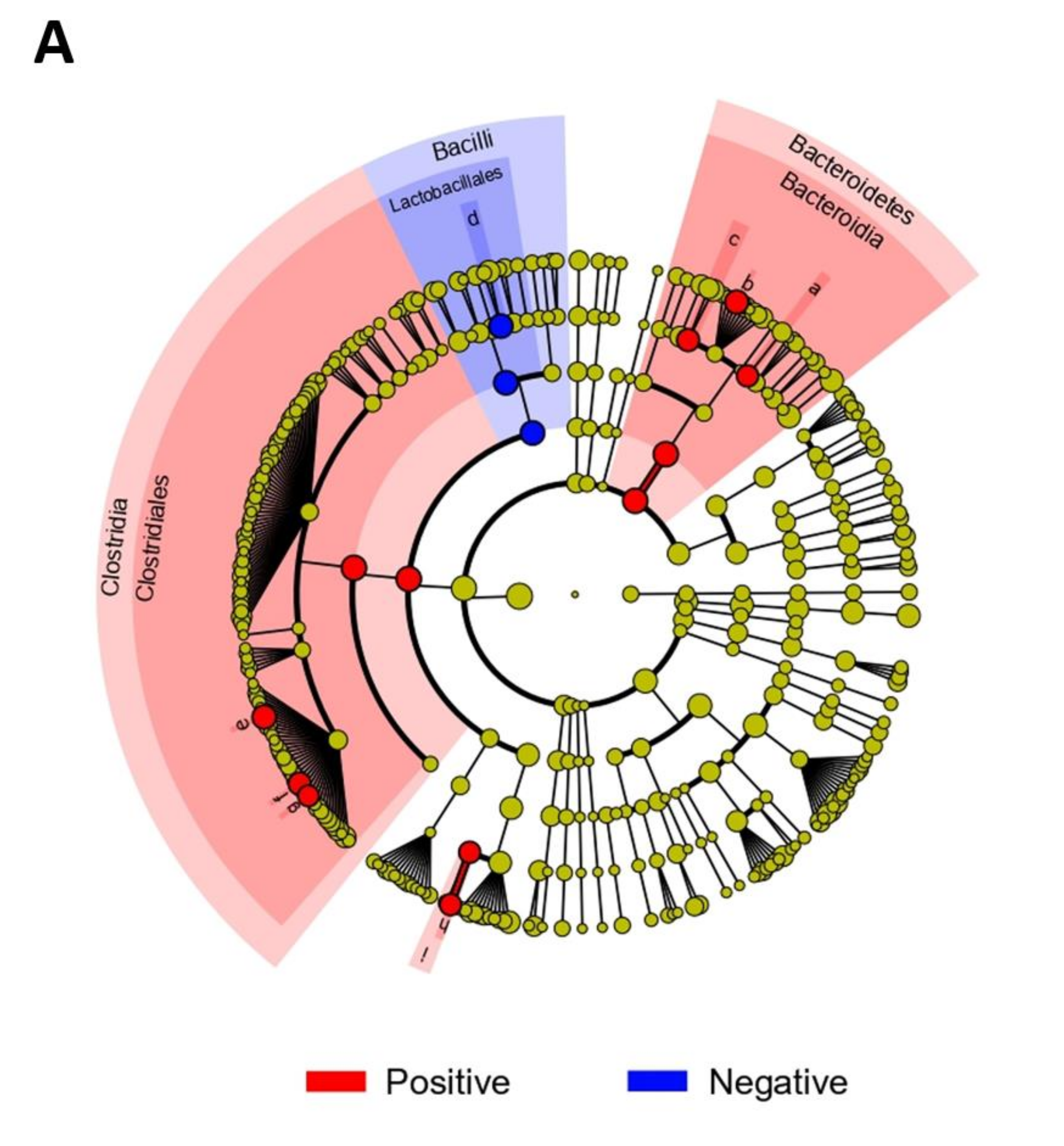
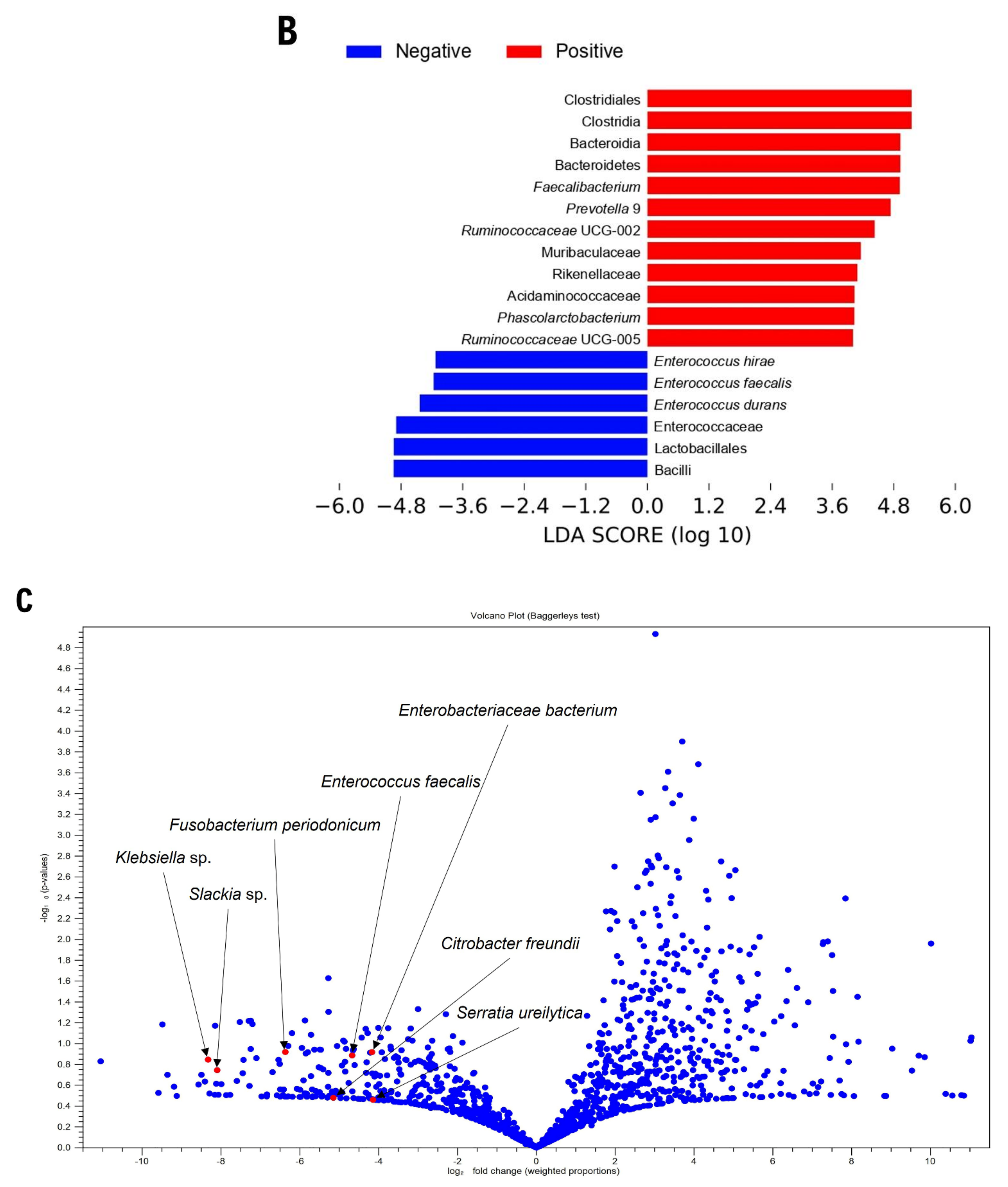
3.2. Gut Microbiome Composition According to Blastocystis Colonization

The bacterial composition of fecal samples from Blastocystis-positive and Blastocyst-is-negative patients was further examined. At the class level, Clostridia were more abundant in the Blastocystis-positive group and Bacilli were more abundant in the Blastocystis-negative group (Figure 2C and Figure S1). At the family level, Ruminococcaceae were more abundant in the Blastocystis-positive group and Enterobacteriaceae were enriched in the Blastocystis-negative group. At the genus level, the Blastocystis-positive group exhibited a greater abundance of Faecalibacterium species, whereas Bacteroides and Enterococcus species were enriched in the Blastocystis-negative group. The mean proportions of Faecalibacterium species, Prevotella 9, and Ruminococcaceae UCG-002 were larger in the Blastocystis-positive group than in the Blastocystis-negative group (Faecalibacterium species, 19.3% vs. 2.4%, p < 0.001; Prevotella 9, 13.1% vs. 2.6%, p = 0.0065; Ruminococcaceae UCG-002, 6.2% vs. 1.0%, p = 0.0007). The Blastocystis-negative group had a larger proportion of Enterococcus species than did the Blastocystis-positive group (17.3% vs. 0.1%, p = 0.0114; Figure S2). Bifidobacterium species were enriched in the diarrheal, but not the non-diarrheal, Blastocystis-positive group. LDA was performed to identify bacterial taxa associated with the Blastocystis colonization (Figure 3). Of them, Faecalibacterium, Prevotella 9, Ruminococcaceae UCG-002, Muribaculaceae, Rikenellaceae, Acidaminococcaceae, Phascolarctobacterium, and Ruminococcaceae UCG-005 were highly enriched in the Blastocystis-positive group, whereas Enterococcus hirae, Enterococcus faecalis, Enterococcus durans, Enterococcaceae, Lactobacillales, and Bacilli were enriched in the Blastocystis-negative group. A volcano plot also showed that several bacterial species, such as Enterobacteriaceae bacterium, Enterococcus faecalis, Fusobacterium periodonicum, Citrobacter freundii, Serratia ureilytica, Klebsiella sp., and Slackia sp., were associated with the Blastocystis-negative group.

4. Discussion

The diversity and bacterial species composition of the microbiota of healthy individuals differ from those of patients with metabolic or infectious diseases. This study provides insight into the association between Blastocystis and gut health in the Korean population. Blastocystis was associated with greater microbiota richness and diversity, indicating that Blastocystis is a potential biomarker of a healthy gut or promoter of gut microbiome eubiosis [8,13]. The high richness and enterotype distribution in Blastocystis carriers indicate that Blastocystis is a component of a stable, resilient, and health-associated microbiota; subtype-level analysis revealed important differences within the genus. In agreement with Audebert et al. [8], our cohort showed greater abundance of the class Clostridia and families Ruminococcaceae and Prevotellaceae in Blastocystis-positive patients, and of Enterobacteriaceae in Blastocystis-negative patients. Prevotella species are dominant colonizers of the human gut in non-westernized populations that consume plant-rich diets [14]. Blastocystis colonization is thus potentially important in the gut microbiota architecture. Among butyrate-producing bacteria, the relative abundance of Faecalibacterium, but not that of Roseburia, was high in our cohort. Differences among studies may be, in part, a result of differences in the techniques used to analyze samples, or in geographic, racial/ethnic, or dietary factors. Such inter-study discrepancies reflect the need to evaluate Blastocystis in a context-specific manner, accounting for the effects of subtype, inflammation, geographical setting, the dietary factor, and other covariates that influence microbiome composition and diversity [14,15]. In addition, differences in gut bacterial richness were less pronounced in the Blastocystis-negative group, regardless of diarrheal status. This finding supports the notion that gut bacterial richness may not be indicative of a healthy gut [16]. The abundance of several Enterococcus species, Enterococcaceae, and Bacilli correlated negatively with Blastocystis colonization. These results are in accordance with the notion that the relative abundance of Enterobacteriaceae or Enterococcaceae correlates positively with gastrointestinal or systemic inflammation [17]. Our data on the correlation of a heathy microbiome, rich in different species of bacteria, and the presence of Blastocystis might be due to gut colonization by this predator protozoa species due to its grazing activity on the bacterial substrate. Together, our results indicate that Blastocystis is associated with a healthy gut microbiota.
Fecal microbiota transplantation (FMT) is used to treat several diseases, including recurrent Clostridium difficile infection and inflammatory bowel disease [18]. Safe FMT requires careful donor selection via questionnaire administration, interviewing, blood testing, and stool examination [19], but no consensus on the effect of luminal parasites, such as Blastocystis, has been reached. Our data partly suggest that colonization may be used as one of the markers for the selection of donors with healthy guts. This notion should be also supported by further studies with a larger population subset.
A recent population-level analysis assessed potential links between Blastocystis subtypes and microbiota–host covariates and quantified microbiota differentiation relative to subtype abundance [20]. In that study, the abundance of subtypes ST3 and ST4 was associated inversely with the relative abundance of Akkermansia, suggesting that these subtypes are associated with a less-healthy host phenotype. In a previous Korean study, subtype ST3 was the major clone (78.3%); thus, we did not focus on the associations of the gut microbiota with specific subtypes. The relative abundance of Akkermansia (>2%) was greater in the Blastocystis-negative group (25.0%, 4/16 patients) than in the Blastocystis-positive group (4.3%, 1/23 patients). Akkermansia is considered to be a health-promoting gastrointestinal isolate and may interact with Blastocystis to promote a healthy gut phenotype.
This pilot study was subject to several limitations. First of all, our dataset is too small to be representative of a true population subset and needs further longitudinal studies enrolling larger numbers of subjects with diverse populations. Second, we did not analyze the associations of Blastocystis subtypes with the gut microbiome due to the small size of the subgroups. Third, we did not evaluate the role of Blastocystis in the structuring of the microbiota or its contribution to intestinal homeostasis. Further longitudinal metagenetic studies in humans or animal models are thus needed. Regardless, our data could serve as the basis for biomarkers of a healthy microbiota and pave the way for an in-depth understanding of the role of Blastocystis in the gut. Further insight into the pathophysiological relevance of Blastocystis may lead to the development of innovative therapeutic solutions, such as supplementation with probiotics.

Supplementary Materials

The following are available online at https://www.mdpi.com/article/10.3390/microorganisms10010034/s1, Figure S1. Stepwise distributions of the gut microbiome according to the colonization of Blastocystis or not. Class-Family-Genus-Species level. At the class level, Clostridia were more abundant in the Blastocystis-positive group and Bacilli were more abundant in the Blastocystis-negative group. At the family level, Ruminococcaceae were more abundant in the Blastocystis-positive group and Enterobacteriaceae were enriched in the Blastocystis-negative group. At the genus level, the Blastocystis-positive group exhibited a greater abundance of Faecalibacterium species, whereas Bacteroides and Enterococcus species were enriched in the Blastocystis-negative group. Stacked bars were generated using the operational taxonomic unit files by CLC Genomics Workbench v. 10.1.1 and CLC Microbial Genomics Module v. 2.5 (Qiagen, Hilden, Germany). Figure S2. Potential markers of Blastocystis colonization. At the genus level, the mean proportions of Faecalibacterium species, Prevotella 9, and Ruminococcaceae UCG-002 were larger in the Blastocystis-positive group than in the Blastocystis-negative group (Faecalibacterium species, 19.3% vs. 2.4%, p < 0.001; Prevotella 9, 13.1% vs. 2.6%, p = 0.0065; Ruminococcaceae UCG-002, 6.2% vs. 1.0%, p = 0.0007). The Blastocystis-negative group had a larger proportion of Enterococcus species than did the Blastocystis-positive group (17.3% vs. 0.1%, p = 0.0114).

Author Contributions

Conceptualization, E.J.W.; methodology, M.-J.K. and Y.J.L.; software, M.-J.K. and Y.J.L.; validation, E.J.W. and T.-J.K.; data curation, E.J.W., T.-J.K., M.-J.K. and Y.J.L.; writing-original draft preparation, E.J.W., T.-J.K., M.-J.K.; supervision, T.-J.K.; funding acquisition, E.J.W. and T.-J.K. All authors have read and agreed to the published version of the manuscript.

Funding

This study was supported by grants from the National Research Foundation of Korea (NRF) Grant funded by the Ministry of Education, Science, and Technology (grant no. NRF-2019M3E5D1A02067953) and by the Chonnam National University Hospital Biomedical Research Institute (grant no. BCRI 19047 and BCRI 19048).

Institutional Review Board Statement

This study was carried out in accordance with all relevant institutional guidelines. The Ethics Committee of Chonnam National University Hospital and Chonnam National University Hwasun Hospital approved this study (IRB CNUH-2015-052; CNUHH-2020-045).

Informed Consent Statement

Written informed consent was waived due to the nature of this study and this was approved by above IRBs.

Data Availability Statement

Data sharing not applicable as no datasets generated and/or analyzed for this study. Data are available upon reasonable request. All data relevant to the study are included in the article or uploaded as supplementary information.

Conflicts of Interest

The authors declare no conflict of interest.

References

  1. Cani, P.D. Human gut microbiome: Hopes, threats and promises. Gut 2018, 67, 1716–1725. [Google Scholar] [CrossRef] [PubMed]
  2. Kho, Z.Y.; Lal, S.K. The Human Gut Microbiome—A Potential Controller of Wellness and Disease. Front. Microbiol. 2018, 9, 1835. [Google Scholar] [CrossRef] [PubMed] [Green Version]
  3. Wu, H.J.; Wu, E. The role of gut microbiota in immune homeostasis and autoimmunity. Gut Microbes 2012, 3, 4–14. [Google Scholar] [CrossRef] [PubMed] [Green Version]
  4. Peterson, C.T.; Sharma, V.; Elmén, L.; Peterson, S.N. Immune homeostasis, dysbiosis and therapeutic modulation of the gut microbiota. Clin. Exp. Immunol. 2015, 179, 363–377. [Google Scholar] [CrossRef] [PubMed] [Green Version]
  5. Verweij, J.J.; Stensvold, C.R. Molecular Testing for Clinical Diagnosis and Epidemiological Investigations of Intestinal Parasitic Infections. Clin. Microbiol. Rev. 2014, 27, 371–418. [Google Scholar] [CrossRef] [PubMed] [Green Version]
  6. Stensvold, C.R.; van der Giezen, M. Associations between Gut Microbiota and Common Luminal Intestinal Parasites. Trends Parasitol. 2018, 34, 369–377. [Google Scholar] [CrossRef] [PubMed]
  7. Lukeš, J.; Stensvold, C.R.; Jirků-Pomajbíková, K.; Parfrey, L.W. Are Human Intestinal Eukaryotes Beneficial or Commensals? PLoS Pathog. 2015, 11, e1005039. [Google Scholar] [CrossRef] [PubMed] [Green Version]
  8. Audebert, C.; Even, G.; Cian, A.; Loywick, A.; Merlin, S.; Viscogliosi, E.; Chabe, M.; Blastocystis Investigation Group. Colonization with the enteric protozoa Blastocystis is associated with increased diversity of human gut bacterial microbiota. Sci. Rep. 2016, 6, 25255. [Google Scholar] [CrossRef] [PubMed]
  9. Scanlan, P.D.; Stensvold, C.R.; Rajilic-Stojanovic, M.; Heilig, H.G.; De Vos, W.M.; O’Toole, P.W.; Cotter, P. The microbial eukaryoteBlastocystisis a prevalent and diverse member of the healthy human gut microbiota. FEMS Microbiol. Ecol. 2014, 90, 326–330. [Google Scholar] [CrossRef] [PubMed] [Green Version]
  10. Beghini, F.; Pasolli, E.; Truong, T.D.; Putignani, L.; Cacciò, S.M.; Segata, N. Large-scale comparative metagenomics of Blastocystis, a common member of the human gut microbiome. ISME J. 2017, 11, 2848–2863. [Google Scholar] [CrossRef] [PubMed]
  11. Kim, M.-J.; Won, E.J.; Kim, S.H.; Shin, J.H.; Chai, J.-Y. Molecular Detection and Subtyping of Human Blastocystis and the Clinical Implications: Comparisons between Diarrheal and Non-diarrheal Groups in Korean Populations. Korean J. Parasitol. 2020, 58, 321–326. [Google Scholar] [CrossRef] [PubMed]
  12. Autier, B.; Gangneux, J.-P.; Robert-Gangneux, F. Evaluation of the AllplexTM Gastrointestinal Panel—Parasite Assay for Protozoa Detection in Stool Samples: A Retrospective and Prospective Study. Microorganisms 2020, 8, 569. [Google Scholar] [CrossRef] [PubMed]
  13. Andersen, L.O.; Bonde, I.; Nielsen, H.B.; Stensvold, C.R. A retrospective metagenomics approach to studyingBlastocystis. FEMS Microbiol. Ecol. 2015, 91, 1–9. [Google Scholar] [CrossRef] [PubMed] [Green Version]
  14. De Filippo, C.; Cavalieri, D.; Di Paola, M.; Ramazzotti, M.; Poullet, J.B.; Massart, S.; Collini, S.; Pieraccini, G.; Lionetti, P. Impact of diet in shaping gut microbiota revealed by a comparative study in children from Europe and rural Africa. Proc. Natl. Acad. Sci. USA 2010, 107, 14691–14696. [Google Scholar] [CrossRef] [PubMed] [Green Version]
  15. Nieves-Ramírez, M.E.; Partida-Rodríguez, O.; Laforest-Lapointe, I.; Reynolds, L.A.; Brown, E.M.; Valdez-Salazar, A.; Morán-Silva, P.; Rojas-Velázquez, L.; Morien, E.; Parfrey, L.W.; et al. Asymptomatic Intestinal Colonization with Protist Blastocystis Is Strongly Associated with Distinct Microbiome Ecological Patterns. mSystems 2018, 3, e00007-18. [Google Scholar] [CrossRef] [PubMed] [Green Version]
  16. Gagliardi, A.; Totino, V.; Cacciotti, F.; Iebba, V.; Neroni, B.; Bonfiglio, G.; Trancassini, M.; Passariello, C.; Pantanella, F.; Schippa, S. Rebuilding the Gut Microbiota Ecosystem. Int. J. Environ. Res. Public Health 2018, 15, 1679. [Google Scholar] [CrossRef] [PubMed] [Green Version]
  17. Liu, J.; Wang, M.; Chen, W.; Ma, J.; Peng, Y.; Zhang, M.; Wang, C.; Yan, G.; Lu, G. Altered Gut Microbiota Taxonomic Compositions of Patients With Sepsis in a Pediatric Intensive Care Unit. Front. Pediatr. 2021, 9, 645060. [Google Scholar] [CrossRef] [PubMed]
  18. Cammarota, G.; Ianiro, G.; Tilg, H.; Rajilic-Stojanovic, M.; Kump, P.; Satokari, R.; Sokol, H.; Arkkila, P.; Pintus, C.; Hart, A.; et al. European consensus conference on faecal microbiota transplantation in clinical practice. Gut 2017, 66, 569–580. [Google Scholar] [CrossRef] [PubMed]
  19. Wang, J.-W.; Kuo, C.-H.; Kuo, F.-C.; Wang, Y.-K.; Hsu, W.-H.; Yu, F.-J.; Hu, H.-M.; Hsu, P.-I.; Wang, J.-Y.; Wu, D.-C. Fecal microbiota transplantation: Review and update. J. Formos. Med. Assoc. 2019, 118 (Suppl. S1), S23–S31. [Google Scholar] [CrossRef] [PubMed]
  20. Tito, R.Y.; Chaffron, S.; Caenepeel, C.; Lima-Mendez, G.; Wang, J.; Vieira-Silva, S.; Falony, G.; Hildebrand, F.; Darzi, Y.; Rymenans, L.; et al. Population-level analysis of Blastocystis subtype prevalence and variation in the human gut microbiota. Gut 2019, 68, 1180–1189. [Google Scholar] [CrossRef] [PubMed] [Green Version]
Figure 1. Boxplots of observed OTU richness and Shannon diversity indices for Blastocystis colonization and diarrheal type. (A) Blastocystis (red dots) and the absence thereof (blue dots). (B) Blastocystis-negative diarrheal (sky-blue dots) and non-diarrheal (blue dots) samples; Blastocystis-positive diarrheal (orange dots) and non-diarrheal (red dots) samples. Statistical analysis was performed by a Mann–Whitney U-test. Plotted are interquartile ranges (IQRs; boxes), medians and means (dark lines and dotted lines, respectively, in the boxes), and the lowest and highest values within 1.5-fold of the IQR of the first and third quartiles (whiskers above and below boxes): *, p < 0.05, ***, p < 0.001.
Microorganisms 10 00034 g001
Figure 2. Gut microbiome composition according to Blastocystis colonization. (A) Unweighted beta diversity analysis showed that the overall bacterial community structure and phylogenetic diversity of the Blastocystis-negative (blue and sky-blue) and Blastocystis-positive (red and orange) groups were distinct. Blue dots: non-diarrheal Blastocystis-negative samples; sky-blue dots: diarrheal Blastocystis-negative samples; red dots: non-diarrheal Blastocystis-positive samples; orange dots: diarrheal Blastocystis-positive samples. (B) Heatmap showing operational taxonomic unit abundance in the Blastocystis-negative (blue) and Blastocystis-positive (red) groups. (C) Of note, the Clostridia class and the Ruminococcaceae family were more abundant in the Blastocystis-positive group, while the Bacilli class and the Enterobacteriaceae family were noticed in the Blastocystis-negative group.
Microorganisms 10 00034 g002
Figure 3. Bacterial taxa associated with the presence or absence of Blastocystis according to linear discriminant analysis (LDA) effect size (LEfSe). (A) LEfSe taxonomic cladogram showing differences in fecal taxa. Dot size is proportional to taxon abundance. Letters correspond to the following taxa: (a) Muribaculaceae, (b) Prevotella 9, (c) Rikenellaceae, (d) Enterococcaceae, (e) Faecalibacterium, (f) Ruminococcaceae UCG-002, (g) Ruminococcaceae UCG-005, (h) Phascolarctobacterium, and (i) Acidaminococcaceae. (B) LDA scores of differentially abundant taxa in the fecal microbiome of the Blastocystis-negative (blue) and Blastocystis-positive (red) groups. Length denotes effect size. p = 0.05, Kruskal–Wallis test; LDA score > 4.0. (C) Volcano plot showing bacterial taxa related to Blastocystis colonization.
Microorganisms 10 00034 g003aMicroorganisms 10 00034 g003b
Publisher’s Note: MDPI stays neutral with regard to jurisdictional claims in published maps and institutional affiliations.

