Article

UME6 Is Involved in the Suppression of Basal Transcription of ABC Transporters and Drug Resistance in the ρ+ Cells of Saccharomyces cerevisiae

Faculty of Biological Science and Technology, Institute of Science and Engineering, Kanazawa University, Kanazawa 920-1192, Japan
Microorganisms 2022, 10(3), 601; https://doi.org/10.3390/microorganisms10030601
Submission received: 16 February 2022 / Revised: 3 March 2022 / Accepted: 7 March 2022 / Published: 10 March 2022
(This article belongs to the Special Issue Trends in Yeast Biochemistry and Biotechnology)

Abstract

:
In Saccharomyces cerevisiae, the Rpd3L complex contains a histone deacetylase, Rpd3, and the DNA binding proteins, Ume6 and Ash1, and acts as a transcriptional repressor or activator. We previously showed that RPD3 and UME6 are required for the activation of PDR5, which encodes a major efflux pump, and pleiotropic drug resistance (PDR) in ρ0/− cells, which lack mitochondrial DNA. However, there are inconsistent reports regarding whether RPD3 and UME6 are required for Pdr5-mediated PDR in ρ+ cells with mitochondrial DNA. Since PDR5 expression or PDR in the ρ+ cells of the rpd3Δ and ume6Δ mutants have primarily been examined using fermentable media, mixed cultures of ρ+ and ρ0/− cells could be used. Therefore, we examined whether RPD3 and UME6 are required for basal and drug-induced PDR5 transcription and PDR in ρ+ cells using fermentable and nonfermentable media. UME6 suppresses the basal transcription levels of the ABC transporters, including PDR5, and drug resistance in ρ+ cells independent of the carbon source used in the growth medium. In contrast, RPD3 is required for drug resistance but did not interfere with the basal PDR5 mRNA levels. UME6 is also required for the cycloheximide-induced transcription of PDR5 in nonfermentable media but not in fermentable media.

1. Introduction

In the yeast Saccharomyces cerevisiae, ATP-binding cassette (ABC) transporters, such as Pdr5, Snq2, and Yor1, efflux a variety of functionally and structurally unrelated compounds, such as fluconazole and cycloheximide from cells [1,2,3,4,5]. The overexpression of the ABC transporters causes multidrug resistance, which is referred to as pleiotropic drug resistance (PDR) in S. cerevisiae [6,7]. The ABC transporters PDR5, SNQ2, YOR1, PDR10, and PDR15 are regulated by the paralogous Zn2Cys6 transcription factors, Pdr1 and/or Pdr3 [1,8,9]. Pdr1 or Pdr3 recognizes the pleiotropic drug response elements (PDRE) DNA consensus motifs in the promoter regions of PDR5, SNQ2, YOR1, PDR10, and PDR15 [9,10,11,12]. PDR3 also has two PDREs in its promoter region that are recognized by Pdr3 and Pdr1 [13]. Thus, PDR3 is subject to positive transcriptional autoregulation by Pdr3 and transcriptional regulation by Pdr1, via these two PDREs [13]. The basal expression levels of PDR5 and SNQ2 and PDR are reduced by PDR1 deletion in ρ+ cells, which contains mitochondrial DNA, compared to PDR3 deletion [10,12]. The disruption of PDR1 and PDR3 substantially reduces the basal expression of their target genes, including PDR5, and causes hypersensitivity to multiple drugs compared with the disruption of either PDR1 or PDR3 [10,12]. Thus, PDR1 and PDR3 have functionally overlapping roles in PDR.
Gain-of-function mutations, such as pdr1-3 and pdr3-7 in Pdr1 and Pdr3, lead to the overexpression of PDR3, PDR5, SNQ2, YOR1, PDR10, and PDR15, and thereby induce the PDR [6,12,14,15,16]. This suggests that the exogenous compounds and drugs lead to the release of Pdr1 and Pdr3 from certain negative regulators because Pdr1 and Pdr3 directly bind a variety of exogenous compounds and drugs [17]. On the other hand, cycloheximide, fluphenazine, or cantharidin induces the transcription of PDR1 and PDR3 [18,19,20]. However, the physiological significance underlying this regulation remains unclear. Interestingly, overproduction of either Pdr1 or Pdr3 in the absence of exogenous drugs can lead to the induction of target gene expression [6,10]. In addition, the PDR5, SNQ2, YOR1, PDR10, or PDR15 mRNA levels were found to be upregulated by the compounds such as cycloheximide, benomyl, fluphenazine, 4-nitroquinolineN-oxide, cantharidin, 2,4-dichlorophenol, or polyoxyethylene-9-laurylether [18,19,20,21,22]. Pdr1 and Pdr3 are also responsible for the transcriptional induction of the ABC transporters such as PDR5, SNQ2, and YOR1 after exposure to drugs or chemical compounds [18,22,23]. The retrograde signaling pathway is strongly activated in ρ0 cells lacking mitochondrial DNA in S. cerevisiae and Candida glabrata, resulting in the enhanced expression of multidrug resistance genes, including PDR5 and CgCdr1, and the elevated PDR. Notably, Pdr3, but not Pdr1, is required for the induction of PDR5 expression in ρ0 cells [10,24].
In S. cerevisiae, there are two Rpd3 complexes, Rpd3S (0.6 MDa) and Rpd3L (1.2 MDa). Both complexes share Rpd3, Sin3, and Ume1 [25]. Rpd3 is a histone deacetylase that participates in chromatin remodeling and transcriptional repression [26,27]. Interestingly, Rpd3 is also required for Hsp90-dependent antifungal drug resistance. The Rpd3L complex contains the DNA binding proteins Ume6 and Ash1 and is thereby recruited by the proteins at the target gene promoters [12,28]. For example, the Rpd3L and Isw2 chromatin remodeling complexes are recruited by Ume6 at early meiotic gene promoters to repress their expression [27,29]. Alternatively, the Rpd3L complex is targeted to the HO promoter by Ash1 to specifically inhibit its expression [25]. In addition to transcriptional repression, Rpd3 is also required for transcriptional activation of osmoresponsive [30] and the DNA damage-inducible genes [31].
We previously showed that RPD3 and UME6 are required for the activation of PDR5 transcription and drug resistance by retrograde signaling in the ρ0 cells of S. cerevisiae [32]. On the other hand, Borecka-Melkusova et al. showed that the ρ+ cells in the rpd3Δ, but not the ume6Δ and ash1Δ strains, displayed sensitivity to cycloheximide at the minimum inhibitory concentration [33]. They also reported that disruption of RPD3 reduces basal PDR5 transcription levels in ρ+ cells [33]. However, Robbins et al. reported that the increased azole susceptibility observed in ρ+ cells of the rpd3Δ strains of Candida albicans and S. cerevisiae is not due to reduced PDR5 mRNA levels but instead due to diminished Hsp90-dependent antifungal drug resistance [34]. In addition, Yibmantasiri et al. reported that the deletion of UME6 in ρ+ cells conferred sensitivity to atorvastatin, cycloheximide, and benomyl but not to ketoconazole, fluconazole, and oligomycin via spot dilution assay [35]. The authors also reported that deletion of UME6 does not reduce Pdr5 expression by western blot analysis [35]. Thus, there are discrepancies regarding whether RPD3 and UME6 are required for PDR5-mediated PDR in ρ+ cells.
However, in the above studies, ρ+ cells grown in yeast extract peptone dextrose (YPD) culture medium containing glucose as the carbon source are used when drug susceptibility and basal PDR5 expression were examined. Since ρ+ cells grown in YPD medium spontaneously generate ρ0/− cells that lack mitochondrial DNA at a rate of 0.1–1% per generation, the above studies could have used mixed cultures of ρ+ and ρ0/− cells [36,37]. ρ0/− cells can grow on YPD culture medium containing glucose as a fermentable carbon source but cannot grow on yeast extract peptone glycerol (YPG) medium containing glycerol as a nonfermentable carbon source. Thus, we can examine the effect of RPD3 and UME6 deletion on PDR5 transcription and PDR in pure ρ+ cells grown on YPG culture medium. Therefore, in this study, we examined whether RPD3 and UME6 are required for basal and drug-induced PDR5 transcription and drug resistance in the ρ+ cells of S. cerevisiae grown in YPG medium. Furthermore, the results obtained in YPG medium were compared with those obtained from ρ+ cells grown in YPD medium. As a result, we show that UME6 suppresses the basal transcription levels of the ABC transporters, including PDR5, and drug resistance in ρ+ cells independent of the carbon source used in the growth medium. In contrast, we also show that RPD3 is required for drug resistance but did not interfere with the basal PDR5 mRNA levels.

2. Materials and Methods

2.1. Yeast Strains and Media

The FY1679-28C (MATa, ura3-52, leu2-D1, trp1-D63, his3-D200, GAL2+) strain was used as the wild-type strain [38]. To construct its derivatives with UME6, RPD3, PDR3, ASH1, or GAT3 deletions, open reading frames of the genes were replaced with KanMX or bleMX6 gene cassettes by PCR-mediated one-step gene disruption in the FY1679-28C background [39]. The strains described above were grown on glycerol-rich YPG agar plates (2% glycerol, 1% yeast extract, 2% bactopeptone, 2% agar) to eliminate ρ0 cells and obtain ρ+ cells.
Yeast cells were grown in YPD medium (2% glucose, 1% yeast extract, 2% bactopeptone) or YPG medium (2% glycerol, 1% yeast extract, 2% bactopeptone) at 30 °C with shaking.

2.2. Spot Dilution Assay

The relative resistance of each yeast strain to fluconazole or cycloheximide was estimated with a spot dilution assay using YPG or YPD media [40,41]. ρ+ cells from each yeast strain were aerobically grown to the logarithmic phase (an OD600 of 0.6–0.9) at 30 °C in YPG or YPD media in triplicate. Five microliters of 10-fold serial dilutions of the YPG cultures containing the same number of cells was spotted on YPG plates with or without 3 μg/mL fluconazole (Nacalai Tesque) (or 0.1 μg/mL cycloheximide (Wako)) and incubated at 30 °C for 21 days. Five microliters of 10-fold serial dilutions of the YPD cultures containing the same number of cells was spotted on YPD plates with or not without 30 μg/mL fluconazole (Nacalai Tesque) (or 0.5 μg/mL cycloheximide (Wako)) and incubated at 30 °C for 21 days.

2.3. Cocultivation of Two Gene Deletion Mutants Replaced with KanMX or bleMX6 Gene Cassettes

One gene deletion mutant replaced with the KanMX gene cassette was cocultivated with the other gene deletion mutant that had been replaced with the bleMX6 gene cassette [32]. ρ+ cells from each mutant corresponding to 0.1 OD600 initial concentration were cocultivated in 10 mL of YPG or YPD media with or without 30 μg/mL fluconazole (or 0.5 μg/mL cycloheximide) [32]. The aliquots of the coculture were recovered immediately before drugs addition (at 0 min) and at various times after drug addition and spread on the YPD plates containing G418 or Zeocin [32,42]. The viability of each strain at each time point was estimated from the colony numbers on the G418 and Zeocin plates [32,42].

2.4. RNA Extraction from the ρ+ Cells in Each Strain Grown to the Logarithmic Phase

ρ+ cells from each yeast strain were grown to the logarithmic phase (an OD600 of 7–9) in YPG or YPD media in duplicate. The cultures were diluted to an OD600 of 0.1 and grown to an OD600 of 0.4–0.8 [32,43,44]. Aliquots of the cultures were recovered. The cells in the aliquots above were pelleted, washed, frozen at –80 °C, and used to extract total RNA [32,43,44]. Total RNA was isolated from the yeast cells using a NucleoSpin RNA kit (TaKaRa) according to the manufacturer’s protocol.

2.5. RNA Extraction from ρ+ Cells before and after Cycloheximide Exposure

ρ+ cells from the wild-type and ume6∆꞉꞉bleMX6 strains were grown to an OD600 of 7–9 in YPG or YPD media, diluted to an OD600 of 0.1, and grown to an OD600 of 0.4–0.8 in duplicate [32,43,44]. Aliquots of each duplicate were harvested just prior to the addition of cycloheximide to the medium. The duplicates were grown for 45 min or 90 min at 30 °C after exposure to cycloheximide (0.2 μg/mL). Aliquots of each duplicate were recovered at 45 min or 90 min after the addition of cycloheximide. The cells in the aliquots above were pelleted, washed, frozen at –80 °C, and used to extract total RNA [32,43,44]. Total RNA was isolated from the yeast cells before and after exposure to cycloheximide using a NucleoSpin RNA Plus kit (TaKaRa) according to the manufacturer’s protocol.

2.6. Real-Time RT–PCR

Reverse transcription of total RNA was performed using the PrimeScript 1st strand cDNA Synthesis Kit (TaKaRa). SYBR Green qRT-PCR for cDNA from the individual duplicate samples was performed using the TB Green® Premix Ex Taq II (TaKaRa) in a Step One Real-time PCR system (Applied Biosystems). A minus reverse transcriptase control was used as the negative control. Serial dilutions of control cDNA were prepared to produce a standard curve for each primer pair. The primers used for qRT-PCR are listed in Table S1. The transcription levels of each target gene were measured by qRT-PCR and normalized to ACT1, which was used as an endogenous control. The normalized levels are shown relative to samples from the wild-type or cycloheximide-untreated wild-type strains, which were set to 1.

2.7. Statistical Analysis

The survival rate of the ume6∆ strain at each time point in Figure 2 was normalized to that at 0 h. A paired t-test was used for statistical analysis in Figure 2. An unpaired Student’s t-test was used for statistical analysis in Figures 3 and 4. Statistical significance was indicated as p < 0.05.

3. Results

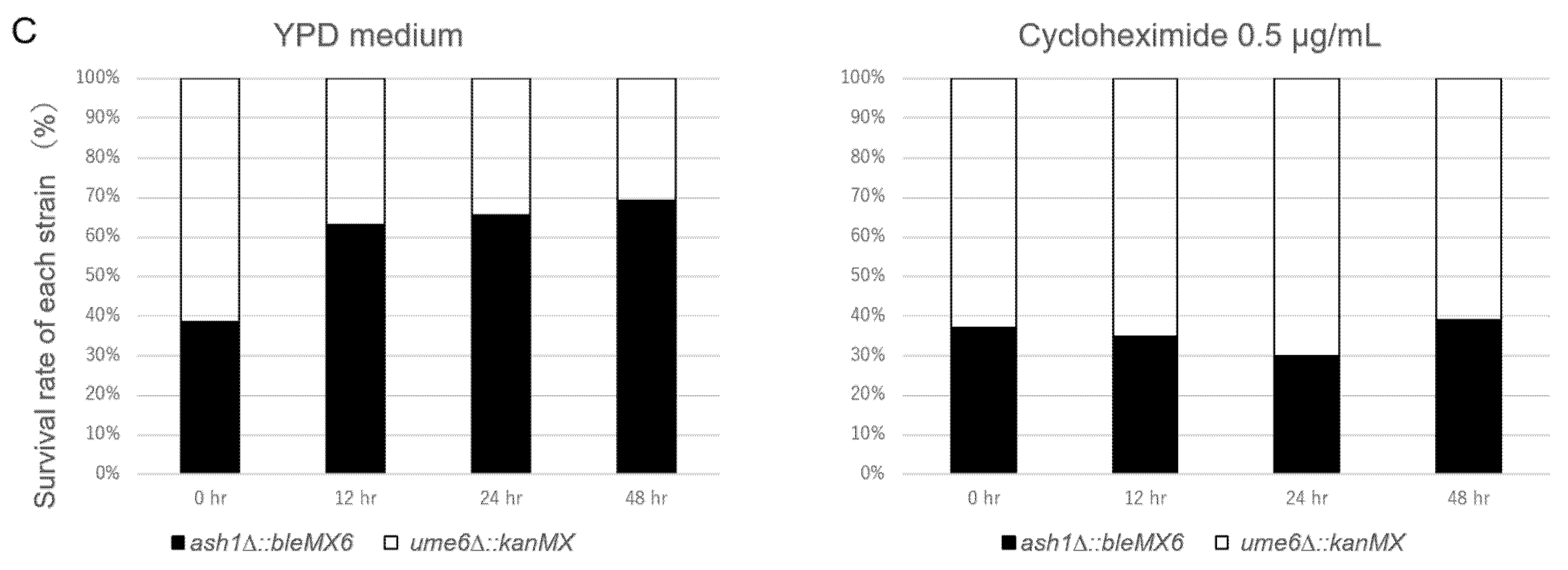
3.1. UME6 Acts as a Negative Regulator to Fluconazole- and Cycloheximide-Resistance in ρ+ Cells, While RPD3 Acts as a Positive Regulator

To examine whether RPD3 and UME6 are responsible for PDR in ρ+ cells of S. cerevisiae, a spot dilution assay using YPG medium containing the nonfermentable carbon source glycerol was carried out to determine the resistance or susceptibility in the ρ+ cells of the rpd3∆ and ume6∆ mutants to the PDR substrates fluconazole and cycloheximide. ρ+ cells of the wild-type strain required a lower concentration of fluconazole and cycloheximide for growth inhibition in YPG medium than in YPD medium, which is consistent with previous reports [45]. The ρ+ cells of the ume6∆꞉꞉bleMX6 mutant were slightly more tolerant to fluconazole and cycloheximide than those in the wild-type, ash1∆꞉꞉bleMX6, pdr3∆꞉꞉bleMX6, and gat3∆꞉꞉bleMX6 strains (Figure 1). However, the ρ+ cells of the rpd3∆ mutant were more susceptible to fluconazole and cycloheximide than those in the wild-type, ume6∆꞉꞉bleMX6, ash1∆꞉꞉bleMX6, pdr3∆꞉꞉bleMX6, and gat3∆꞉꞉bleMX6 strains (Figure 1).
The oligomycin susceptibility of the yeasts was tested on nonfermentable media like YPG medium [3,46]. However, Jensen et al. reported impaired endoplasmic reticulum (ER) to Golgi trafficking of Pdr5 in the rpd3Δ strain of ρ+ S. cerevisiae in nonfermentable medium with glycerol and ethanol as the carbon sources but not in YPD medium [45]. In addition, the PDR5 mRNA levels are downregulated in the ρ+ cells of S. cerevisiae limited for glucose [8]. These reports indicate that the expression of the regulatory machinery of PDR5 in ρ+ cells could be different between YPG and YPD cultures. Therefore, to examine whether RPD3 and UME6 are involved in PDR in ρ+ cells cultured in YPD medium in the same way as that in YPG medium, a similar spot dilution assay as that described above was conducted in YPD plates with or without 30 μg/mL fluconazole (or 0.5 μg/mL cycloheximide). As a result, there were no significant differences in fluconazole and cycloheximide resistance between the wild-type, ume6∆꞉꞉bleMX6, ash1∆꞉꞉bleMX6, pdr3∆꞉꞉bleMX6, and gat3∆꞉꞉bleMX6 strains in YPD plates (Figure S1). However, significantly higher susceptibilities to fluconazole and cycloheximide were found in the rpd3∆ mutant than those found in the wild-type strain (Figure S1). The cycloheximide insusceptibility found in the ρ+ cells of the ume6∆ and ash1∆ mutants in YPD medium was consistent with reports by Borecka-Melkusova et al. [33]. On the other hand, fluconazole insusceptibility, but not cycloheximide insusceptibility, in the ρ+ cells of the ume6∆ mutant in YPD was consistent with reports by Yibmantasiri et al. [35].
In the spot dilution assay above, the ρ+ cells of the ume6∆ mutant displayed slightly more tolerance to fluconazole and cycloheximide in YPG plates than the ρ+ cells of the wild-type, pdr3∆, ash1∆, and gat3∆ strains (Figure 1). To detect a slight difference in drug resistance between two mutant strains, we previously used a cocultivation assay [32]. Therefore, we further investigated whether the ρ+ cells of the ume6∆ mutant confer fluconazole and cycloheximide tolerance in a cocultivation assay using YPG medium. The ash1∆ and gat3∆ mutants displayed an equal tolerance to fluconazole and cycloheximide compared to the wild-type strain in the spot dilution assay (Figure 1), and thereby, the ash1∆ and gat3∆ mutants were used as controls for the ume6∆ mutant in the cocultivation assay. ρ+ cells of two mutant strains, ash1∆꞉꞉kanMX and ume6∆꞉꞉bleMX6, were cocultivated in the presence and absence of 0.5 μg/mL cycloheximide in YPG medium. The number of viable cells in each mutant strain in the coculture was estimated by spreading the cells on YPD plates containing G418 or Zeocin. We found that the ρ+ cells of the ume6∆꞉꞉bleMX6 strain were eliminated from the coculture over time in YPG medium without cycloheximide but accounted for a large percentage of the viable cells in the coculture over time in YPG medium containing 0.5 μg/mL cycloheximide (p < 0.05) (Figure 2A). Similarly, the ρ+ cells of the ume6∆꞉꞉KanMX strain were eliminated from the cocultures of the ume6∆꞉꞉KanMX and ash1∆꞉꞉bleMX6 strains in YPG medium over time in the absence of cycloheximide but selected in the presence of 0.5 μg/mL cycloheximide (data not shown). In addition, the ρ+ cells of the ume6∆꞉꞉KanMX strain were eliminated from the coculture over time in the absence of fluconazole but not in the presence of fluconazole when cocultivated with gat3∆꞉꞉bleMX6 in YPG medium with or without 30 μg/mL fluconazole (Figure 2B). Furthermore, similar results were observed for the ρ+ cells of the gat3∆꞉꞉KanMX and ume6∆꞉꞉bleMX6 mutants cocultivated in YPG medium with or without 30 μg/mL fluconazole (data not shown).
We next examined whether the higher cycloheximide and fluconazole resistance in ume6∆ is dependent on the carbon source used in the growth medium. Rather than using the coculture grown in YPG medium, the survival rate of each strain using the coculture grown in YPD medium was estimated in the same way. Consequently, the ρ+ cells of the ume6∆꞉꞉KanMX strain were eliminated from the coculture over time in the absence of cycloheximide but not in the presence of 0.5 μg/mL cycloheximide when cocultivated with the ash1∆꞉꞉bleMX6 mutant in YPD medium (p < 0.05) (Figure 2C). Furthermore, similar results were observed for the ρ+ cells of the gat3∆꞉꞉KanMX and ume6∆꞉꞉bleMX6 strains cocultivated in YPD medium with or without 30 μg/mL fluconazole (or 0.5 μg/mL cycloheximide) (data not shown). These results indicate that, in contrast to previous reports, UME6 acts as a silencer rather than an enhancer of drug resistance in ρ+ cells of S. cerevisiae independent of the carbon source used in the growth medium.

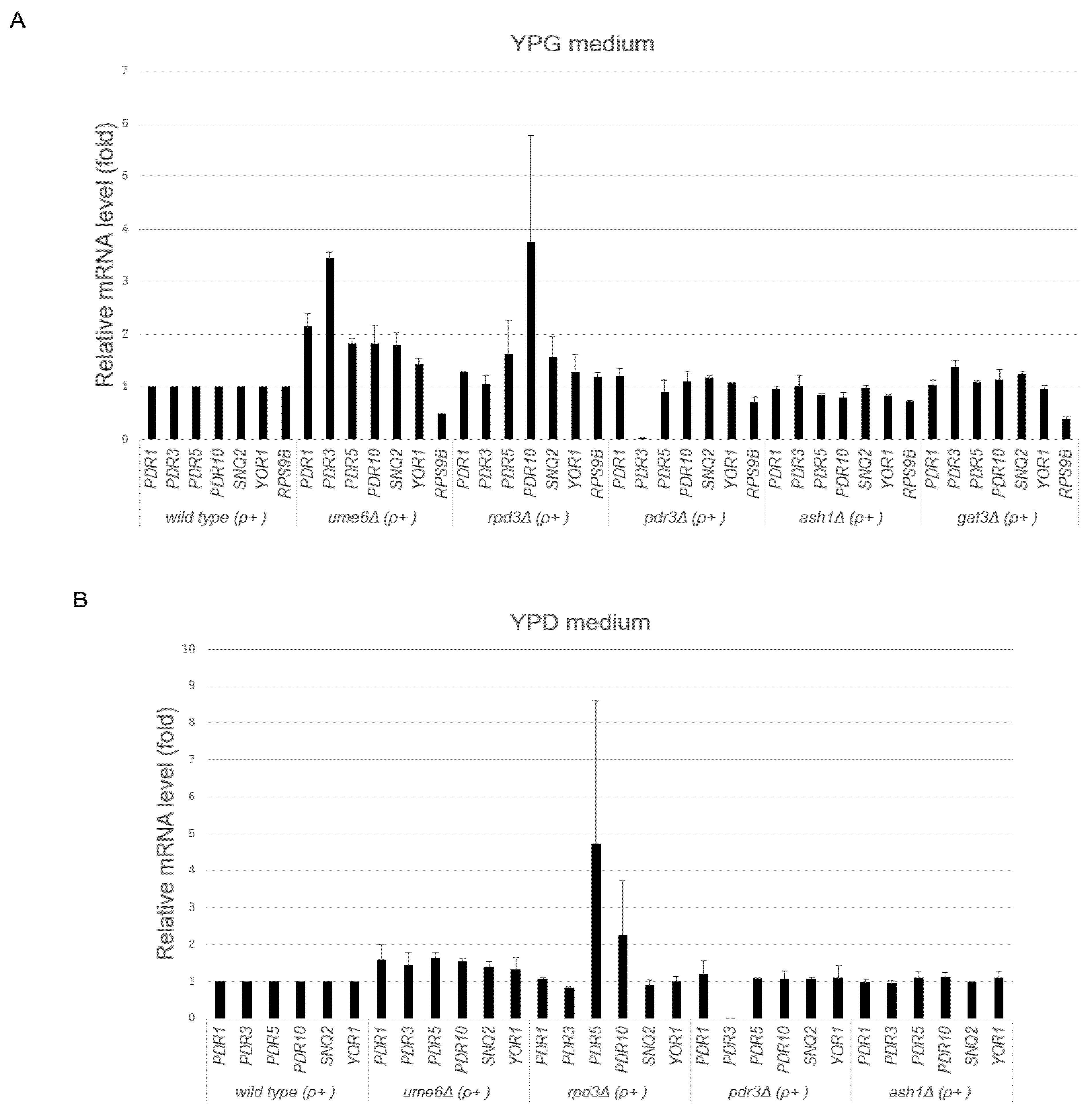
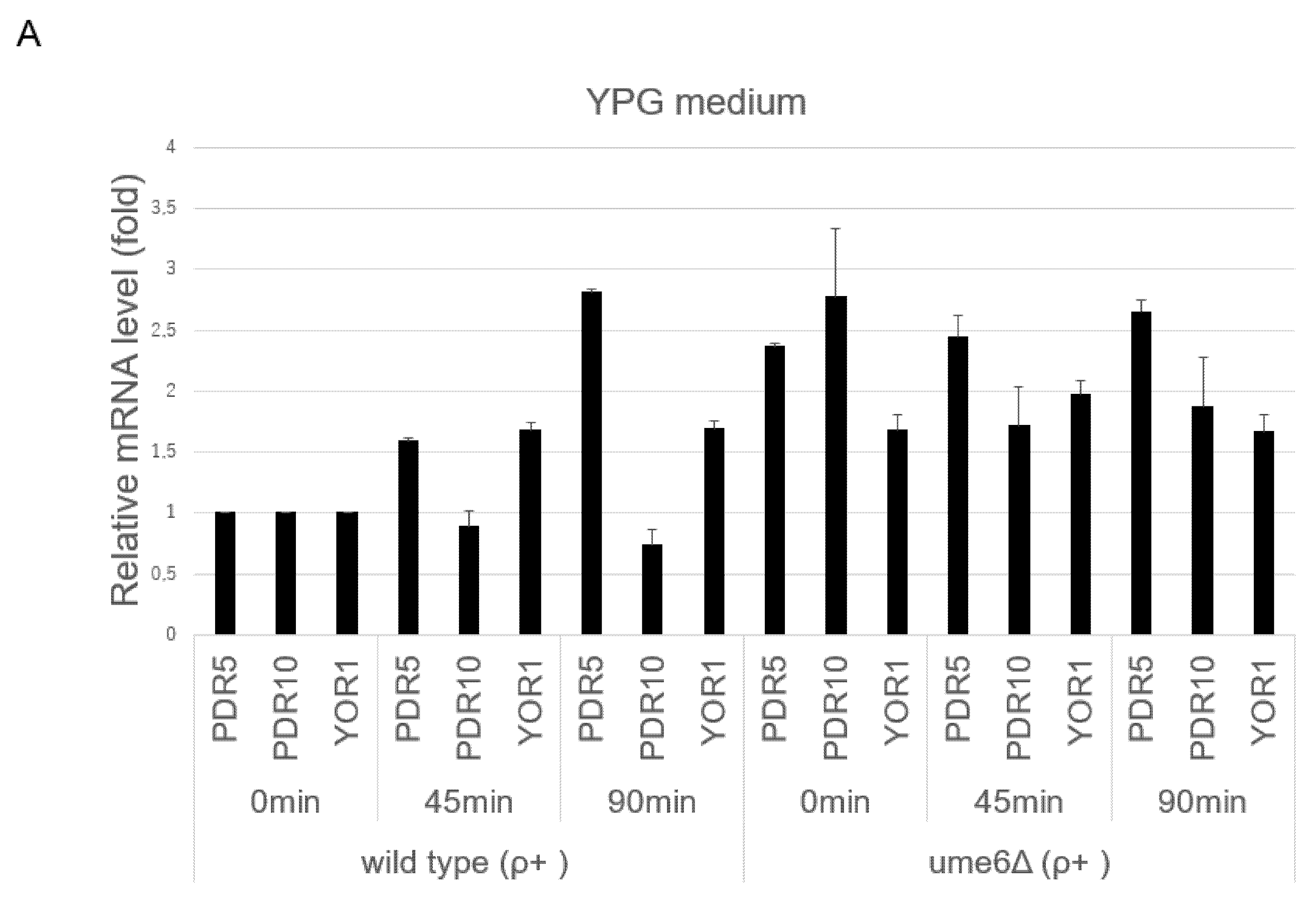
3.2. UME6 Is Required for the Suppression of Basal ABC Transporter mRNA Levels
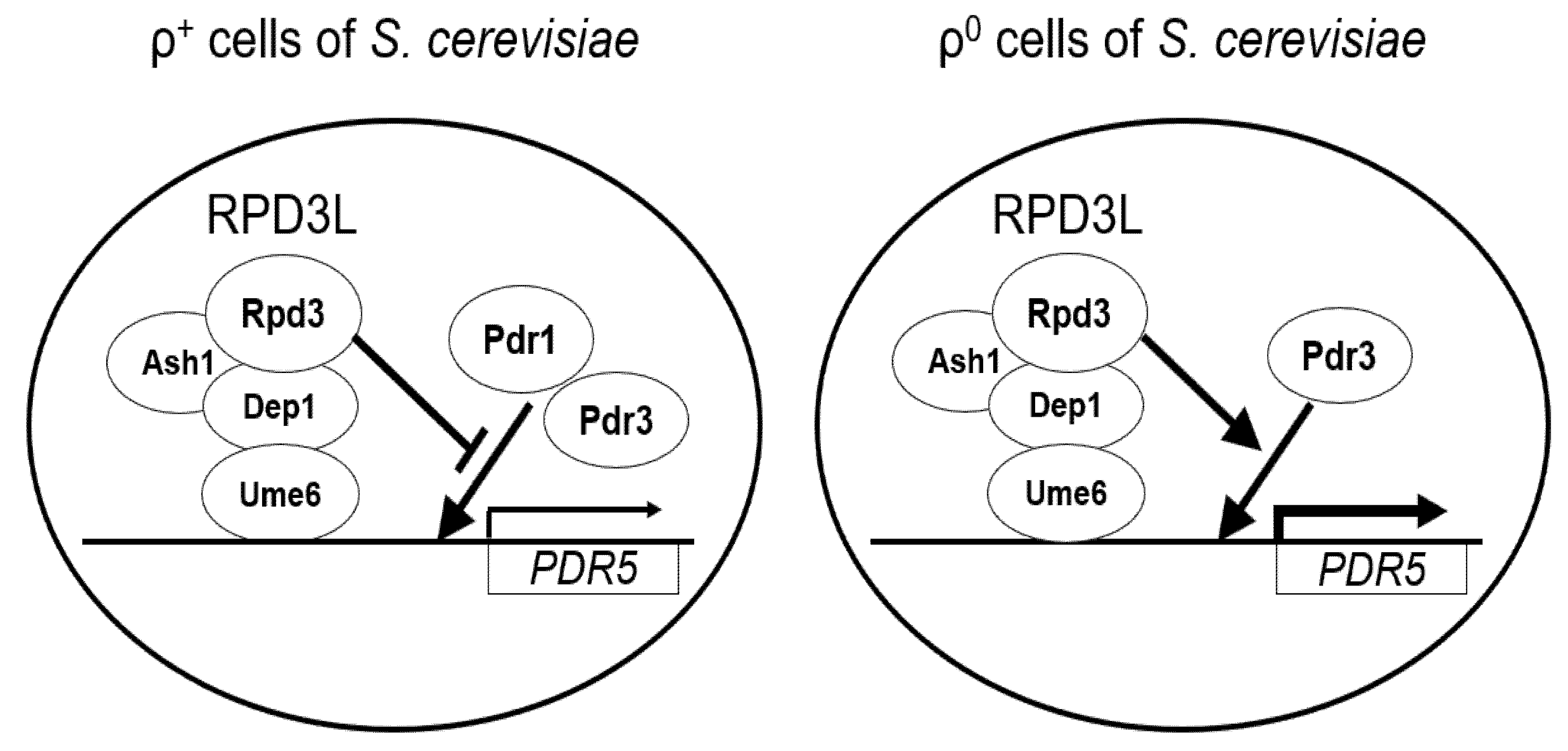
Decreased drug resistance was found in the ρ+ cells of the rpd3∆ strain in the spot dilution assay, while increased drug resistance in the ρ+ cells of the ume6∆ strain was found in the spot dilution and cocultivation assays. These results suggested that the transcriptional levels of ABC transporters such as PDR5 are reduced in the ρ+ cells of the rpd3∆ mutant and increased in the ρ+ cells of the ume6∆ mutant. Thus, we investigated the mRNA levels of ABC transporters PDR5, PDR10, SNQ2, and YOR1 and their transcriptional regulators PDR1 and PDR3 in the ρ+ cells of the wild-type, ume6∆, rpd3∆, pdr3∆, ash1∆, and gat3∆ strains grown in YPG medium to the logarithmic phase by real-time RT-PCR. PDR1, PDR3, PDR5, SNQ2, and YOR1 but not PDR10 expression was significantly induced in the ume6∆ strain compared with the wild-type strain (p < 0.05), while RPS9B expression, used as a control, was downregulated in the ume6∆ strain compared with the wild-type strain (Figure 3A). The transcriptional induction of these ABC transporters and their transcriptional regulators can explain the increased tolerance of the ume6∆ mutant to fluconazole and cycloheximide in the spot dilution and cocultivation assays using YPG medium. In contrast, PDR1 expression, but not PDR3, PDR5, PDR10, SNQ2, or YOR1 expression, was significantly more induced in the rpd3∆ strain than in the wild-type strain (p < 0.05) (Figure 3A). The lack of significant changes in expression of the ABC transporters, including PDR5, between the wild-type and rpd3∆ strains cannot explain the high susceptibility of the rpd3∆ mutant to fluconazole and cycloheximide in the spot dilution assays using YPG plates (Figure 1 and Figure 3A). Therefore, the susceptibility of the rpd3∆ mutant to fluconazole and cycloheximide may result from impaired Hsp90-dependent antifungal drug resistance and ER-to-Golgi trafficking of Pdr5 in YPG medium and not from PDR5 transcriptional reduction [34,45]. In contrast to the ume6∆ mutant, the PDR5 and YOR1 mRNA levels were significantly reduced in the ash1∆ mutant compared with the wild-type strain (p < 0.05) (Figure 3A).
Next, we investigated the mRNA levels of the ABC transporters PDR5, PDR10, SNQ2, and YOR1 and their transcriptional regulators PDR1 and PDR3 in ρ+ cells of the wild-type, ume6∆, rpd3∆, pdr3∆, and ash1∆ strains grown to the logarithmic phase in YPD medium in the same way. In contrast to the results obtained by real-time RT-PCR using YPG medium, only the basal PDR5 and PDR10 mRNA levels were significantly upregulated in the ume6∆ strain compared with the wild-type strain in YPD medium (p < 0.05) (Figure 3B). In addition, the degrees of upregulation of basal transcription of ABC transporters and their regulators in the ume6∆ strain are relatively lower in YPD medium than in YPG medium (Figure 3A,B). However, the increased drug resistance of the ume6∆ mutant shown in the cocultivation assays using YPD can be explained by the induction in the PDR5 mRNA level. In contrast, there was no significant difference in mRNA levels of the ABC transporters and their regulators between ρ+ cells of the wild-type and rpd3∆ strains grown in YPD medium (p > 0.05) (Figure 3B). Therefore, the susceptibility of the rpd3∆ mutant to fluconazole and cycloheximide found in the spot dilution assays using YPD plates may result from impaired Hsp90-dependent antifungal drug resistance but not from the reduced PDR5 mRNA level [34]. In addition, different from the results obtained by real-time RT-PCR using YPG medium, no significant changes in expression of the ABC transporters and their regulators were found between the wild-type and the ash1∆ strains in YPD medium (Figure 3B).
Furthermore, we investigated whether transcriptional activation of the ABC transporters following cycloheximide exposure occurs in the ρ+ cells of the ume6∆ mutant in YPG and YPD media using real-time RT-PCR. In YPG and YPD media, the ABC transporter mRNA levels in the ume6∆ strain at 0 min were higher than those in the wild-type strain at 0 min (p < 0.05), which is consistent with the results in Figure 3A,B (Figure 4A,B). This also suggested that UME6 is required for the repression of basal transcription of the ABC transporters, including PDR5, in ρ+ cells, independent of the media used. In addition, the degree of upregulation of PDR5 transcription in the ume6∆ strain at 0 min was relatively lower in YPD medium than in YPG medium, which is consistent with the results in Figure 3A,B (Figure 4A,B). On the other hand, the ABC transporter mRNA levels, except for PDR10, in the wild-type strain were significantly higher at 45 min than at 0 min in YPG and YPD media (p < 0.05) (Figure 4A,B). In contrast, the PDR5, PDR10, and YOR1 mRNA levels in the ume6∆ strain were not different between 0 min and 45 min in YPG medium (p > 0.05) (Figure 4A). In YPD medium, the PDR5, but not SNQ2, mRNA levels in the ume6∆ strain were significantly higher at 45 min than at 0 min (p < 0.05) (Figure 4B). These results suggested that UME6 is required for the intact induction of PDR5 and YOR1 transcription in ρ+ cells after drug exposure in YPG medium but only for the intact induction of SNQ2 in YPD medium.

4. Discussion

In this report, we have shown that UME6 suppresses the basal transcription levels of the ABC transporters, including PDR5, and drug resistance in the ρ+ cells of S. cerevisiae independent of the carbon source used in the growth medium while RPD3 does not interfere with basal PDR5 transcription but contributes to drug resistance. The results found for RPD3 in this study were consistent with previous reports [34]. However, it has not been previously reported that UME6 suppresses basal PDR5 mRNA levels and drug resistance in the ρ+ cells of S. cerevisiae. ρ+ cells of the ume6∆ strain conferred more resistance to fluconazole and cycloheximide than those of the wild-type strain in the spot dilution assay using YPG plates but not using YPD plates. In addition, although the ρ+ cells of the ume6∆ mutant were eliminated from the coculture over time in both YPG and YPD media without cycloheximide, these cells accounted for the majority of the coculture over time in YPG medium containing 0.5 μg/mL cycloheximide (Figure 2A) while maintaining a constant survival rate over time in YPD medium containing 0.5 μg/mL cycloheximide (Figure 2C). These results can be explained by the appearance of ρ0 cells within the ρ+ cells in the wild-type and ume6∆ strains in YPD medium because ρ0 cells of the ume6∆ mutant are more sensitive to fluconazole and cycloheximide than the ρ0 cells of the wild-type strain [32]. Inverse transcriptional regulation of PDR5 is reported between the ρ0 cells of the ume6∆ and ash1∆ mutants, in which the PDR3 and PDR5 mRNA levels are downregulated in the ρ0 cells of the ume6∆ mutant but upregulated in the ash1∆ mutant [32]. In addition, the fold expression levels of most of the ABC transporters in the ume6∆ mutant compared to the wild-type strain were downregulated in YPD medium compared to YPG medium, while those in the ash1∆ mutant compared to the wild-type strain were upregulated in YPD medium compared to YPG medium (Figure 3A,B and Figure 4A,B). Thus, this inverse transcriptional correlation in the ρ+ cells of the ume6∆ and ash1∆ mutants between YPD and YPG media can also be explained by the occurrence of ρ0 cells within the ρ+ cells of the ume6∆ and ash1∆ mutants in YPD medium. As histone deacetylation leads to transcriptional repression and activation, Ume6 may serve as a silencer of PDR5 expression in ρ+ cells and an enhancer of PDR5 expression in ρ0 cells [27,47]. Furthermore, the inverse transcriptional regulation of the PDR5 between the ume6∆ and ash1∆ mutants independent of ρ+ and ρ0 cells may be caused by exclusive and elevated recruitment of the Rpd3L complex to the target regions by Ume6 in the ash1∆ mutant. Considering the above, due to the relatively low suppression levels of basal PDR5 transcription and drug resistance by UME6 in YPD medium, which could be led by the occurrence of ρ0 cells within ρ+ cells, the roles of UME6 as a negative regulator of basal PDR5 transcription and drug resistance in ρ+ cells may have not been reported. In such cases, a co-cultivation assay but not a spot dilution assay may be useful for detecting a slight difference in drug resistance.
It is currently unknown how Ume6 suppresses the basal transcriptional expression of PDR5 and drug resistance in ρ+ cells and inversely enhances basal PDR5 expression and drug resistance in ρ0 cells [32]. As Ume6 binds to the promoter region of the ABC transporters such as PDR5, PDR10, and YOR1 in ρ+ cells, Ume6 may directly mediate the repression of the ABC transporters by recruiting the Rpd3L and Isw2 chromatin remodeling complexes (Figure 5) [48]. Furthermore, suppression of the ABC transporter transcription and drug resistance by Ume6 in ρ+ cells may be indirectly caused by changes in the expression of the regulators of ABC transporters, such as Pdr1 and Pdr3.
The transcriptional mediator complex serves as a link between sequence-specific transcription factors and the RNA polymerase II machinery [49]. There are two different forms in the transcriptional mediator complex in S. cerevisiae: the core Mediator and L-Mediator. The L-Mediator complex contains the core Mediator complex and the Cdk8 subcomplex. The Cdk8 subcomplex consists of Med12 (Srb8), Med13 (Srb9), the cyclin-dependent kinase Cdk8 (Srb10), and cyclin C (Srb11). The Cdk8 subcomplex has both a negative and a positive role in gene transcription [49,50,51]. Pdr1 and Pdr3 can bind to the KIX domain of a mediator subunit called Med15/Gal11 of the core Mediator and L-Mediator [17]. Deletion of Med12 from the Cdk8 complex completely suppresses the induction of PDR5 expression in ρ0 cells but not in ρ+ cells, indicating a difference in the regulatory machinery of PDR5 transcription between ρ+ and ρ0 cells [52]. This difference may be relevant to the difference in transcriptional regulation of PDR5 by Ume6 between ρ0 and ρ+ cells.
This study indicated that UME6 is required for the intact enhancement of PDR5 and YOR1 transcription after cycloheximide exposure in YPG medium and for intact cycloheximide-induced transcription of SNQ2, but not PDR5, in YPD medium. This result suggested that basal transcriptional repression of the ABC transporters, including PDR5, by UME6 is lifted in response to drug exposure in YPG medium. However, it is unknown why the intact induction of PDR5, but not SNQ2, in the ume6∆ mutant occurs after cycloheximide exposure in YPD medium (Figure 4B). This may result from that cycloheximide can distort measurements of mRNA levels [53]. However, which of upregulation in basal PDR5 transcription or intact drug-induced PDR5 upregulation in the ume6∆ mutant is more effective for drug resistance in YPD medium? Despite intact cycloheximide-induced transcription of PDR5 in the ume6∆ strain in YPD medium, there was no difference in cycloheximide resistance between the ρ+ cells of the wild-type and ume6∆ strains in the spot dilution assay using YPD plates, while a little difference in the cocultivation assay using YPD plates. In addition, upregulation of basal PDR5 transcription levels in the ume6∆ strain compared to the wild-type strain was relatively lower in YPD medium than in YPG medium. Therefore, the upregulation of basal PDR5 transcription in the ume6∆ strain may be more important for drug resistance than intact transcriptional upregulation of PDR5 in the ume6∆ strain following cycloheximide exposure.

5. Conclusions

This study investigated the roles of RPD3 and UME6 in basal and drug-induced transcription of the ABC transporters, including PDR5, and PDR in the ρ+ cells of S. cerevisiae. Using spot dilution and cocultivation assays and YPG and YPD media, we have shown that RPD3 contributes to drug resistance in ρ+ cells, while in contrast to the previous reports, UME6 contributes to the suppression of drug resistance in ρ+ cells, independent of the carbon source used in the growth medium. In addition, using a real-time PCR assay, we have shown that RPD3 does not interfere with the basal PDR5 mRNA level in ρ+ cells, while UME6 is involved in the suppression of basal transcription of some ABC transporters, including PDR5, in ρ+ cells regardless of the carbon source utilized. We have also described that UME6 contributes to an intact transcriptional enhancement of PDR5 in cycloheximide-exposed ρ+ cells in YPG medium but not in YPD medium. This and our previous work will provide useful knowledge on yeast multidrug resistance via the transcriptional regulation of efflux genes by UME6 in ρ+ and ρ0 cells.

Supplementary Materials

The following are available online at https://www.mdpi.com/article/10.3390/microorganisms10030601/s1, Table S1: Primers used for qRT-PCR, Figure S1: Cycloheximide resistance of ρ+ cells of the wild-type and its derivative strains.

Funding

This work was supported by Kanazawa University.

Institutional Review Board Statement

Not applicable.

Informed Consent Statement

Not applicable.

Data Availability Statement

All data generated or analysed during this study are included in this published article.

Acknowledgments

I thank Takumu Nishihara at the School of Electrical and Computer Engineering, Kanazawa University, for technical assistance.

Conflicts of Interest

The author declares no conflict of interest.

References

  1. Decottignies, A.; Grant, A.M.; Nichols, J.W.; de Wet, H.; McIntosh, D.B.; Goffeau, A. ATPase and multidrug transport activities of the overexpressed yeast ABC protein Yor1p. J. Biol. Chem. 1998, 273, 12612–12622. [Google Scholar] [CrossRef] [PubMed] [Green Version]
  2. Harris, A.; Wagner, M.; Du, D.; Raschka, S.; Nentwig, L.M.; Gohlke, H.; Smits, S.H.J.; Luisi, B.F.; Schmitt, L. Structure and efflux mechanism of the yeast pleiotropic drug resistance transporter Pdr5. Nat. Commun. 2021, 12, 5254. [Google Scholar] [CrossRef] [PubMed]
  3. Katzmann, D.J.; Hallstrom, T.C.; Voet, M.; Wysock, W.; Golin, J.; Volckaert, G.; Moye-Rowley, W.S. Expression of an ATP-binding cassette transporter-encoding gene (YOR1) is required for oligomycin resistance in Saccharomyces cerevisiae. Mol. Cell. Biol. 1995, 15, 6875–6883. [Google Scholar] [CrossRef] [Green Version]
  4. Kumari, S.; Kumar, M.; Gaur, N.A.; Prasad, R. Multiple roles of ABC transporters in yeast. Fungal Genet. Biol. 2021, 150, 103550. [Google Scholar] [CrossRef] [PubMed]
  5. Godinho, C.P.; Dias, P.J.; Ponçot, E.; Sá-Correia, I. The Paralogous Genes PDR18 and SNQ2, Encoding Multidrug Resistance ABC Transporters, Derive From a Recent Duplication Event, PDR18 Being Specific to the Saccharomyces Genus. Front. Genet. 2018, 9, 476. [Google Scholar] [CrossRef]
  6. Carvajal, E.; Van Den Hazel, H.B.; Cybularz-Kolaczkowska, A.; Balzi, E.; Goffeau, A. Molecular and phenotypic: Characterization of yeast PDR1 mutants that show hyperactive transcription of various ABC multidrug transporter genes. Mol. Gen. Genet. 1997, 256, 406–415. [Google Scholar] [CrossRef] [PubMed] [Green Version]
  7. Buechel, E.R.; Pinkett, H.W. Transcription factors and ABC transporters: From pleiotropic drug resistance to cellular signaling in yeast. FEBS Lett. 2020, 594, 3943–3964. [Google Scholar] [CrossRef]
  8. Mamnun, Y.M.; Schüller, C.; Kuchler, K. Expression regulation of the yeast PDR5 ATP-binding cassette (ABC) transporter suggests a role in cellular detoxification during the exponential growth phase. FEBS Lett. 2004, 559, 111–117. [Google Scholar] [CrossRef] [Green Version]
  9. Wolfger, H.; Mahé, Y.; Parle-McDermott, A.; Delahodde, A.; Kuchler, K. The yeast ATP binding cassette (ABC) protein genes PDR10 and PDR15 are novel targets for the Pdr1 and Pdr3 transcriptional regulators. FEBS Lett. 1997, 418, 269–274. [Google Scholar] [CrossRef] [Green Version]
  10. Katzmann, D.J.; Burnett, P.E.; Golin, J.; Mahé, Y.; Moye-Rowley, W.S. Transcriptional control of the yeast PDR5 gene by the PDR3 gene product. Mol. Cell. Biol. 1994, 14, 4653–4661. [Google Scholar]
  11. Katzmann, D.J.; Hallstrom, T.C.; Mahé, Y.; Moye-Rowley, W.S. Multiple Pdr1p/Pdr3p binding sites are essential for normal expression of the ATP binding cassette transporter protein-encoding gene PDR5. J. Biol. Chem. 1996, 271, 23049–23054. [Google Scholar] [CrossRef] [Green Version]
  12. Mahé, Y.; Parle-McDermott, A.; Nourani, A.; Delahodde, A.; Lamprecht, A.; Kuchler, K. The ATP-binding cassette multidrug transporter Snq2 of Saccharomyces cerevisiae: A novel target for the transcription factors Pdr1 and Pdr3. Mol. Microbiol. 1996, 20, 109–117. [Google Scholar] [CrossRef] [PubMed]
  13. Delahodde, A.; Delaveau, T.; Jacq, C. Positive autoregulation of the yeast transcription factor Pdr3p, which is involved in control of drug resistance. Mol. Cell. Biol. 1995, 15, 4043–4051. [Google Scholar] [CrossRef] [PubMed] [Green Version]
  14. DeRisi, J.; van den Hazel, B.; Marc, P.; Balzi, E.; Brown, P.; Jacq, C.; Goffeau, A. Genome microarray analysis of transcriptional activation in multidrug resistance yeast mutants. FEBS Lett. 2000, 470, 156–160. [Google Scholar] [CrossRef] [Green Version]
  15. Devaux, F.; Carvajal, E.; Moye-Rowley, S.; Jacq, C. Genome-wide studies on the nuclear PDR3-controlled response to mitochondrial dysfunction in yeast. FEBS Lett. 2002, 515, 25–28. [Google Scholar] [CrossRef] [Green Version]
  16. Nourani, A.; Papajova, D.; Delahodde, A.; Jacq, C.; Subik, J. Clustered amino acid substitutions in the yeast transcription regulator Pdr3p increase pleiotropic drug resistance and identify a new central regulatory domain. Mol. Gen. Genet. 1997, 256, 397–405. [Google Scholar] [CrossRef]
  17. Thakur, J.K.; Arthanari, H.; Yang, F.; Pan, S.J.; Fan, X.; Breger, J.; Frueh, D.P.; Gulshan, K.; Li, D.K.; Mylonakis, E.; et al. A nuclear receptor-like pathway regulating multidrug resistance in fungi. Nature 2008, 452, 604–609. [Google Scholar] [CrossRef]
  18. Fardeau, V.; Lelandais, G.; Oldfield, A.; Salin, H.N.; Lemoine, S.; Garcia, M.; Tanty, V.; Le Crom, S.; Jacq, C.; Devaux, F. The central role of PDR1 in the foundation of yeast drug resistance. J. Biol. Chem. 2007, 282, 5063–5074. [Google Scholar] [CrossRef] [Green Version]
  19. Miranda, M.N.; Masuda, C.A.; Ferreira-Pereira, A.; Carvajal, E.; Ghislain, M.; Montero-Lomelí, M. The serine/threonine protein phosphatase Sit4p activates multidrug resistance in Saccharomyces cerevisiae. FEMS Yeast Res. 2010, 10, 674–686. [Google Scholar] [CrossRef] [Green Version]
  20. Swagatika, S.; Tomar, R.S. ABC transporter Pdr5 is required for cantharidin resistance in Saccharomyces cerevisiae. Biochem. Biophys. Res. Commun. 2021, 553, 141–147. [Google Scholar] [CrossRef]
  21. Cui, Z.; Shiraki, T.; Hirata, D.; Miyakawa, T. Yeast gene YRR1, which is required for resistance to 4-nitroquinoline N-oxide, mediates transcriptional activation of the multidrug resistance transporter gene SNQ2. Mol. Microbiol. 1998, 29, 1307–1315. [Google Scholar] [CrossRef] [PubMed]
  22. Schüller, C.; Mamnun, Y.M.; Wolfger, H.; Rockwell, N.; Thorner, J.; Kuchler, K. Membrane-active compounds activate the transcription factors Pdr1 and Pdr3 connecting pleiotropic drug resistance and membrane lipid homeostasis in saccharomyces cerevisiae. Mol. Biol. Cell 2007, 18, 4932–4944. [Google Scholar] [CrossRef] [PubMed] [Green Version]
  23. Teixeira, M.C.; Sá-Correia, I. Saccharomyces cerevisiae resistance to chlorinated phenoxyacetic acid herbicides involves Pdr1p-mediated transcriptional activation of TPO1 and PDR5 genes. Biochem. Biophys. Res. Commun. 2002, 292, 530–537. [Google Scholar] [CrossRef]
  24. Hallstrom, T.C.; Moye-Rowley, W.S. Multiple signals from dysfunctional mitochondria activate the pleiotropic drug resistance pathway in Saccharomyces cerevisiae. J. Biol. Chem. 2000, 275, 37347–37356. [Google Scholar] [CrossRef] [PubMed] [Green Version]
  25. Carrozza, M.J.; Florens, L.; Swanson, S.K.; Shia, W.J.; Anderson, S.; Yates, J.; Washburn, M.P.; Workman, J.L. Stable incorporation of sequence specific repressors Ash1 and Ume6 into the Rpd3L complex. Biochim. Biophys. Acta 2005, 1731, 77–87. [Google Scholar] [CrossRef] [PubMed]
  26. Rundlett, S.E.; Carmen, A.A.; Suka, N.; Turner, B.M.; Grunstein, M. Transcriptional repression by UME6 involves deacetylation of lysine 5 of histone H4 by RPD3. Nature 1998, 392, 831–835. [Google Scholar] [CrossRef]
  27. Goldmark, J.P.; Fazzio, T.G.; Estep, P.W.; Church, G.M.; Tsukiyama, T. The Isw2 chromatin remodeling complex represses early meiotic genes upon recruitment by Ume6p. Cell 2000, 103, 423–433. [Google Scholar] [CrossRef] [Green Version]
  28. David, K.; Kevin, S. Repression by Ume6 involves recruitment of a complex containing Sin3 corepressor and Rpd3 histone deacetylase to target promoters. Cell 1997, 89, 365–371. [Google Scholar]
  29. Fazzio, T.G.; Kooperberg, C.; Goldmark, J.P.; Neal, C.; Basom, R.; Delrow, J.; Tsukiyama, T. Widespread collaboration of Isw2 and Sin3-Rpd3 chromatin remodeling complexes in transcriptional repression. Mol. Cell. Biol. 2001, 21, 6450–6460. [Google Scholar] [CrossRef] [Green Version]
  30. Pérez-Martínez, M.E.; Benet, M.; Alepuz, P.; Tordera, V. Nut1/Hos1 and Sas2/Rpd3 control the H3 acetylation of two different sets of osmotic stress-induced genes. Epigenetics 2020, 15, 251–271. [Google Scholar] [CrossRef]
  31. Sharma, V.M.; Tomar, R.S.; Dempsey, A.E.; Reese, J.C. Histone deacetylases RPD3 and HOS2 regulate the transcriptional activation of DNA damage-inducible genes. Mol. Cell. Biol. 2007, 27, 3199–3210. [Google Scholar] [CrossRef] [PubMed] [Green Version]
  32. Yamada, Y. RPD3 and UME6 are involved in the activation of PDR5 transcription and pleiotropic drug resistance in ρ0 cells of Saccharomyces cerevisiae. BMC Microbiol. 2021, 21, 311. [Google Scholar] [CrossRef] [PubMed]
  33. Borecka-Melkusova, S.; Kozovska, Z.; Hikkel, I.; Dzugasova, V.; Subik, J. RPD3 and ROM2 are required for multidrug resistance in Saccharomyces cerevisiae. FEMS Yeast Res. 2008, 8, 414–424. [Google Scholar] [CrossRef] [Green Version]
  34. Robbins, N.; Leach, M.D.; Cowen, L.E. Lysine Deacetylases Hda1 and Rpd3 Regulate Hsp90 Function thereby Governing Fungal Drug Resistance. Cell Rep. 2012, 2, 878–888. [Google Scholar] [CrossRef] [PubMed] [Green Version]
  35. Yibmantasiri, P.; Bircham, P.W.; Maass, D.R.; Bellows, D.S.; Atkinson, P.H. Networks of genes modulating the pleiotropic drug response in Saccharomyces cerevisiae. Mol. Biosyst. 2014, 10, 128–137. [Google Scholar] [CrossRef]
  36. Knorre, D.A.; Azbarova, A.V.; Galkina, K.V.; Feniouk, B.A.; Severin, F.F. Replicative aging as a source of cell heterogeneity in budding yeast. Mech. Ageing Dev. 2018, 176, 24–31. [Google Scholar] [CrossRef]
  37. Kochmak, S.A.; Knorre, D.A.; Sokolov, S.S.; Severin, F.F. Physiological scenarios of programmed loss of mitochondrial DNA function and death of yeast. Biochemistry 2011, 76, 167–171. [Google Scholar] [CrossRef]
  38. Berman, J.; Krysan, D.J. Drug resistance and tolerance in fungi. Nat. Rev. Microbiol. 2020, 18, 319–331. [Google Scholar] [CrossRef]
  39. Hentges, P.; Van Driessche, B.; Tafforeau, L.; Vandenhaute, J.; Carr, A.M. Three novel antibiotic marker cassettes for gene disruption and marker switching in Schizosaccharomyces pombe. Yeast 2005, 22, 1013–1019. [Google Scholar] [CrossRef]
  40. Garcia, J.M.; Schwabe, M.J.; Voelker, D.R.; Riekhof, W.R. A functional genomic screen in Saccharomyces cerevisiae reveals divergent mechanisms of resistance to different alkylphosphocholine chemotherapeutic agents. G3 2021, 11, jkab233. [Google Scholar] [CrossRef]
  41. Huang, Z.; Dai, H.; Zhang, X.; Wang, Q.; Sun, J.; Deng, Y.; Shi, P. BSC2 induces multidrug resistance via contributing to the formation of biofilm in Saccharomyces cerevisiae. Cell. Microbiol. 2021, 23, e13391. [Google Scholar] [CrossRef] [PubMed]
  42. Onda, M.; Ota, K.; Chiba, T.; Sakaki, Y.; Ito, T. Analysis of gene network regulating yeast multidrug resistance by artificial activation of transcription factors: Involvement of Pdr3 in salt tolerance. Gene 2004, 332, 51–59. [Google Scholar] [CrossRef] [PubMed]
  43. Elsztein, C.; de Lucena, R.M.; de Morais, M.A. The resistance of the yeast Saccharomyces cerevisiae to the biocide polyhexamethylene biguanide: Involvement of cell wall integrity pathway and emerging role for YAP1. BMC Mol. Biol. 2011, 12, 38. [Google Scholar] [CrossRef] [PubMed] [Green Version]
  44. Wang, X.; Yue, Z.; Xu, F.; Wang, S.; Hu, X.; Dai, J.; Zhao, G. Coevolution of ribosomal RNA expansion segment 7L and assembly factor Noc2p specializes the ribosome biogenesis pathway between Saccharomyces cerevisiae and Candida albicans. Nucleic Acids Res. 2021, 49, 4655–4667. [Google Scholar] [CrossRef]
  45. Jensen, A.N.; Chindaudomsate, W.; Thitiananpakorn, K.; Mongkolsuk, S.; Jensen, L.T. Improper protein trafficking contributes to artemisinin sensitivity in cells lacking the KDAC Rpd3p. FEBS Lett. 2014, 588, 4018–4025. [Google Scholar] [CrossRef] [Green Version]
  46. Hallstrom, T.C.; Katzmann, D.J.; Torres, R.J.; Sharp, W.J.; Moye-Rowley, W.S. Regulation of transcription factor Pdr1p function by an Hsp70 protein in Saccharomyces cerevisiae. Mol. Cell. Biol. 1998, 18, 1147–1155. [Google Scholar] [CrossRef] [Green Version]
  47. Bernstein, B.E.; Tong, J.K.; Schreiber, S.L. Genome wide studies of histone deacetylase function in yeast. Proc. Natl. Acad. Sci. USA 2000, 97, 13708–13713. [Google Scholar] [CrossRef] [Green Version]
  48. Venters, B.J.; Wachi, S.; Mavrich, T.N.; Andersen, B.E.; Jena, P.; Sinnamon, A.J.; Jain, P.; Rolleri, N.S.; Jiang, C.; Hemeryck-Walsh, C.; et al. A Comprehensive Genomic Binding Map of Gene and Chromatin Regulatory Proteins in Saccharomyces. Mol. Cell 2011, 41, 480–492. [Google Scholar] [CrossRef] [Green Version]
  49. Horvath, R.; Hawe, N.; Lam, C.; Mestnikov, K.; Eji-Lasisi, M.; Rohde, J.; Sadowski, I. TORC1 signaling modulates Cdk8-dependent GAL gene expression in Saccharomyces cerevisiae. Genetics 2021, 219, iyab168. [Google Scholar] [CrossRef]
  50. Gonzalez, D.; Hamidi, N.; Del Sol, R.; Benschop, J.J.; Nancy, T.; Li, C.; Francis, L.; Tzouros, M.; Krijgsveld, J.; Holstege, F.C.; et al. Suppression of Mediator is regulated by Cdk8-dependent Grr1 turnover of the Med3 coactivator. Proc. Natl. Acad. Sci. USA 2014, 111, 2500–2505. [Google Scholar] [CrossRef] [Green Version]
  51. Osman, S.; Mohammad, E.; Lidschreiber, M.; Stuetzer, A.; Bazsó, F.L.; Maier, K.C.; Urlaub, H.; Cramer, P. The Cdk8 kinase module regulates interaction of the mediator complex with RNA polymerase II. J. Biol. Chem. 2021, 296, 100734. [Google Scholar] [CrossRef] [PubMed]
  52. Shahi, P.; Gulshan, K.; Näär, A.M.; Moye-Rowley, W.S. Differential Roles of Transcriptional Mediator Subunits in Regulation of Multidrug Resistance Gene Expression in Saccharomyces cerevisiae. Mol. Biol. Cell 2010, 21, 2469–2482. [Google Scholar] [CrossRef] [PubMed] [Green Version]
  53. Santos, D.A.; Shi, L.; Tu, B.P.; Weissman, J.S. Cycloheximide can distort measurements of mRNA levels and translation efficiency. Nucleic Acids Res. 2019, 47, 4974–4985. [Google Scholar] [CrossRef] [PubMed] [Green Version]
Figure 1. The ρ+ cells of the ume6∆ mutant are more tolerant to fluconazole and cycloheximide than those of the wild type on YPG plates. Fluconazole and cycloheximide resistance of ρ+ cells of the wild-type strain (FY1679-28C) and its derivative strains, ume6∆꞉꞉bleMX6, ash1∆꞉꞉bleMX6, pdr3∆꞉꞉bleMX6, and rpd3∆꞉꞉bleMX6, and gat3∆꞉꞉bleMX6, were determined by spot dilution assay in YPG plates with or without 3 μg/mL fluconazole (or 0.1 μg/mL cycloheximide).
Microorganisms 10 00601 g001
Figure 2. The ρ+ cells of the ume6∆ mutant are more tolerant to fluconazole and cycloheximide than those of the ash1∆ and gat3∆ mutants in the cocultivation assays. (A) Changes in the survival rates of each strain after cocultivation of the ume6∆꞉꞉bleMX6 and ash1∆꞉꞉KanMX strains in YPG medium with (right) and without (left) of 0.5 μg/mL cycloheximide. (B) Rates of viable cells of each strain after cocultivation of the gat3∆꞉꞉bleMX6 and ume6∆꞉꞉KanMX strains using YPG medium with (right) and without (left) 30 μg/mL fluconazole. (C) Changes in the survival rates of each strain after cocultivation of the ash1∆꞉꞉bleMX6 and ume6∆꞉꞉KanMX strains in YPD medium with (right) and without (left) of 0.5 μg/mL cycloheximide.
Microorganisms 10 00601 g002aMicroorganisms 10 00601 g002b
Figure 3. Transcription levels of ABC transporters and their regulators in ρ+ cells of the wild-type and mutant strains in the logarithmic growth phase. (A) ρ+ cells of the wild-type, ume6∆, rpd3∆, pdr3∆, ash1∆, and gat3∆ strains were grown to the log phase in YPG medium. Relative PDR1, PDR3, PDR5, PDR10, SNQ2, YOR1, and RPS9B mRNA levels were determined by qRT-PCR. (B) Relative PDR1, PDR3, PDR5, PDR10, SNQ2, and YOR1 mRNA levels in ρ+ cells of the wild-type, ume6∆, rpd3∆, pdr3∆, and ash1∆ strains grown to the log phase in YPD medium were determined by qRT-PCR.
Microorganisms 10 00601 g003
Figure 4. Changes in the transcriptional expression of PDR5, PDR10, YOR1, and SNQ2 in the ρ+ cells of the wild-type and ume6∆ strains in YPG and YPD media after the addition of cycloheximide. (A) The relative fold levels of PDR5, PDR10, and YOR1 mRNA in the ρ+ cells of the wild-type and ume6∆ strains after exposure to cycloheximide (0.2 μg/mL) for 45 min and 90 min in YPG medium were quantified by qRT-PCR. (B) The relative fold levels of PDR5 and SNQ2 mRNA in the ρ+ cells of the wild-type and ume6∆ strains at 45 min after the addition of cycloheximide (0.2 μg/mL) in YPD medium were quantified by qRT-PCR.
Microorganisms 10 00601 g004aMicroorganisms 10 00601 g004b
Figure 5. A simplified diagram of the regulation of basal transcription of PDR5 by Ume6 and Rpd3.
Microorganisms 10 00601 g005
Publisher’s Note: MDPI stays neutral with regard to jurisdictional claims in published maps and institutional affiliations.

