Article

Dihydrolipoamide Acetyltransferase AceF Influences the Type III Secretion System and Resistance to Oxidative Stresses through RsmY/Z in Pseudomonas aeruginosa

1
State Key Laboratory of Medicinal Chemical Biology, Key Laboratory of Molecular Microbiology and Technology of the Ministry of Education, Department of Microbiology, College of Life Sciences, Nankai University, Tianjin 300071, China
2
Department of Immunology of Infectious Diseases, Hirszfeld Institute of Immunology and Experimental Therapy, Polish Academy of Sciences, ul. R. Weigla 12, 53-114 Wroclaw, Poland
*
Author to whom correspondence should be addressed.
Microorganisms 2022, 10(3), 666; https://doi.org/10.3390/microorganisms10030666
Submission received: 11 January 2022 / Revised: 10 March 2022 / Accepted: 17 March 2022 / Published: 21 March 2022
(This article belongs to the Section Molecular Microbiology and Immunology)

Abstract

:
Carbon metabolism plays an important role in bacterial physiology and pathogenesis. The type III secretion system (T3SS) of Pseudomonas aeruginosa is a virulence factor that contributes to acute infections. It has been demonstrated that bacterial metabolism affects the T3SS. Meanwhile, expression of T3SS genes is negatively regulated by the small RNAs RsmY and RsmZ. In this study, we studied the relationship between the dihydrolipoamide acetyltransferase gene aceF and the T3SS. Our results reveal an upregulation of RsmY and RsmZ in the aceF mutant, which represses the expression of the T3SS genes. Meanwhile, the aceF mutant is more tolerant to hydrogen peroxide. We demonstrate that the expression levels of the catalase KatB and the alkyl hydroperoxide reductase AhpB are increased in the aceF mutant. The simultaneous deletion of rsmY and rsmZ in the aceF mutant restored the expression levels of katB and ahpB, as well as bacterial susceptibility to hydrogen peroxide. Thus, we identify a novel role of AceF in the virulence and oxidative response of P. aeruginosa.

1. Introduction

Pseudomonas aeruginosa is a Gram-negative bacterial pathogen that causes various chronic and acute infections in humans [1]. The bacterium harbors an arsenal of virulence determinants that play important roles in infections [1]. The type III secretion system (T3SS) is a syringe-like complex that directly translocates effector proteins into the host cell cytosol, which leads to the malfunction or death of the cell [2,3]. The expression of the T3SS genes is activated under Ca2+ depletion (e.g., by EGTA) or contact with host cells [2,3]. Four major T3SS effectors have been identified in P. aeruginosa, namely, ExoY, ExoT, ExoU and ExoS [2]. ExoU has been shown to function as a phospholipase, which directly damages the host cell membrane and causes rapid necrotic death [4]. P. aeruginosa strains with the ability to inject ExoU are usually associated with poor clinical outcomes and increased mortality rates [5].
The T3SS machinery and effector genes are regulated by the master regulator ExsA [6]. The exsA gene is located in an exsC-exsE-exsB-exsA operon. The transcription of the exsA gene is driven by its own promoter (PexsA) and the exsC promoter (PexsC), which are positively regulated by Vfr and ExsA, respectively [7,8,9]. In addition to transcriptional regulators, small RNAs (sRNAs) are involved in the regulation of T3SS genes [10]. The sRNAs RsmY and RsmZ repress the expression of the T3SS genes by sequestering RsmA, a post-transcriptional regulator that positively regulates the T3SS genes and represses the type VI secretion systems (T6SSs) and biofilm formation [11,12,13]. The expression of rsmY and rsmZ is controlled by the GacS-GacA two-component system [14], whose function is repressed by RetS and activated by LadS [15]. The sensor kinase GacS phosphorylates the response regulator GacA, which binds to the promoters of rsmY and rsmZ and activates their transcription [14]. RetS is a hybrid two-component regulator with a sensor kinase domain and two response regulator domains [16]. It has been demonstrated that RetS inhibits the GacS activity through three mechanisms: (1) the siphoning of phosphates from the catalytic domain of GacS; (2) the inhibition of GacS autophosphorylation through direct interaction; and (3) the dephosphorylation of the receiver domain of GacS [16,17,18,19]. A recent study identified host-derived mucin glycans as ligands of RetS. The presence of mucin glycans represses the GacS-GacA two-component system through RetS [20]. Meanwhile, the GacS-GacA two-component system is positively regulated by two hybrid histidine kinases, PA1611 and LadS. PA1611 directly binds to RetS, which counteracts the repressive effect of RetS on GacS [21]. LadS activates the GacS-GacA two-component system by directly phosphorylating GacS [15].
Metabolism plays important roles in bacterial virulence [22]. It has been demonstrated that tryptophan and histidine utilization interfere with the expression of the T3SS genes in P. aeruginosa [23,24]. The catabolite repression control protein Crc is required for the expression of the T3SS genes in P. aeruginosa [25], indicating a relationship between carbon metabolism and the T3SS. The function of Crc is antagonized by a small RNA (sRNA), CrcZ [26]. In our previous screening for T3SS-related carbon metabolism genes, we found that triosephosphate isomerase (TpiA) affects T3SS gene expression through CrcZ [22]. The isocitrate lyase is required for the expression of the T3SS genes under oxygen-limited conditions [25]. Dacheux et al. found that transposon insertions in genes encoding the pyruvate dehydrogenase (PDH) subunits AceE and AceF (previously called AceA and AceB) resulted in the defective expression of the T3SS genes upon Ca2+ depletion, which was restored by expression of exsA in trans [27]. A mutation in the E1 subunit (AceA/AceE) of pyruvate dehydrogenase resulted in the defective expression of exoS in response to Ca2+ depletion in a minimal medium containing 0.1% tryptone and glutamate [28]. In this study, we demonstrate that the dihydrolipoamide acetyltransferase AceF inversely influences the T3SS and bacterial tolerance to oxidative stress through RsmY/Z.

2. Materials and Methods

2.1. Bacterial Strains and Plasmids

The bacterial strains, primers and plasmids used in this study are shown in Table S1. All strains were cultured in Luria–Bertani (LB) broth with agitation at 200 rpm at 37 °C. Antibiotics were used as follows: for Escherichia coli, ampicillin at 100 µg/mL, tetracycline at 10 µg/mL; for P. aeruginosa, tetracycline at 50 µg/mL, carbenicillin at 150 µg/mL. Construction of the aceF gene deletion mutant in the wild-type Pseudomonas aeruginosa reference strain PA14 was performed as described previously [29]. A 1005 bp fragment and a 1152 bp fragment that are upstream and downstream of the aceF open reading frame were amplified by PCR, respectively. The PCR products were ligated to the plasmid pEX18Tc. The resultant plasmid was transferred into an E. coli conjugation donor strain S17-1 and then transferred to wild-type PA14 by conjugation. The PA14 strains with the plasmid integrated into the chromosome (single-crossover mutant) were selected by tetracycline. The single-crossover mutants were grown in LB overnight and plated on plates with 5% sucrose. The aceF deletion mutants (double-crossover mutants) were screened by PCR with primers targeting the coding region of aceF (Table S1). Other chromosomal gene mutations were generated in the same way. For the overexpression of aceF and exsA, the coding regions with their native ribosome binding sequences were amplified by PCR using the PA14 chromosomal DNA as the template with the primers shown in Table S1. The PCR products were cloned into the pUCP20 plasmid in which the transcription of the genes is driven by the tac promoter on the plasmid.

2.2. Cytotoxicity Assay

Bacterial cytotoxicity was measured by examining the survival of the A549 cells following P. aeruginosa infection. A549 cells were cultured in RPMI 1640 medium containing 10% (v/v) heat-inactivated fetal bovine serum (hiFBS), penicillin (100 mg/mL) and streptomycin (100 mg/mL) at 37 °C with 5% CO2. Then, 1 × 10⁵ cells were seeded into each well of a 24-well plate 20 h before bacterial infection. Overnight bacterial cultures were diluted into fresh LB and cultured at 37 °C to an OD600 of 1.0. Then, bacterial cells were washed once with PBS and resuspended in PBS. A549 cells were infected with the bacteria at a multiplicity of infection (MOI) of 50. After 3 h of infection, the medium was removed, and each well was washed with PBS twice and stained with 0.25% crystal violet at room temperature for 15 min. An amount of 0.2 mL 95% ethanol was added to each well, followed by incubation for 30 min at room temperature with gentle shaking. Each sample was then subjected to measurement for OD590 using a Varioskan Flash multimode microplate reader (Thermo Scientific, Vantaa, Finland).

2.3. Murine Acute Pneumonia Model

The animal experiments were in accordance with Chinese National and Nankai University guidelines for animal research. The protocol was approved by the animal care and use committee of Nankai University College of Life Sciences with the permit number NK-04-2012. The animal facility in Nankai University College of Life Sciences has been approved by Tianjin Municipal Science and Technology Bureau with the permission number SYXK 2019-0003. The animal care and use committee of Nankai University College of Life Sciences has been authorized by the local (Tianjin) government to review and approve animal experiment protocols. The animal facility and protocols are inspected by the local government regularly. The murine acute pneumonia model was performed as previously described [30,31]. The bacteria were cultured to OD600 of 1.0. Bacterial cultures were washed twice with PBS and resuspended to 2 × 10⁸ CFU/mL. BALB/c mice (6 weeks, female) were anesthetized with 90 µL of 7.5% chloral hydrate by intraperitoneal injection and then intranasally inoculated with 4 × 10⁶ CFU bacteria. The mice were sacrificed 12 h post infection (hpi) by asphyxiation with carbon dioxide. The euthanasia of mice was performed following the guidelines from the American Veterinary Medical Association (AVMA) for the euthanasia of animals: 2020 Edition (https://www.avma.org/KB/Policies/Documents/euthanasia.pdf, accessed on 10 January 2022). Less than four mice were placed in a cage. Carbon dioxide was introduced with a flow rate of 3–4 L/min until the mice were unconscious. The death of each mouse was confirmed by respiratory and cardiac arrest. The lungs were isolated and homogenized in PBS containing 1% peptone. The homogenate was serially diluted in LB and then plated on LB plates. The plates were incubated at 37 °C for 20 h before colony counting.

2.4. Histology

After infection with the indicated strains (4 × 10⁶ CFU/mouse) for 12 h, the lungs of mice were removed and fixed with 4% paraformaldehyde (Servicebio, Wuhan, China) for 12 h, followed by dehydration in ethanol. The lungs were then embedded in paraffin and sectioned, followed by staining with hematoxylin and eosin. The samples were observed with microscopy as previously described [32].

2.5. Real-Time qPCR

Bacteria were cultured overnight and then diluted in fresh LB until they reached an OD600 of 1.0. Total RNA was isolated using the ZOMANBIO RNA Rapid Extraction Kit (Zomanbio, Beijing, China). cDNA was synthesized with reverse transcriptase and random primers (TaKaRa, Dalian, China). SYBR II green Supermix (TaKaRa, Dalian, China) was used to perform real-time qPCR (RT–qPCR). To ensure the reliability of the results, we used the housekeeping genes rpsL and PA1805 as the internal controls for each RT–qPCR assay [33,34].

2.6. β-Galactosidase Assay

Overnight bacterial cultures were diluted in LB and grown to an OD600 of 1.0 at 37 °C. A 500 µL volume of the bacterial cultures was resuspended in Z-buffer (8.5 g/L NaH2PO4, 4.8 g/L Na2HPO4, 0.75 g/L KCl, 0.12 g/L MgSO4, and 0.35% (v/v) β-mercaptoethanol, pH 7.0). The OD600 of the bacterial suspensions were measured. Then, 10 µL of 0.1% SDS and 10 µL of chloroform were added to the bacterial suspension and vortexed for 15 s. Then, the mixture was incubated at 37 °C immediately after adding 100 μL of 4 mg/mL orthonitrophenyl-galactopyranoside (ONPG) (BBI Life Sciences, Shanghai, China). When the color of the mixture turned yellow, the reaction was stopped by addition of a 500 μL volume of 1 M Na2CO3. OD420 of the reaction mixtures was measured. The β-galactosidase activities (Miller units) = (1000 × OD420)/T/500/OD600; T represents reaction time [35].

2.7. H2O2 Susceptibility Assay

Bacteria were grown at 37 °C to an OD600 of 1.0. The bacterial cells were washed twice with PBS. After resuspension in PBS, the bacteria were incubated in the presence or absence of 300 mM H2O2 for 1 h at 37 °C. The bacterial survival rate = (live bacterial number with H2O2 treatment)/(live bacterial number without H2O2 treatment).

3. Results

3.1. Mutation in the aceF Gene Reduces the Expression of Type III Secretion System Genes

In our previous screening for metabolic genes that influence bacterial cytotoxicity, we found that an aceF::Tn mutant displayed defective cytotoxicity [22]. AceF, AceE and LpdG form the pyruvate dehydrogenase that connects the glycolysis pathway and the tricarboxylic acid cycle (Figure 1). To verify the role of AceF in bacterial cytotoxicity, we constructed an aceF in-frame deletion mutant. The deletion of the aceF gene reduced the cytotoxicity, which was restored by complementation with an aceF gene (Figure 2A). In P. aeruginosa, the T3SS plays an important role in bacterial cytotoxicity. In agreement with the reduced cytotoxicity, the mutation of aceF reduced the expression of the T3SS genes, including the regulatory genes exsA and exsC, the structural gene pcrV and the effector genes exoU, exoT and exoY (Figure 2B, Figure S1). Complementation with the aceF gene or the overexpression of the exsA gene restored the expression of these genes (Figure 2B). These results suggest that AceF affects bacterial cytotoxicity by regulating the expression of the T3SS genes.

3.2. AceF Affects the Expression of the T3SS Genes through RsmY and RsmZ

Given the role of AceF in carbon metabolism, we suspected that CrcZ might be involved in the regulation of the T3SS in ΔaceF. The CrcZ level in the ΔaceF mutant was 1, which is 9-fold lower than that in the wild-type PA14 (Figure S2). Since CrcZ antagonists the function of Crc that positively regulates the T3SS, the downregulation of CrcZ in the ΔaceF mutant is unlikely to cause the defective T3SS. We next examined the expression levels of RsmY and RsmZ. qRT–PCR revealed upregulation of RsmY and RsmZ in the ΔaceF mutant (Figure 3A). By using transcriptional fusions between a lacZ reporter gene and the promoters of rsmY and rsmZ (PrsmY-lacZ and PrsmZ-lacZ), we found that the promoter activities of rsmY and rsmZ were enhanced in the ΔaceF mutant (Figure 3B). To examine whether the upregulation of RsmY/Z leads to the repression of the T3SS, we deleted rsmY and rsmZ in the ΔaceF mutant, which restored the expression of exsA and exoU and bacterial cytotoxicity (Figure 4A,B). However, compared to the ΔaceFΔrsmY/Z triple mutant, the pcrV mRNA levels were higher in the ΔrsmY/Z mutant in the absence and presence of EGTA, and the mRNA level of exsC was higher in the ΔrsmY/Z mutant in the presence of EGTA (Figure 4B). These results indicate that AceF might regulates the transcription or stabilities of pcrV and exsC through an RsmY/Z-independent mechanism. Collectively, these results demonstrate that AceF affects the T3SS and cytotoxicity mainly through RsmY/Z.

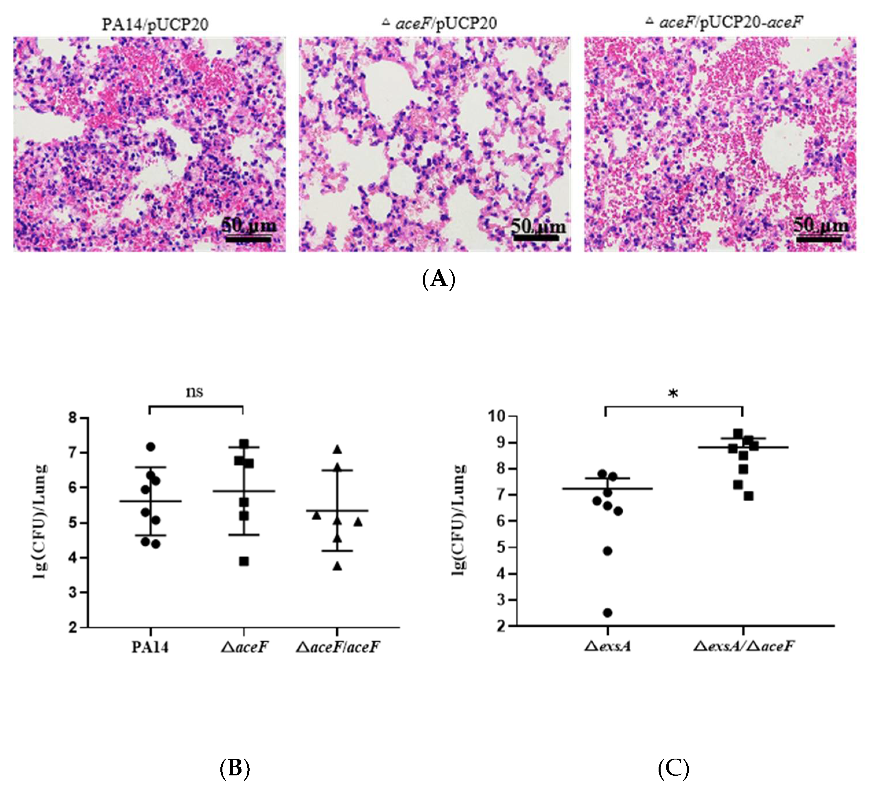
3.3. Role of AceF in Bacterial Virulence in an Acute Pneumonia Model

Since the P. aeruginosa T3SS plays an essential role in acute infections, we examined the virulence of the ΔaceF mutant in a murine acute pneumonia model by inoculating 4 × 10⁶ CFU of bacteria into each mouse. A pathology analysis of the infected lungs showed that the mutation of aceF alleviated the host inflammatory response, which is in agreement with the defective T3SS in the ΔaceF mutant (Figure 5A). However, the bacterial loads of the lungs infected by the ΔaceF mutant were similar to those infected by wild-type PA14 (Figure 5B). These results indicate that mutations in aceF might enhance a mechanism that promotes the bacterial survival independent of the T3SS. To test our hypothesis, we compared the bacterial loads of a ΔexsA mutant and a ΔexsAΔaceF double mutant. The deletion of exsA significantly attenuated the bacterial virulence. At the dose of 4 × 10⁶ CFU per mouse, both the ΔexsA and ΔexsAΔaceF mutants were cleared at 12 hpi (data not shown). Thus, we increased the dose to 4 × 10⁸ CFU per mouse. The bacterial loads in the ΔexsAΔaceF mutant infected mice were higher than those infected by the ΔexsA mutant (Figure 5C). Given that the P. aeruginosa T3SS preferentially targets bactericidal neutrophils in the acute pneumonia model [36,37,38], these results suggest that the T3SS-defective ΔaceF mutant might be more tolerant to the killing mechanisms of neutrophils.

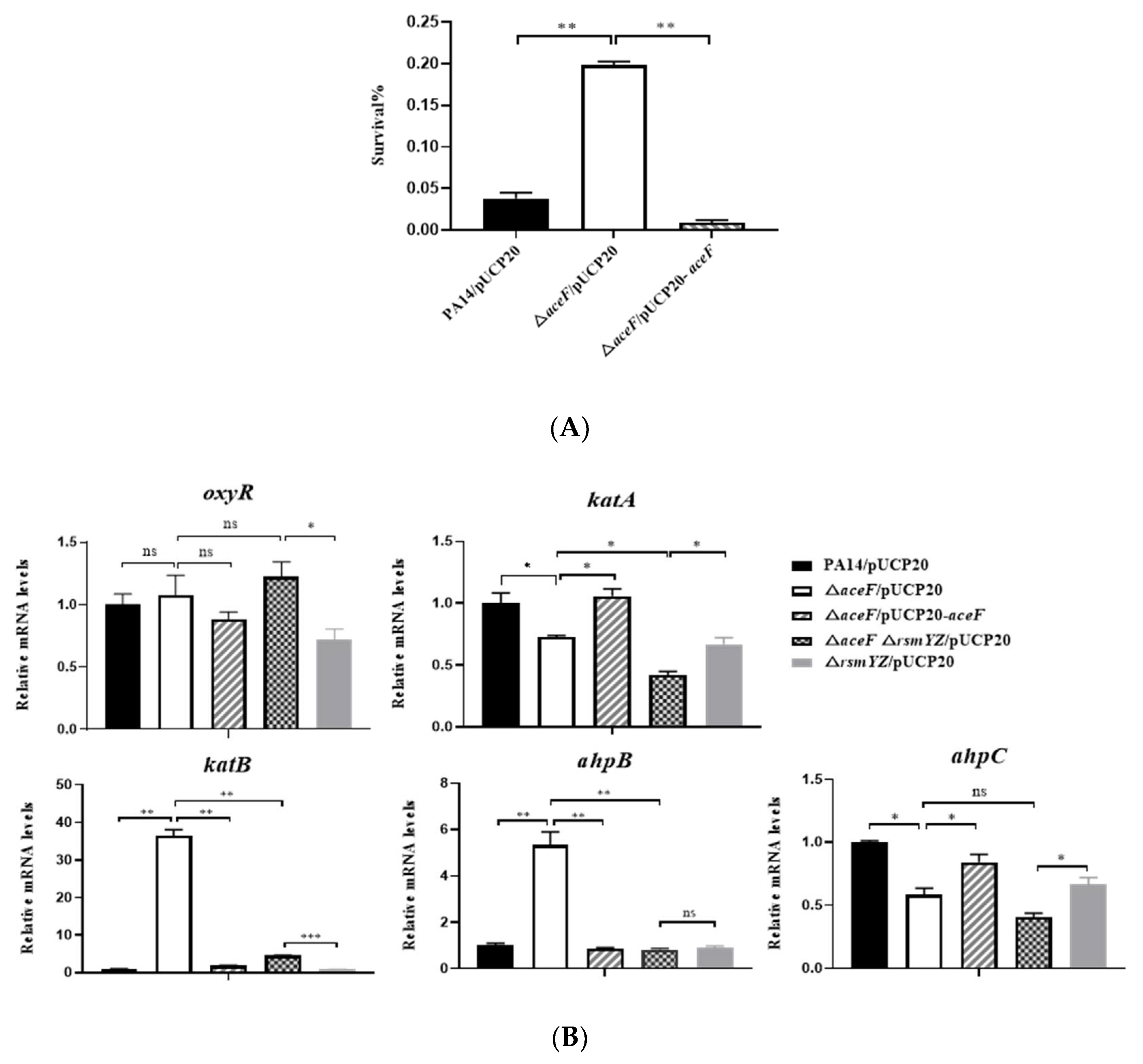
3.4. Mutation of the aceF gene Increases Bacterial Tolerance to Hydrogen Peroxide

The generation of reactive oxygen species (ROS) is a major bactericidal mechanism of neutrophils [39,40]. P. aeruginosa strains that are defective in the oxidative stress response have been shown to be attenuated in virulence [41,42]. We thus examined bacterial survival following H2O2 treatment. Compared to wild-type PA14, the ΔaceF mutant displayed a higher survival rate (Figure 6A).
To understand the mechanism of the increased tolerance to H2O2, we examined the expression of genes involved in bacterial oxidative stress response, including the regulator gene oxyR, catalase genes katA and katB and alkyl hydroperoxide reductase genes ahpB and ahpC. The mutation of the aceF gene resulted in the upregulation of ahpB and katB and reduced the mRNA levels of katA and ahpC by 1.4- and 1.7-fold (Figure 6B). The deletion of ahpB and katB reduced the H2O2 tolerance of the ΔaceF mutant, which reduced the differences in survival between wild-type PA14 and the ΔaceF mutant from 26.8-fold to 2- and 4.2-fold, respectively (Figure 6C). These results indicate that the upregulation of ahpB and katB contributes to the increased H2O2 tolerance of the ΔaceF mutant.
We next examined whether RsmY/Z are involved in the regulation of ahpB and katB in the ΔaceF mutant. The deletion of rsmY/Z reduced the expression levels of ahpB and katB (Figure 6B) as well as bacterial survival following H2O2 treatment (Figure 6D). These results demonstrate that the upregulation of RsmY/Z increases the expression of ahpB and katB and subsequent H2O2 tolerance in the ΔaceF mutant.

4. Discussion

In this study, we found that the mutation of the dihydrolipoamide acetyltransferase gene aceF in P. aeruginosa resulted in defective T3SS and enhanced H2O2 tolerance, which is due to the upregulation of RsmY/Z. By using PrsmY-lacZ and PrsmZ-lacZ transcriptional fusions, we found that the activities of the rsmY/Z promoters were elevated in the ΔaceF mutant. The transcription of rsmY/Z is under the direct regulation of the two-component regulatory system GacS-GacA. However, we did not observe an upregulation of gacS or gacA in the ΔaceF mutant (data not shown). This warrants further studies to examine whether the activity of GacS-GacA is enhanced (e.g., phosphorylation of GacA) or whether other regulatory genes are involved in the regulation of rsmY/Z.
P. aeruginosa produces catalases and alkyl hydroperoxide reductases to detoxify ROS [43]. The expression of these oxidative response genes is activated by the transcriptional regulator OxyR upon oxidative stresses [44]. OxyR is an LysR-type transcriptional regulator that consists of an N-terminal DNA-binding domain and a C-terminal regulatory domain. The presence of H2O2 leads to the formation of an intramolecular disulfide bond in the C-terminal regulatory domain, which results in the activation of OxyR [45,46]. In the ΔaceF mutant, ahpB and katB were upregulated, and ahpC and katA were slightly downregulated. However, the mRNA levels of oxyR were similar to wild-type PA14. These results suggest that AceF is unlikely to influence the protein level or function of OxyR. Since the deletion of rsmY and rsmZ reduced the expression levels of ahpB and katB, it might be possible that an unknown regulatory gene is involved in the regulation of rsmY and rsmZ and subsequent ahpB and katB in the ΔaceF mutant. In addition to OxyR, the stress sigma factor RpoS positively regulates the catalase genes katA and katB [47,48]. In Pseudomonas fluorescens, the expression of rpoS is activated by GacA and repressed by RsmA [49]. Further studies are warranted to examine whether the mutation of aceF affects the level of RpoS. The stringent response has been shown to control the expression of the catalase genes in P. aeruginosa and promotes the bacterial survival under oxidative stresses [47,50]. The stringent response is controlled by the molecule guanosine tetra- and penta-phosphate (p)ppGpp [51]. The homeostasis of (p)ppGpp is controlled by two enzymes, namely RelA and SpoT. RelA is a (p)ppGpp synthetase that is activated by amino acid starvation. SpoT functions as both (p)ppGpp synthetase and hydrolase [52]. It has been demonstrated that in E. coli, acyl carrier protein (Acyl-ACP) activates the (p)ppGpp hydrolysis activity of SpoT, whereas uncharged ACP activates the (p)ppGpp synthetase activity [53], indicating a connection between carbon metabolism and the stringent response. The pyruvate dehydrogenase connects the glycolysis and the TCA cycle by converting pyruvates to acetyl-CoA that influx into the TCA cycle. Thus, the mutation of aceF might impede the TCA cycle, which might affect the activity of SpoT. Further studies are needed to examine whether the stringent response is activated in the ΔaceF mutant.
Previously, we found that the mutation of another carbon metabolism gene eno results in the downregulation of ahpB and ahpC, which increases bacterial susceptibility to oxidative stresses [32]. The eno encoded enolase is a glycolytic enzyme that reversibly catalyzes the dehydration of 2-phosphoglycerate to phosphoenolpyruvate [54]. In addition, enolase interacts with ribonuclease (RNaseE), polynucleotide phosphorylase (PNPase) and the RNA helicase RhlB to form the RNA degradosome, which plays important roles in RNA processing and degradation [55,56]. Further studies are warranted to elucidate whether the downregulation of ahpB and ahpC is due to defective assembly of the RNA degradosome or carbon metabolism.
Overall, our results demonstrated that the mutation of aceF induces the expression of the sRNA RsmYZ, which represses the expression of T3SS genes and increases bacterial tolerance to H2O2. Previous and current studies have revealed that interruptions of carbon metabolism in different reactions result in distinct expression patterns of bacterial virulence factors, demonstrating a complex relationship between bacterial metabolism and virulence.

Supplementary Materials

The following are available online at https://www.mdpi.com/article/10.3390/microorganisms10030666/s1, Figure S1: Expression levels of exoT and exoY. Figure S2: Expression levels of crcZ. Table S1: The bacterial strains, plasmids and primers used in this study [22,29,57,58,59,60].

Author Contributions

Conceptualization, X.P. and W.W.; methodology, X.P., W.W., W.S. and H.L.; validation, X.P., W.W., H.L., Z.C., and F.B.; formal analysis, X.P., W.W., H.L. and Y.J.; investigation, H.L., Y.X. and Z.T.; resources, X.P.; data curation, X.P. and H.L.; writing—original draft preparation, X.P. and H.L.; writing—review and editing, W.W. and W.S.; visualization, X.P.; supervision, X.P. and W.S.; project administration, X.P.; funding acquisition, X.P., F.B., Z.C. and W.W. All authors have read and agreed to the published version of the manuscript.

Funding

This research was funded by the National Key Research and Development Project of China (2017YFE0125600, 82061148018), the National Science Foundation of China (31900115, 32170177, 32170199, 31970179 and 31970680), the Tianjin Municipal Science and Technology Commission (19JCYBJC24700), and the Fundamental Research Funds for the Central Universities (63213121). The funders had no role in the study design, data collection and interpretation, or the decision to submit the work for publication.

Institutional Review Board Statement

The animal study protocol was approved by the Institutional Ethics Committee of Nankai University College of Life Sciences (protocol code NK-04-2012, approved on 08-05-2012).

Conflicts of Interest

The authors declare no conflict of interest.

References

  1. Driscoll, J.A.; Brody, S.L.; Kollef, M.H. The Epidemiology, Pathogenesis and Treatment of Pseudomonas aeruginosa Infections. Drugs 2007, 67, 351–368. [Google Scholar] [CrossRef] [PubMed]
  2. El-Solh, A.A.; Hattemer, A.; Hauser, A.R.; Alhajhusain, A.; Vora, H. Clinical outcomes of type III Pseudomonas aeruginosa bacteremia. Crit. Care Med. 2012, 40, 1157–1163. [Google Scholar] [CrossRef] [PubMed]
  3. Hauser, A.R. The type III secretion system of Pseudomonas aeruginosa: Infection by injection. Nat. Rev. Genet. 2009, 7, 654–665. [Google Scholar] [CrossRef] [Green Version]
  4. Bagayoko, S.; Leon-Icaza, S.A.; Pinilla, M.; Hessel, A.; Meunier, E. Phospholipid peroxidation fuels ExoU phospholipase-dependent cell necrosis and supports Pseudomonas aeruginosa-driven pathology. bioRxiv 2021, 17, e1009927. [Google Scholar]
  5. Hauser, A.R.; Cobb, E.; Bodí, M.; Mariscal, D.; Vallés, J.; Engel, J.N.; Rello, J. Type III protein secretion is associated with poor clinical outcomes in patients with ventilator-associated pneumonia caused by Pseudomonas aeruginosa. Crit. Care Med. 2002, 30, 521–528. [Google Scholar] [CrossRef]
  6. Brutinel, E.D.; Yahr, T.L. Control of gene expression by type III secretory activity. Curr. Opin. Microbiol. 2008, 11, 128–133. [Google Scholar] [CrossRef] [Green Version]
  7. Marsden, A.E.; Intile, P.J.; Schulmeyer, K.H.; Simmons-Patterson, E.R.; Urbanowski, M.L.; Wolfgang, M.C.; Yahr, T.L. Vfr Directly Activates exsA Transcription To Regulate Expression of the Pseudomonas aeruginosa Type III Secretion System. J. Bacteriol. 2016, 198, 1442–1450. [Google Scholar] [CrossRef] [Green Version]
  8. Thibault, J.; Faudry, E.; Ebel, C.; Attree, I.; Elsen, S. Anti-activator ExsD Forms a 1:1 Complex with ExsA to Inhibit Transcription of Type III Secretion Operons. J. Biol. Chem. 2009, 284, 15762–15770. [Google Scholar] [CrossRef] [Green Version]
  9. McMackin, E.A.W.; Djapgne, L.; Corley, J.M.; Yahr, T.L. Fitting Pieces into the Puzzle of Pseudomonas aeruginosa Type III Secretion System Gene Expression. J. Bacteriol. 2019, 201, e00209-19. [Google Scholar] [CrossRef] [Green Version]
  10. Janssen, K.H.; Corley, J.M.; Djapgne, L.; Cribbs, J.T.; Voelker, D.; Slusher, Z.; Nordell, R.; Regulski, E.E.; Kazmierczak, B.I.; McMackin, E.W.; et al. Hfq and sRNA 179 Inhibit Expression of the Pseudomonas aeruginosa cAMP-Vfr and Type III Secretion Regulons. MBio 2020, 11, e00363-20. [Google Scholar] [CrossRef]
  11. Janssen, K.H.; Diaz, M.R.; Golden, M.; Graham, J.W.; Yahr, T.L. Functional analyses of the RsmY and RsmZ small non-coding regulatory RNAs in Pseudomonas aeruginosa. J. Bacteriol. 2018, 200, e00736-17. [Google Scholar] [PubMed] [Green Version]
  12. Allsopp, L.P.; Wood, T.E.; Howard, S.A.; Maggiorelli, F.; Nolan, L.M.; Wettstadt, S.; Filloux, A. RsmA and AmrZ orchestrate the assembly of all three type VI secretion systems in Pseudomonas aeruginosa. Proc. Natl. Acad. Sci. USA 2017, 114, 7707–7712. [Google Scholar] [CrossRef] [Green Version]
  13. Irie, Y.; Starkey, M.; Edwards, A.N.; Wozniak, D.J.; Romeo, T.; Parsek, M.R. Pseudomonas aeruginosa biofilm matrix polysaccharide Psl is regulated transcriptionally by RpoS and post-transcriptionally by RsmA. Mol. Microbiol. 2010, 78, 158–172. [Google Scholar] [CrossRef] [PubMed] [Green Version]
  14. Lapouge, K.; Schubert, M.; Allain, F.H.-T.; Haas, D. Gac/Rsm signal transduction pathway of γ-proteobacteria: From RNA recognition to regulation of social behaviour. Mol. Microbiol. 2008, 67, 241–253. [Google Scholar] [CrossRef] [PubMed]
  15. Chambonnier, G.; Roux, L.; Redelberger, D.; Fadel, F.; Bordi, C. The Hybrid Histidine Kinase LadS Forms a Multicomponent Signal Transduction System with the GacS/GacA Two-Component System in Pseudomonas aeruginosa. PLoS Genet. 2016, 12, e1006032. [Google Scholar] [CrossRef] [PubMed] [Green Version]
  16. Goodman, A.L.; Merighi, M.; Hyodo, M.; Ventre, I.; Filloux, A.; Lory, S. Direct interaction between sensor kinase proteins mediates acute and chronic disease phenotypes in a bacterial pathogen. Genes Dev. 2009, 23, 249–259. [Google Scholar] [CrossRef] [Green Version]
  17. Ryan Kaler, K.M.; Nix, J.C.; Schubot, F.D. RetS inhibits Pseudomonas aeruginosa biofilm formation by disrupting the canonical histidine kinase dimerization interface of GacS. J. Biol. Chem. 2021, 297, 101193. [Google Scholar] [CrossRef]
  18. Francis, V.I.; Waters, E.; Finton-James, S.E.; Gori, A.; Kadioglu, A.; Brown, A.R.; Porter, S.L. Multiple communication mechanisms between sensor kinases are crucial for virulence in Pseudomonas aeruginosa. Nat. Commun. 2018, 9, 2219. [Google Scholar] [CrossRef]
  19. Laskowski, M.A.; Kazmierczak, B.I. Mutational analysis of RetS, an unusual sensor kinase-response regulator hybrid required for Pseudomonas aeruginosa virulence. Infect. Immun. 2006, 74, 4462–4473. [Google Scholar] [CrossRef] [Green Version]
  20. Wang, B.X.; Wheeler, K.M.; Cady, K.C.; Lehoux, S.; Cummings, R.D.; Laub, M.T.; Ribbeck, K. Mucin Glycans Signal through the Sensor Kinase RetS to Inhibit Virulence-Associated Traits in Pseudomonas aeruginosa. Curr. Biol. 2021, 31, 90–102. [Google Scholar] [CrossRef]
  21. Kong, W.; Lin, C.; Zhao, J.; Shen, T.; Surette, M.G.; Shen, L.; Duan, K.J. Hybrid sensor kinase PA1611 in Pseudomonas aeruginosa regulates transitions between acute and chronic infection through direct interaction with RetS. Mol. Microbiol. 2013, 88, 784–797. [Google Scholar] [CrossRef] [PubMed]
  22. Xia, Y.; Wang, D.; Pan, X.; Xia, B.; Weng, Y.; Long, Y.; Ren, H.; Zhou, J.; Jin, Y.; Bai, F.; et al. TpiA is a Key Metabolic Enzyme That Affects Virulence and Resistance to Aminoglycoside Antibiotics through CrcZ in Pseudomonas aeruginosa. MBio 2020, 11, e02079-19. [Google Scholar] [CrossRef] [PubMed] [Green Version]
  23. Shen, D.-K.; Filopon, D.; Chaker, H.; Boullanger, S.; Derouazi, M.; Polack, B.; Toussaint, B. High-cell-density regulation of the Pseudomonas aeruginosa type III secretion system: Implications for tryptophan catabolites. Microbiology 2008, 154, 2195–2208. [Google Scholar] [CrossRef] [Green Version]
  24. Rietsch, A.; Wolfgang, M.C.; Mekalanos, J.J. Effect of Metabolic Imbalance on Expression of Type III Secretion Genes in Pseudomonas aeruginosa. Infect. Immun. 2004, 72, 1383–1390. [Google Scholar] [CrossRef] [PubMed] [Green Version]
  25. Chung, J.C.S.; Rzhepishevska, O.; Ramstedt, M.; Welch, M. Type III secretion system expression in oxygen-limited Pseudomonas aeruginosa cultures is stimulated by isocitrate lyase activity. Open Biol. 2013, 3, 120131. [Google Scholar] [CrossRef] [PubMed] [Green Version]
  26. Sonnleitner, E.; Abdou, L.; Haas, D. Small RNA as global regulator of carbon catabolite repression in Pseudomonas aeruginosa. Proc. Natl. Acad. Sci. USA 2009, 106, 21866–21871. [Google Scholar] [CrossRef] [PubMed]
  27. Naftalin, R.J.; Neale, H.; Simmons, N.L. Proceedings: Methods used to estimate the unidirectional fluxes of sodium simultaneously across the mucosal and serosal borders of rabbit ileum. J. Physiol. 1975, 252, 7P–8P. [Google Scholar] [PubMed]
  28. Rietsch, A.; Mekalanos, J.J. Metabolic regulation of type III secretion gene expression in Pseudomonas aeruginosa. Mol. Microbiol. 2005, 59, 807–820. [Google Scholar] [CrossRef] [PubMed]
  29. Hoang, T.T.; Karkhoff-Schweizer, R.R.; Kutchma, A.J.; Schweizer, H.P. A broad-host-range Flp-FRT recombination system for site-specific excision of chromosomally-located DNA sequences: Application for isolation of unmarked Pseudomonas aeruginosa mutants. Gene 1998, 212, 77–86. [Google Scholar] [CrossRef]
  30. Wu, W.; Huang, J.; Duan, B.; Traficante, D.C.; Hong, H.; Risech, M.; Lory, S.; Priebe, G.P. Th17-stimulating protein vaccines confer protection against Pseudomonas aeruginosa pneumonia. Am. J. Respir. Crit. Care Med. 2012, 186, 420–427. [Google Scholar] [CrossRef] [Green Version]
  31. Chang, L.; Pan, X.; Xia, B.; Fei, C.; Jin, Y.; Fang, B.; Gregory, P.; Cheng, Z.; Jin, S.; Wu, W. Construction of a Protective Vaccine Against Lipopolysaccharide-Heterologous Pseudomonas aeruginosa Strains Based on Expression Profiling of Outer Membrane Proteins During Infection. Front. Immunol. 2018, 9, 1737. [Google Scholar]
  32. Weng, Y.; Chen, F.; Liu, Y.; Zhao, Q.; Chen, R.; Pan, X.; Liu, C.; Cheng, Z.; Jin, S.; Jin, Y.; et al. Pseudomonas aeruginosa Enolase Influences Bacterial Tolerance to Oxidative Stresses and Virulence. Front. Microbiol. 2016, 7, 1999. [Google Scholar] [CrossRef] [PubMed] [Green Version]
  33. Savli, H.; Karadenizli, A.; Kolayli, F.; Gundes, S.; Ozbek, U.; Vahaboglu, H. Expression stability of six housekeeping genes: A proposal for resistance gene quantification studies of Pseudomonas aeruginosa by real-time quantitative RT-PCR. J. Med. Microbiol. 2003, 52, 403–408. [Google Scholar] [CrossRef] [PubMed] [Green Version]
  34. Son, M.S.; Matthews, W.J., Jr.; Kang, Y.; Nguyen, D.T.; Hoang, T.T. In Vivo Evidence of Pseudomonas aeruginosa Nutrient Acquisition and Pathogenesis in the Lungs of Cystic Fibrosis Patients. Infect. Immun. 2007, 75, 5313–5324. [Google Scholar] [CrossRef] [Green Version]
  35. Miller, J.H. Assay of β-Galactosidase. In Experiments in Molecular Genetics; Cold Spring Harbor Laboratory Press: Cold Spring Harbor, NY, USA, 1972. [Google Scholar]
  36. Diaz, M.H.; Hauser, A.R. Pseudomonas aeruginosa Cytotoxin ExoU Is Injected into Phagocytic Cells during Acute Pneumonia. Infect. Immun. 2010, 78, 1447–1456. [Google Scholar] [CrossRef] [Green Version]
  37. Howell, H.A.; Logan, L.K.; Hauser, A.R. Type III Secretion of ExoU Is Critical during Early Pseudomonas aeruginosa Pneumonia. MBio 2013, 4, e00032-13. [Google Scholar] [CrossRef] [Green Version]
  38. Rangel, S.M.; Diaz, M.H.; Knoten, C.A.; Zhang, A.; Hauser, A.R. The Role of ExoS in Dissemination of Pseudomonas aeruginosa during Pneumonia. PLoS Pathog. 2015, 11, e1004945. [Google Scholar] [CrossRef] [Green Version]
  39. Arai, T.; Kamiya, J.; Nagino, M.; Uesaka, K.; Yuasa, N.-H.; Oda, K.; Sano, T.; Yoshikai, Y.; Nimura, Y. Bilirubin impairs neutrophil bactericidal activity through the antioxidant mechanism in vitro. Gastroenterology 2000, 118, A1332. [Google Scholar] [CrossRef]
  40. Ziltener, P.; Reinheckel, T.; Oxenius, A. Neutrophil and Alveolar Macrophage-Mediated Innate Immune Control of Legionella pneumophila Lung Infection via TNF and ROS. PLoS Pathog. 2016, 12, e1005591. [Google Scholar] [CrossRef]
  41. Vogt, S.L.; Green, C.; Stevens, K.M.; Day, B.; Erickson, D.L.; Woods, D.E.; Storey, D.G. The Stringent Response Is Essential for Pseudomonas aeruginosa Virulence in the Rat Lung Agar Bead and Drosophila melanogaster Feeding Models of Infection. Infect. Immun. 2011, 79, 4094–4104. [Google Scholar] [CrossRef] [Green Version]
  42. Mohamed, F.A.; Shaker, G.H.; Askoura, M.M. Oxidative Stress Influences Pseudomonas aeruginosa Susceptibility to Antibiotics and Reduces Its Pathogenesis in Host. Curr. Microbiol. 2020, 77, 479–490. [Google Scholar] [CrossRef] [PubMed]
  43. Lee, J.S.; Heo, Y.J.; Lee, J.K.; Cho, Y.H. KatA, the major catalase, is critical for osmoprotection and virulence in Pseudomonas aeruginosa PA14. Infect. Immun. 2005, 73, 4399–4403. [Google Scholar] [CrossRef] [PubMed] [Green Version]
  44. Ochsner, U.A.; Vasil, M.L.; Alsabbagh, E.; Parvatiyar, K.; Hassett, D.J. Role of the Pseudomonas aeruginosa oxyR-recG operon in oxidative stress defense and DNA repair: OxyR-dependent regulation of katB-ankB, ahpB, and ahpC-ahpF. J. Bacteriol. 2000, 182, 4533–4544. [Google Scholar] [CrossRef] [Green Version]
  45. Jo, I.; Chung, I.Y.; Bae, H.W.; Kim, J.S.; Song, S.; Cho, Y.H.; Ha, N.C. Structural details of the OxyR peroxide-sensing mechanism. Proc. Natl. Acad. Sci. USA 2015, 112, 6443–6448. [Google Scholar] [CrossRef] [Green Version]
  46. Wei, Q.; Le, M.; Andreas, D.; Falk, H.; Warunya, P.; Ameer, E.; Sebastian, S.; Stéphane, P.; Daniel, C.; Daniel, H. Global regulation of gene expression by OxyR in an important human opportunistic pathogen. Nucleic Acids Res. 2012, 40, 4320–4333. [Google Scholar] [CrossRef] [PubMed]
  47. Khakimova, M.; Ahlgren, H.G.; Harrison, J.; English, A.M.; Nguyen, D. The Stringent Response Controls Catalases in Pseudomonas aeruginosa and Is Required for Hydrogen Peroxide and Antibiotic Tolerance. J. Bacteriol. 2013, 195, 2011–2020. [Google Scholar] [CrossRef] [Green Version]
  48. Xia, Y.; Weng, Y.; Xu, C.; Wang, D.; Pan, X.; Tian, Z.; Xia, B.; Li, H.; Chen, R.; Liu, C.; et al. Endoribonuclease YbeY Is Essential for RNA Processing and Virulence in Pseudomonas aeruginosa. MBio 2020, 11, e00659-20. [Google Scholar] [CrossRef]
  49. Stephan, H.; Claudio, V.; Cécile, G.-B.; Dieter, H. Role of the stress sigma factor RpoS in GacA/RsmA-controlled secondary metabolism and resistance to oxidative stress in Pseudomonas fluorescens CHA0. FEMS Microbiol. Lett. 2005, 243, 251–258. [Google Scholar]
  50. Pletzer, D.; Blimkie, T.; Wolfmeier, H.; Li, Y.; Baghela, A.; Lee, A.H.Y.; Falsafi, R.; Hancock, R.E.W. The Stringent Stress Response Controls Proteases and Global Regulators under Optimal Growth Conditions in Pseudomonas aeruginosa. MSystems 2020, 5, e00495-20. [Google Scholar] [CrossRef]
  51. Chowdhury, N.; Kwan, B.W.; Wood, T.K. Persistence Increases in the Absence of the Alarmone Guanosine Tetraphosphate by Reducing Cell Growth. Sci. Rep. 2016, 6, 20519. [Google Scholar] [CrossRef]
  52. Battesti, A.; Bouveret, E. Bacteria possessing two RelA/SpoT-like proteins have evolved a specific stringent response involving the acyl carrier protein-SpoT interaction. J. Bacteriol. 2009, 191, 616–624. [Google Scholar] [CrossRef] [PubMed] [Green Version]
  53. Battesti, A.; Bouveret, E. Acyl carrier protein/SpoT interaction, the switch linking SpoT-dependent stress response to fatty acid metabolism. Mol. Microbiol. 2006, 62, 1048–1063. [Google Scholar] [CrossRef] [PubMed]
  54. Sekowska, A.; Dénervaud, V.; Ashida, H.; Michoud, K.; Haas, D.; Yokota, A.; Danchin, A. Bacterial variations on the methionine salvage pathway. BMC Microbiol. 2004, 4, 9. [Google Scholar] [CrossRef] [PubMed] [Green Version]
  55. Carpousis, A.J. The RNA degradosome of Escherichia coli: An mRNA-degrading machine assembled on RNase E. Annu. Rev. Microbiol. 2007, 61, 71–87. [Google Scholar] [CrossRef] [Green Version]
  56. Mildenhall, K.B.; Wiese, N.; Chung, D.; Maples, V.F.; Mohanty, B.K.; Kushner, S.R. RNase E-based degradosome modulates polyadenylation of mRNAs after Rho-independent transcription terminators in Escherichia coli. Mol. Microbiol. 2016, 101, 645–655. [Google Scholar] [CrossRef] [Green Version]
  57. Liberati, N.T.; Urbach, J.M.; Miyata, S.; Lee, D.G.; Drenkard, E.; Wu, G.; Villanueva, J.; Wei, T.; Ausubel, F.M. Anordered, nonredundant library of Pseudomonas aeruginosa strain PA14 transposon insertion mutants. Proc. Natl. Acad. Sci. USA 2006, 103, 2833–2838. [Google Scholar] [CrossRef] [Green Version]
  58. Olsen, R.H.; DeBusscher, G.; McCombie, W.R. Development of broad-host-range vectors and gene banks: Self-cloning of the Pseudomonas aeruginosa PAO chromosome. J. Bacteriol. 1982, 150, 60–69. [Google Scholar] [CrossRef] [Green Version]
  59. Li, K.; Xu, C.; Jin, Y.; Sun, Z. SuhB is a regulator of multiple virulence genes and essential for pathogenesis of Pseudomonas aeruginosa. mBio 2013, 4, e00419-13. [Google Scholar] [CrossRef] [Green Version]
  60. Chen, R.; Weng, Y.; Feng, Z. Polynucleotide Phosphorylase Regulates Multiple Virulence Factors and the Stabilities of Small RNAs RsmY/Z in Pseudomonas aeruginosa. Front. Microbiol. 2016, 7, 247. [Google Scholar] [CrossRef]
Figure 1. AceE, AceF and LpdG link glycolysis and the tricarboxylic acid (TCA) cycle.
Microorganisms 10 00666 g001
Figure 2. AceF influences the T3SS. (A) Cytotoxicity of wild-type PA14, the ΔaceF mutant, and the mutant complemented with an aceF gene or overexpressing the exsA gene. A549 cells were infected with the bacteria for 3 h (MOI = 50). The cytotoxicity was determined by measuring crystal violet-stained cells. (B) mRNA levels of the T3SS genes. Bacteria were grown in the presence and absence of 5 mM EGTA for 4 h. The relative mRNA levels of pcrV, exsA, exsC and exoU were determined by real-time PCR. Data shown represent mean ± standard error of mean from three samples. * p < 0.05; ** p < 0.01 by ANOVA.
Microorganisms 10 00666 g002
Figure 3. Mutation of the aceF gene upregulates rsmY and rsmZ. (A) The bacteria were grown in LB with or without 5 mM EGTA to an OD600 of 1. The RNA levels of rsmY and rsmZ were determined by real-time PCR. (B) PA14, the ΔaceF mutant and the complemented strain containing the PrsmY-lacZ (left) and PrsmZ-lacZ (right) transcriptional fusion were cultured in LB with or without 5 mM EGTA to an OD600 of 1, followed by determination of the β-galactosidase activity. Data shown represent mean ± standard error of mean from three samples. * p < 0.05; ** p < 0.01 by ANOVA.
Microorganisms 10 00666 g003
Figure 4. RsmY and RsmZ repress the T3SS in the ΔaceF mutant. (A) Cytotoxicity of wild-type PA14, the ΔaceF mutant, the complemented strain, the ΔaceFΔrsmYZ triple mutant and ΔrsmYZ double mutant. A549 cells were infected with the bacteria for 3 h (MOI = 50). The cytotoxicity was determined by measuring crystal violet-stained cells. (B) mRNA levels of the T3SS genes. Bacteria were grown in the presence and absence of 5 mM EGTA for 4 h. The relative mRNA levels of pcrV, exsA, exsC and exoU were determined by real-time PCR. Data shown represent mean ± standard error of mean from three samples. * p < 0.05; ** p < 0.01; *** p < 0.001 by ANOVA.
Microorganisms 10 00666 g004
Figure 5. Mutation of the aceF gene affects bacterial pathogenesis in a murine acute pneumoniae model. (A,B) Each mouse was intranasally inoculated with 4 × 10⁶ CFU of bacteria. (A) At 12 hpi, the lung sections were observed with a 20× objective lens following hematoxylin and eosin staining. (B) Bacterial numbers in the lungs from the infected mice at 12 hpi. (C) Each mouse was infected with 4 × 10⁸ CFU of ΔexsA or ΔexsAΔaceF. At 12 hpi, the bacterial numbers in the lungs were determined. ns, not significant; * p < 0.05 by ANOVA.
Microorganisms 10 00666 g005
Figure 6. RsmY and RsmZ increase bacterial tolerance to hydrogen peroxide in the ΔaceF mutant. (A) Survival rates of bacteria following treatment with 300 mM H2O2 for 1 h. (B) The RNA levels of oxyR, katA, katB, ahpB and ahpC were determined by real-time PCR. (C,D) survival rates of bacteria following treatment with 300 mM H2O2 for 1 h. Data shown represent mean ± standard error of mean from three samples. * p < 0.05; ** p < 0.01; *** p < 0.001 by ANOVA.
Microorganisms 10 00666 g006aMicroorganisms 10 00666 g006b
Publisher’s Note: MDPI stays neutral with regard to jurisdictional claims in published maps and institutional affiliations.

