Article

The Microbiota–Gut–Brain Axis: Gut Microbiota Modulates Conspecific Aggression in Diversely Selected Laying Hens

1
Department of Animal Sciences, Purdue University, West Lafayette, IN 47907, USA
2
USDA-Agricultural Research Service, Avian Disease and Oncology Laboratory, East Lansing, MI 48823, USA
3
USDA-Agricultural Research Service, Livestock Behavior Research Unit, West Lafayette, IN 47907, USA
*
Authors to whom correspondence should be addressed.
Microorganisms 2022, 10(6), 1081; https://doi.org/10.3390/microorganisms10061081
Submission received: 19 April 2022 / Revised: 18 May 2022 / Accepted: 19 May 2022 / Published: 24 May 2022
(This article belongs to the Special Issue Gut Microbiome of Farm Animals in Health and Disease)

Abstract

:
The gut microbiota plays an important role in regulating brain function, influencing psychological and emotional stability. The correlations between conspecific aggression, gut microbiota, and physiological homeostasis were further studied in inbred laying chicken lines, 63 and 72, which were diversely selected for Marek’s disease, and they also behave differently in aggression. Ten sixty-week-old hens from each line were sampled for blood, brain, and cecal content. Neurotransmitters, cytokines, corticosterone, and heterophil/lymphocyte ratios were determined. Cecal microbiota compositions were determined by bacterial 16s rRNA sequencing, and functional predictions were performed. Our data showed that the central serotonin and tryptophan levels were higher in line 63 compared to line 72 (p < 0.05). Plasma corticosterone, heterophil/lymphocyte ratios, and central norepinephrine were lower in line 63 (p < 0.05). The level of tumor necrosis factor α tended to be higher in line 63. Faecalibacterium, Oscillibacter, Butyricicoccus, and Bacteriodes were enriched in line 63 birds, while Clostridiales vadin BB60, Alistipes, Mollicutes RF39 were dominated in line 72. From the predicted bacterial functional genes, the kynurenine pathway was upregulated in line 72. These results suggested a functional linkage of the line differences in serotonergic activity, stress response, innate immunity, and gut microbiota populations.

1. Introduction

The gut microbiota plays an important role in the bidirectional interactions between the gut and brain. The microbiota–gut–brain axis involves multiple pivotal pathways, including the autonomic nervous system, the enteric nervous system, the neuroendocrine system, and the immune system [1,2]. The disturbance of the commensal gut microbiome not only causes many intestinal diseases but also impairs brain function, leading to neuropsychiatric disorders with abnormal behaviors [1,3]. Studies conducted in humans and rodents have demonstrated the significant changes in gut microbiota composition between healthy individuals and those with psychological and emotional disorders [4,5]. It has been shown that gut microbiota can affect depressive-like and anxiety-like behaviors directly or indirectly through the regulation of brain functions, such as the activation of the hypothalamic–pituitary–adrenal (HPA) axis, via synthesizing and releasing various neurotransmitters, neuropeptides, neuroactive and immune factors [6,7].
One possible mechanism of gut microbiota influencing psychiatric health is through the tryptophan (TRP) and serotonin (5-HT) metabolism. The function of the central serotonergic system is highly correlated to depression, aggressiveness, impulsivity, and anger-related personality traits in both humans and animals [8,9]. The raphe nuclei are the primary location in the brain for 5-HT production, while the majority of peripheral 5-HT produced by enterochromaffin (EC) cells cannot pass the blood–brain barrier (BBB). Tryptophan passes through the BBB, directly affecting the 5-HT synthesis in the brain, and consequently influencing host behaviors [10,11,12]. Individuals who have diminished levels of 5-HT and 5-hydroxyindoleacetic acid (5HIAA) in the brain are generally associated with increased psychological disorders and related aggression [13,14,15,16]. Previous research has reported that low aggressive mice have higher serotonergic activations compared to high aggressive ones [17,18]. The linkage between 5-HT and psychological disorders has been intensively investigated through the modification of serotonergic regulatory genes and serotonin receptors, as well as the administration of 5-HT reuptake inhibitors [19,20,21]. However, the gut microbiota influencing serotonergic activity and related psychological health has not been thoroughly investigated. In the peripheral system, TRP, as the precursor of 5-HT, can be directly regulated by certain commensal gut microbiota through TRP synthesis, degradation and 5-HT production [22,23]. Commensal bacteria belonging to Clostridium, Ruminococcus, Blautia, and Lactobacillus have been identified to be able to degrade TRP to tryptamine, a monoamine that is structurally similar to 5-HT [24,25]. Studies on enterochromaffin cell cultures have proved that 1-trytophan hydroxylase (TPH1), which converts peripheral TRP to 5-HT, is induced by short chain fatty acid (SCFA) derived from bacteria [26]. Similarly, the enzyme that converts TRP to N-formylkynurenine, IDO-1, is also induced by commensal gut microbiota [27]. This hypothesis was supported by an acute TRP depletion study conducted in specific pathogen-free (SPF) and germ-free (GF) mice. Compared to GF mice, SPF mice had higher plasma levels of TRP, with less depressive-like behaviors [28].
Inflammation-associated brain dysfunction is another key factor associated with abnormal behaviors, including aggression [29]. A positive relationship between aggression and inflammatory markers, such as interleukin (IL)-6, IL-1ß, IL-2, and tumor necrosis factor (TNF-α), has been reported in both humans and various animals [30,31]. An impaired intestinal barrier increases gut permeability (leaky gut), leading to pathogens and toxic components, such as lipopolysaccharides (LPS), being translocated or diffused systemically, which is one of the reasons for inflammation [32,33]. The leaked bacteria and derived LPS can elevate a series of immune-inflammatory reactions, which are as follows: activating the innate immune system to release pro-inflammatory cytokines and chemokines [34]; and these immune bioactive chemicals can further cross the BBB, causing neuroinflammation; consequently, activating the HPA and SMA (sympathetic-medullary-adrenal) stress response systems [34,35]. Studies in rodents, for example, have shown that GF mice have increased stress responses compared to the mice colonized with a microbial community, which indicates the importance of gut microbiota in regulating stress responses [36,37]. Increased corticotropin releasing hormone (CRH) gene expression and circulating corticosterone (CORT) levels in GF mice are correlated with depression and anxiety-like behaviors [28,38]. Alterations of CORT and catecholamines’ (epinephrine, EP, norepinephrine, NEP, and dopamine, DA) synthesis and release have been identified as the final common pathways in regulating animals’ pathophysiological statue and behavior exhibition [39].
Intensive poultry production systems are continuously developed to meet the increasing food demands for an expanding human population [40,41]. However, social stress-induced injurious pecking and cannibalism have become the top health and welfare issues in laying hens reared in all the current housing environments, including both cage and cage-free systems [42,43]. Beak trimming, a routine practice for controlling aggression in the poultry egg industry, has been criticized to cause pain (acute, chronic or both), stress and beak trimming-induced beak malfunction [44]. Understanding the mechanisms of injurious pecking and aggression are critical for developing beak trimming alternatives to improve animal welfare. Various dietary supplements (prebiotics, probiotics, and synbiotics) and fecal microbiota transplantation have been used as bacteriotherapy (or psychomicrobiotic) in humans to target gut microbiota for treating depression, anxiety, autism, and neuropsychiatric disorders [45,46,47]. However, different outcomes have been reported, which could be affected by multiple factors, including host genetics effects on the function of the microbiota–gut–brain axis [48,49]. In chickens, several studies have given arguments about the influence of gut microbiota on modifying host physiology and behaviors. For example, van der Eijk et al. [50] reported that early-life microbiota transplantation influenced feather pecking behaviors, and van Staaveren et al. [51] indicated the potential for Lactobacillus spp. as a therapy for feather pecking behaviors. The chicken has been used as a model in developmental biology for over a century for its advantages, such as high productivity, short breeding time, simple management, and a complete genome map [52]. As a result, chickens may very well continue to offer a good animal model for investigating the correlation between genetic variants, gut microbiota population, pathophysiology, and behavioral exhibition associated with psychological and emotional status.
Numerous studies have found that aggression is heritable [53,54,55]. Differences in injurious behaviors were found in two highly inbred lines of white leghorns selected for resistance (line 63) or susceptibility (line 72) to Marek’s disease (the Avian Disease and Oncology Laboratory, USDA-ARS, East Lansing, MI, USA) [56]. The line differences in production performance and immune functions have also been reported previously [57,58,59]. Compared to line 63 birds, the line 72 birds have a higher number of CD4+ T cells but lower number of CD8+ T cells [60,61]. In addition, the production of cytokine (IL-6 and IL-18) mRNAs in response to Marek’s disease virus infection is different between the two inbred lines, of which the line 72 expresses higher levels of both cytokines than the line 63 [62]. Line differences in social stress-induced aggressive behaviors have also been reported. Compared with chickens from line 72, chickens from line 63 showed fewer aggressive behaviors, including aggressive pecks and fights, as well as duration [15,63]. The difference in behavior expression could be related to line differences in serotonergic activities and the unique distributions of 5-HT1A and 1B receptors [15]. The objectives of the current experiment were to profile the gut microbiota in both of the inbred lines and to evaluate the line’s unique correlations between aggression, gut microbiota, and physiological homeostasis.

2. Materials and Methods

2.1. Animals and Sample Collection

Two highly inbred lines, 63 and 72, were used in the current experiment. Chickens from each line were raised in multiple hen colony cages and raised under the same conditions on the USDA, Agricultural Research Service, Avian Disease and Oncology Laboratory (ADOL) specific-pathogen-free farm at East Lansing, Michigan. All these chickens were managed following preapproved standard operating procedures established by the Institutional Animal Care and Use Committee (IACUC, USDA-ARS-ADOL, MI).
Ten sixty-weekk old hens per line were randomly taken for sample collection. A 10-mL blood sample was collected from each chicken through cardiac puncture (n = 10 per group), following sodium pentobarbital sedation (30 mg/kg body weight, i.v.). After blood collection, the chickens were euthanized immediately via cervical dislocation. The raphe nucleus and cecal contents were collected following euthanasia. The blood samples were centrifuged at 700× g for 20 min at 4 °C. The brain tissues were immediately put on dry ice, while the cecal content samples were snap frozen in liquid nitrogen. All the samples were stored at −80 °C until later analysis.

2.2. Central Monoamines

The monoamines of NEP, EP, TRP, 5-HT and 5-HIAA of the raphe nuclei were analyzed using high performance liquid chromatography (UltiMateTM 3000 RSLCnano System, Thermo Fisher Scientific Inc., Waltham, MA, USA), following previously described procedures [64]. Briefly, each tissue was weighed and homogenized in ice-cold 0.2 M perchloric acid (PCA) at a 10:1 ratio. The homogenized samples were centrifuged at 18,187× g for 15 min at 4 °C. The supernatant of each sample was transferred to a new tube and mixed with the mobile phase (Catalog #: NC0351268, Thermo Fisher Scientific Inc., Waltham, MA, USA) at a 1:1 ratio. The mixture was vortexed for 1 min and centrifuged at 18,187× g for 10 min at 4 °C. The supernatant was drawn and filtered through a 0.2 µm polyvinylidene fluoride filter into a sample vial. The mobile phase flow rate was 0.8 mL/min, and the cell potentials were set at 0 mV, 225 mV, and 550 mV. Each sample was tested twice at 10 µL per injection. Each monoamine concentration was calculated according to its corresponding standard and reported as ng/g.

2.3. Plasma Serotonin and Tryptophan

The plasma concentrations of 5-HT and TRP were detected in duplicate using commercially available enzyme-linked immunosorbent assay (ELISA) kits (MyBioSource cat #: MBS016644 and MBS038462, respectively) by following the corresponding protocols. Briefly, 50 µL of standards or samples were added to each well of the antibody pre-coated plate, then 100 µL of horseradish peroxidase-conjugate reagent was added, and the plate was incubated for 60 min at 37 °C. After incubation, the plate was washed and 50 µL of chromogen solutions A and B were added. The plate was placed at dark for 15 min at 37 °C, and then the reaction was stopped by adding a stop solution. The optical density of each sample was read at 450 nm (Epoch Microplate Spectrophotometer, BioTek, Winooski, VT, USA). All the samples were measured in duplicates with a CV ≤ 15% [65].

2.4. Plasma Cytokines and Immunoglobulin G

The plasma levels of IL-6, IL-2, IL-10, tumor necrosis factor (TNF)- α (Catalog#: MBS037319, MBS005165, MBS007312, MBS9713430, MyBioSource, San Diego, CA, USA), and immunoglobulin G (IgG) (Catalog#: E33-104, Bethyl, Montgomery, TX, USA) were measured using the commercially available ELISA kits following each manufacturer’s protocol. The optical density of each sample was read at 450 nm (Epoch Microplate Spectrophotometer, BioTek, Winooski, VT, USA). All the samples were measured in duplicates with a CV ≤ 15% [65].

2.5. Plasma Corticosterone and Heterophil to Lymphocyte Ratio

Plasma corticosterone (CORT) concentration was measured using a commercially available radioimmunoassay kit (Catalog #: 07120103, MP Biomedicals, Solon, OH, USA). The total plasma CORT concentration was analyzed in duplicate following the protocol described by [66].
The H/L ratio was determined from 2 blood smears per bird. A thin layer of blood was applied on the surfaces of glass slides and then air dried overnight. The slides were stained with Wright’s staining solution. One-hundred cells, i.e., heterophils and lymphocytes, were differentiated and counted for each slide (total two-hundred cells per bird) using a microscope with 4000× magnification. The H/L ratio was calculated as the number of heterophils/the number of lymphocytes [65].

2.6. DNA Extraction and Sequencing

The total bacterial community DNA from frozen cecal contents were extracted using a QIAamp Fast DNA Stool Mini Kit (Qiagen, Hilden, Germany). Briefly, 200 mg of the cecal content sample was homogenized with 1 mL of InhibitEX buffer and 25 uL of proteinase K. The supernatant was mixed with buffer AL and incubated at 70 °C for 10 min. Then, the mixture was purified by adding buffer AW1 and AW2 and eluted in 200 uL of ATE buffer. The total DNA concentration was determined using NanoDrop (ThermoScientific, ND-1000 Spectrophotometer).
Sample 16S rRNA amplicon sequencing was performed by the Environmental Sample Preparation and Sequencing Facility (ESPSF) of Argonne National Laboratory (Chicago, IL, USA). Specifically, the V4 region of the 16S rRNA gene (515F-806R) was PCR amplified with region-specific primers that included sequencer adapter sequences used in the Illumina MiSeq flowcell [67,68]. Briefly, the sample DNA was amplified in a 25 µL PCR reaction system that contains 9.5 µL of MO BIO PCR Water (Certified DNA-Free), 12.5 µL of QuantaBio’s AccuStart II PCR ToughMix (2× concentration, 1× final), 1 µL each Golay barcode tagged Forward Primer and Reverse Primer (5 µM concentration, 200 pM final), and 1 µL of template DNA. The conditions for PCR are as follows: 94 °C for 3 min to denature the DNA, with 35 cycles at 94 °C for 45 s, 50 °C for 60 s, and 72 °C for 90 s; with a final extension of 10 min at 72 °C to ensure complete amplification. The amplicons are then quantified using PicoGreen (Invitrogen) and a plate reader (Infinite® 200 PRO, Tecan). Once quantified, the volumes of each of the products are equally pooled into a single tube. The pool was cleaned up using AMPure XP Beads (Beckman Coulter), and then quantified using a fluorometer (Qubit, Invitrogen). After quantification, the molarity of the pool was determined and diluted down to 2 nM, denatured, and then diluted to a final concentration of 6.75 pM with a 10% PhiX spike for sequencing on the Illumina MiSeq. The amplicons were then sequenced on a 151 bp × 12 bp × 151 bp MiSeq run, using customized sequencing primers and procedures [68].

2.7. Data Processing and Analysis

2.7.1. Physiological Data

The physiological data were subjected to one-way ANOVA using the MIXED method of SAS 9.4 software (SAS Institute Inc., Cary, NC, USA), with the genetic line as the major factor. Data transformation (Box-Cox or log transformation) was performed to improve normality [69]. The statistical trend was similar for both the transformed and untransformed data; thus, the untransformed data were reported, and presented as least square means (LSMeans) ± standard error of the mean (SEM). Statistical difference was reported when p ≤ 0.05, and a trend was reported when 0.05 < p ≤ 0.10.

2.7.2. 16S rRNA Sequencing Data

The 16S rRNA sequencing data (available at the NCBI SRA database, BioProject accession number PRJNA762159) were analyzed using QIIME2 (version 2020.2) and R studio (version 1.3.1073). The fastq files of the sequenced data were de-multiplexed, quality filtered, and closed reference amplicon sequence variants (ASVs) picking was performed using DADA2, according to the QIIME2 pipeline [70]. In brief, both forward and reverse reads were first trimmed at position (0, 150) and filtered out all the reads with more than 2 expected errors. The filtered reads were then dereplicated and denoised with DADA2 default parameters. After screening and filtering, a total of 497,262 sequences (original sequence was 511,408) remained in the analysis. The table of representative sequences in the samples was produced in QIIME2 and compared with the reference sequence (SILVA database release 132) to produce a feature table containing the frequencies of each taxon per sample [71]. The data were subsampled to 12,804 reads per sample. The measures of alpha diversity (phylogenetic distance, observed ASVs, pilou evenness and Shannon index) were checked for normality and analyzed using ANOVA with a Tukey’s test. A community beta diversity analysis (unweighted Unifrac, weighted Unifrac, Bray–Curtis similarity and Jaccard similarity index) between the samples was performed in R with the “vegan” package [72]. PERMANOVA testing was completed using the adonis function QIIME2 plugin. A multiple test correction q-value was calculated for beta diversity Tukey’s p-values and Spearman p-values using the Benjamini–Hochberg procedure to control the false discovery rate (FDR). The output matrices were ordinated using principal coordinate analysis (PCoA) in R software. A differential relative abundance analysis of the microbiota composition was carried out with negative binomial regression, using the DESeq2 function within the Phyloseq package (Padjusted < 0.05), where negative and positive log2 fold changes were defined as corresponding groups, respectively [73].
PICRUST2 [74] (https://github.com/picrust/picrust2 (accessed on 1 December 2020)) was used to predict the functional profiles based on 16S rRNA sequences between the two genetic lines. The plugin q2-picrust2 was applied in QIIME2 (version 2019.10). MetaCyc pathway abundances and Kyoto Encyclopedia of Genes and Genomes (KEGG) Orthology (KO) were predicted based on the Kyoto KEGG database between the two strains and analyzed with the DESeq2 method (Padjusted < 0.05). All the sequencing data were visualized using the R package “ggplot2” [75].

2.8. Correlation Analysis

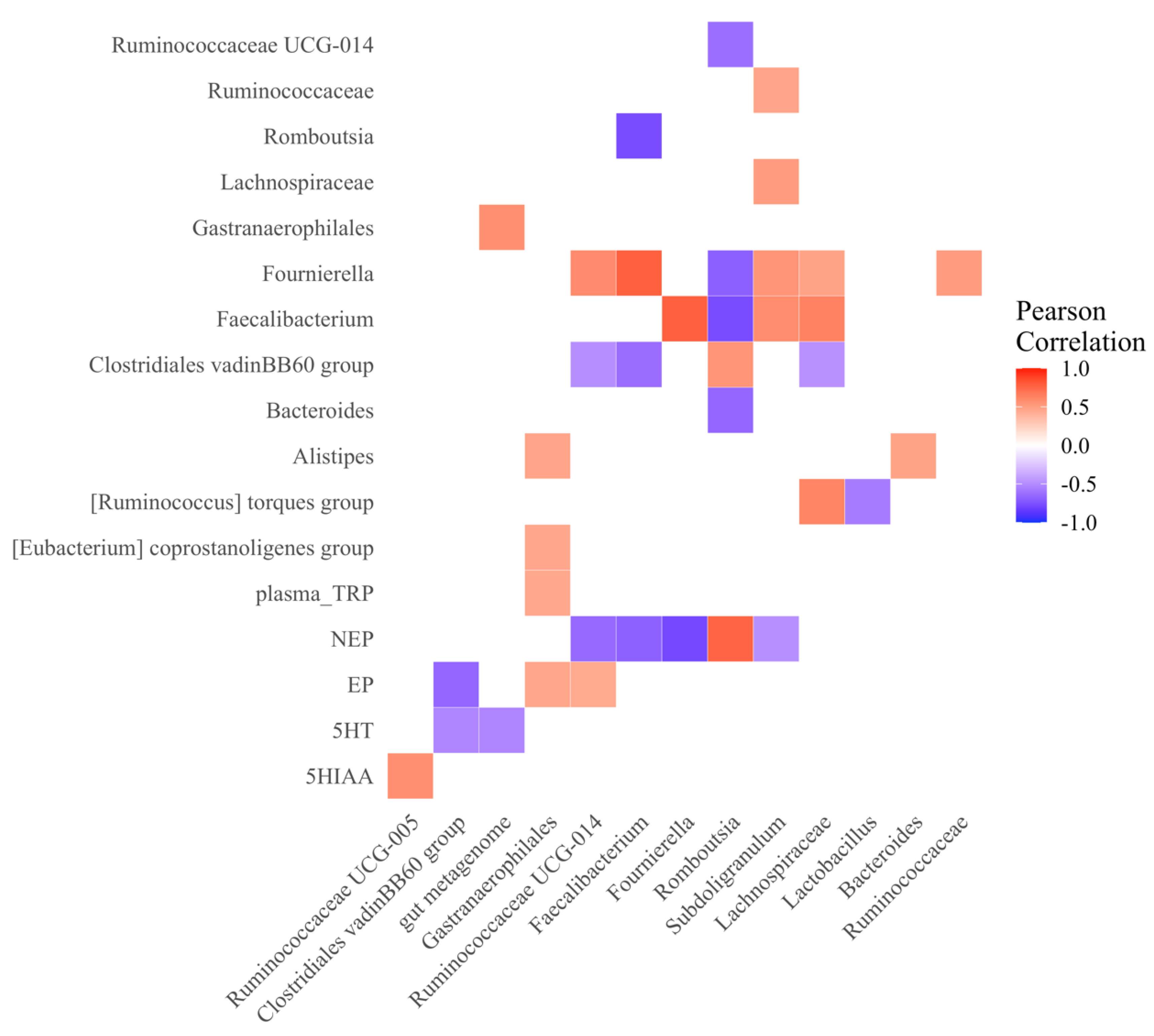
The relationships between the genus level of the microbiome (relative abundance > 1%), brain neurotransmitters, peripheral 5-HT and TRP, and CORT in each line were calculated for the Spearman’s rank correlation coefficients using the “psych” package in R [76], and the correction was made using Benjamini–Hochberg FDR. For visualization, the “ggplot2” in R was used, where significant (p < 0.05) positive and negative correlations are represented in red and blue, respectively.

3. Results

3.1. Physiological Parameters

3.1.1. Central Monoamines Levels

In the raphe nucleus, line 63-hens tended to have higher levels of EP (p = 0.06, Figure 1A), but lower levels of NEP compared to line 72-hens (p = 0.002, Figure 1B). Regarding the serotonergic system, line 63-hens had higher levels of 5-HT (p = 0.01, Figure 1C) but no difference in 5HIAA (p = 0.16, Figure 1D), resulting in a lower 5HIAA/5-HT conversion rate (p = 0.04, Figure 1E) in 63-hens. Compared to line 72-hens, the line 63-hens also tended to have a higher level of TRP than line 72-hens (p = 0.08, Figure 1F).

3.1.2. Peripheral Serotonergic, Stress and Immune Parameters

In the blood samples, TRP was higher in line 63-hens than in line 72-hens (p = 0.03, Figure 2A). No difference in plasma 5-HT was found between the two lines (Table 1). The plasma CORT level was lower in line 63-hens than line 72-hens (p = 0.04, Table 1). The heterophil to lymphocyte ratio was also lower in line 63 than line 72 (p < 0.0001, Table 1). The hens from the line 63 tended to have higher TNF-α than those from line 72 (p = 0.09). No difference was found in IL-6, IL-2, IL-10, and IgG (p > 0.05) between the lines (Table 2).

3.2. Microbiota Profile

3.2.1. Alpha Diversity

The Faith phylogenetic distance was lower in line 63-hens than line 72-hens (p = 0.03, Figure 2A). The observed ASV, Shannon index and Pielou evenness did not differ between the line 63 and line 72 (p > 0.05, Figure 2B–D).

3.2.2. Beta Diversity

Regarding the beta-diversity between the two genetic lines, with the p-value adjusted using the Benjamini–Hochberg FDR correction approach, there were significant differences in Jaccard (p = 0.001, q = 0.001, Figure 3A), Bray–Curtis (p = 0.001, q = 0.001, Figure 3B), weighted UniFrac (p = 0.002, q = 0.004, Figure 3C) and unweighted UniFrac (p = 0.001, q = 0.002, Figure 3D) distances.

3.2.3. Taxonomic Composition

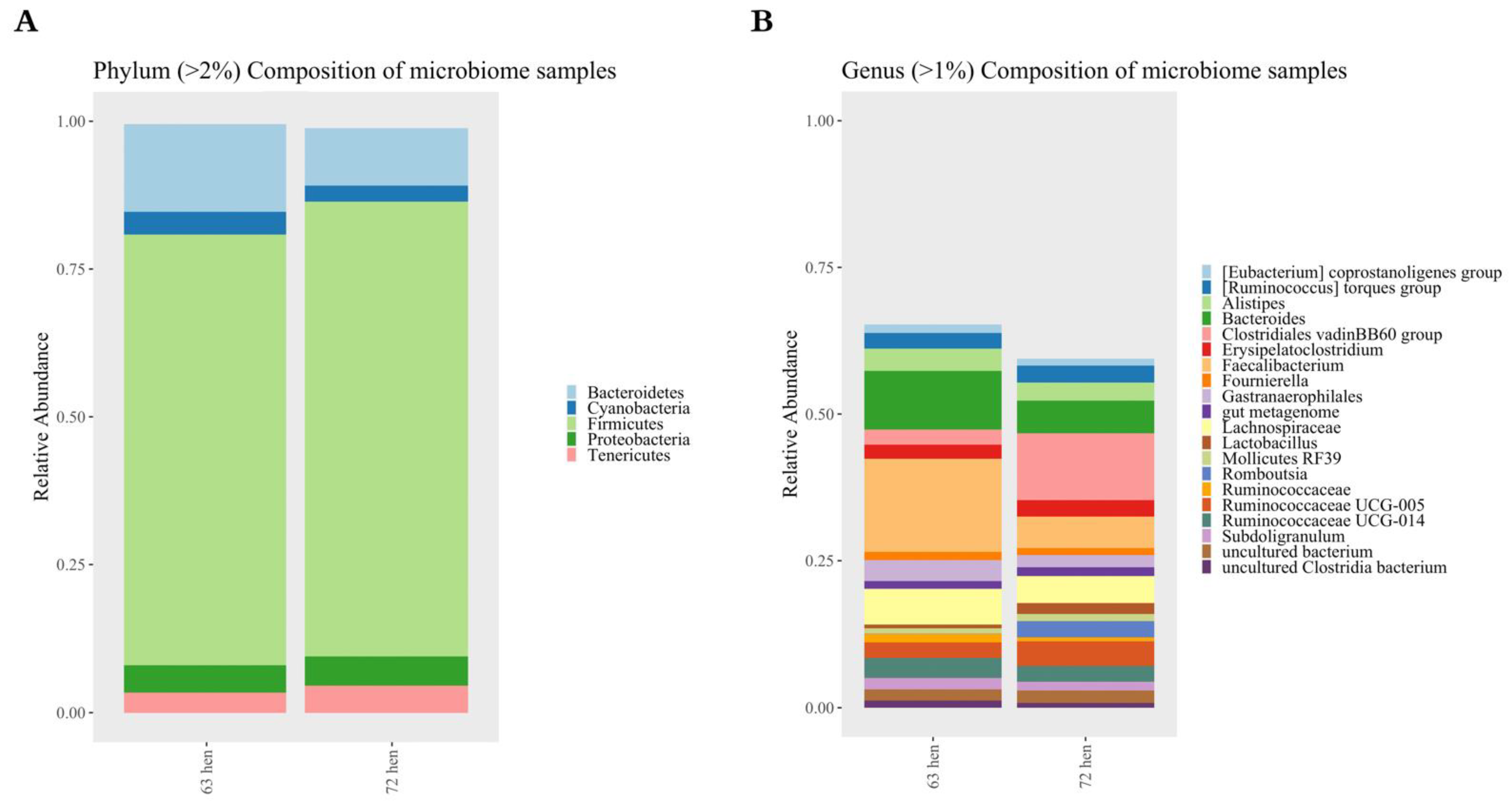
At the phylum level, the majority of 16S rRNA amplicons belonged to five main phyla, including the following: Bacteroidetes, Cyanobacteria, Firmicutes, Proteobacteria, and Tenericutes (Figure 4A). Bacteroidetes and Firmicutes were two most abundant phyla in both lines.
At the genus level, a deeper analysis using DESeq2 identified 119 out of 993 ASVs that significantly differed between the line 63 and line 72 hens (Padjusted < 0.05, Figure 5). With a log2 fold-change larger than 2, Faecalibacterium, (Eubacterium) hallii group, Oscillibacter, Butyricicoccus, Anaeroplasma, Bacteriodes, Parasutterella, Peptococcus, Ruminiclostridium 5, Ruminococcaceae, Ruminococcaceae UCG-005, Ruminococcaceae UCG-008, Ruminococcaceae UCG-009 and Tyzzerella were more enriched in line 63, while Alistipes, Anaerofilum, Anaerostipes, Clostridiales vadinBB60 group, Clostridium sensu stricto 1, Dielma, Family XIII AD3011 group, Lachnoclostridium, Lachnospiraceae FCS020 group, Methanomassiliicoccus, Mollicutes RF39, Oscillospira, Romboutsia, Ruminococcaceae UCG-010, Ruminococcus 1 and Ruminococcus 2 were more enriched in line 72.
More ASVs from the genus Fournierella (two out of three ASVs), Lachnospiraceae (nine out of thirteen ASVs), Ruminococcaceae UCG-014 (four out of seven ASVs), were enriched in line 63, while more ASVs from the genus Lactobacillus (three out of four ASVs), and Christensenellaceae R-7 group (two out of three ASVs) were more enriched in line 72.

3.2.4. Metagenome Functions Prediction

Predicted functional metagenomic profiles based on MetaCyc pathways were generated using PICRUSt2. The comparisons between the lines 63 and 72 resulted in the identification of 57 metabolic pathways that differed between the lines of hens (p < 0.05, log2 fold change > 1 shown in Figure 6). The predicted metagenomic function pathways, including antibiotic resistance, generation of precursor metabolites and energy, carbocylate degradation, inorganic nutrient metabolism and biosynthesis pathways (coenzyme A, amino acid, aromatic compound, cofactor, carrier and vitamin, fatty acid and lipid, and secondary metabolite) were increased in line 72, while CO2 fixation and biosynthesis pathways (carbohydrate, and nucleoside and nucleotide) were increased in line 63.
Specifically, the enzymes related to the TRP metabolism pathway varied between the two lines. In line 72 hens, tryptophan 2, 3-dioxygenase (K00453: TDO2, log2 fold change = −2.35, p < 0.0001), tryptophanase (K01667: tnaA, log2 fold change = −0.41, p = 0.003), indolepyruvate decarboxylase (K04103: ipdC, log2 fold change = −1.59, p = 0.02) and monoamine oxidase (K00274: MAO, log2 fold change = −3.10, p = 0.01) were detected more often, while aldehyde dehydrogenase (K00128: ALDH, log2 fold change = 0.27, p = 0.01) was increased in line 63 hens (Figure 7).

3.2.5. Microbial and Neurotransmitter Correlations

The correlations within and between the relative abundance of individual bacterial genera and concentrations of physiological relevant molecules (Figure 8) show that brain 5-HT (r = −0.53, p = 0.02) and EP (r = −0.66, p = 0.002) were negatively correlated with the Clostridiales vadin BB60 group, which was the most prominent bacteria in line 72; and brain NEP was negatively correlated to Faecalibacterium (r = −0.69, p = 0.001), Fournierella (r = −0.78, p < 0.0001), but positively correlated to Romboutsia (r = 0.76, p = 0.003).

4. Discussion

The gut microbiome plays an important role in regulating physiological and behavioral homeostasis [1]. The data in this study showed that the gut microbiota (microbiome) community composition and diversity are correlated with the differences in synthesis of central and peripheral TRP and 5-HT, cytokines, and stress indicators between the two genetic lines of hens diversely selected for or against Marek’s disease, or viral-induced tumor formation. These differences are also correlated with each line’s unique characteristics of productivity, disease resistance, and aggression reported previously [14,15,54,56,57,58].
The implications of serotonergic functions in behaviors have been the subject of many studies in both humans and animals in the last century. The outcomes from multiple experimental methods, including neurosurgical lesion of the raphe nucleus, and pharmacological or genetic inhibition of 5-HT synthesis, reuptake, and binding capability of 5-HT receptors, indicate that the hypofunction of the central serotonergic system is associated with increased aggressive and impulsive behaviors [77,78]. Previous studies in our laboratory have demonstrated some line differences in serotonergic activity and aggressive behaviors between the line 63 and 72 chickens. The chickens from line 72 exhibited increased aggressive behaviors with lower brain 5-HT concentrations, compared to line 63 chickens [15,54]. In the current study, we also found that hens from the line 72 (high aggressive) had lower levels of 5-HT and TRP in the raphe nuclei than that of the line 63 hens (low aggressive). A similar genetic variant in brain monoamines was also reported between highly aggressive and non-aggressive mice, in which excessively aggressive mice had low levels of 5-HT in the prefrontal cortex [17]. A similar heritable pattern of TRP was also found in the peripheral system, where hens from line 63 had higher plasma TRP than line 72. Tryptophan, the major precursor of 5-HT, can pass the BBB and be directly involved in brain 5-HT production [5,79,80]. The tryptophan-serotonin deficiency hypothesis of aggression has been evidenced in patients with depression [81,82] and a naturalistic 5-HT deficient animal model, the tryptophan hydroxylase 2 knockin mouse [83]. Unlike TRP, no difference was found in circulating 5-HT between the line 63 and line 72 hens. Because 5-HT cannot cross the BBB, whether the level of peripheral 5-HT contributes to aggression is unknown. In addition, the pathophysiological roles of 5-HT in behavioral and motivational regulation are different between the central and peripheral serotonergic systems [13,84]. Contradicting trends of blood 5-HT concentrations have been reported in association with behavioral dysfunctions, including aggressiveness. Studies comparing aggressive and non-aggressive dogs showed lower serum concentrations of 5-HT in aggressive dogs [85]. However, increased circulating 5-HT was found in an aggressive strain in laying hens [86] and in human patients with severe psychological illness and related aggression [87].
The difference in TRP and 5-HT synthesis and metabolism between the two genetic lines could be attributable to the commensal microbiota variance. Approximately 90% of 5-HT is produced by the gut enterochromaffin cells (EC), and gut microbiota plays a crucial role in TRP synthesis, degradation and 5-HT production, which consequently impact peripheral and central 5-HT concentrations [88,89,90]. At the genus level, many of the bacterial genera were different between the line 63 and line 72 hens. Faecalibacterium, the most prominent genera that was enriched in line 63, has been associated with the behavior of the host. Patients suffering from bipolar depression and major depressive disorder (MDD) have reduced levels of Faecalibacterium [91,92]. In a clinical study with multiple cohorts, the fecal level of Faecalibacterium prausnitzii (the sole species of Faecalibacterium genus) was negatively correlated with the severity of depression between depressive and healthy subjects [93]. More ASVs were enriched in the genus Lachnospiraceae, another beneficial microbiota, which was also found in line 63 birds, and its function is closely related to 5-HT and TRP metabolism. A mouse model of autism with deficient TRP and 5-HT metabolism in the intestines has defined a significant decrease in Lachnospiraceae taxa [94]. Other studies conducted in depression, bipolar and anhedonia susceptible patients, who generally have decreased serotonergic activities, reported decreased Lachnospiraceae abundance compared with the healthy controls [95,96,97]. Merging data have suggested the potential interventions of Lachnospiraceae and Faecalibacterium genera for the treatment of psychological disorders, including anxiety, depression, and autism [98,99]. Other enriched bacteria in the line 63, Eubacterium hallii group was also found to have a lower abundance in psychological disorder patients than healthy controls [100,101,102]. Microbial metabolites, including SCFA and long chain fatty acids (LCFA), are known to affect psychological health. Some of the genera, such as Faecalibacterium and Butyricicoccus, are butyrate-producing organisms. SCFA producers can directly stimulate 5-HT secretion [50,103,104]. In the current study, central 5-HT, and both central and peripheral TRP, were found to be higher in the line 63 hens, together with the fact that line 63 birds were less aggressive than line 72 [15]; this could partly be due to the higher relative abundance of the beneficial bacteria composition in the gut of line 63 birds.
In line 72, the more dominant genera, Anaerostipes, Clostridium and Lachnoclostridium are consistently found to be higher in MDD patients [105]. The genus Romboutsia is highly involved in metabolic capabilities, including carbohydrate utilization, single amino acids fermentation, and metabolic end products [106]. The enrichment of this bacteria could directly lead to the heavier body weight and early sexual maturation found in line 72 birds. However, another dominated genus Clostridiales vadin BB60 group has been poorly classified for its biological effectiveness [107,108]. To our knowledge, the Clostridiales vadin BB60 group is only defined to be associated with an increased risk of developing alopecia universalis, an autoimmune disorder [108].
Elevated stress hormones likely also contribute to the lower serotonergic activity in the 72 birds. In the current study, we observed that the line 72 hens had a higher plasma CORT and H/L ratio compared to 63 hens, the two common stress indicators used in avian species, which suggested an elevated stress status in the line 72 hens. The difference in the stress responding pattern is also indicated by the different changes in neurotransmitters, where the NEP level in the raphe nuclei was significantly higher in the line 72 hens, compared to the line 63. The activation of the HPA stimulates the synthesis of CRH and further regulates central NEP and peripheral CORT [109,110]. Both NEP and CORT, as stress hormones, could contribute to the behavior variance between the two genetic lines. In human and rodent studies, the hypersecretion of NEP and cortisol/corticosterone are found in most stress-related psychiatric disorders with suppressed central 5-HT activity [111,112]. Moreover, elevated stress hormones can also lead to disrupted gut barrier function and altered commensal bacteria [113]. An increase in proinflammatory bacteria, such as Helicobacter and Streptococcus, and a decrease in anti-inflammatory bacteria, such as Roseburia and Lachnospiraceae species, are observed in individuals under stress conditions [114]. In the current experiment, the levels of NEP had a positive correlation with the Clostridiales VadinBB60 group (enriched in line 72) but a negative correlation with Faecalibacterium and Fournierella (both enriched in line 63).
The immune system plays an important role in both brain and the gut integrity and is involved in regulating the function of the microbiota–gut–brain axis via the immune-brain connections [115]. The gut microbiota and related metabolites are the key regulators by which the GIT communicates with the immune system [116,117]. Recent studies have suggested that the role for the gut microbiota in the regulation of inflammation is to modulate the differentiation of various types of both immune and inflammatory cells for cytokine production and hematopoiesis [118,119,120]. For example, polysaccharide A (PSA) derived from Bacteroides fragilis directly influences the immune system, leading to systemic T cell deficiencies and Th1/Th2 imbalances in lymphoid tissues [121]. A Lactobacillus cripatus-based probiotic enhances the anti-inflammatory response of both macrophages and epithelial cells by releasing anti-inflammatory cytokine, IL-10, and reducing proinflammatory cytokines, TNF-α and IL-6 [122]. Faecalibacterium prausnitzii administration increases the IL-10 level and reduces IL-6 as well as CORT levels in depression-like and anxiety-like rats [123]. However, the other enriched genera in line 63 Anaeroplasma and Peptococcus were opportunistic pathogens, which elicited various host immune responses in human diseases [124,125]. In the current study, a trend of increased proinflammatory cytokine, TNF-α, was determined in the less aggressive 63-hens compared to the 72-hens. On the other side, the more abundant bacteria strains in the line 72 could have an important immunomodulation role in the intestinal mucosa, such as some beneficial Lactobacillus species [126]. For example, Lactobacillus rhamnosus and Lactobacillus gasseri have shown promising anti-inflammatory abilities by inhibiting proinflammatory cytokines [127,128,129]. Thus, the proinflammatory cytokine might be lower due to the presence of these beneficial Lactobacillus strains. More research is needed to further identify the correlation between cytokine production and gut microbiota in the two genetic lines.
PICRUSt2 prediction indicates the different metabolic pathways involved in energy and nutrition metabolism between the line 63 and line 72 chickens. The majority of the pathways related to biosynthesis and energy metabolism were increased in relative abundance in line 72, which could contribute to the growth and production differences between the two genetic lines. Importantly, the pathway of TRP metabolism varied between the two lines. In line 72, the predicted abundance of tryptophanase (tnaA) and indolepyruvate decarboxylase (ipdC) genes increased, by which TRP can be catalyzed into indole, pyruvate, and or ammonium [130]. In addition, the abundance of 2, 3-dioxygenase (TDO2), the rate-limiting enzyme to metabolize TRP to kynurenine, also had a log2 fold increase of over 2 in the line 72 birds. These results indicate that there could be a higher degradation rate of TRP to kynurenines through the activated kynurenine pathway, rather than 5-HT synthesis [131]. The dysregulation of TRP metabolism, with shifts in the balance between the kynurenine and 5-HT pathways, is associated with psychiatric and neurological disorders [132,133]. The deletion of TDO2 in mice resulted in lower anxiety-like behaviors than the wild type [130]. Imbeault et al. [134] also reported reduced anxiety and cognitive deficits after the inhibition of TDO2 expression. The higher TDO2 gene abundance predicted in line 72 might be a possible factor that contributes to the higher aggression reported previously [15]. Additionally, corticosteroid is known to enhance the gene expression of the enzyme TDO2 [135], which agrees with the higher CORT level found in the line 72 birds in the current study. Moreover, monoamine oxidase (MAO) also increased in abundance in line 72; this could be one of the reasons that they have lower central and peripheral 5-HT levels. However, these hypotheses need to be tested in future studies.
The current results discovered that the ubiquity of host-associated microorganisms indicate that microbes play an important role in chicken behavior, and chickens’ aggression could be reduced by modifying their microbial composition via the microbiota–gut–brain axis. However, the most behavioral differences found among chickens are affected by multiple factors, including the rearing systems. The different housing systems with inherently different designs cause chickens to exhibit different behaviors [43,136]. This approach deserves further attention under the current commercial conditions, i.e., chickens kept in both the enriched cage system and the cage-free systems (various aviaries), with the aim that eventually management practices, such as beak trimming, will no longer be necessary.
In conclusion, this work has identified the unique correlations of gut bacteria and related metabolic pathways with the different pattens of stress response, neurotransmitter expression, and innate immunity between the two diversely selected lines of chickens. Compared with the aggressive line 72, the line 63 has inherently higher central tryptophan-serotonergic activities, lower stress hormones and increased certain major beneficial bacteria, such as Faecalibacterium and Lachnospiraceae. In addition, the metagenomic function predicted certain gut microbes that could be related to the tryptophan-serotonergic metabolic differences between the two lines. The findings of this study have provided new signals for developing novel biotherapy strategies for controlling aggression in laying hens by identifying gut bacteria and their metabolites correlated with behavioral repertoire.

Author Contributions

Conceptualization, J.H., T.A.J., H.Z. and H.-W.C.; Methodology, J.H., T.A.J., H.Z. and H.-W.C.; Data Analysis, J.H. and T.A.J.; Writing—Original Draft Preparation, J.H.; Writing—Review and Editing, T.A.J., H.Z. and H.-W.C. All authors have read and agreed to the published version of the manuscript.

Funding

This research received no external funding.

Institutional Review Board Statement

All procedures of animal care used in this experiment were approved by the Purdue Animal Use and Care Committee (PACUC Number: 1712001657).

Informed Consent Statement

Not applicable.

Data Availability Statement

The 16S rRNA sequencing data are available at the NCBI SRA database, BioProject accession number PRJNA762159.

Acknowledgments

We would like to thank the staff and graduate students of the Livestock Behavior Research Unit, Avian Disease and Oncology Laboratory, USDA-ARS and the Department of Animal Sciences of Purdue University, Environmental Sample Preparation and Sequencing Facility of Argonne National Laboratory for their assistance in conducting this study. The mention of trade names or commercial products in this article is solely for the purpose of providing specific information and does not imply recommendation or endorsement of the USDA. The USDA is an equal opportunity provider and employer.

Conflicts of Interest

The authors declare no conflict of interest.

References

  1. Cryan, J.F.; O’Mahony, S.M. The microbiome-gut-brain axis: From bowel to behavior. Neurogastroenterol. Motil. 2011, 23, 187–192. [Google Scholar] [CrossRef] [PubMed]
  2. Łoniewski, I.; Misera, A.; Skonieczna-Żydecka, K.; Kaczmarczyk, M.; Kaźmierczak-Siedlecka, K.; Misiak, B.; Marlicz, W.; Samochowiec, J. Major Depressive Disorder and gut microbiota—Association not causation. A scoping review. Prog. Neuro-Psychopharmacol. Biol. Psychiatry 2021, 106, 110111. [Google Scholar] [CrossRef] [PubMed]
  3. Bioque, M.; González-Rodríguez, A.; Garcia-Rizo, C.; Cobo, J.; Monreal, J.A.; Usall, J.; Soria, V.; Labad, J. Targeting the microbiome-gut-brain axis for improving cognition in schizophrenia and major mood disorders: A narrative review. Prog. Neuro-Psychopharmacol. Biol. Psychiatry 2021, 105, 110130. [Google Scholar] [CrossRef]
  4. Li, N.; Wang, Q.; Wang, Y.; Sun, A.; Lin, Y.; Jin, Y.; Li, X. Fecal microbiota transplantation from chronic unpredictable mild stress mice donors affects anxiety-like and depression-like behavior in recipient mice via the gut microbiota-inflammation-brain axis. Stress 2019, 22, 592–602. [Google Scholar] [CrossRef] [PubMed]
  5. Lukic, I.; Getselter, D.; Ziv, O.; Oron, O.; Reuveni, E.; Koren, O.; Elliott, E. Antidepressants affect gut microbiota and Ruminococcus flavefaciens is able to abolish their effects on depressive-like behavior. Transl. Psychiatry 2019, 9, 133. [Google Scholar] [CrossRef] [PubMed]
  6. Sandhu, K.V.; Sherwin, E.; Schellekens, H.; Stanton, C.; Dinan, T.; Cryan, J.F. Feeding the microbiota-gut-brain axis: Diet, microbiome, and neuropsychiatry. Transl. Res. 2017, 179, 223–244. [Google Scholar] [CrossRef] [PubMed]
  7. Dodiya, H.B.; Forsyth, C.B.; Voigt, R.M.; Engen, P.A.; Patel, J.; Shaikh, M.; Green, S.J.; Naqib, A.; Roy, A.; Kordower, J.H.; et al. Chronic stress-induced gut dysfunction exacerbates Parkinson’s disease phenotype and pathology in a rotenone-induced mouse model of Parkinson’s disease. Neurobiol. Dis. 2020, 135, 104352. [Google Scholar] [CrossRef]
  8. Zimmermann, M.; Grabemann, M.; Mette, C.; Abdel-Hamid, M.; Ueckermann, J.; Kraemer, M.; Wiltfang, J.; Kis, B.; Zepf, F.D. The Effects of Acute Tryptophan Depletion on Reactive Aggression in Adults with Attention-Deficit/Hyperactivity Disorder (ADHD) and Healthy Controls. PLoS ONE 2012, 7, e32023. [Google Scholar] [CrossRef]
  9. Coccaro, E.F.; Fanning, J.R.; Phan, K.L.; Lee, R. Serotonin and impulsive aggression. CNS Spectr. 2015, 20, 295–302. [Google Scholar] [CrossRef]
  10. Zelante, T.; Iannitti, R.G.; Cunha, C.; De Luca, A.; Giovannini, G.; Pieraccini, G.; Zecchi, R.; D’Angelo, C.; Massi-Benedetti, C.; Fallarino, F.; et al. Tryptophan catabolites from microbiota engage aryl hydrocarbon receptor and balance mucosal reactivity via interleukin-22. Immunity 2013, 39, 372–385. [Google Scholar] [CrossRef] [Green Version]
  11. Yano, J.M.; Yu, K.; Donaldson, G.P.; Shastri, G.G.; Ann, P.; Ma, L.; Nagler, C.R.; Ismagilov, R.F.; Mazmanian, S.K.; Hsiao, E.Y. Indigenous Bacteria from the Gut Microbiota Regulate Host Serotonin Biosynthesis. Cell 2015, 161, 264–276. [Google Scholar] [CrossRef] [PubMed] [Green Version]
  12. Roager, H.M.; Licht, T.R. Microbial tryptophan catabolites in health and disease. Nat. Commun. 2018, 9, 3294. [Google Scholar] [CrossRef] [PubMed] [Green Version]
  13. Pietraszek, M.; Takada, Y.; Yan, D.; Urano, T.; Serizawa, K.; Takada, A. Relationship between serotonergic measures in periphery and the brain of mouse. Life Sci. 1992, 51, 75–82. [Google Scholar] [CrossRef]
  14. Dennis, R.; Zhang, H.M.; Cheng, H.W. Effect of Selection for Resistance and Susceptibility to Viral Diseases on Concentrations of Dopamine and Immunological Parameters in Six-Week-Old Chickens. Poult. Sci. 2006, 85, 2135–2140. [Google Scholar] [CrossRef]
  15. Dennis, R.L.; Cheng, H.-W. Differential serotonergic mediation of aggression in roosters bred for resistance and susceptibility to Marek’s disease. Br. Poult. Sci. 2014, 55, 13–20. [Google Scholar] [CrossRef]
  16. Bienenstock, J.; Kunze, W.; Forsythe, P. Microbiota and the gut–brain axis. Nutr. Rev. 2015, 73 (Suppl. 1), 28–31. [Google Scholar] [CrossRef]
  17. Caramaschi, D.; de Boer, S.F.; Koolhaas, J.M. Differential role of the 5-HT1A receptor in aggressive and non-aggressive mice: An across-strain comparison. Physiol. Behav. 2007, 90, 590–601. [Google Scholar] [CrossRef] [Green Version]
  18. Homberg, J.R.; Pattij, T.; Janssen, M.C.W.; Ronken, E.; De Boer, S.F.; Schoffelmeer, A.N.M.; Cuppen, E. Serotonin transporter deficiency in rats improves inhibitory control but not behavioural flexibility. Eur. J. Neurosci. 2007, 26, 2066–2073. [Google Scholar] [CrossRef] [Green Version]
  19. Olivier, B.; van Oorschot, R. 5-HT1B receptors and aggression: A review. Eur. J. Pharmacol. 2005, 526, 207–217. [Google Scholar] [CrossRef]
  20. Albert, P.R.; Le François, B.; Millar, A.M. Transcriptional dysregulation of 5-HT1A autoreceptors in mental illness. Mol. Brain 2011, 4, 21. [Google Scholar] [CrossRef] [Green Version]
  21. Albert, P.R.; Vahid-Ansari, F. The 5-HT1A receptor: Signaling to behavior. Biochimie 2019, 161, 34–45. [Google Scholar] [CrossRef] [PubMed]
  22. O’Mahony, S.M.; Clarke, G.; Borre, Y.E.; Dinan, T.G.; Cryan, J.F. Serotonin, tryptophan metabolism and the brain-gut-microbiome axis. Behav. Brain Res. 2015, 277, 32–48. [Google Scholar] [CrossRef] [PubMed]
  23. Gao, K.; Mu, C.-L.; Farzi, A.; Zhu, W.-Y. Tryptophan Metabolism: A Link Between the Gut Microbiota and Brain. Adv. Nutr. 2020, 11, 709–723. [Google Scholar] [CrossRef] [PubMed]
  24. Williams, B.B.; Van Benschoten, A.H.; Cimermancic, P.; Donia, M.S.; Zimmermann, M.; Taketani, M.; Ishihara, A.; Kashyap, P.C.; Fraser, J.S.; Fischbach, M.A. Discovery and Characterization of Gut Microbiota Decarboxylases that Can Produce the Neurotransmitter Tryptamine. Cell Host Microbe 2014, 16, 495–503. [Google Scholar] [CrossRef] [Green Version]
  25. Agus, A.; Planchais, J.; Sokol, H. Gut Microbiota Regulation of Tryptophan Metabolism in Health and Disease. Cell Host Microbe 2018, 23, 716–724. [Google Scholar] [CrossRef] [Green Version]
  26. Cryan, J.F.; Dinan, T.G. Mind-altering microorganisms: The impact of the gut microbiota on brain and behaviour. Nat. Rev. Neurosci. 2012, 13, 701–712. [Google Scholar] [CrossRef]
  27. Gensollen, T.; Iyer, S.S.; Kasper, D.L.; Blumberg, R.S. How colonization by microbiota in early life shapes the immune system. Science 2016, 352, 539–544. [Google Scholar] [CrossRef] [Green Version]
  28. Clarke, G.; Grenham, S.; Scully, P.; Fitzgerald, P.; Moloney, R.D.; Shanahan, F.; Dinan, T.G.; Cryan, J.F. The microbiome-gut-brain axis during early life regulates the hippocampal serotonergic system in a sex-dependent manner. Mol. Psychiatry 2013, 18, 666–673. [Google Scholar] [CrossRef] [Green Version]
  29. Fanning, J.R.; Lee, R.; Gozal, D.; Coussons-Read, M.; Coccaro, E.F. Childhood trauma and parental style: Relationship with markers of inflammation, oxidative stress, and aggression in healthy and personality disordered subjects. Biol. Psychol. 2015, 112, 56–65. [Google Scholar] [CrossRef]
  30. Kraus, M.R.; Schäfer, A.; Faller, H.; Csef, H.; Scheurlen, M. Psychiatric symptoms in patients with chronic hepatitis C receiving interferon alfa-2b therapy. J. Clin. Psychiatry 2003, 64, 708–714. [Google Scholar] [CrossRef]
  31. Hassanain, M.; Bhatt, S.; Zalcman, S.; Siegel, A. Potentiating role of interleukin-1β (IL-1β) and IL-1β type 1 receptors in the medial hypothalamus in defensive rage behavior in the cat. Brain Res. 2005, 1048, 1–11. [Google Scholar] [CrossRef] [PubMed]
  32. Van De Wouw, M.; Boehme, M.; Lyte, J.M.; Wiley, N.; Strain, C.; O’Sullivan, O.; Clarke, G.; Stanton, C.; Dinan, T.G.; Cryan, J.F. Short-chain fatty acids: Microbial metabolites that alleviate stress-induced brain-gut axis alterations. J. Physiol. 2018, 596, 4923–4944. [Google Scholar] [CrossRef] [PubMed]
  33. Halverson, T.; Alagiakrishnan, K. Gut microbes in neurocognitive and mental health disorders. Ann. Med. 2020, 52, 423–443. [Google Scholar] [CrossRef] [PubMed]
  34. Obrenovich, M.E.M. Leaky Gut, Leaky Brain? Microorganisms 2018, 6, 107. [Google Scholar] [CrossRef] [PubMed] [Green Version]
  35. Kuehne, S.A.; Cartman, S.T.; Heap, J.T.; Kelly, M.L.; Cockayne, A.; Minton, N.P. The role of toxin A and toxin B in Clostridium difficile infection. Nature 2010, 467, 711–713. [Google Scholar] [CrossRef] [PubMed] [Green Version]
  36. Sudo, N.; Chida, Y.; Aiba, Y.; Sonoda, J.; Oyama, N.; Yu, X.-N.; Kubo, C.; Koga, Y. Postnatal microbial colonization programs the hypothalamic-pituitary-adrenal system for stress response in mice. J. Physiol. 2004, 558 Pt 1, 263–275. [Google Scholar] [CrossRef]
  37. Bailey, M.T.; Dowd, S.; Galley, J.D.; Hufnagle, A.R.; Allen, R.G.; Lyte, M. Exposure to a social stressor alters the structure of the intestinal microbiota: Implications for stressor-induced immunomodulation. Brain Behav. Immun. 2011, 25, 397–407. [Google Scholar] [CrossRef] [Green Version]
  38. Crumeyrolle-Arias, M.; Jaglin, M.; Bruneau, A.; Vancassel, S.; Cardona, A.; Daugé, V.; Naudon, L.; Rabot, S. Absence of the gut microbiota enhances anxiety-like behavior and neuroendocrine response to acute stress in rats. Psychoneuroendocrinology 2014, 42, 207–217. [Google Scholar] [CrossRef]
  39. Dallman, M.F.; Akana, S.F.; Levin, N.; Walker, C.-D.; Bradbury, M.J.; Suemaru, S.; Scribner, K.S. Corticosteroids and the Control of Function in the Hypothalamo-Pituitary-Adrenal (HPA) Axisa. Ann. N. Y. Acad. Sci. 1994, 746, 22–31. [Google Scholar] [CrossRef]
  40. Pollard, C.M.; Booth, S. Addressing Food and Nutrition Security in Developed Countries. Int. J. Environ. Res. Public Health 2019, 16, 2370. [Google Scholar] [CrossRef] [Green Version]
  41. Pollard, C.M.; Booth, S. Food Insecurity and Hunger in Rich Countries—It Is Time for Action against Inequality. Int. J. Environ. Res. Public Health 2019, 16, 1804. [Google Scholar] [CrossRef] [PubMed] [Green Version]
  42. Lay, D.C., Jr.; Fulton, R.M.; Hester, P.Y.; Karcher, D.M.; Kjaer, J.B.; Mench, J.A.; Mullens, B.A.; Newberry, R.C.; Nicol, C.J.; O’Sullivan, N.P.; et al. Hen welfare in different housing systems. Poult. Sci. 2011, 90, 278–294. [Google Scholar] [CrossRef] [PubMed]
  43. Janczak, A.M.; Riber, A.B. Review of rearing-related factors affecting the welfare of laying hens. Poult. Sci. 2015, 94, 1454–1469. [Google Scholar] [CrossRef] [PubMed]
  44. Cheng, H. Morphopathological changes and pain in beak trimmed laying hens. World Poult. Sci. J. 2006, 62, 41–52. [Google Scholar] [CrossRef]
  45. Barbosa, R.S.D.; Vieira-Coelho, M.A. Probiotics and prebiotics: Focus on psychiatric disorders—A systematic review. Nutr. Rev. 2020, 78, 437–450. [Google Scholar] [CrossRef] [PubMed]
  46. Generoso, J.S.; Giridharan, V.V.; Lee, J.; Macedo, D.; Barichello, T. The role of the microbiota-gut-brain axis in neuropsychiatric disorders. Braz. J. Psychiatr. 2021, 43, 293–305. [Google Scholar] [CrossRef]
  47. Noonan, S.; Zaveri, M.; Macaninch, E.; Martyn, K. Food & mood: A review of supplementary prebiotic and probiotic interventions in the treatment of anxiety and depression in adults. BMJ Nutr. Prev. Health 2020, 3, 351–362. [Google Scholar] [CrossRef]
  48. Evrensel, A.; Tarhan, K.N. Emerging role of Gut-microbiota-brain axis in depression and therapeutic implication. Prog. Neuro-Psychopharmacol. Biol. Psychiatry 2020, 106, 110138. [Google Scholar] [CrossRef]
  49. Tremblay, A.; Lingrand, L.; Maillard, M.; Feuz, B.; Tompkins, T.A. The effects of psychobiotics on the microbiota-gut-brain axis in early-life stress and neuropsychiatric disorders. Prog. Neuro-Psychopharmacol. Biol. Psychiatry 2021, 105, 110142. [Google Scholar] [CrossRef]
  50. Van Der Eijk, J.A.J.; Rodenburg, T.B.; De Vries, H.; Kjaer, J.B.; Smidt, H.; Naguib, M.; Kemp, B.; Lammers, A. Early-life microbiota transplantation affects behavioural responses, serotonin and immune characteristics in chicken lines divergently selected on feather pecking. Sci. Rep. 2020, 10, 2750. [Google Scholar] [CrossRef]
  51. Van Staaveren, N.; Krumma, J.; Forsythe, P.; Kjaer, J.B.; Kwon, I.Y.; Mao, Y.-K.; West, C.; Kunze, W.; Harlander-Matauschek, A. Cecal motility and the impact of Lactobacillus in feather pecking laying hens. Sci. Rep. 2020, 10, 12978. [Google Scholar] [CrossRef] [PubMed]
  52. Burt, D.W. Emergence of the Chicken as a Model Organism: Implications for Agriculture and Biology. Poult. Sci. 2007, 86, 1460–1471. [Google Scholar] [CrossRef] [PubMed]
  53. Caspi, A.; McClay, J.; Moffitt, T.E.; Mill, J.; Martin-Moreno, L.; Craig, I.W.; Taylor, A.; Poulton, R. Role of Genotype in the Cycle of Violence in Maltreated Children. Science 2002, 297, 851–854. [Google Scholar] [CrossRef] [PubMed]
  54. Dennis, R.L.; Chen, Z.Q.; Cheng, H.W. Serotonergic Mediation of Aggression in High and Low Aggressive Chicken Strains. Poult. Sci. 2008, 87, 612–620. [Google Scholar] [CrossRef]
  55. Tiihonen, J.; Rautiainen, M.-R.; Ollila, H.; Repotiihonen, E.; Virkkunen, M.; Palotie, A.; Pietilainen, O.; Kristiansson, K.; Joukamaa, M.; Lauerma, H.; et al. Genetic background of extreme violent behavior. Mol. Psychiatry 2015, 20, 786–792. [Google Scholar] [CrossRef] [Green Version]
  56. Bacon, L.; Palmquist, D. Chicken lines differ in production of interferon-like activity by peripheral white blood cells stimulated with phytohemagglutinin. Poult. Sci. 2002, 81, 1629–1636. [Google Scholar] [CrossRef]
  57. Bacon, L.D.; Hunt, H.D.; Cheng, H.H. A Review of the Development of Chicken Lines to Resolve Genes Determining Resistance to Diseases. Poult. Sci. 2000, 79, 1082–1093. [Google Scholar] [CrossRef]
  58. Bacon, L.D.; Hunt, H.D.; Cheng, H.H. Genetic Resistance to Marek’s Disease. In Marek’s Disease; Current Topics in Microbiology and Immunology; Springer: Berlin/Heidelberg, Germany, 2001; Volume 255, pp. 121–141. [Google Scholar] [CrossRef]
  59. Xu, M.; Xu, C.; Liu, F.; Shen, X.; Meng, J.; Chen, H.; Yang, J.; Zhou, P.; Gao, R.; Gan, S. Effects of active immunization with newly modified GnRH peptides on spermatogenesis and production performance of Holstein bulls. Biol. Reprod. 2018, 99, 461–472. [Google Scholar] [CrossRef]
  60. Burgess, S.C.; Basaran, B.H.; Davison, T.F. Resistance to Marek’s Disease Herpesvirus-induced Lymphoma is Multiphasic and Dependent on Host Genotype. Vet. Pathol. 2001, 38, 129–142. [Google Scholar] [CrossRef] [Green Version]
  61. Yu, H.; Babiuk, L.A.; Hurk, S.; van Drunen, L.-V.D. Strategies for loading dendritic cells with hepatitis C NS5a antigen and inducing protective immunity. J. Viral Hepat. 2008, 15, 459–470. [Google Scholar] [CrossRef]
  62. Kaiser, P.; Underwood, G.; Davison, F. Differential Cytokine Responses following Marek’s Disease Virus Infection of Chickens Differing in Resistance to Marek’s Disease. J. Virol. 2003, 77, 762–768. [Google Scholar] [CrossRef] [PubMed] [Green Version]
  63. Dennis, R.; Zhang, H.M.; Bacon, L.D.; Estevez, I.; Cheng, H.W. Behavioral and physiological features of chickens diversely selected for resistance to Avian Disease. 1. Selected inbred lines differ in behavioral and physical responses to social stress. Poult. Sci. 2004, 83, 1489–1496. [Google Scholar] [CrossRef] [PubMed]
  64. Hu, J.Y. Thermal Perches as Cooling Devices for Reducing Heat Stress in Caged Laying Hens. Ph.D. Dissertation, Purdue University, West Lafayette, IN, USA, 2017. [Google Scholar]
  65. Hu, J.; Hester, P.; Makagon, M.; Xiong, Y.; Gates, R.; Cheng, H. Effect of cooled perches on physiological parameters of caged White Leghorn hens exposed to cyclic heat. Poult. Sci. 2019, 98, 2317–2325. [Google Scholar] [CrossRef] [PubMed]
  66. Cheng, H.W.; Dillworth, G.; Singleton, P.; Chen, Y.; Muirt, W.M. Effects of Group Selection for Productivity and Longevity on Blood Concentrations of Serotonin, Catecholamines, and Corticosterone of Laying Hens. Poult. Sci. 2001, 80, 1278–1285. [Google Scholar] [CrossRef]
  67. Caporaso, J.G.; Lauber, C.L.; Walters, W.A.; Berg-Lyons, D.; Huntley, J.; Fierer, N.; Owens, S.M.; Betley, J.; Fraser, L.; Bauer, M.; et al. Ultra-high-throughput microbial community analysis on the Illumina HiSeq and MiSeq platforms. ISME J. 2012, 6, 1621–1624. [Google Scholar] [CrossRef] [Green Version]
  68. Caporaso, J.G.; Lauber, C.L.; Walters, W.A.; Berg-Lyons, D.; Lozupone, C.A.; Turnbaugh, P.J.; Fierer, N.; Knight, R. Global patterns of 16S rRNA diversity at a depth of millions of sequences per sample. Proc. Natl. Acad. Sci. USA 2011, 108 (Suppl. S1), 4516–4522. [Google Scholar] [CrossRef] [Green Version]
  69. Caliński, T.; Steel, R.G.D.; Torrie, J.H. Principles and Procedures of Statistics: A Biometrical Approach. Biometrics 1981, 37, 859–860. [Google Scholar] [CrossRef]
  70. Callahan, B.J.; Sankaran, K.; Fukuyama, J.A.; McMurdie, P.J.; Holmes, S.P. Bioconductor Workflow for Microbiome Data Analysis: From raw reads to community analyses. F1000Research 2016, 5, 1492. [Google Scholar] [CrossRef]
  71. Quast, C.; Pruesse, E.; Yilmaz, P.; Gerken, J.; Schweer, T.; Yarza, P.; Peplies, J.; Glöckner, F.O. The SILVA Ribosomal RNA Gene Database Project: Improved Data Processing and Web-Based Tools. Nucleic Acids Res. 2013, 41, D590–D596. [Google Scholar] [CrossRef]
  72. Oksanen, F.J. Vegan: Community Ecology Package. R Package Version 2.5-7. 2020. Available online: https://cran.r-project.org/web/packages/vegan/vegan.pdf (accessed on 1 December 2020).
  73. Love, M.I.; Huber, W.; Anders, S. Moderated estimation of fold change and dispersion for RNA-seq data with DESeq2. Genome Biol. 2014, 15, 550. [Google Scholar] [CrossRef] [Green Version]
  74. Douglas, G.M.; Maffei, V.J.; Zaneveld, J.R.; Yurgel, S.N.; Brown, J.R.; Taylor, C.M.; Huttenhower, C.; Langille, M.G.I. PICRUSt2 for prediction of metagenome functions. Nat. Biotechnol. 2020, 38, 685–688. [Google Scholar] [CrossRef] [PubMed]
  75. Wickham, H. ggplot2: Elegant Graphics for Data Analysis; Springer-Verlag: New York, NY, USA, 2016; ISBN 978-3-319-24277-4. Available online: https://ggplot2.tidyverse.org (accessed on 1 December 2020).
  76. Revelle, W. Package ‘Psych’. Version 2.1.3. 2021. Available online: https://cran.r-project.org/web/packages/psych/psych.pdf (accessed on 1 December 2020).
  77. Palumbo, S.; Mariotti, V.; Iofrida, C.; Pellegrini, S. Genes and Aggressive Behavior: Epigenetic Mechanisms Underlying Individual Susceptibility to Aversive Environments. Front. Behav. Neurosci. 2018, 12, 117. [Google Scholar] [CrossRef] [PubMed]
  78. Runions, K.C.; Morandini, H.; Rao, P.; Wong, J.W.Y.; Kolla, N.J.; Pace, G.; Mahfouda, S.; Hildebrandt, C.S.; Stewart, R.; Zepf, F.D. Serotonin and aggressive behaviour in children and adolescents: A systematic review. Acta Psychiatr. Scand. 2019, 139, 117–144. [Google Scholar] [CrossRef] [PubMed]
  79. Browne, C.; Clarke, G.; Dinan, T.G.; Cryan, J.F. An effective dietary method for chronic tryptophan depletion in two mouse strains illuminates a role for 5-HT in nesting behaviour. Neuropharmacology 2012, 62, 1903–1915. [Google Scholar] [CrossRef]
  80. Lu, F.; Zhang, Y.; Trivedi, A.; Jiang, X.; Chandra, D.; Zheng, J.; Nakano, Y.; Uyghurturk, D.A.; Jalai, R.; Onur, S.G.; et al. Fluoride related changes in behavioral outcomes may relate to increased serotonin. Physiol. Behav. 2019, 206, 76–83. [Google Scholar] [CrossRef]
  81. Lindseth, G.; Helland, B.; Caspers, J. The Effects of Dietary Tryptophan on Affective Disorders. Arch. Psychiatr. Nurs. 2015, 29, 102–107. [Google Scholar] [CrossRef] [Green Version]
  82. Jenkins, T.A.; Nguyen, J.C.D.; Polglaze, K.E.; Bertrand, P.P. Influence of Tryptophan and Serotonin on Mood and Cognition with a Possible Role of the Gut-Brain Axis. Nutrients 2016, 8, 56. [Google Scholar] [CrossRef]
  83. Jacobsen, J.P.R.; Medvedev, I.O.; Caron, M.G. The 5-HT deficiency theory of depression: Perspectives from a naturalistic 5-HT deficiency model, the tryptophan hydroxylase 2 Arg 439 His knockin mouse. Philos. Trans. R. Soc. Lond B Biol. Sci. 2012, 367, 2444–2459. [Google Scholar] [CrossRef] [Green Version]
  84. Manchia, M.; Carpiniello, B.; Valtorta, F.; Comai, S. Serotonin Dysfunction, Aggressive Behavior, and Mental Illness: Exploring the Link Using a Dimensional Approach. ACS Chem. Neurosci. 2017, 8, 961–972. [Google Scholar] [CrossRef]
  85. Rosado, B.; García-Belenguer, S.; Palacio, J.; Chacón, G.; Villegas, A.; Alcalde, A.I. Serotonin transporter activity in platelets and canine aggression. Vet. J. 2010, 186, 104–105. [Google Scholar] [CrossRef]
  86. Cheng, H.; Muir, W.M. The effects of genetic selection for survivability and productivity on chicken physiological homeostasis. World Poult. Sci. J. 2005, 61, 15. [Google Scholar] [CrossRef]
  87. Luykx, J.; Bakker, S.C.; Lentjes, E.; Neeleman, M.; Strengman, E.; Mentink, L.; Deyoung, J.; De Jong, S.; Sul, J.H.; Eskin, E.; et al. Genome-wide association study of monoamine metabolite levels in human cerebrospinal fluid. Mol. Psychiatry 2014, 19, 228–234. [Google Scholar] [CrossRef] [PubMed]
  88. Costedio, M.M.; Hyman, N.; Mawe, G.M. Serotonin and Its Role in Colonic Function and in Gastrointestinal Disorders. Dis. Colon Rectum 2007, 50, 376–388. [Google Scholar] [CrossRef] [PubMed]
  89. McLean, P.G.; Borman, R.A.; Lee, K. 5-HT in the enteric nervous system: Gut function and neuropharmacology. Trends Neurosci. 2007, 30, 9–13. [Google Scholar] [CrossRef] [PubMed]
  90. Desbonnet, L.; Garrett, L.; Clarke, G.; Bienenstock, J.; Dinan, T.G. The probiotic Bifidobacteria infantis: An assessment of potential antidepressant properties in the rat. J. Psychiatr. Res. 2008, 43, 164–174. [Google Scholar] [CrossRef]
  91. Jiang, H.; Ling, Z.; Zhang, Y.; Mao, H.; Ma, Z.; Yin, Y.; Wang, W.; Tang, W.; Tan, Z.; Shi, J.; et al. Altered fecal microbiota composition in patients with major depressive disorder. Brain Behav. Immun. 2015, 48, 186–194. [Google Scholar] [CrossRef] [Green Version]
  92. Aizawa, E.; Tsuji, H.; Asahara, T.; Takahashi, T.; Teraishi, T.; Yoshida, S.; Ota, M.; Koga, N.; Hattori, K.; Kunugi, H. Possible association of Bifidobacterium and Lactobacillus in the gut microbiota of patients with major depressive disorder. J. Affect. Disord. 2016, 202, 254–257. [Google Scholar] [CrossRef]
  93. Valles-Colomer, M.; Falony, G.; Darzi, Y.; Tigchelaar, E.F.; Wang, J.; Tito, R.Y.; Schiweck, C.; Kurilshikov, A.; Joossens, M.; Wijmenga, C.; et al. The neuroactive potential of the human gut microbiota in quality of life and depression. Nat. Microbiol. 2019, 4, 623–632. [Google Scholar] [CrossRef]
  94. Golubeva, A.V.; Joyce, S.A.; Moloney, G.; Burokas, A.; Sherwin, E.; Arboleya, S.; Flynn, I.; Khochanskiy, D.; Moya-Pérez, A.; Peterson, V.; et al. Microbiota-related Changes in Bile Acid & Tryptophan Metabolism are Associated with Gastrointestinal Dysfunction in a Mouse Model of Autism. EBioMedicine 2017, 24, 166–178. [Google Scholar] [CrossRef] [Green Version]
  95. Flowers, S.A.; Evans, S.J.; Ward, K.M.; McInnis, M.G.; Ellingrod, V.L. Interaction between Atypical Antipsychotics and the Gut Microbiome in a Bipolar Disease Cohort. Pharmacotherapy 2017, 37, 261–267. [Google Scholar] [CrossRef]
  96. Sun, J.; Wang, F.; Hu, X.; Yang, C.; Xu, H.; Yao, Y.; Liu, J. Clostridium butyricum Attenuates Chronic Unpredictable Mild Stress-Induced Depressive-Like Behavior in Mice via the Gut-Brain Axis. J. Agric. Food Chem. 2018, 66, 8415–8421. [Google Scholar] [CrossRef] [PubMed]
  97. Yang, C.; Fang, X.; Zhan, G.; Huang, N.; Li, S.; Bi, J.; Jiang, R.; Yang, L.; Miao, L.; Zhu, B.; et al. Key role of gut microbiota in anhedonia-like phenotype in rodents with neuropathic pain. Transl. Psychiatry 2019, 9, 57. [Google Scholar] [CrossRef] [PubMed] [Green Version]
  98. Rook, G.A.W.; Raison, C.L.; Lowry, C.A. Childhood Microbial Experience, Immunoregulation, Inflammation, and Adult Susceptibility to Psychosocial Stressors and Depression. In Inflammation and Immunity in Depression Basic Science and Clinical Applications; Baune, B.T., Ed.; Academic Press: Washington, DC, USA, 2018; pp. 17–44. [Google Scholar]
  99. Dahiya, D.K.; Dangi, A.K.; Shandilya, U.K.; Puniya, A.K.; Shukla, P. New-Generation Probiotics: Perspectives and Applications. In Microbiome and Metabolome in Diagnosis, Therapy, and Other Strategic Applications; Faintuch, E.D.S.J., Faintuch, S., Eds.; Academic Press: Washington, DC, USA, 2019; pp. 417–424. [Google Scholar]
  100. De Angelis, M.; Piccolo, M.; Vannini, L.; Siragusa, S.; De Giacomo, A.; Serrazzanetti, D.I.; Cristofori, F.; Guerzoni, M.E.; Gobbetti, M.; Francavilla, R. Fecal Microbiota and Metabolome of Children with Autism and Pervasive Developmental Disorder Not Otherwise Specified. PLoS ONE 2013, 8, e76993. [Google Scholar] [CrossRef] [PubMed] [Green Version]
  101. Chen, Y.-H.; Bai, J.; Wu, D.; Yu, S.-F.; Qiang, X.-L.; Bai, H.; Wang, H.-N.; Peng, Z.-W. Association between fecal microbiota and generalized anxiety disorder: Severity and early treatment response. J. Affect. Disord. 2019, 259, 56–66. [Google Scholar] [CrossRef]
  102. Averina, O.V.; Zorkina, Y.A.; Yunes, R.A.; Kovtun, A.S.; Ushakova, V.M.; Morozova, A.Y.; Kostyuk, G.P.; Danilenko, V.N.; Chekhonin, V.P. Bacterial Metabolites of Human Gut Microbiota Correlating with Depression. Int. J. Mol. Sci. 2020, 21, 9234. [Google Scholar] [CrossRef]
  103. Fukumoto, S.; Tatewaki, M.; Yamada, T.; Fujimiya, M.; Mantyh, C.; Voss, M.; Eubanks, S.; Harris, M.; Pappas, T.; Takahashi, T. Short-chain fatty acids stimulate colonic transit via intraluminal 5-HT release in rats. Am. J. Physiol. Regul. Integr. Comp. Physiol. 2003, 284, R1269–R1276. [Google Scholar] [CrossRef] [Green Version]
  104. Chang, S.-C.; Shen, M.-H.; Liu, C.-Y.; Pu, C.-M.; Hu, J.-M.; Huang, C.-J. A gut butyrate-producing bacterium Butyricicoccus pullicaecorum regulates short-chain fatty acid transporter and receptor to reduce the progression of 1,2-dimethylhydrazine-associated colorectal cancer. Oncol. Lett. 2020, 20, 327. [Google Scholar] [CrossRef]
  105. Cheung, S.G.; Goldenthal, A.R.; Uhlemann, A.-C.; Mann, J.J.; Miller, J.M.; Sublette, M.E. Systematic Review of Gut Microbiota and Major Depression. Front. Psychiatry 2019, 10, 34. [Google Scholar] [CrossRef] [Green Version]
  106. Gerritsen, J.; Hornung, B.; Ritari, J.; Paulin, L.; Rijkers, G.T.; Schaap, P.J.; de Vos, W.M.; Smidt, H. A Comparative and functional ge-nomics analysis of the genus romboutsia provides insight into adaptation to an intestinal lifestyle. BioRxiv 2019, 845511. [Google Scholar] [CrossRef] [Green Version]
  107. Richards, P.; Fothergill, J.; Bernardeau, M.; Wigley, P. Development of the Caecal Microbiota in Three Broiler Breeds. Front. Vet. Sci. 2019, 6, 201. [Google Scholar] [CrossRef]
  108. Moreno-Arrones, O.; Serrano-Villar, S.; Perez-Brocal, V.; Saceda-Corralo, D.; Morales-Raya, C.; Rodrigues-Barata, R.; Moya, A.; Jaen-Olasolo, P.; Vano-Galvan, S. Analysis of the gut microbiota in alopecia areata: Identification of bacterial biomarkers. J. Eur. Acad. Dermatol. Venereol. 2019, 34, 400–405. [Google Scholar] [CrossRef]
  109. Leonard, B.E. Stress, Norepinephrine and depression. J. Psychiatry Neurosci. 2001, 26, S11–S16. [Google Scholar] [CrossRef] [PubMed] [Green Version]
  110. Wood, S.K.; Valentino, R.J. The brain norepinephrine system, stress and cardiovascular vulnerability. Neurosci. Biobehav. Rev. 2017, 74, 393–400. [Google Scholar] [CrossRef] [PubMed] [Green Version]
  111. Wood, S.K.; Bhatnagar, S. Resilience to the effects of social stress: Evidence from clinical and preclinical studies on the role of coping strategies. Neurobiol. Stress 2015, 1, 164–173. [Google Scholar] [CrossRef] [PubMed] [Green Version]
  112. Wood, S.K.; McFadden, K.V.; Grigoriadis, D.; Bhatnagar, S.; Valentino, R.J. Depressive and cardiovascular disease comorbidity in a rat model of social stress: A putative role for corticotropin-releasing factor. Psychopharmacology 2012, 222, 325–336. [Google Scholar] [CrossRef] [PubMed] [Green Version]
  113. Maes, M.; Kubera, M.; Leunis, J.-C.; Berk, M.; Geffard, M.; Bosmans, E. In depression, bacterial translocation may drive inflammatory responses, oxidative and nitrosative stress (O&NS), and autoimmune responses directed against O&NS-damaged neoepitopes. Acta Psychiatr. Scand. 2013, 127, 344–354. [Google Scholar] [CrossRef] [PubMed]
  114. Gao, X.; Cao, Q.; Cheng, Y.; Zhao, D.; Wang, Z.; Yang, H.; Wu, Q.; You, L.; Wang, Y.; Lin, Y.; et al. Chronic stress promotes colitis by disturbing the gut microbiota and triggering immune system response. Proc. Natl. Acad. Sci. USA 2018, 115, E2960–E2969. [Google Scholar] [CrossRef] [PubMed] [Green Version]
  115. Bengmark, S. Gut microbiota, immune development and function. Pharmacol. Res. 2013, 69, 87–113. [Google Scholar] [CrossRef]
  116. Rooks, M.G.; Garrett, W.S. Gut microbiota, metabolites and host immunity. Nat. Rev. Immunol. 2016, 16, 341–352. [Google Scholar] [CrossRef]
  117. Cohen, L.J.; Esterhazy, D.; Kim, S.-H.; Lemetre, C.; Aguilar, R.; Gordon, E.A.; Pickard, A.; Cross, J.R.; Emiliano, A.B.; Han, S.M.; et al. Commensal bacteria make GPCR ligands that mimic human signalling molecules. Nature 2017, 549, 48–53. [Google Scholar] [CrossRef] [Green Version]
  118. Schirmer, M.; Smeekens, S.P.; Vlamakis, H.; Jaeger, M.; Oosting, M.; Franzosa, E.A.; ter Horst, R.; Jansen, T.; Jacobs, L.; Bonder, M.J.; et al. Linking the Human Gut Microbiome to Inflammatory Cytokine Production Capacity. Cell 2016, 167, 1897. [Google Scholar] [CrossRef] [PubMed] [Green Version]
  119. Geva-Zatorsky, N.; Sefik, E.; Kua, L.; Pasman, L.; Tan, T.G.; Ortiz-Lopez, A.; Yanortsang, T.B.; Yang, L.; Jupp, R.; Mathis, D.; et al. Mining the Human Gut Microbiota for Immunomodulatory Organisms. Cell 2017, 168, 928–943.e11. [Google Scholar] [CrossRef] [PubMed] [Green Version]
  120. Brandsma, E.; Kloosterhuis, N.J.; Koster, M.; Dekker, D.C.; Gijbels, M.J.; van der Velden, S.; Ríos-Morales, M.; Van Faassen, M.J.; Loreti, M.G.; De Bruin, A.; et al. A Proinflammatory Gut Microbiota Increases Systemic Inflammation and Accelerates Atherosclerosis. Circ. Res. 2019, 124, 94–100. [Google Scholar] [CrossRef] [PubMed]
  121. Mazmanian, S.K.; Liu, C.H.; Tzianabos, A.O.; Kasper, D.L. An Immunomodulatory Molecule of Symbiotic Bacteria Directs Maturation of the Host Immune System. Cell 2005, 122, 107–118. [Google Scholar] [CrossRef] [Green Version]
  122. Rizzo, A.; Fiorentino, M.; Buommino, E.; Donnarumma, G.; Losacco, A.; Bevilacqua, N. Lactobacillus crispatus mediates anti-inflammatory cytokine interleukin-10 induction in response to Chlamydia trachomatis infection in vitro. Int. J. Med. Microbiol. 2015, 305, 815–827. [Google Scholar] [CrossRef]
  123. Hao, Z.; Wang, W.; Guo, R.; Liu, H. Faecalibacterium prausnitzii (ATCC 27766) has preventive and therapeutic effects on chronic unpredictable mild stress-induced depression-like and anxiety-like behavior in rats. Psychoneuroendocrinology 2019, 104, 132–142. [Google Scholar] [CrossRef]
  124. Yang, C.; Chalasani, G.; Ng, Y.-H.; Robbins, P.D. Exosomes released from mycoplasma infected tumor cells activate inhibitory B cells. PLoS ONE 2012, 7, e36138. [Google Scholar] [CrossRef] [Green Version]
  125. Chen, Q.; He, Z.; Zhuo, Y.; Li, S.; Yang, W.; Hu, L.; Zhong, H. Rubidium chloride modulated the fecal microbiota community in mice. BMC Microbiol. 2021, 21, 46. [Google Scholar] [CrossRef]
  126. Delcenserie, V.; Martel, D.; Lamoureux, M.; Amiot, J.; Boutin, Y.; Roy, D. Immunomodulatory effects of probiotics in the intestinal tract. Curr. Issues Mol. Biol. 2008, 10, 37–54. [Google Scholar]
  127. Li, N.; Russell, W.M.; Douglas-Escobar, M.; Hauser, N.; Lopez, M.; Neu, J. Live and Heat-Killed Lactobacillus rhamnosus GG: Effects on Proinflammatory and Anti-Inflammatory Cytokines/Chemokines in Gastrostomy-Fed Infant Rats. Pediatr. Res. 2009, 66, 203–207. [Google Scholar] [CrossRef] [Green Version]
  128. Oh, N.S.; Joung, J.Y.; Lee, J.Y.; Kim, Y. Probiotic and anti-inflammatory potential of Lactobacillus rhamnosus 4B15 and Lactobacillus gasseri 4M13 isolated from infant feces. PLoS ONE 2018, 13, e0192021. [Google Scholar] [CrossRef] [PubMed]
  129. Wang, T.; Yan, H.; Lu, Y.; Li, X.; Wang, X.; Shan, Y.; Yi, Y.; Liu, B.; Zhou, Y.; Lü, X. Anti-obesity effect of Lactobacillus rhamnosus LS-8 and Lactobacillus crustorum MN047 on high-fat and high-fructose diet mice base on inflammatory response alleviation and gut microbiota regulation. Eur. J. Nutr. 2019, 59, 2709–2728. [Google Scholar] [CrossRef] [PubMed]
  130. Knott, P.J.; Curzon, G. Free Tryptophan in Plasma and Brain Tryptophan Metabolism. Nature 1972, 239, 452–453. [Google Scholar] [CrossRef] [PubMed]
  131. Hattori, S.; Takao, K.; Funakoshi, H.; Miyakawa, T. Comprehensive behavioral analysis of tryptophan 2,3-dioxygenase (Tdo2) knockout mice. Neuropsychopharmacol. Rep. 2018, 38, 52–60. [Google Scholar] [CrossRef] [PubMed] [Green Version]
  132. Schwarcz, R.; Bruno, J.P.; Muchowski, P.J.; Wu, H.-Q. Kynurenines in the mammalian brain: When physiology meets pathology. Nat. Rev. Neurosci. 2012, 13, 465–477. [Google Scholar] [CrossRef] [PubMed]
  133. Erhardt, S.; Schwieler, L.; Imbeault, S.; Engberg, G. The kynurenine pathway in schizophrenia and bipolar disorder. Neuropharmacology 2017, 112 Pt B, 297–306. [Google Scholar] [CrossRef]
  134. Imbeault, S.; Goiny, M.; Liu, X.; Erhardt, S. Effects of IDO1 and TDO2 inhibition on cognitive deficits and anxiety following LPS-induced neuroinflammation. Acta Neuropsychiatr. 2020, 32, 43–53. [Google Scholar] [CrossRef]
  135. Oxenkrug, G.F. Tryptophan kynurenine metabolism as a common mediator of genetic and environmental impacts in major depressive disorder: The serotonin hypothesis revisited 40 years later. Isr. J. Psychiatry Relat. Sci. 2010, 47, 56–63. [Google Scholar] [CrossRef] [Green Version]
  136. Campbell, D.L.M.; de Haas, E.N.; Lee, C. A review of environmental enrichment for laying hens during rearing in relation to their behavioral and physiological development. Poult. Sci. 2019, 98, 9–28. [Google Scholar] [CrossRef]
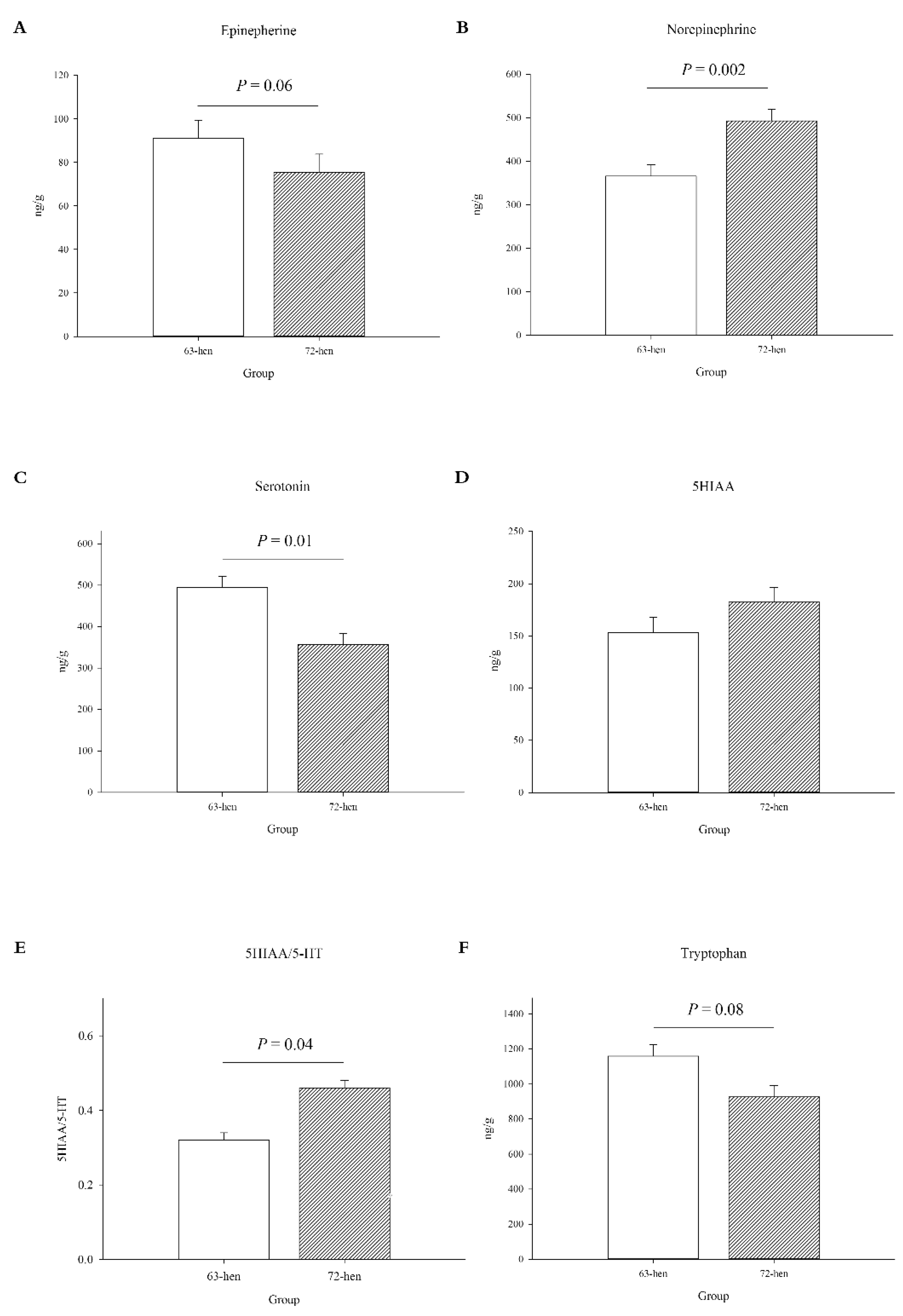
Figure 1. The measured neurotransmitters and neuropeptides in the raphe nucleus of two diversely selected chicken lines. (A), epinephrine (EP), (B), norepinephrine (NEP), (C), serotonin (5-HT), (D), 5-hydroxyindoleacetic acid (5HIAA), (E), 5HIAA/5-HT ratio, (F), tryptophan (TRP). Data are presented as: LSM + SEM (n = 10).
Microorganisms 10 01081 g001
Figure 2. Alpha diversity of cecal content bacteria in two diversely selected chicken lines. Box-and-whisker plots of diversity within samples. (A): estimated number of amplicon sequence variants (ASVs) calculated by the Chao1 index, (B): alpha diversity calculated by the Shannon index, (C): species evenness calculated by the Pielou’s evenness measures; and (D): phylogenetic diversity calculated by Faith’s phylogenetic diversity (PD).
Microorganisms 10 01081 g002
Figure 3. Principal coordinates analysis (PCoA), which shows the similarity between two diversely selected chicken lines. (A): Bray–Curtis similarity, (B): Jaccard similarity, (C): unweighted UniFrac, and (D): weighted UniFrac distances with 95% confidence ellipses.
Microorganisms 10 01081 g003
Figure 4. The different cecal bacterial compositions between two diversely selected chicken lines (n = 10). (A) phyla, and (B) genera analyses that compose an average of >2% and >1%, respectively, of the community, while other minor phyla or genera are not shown.
Microorganisms 10 01081 g004
Figure 5. Pairwise comparisons (DESeq2 analysis) between two diversely selected chicken lines (n = 10). Differential abundant OTUs (p < 0.05) in 63-hen and 72-hen are shown. OTUs were assigned to genus (x-axis) and phylum level (colors). Positive “log2 Fold Change” values (y-axis) indicate higher abundance in 63-hens, and negative values indicate higher abundance in 72-hens.
Microorganisms 10 01081 g005
Figure 6. MetaCyc pathway difference predictions using 16S rRNA data with PICRUSt2 between hens from line 63 and line 72 (>1 log2 fold change). Positive “log2 fold change” values (x-axis) indicate higher expression of certain pathways in 63-hens, and negative values indicate higher expression in 72-hens.
Microorganisms 10 01081 g006
Figure 7. Relative abundance of tryptophan metabolism on KEGG orthology between diversely selected 63-hens and 72-hens. Positive “log2 fold change” values (x-axis) indicate higher expression of certain pathways in 63-hens and negative values indicate higher expression in 72-hens.
Microorganisms 10 01081 g007
Figure 8. Heatmap showing the Spearman’s rank correlation coefficients between genera abundance and physiological parameters. Blue and red colors indicate significant negative and positive correlations, respectively (p < 0.05).
Microorganisms 10 01081 g008
Table 1. Peripheral serotonin, tryptophan, corticosterone and heterophil/lymphocyte ratio levels between two diversely selected chicken lines.
f5-HT (ng/mL) 1TRP (µmol/mL) 1CORT (ng/mL) 1H/L Ratio 1
63-hen61.38171.52 a8.44 b0.16 b
72-hen59.46121.42 b9.75 a0.50 a
SEM3.7915.371.510.04
n 210101010
p-Value0.730.030.05<0.0001
a,b Least squares means within a column for the 2 strains lacking a common superscript differ (p < 0.05). 1 5-HT: serotonin, TRP: tryptophan, CORT: corticosterone, H/L ratio: heterophil to lymphocyte ratio. 2 Average number of observations per least squares mean.
Table 2. Peripheral immunoglobulin G and cytokine levels between two diversely selected chicken lines.
IgG (mg/mL) 1IL-6 (pg/mL) 1IL-2 (pg/mL) 1IL-10 (pg/mL) 1TNF-α (ng/mL) 1
63-hen12.028.1460.099.3736.65
72-hen12.927.5671.6513.1330.73
SEM0.731.6312.81.642.37
n 21010101010
p-value0.540.810.540.120.09
1 IgG: immunoglobulin G, IL: interleukin, TNF-α: tumor necrosis factor alpha. 2 Average number of observations per least squares mean.
Publisher’s Note: MDPI stays neutral with regard to jurisdictional claims in published maps and institutional affiliations.