Article

Combined Action of Antibiotics and Bacteriocins against Vancomycin-Resistant Enterococci

by
Jorge Enrique Vazquez Bucheli
1,2,
Joanna Ivy Irorita Fugaban
2,†,
Wilhelm Heinrich Holzapfel
1 and
Svetoslav Dimitrov Todorov
2,*
1
Human Effective Microbes Laboratory, Department of Advanced Convergence, Handong Global University, Pohang 37554, Korea
2
ProBacLab, Department of Advanced Convergence, Handong Global University, Pohang 37554, Korea
*
Author to whom correspondence should be addressed.
Current address: National Food Institute, Technical University of Denmark, Building 202, Rm. 3.234, Kemitorvet, 2800 Kongens Lyngby, Denmark.
Microorganisms 2022, 10(7), 1423; https://doi.org/10.3390/microorganisms10071423
Submission received: 16 June 2022 / Revised: 6 July 2022 / Accepted: 13 July 2022 / Published: 14 July 2022
(This article belongs to the Special Issue Bacteriocins: Academic Advances and Immediate Applications)

Abstract

:
Antibiotics have been one of the most important discoveries in the area of applied medical microbiology; however, as a result of various factors, we are currently facing a dramatic and relatively dangerous increase in the number of cases of antibiotic resistance, and the need for new types of antimicrobials continues to grow. New approaches are needed to combat antibiotic-resistant pathogens. Bacteriocins, as part of the group of antimicrobial peptides, can be considered as alternatives and/or complements to known antibiotics. Their narrow spectra of activity can be explored for the control of various pathogens, such as vancomycin-resistant enterococci (VRE), as single therapies or in combination with known antibiotics. In the present study, we isolated bacteriocins from different lactic acid bacteria (LAB) strains, including Enterococcus and Pediococcus, and explored the possible synergistic inhibition of growth by bacteriocins and vancomycin. It was observed in the growth dynamics with previously selected VRE strains that the bacteriocins had a high specificity and a promising inhibitory effect against the VRE strains, and these results were validated by a propidium iodide viability test using flow cytometry. The data obtained indicate that the selected bacteriocins can be used to control VRE in the food industry or even as an alternative treatment to combat infections with antibiotic-resistant bacteria.

1. Introduction

Antibiotic resistance is an emerging public health problem. This has triggered the realization of an urgent need for the development of new antimicrobials and, concomitantly, also the development of rapid and sensitive approaches for the identification of representative pathogens and mechanisms of antibiotic resistance [1]. Since the discovery of antibiotics and their commercialization, the scientific community also invariably evaluated the different negative aspects of the clinical application of antibiotics, resulting in the development of antibiotic-resistant strains and their predominant spread, especially in the hospital environment [2]. The presence of antibiotic-resistance genetic determinants is a natural phenomenon; however, and therefore, the irresponsible use of these agents from the second half of the 20th century accelerated the selective pressure of the antibiotic-resistant mutants and their survival and establishment as the predominant representatives in several ecosystems. In the recent past, antibiotics were applied at large scale, not just for the treatment of infectious diseases but also frequently and unnecessarily for the prevention of potential medical complications as growth promotors and for preventive treatment in the veterinary sector and in agricultural practices to increase production yields. The uncontrolled release of antibiotics in the environment contributed to the reduction of several human and animal pathogens; however, the lack of regulations on their use had negative consequences in the preselection and survival of antibiotic-resistant mutants and allowed them to predominate in the environment [3]. A similar scenario is typical of the current hospital environment, where multidrug-resistant bacterial pathogens have passively been preselected by the constant application of antibiotics, thus developing into a serious health threat. Among these resistant pathogens, vancomycin-resistant enterococci (VRE) and methicillin-resistant Staphylococcus aureus (MRSA), are recognized worldwide as bacteria of major health concern [4].
Bacteriocins, a non-posttranslational-modified antimicrobial peptide produced via ribosomal machinery of the bacterial cells [5] were suggested as a potential alternative and/or addendum to conventional antibiotics [6,7]. The narrow antimicrobial spectrum of bacteriocins is a characteristic that gives them an advantage in combating human and animal pathogens [8]. Considered safe, bacteriocins are characterized by a general low toxicity [7,9]. Their application was originally explored as bio-preservatives, and nisin, in particular, is approved in several countries for application in the food industry [10,11]. However, in the last decade, scientific interest has increasingly focused on the application of bacteriocins as an alternative and/or additive to antibiotics in the combat against emerging pathogens in human and veterinary practices. The applications of native and modified bacteriocins have been proposed as alternative treatments for Mycobacterium tuberculosis [12], MRSA [13], and VRE [14,15,16]. Moreover, the combined application of bacteriocins and antibiotics or essential oils were proposed as an alternative for the reduction of the required dose of antibiotics for application in human and veterinary medicine [12,17]. Will bacteriocins be the next generation antimicrobials in the treatment of relevant human and animal pathogens? However, related to the application of chemical preservatives in the food industry, a concern has been raised about the possible use of sublethal doses of non-antibiotic antimicrobials and their role in increasing MIC for antibiotics [17]. Questions on possible indirect interactions are of particular concern in view of potentially increasing problems with antibiotic resistance. Do bacteriocins need to be regarded as safe antimicrobials, indifferent to the antibiotic resistance phenomena, and could they even be considered as alternatives and maybe as new generation “antibiotics”? Already Heilbronner et al. [18] suggested that bacteriocins can be considered safe, and due to their effective antimicrobial activity, they could be attractive alternatives for precision therapy and prevention of bacterial infection. Moreover, the correct answer might be that we would need to predict the possible negative consequences, explore these possible challenging scenarios, and attempt to perform deeper research to avoid any future negative consequences. The aim of this study was to explore the possibility of effective combat of VRE by applying bacteriocins and to evaluate the safe applications of bacteriocins and their influence on the development of antibiotic resistance in VRE strains.

2. Materials and Methods

2.1. Differentiation and Identification of VRE Strains and Detection of the Type of Vancomycin Resistance

Vancomycin-resistant enterococci (VRE), previously isolated from hospital patients and fully characterized, were kindly provided by the Laboratory of Antibiotics (kindly provided from Prof. Kwak, Handong Global University, Pohang, Republic of Korea). In the present study, we have differentiated the VRE based on repPCR with primer (GTG)5, according to Valledor et al. [19]; these were also screened for the presence of vancomycin-resistant genes, including vanA, vanB, vanC, vanD, vanE, and vanG, according to Valledor et al. [19]. Further identification of these VRE strains was based on 16S rRNA partial gene sequencing at SolGent Analysis Service (Daejeon, Republic of Korea) as a commercial service, in addition to further biochemical and physiological tests, as recommended by Bergey’s Manual of Systematic Bacteriology [20].
The VRE strains were cultured in 10 mL MRS broth (Difco, Franklin Lakes, NJ, USA) at 37 °C for 24 h; the cells were harvested by centrifugation (4000× g, 10 min, 4 °C), and the DNA was extracted using ZR Fungal/Bacterial DNA Kit (Zymo Research, Irvine, CA, USA), according to the manufacturer’s recommendations. The purity and concentration of the DNA were evaluated spectrophotometrically on SPECTROstar Nano nanodrop (BMG LABTECH, Ortenberg, Germany). All PCR reactions were performed in a Veriti 96-well Thermal Cycler, Applied Biosystems (Thermo Scientific, Waltham, MA, USA). The generated amplicons were separated and visualized on 1.0–2.0% agarose (Sigma-Aldrich, St. Louis, MO, USA) in the presence of 0.02 μL mL−1 of SYBR® Safe (Thermo Scientific) in 1× TAE buffer at 100 V for 1 h (GH-200 Genera Biosystems, Victoria, Australia; Elite 300 Plus Power Supply, Wealtec Bioscience Co., Ltd., Taipei, Taiwan). The gel was visualized using the Omega Lum™G gel documenter (Aplegen, Inc., Pleasanton CA, USA).

2.2. Screening for Bacteriocins Effective against VRE Strains

Bacteriocinogenic LAB from the culture collection of the ProBacLab (Handong Global University, Pohang, Republic of Korea) were evaluated for bacteriocinogenic activity against a selected panel of VRE strains, according to the recommendations of dos Santos et al. [21]. Bacteriocinogenic strains were cultured in 10 mL MRS broth at 37 °C for 24 h, and cell-free supernatants were obtained by centrifugation (4000× g, 10 min, 4 °C), heat treated at 80 °C for 10 min with the aim to deactivate potentially produced extracellular proteases and hydrogen peroxide, and the pH was adjusted to 6.0–7.0 with 1 M NaOH. Aliquots of 10 μL from the previously described cell-free supernatant were spotted on the surface of previously prepared plates containing BHI (Difco) supplemented with 1% agar and the studied VRE strains on an individual basis at a final concentration around 105 CFU mL−1. Plates were incubated for 24 h at 37 °C and observed for the formation of inhibition zones around spotted material. Inhibition zones larger than 3 mm were considered as evidence for bacteriocinogenic activity against the VRE strains. In addition, the titer of the bacteriocin activity, expressed in AU mL−1, was determined according to dos Santos et al. [21]. The cell-free supernatants prepared, as described before, were serially diluted in 100 mM potassium phosphate buffer at pH 6.5, and the dilutions were spotted on the surface of the previously prepared plates with the incorporated VRE strains, as described before. The titer of bacteriocin activity expressed in AU/mL was calculated as (Dn)/p, where D is the dilution factor used in the serial dilution, n is the first dilution with no inhibition zone, and p is the volume of the cell-free supernatant spotted in mL.

2.3. Partial Purification of the Bacteriocins

The selected bacteriocinogenic strains were grown in 500 mL MRS broth at 37 °C for 24 h, and the cell free supernatants were obtained and heat treated, and the bacteriocin activity (AU mL−1) was determined, as described before. Partial bacteriocin purification was performed, according to Todorov et al. [22], including ammonium sulfate precipitation (60% saturation) at 4 °C for 4 h with continuous shaking followed by centrifugation at 10,000× g, 60 min, 4 °C. The pellet was resuspended in 1/10 from the original volume in 25 mM potassium phosphate buffer pH 6.5, and the bacteriocin activity (AU mL−1) of the obtained suspension and supernatant was determined, as described before. In the next step, hydrophobic chromatography was performed on SepPakC18 with a step gradient of 20, 40, 60, and 80% isopropanol in 25 mM potassium phosphate buffer at pH 6.5. The obtained fractions were collected and evaluated for bacteriocin activity (AU mL−1), as described before, and preserved at −20 °C for further studies. All bacteriocin tests were performed against Listeria monocytogenes ATCC15313 and Enterococcus faecium VRE 9, 18, 19, and 23.

2.4. Determination of MIC for Vancomycin, Other Antibiotics, and Semi-Purified Bacteriocins for Enterococcus faecium Strains VRE 9, 18, 19, and 23

The antimicrobial susceptibility testing of the selected VRE strains (Cheil Jedang Pharma Co., Seoul, Republic of Korea), against vancomycin, ampicillin, chloramphenicol, gentamicin, and kanamycin (Sigma-Aldrich) was performed according to the recommendations of the Clinical and Laboratory Standards Institute (CLSI, 2016, www.clsi.org (accessed on 3 June 2021)) on Performance Standards for Antimicrobial Susceptibility Testing for Enterococcus spp. on cation-adjusted Mueller–Hinton broth (Sigma-Aldrich) supplemented with MRS (5.0 g L−1). The experimental assay was performed in a 96-well microplate (SPL Life sciences, Pocheon-si, Gyeonggi-do, Republic of Korea) and performed in 10 two-fold dilutions of the antibiotics and selected semi-purified bacteriocins (40, 60, and 80 isopropanol fractions) and controls (growth and sterility controls). The bacterial inoculums were adjusted to 0.5 McFarland units (corresponding to approximately 107 CFU mL−1) and distributed accordingly to obtain a final concentration of 105 CFU mL−1. The plates were incubated at 35 ± 1 °C for 18 h. Interpretation of the results was focused on the lowest concentration with complete bacterial growth inhibition and was recorded as the MIC and analyzed according to the standards set for Enterococcus spp. [23,24].

2.5. Evaluation of Proportions between Resistant and Susceptible Mutants in the Population of the VRE Strains

For the identification of resistant and susceptible E. faecium strain, VRE19, previously isolated from a hospital in South Korea, 100 subcultures were evaluated. The subcultures of this strain were grown on 1.5% MRS agar plates, and two mirror plates were made from the original plate, supplemented with 64 and 32 µg mL−1 of vancomycin, respectively. The plates were cultured for 48 h at 37 °C under aerobic conditions and based on the observed bacterial growth, the subcultures were classified as resistant if the bacteria grew just in the mirror plates supplemented with vancomycin and susceptible if the bacteria only grew on the MRS plates without added antibiotic.

2.6. Effect of Individual and Combined Application of Bacteriocin and Antibiotics on VRE Strains

Antimicrobials commonly used in bacterial infections therapy were tested, according to the EFSA protocol [25]. Ampicillin, chloramphenicol, gentamicin, kanamycin, and vancomycin were used in double-fold dilutions starting from 128 to 0.25 µg mL−1 and added to 10 mL of LSM broth (IsoSensitest broth (90%) and MRS broth (10%), adjusted to pH 6.7). The VRE strains were inoculated to the previously prepared media supplemented with antibiotics and cultured at 37 °C for 24 h under aerobic conditions. The lowest concentration of each antibiotic that inhibits the visible growth of bacteria was determined after 24 h. The susceptibility cut-off values were defined according to the EFSA standards [25].
The effect of combinations of the bacteriocin and antibiotics was determined in a 96-well microplate (SPL Life sciences), where the horizontal wells contained sequentially reduced concentrations of the tested bacteriocin (from 1600 AU mL−1 to 6.25 AU mL−1), and the vertical wells contained different concentrations of vancomycin (from 1 to 64 μg mL−1). The wells were supplemented with MRS seeded with the E. faecium VRE 9, 18, 19, and 23 strains, respectively. Over regular intervals (6, 9, 18, 21, and 24 h) bacterial growth was evaluated by changes in the OD at 600 nm determined in a SPECTROstar (BMG LABTECH, Ortenberg, Germany).

2.7. Fluorescent In Situ Hybridization

The hybridization method was based on the procedure of Azevedo et al. [26] and Oliviera et al. [27] with slight modifications. For all strains, the growth medium was removed by centrifuging the culture at 10,000× g for 5 min and resuspending in 1.5 mL of 1× PBS. The washed bacteria were collected by centrifugation at 10,000× g for 5 min and resuspended in 1.5 mL of 4% paraformaldehyde (pH 7.2), and incubated for 1 h at room temperature, followed by centrifugation (10,000× g for 5 min) and resuspension of the bacteria in 1.5 mL of 50% ethanol and subsequently incubated for 1 h at −20 °C. A total of 20 μL of the fixed bacteria at an OD600 nm 0.5–0.8 was washed in 480 μL of 1× PBS and centrifuged at 10,000× g for 5 min. The pellet was then resuspended in the hybridization buffer of [10% (w/v)] dextran sulphate, 10 mM sodium chloride, 30% (v/v) formamide, 0.1% (w/v) sodium pyrophosphate, 0.2% (w/v) polyvinylpyrrolidone, 0.2% ficoll (w/v), 5 mM disodium EDTA, 0.1% (v/v), and 0.1% Triton X-100, 50 mM Tris-HCl) with 200 nM of the specific probe, and the bacteria were incubated at 75 °C, for 2 h. The hybridized bacteria were collected by centrifugation at 10,000× g for 5 min, and the pellet was resuspended in 500 μL of wash buffer pH 9.0 (5 mM Tris base, 15 mM sodium chloride, 1% (v/v) Triton-X-100) and incubated for 1 h at room temperature. After incubation, the bacteria were collected by centrifugation at 10,000× g for 5 min and resuspended in 200 μL DW.

2.8. FLOW Cytometry Analysis

A ZE5 flow cytometer (Bio-Rad, Hercules, CA, USA) was used to measure the forward scattering and fluorescence of microbial cells. These parameters were acquired as pulse height signals for 10,000 events at a rate of 1 μL per second and at a fluorescence wavelength of 647 nm. Prior to analysis, quality control was performed using ZE-series QC beads (Bio-Rad, #12004403). The instrument tubing was cleaned by sequentially using 1% bleach, 70% ethanol, and sterile PBS. Data were acquired using the Everest software package v1.4.

2.9. Virulence Genes in VRE

The DNA extracted, as described previously, was analyzed by PCR for the presence of the selected virulence genes (asa1, cyt, efaA, esp, hyl, and IS16), according to the recommendations of EFSA [26]. The PCR reactions were performed in a Veriti 96-well Thermal Cycler, Applied Biosystems (Thermo Scientific, Waltham, MA, USA). The primer sequences and amplification conditions were applied, according to Fugaban et al. [15]. The amplicons were separated on 1.5% (w/v) agarose gels in 1× TAE and visualized, as described before.

2.10. Determining the Changes in the MIC after Exposure to Low Concentrations of Bacteriocin

To evaluate the long-term effect of bacteriocin treatment on the virulence potential of VRE, the E. faecium strains VRE 9, 18, 19, and 23 were cultured in 10 mL MRS broth, supplemented with sublethal doses of the semi-purified bacteriocins (50 AU mL−1) at 37 °C for 24 h and sub-cultured consequently for 30 days. At the beginning of the experiment (day zero) and after 30 days, the MIC of the E. faecium VRE 9, 18, 19, and 23 strains receiving different bacteriocins were determined, according to the method described previously in this manuscript. The MICs at day zero and after 30 days were compared. The experiment was repeated on two independent occasions.

3. Results and Discussion

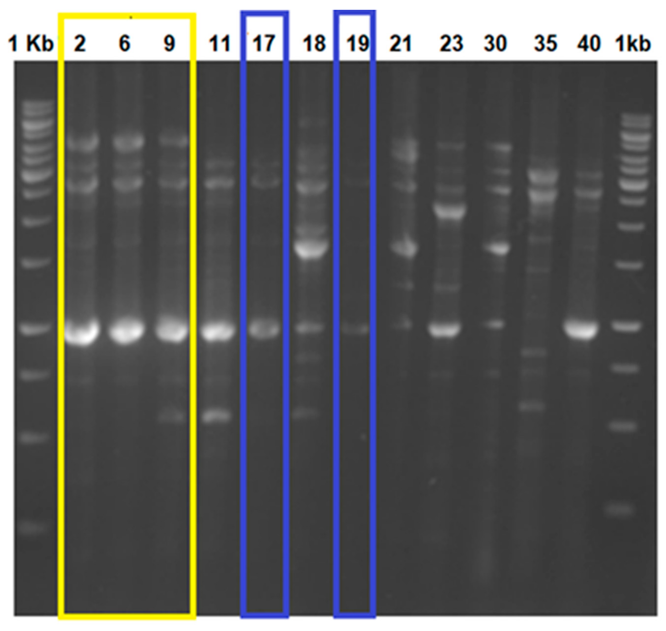
3.1. Differentiation and Identification of VRE Strains and Detection of the Type of Vancomycin Resistance

Twelve (12) vancomycin-resistant Enterococcus (VRE) strains, previously isolated from patients, were kindly provided by Prof. Kwak (Laboratory of Antibiotics, Handong Global University, Pohang, Republic of Korea) and were differentiated by fingerprinting using repPCR (Figure 1). The unique strains were selected and identified as E. faecium 9, 18, 19, and 23 based on the 16S rRNA sequences (commercial service from SolGent South Korea) and by applying additional morphological, physiological, and biochemical tests recommended by Bergey’s Manual [20]. It was interesting to observe that some of the evaluated VRE isolates generated similar repPCR profiles (Figure 1), which may be evidence that the same strain has been isolated on multiple occasions This suggests that specific resistant VRE strains are well established in healthcare units. Additional studies should be performed with more clinical samples and a relevant number of isolates to confirm our preliminary hypothesis and to investigate if the mentioned strains are associated with specific healthcare units or possibly have also spread to larger regions. Interestingly, all evaluated and sequenced cultures were identified as E. faecium, except for VRE2, which was identified as Enterococcus lactis. Identification of the enterococci by 16S rRNA is not definitive, and additional biochemical and physiological tests are required. In general, vancomycin resistance is associated with E. faecalis [28]; however, other representatives of the genus Enterococcus can also be carriers of vancomycin-resistant genes.
The VRE strains selected for this study were evaluated for the presence of different vancomycin-resistance-associated genes (vanA, vanB, vanC, vanD, vanE, and vanG), and the predominant presence of vanA and vanB were recorded. Moreover, vanD was also recorded in strain E. faecium VRE17. According to previous reports, vancomycin-resistant genes can be equally distributed on the bacterial chromosome and the plasmid DNA [29]. Different suggestions were hypothesized related to the relevance of the genetic locations of vancomycin-resistant genes, from serious to relatively low risk for possible gene transfer and spread around other bacterial species via horizontal and vertical gene transfer [30,31]. Moreover, Suvorov [32] discussed the relevant scenarios, such as genetic transfer in the GIT of humans and animals or in food production environments. Healthcare units, however, need to be considered as a principal environment for the relevance of VRE strains based on the extensive application of antibiotics and their role in the selection and domestication of VRE mutants.

3.2. Virulence Genes in Enterococcus faecium Strains VRE 8, 18, 19, and 23

A major safety concern of VRE strains has been associated with the genetic determinants for vancomycin resistance. However, several other virulence factors can be recorded in both vancomycin-resistant and non-vancomycin-resistant enterococci. The DNA extracted, as described previously, was analyzed by PCR for the presence of the selected virulence genes (cyt, asa1, efaA, esp, hyl, and IS16). The PCR results indicated the presence of efaA, esp, and hyl genes in all the tested VRE strains; yet, cyt and asa1 were absent in these four strains. The virulence factor, efaA, is of clinical importance and has been typically associated with cases of endocarditis [33]. The protein, esp, is an adhesin that allows biofilm formation and colonization of epithelial cells in the urinary tract; it has been associated with VRE outbreaks, suggesting a key role in nosocomial infections [34].
Padilla and Lobos [35] reported that of 78 Enterococcus strains isolated from water wells for human consumption, those with the greatest number of virulence factors showed greater resistance to antibiotics. This correlation could be due to the irresponsible use of antibiotics, resulting in the selection of strains in the environment with greater pathogenicity and highlighting the importance of the use of alternatives for the control of pathogenic bacteria.

3.3. Screening for Bacteriocins Effective against VRE Strains

The strategy of the selection of new antimicrobials for the combat of the VRE is based on the search of inhibitory molecules that will effectively kill the target organisms by acting via a different action mechanism and that is associated with low cytotoxicity but with a specific spectrum of effective activity. In general, the development of new antibiotics aims at a broad-spectrum mode of action but may be associated with several side effects [36]. Most of the proposed new antibiotics are effective against VRE and can be applied in clinical practices. However, their broad phylogenetic spectrum generally results in the disruption of numerous beneficial gut microbiota. This may have some serious health consequences, especially with long-term applications [37]. One of the potential alternatives in the screening for potentially effective antimicrobials against VRE could be the use of bacteriocins. Based on their narrow spectrum of activity [38] and specificity in the bactericidal mode of action [9], several reports considered them as relevant candidates for clinical trials towards application as alternatives or as synergetic partners to antibiotics in the treatment of VRE-associated infections [39].
In our study, we evaluated four strains, previously described as bacteriocin producers, for their effective inhibition/killing of VRE bacteria (Table 1, Figure 2). Pediococcus acidilactici ST3522BG, P. pentosaceus ST3633BG, E. faecium ST651ea, E. faecium ST7119ea, and E. faecium ST7319ea formerly isolated from silage and from Korean fermented food products [15,40].
The bacteriocinogenic potential of the evaluated LAB strains versus selected VRE is presented in Table 1. The comparison of the inhibitory potential versus VRE, as well taking into consideration additional bacteriocinogenic properties of the tested bacteriocins [15,40], identified three bacteriocins (produced by P. acidilactici ST3522BG, P. pentosaceus ST3633BG, and E. faecium ST7319ea) that were selected for future studies.

3.4. Partial Purification of the Bacteriocins

By a combination of precipitation with ammonium sulfate at 60% saturation followed by hydrophobic chromatography on SepPak C18 with elution with a stepwise gradient of isopropanol in 25 mM potassium phosphate buffer at pH 6.5, fractions of bacteriocins from P. acidilactici ST3522BG, P. pentosaceus ST3633BG, and E. faecium ST7319ea were obtained predominantly in the 60% and 80% isopropanol fractions. The combination of ammonium sulfate precipitation and hydrophobic chromatography on SpePakC18 can be considered a highly successful approach for the partial purification of bacteriocins produced by LAB and was applied formerly in purification protocols by Todorov et al. [22], Metivier et al. [41], Bughaloo-Vial et al. [42], Song et al. [43], and Surovtsev et al. [44]. By nature, all bacteriocins are proteinaceous, and ammonium sulfate precipitation combined with the level of saturation gives excellent results for the preliminary extraction of these proteinaceous molecules with a specific range of molecular sizes [45]. The use of hydrophobic SepPakC18 columns exploits the hydrophobic character of most bacteriocins. By further application of an isopropanol gradient, these antimicrobial peptides can be eluted at a specific point [44]. These approaches were previously applied in the partial purification of several bacteriocins [22,41,42,43,44]. The obtained active fractions can be applied for the purpose of analytical experiments or can be subjected to further purification, depending on the objective of the research.

3.5. Determination of MIC for Vancomycin and Semi-Purified Bacteriocins against Selected VRE Strains

For the determination of the MICs of the VRE strains to vancomycin and the previously prepared semi-purified bacteriocins the recommendations of CLSI were followed; the obtained results are summarized in Table 2. The purpose of this experiment was to determine the break point for the possible inhibitory application of vancomycin and semi-purified bacteriocins and to plan the experiment for the combined antibiotic–bacteriocin application. As expected, lethal concentrations of vancomycin exceeded levels of 128 μg mL−1 for all tested VRE strains. By contrast, even at 200 AU mL−1, the studied bacteriocins inhibited/killed the studied VRE strains. Similar results have formerly been reported for the lethal activity of different bacteriocins against VRE strains [19,46,47]. It is known that vancomycin uses lipid II as a docking molecule for the initial contact with target cells; however, additional steps in the antibiotic mode of action were required to effectively kill the target cells [48]. In a similar way, bacteriocins from class IIa, belonging to the two studied pediocins and the one enterocin, are associated with the recognition of lipid II and pore formation as part of the killing mode of action [49]. Comparison and similarity of the initial steps of the use of the same receptor and better effectiveness of the bacteriocins in the successful killing of VRE can be considered as a solid argument towards deepening the exploration for the potential application of bacteriocins as an alternative to conventional antibiotics in the therapy of antibiotic-resistant pathogens.

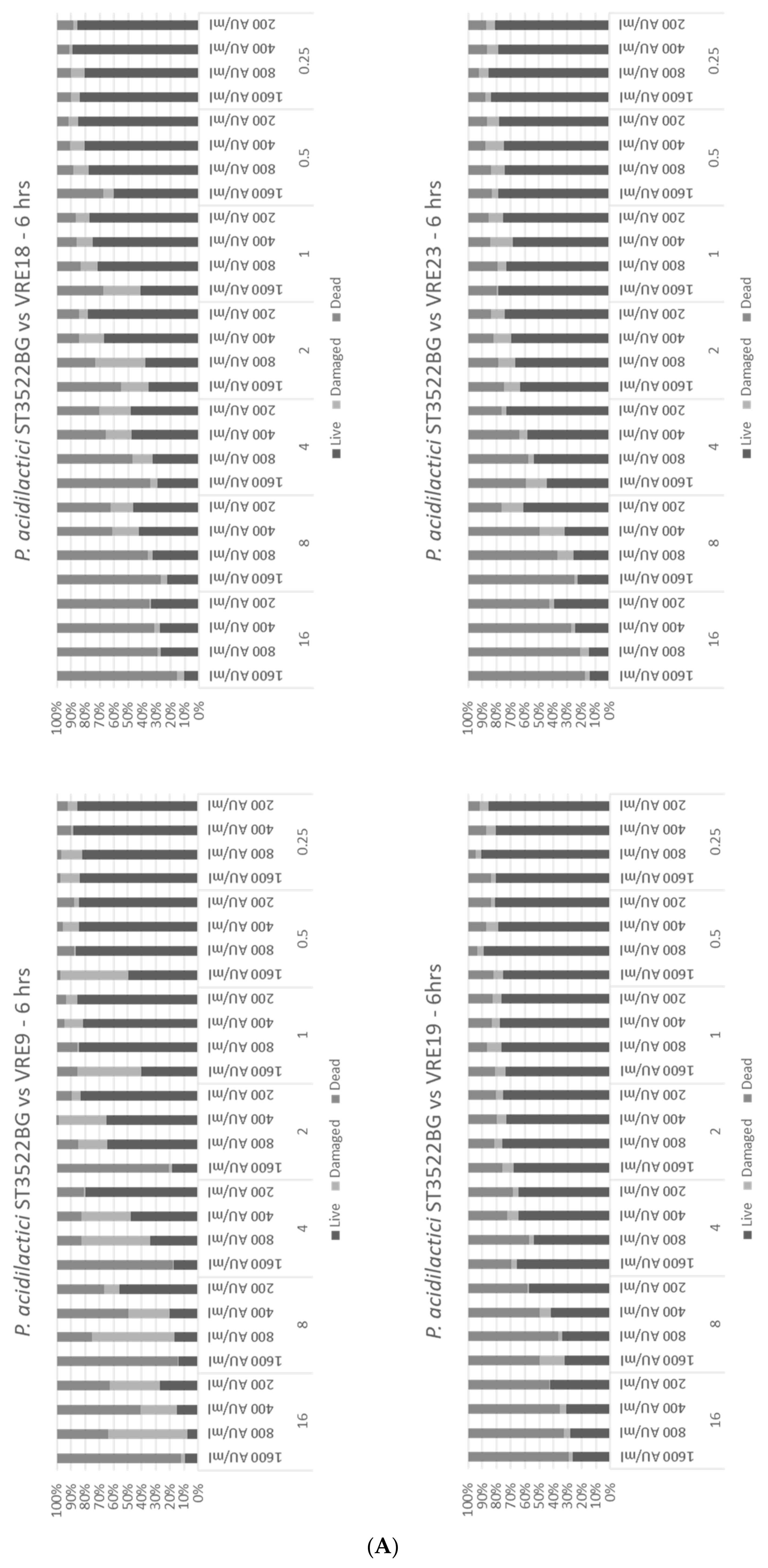
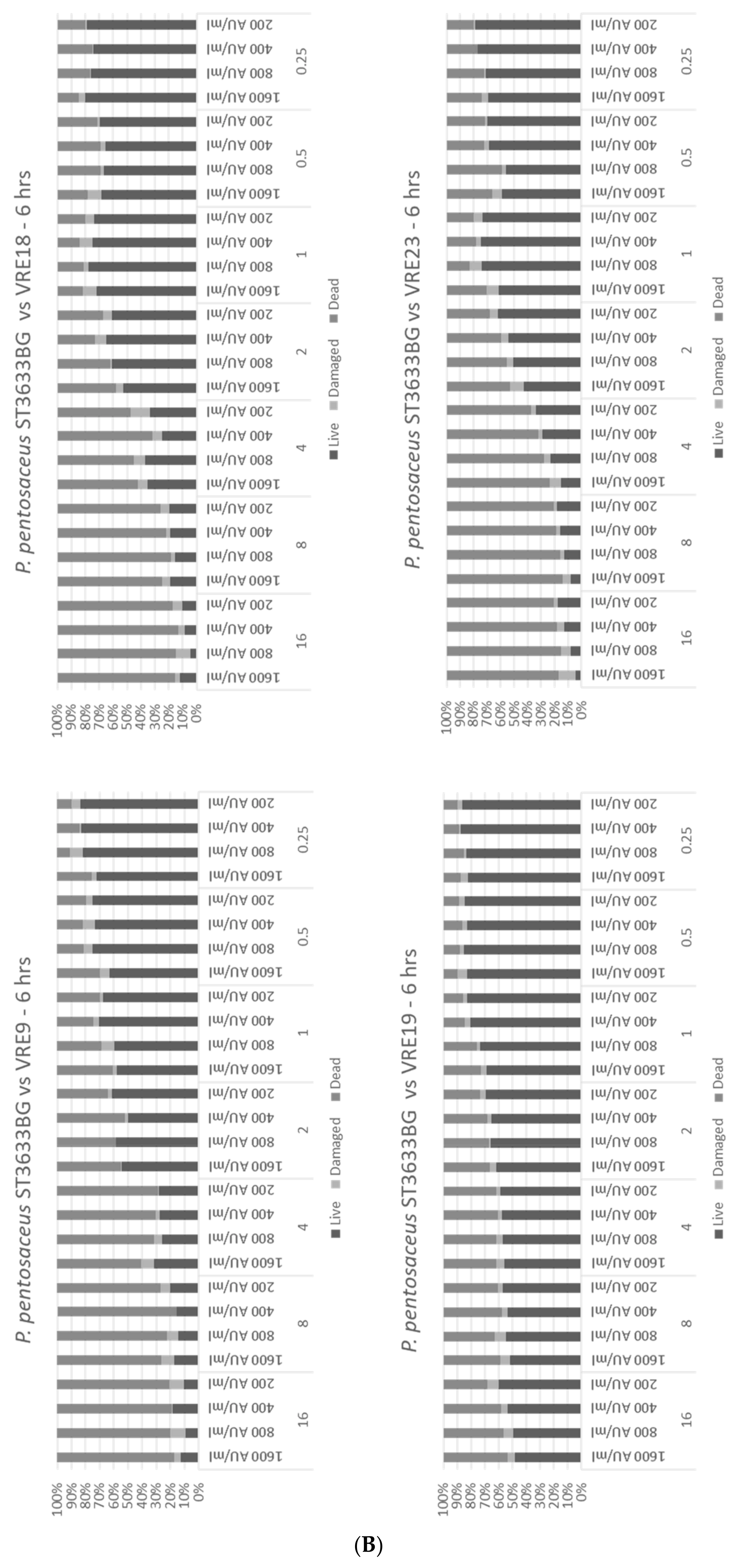
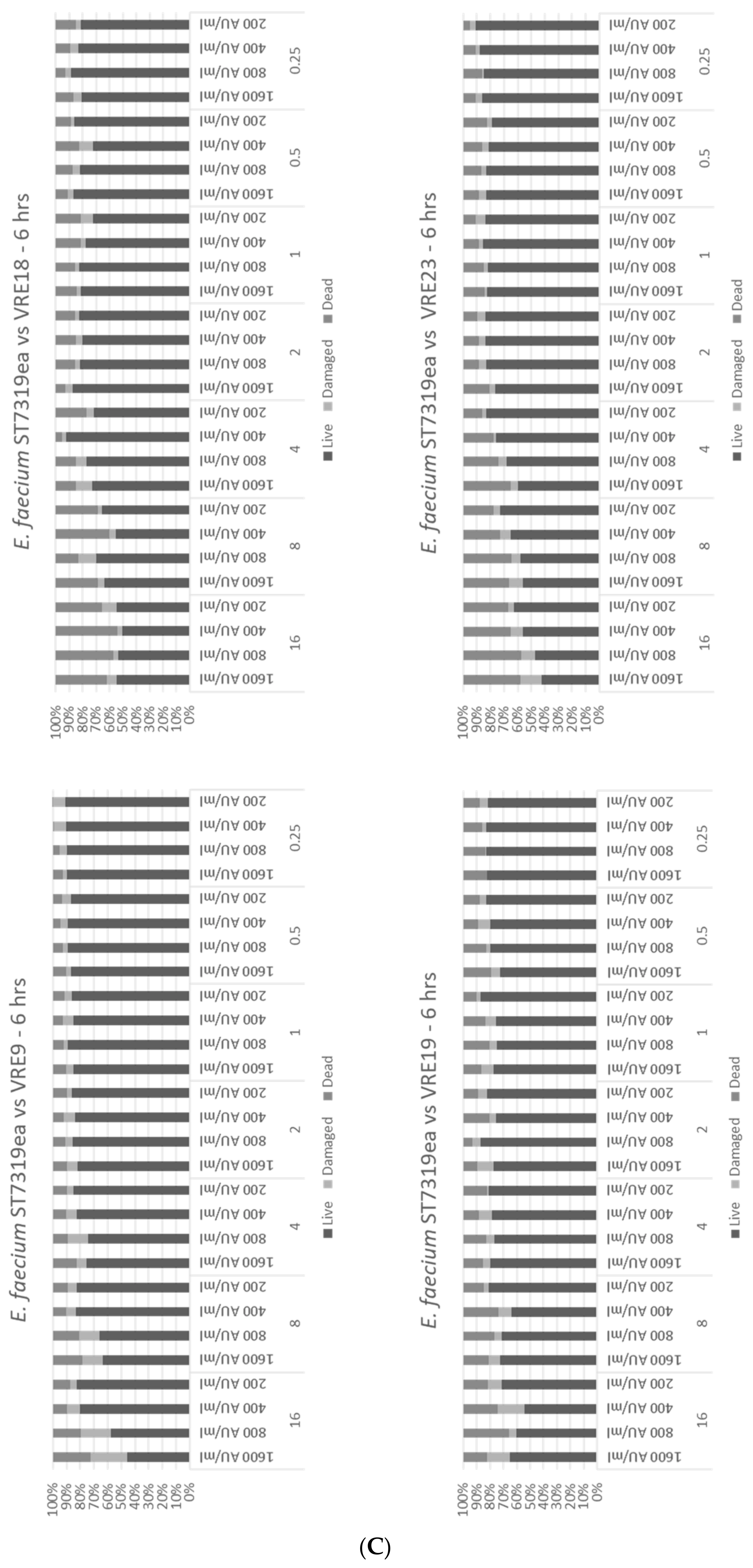
3.6. Effect of Bactriocins, Dead, Resistant, and Susceptible Strains in the Population of the Studied VRE Strains

The presence of antibiotics can be considered as a preselection factor for survival and predomination of mutant-carrying, antibiotic-resistant genes [50]. This is one of the reasons that the hospital environment is considered as an “incubator” for antibiotic-resistant mutants [51]. The inappropriate application of antibiotics in combination with the natural process of development of resistance into the pathogenic bacteria, the driving force of selectively killing susceptible pathogen variants, and the survival of resistant mutants, as well as the reduction of natural intraspecies competition, can lead to the establishment of predominating resistant pathogen populations [52].
The analysis of the population of VRE strains showed that not all colonies could be characterized as antibiotic resistant (Figure 3). The presence of damaged cells were observed when low levels of antimicrobials were applied. Flow cytometry is an appropriate approach to evaluate dead, damaged, and live cells, and based on the last, conclusions about efficacies of applied antimicrobials can be drawn. This result was not surprising since, from a selective point of view, the absence of a lethal factor can be the driving argument for loss of the appropriate defense mechanism(s). In other words, the absence of antibiotics may result in a reduction of the defense system and decreasing levels of MIC. However, the entire process is random and does not cover all cells in the population. Always, a small part may still retain the defense mechanisms, and in return for the killing factor (antibiotic), this can be effective only against susceptible mutants, while the resistant mutants will survive and soon become predominant under a prevailing selective factor.
As previously indicated, we hypothesize that by the combined treatment with antibiotics and bacteriocins, it should be possible to kill VRE pathogens more effectively by increasing the susceptibility of the antibiotic-resistant mutants. We observed that combining the bacteriocins from P. acidilactici ST3522BG, P. pentosaceus ST3633BG, and E. faecium ST7319ea with vancomycin had an enhanced antimicrobial effect on the VRE strains (Figure 3) such as was the case for the bacteriocins from P. acidilactici ST3522BG. At the highest concentrations of bacteriocin and vancomycin, an inhibition of up to 90% was determined; when the bacteriocins were diluted to a concentration of 200 AU mL−1 with the same concentration of antibiotic, 60% of VRE cells were either damaged or killed (Figure 3A).
The treatment of vancomycin mixed with the semi-purified bacteriocin obtained from E. faecium ST7319ea resulted in a low-inhibition level of the VRE strains (Figure 3C); this could be due to both the bacteriocin and vancomycin having a similar site of action. The target of vancomycin is lipid II; coupling of vancomycin would, therefore, inhibit the synthesis of the peptidoglycan. In a similar fashion, lipid II is a docking site for nisin [53]. For this reason, it is essential to first determine the mechanism of action of this bacteriocin.

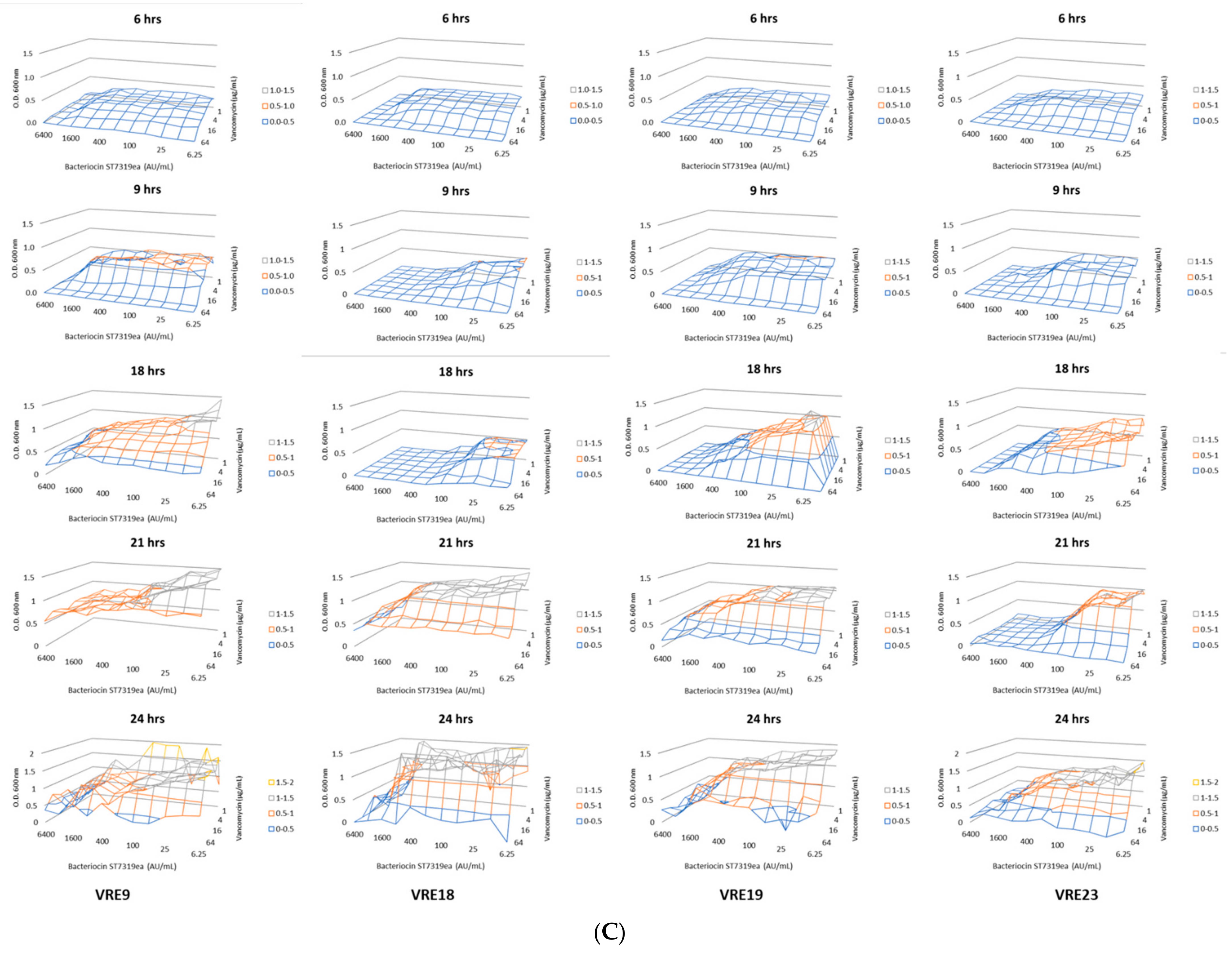
3.7. Effect of Individual and Combined Application of Bacteriocin and Vancomycin on VRE

The application of a combination of antibiotics and bacteriocins for the control of human and animal pathogens has been explored before. The combined application of ciprofloxacin and bacteriocin produced by P. pentosaceus ST44AM showed a synergistic effect against Listeria ivanovii subsp. ivanovii ATCC 19119 [54]. Moreover, the combination between ciprofloxacin and five bacteriocins produced by Lb. plantarum ST69BZ, E. faecium ST62BZ, and L. lactis strains, ST63BZ, ST611BZ, and ST612BZ (all isolated from boza) showed synergistic effects in combination [55]. This may be supported by the fact that pore formation by these bacteriocins could facilitate the antibiotic killing action against sensitive bacterial cells. Alternatively, the weakness of the pathogen population as a result of the combined application of two different antimicrobials with a different mode of action may result in more effective lethal activity against pathogens.
The combined application of vancomycin and semi-purified bacteriocins obtained from P. acidilactici ST3522BG, P. pentosaceus ST3633BG, and E. faecium ST7319ea showed a possible synergistic interaction and improved the inhibition of the VRE test strains. Moreover, the killing effect was observed by following the bacterial growth of VRE exposed to different combinations of vancomycin (from 0.25 to 128 μg mL−1) and bacteriocins produced by P. acidilactici ST3522BG, P. pentosaceus ST3633BG, and E. faecium ST7319ea (from 200 to 1600 AU mL−1) (Figure 4A–C). The destruction effect by applying the Fish-Flow approach, was the detection of live, damaged, and dead cells; these results complemented previous observations by monitoring the bacterial growth (Figure 3A–C).
The evaluation of the combined effect of antibiotics and bacteriocins by Fish-Flow is a novel approach; it complements the previously explored microtiter plates for evaluation of bacterial growth, such as VRE or other relevant pathogens, when exposed to a combination of antibiotics and bacteriocins or other antimicrobials. The Fish-Flow approach can be considered as more precise since it clearly distinguishes between the dead, damaged, or living cells after treatment, respectively. Thereby, more clarity is provided on the effect of combined applications of antimicrobials, resulting in bactericidal, bacteriostatic, or no effect on the test organisms. Moreover, the use of specific probes can increase the reliability of the results because the obtained results will clearly represent the viability of the targeted test organisms for which the probes were designed. Thereby, misinterpretations of the results can be reduced, e.g., in case of potential bacterial contamination. Moreover, a possible clinical application may be to evaluate the effect of different ratios of antibiotics and bacteriocins (or other antimicrobials or even including a combination of antibiotics with a different mode of action) on fecal samples from patients diagnosed with VRE infections. This may enable the prediction of appropriate proportions of different antimicrobials for the effective treatment against the elimination of pathogens.

3.8. Following the Changes in the MICs after Exposure to Low Concentrations of Bacteriocin

It is possible that the continuous exposure of bacteria to bacteriocins could result in the selection of innate or acquired resistance of bacteria, as has already been reported for conventional antibiotics [56]. To study the possible acquisition of resistance of the VRE strains against bacteriocins, a long-term application was carried out where no significant change in the MICs was observed during the experimental period (Table 2). It was observed that the VRE inhibition levels after 30 days of exposure to bacteriocins were the same or very similar to those obtained on day 0, as was also observed in previous experiments.
This could indicate that the VRE strains studied do not have innate mechanisms of resistance to these bacteriocins. However, it is necessary to study the models for a longer period of time to know in detail the possible mutations that may cause the acquisition of a resistance mechanism against these molecules.

4. Conclusions

The necessity for new effective antimicrobials against emerging antibiotic-resistant pathogens is a fact and has become an issue of increasing importance since the beginning of this century. Bacteriocins, as part of the group of antimicrobial peptides, can be considered as an alternative and/or complement to known antibiotics. Our study reaffirms the importance of bacteriocins in the control of pathogenic bacteria. Moreover, our results suggest a possible synergistic effect of bacteriocins when combined with vancomycin, which offers a new approach of combat antibiotic resistance; the specific spectrum of activity would make them a good option for the treatment of already diagnosed specific infections and in the case of the food industry, thus, helping to inhibit the proliferation of resistant bacteria. Although more studies are needed to characterize these bacteriocins and their spectrum of activity in detail, we consider them to be a reliable option for the control of pathogenic microorganisms.

Author Contributions

Conceptualization: S.D.T.; Experimental work: J.E.V.B. and J.I.I.F.; Funds: W.H.H. and S.D.T.; Writing of the manuscript: J.E.V.B. and S.D.T.; Corrections and editing: W.H.H. and S.D.T. All authors have read and agreed to the published version of the manuscript.

Funding

Grant from the National Research Foundation (NRF) funded by the Ministry of Science and ICT (NRF-2018M3A9F3021964), Seoul, Republic of Korea.

Institutional Review Board Statement

Not applicable.

Informed Consent Statement

Not applicable.

Data Availability Statement

All data associated with this paper are available upon request.

Acknowledgments

To Handong Global University, Pohang, Republic of Korea for providing the research facilities. The authors acknowledge Mia Miau for her unconditional support.

Conflicts of Interest

The authors declare no conflict of interest.

References

  1. Murray, C.J.; Ikuta, K.S.; Sharara, F.; Swetschinski, L.; Aguilar, G.R.; Gray, A.; Han, C.; Bisignano, C.; Rao, P.; Wool, E.; et al. Global burden of bacterial antimicrobial resistance in 2019: A systematic analysis. Lancet 2022, 399, 629–655. [Google Scholar] [CrossRef]
  2. Petrovich, M.L.; Zilberman, A.; Kaplan, A.; Eliraz, G.R.; Wang, Y.; Langenfeld, K.; Duhaime, M.; Wigginton, K.; Poretsky, R.; Avisar, D.; et al. Microbial and viral communities and their antibiotic resistance genes throughout a hospital wastewater treatment system. Front. Microbiol. 2020, 11, 153. [Google Scholar] [CrossRef] [PubMed] [Green Version]
  3. Zhuang, M.; Achmon, Y.; Cao, Y.; Liang, X.; Chen, L.; Wang, H.; Siame, B.A.; Leung, K.Y. Distribution of antibiotic resistance genes in the environment. Environ. Pollut. 2021, 285, 117402. [Google Scholar] [CrossRef] [PubMed]
  4. World Health Organization. Global Priority List of Antibiotic-Resistant Bacteria to Guide Research, Discovery, and Development of New Antibiotics. 2017. Available online: https://www.who.int/medicines/publications/WHO-PPL-Short_Summary_25Feb-ET_NM_WHO.pdf (accessed on 31 January 2022).
  5. Chikindas, M.L.; Weeks, R.; Drider, D.; Chistyakov, V.A.; Dicks, L.M. Functions and emerging applications of bacteriocins. Curr. Opin. Biotechnol. 2018, 49, 23–28. [Google Scholar] [CrossRef]
  6. Pircalabioru, G.G.; Popa, L.; Marutescu, L.; Gheorghe, I.; Popa, M.; Barbu, I.C.; Cristescu, R.; Chifiriuc, M.-C. Bacteriocins in the era of antibiotic resistance: Rising to the challenge. Pharmaceuticals 2021, 13, 196. [Google Scholar] [CrossRef]
  7. Todorov, S.D.; Franco, B.D.G.M.; Tagg, J.R. Bacteriocins of Gram-positive bacteria having activity spectra extending beyond closely-related species. Benef. Microbes 2019, 10, 315–328. [Google Scholar] [CrossRef]
  8. Mills, S.; Ross, R.P.; Hill, C. Bacteriocins and bacteriophage; a narrow-minded approach to food and gut microbiology. FEMS Microbiol. Rev. 2017, 41 (Suppl. S1), S129–S153. [Google Scholar] [CrossRef]
  9. Soltani, S.; Hammami, R.; Cotter, P.D.; Rebuffat, S.; Said, L.B.; Gaudreau, H.; Bédard, F.; Biron, E.; Drider, D.; Fliss, I. Bacteriocins as a new generation of antimicrobials: Toxicity aspects and regulations. FEMS Microbiol. Rev. 2020, 45, fuaa039. [Google Scholar] [CrossRef]
  10. Favaro, L.; Penna, A.B.; Todorov, S. Bacteriocinogenic LAB from cheeses—Application in biopreservation? Trends Food. Sci. Technol. 2015, 41, 37–48. [Google Scholar] [CrossRef]
  11. Mokoena, M.; Omatola, C.; Olaniran, A. Applications of lactic acid bacteria and their bacteriocins against food spoilage microorganisms and foodborne pathogens. Molecules 2021, 26, 7055. [Google Scholar] [CrossRef]
  12. Aguilar-Pérez, C.; Gracia, B.; Rodrigues, L.; Vitoria, A.; Cebrián, R.; Deboosère, N.; Song, O.-R.; Brodin, P.; Maqueda, M.; Aínsa, J.A. Synergy between circular bacteriocin AS-48 and ethambutol against Mycobacterium tuberculosis. Antimicrob. Agents Chemother. 2018, 62, e00359-18. [Google Scholar] [CrossRef] [PubMed] [Green Version]
  13. Okuda, K.-I.; Zendo, T.; Sugimoto, S.; Iwase, T.; Tajima, A.; Yamada, S.; Sonomoto, K.; Mizunoe, Y. Effects of Bacteriocins on Methicillin-Resistant Staphylococcus aureus Biofilm. Antimicrob. Agents Chemother. 2013, 57, 5572–5579. [Google Scholar] [CrossRef] [PubMed] [Green Version]
  14. Phumisantiphong, U.; Siripanichgon, K.; Reamtong, O.; Diraphat, P. A novel bacteriocin from Enterococcus faecalis 478 exhibits a potent activity against vancomycin-resistant enterococci. PLoS ONE 2017, 12, e0186415. [Google Scholar] [CrossRef] [PubMed] [Green Version]
  15. Fugaban, J.; Vazquez Bucheli, J.E.; Holzapfel, W.H.; Todorov, S.D. Characterization of partially purified bacteriocins produced by Enterococcus faecium strains isolated from soybean paste active against Listeria spp. and vancomycin-resistant enterococci. Microorganisms 2021, 9, 1085. [Google Scholar] [CrossRef]
  16. Valledor, S.J.D.; Dioso, C.M.; Bucheli, J.E.V.; Park, Y.J.; Suh, D.H.; Jung, E.S.; Kim, B.; Holzapfel, W.H.; Todorov, S.D. Characterization and safety evauation of two beneficial, enterocin-producing Enterococcus faecium strains isolated from kimchi, a Korean fermented cabbage. Food Microbiol. 2022, 102, 103886. [Google Scholar] [CrossRef] [PubMed]
  17. Montalbán-López, M.; Cebrián, R.; Galera, R.; Mingorance, L.; Martín-Platero, A.; Valdivia, E.; Martínez-Bueno, M.; Maqueda, M. Synergy of the bacteriocin AS-48 and antibiotics against uropathogenic Enterococci. Antibiotics 2020, 9, 567. [Google Scholar] [CrossRef]
  18. Heilbronner, S.; Krismer, B.; Brötz-Oesterhelt, H.; Peschel, A. The microbiome-shaping roles of bacteriocins. Nat. Rev. Microbiol. 2021, 19, 726–739. [Google Scholar] [CrossRef]
  19. Valledor, S.J.D.; Bucheli, J.E.V.; Holzapfel, W.H.; Todorov, S.D. Exploring beneficial properties of the bacteriocinogenic Enterococcus faecium ST10Bz strain isolated from boza, a Bulgarian cereal-based beverage. Microorganisms 2020, 8, 1474. [Google Scholar] [CrossRef]
  20. Vos, P.D.; Garrity, G.M.; Jones, D.; Krieg, N.R.; Ludwig, W.; Rainey, F.A.; Schleifer, K.-H.; Whitman, W.B.; Parte, A.C.; Michael, G.; et al. Bergeys Manual of Systematic Bacteriology: The Firmicutes; Springer: London, UK, 2009. [Google Scholar]
  21. dos Santos, K.M.O.; de Matos, C.R.; Salles, H.O.; Franco, B.D.G.D.M.; Arellano, K.; Holzapfel, W.H.; Todorov, S.D. Exploring beneficial/virulence properties of two dairy-related strains of Streptococcus infantarius subsp. infantarius. Probiotics Antimicrob. Proteins 2020, 12, 1524–1541. [Google Scholar] [CrossRef]
  22. Todorov, S.; Onno, B.; Sorokine, O.; Chobert, J.; Ivanova, I.; Dousset, X. Detection and characterization of a novel antibacterial substance produced by Lactobacillus plantarum ST31 isolated from sourdough. Int. J. Food Microbiol. 1999, 48, 167–177. [Google Scholar] [CrossRef]
  23. EFSA (European Food Safety Authority) and ECDC (European Centre for DiseasePrevention and Control). The European Union summary report on antimicrobial resistance inzoonotic and indicator bacteria from humans, animals and food in 2017. EFSA J. 2019, 17, 5598. [Google Scholar] [CrossRef]
  24. Wiegand, I.; Hilpert, K.; Hancock, R.E.W. Agar and broth dilution methods to determine the minimal inhibitory concentration (MIC) of antimicrobial substances. Nat. Protoc. 2008, 3, 163–175. [Google Scholar] [CrossRef] [PubMed]
  25. EFSA Panel on Additives and Products or Substances used in Animal Feed (FEEDAP); Guidance on the assessment of bacterial susceptibility to antimicrobials of human and veterinary importance. EFSA J. 2012, 10, 2740. [CrossRef]
  26. Azevedo, A.S.; Gerola, G.P.; Baptista, J.; Almeida, C.; Peres, J.; Mergulhão, F.J.; Azevedo, N.F. Increased intraspecies diversity in Escherichia coli biofilms promotes cellular growth at the expense of matrix production. Antibiotics 2020, 9, 818. [Google Scholar] [CrossRef]
  27. Oliveira, R.; Azevedo, A.; Mendes, L. Application of nucleic acid mimics in fluorescence in situ hybridization. Meth. Mol. Biol. 2021, 2246, 69–86. [Google Scholar] [CrossRef]
  28. Ahmed, M.; Baptiste, K. Vancomycin-Resistant Enterococci: A review of antimicrobial resistance mechanisms and perspectives of human and animal health. Microb. Drug. Resist. 2018, 24, 590–606. [Google Scholar] [CrossRef] [Green Version]
  29. Suppola, J.P.; Kolho, E.; Salmenlinna, S.; Tarkka, E.; Vuopio-Varkila, J.; Vaara, M. vanA and vanB incorporate into an endemic ampicillin-resistant vancomycin-sensitive enterococcus faecium strain: Effect on interpretation of clonality. J. Clin. Microbiol. 1999, 37, 3934–3939. [Google Scholar] [CrossRef] [Green Version]
  30. Sivertsen, A.; Pedersen, T.; Larssen, K.W.; Bergh, K.; Rønning, T.G.; Radtke, A.; Hegstad, K. A silenced vanA gene cluster on a transferable plasmid caused an outbreak of vancomycin-variable enterococci. Antimicrob. Agents Chemother. 2016, 60, 4119–4127. [Google Scholar] [CrossRef] [Green Version]
  31. Hashimoto, Y.; Kita, I.; Suzuki, M.; Hirakawa, H.; Ohtaki, H.; Tomita, H. First report of the local spread of vancomycin-resistant enterococci ascribed to the interspecies transmission of a vanA gene cluster-carrying linear plasmid. mSphere 2020, 5, e00102-20. [Google Scholar] [CrossRef] [Green Version]
  32. Suvorov, A. What is wrong with enterococcal probiotics? Probiotics Antimicrob. Proteins 2020, 12, 1–4. [Google Scholar] [CrossRef]
  33. Creti, R.; Imperi, M.; Bertuccini, L.; Fabretti, F.; Orefici, G.; Di Rosa, R.; Baldassarri, L. Survey for virulence determinants among Enterococcus faecalis isolated from different sources. J. Med. Microbiol. 2004, 53, 13–20. [Google Scholar] [CrossRef] [PubMed] [Green Version]
  34. Willems, R.J.; Homan, W.; Top, J.; van Santen-Verheuvel, M.; Tribe, D.; Manzioros, X.; Gaillard, C.; Vandenbroucke-Grauls, C.M.; Mascini, E.M.; van Kregten, E.; et al. Variant esp gene as a marker of a distinct genetic lineage of vancomycin resistant Enterococcus faecium spreading in hospitals. Lancet 2001, 357, 853–855. [Google Scholar] [CrossRef]
  35. Padilla, C.; Lobos, O. Virulence, bacterocin genes and antibacterial susceptibility in Enterococcus faecalis strains isolated from water wells for human consumption. SpringerPlus 2013, 2, 43. [Google Scholar] [CrossRef] [Green Version]
  36. Wiens, J.; Snyder, G.; Finlayson, S.; Mahoney, M.; Celi, L. Potential adverse effects of broad-spectrum antimicrobial exposure in the intensive care unit. Open Forum Infect. Dis. 2017, 5, ofx270. [Google Scholar] [CrossRef] [Green Version]
  37. Heianza, Y.; Ma, W.; Li, X.; Cao, Y.; Chan, A.T.; Rimm, E.B.; Hu, F.B.; Rexrode, K.M.; Manson, J.E.; Qi, L. Duration and life-stage of antibiotic use and risks of all-cause and cause-specific mortality. Prospective cohort study. Circ. Res. 2020, 126, 364–373. [Google Scholar] [CrossRef] [PubMed]
  38. Drider, D.; Bendali, F.; Naghmouchi, K.; Chikindas, M.L. Bacteriocins: Not only antibacterial agents. Probiotics Antimicrob. Proteins 2016, 8, 177–182. [Google Scholar] [CrossRef]
  39. Van Heel, A.; Montalban-Lopez, M.; Kuipers, O. Evaluating the feasibility of lantibiotics as an alternative therapy against bacterial infections in humans. Expert. Opin. Drug. Metabol. Toxicol. 2011, 7, 675–680. [Google Scholar] [CrossRef] [Green Version]
  40. Fugaban, J.I.I.; Bucheli, J.E.V.; Park, Y.J.; Suh, D.H.; Jung, E.S.; Franco, B.D.G.D.M.; Ivanova, I.V.; Holzapfel, W.H.; Todorov, S.D. Antimicrobial properties of Pediococcus acidilactici and Pediococcus pentosaceus isolated from silage. J. Appl. Microbiol. 2022, 132, 311–330. [Google Scholar] [CrossRef]
  41. Metivier, A.; Pilet, M.-F.; Dousset, X.; Sorokine, O.; Anglade, P.; Zagorec, M.; Piard, J.-C.; Marlon, D.; Cenatiempo, Y.; Fremaux, C. Divercin V41, a new bacteriocin with two disulphide bonds produced by Carnobacterium divergens V41: Primary structure and genomic organization. Microbiology 1998, 144, 2837–2844. [Google Scholar] [CrossRef] [Green Version]
  42. Bhugaloo-Vial, P.; Grajek, W.; Dousset, X.; Boyaval, P. Continuous bacteriocin production with high cell density bioreactors. Enzym. Microb. Technol. 1997, 21, 450–457. [Google Scholar] [CrossRef]
  43. Song, D.; Zhu, M.; Gu, Q. Purification and characterization of plantaricin ZJ5, a new bacteriocin produced by Lactobacillus plantarum ZJ5. PLoS ONE 2014, 9, e105549. [Google Scholar] [CrossRef] [PubMed]
  44. Surovtsev, V.; Borzenkov, V.; Levchuk, V. Purification of bacteriocins by chromatographic methods. Appl. Biochem. Microbiol. 2015, 51, 881–886. [Google Scholar] [CrossRef]
  45. Oh, S.; Kim, S.; Worobo, R. Characterization and purification of a bacteriocin produced by a potential probiotic culture, Lactobacillus acidophilus 30SC. J. Dairy Sci. 2000, 83, 2747–2752. [Google Scholar] [CrossRef]
  46. Aunpad, R.; Na-Bangchang, K. Pumilicin 4, a novel bacteriocin with anti-MRSA and anti-VRE activity produced by newly isolated bacteria Bacillus pumilus strain WAPB4. Curr. Microbiol. 2007, 55, 308–313. [Google Scholar] [CrossRef] [PubMed]
  47. Abanoz, H.; Kunduhoglu, B. Antimicrobial activity of a bacteriocin produced by Enterococcus faecalis KT11 against some pathogens and antibiotic-resistant bacteria. Korean J. Food Sci. Anim. Res. 2018, 38, 1064–1079. [Google Scholar] [CrossRef] [PubMed] [Green Version]
  48. Pérez-Ramos, A.; Madi-Moussa, D.; Coucheney, F.; Drider, D. Current knowledge of the mode of action and immunity mechanisms of LAB-bacteriocins. Microorganisms 2021, 9, 2107. [Google Scholar] [CrossRef]
  49. Bauer, R.; Dicks, L. Mode of action of lipid II-targeting lantibiotics. Int. J. Food Microbiol. 2005, 101, 201–216. [Google Scholar] [CrossRef]
  50. Woodford, N.; Ellington, M. The emergence of antibiotic resistance by mutation. Clin. Microbiol. Infect. 2007, 13, 5–18. [Google Scholar] [CrossRef] [Green Version]
  51. Chng, K.R.; Li, C.; Bertrand, D.; Ng, A.H.Q.; Kwah, J.S.; Low, H.M.; Tong, C.; Natrajan, M.; Zhang, M.H.; Xu, L.; et al. Cartography of opportunistic pathogens and antibiotic resistance genes in a tertiary hospital environment. Nat. Med. 2020, 26, 941–951. [Google Scholar] [CrossRef]
  52. Zhao, X.; Drlica, K. Restricting the selection of antibiotic-resistant mutants: A general strategy derived from fluoroquinolone studies. Clin. Infect. Dis. 2001, 33, S147–S156. [Google Scholar] [CrossRef] [Green Version]
  53. Cleveland, J.; Montville, T.; Nes, I.; Chikindas, M. Bacteriocins: Safe, natural antimicrobials for food preservation. Int. J. Food Microbiol. 2001, 71, 1–20. [Google Scholar] [CrossRef]
  54. Todorov, S.; Dicks, L. Bacteriocin production by Pediococcus pentosaceus isolated from marula (Scerocarya birrea). Int. J. Food Microbiol. 2009, 132, 117–126. [Google Scholar] [CrossRef] [PubMed]
  55. Todorov, S. Diversity of bacteriocinogenic lactic acid bacteria isolated from boza, a cereal-based fermented beverage from Bulgaria. Food Control. 2010, 21, 1011–1021. [Google Scholar] [CrossRef]
  56. Bastos, M.; Coelho, M.; Santos, O. Resistance to bacteriocins produced by Gram-positive bacteria. Microbiology 2015, 161, 683–700. [Google Scholar] [CrossRef] [PubMed] [Green Version]
Figure 1. Fingerprints of the collected VRE strains from clinical origin. RepPCR was used to differentiate the strains and discard those belonging to the same group (colored boxes). The 1 kb ladder is from Fermentas.
Microorganisms 10 01423 g001
Figure 2. Selection of antibiotic resistance in CFU. To determine whether the isolated CFUs were VRE, mirror MRS plates were made without and with 128 µg mL−1 or 64 µg mL−1 vancomycin (final concentration). Results are the average of the four independent experiments.
Microorganisms 10 01423 g002
Figure 3. Viability test of VRE strains by flow cytometry. VRE strains (E. faecium VRE9, E. faecium VRE18, E. faecium VRE19, and E. faecium VRE23, as indicated)—were challenged with different concentrations of vancomycin between 0.5 and 16 µg mL−1 and bacteriocins produced by (A): P. acidilactici ST3522BG, (B): P. pentosaceus ST3633BG, and (C): E. faecium ST7319ea with concentrations between 1600 and 200 AU mL−1, as indicated. After 6 h incubation at 37 °C, cultures were analyzed by flow cytometry.
Microorganisms 10 01423 g003aMicroorganisms 10 01423 g003bMicroorganisms 10 01423 g003c
Figure 4. Viability test of VRE strains (E. faecium VRE9, E. faecium VRE18, E. faecium VRE19, and E. faecium VRE23, as indicated) by spectrophotometry. VRE strains were challenged with different concentrations of vancomycin (1.0 to 64 µg mL−1) and bacteriocins produced by (A): P. acidilactici ST3522BG, (B): P. pentosaceus ST3633BG, and (C): E. faecium ST7319ea (6.25 to 6400 AU mL−1). Cultures were incubated for 24 h and analyzed at 6, 9, 18, 21, and 24 h by spectrophotometry, as indicated on the figures.
Microorganisms 10 01423 g004aMicroorganisms 10 01423 g004bMicroorganisms 10 01423 g004c
Table 1. Evaluation of bacteriocinogenic potential against clinical Enterococcus faecium VRE strains. The selected bacteriocin produced by P. acidilactici ST3522BG, P. pentosaceus ST3633BG, E. faecium ST7119ea, E. faecium ST651ea, and E. faecium ST7319ea were tested versus the investigated VRE strains. Growth inhibition was presented by measuring the inhibition zones, expressed in millimeters. (-): no growth inhibition. L. monocytogenes ATCC 15313 and L. innocua ATCC 33090 served as controls. Results are average values from three independent repetitions.
SpeciesStrain IDBacteriocin (mm Inhibition Zones)
P acidilactici ST3522BGP. pentosaceus ST3633BGE. faecium ST7119eaE. faecium ST651eaE. faecium ST7319ea
E. lactisVRE2-----
E. faeciumVRE61418--6
VRE91516---
VRE1715161599
VRE211414---
VRE351214--6
VRE401315111315
VRE18141712147
VRE231719141415
VRE111618161716
VRE191517121415
VRE301415131414
L. monocytogenesATCC153131417-45
L. innocuaATCC330901718151517
Table 2. Long-term exposition of selected VRE strains to the bacteriocins produced by P. acidilactici ST3522BG, P. pentosaceus ST3633BG, and E. faecium ST7319ea. The test organisms were grown in the MRS supplemented with 50 AU mL−1 (final concentration) for 30 days and at 37 °C, and consequently, MIC was evaluated. In bold were highlighted VRE strains and antibiotic’s MIC from initial screening applied in long term exposure to sublethal doses of studied bacteriocins.
VRE StrainAmpicillinChloramphenicolGentamicinKanamycinVancomycin
Initial MIC (before Exposure to Sublethal Doses of Bacteriocins)
212816≥128≥128≥128
6642≥128128≥128
91284≥128≥128≥128
11644≥128≥128≥128
17644≥128≥128≥128
18328≥128≥128≥128
19324≥128≥128≥128
21324≥128≥128≥128
2312832≥128≥128≥128
303241664128
35648≥128≥128≥128
40648326464
MIC after Exposure to Bacteriocin Produced by P. acidilactici ST3522BG
91284≥128≥128≥128
18328≥128128≥128
19324≥128≥128≥128
2312816≥128128≥128
MIC after exposure to bacteriocin produced by P. pentosaceus ST3633BG
91284≥128128≥128
18328≥128≥128≥128
19324≥128≥128≥128
2312816≥128128≥128
MIC after exposure to bacteriocin produced by E. faecium ST7319ea
91284≥128≥128≥128
18328≥128≥128≥128
19324≥128128≥128
231288≥128128≥128
Publisher’s Note: MDPI stays neutral with regard to jurisdictional claims in published maps and institutional affiliations.

