Communication

Comparison of Virulence between Two Main Clones (ST11 and ST307) of Klebsiella pneumoniae Isolates from South Korea

1
Department of Microbiology, Sungkyunkwan University School of Medicine, Suwon 16419, Korea
2
Department of Precision Medicine, Graduate School of Basic Medical Science, Sungkyunkwan University School of Medicine, Suwon 16419, Korea
3
Department of Immunology, Sungkyunkwan University School of Medicine, Suwon 16419, Korea
*
Authors to whom correspondence should be addressed.
These authors contributed equally to this work.
Microorganisms 2022, 10(9), 1827; https://doi.org/10.3390/microorganisms10091827
Submission received: 25 August 2022 / Revised: 2 September 2022 / Accepted: 10 September 2022 / Published: 13 September 2022
(This article belongs to the Section Antimicrobial Agents and Resistance)

Abstract

:
In this study, we investigate the characteristics of two main clones of carbapenemase-producing Klebsiella pneumoniae isolates from South Korea, ST11 and ST307, including carbapenem-susceptible isolates. Antibiotic susceptibility, serotype or wzi allelic type, the presence of virulence genes, and virulence with respect to serum resistance and macrophage internalization were determined for ST11 and ST307 isolates. ST11 isolates had a wide range of characteristics, including serotype and virulence, compared with those of homogeneous ST307 isolates. The wzi14 or K14 type had higher virulence than that of other serotypes among the ST11 isolates, and the homogeneous ST307 isolates showed similar virulence level as that of the wzi14-type ST11 isolates. Our data suggest that it is necessary to monitor not only the introduction and spread of a specific clone, but also its detailed serotype.

1. Introduction

Carbapenem resistance by carbapenemase has caused antibiotic treatment failure and has threatened global public health in Enterobacteriaceae [1]. In particular, Klebsiella pneumoniae carbapenemase (KPC)-2 is the most prevalent carbapenemase in K. pneumoniae isolates from South Korea [2]. ST11 is the main clone of KPC-2-producing K. pneumoniae isolates from South Korea [3,4]. However, a recent outbreak of ST307 isolates was reported, and its dissemination was identified in South Korea [5,6].
Several virulence factors such as capsule, lipopolysaccharide, siderophores, and fimbriae were identified in K. pneumoniae [7], and pathogenicity varies from clone to clone [8].
In our previous study, we identified two main clones of KPC-2-producing Klebsiella pneumoniae isolates from a hospital in South Korea, namely, ST11 and ST307 [9], which showed variations in resistance profiles for some antibiotics, such as gentamicin and trimethoprim-sulfamethoxazole. In addition, ST11 appeared to be more virulent than ST307. However, virulence tests were only performed for each of the two carbapenem-resistant isolates, limiting the reliability of the conclusion.
In this study, we investigate the characteristics of the two clones by expanding the number of isolates, including carbapenem-susceptible K. pneumoniae. We found that virulence may vary depending on the serotype, even within a particular clone.

2. Materials and Methods

2.1. K. pneumoniae Isolates and Genotyping

A total of 38 K. pneumoniae isolates from South Korea are examined in this study: 18 belonging to ST11 and 20 belonging ST307. Of the ST11 and ST307 isolates, 8 and 10 isolates were carbapenem-resistant, respectively. All the carbapenem-resistant isolates produced KPC-2, which was detected via polymerase chain reaction (PCR) amplification and Sanger sequencing [10]. Other metallo-β-lactamases were not identified (Supplementary Table S1). Genotypes of K. pneumoniae isolates were determined using the multilocus sequence typing method, as described previously [11]. Capsular polysaccharide types were identified using wzi sequencing [12]. The presence of the accessory gene-encoded virulence factors rmpA, kfu, iro, ybtS, iutA, allS, and clbB was detected using PCR (Supplementary Table S1) [13,14,15].

2.2. Antibiotic Susceptibility Testing and String Test

In vitro antibiotic susceptibility testing was performed on all K. pneumoniae isolates using the broth microdilution method according to the Clinical and Laboratory Standard Institute (CLSI) guidelines [16]. Ten antimicrobial agents were tested: imipenem, meropenem, cefepime, trimethoprim–sulfamethoxazole, aztreonam, kanamycin, gentamicin, ciprofloxacin, colistin, and tigecycline. CLSI susceptibility breakpoints were employed for all antimicrobial agents except tigecycline. For tigecycline, the Food and Drug Administration breakpoints were used: susceptible, minimal inhibitory concentration (MIC), ≤2 mg/L; intermediate, MIC, 4 mg/L; and resistant, MIC, ≥8 mg/L. E. coli ATCC 25922 and Pseudomonas aeruginosa ATCC 27853 were used as control strains.
The hypermucoviscosity of K. pneumoniae isolates was evaluated using the string test [17]. Overcultured bacterial colonies were plated on blood agar plates using an inoculation loop, and isolates that formed viscous strings >5 mm in length were considered to be hypermucoviscous.

2.3. Serum Resistance Assay

For all K. pneumoniae isolates included in this study, a serum resistance assay was performed as described previously with slight modifications [18]. Normal human serum (NHS; Innovative Research, Novi, MI, USA) was used to treat mid-log phase bacterial cultures, and heat-inactivated human serum (HIS) was used as a control to determine the bactericidal effect of NHS. After 3 h of incubation at 37 °C with shaking, the mixtures were serially diluted and plated on blood agar. The number of colony-forming units (CFUs) that survived after NHS treatment was compared with that of CFUs that survived after HIS treatment. All assays were performed in triplicate.

2.4. Macrophage Infection Assay

The virulence of K. pneumoniae in mammalian cells was examined using mouse macrophage cell line RAW 264.7 as described previously [9]. The cells were cultured in Dulbecco’s Modified Eagle’s Medium (Corning, Coring, NY, USA) supplemented with 10% fetal bovine serum (Cytiva, Marlborough, MA, USA), 1% antibiotic–antimycotic solution (Gibco, Waltham, MA, USA), and 1% L-glutamine (Lonza, Basel, Switzerland). The cells were then seeded in a 24-well cell culture plate (SPL Life Sciences, Gyeonggi-do, Korea) at a density of 5 × 10⁵ cells per well and incubated for 20 h prior to bacterial infection. Duplicate RAW 264.7 monolayers were challenged with each K. pneumoniae isolate in two replicate 24-well plates. Overnight cultured bacteria were adequately diluted to infect RAW 264.7 cells at a multiplicity of infection of 20. Infected macrophages were incubated for 30 min to permit phagocytosis. The cell culture media were replaced with media that additionally contained 150 mg/L gentamicin to eliminate extracellular bacteria, followed by incubation for 1 h. After incubation, one of the replicate plates was washed with Dulbecco’s Phosphate-Buffered Saline (DPBS; Welgene, Gyeongsan, Korea) and lysed with 1% Triton X-100/DPBS. Meanwhile, the other plate was further incubated with media containing 15 mg/L gentamicin for 20 h, and then washed and lysed with 1% Triton X-100/DPBS. Lysates were sufficiently diluted, and 10 μL of each diluent was dropped onto Luria-Bertani agar plates for measurement of bacterial cells. Macrophage-internalized bacteria were estimated by dividing the CFU of lysates without the additional 20 h incubation with that of lysates with further incubation.

2.5. Statistical Analysis

Statistical analysis was performed using GraphPad Prism (version 8.3.0; GraphPad Software, San Diego, CA, USA). The chi-squared test was used to evaluate the difference in the antibiotic susceptibility test. Student’s t-test and analysis of variance (ANOVA) with Bonferroni’s multiple-comparison test were used to compare differences in serum resistance and macrophage internalization. Statistical significance was set at p < 0.05 (* p < 0.05 and ** p < 0.01).

3. Results

In this study, we included carbapenem-susceptible K. pneumoniae isolates of ST11 and ST307 in addition to other carbapenem-resistant isolates. As in the previous study, ST11 isolates had heterogeneous wzi alleles (i.e., serotypes; Table 1). wzi typing was used for K-serotyping. Although all K types could not be deduced by wzi sequences, wzi sequencing represents a powerful strain typing method in K. pneumoniae. Although all ST307 carbapenem-resistant and -susceptible isolates showed the wzi173 type, ST11 isolates had three main wzi alleles, namely, wzi14 (K14; five isolates), wzi50 (six isolates), and wzi39 (K39; five isolates), and two minor wzi alleles, namely, wzi75 (KL105) and wzi123 (KL136). The wzi50-type alleles were found in both carbapenem-resistant and -susceptible ST11 isolates. In contrast, wzi14-type alleles were identified only in carbapenem-resistant isolates, and wzi39-type alleles were identified only in carbapenem-susceptible isolates. In one ST307 carbapenem-resistant isolate (SCH2106-08), a nonsense mutation of wzi (Gln20*) was found resulting in premature termination. No hypermucoviscosity was observed in all K. pneumoniae isolates included in this study, as revealed by the string test.
Antibiotic susceptibility was not different between the two clones (Supplementary Table S2). The resistance rates to trimethoprim–sulfamethoxazole (77.8% vs. 95.0%), kanamycin (38.9% vs. 55.0%), and gentamicin (33.3% vs. 50.0%) were higher in ST307 than those in ST11, but were not significantly different.
Regarding the virulence genes, ST307 isolates showed very simple distribution. In the ST307 isolates, except for the four carbapenem-susceptible ones, only ybtS encoding yersiniabactin was detected (Table 1). In contrast, ST11 isolates had a wide distribution of virulence genes. ybtS was identified in 12 ST11 isolates; seven out of eight ST11 carbapenem-resistant isolates possessed it. rmpA2 and iutA, which encode a regulator of mucoid phenotype genes and an aerobactin receptor, respectively, were found simultaneously in three ST11 carbapenem-resistant isolates. All three isolates positive to rmpA2 and iutA represented the wzi14-type allele. However, a frameshift mutation in rmpA2, Asn118Thr, was identified in the three rmpA2-positive isolates, leading to premature termination. This may explain the nonmucoid phenotype displayed by the isolates despite the presence of rmpA2. clbB, which encodes colibactin, was detected in the other four ST11 isolates. Meanwhile, kfu and iro, which are associated with ferric iron uptake and salmochelin production, respectively, were not detected in any isolates.
The survival rates against human serum were also highly diverse in ST11 isolates, whereas ST307 had a relatively narrow range of survival rates (Figure 1A). The survival rates against human serum were not significantly different between the two clones. However, different results were obtained when the ST11 isolates were divided according to wzi allele type. The ST11 isolates with wzi14-type allele showed a significantly higher survival rate against human serum than those with wzi50-type and wzi39-type alleles (Figure 1B).
Internalization rates into the macrophage using mouse macrophage cell line RAW 264.7 were measured. As in the survival rates against human serum, internalization rates were more heterogeneous in ST11 isolates than those in ST307 isolates (Figure 2A). The survival rates of ST307 were lower than those of ST11 isolates, possibly indicating higher virulence of ST307 isolates, although the difference was not significant. ST11 isolates with the wzi14-type allele showed lower internalization rates compared with those with other wzi alleles (Figure 2B).
Furthermore, we analyzed the survival and internalization rates between ybtS-positive and ybtS-negative isolates, since ybtS is distributed in both clones (Figure 3). ybtS-positive isolates showed significantly higher survival rates against human serum than those of ybtS-negative isolates (p value = 0.0188; Figure 3A). In the macrophage infection experiment, fewer internalized cells were ybtS-positive isolates than ybtS-negative isolates, although the difference was not significant (p value = 0.0681; Figure 3B).

4. Discussion

In our previous study, we compared two main clones of KPC-2-producing K. pneumoniae isolates form South Korea, ST11 and ST307 [9]. The two clones showed different characteristics. ST11 carbapenem-resistant isolates showed higher MICs for carbapenems. The two clones showed different serotypes and plasmid types. ST11 isolates represent diverse incompatibility types compared to the homogenous plasmids of ST307 KPC-2-producing isolates, supporting that ST307 has been recently introduced in South Korea [6]. We hypothesized that ST11 may be more virulent than ST307 on the basis of the serum-resistance and macrophage-infection assays of the two isolates.
In this study, we investigated more isolates, including carbapenem-susceptible isolates. No difference in virulence related to serum resistance and macrophage internalization was observed between the two clones. In our previous study, the difference between KPC-2-producing ST11 and ST307 isolates might have been due to selection bias [9]. However, survival rates against human serum and internalization rates of ST11 isolates were diverse, which was shown in other features including the serotype.
Further analysis of ST11 isolates by serotype revealed differences in virulence. Specifically, wzi14-type or serotype K14 showed higher virulence than the other serotypes did. All five wzi14-type ST11 isolates contained ybtS, three of which possessed additional virulence genes rmpA2 and iutA. Although the high virulence with respect to serum resistance and macrophage internalization would be responsible for the feature of the capsule (i.e., serotype) in K. pneumoniae [7], the presence of more virulence genes also contributes to the pathogenicity of the isolates. In our analysis, the ybtS-positive isolates had higher serum resistance-related virulence than that of the ybtS-negative isolates.
Overall, multiple serotypes of the ST11 clone means that various capsular polysaccharide loci were transferred into the isolates, suggesting that this clone has existed and disseminated not just recently, at least in South Korea. In contrast, ST307 was identified as a clone that had recently been introduced into South Korea, regardless of whether KPC-2 is produced or not. Notably, the virulence of the ST307 isolates was as high as that of the wzi14-type isolates of ST11. Although the KPC-2-producing ST307 isolate may have been introduced independently from the susceptible ST307 isolate, the possibility that the plasmid bearing blaKPC-2 was introduced into the susceptible ST307 isolate after it had been introduced into South Korea cannot be excluded. Recently, ST307 carbapenem-resistant K. pneumoniae isolates have been globally identified [19,20,21,22], which may have been facilitated by conjugable-plasmid-bearing blaKPC-2 [23]. Global genomic studies of ST307 isolates, including carbapenem-susceptible and -resistant isolates, would reveal the evolution of this recently emerged high-risk K. pneumoniae clone.
In this study, we compared the virulence of two main clones of K. pneumoniae isolates from South Korea. ST11 isolates had diverse features including serotypes, and a particular serotype, wzi14, showed higher virulence than other serotypes of the clone did. Meanwhile, ST307 isolates exhibited homogeneous features and a similar virulence level as that of the wzi14-type ST11 isolates. Continuous monitoring of the transmission of these two major clones is needed, and research on the elaboration of treatments including antibiotic selection according to the characteristics of each clone is required.

Supplementary Materials

The following supporting information can be downloaded at: https://www.mdpi.com/article/10.3390/microorganisms10091827/s1. Table S1. Primers used in this study; Table S2. Antibiotic susceptibility of the ST11 and ST307 K. pneumoniae isolates investigated in this study. The references cited in Supplementary Materials can be founded in [10,12,13,14,15,24,25].

Author Contributions

Y.Y.C., J.H.K., H.K., K.S.K., and S.J.I. designed the experiments. Y.Y.C., J.H.K., and J.L. performed the experiments. Y.Y.C., J.H.K., H.K., J.L., K.S.K. and S.J.I. analyzed the data. Y.Y.C., J.H.K. and K.S.K. wrote the manuscript. All authors have read and agreed to the published version of the manuscript.

Funding

This research was supported by the Basic Science Research Program of the National Research Program of the National Research Foundation (NRF) funded by the Korean government (MSIT) (grant number, 2022R1A2B5B02001716) and Korea Medical Institute (KMI).

Data Availability Statement

Not applicable.

Acknowledgments

Klebsiella pneumoniae isolates used in this study were obtained from Yu Mi Wi of Samsung Changwon Hospital, Sungkyunkwan University School of Medicine (Changwon, South Korea) and the Asian Bacterial Bank (ABB) of the Asia Pacific Foundation for Infectious Diseases (APFID).

Conflicts of Interest

The authors declare no conflict of interest.

References

  1. Potter, R.F.; D’Souza, A.W.; Dantas, G. The rapid spread of carbapenem-resistant Enterobacteriaceae. Drug Resist. Updates 2016, 29, 30–46. [Google Scholar] [CrossRef]
  2. Kim, S.H.; Kim, G.R.; Jeong, J.; Kim, S.; Shin, J.H. Prevalence and characteristics of carbapenemase-producing Enterobacteriaceae in three tertiary-care Korean university hospitals between 2017–2018. Jpn J. Infect. Dis. 2020, 73, 431–436. [Google Scholar] [CrossRef]
  3. Rhee, J.Y.; Park, Y.K.; Shin, J.Y.; Choi, J.Y.; Lee, M.Y.; Peck, K.R.; Song, J.H.; Ko, K.S. KPC-producing extreme drug-resistant Klebsiella pneumoniae isolate from a patient with diabetes mellitus and chronic renal failure on hemodialysis in South Korea. Antimicrob. Agents Chemother. 2010, 54, 2278–2279. [Google Scholar] [CrossRef]
  4. D’Souza, R.; Pinto, N.A.; Hwang, I.; Younjee, H.; Cho, Y.; Kim, H.; Yong, D.; Choi, J.; Lee, K.; Chong, Y. Molecular epidemiology and resistome analysis of multidrug-resistant ST11 Klebsiella pneumoniae strain containing multiple copies of extended-spectrum β-lactamase genes using whole-genome sequencing. New Microbiol. 2017, 40, 38–44. [Google Scholar]
  5. Kim, J.O.; Song, S.A.; Yoon, E.J.; Shin, J.H.; Lee, H.; Jeong, S.H.; Lee, K. Outbreak of KPC-2-producing Enterobacteriaceae caused by clonal dissemination of Klebsiella pneumoniae ST307 carrying an IncX3-type plasmid harboring a truncated Tn4401a. Diagn. Microbiol. Infect. Dis 2017, 87, 343–348. [Google Scholar] [CrossRef]
  6. Yoon, E.J.; Kim, J.O.; Kim, D.; Lee, H.; Yang, J.W.; Lee, K.J.; Jeong, S.H. Klebsiella pneumoniae carbapenemase producers in South Korea between 2013 and 2015. Front. Microbiol. 2018, 9, 56. [Google Scholar] [CrossRef]
  7. Ko, K.S. The contribution of capsule polysaccharide genes to virulence of Klebsiella pneumoniae. Virulence 2017, 8, 485–486. [Google Scholar] [CrossRef]
  8. Wyres, K.L.; Lam, M.M.C.; Holt, K.E. Population genomics of Klebsiella pneumoniae. Nat. Rev. Microbiol. 2020, 18, 344–359. [Google Scholar] [CrossRef]
  9. Kim, J.H.; Cho, Y.Y.; Choi, J.Y.; Wi, Y.M.; Ko, K.S. Two distinct genotypes of KPC-2-producing Klebsiella pneumoniae isolates from South Korea. Antibiotics 2021, 10, 911. [Google Scholar] [CrossRef]
  10. Mlynarcik, P.; Roderova, M.; Kolar, M. Primer evaluation for PCR and its application for detection of carbaepenmase Enterobacteriaceae. Jundishapur. J. Microbiol. 2016, 9, e29314. [Google Scholar] [CrossRef]
  11. Diancourt, L.; Passet, V.; Verhoef, J.; Grimont, P.A.; Brisse, S. Multilocus sequence typing of Klebsiella pneumoniae nosocomial isolates. J. Clin. Microbiol. 2005, 43, 4178–4182. [Google Scholar] [CrossRef] [PubMed] [Green Version]
  12. Brisse, S.; Passet, V.; Haugaard, A.B.; Babosan, A.; Kassis-Chikhani, N.; Struve, C.; Decré, D. wzi gene sequencing, a rapid method for determination of capsular type for Klebsiella strains. J. Clin. Microbiol. 2013, 51, 4073–4078. [Google Scholar] [CrossRef] [PubMed]
  13. Compain, F.; Babosan, A.; Brisse, S.; Genel, N.; Audo, J.; Ailloud, F.; Kassis-Chikhani, N.; Arlet, G.; Decré, D. Multiplex PCR for detection of seven virulence factors and K1/K2 capsular serotypes of Klebsiella pneumoniae. J. Clin. Microbiol. 2014, 52, 4377–4380. [Google Scholar] [CrossRef] [PubMed]
  14. Lee, I.R.; Molton, J.S.; Wyres, K.L.; Gorrie, C.; Wong, J.; Hoh, C.H.; Teo, J.; Kalimunddin, S.; Lye, D.C.; Archuleta, S.; et al. Differential host susceptibility and bacterial virulence factors driving Klebsiella liver abscess in an ethnically diverse population. Sci. Rep. 2016, 6, 29316. [Google Scholar] [CrossRef]
  15. Khaertynov, K.; Anokhin, V.A.; Rizvanov, A.A.; Davidyuk, Y.N.; Semyeonova, D.R.; Lubin, S.A.; Skvortsova, N.N. Virulence factors and antibiotic resistance of Klebsiella pneumoniae strains isolated from neonates with sepsis. Front. Med. 2018, 5, 225. [Google Scholar] [CrossRef]
  16. Performance Standards for Antimicrobial Susceptibility Testing; 31st Informational Supplement. Document M100-S31; Clinical and Laboratory Standards Institute (CLSI): Wayne, PA, USA, 2021.
  17. Shon, A.S.; Bajwa, R.P.S.; Russo, T.A. Hypervirulent (hypermucoviscous) Klebsiella pneumoniae: A new and dangerous breed. Virulence 2013, 4, 107–118. [Google Scholar] [CrossRef]
  18. Lee, H.; Baek, J.Y.; Kim, S.Y.; Jo, H.; Kang, K.; Ko, J.H.; Cho, S.Y.; Chung, D.R.; Peck, K.R.; Song, J.H.; et al. Comparison of virulence between matt and mucoid colonies of Klebsiella pneumoniae coproducing NDM-1 and OXA-232 isolated from a single patient. J. Microbiol. 2018, 56, 665–672. [Google Scholar] [CrossRef]
  19. Cejas, D.; Elena, A.; Guevara Nuñez, D.; Sevillano Platero, P.; De Paulis, A.; Magariños, F.; Alfonso, C.; Berger, M.A.; Fernández-Canigia, L.; Gutkind, G.; et al. Changing epidemiology of KPC-producing Klebsiella pneumoniae in Argentina: Emergence of hypermucoviscous ST25 and high-risk clone ST307. J. Glob. Antimicrob. Resist. 2019, 18, 238–242. [Google Scholar] [CrossRef]
  20. Rojas, R.; Macesic, N.; Tolari, G.; Guzman, A.; Uhlemann, A.C. Multidrug-resistant Klebsiella pneumoniae ST307 in traveler returning from Puerto Rico to Dominican Republic. Emerg. Infect. Dis. 2019, 25, 1583–1585. [Google Scholar] [CrossRef]
  21. Magi, G.; Tontarelli, F.; Caucci, S.; Sante, L.D.; Brenciani, A.; Morroni, G.; Giovanetti, E.; Menzo, S.; Mingoia, M. High prevalence of carbapenem-resistant Klebsiella pneumoniae ST307 recovered from fecal samples in an Italian hospital. Future Microbiol. 2021, 16, 703–711. [Google Scholar] [CrossRef]
  22. Yang, Y.; Yang, Y.; Chen, G.; Lin, M.; Chen, Y.; He, R.; Galvão, K.N.; El-Gawad El-Sayed Ahmed, M.A.; Roberts, A.P.; Wu, Y.; et al. Molecular characterization of carbapenem-resistant and virulent plasmids in Klebsiella pneumoniae from patients with bloodstream infections in China. Emerg. Microbes Infect. 2021, 10, 700–709. [Google Scholar] [CrossRef]
  23. Lee, M.; Choi, T.J. Antimicrobial resistance caused by KPC-2 encoded by promiscuous plasmids of the Klebsiella pneumoniae ST307 strain. Ann. Lab. Med. 2021, 41, 86–94. [Google Scholar] [CrossRef] [PubMed]
  24. Hatrongjit, R.; Kerdsin, A.; Akeda, Y.; Hamada, S. Detection of plasmid-mediated colistin-resistant and carbapenem-resistant genes by multiplex PCR. MethodsX 2018, 5, 532–536. [Google Scholar] [CrossRef]
  25. Weill, F.X.; Lailler, R.; Praud KKérouanton, A.; Fabre, L.; Brisabois, A.; Grimont, P.A.D.; Cloeckaert, A. Emergence of extended-spectrum-β-lactamase (CTX-M-9)-producing multiresistant strains of Salmonella enterica serotype Virchow in poultry and humans in France. J. Clin. Microbiol. 2004, 42, 5767–5773. [Google Scholar] [CrossRef] [PubMed] [Green Version]
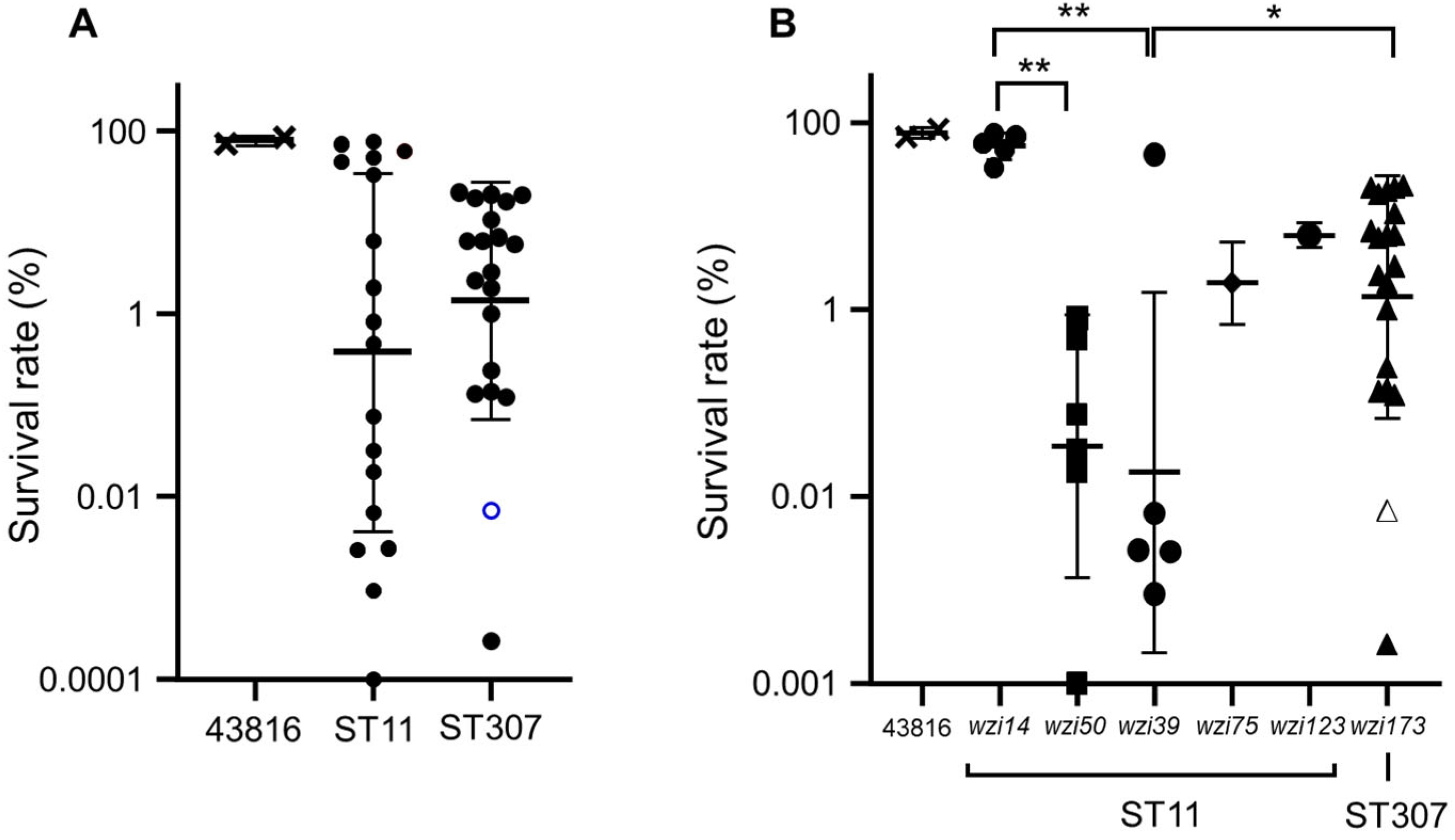
Figure 1. Results of serum resistance assay. (A) Survival rates of ST11 and ST307 isolates were compared using Student’s t-test. (B) Survival rates were compared among the wzi allele types using analysis of variance (ANOVA) with Bonferroni’s multiple-comparison test. The (A) blank circle and (B) blank triangle in ST307 represent the survival rate of SCH2106-08, in which a C58T mutation leading to Q20* in the wzi gene was detected. * p < 0.05, ** p < 0.091.
Microorganisms 10 01827 g001
Figure 2. Results of the macrophage infection experiment. Only gentamicin-susceptible isolates were included in this experiment, as gentamicin was used to eliminate extracellular bacteria. (A) Macrophage internalization rates in ST11 and ST307 isolates analyzed using Student’s t-test. (B) Internalization rates were compared among the wzi allele types using ANOVA with Bonferroni’s multiple-comparison test. * p < 0.05.
Microorganisms 10 01827 g002
Figure 3. (A) Serum resistance and (B) macrophage internalization were compared between the ybtS-positive and -negative isolates regardless of clones. Student’s t-test was used for statistical analysis. * p < 0.05.
Microorganisms 10 01827 g003
Table 1. Sources, wzi alleles, and virulence genes of two K. pneumoniae clones from South Korea, ST11 and ST307.
GenotypeCarbapenemIsolate NumberSourceMBL and ESBLwzi AlleleVirulence Genes
ST11Carbapenem-resistantSCH2104-31BileKPC-214rmpA2, ybtS, iutA
SCH2104-32Voided urineKPC-2, CTX-M-1514rmpA2, ybtS, iutA
SCH2106-16Rectal swabKPC-250ybtS
SCH2107-22Catheterized urineKPC-2, CTX-M-1550ybtS
SCH2108-30SputumKPC-214rmpA2, ybtS, iutA
SCH2104-17Catheterized urineKPC-214ybtS
SCH2108-43SputumKPC-250
SCH2108-44Bronchial washingKPC-275ybtS
Carbapenem-susceptibleSCH2107-06BileCTX-M-950ybtS, clbB
SCH2108-31ND aCTX-M-1514ybtS
B0706-169NDCTX-M-1550ybtS, clbB
B0708-216NDCTX-M-1539
K01-Bact-08-03094NDCTX-M-1539
K01-Bact-08-10058NDCTX-M-1550ybtS, clbB
K01-Bact-08-12164NDCTX-M-1539
K01-Bact-08-12216NDCTX-M-1539
K01-Bact-08-12226NDCTX-M-1539
726UrineCTX-M-15123ybtS, clbB
ST307Carbapenem-resistantSCH2101-15Ascitic fluidKPC-2, CTX-M-15173ybtS
SCH2102-16BileKPC-2, CTX-M-15173ybtS
SCH2102-30Rectal swabKPC-2, CTX-M-15173ybtS
SCH2104-07Voided urineKPC-2, CTX-M-15173ybtS
SCH2104-33Rectal swabKPC-2, CTX-M-15173ybtS
SCH2105-10Catheterized urineKPC-2, CTX-M-15173ybtS
SCH2105-20Rectal swabKPC-2, CTX-M-15173ybtS
SCH2106-08Rectal swabKPC-2, CTX-M-15173 bybtS
SCH2108-07BloodKPC-2, CTX-M-15173ybtS
SCH2109-15SputumKPC-2, CTX-M-15173ybtS
Carbapenem-susceptibleSCH2012-07BloodCTX-M-15173ybtS
SCH2012-19BloodCTX-M-15173ybtS
SCH2107-01BloodCTX-M-15173ybtS
SCH2107-07BloodCTX-M-15173ybtS
SCH2107-08BloodCTX-M-15173
SCH2107-19BloodNone173
925Peritoneal fluidNone173ybtS
SCH-CR31Peritoneal fluidNone173ybtS
633StoolNone173
SCH2106-18BloodNone173
a ND, not described. b C58T mutation led to Q20*, resulting in premature stop in wzi gene.
Publisher’s Note: MDPI stays neutral with regard to jurisdictional claims in published maps and institutional affiliations.