Article

Clostridioides difficile from Fecally Contaminated Environmental Sources: Resistance and Genetic Relatedness from a Molecular Epidemiological Perspective

1
Department of Microbiology–Biotechnology, Faculty of Technology, University of Applied Sciences Emden/Leer, 26723 Emden, Germany
2
Institute of Medical Microbiology and Hygiene, Saarland University Medical Center, 66421 Homburg, Germany
3
German National Reference Center for Clostridioides Difficile, 66421 Homburg, Germany
4
Institute of Hygiene, University of Münster, 48149 Münster, Germany
*
Author to whom correspondence should be addressed.
Microorganisms 2023, 11(10), 2497; https://doi.org/10.3390/microorganisms11102497
Submission received: 4 September 2023 / Revised: 28 September 2023 / Accepted: 28 September 2023 / Published: 5 October 2023

Abstract

:
Clostridioides difficile is the most important pathogen causing antimicrobial-associated diarrhea and has recently been recognized as a cause of community-associated C. difficile infection (CA-CDI). This study aimed to characterize virulence factors, antimicrobial resistance (AMR), ribotype (RT) distribution and genetic relationship of C. difficile isolates from diverse fecally contaminated environmental sources. C. difficile isolates were recovered from different environmental samples in Northern Germany. Antimicrobial susceptibility testing was determined by E-test or disk diffusion method. Toxin genes (tcdA and tcdB), genes coding for binary toxins (cdtAB) and ribotyping were determined by PCR. Furthermore, 166 isolates were subjected to whole genome sequencing (WGS) for core genome multi-locus sequence typing (cgMLST) and extraction of AMR and virulence-encoding genes. Eighty-nine percent (148/166) of isolates were toxigenic, and 51% (76/148) were positive for cdtAB. Eighteen isolates (11%) were non-toxigenic. Thirty distinct RTs were identified. The most common RTs were RT127, RT126, RT001, RT078, and RT014. MLST identified 32 different sequence types (ST). The dominant STs were ST11, followed by ST2, ST3, and ST109. All isolates were susceptible to vancomycin and metronidazole and displayed a variable rate of resistance to moxifloxacin (14%), clarithromycin (26%) and rifampicin (2%). AMR genes, such as gyrA/B, blaCDD-1/2, aph(3′)-llla-sat-4-ant(6)-la cassette, ermB, tet(M), tet(40), and tetA/B(P), conferring resistance toward fluoroquinolone, beta-lactam, aminoglycoside, macrolide and tetracycline antimicrobials, were found in 166, 137, 29, 32, 21, 72, 17, and 9 isolates, respectively. Eleven “hypervirulent” RT078 strains were detected, and several isolates belonged to RTs (i.e., RT127, RT126, RT023, RT017, RT001, RT014, RT020, and RT106) associated with CA-CDI, indicating possible transmission between humans and environmental sources pointing out to a zoonotic potential.

1. Introduction

Clostridioides difficile (formerly Clostridium difficile) is a Gram-positive, anaerobic, spore-forming, toxin-producing, rod-shaped bacterium, which can cause diarrhea but also more severe disease, such as pseudomembranous colitis and even toxic megacolon [1,2]. CDI usually occurs after antibiotic exposure when the normal gut microbiota is disrupted, giving vegetative and spores of C. difficile the ability to thrive. Treatment with antimicrobials, including penicillins, cephalosporins, fluoroquinolones and the macrolide–lincosamide–streptogramin B (MLSB) antimicrobials, is considered a high risk factor for CDI development [3,4,5].
The pathogenicity of C. difficile strains is predominately dependent on the release of two toxins; toxin A (tcdA) and toxin B (tcdB), which contribute to CDI and the respective genes, are encoded on a 19.6 kb pathogenicity locus (PaLoc) together with the regulatory components, TcdR, TcdC and TcdE [6]. Additionally, binary toxin (CDT) encoded by cdtAB is associated with so called “hypervirulent” strains [7]. Besides CDT, these “hypervirulent” strains might harbor mutations in the toxin repressor gene tcdC, leading to a higher toxin production [8].
C. difficile can be characterized by PCR ribotyping on a molecular level, and several ribotypes (RTs) are of epidemiologic importance. For instance, nosocomial CDI is often associated with “hypervirulent” RT027, which has been frequently found in hospital settings and outbreaks, especially in Europe, North America and to some extent in Asian countries [9,10,11]. Furthermore, other “hypervirulent” RTs, such as RT023, RT078, RT126, RT127, and RT176, are known [12,13,14,15]. Of note, RT078 is more commonly associated with community associated (CA)-CDI. In previous years, the zoonotic potential of C. difficile has been under scientific investigation. Several studies have reported that the environment, including animals and food, can be considered as a potential source of CA-CDI [7,16,17,18]. However, up to this date, these reservoirs and C. difficile transmission outside the hospital environment are not fully understood.
In recent years, diverse toxigenic C. difficile strains were recovered from a broad variety of environmental sources (e.g., food, soil, water, wastewater treatment plants (WWTPs), and animal manure) and from different animal species (e.g., cattle, pig and poultry). This includes common RTs, which are frequently encountered in human disease, such as RT001, RT005, RT014/RT020, RT078, and RT126, [19,20,21,22,23]. The prevalence of RT078, being commonly encountered in pigs, has been one of the frequent RTs in 34 European countries in the year 2008, with 8% [12] with decreasing tendency.
Animal manure and sewage sludge often contains C. difficile spores after being treated by digestion or composting in digesters or biogas plants [22,24,25]. Subsequently, the disposal of animal manure and feces, manure-, biogas plant- and thermophilic digester-derived materials or digested sewage sludge as agricultural fertilizers might contribute to environmental contamination with C. difficile.
Exemplified for RT078, strains from both humans and animals are genetically related based on subtyping techniques, such as whole genome sequencing (WGS) following by subsequent phylogenetic analysis [13,26,27,28], which demonstrates evidence for zoonotic transmission of C. difficile between humans and animals. In particular, WGS provides more-in-depth information about genetic diversity and relatedness resulting in a better understanding of the source and the evolution of C. difficile contributing to the current molecular CDI epidemiology [29].
Furthermore, the rapid resistance formation in C. difficile strains poses a significant threat to global health, driven by the increased use of antimicrobials as a treatment against other intestinal pathogens [3], and is known to promote CDI. Several recent studies have reported the emergence of virulent-resistant bacterial pathogens from a variety of sources, increasing the need for the appropriate use of antimicrobial agents. In C. difficile, accessory antimicrobial resistance (AMR) genes are often located on mobile genetic elements (MGEs) (i.e., conjugative and mobilizable transposons, plasmids, and prophages). They can be transferred via horizontal gene transfer (HGT), within toxigenic and non-toxigenic C. difficile strains [30] as well as other bacterial species (i.e., Bacillus subtilis and Enterococcus faecalis) [31,32]. In this study, the strain composition and corresponding phenotypic and genotypic antimicrobial resistance and virulence-associated factors were evaluated giving insight into the molecular epidemiology of C. difficile of environmental origin from Northern Germany. In a second step, the genetic relationship between C. difficile isolates was determined by using core genome multi-locus sequence typing (cgMLST) based on WGS to show possible epidemiologic intersections.

2. Materials and Methods

2.1. Isolation and Identification of C. difficile

C. difficile isolates used in the present study were recovered from various environmental samples, such as WWTP samples (raw sewage, sewage sludge, activated sewage sludge, and digested sewage sludge), calf feces, cattle feces-contaminated soil, thermophilic digesters for treating biowaste and sewage sludge and digested sewage sludge-amended soils as previously described [22]. Briefly, environmental samples were inoculated in C. difficile selective (CD) broth, consisting of proteose peptone 40 g/L, fructose 6.0 g/L, Na2HPO4 5.0 g/L, KH2PO4 1.0 g/L, MgSO4·7H2O 0.1 g/L and NaCl 2.0 g/L. Inoculated CD broths were supplemented with (12 mg/L) norfloxacin (Sigma-Aldrich Chemie GmbH, Munich, Germany) and (32 mg/L) moxalactam (Biomol GmbH, Hamburg, Germany) and 0.1% sodium taurocholate (Carl Roth GmbH & Co. KG, Karlsruhe, Germany) for spore germination. All inoculated CD broths were prepared anaerobically in an anaerobic chamber (Coy Laboratory Products, Inc. Los Angeles, CA, USA) and flushed with a gas mixture (80% N2 and 20% CO2). All inoculated CD broths were incubated at 37 °C for 7–10 days. Each incubated CD broth was then mixed with an equal volume of absolute alcohol (1:1) and incubated at room temperature for 50–60 min. The mixtures were then centrifuged at 4000 rpm for 10 min and the supernatant discarded. The pellet was resuspended in 1× phosphate-buffered saline (PBS) and plated on Clostridium difficile agar (CDA, Fisher Scientific GmbH, Schwerte, Germany) supplemented with 7% defibrinated horse blood (Fisher Scientific GmbH, Schwerte, Germany), (12 mg/mL) norfloxacin, (32 mg/mL) moxalactam and 0.1% sodium taurocholate. All plates were incubated anaerobically in anaerobic jars (Schuett-Biotec GmbH, Göttingen, Germany) (80% N2, 10% H2 and 10% CO2) at 37 °C for 2–5 days. Selected colonies were evaluated by morphology and confirmed by the Oxoid C. difficile latex agglutination test (Fisher Scientific GmbH, Schwerte, Germany). The final confirmation was made by analyzing the specific housekeeping gene, triose phosphate isomerase (tpi), as previously described by Leeme et al. [33].

2.2. PCR-Ribotyping and Toxin Genotyping

PCR ribotyping was conducted as described previously [34]. In short, a standardized ESCMID (European Society of Clinical Microbiology and Infectious Diseases) protocol was utilized together with capillary gel electrophoresis. The obtained C. difficile isolates were characterized for toxin A (tcdA), toxin B (tcdB) and binary toxins (CDT, cdtA B) by conventional PCR [35], and results were confirmed by analyzing the genome of C. difficile strains (see below in Section 2.4).

2.3. Antimicrobial Susceptibility Testing

Antimicrobial susceptibility testing was performed by epsilometry (E-test) and agar disk diffusion as described previously with a McFarland value of 4.0 on Columbia agar (Becton Dickinson, Heidelberg, Germany) [34]. For metronidazole (nitroimidazole), vancomycin (glycopeptide) and moxifloxacin (fluoroquinolone), epsilometry tests were derived from Liofilchem (Roseto degli Abruzzi, Italy) while, for clarithromycin (macrolide) and rifampicin (rifamycin), antibiotic disks originated from Becton Dickinson (Heidelberg, Germany).

2.4. Whole Genome Sequencing and Data Analysis

To determine the genetic relationship of the C. difficile isolates, 166 isolates were subjected to WGS using the Pacific Biosciences long-read platform Sequel IIe (Pacific Biosciences Inc., Menlo Park, CA, USA) and were subsequently de novo-assembled using the SMRT Link software versions 10 and 11 (Pacific Biosciences Inc.) as described recently [36]. For molecular subtyping and to determine the genetic relationship of the different isolates, the cgMLST approach as described elsewhere was applied [37]. Using the Ridom SeqSphere+ software version 9 (Ridom GmbH, Münster, Germany), the cgMLST genes were extracted, and a minimum-spanning tree was constructed to display the genotypic clustering. For backwards compatibility, the “classical” MLST Sequence Types (STs) were extracted in accordance to the C. difficile MLST database of the PubMLST website (https://pubmlst.org/organisms/clostridioides-difficile/. Accessed 15 November 2022). In addition to the minimum-spanning tree analysis, all single nucleotide polymorphisms (SNPs) were extracted from the cgMLST target genes that were present in all strains investigated, and a phylogenetic tree (neighbor-joining tree) was constructed using the SeqSphere+ software. Subsequent graphical representation was done using the iTOL tool version 5 [38]. For further in-depth analysis, the WGS datasets were annotated using the RAST server (the rapid annotation using subsystem technology) version 2.0 (https://rast.nmpdr.org/. Accessed 15 November 2022) [39]. AMR genes were identified by screening contigs with the CARD version 2 (the comprehensive antibiotic resistance databases) using resistance gene identifier (RGI) (https://card.mcmaster.ca/. Accessed 11 April 2023), BacAnt [40], ResFinder 4.1 (https://cge.food.dtu.dk/services/ResFinder/. Accessed 11 April 2023) [41], ARG-ANNOT [42] and Vrprofile2 [43]. The genomes were further analyzed for the presence of known point mutations associated with resistance to fluoroquinolones (e.g., substitution in GyrA and GyrB subunit of the gyrase enzyme) and rifampicin (substitution in RpoB enzyme) using CARD and Snippy v.4.6.0 (https://github.com/tseemann/snippy. Accessed 25 November 2022), respectively.
The toxin genes were identified by using the virulence factors database from BacAnt [40] as well as by annotation provided by the RAST server (https://rast.nmpdr.org/. Accessed 15 November 2022).
All contig sequences generated were submitted to NCBI GenBank under BioProject number (PRJNA1011814).

3. Results

The collection of environmental C. difficile isolates, which were characterized phenotypically and genotypically in the current study, was obtained from different environmental sources in the Northern region of Germany as described previously [22]. The isolates were characterized for antimicrobial susceptibility patterns, and the genomic characterization was assessed for the RT diversity and the prevalence of virulence-encoding genes and AMR genes. In addition, the genetic relatedness among C. difficile isolates was performed using cgMLST based on WGS.

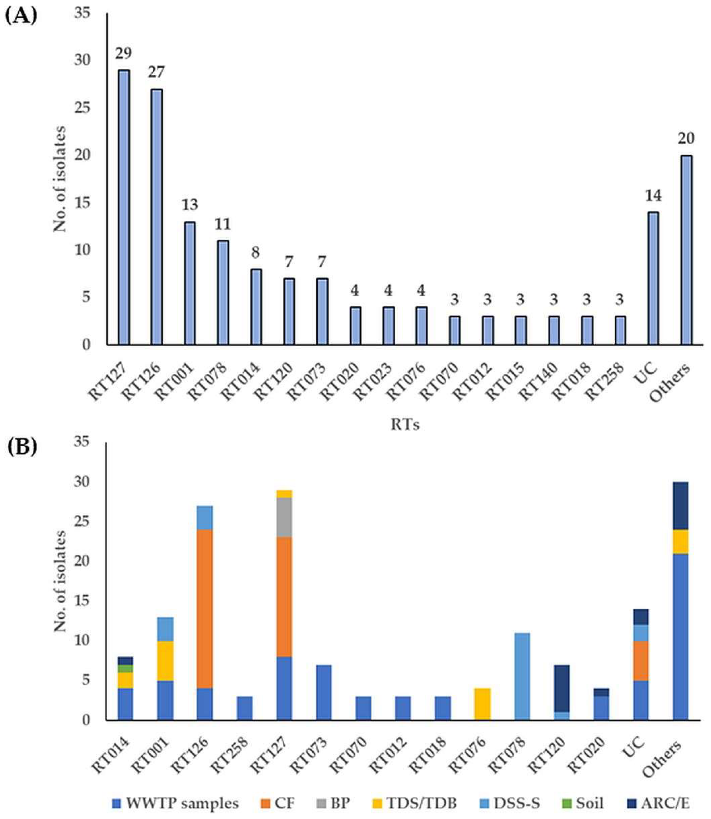
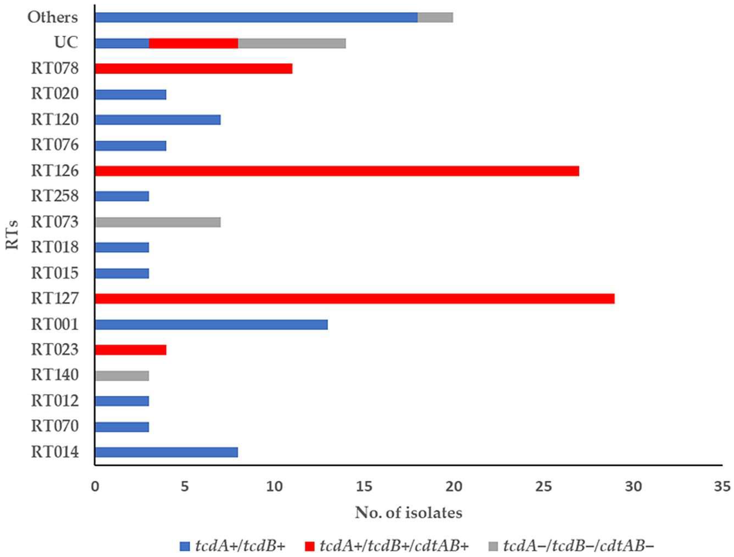
3.1. Toxin-Encoding Genes and PCR Ribotypes of C. difficile Strains

In total, 166 C. difficile isolates were obtained, 148 (89%) isolates were toxigenic, comprised of tcdA+/tcdB+ [72, (49%)], tcdA+/tcdB+/cdtAB+ [76, (51%)] and [18, (11%)] as non-toxigenic isolates (tcdA/tcdB/cdtAB) (Tables S1, S3 and S5). Toxigenic strains could be isolated from almost all environmental samples, (33% in municipal WWTP samples or in feces of calves with 24%).
A total of 30 different RT profiles were identified with remaining 14 isolates that could not be classified (UC). Most predominant RTs were RT127 [29, (17%)], RT126 [27, (16%)], RT001 [13, (8%)], RT078 [11, (7%)], and RT014 [8, (5%)], followed by RT120 and RT073 [7, (4%), each] (Figure 1A, Table S2). Among these RTs, municipal WWTP samples, including raw sewage (RS), raw sewage sludge (RSS), digested sewage sludge (DSS), and activated sewage sludge (ASS), showed the greatest diversity (24 different RTs), followed by anaerobic lab scale bioreactors treating sewage sludge supplemented with or without canola lecithin (control/experiment) (ARC/E) (9 RTs), thermophilic digester for treating sewage sludge or biowaste (TDS/TDB) (6 RTs), digested sewage sludge-amended soils (DSS-S) (4 RTs) and calf feces (CF) (2 RTs) (Figure 1B, Table S2). “Hypervirulent” RT027 was absent, however, RT078 was identified only in C. difficile isolates recovered from DSS-S [11/20, (55%)] (Figure 1B).
RT126 was found more frequently in isolates from CF [20/40, (50%)], whereas RT127 was predominant in isolates from CF, RSS and biogas plant digestate (BP) [15/40, (38%), 8/23, (35%) and 5/5, (100%), respectively]. RT014 and RT020 were only detected in municipal WWTP samples, TDS/TDB, ARC and cattle feces-contaminated soil, and the prevalence indicates the ubiquitous distribution of this RT (Table S2). Some RTs were rarely identified. This included strains from RS (RT073), DSS (RT258, RT106, and RT103), from TDS (RT076), RS/DSS (RT018), RS/RSS/TDB (RT023), and DSS-S/ARE (RT120) (Figure 1B, Table S2).
The toxin-encoding gene profiles of each RT are shown in Figure 2. The most common non-toxigenic strains were RT073 and RT140 [7/18, (39%) and 3/18 (17%), respectively]. The tcdA+/tcdB+ was frequently found in C. difficile RTs RT001, RT014, RT120, and RT020 [13/72, (18%), 8/72, 11%), 7/72, (10%), and 4/72, (6%), respectively] while the tcdA+/tcdB+/cdtAB+ was identified in C. difficile RTs RT126, RT127, RT078, and RT023 [27/76, (36%), 29/76, (38%), 11/76, (14%), and 4/76, (5%), respectively] (Figure 2, Table S3).

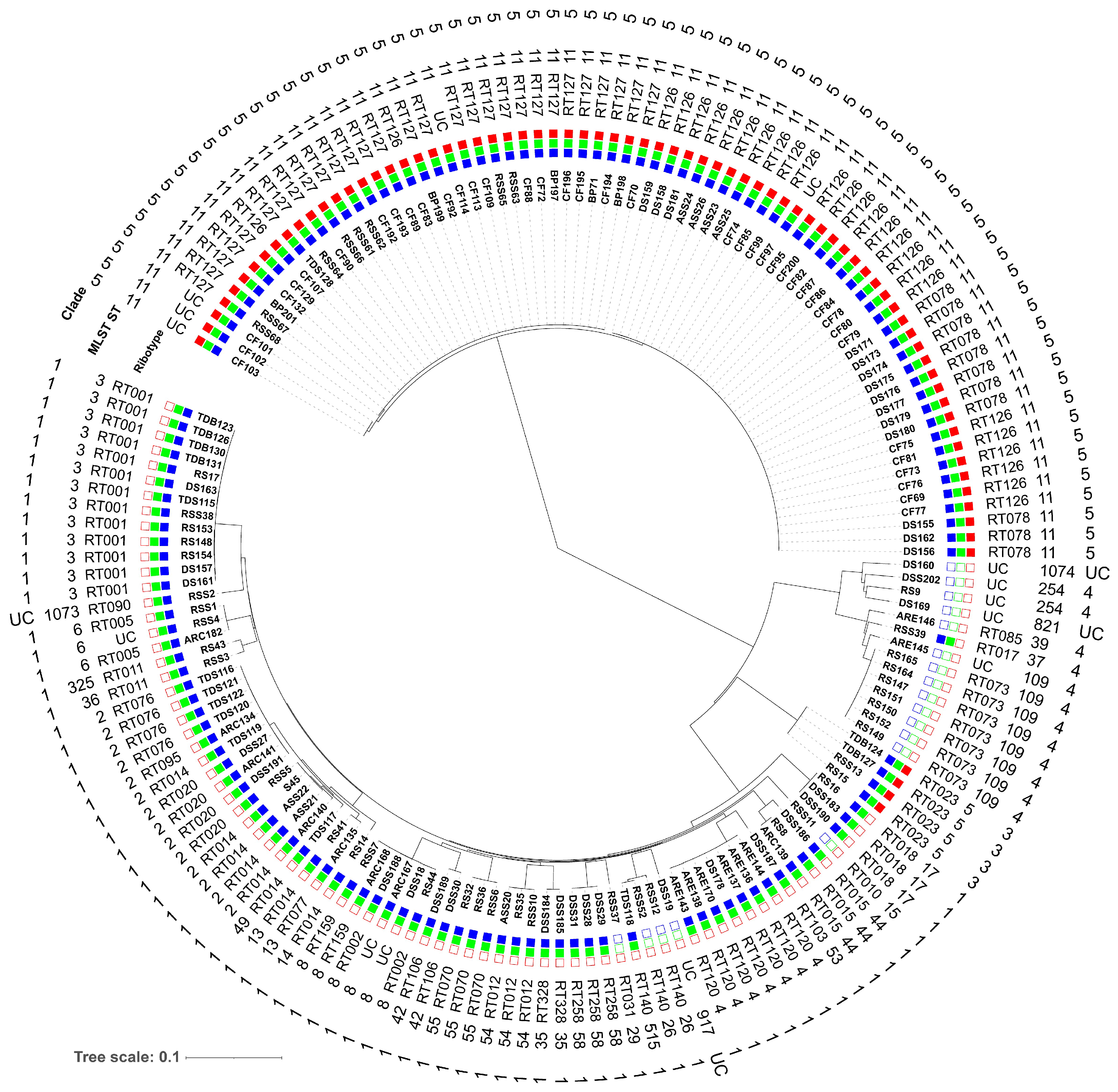
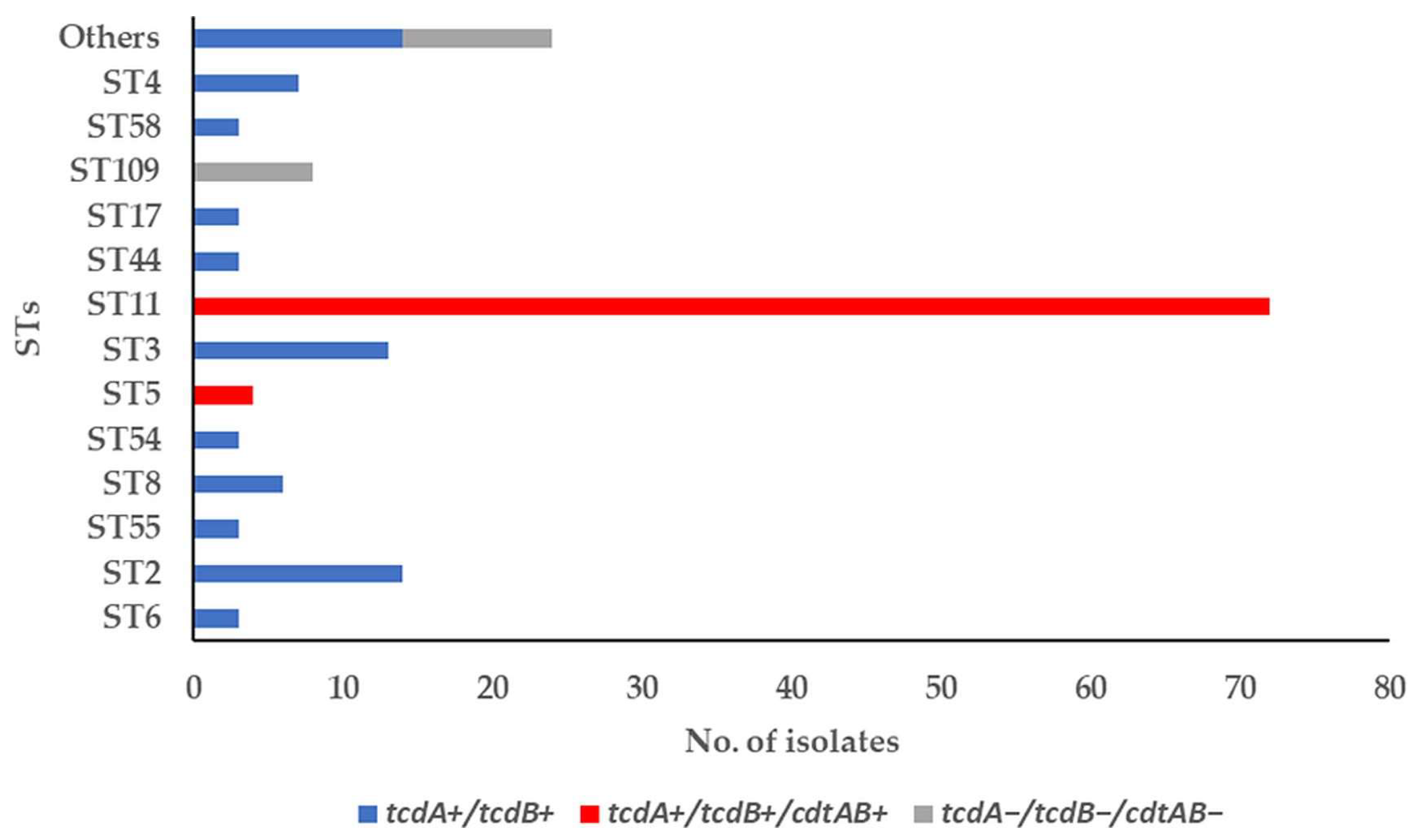
3.2. Molecular Subtyping, Molecular Epidemiology and Association with RTs and Toxin Genes

Using MLST, 166 C. difficile isolates were classified into 32 different sequence types (STs) (Figure 3A, Table S4). Strains belonging to ST11 were the most common, accounting for [72/ (43%)], followed by those belonging to ST2, ST3, ST109, ST4, and ST8 [14, (8%), 13, (8%), 8, (5%), 7, (4%), and 6, (4%), respectively] (Figure 3A, Table S4). The ST11 was most prevalent in C. difficile strains from CF, DSS-S and municipal WWTP samples [40/72, (56%), 14/72 (19%) and 12/72, (17%), respectively] (Figure 3B, Table S4). The ST109 was found only in non-toxigenic C. difficile isolates from RS and ARE [7/8, (88%) and 1/8, (13%), respectively] while the ST3 was found in isolates from TDS/TDB, RS, and DSS-S [5/13, (38%), 4/13, (31%), and 3/13, (23%), respectively]. ST4 was identified in strains from ARE [6/7, (86%)], whereas ST2 in strains from municipal WWTP samples (RSS, DSS, and ASS), TDS, ARC, and cattle feces-contaminated soil [5/14, (36%), 5/14, (36%), 3/14, (21%), and 1/14, (7%), respectively]. The ST17 was identified only in municipal WWTP samples (RS and DSS) (Figure 3B, Table S4). The remaining STs were represented by one or two isolates.
The results of cgMLST typing and subsequent clustering of the 166 isolates from environmental samples are shown in Figure 4. A minimum-spanning tree was constructed based on the allelic profiles of up to 2147 target genes to display the genotype clustering. Differences detected among the isolates ranged from 0–1944 alleles. In total, cgMLST resulted in discrimination of 98 different genotypes. Of these, 19 genotypes were shared among ≥2 isolates; the remaining 79 genotypes were singletons. Using the cluster threshold of ≤6 cgMLST alleles distance, according to Bletz et al. [37], all isolates formed 20 genotyping clusters consisting of 2 to 32 isolates. The largest cluster consisting of 32 isolates was dominated by isolates of RT127, the second largest cluster (n = 25) comprised isolates of RT078 and RT126 (Figure 4, Table S1). Interestingly, genotypes of these two clusters isolates belonged to the same ST11 but differed in cgMLST target genes and their RTs. For instance, RT126 and RT127 isolates differed in 153 cgMLST alleles. Conversely, clustering results indicate that RT126 (10 and 4 isolates from CF and ASS, respectively) is closely related to 11 isolates (RT078) from DSS-S. In addition, among the 32 isolates of the largest cluster, the samples originate from CF, BP, RSS, and TDS. Here, the isolates were distributed based on environmental sources. Also, 14 isolates belonged to the ST2, including different RTs, but the isolate S45 (RT014) showed only one allelic difference from the isolate RSS5 (RT020) whereas the other two isolates (ASS21 and ASS22) remained at 15 allele differences (Figure 4, Table S1).
The SNPs were extracted within the cgMLST dataset to achieve a more in-depth phylogenetic analysis of the 166 C. difficile isolates. In total, 26,636 SNPs were extracted and used to construct a phylogenetic neighbor-joining tree (Figure 5). Here, C. difficile isolates were grouped by their STs, and four related clusters were displayed. The MLST relationship of the C. difficile isolates formed four clades (1, 3, 4, and 5). Clade 1 consists of 21 different STs and clade 4 of four different STs whereas clades 3 and 5 represent one ST each. Clade 1 frequency was higher in municipal WWTP samples. In contrast, clade 5 was more frequent in strains isolated from feces of calves than in municipal WWTP samples (Figure 5, Table S1). Furthermore, some genomes with indistinguishable cgMLST alleles were assigned to multiple RTs, including RT078/RT126 (ST11, clade 5), RT002/RT159 (ST8, clade1), RT077/RT014 (ST13, clade 1), and RT014/RT020/RT076/RT095 (ST2, clade 1). In these cases, several RTs were assigned to different STs and closely related clades (Figure 5, Table 1).
The assignment of C. difficile RTs with the STs and MLST clades are also shown in Table 1. The majority of STs correspond to one RT while some correspond to multiple RTs. Four distinct STs were identified in the RT014 collection (STs, 2, 13, 14, and 49; clade 1) while two STs were identified in the RT011 (STs, 36 and 325; clade 1) and in the RT140 (STs, 26 and 515; clade 1). The ST2 has been associated with different RTs, RT020, RT014, ST076, and ST095 in clade 1 (Table 1).
Interestingly, non-toxigenic strains were found more frequently in clades 4 and 1 while toxigenic strains tcdA+/tcdB+ and tcdA+/tcdB+/cdtAB+ were associated with clade 1 and clades 3 and 5, respectively (Figure 5 and Table S1). The toxin-encoding gene profiles of each ST are included in Figure 6. The tcdA+/tcdB+/cdtAB+ and the tcdA+/tcdB+ isolates were the dominant profiles [76, (46%) and 72, (43%), respectively]. Of 72 toxigenic strains (tcdA+/tcdB+), 14 (19%), 13 (18%), 7 (10%), and 6 (8%) could be associated with four different STs, ST2, ST3, ST4, and ST8, respectively, all corresponding to clade 1. Whereas 72 out of 76 tcdA+/tcdB+/cdtAB+ strains could be assigned with two different STs, ST11 (clade 5, 95%) and ST5 (clade 3, 5%), respectively (Figure 6, Table S5). Several isolates belonged to STs previously associated with human CA-CDI. The non-toxigenic strains were frequently associated with ST109 [8/18, (44%)] (Figure 6).

3.3. Antimicrobial Susceptibility

The antimicrobial susceptibility of 166 C. difficile isolates to five tested antibiotics and their corresponding RTs and STs is shown in Table 2 and Table S1. All C. difficile strains were susceptible to metronidazole and vancomycin. Overall resistance towards clarithromycin, moxifloxacin and rifampicin was encountered in these strains as follows: 26% (43), 14% (23), and 2% (3), respectively. The most clarithromycin (CLR)-resistant strains were found in CF [18/43, (42%)], municipal WWTP samples [13/43, (30%)], and DSS-S [9/43, (21%)]. In addition, moxifloxacin (MXF)-resistant strains were found in DSS-S and CF [10/23, (43%) and 9/23, (39%), respectively]. The highest number of CLR- and MXF-resistance were observed in C. difficile ST11 strains [29/72, (40%) and 17/72, (24%), respectively] (Table 2).

3.4. Antimicrobial Resistance (AMR) Genes

All 166 C. difficile strains harbored at least four accessory AMR genes (Table S1). The most common accessory AMR genes were gyrA and gyrB, conferring fluoroquinolone resistance and found in all strains, caused via mutations in the quinolone resistance determining regions (QRDRs) of DNA gyrase subunits A (gyrA) and/or B (gyrB) (not separately shown in Figure 7). The blaCDD-1 encoding beta-lactamase could be detected in 137 strains (83%) whereas the blaCDD-2 gene was found only in 29 strains (17%). The second most abundant resistance gene is tet(M) detected in 72 strains (43%) and conferring tetracycline resistance by protecting the ribosomal protection protein. The aph(3′)-IIIa gene encoding aminoglycoside resistance was found in 64 strains (39%) whereas ant(6)-la gene conferring also aminoglycoside resistance was found in 36 strains (22%). The sat-4 gene encoding streptothricin resistance was found in 32 strains (19%), and ermB encoding a methylase enzyme that protects the 23S rRNA from the binding of the MLSB group antimicrobials was found in 21 strains (13%) (Figure 7A).
Six different tetracycline (tet) resistance genes were identified in 85 (51%) out of 166 isolates. Among those tet resistance genes, tet(M), tet(40), tet(M)+tet(40), tetA(P)+tetB(P), tet(O), and tet(L) were found in 72, (85%), 17, (20%), 15, (18%), 9, (11%), 2, (2%), and 1, (1%) isolates, respectively (Figure 7A). The tet(M) gene was the most common in isolates recovered from CF and municipal WWTP samples, accounting for [37/72, (51%) and 21/72, (29%), respectively], followed by DSS-S and BP [6/72, (8%) and 5/72, (7%), respectively]. Whereas tet(M)+tet(40) was more dominant in RT126 isolates from CF and municipal WWTP samples [9/15, (60%), and 4/15, (27%), respectively]. In addition, tet(M) was mostly identified in toxigenic C. difficile RT126/127 and non-toxigenic C. difficile RT140 strains, belonging to ST11 and ST26/515, respectively (Figure 7B). The tet(40) gene was found only in RT126 and RT078 (ST11) strains [15/17, (88%) and 2/17, (12%), respectively] isolated from CF, ASS, and DSS-S. Interestingly, tetA(P)+tetB(P) was identified only in isolates from RS and DSS-S [7/9, (78%) and 2/9, (22%), respectively], and those strains belonged to ST109 (RT073, non-toxigenic strains) and ST3 (RT001, toxigenic strains), respectively (Figure 7B).
Beside isolates carrying more than one tetracycline resistances gene, it was also observed that C. difficile isolates harbor one or more genes belonging to an aminoglycoside-streptothricin resistance cassette (aph(3′)-IIIa-sat-4-ant(6)-la). Thirty-two strains carried the complete cassette, belonging to ST11 (RT126 and RT078), suggesting that this cluster associated with ST11 while 32 and 4 strains carried only aph(3′)-IIIa and ant(6)-la, respectively (Figure 7B, Table S1). For aminoglycoside resistance, aac(6′)-aph(2′’) gene was identified in [18/166, (11%)] strains, 15 of them were found in isolates from municipal WWTP samples. This gene is frequently associated with two different STs, ST109 and ST54 (Figure 7B).
In addition, another series of genes related to vancomycin resistance, vanZ1, vanS, vanG and vanT cluster [53/166, (32%)], vanS, vanG, and vanT cluster [22/166, (13%)] or only vanZ1 gene [84/166, (51%)] were found in C. difficile strains. However, all these isolates, which carried vancomycin resistance clusters, displayed high sensitivity towards vancomycin.

4. Discussion

The impact of environmental sources for CDI development is still poorly understood. The presence of toxigenic or non-toxigenic C. difficile has been documented in different environmental sources outside healthcare institutions, such as animal feces, manure, soil, food, and municipal WWTPs [17,21,22,24,44], which could be served as potential sources of CA-CDI.
In the present study, a large strain diversity was evident with several strains being of higher epidemiologic importance. In particular, RT014 and RT020 as one of the most often encountered RTs in human disease could be detected together with RT001 which is considered to be a nosocomially associated strain [12]. Furthermore, RT001 and RT014 were one of the most frequently detected in isolates from poultry meat in Germany [19]. RT014 was also detected in soil samples being located next to a dairy farm [45]. RT014 and RT020 were the predominant RT among soil isolates obtained from home gardens in Western Australia [46] and poultry feces [20].
On the other hand, strains that harbor the binary toxin, such as RT126, RT127 and RT078, were present as well. Of note, RT127 was a major clinical strain in Northwestern Taiwan for the years 2009–2015 [14] and was the most numerous RT detected in this study. Moreover, this RT was most frequently found in toxigenic isolates (50.2%) with CDT among obtained RTs from a calf farm in Australia [47].
A similar situation is given for RT126. RT126 was predominately detected in the feces of calves. RT126 has already been described in cattle [21,44] and pigs [44,48]. Furthermore, RT126 has been observed as one of the predominant RTs in a veal calf farm in Belgium [49]. In Spain, RT126 is one of the most common RTs among clinical isolates [48], and RT126 was also identified in clinical isolates in Southern Taiwan [50]. In a study carried out by Primavilla et al. [51] in hospital food in central Italy, RT126 was also the second most frequently detected RT in CDI cases.
Interestingly, RT027 could not be detected in contrast to RT078, being identified with a high prevalence in DSS-S (7%). Of note, RT078, which is commonly associated with CA-CDI, was isolated from 19%, 8%, 35%, and 60% of primary sludge, digested sludge, biosolids, and river sediments, respectively [25], suggesting that RT078 strains might have resistance mechanisms that could enhance its survival during sewage sludge treatment. Furthermore, the RT078 is frequently reported in farm animals, such as cattle [21,44,52,53], poultry [54], and pigs [26,44,55]. Its epidemiologic importance concerning humans might be illustrated in that RT078 was among the five most frequently encountered RTs in Europe [56]. Furthermore, subtyping data conclude a potential for ongoing zoonotic transmission [18,27,44].
RT023 was identified in 2% of isolates being obtained from RS, RSS, and TDB samples. RT023 prevalence, isolated from humans in Europe, was ~3% [12]. Interestingly, RT018 was found in three isolates recovered from municipal WWTP samples (RS and DSS). In the past, RT018 has been associated with a C. difficile outbreak in Southern Germany [57]. More importantly, RT018 is considered to be the most predominant RT in Northern Italy with prevalence rates exceeding 40% [58].
Non-toxigenic C. difficile strains were identified in particular RT073 (ST109) and RT140 (ST26 and ST515), with prevalence of 4% and 2%, being obtained from RS and (RSS and TDS), respectively. Beside these RTs, one strain each could be assigned to RT010 (ST15) and RT031 (ST29). The presence of non-toxigenic strains is a common finding. Janezic et al. [59] observed that non-toxigenic isolates were commonly found in the environment (30.8%) in comparison to humans (6.5%) and animals (7.7%). Heise et al. [19] observed that several different RTs belonged to non-toxigenic strains, such as RT010, RT205, RT578, RT629, and RT701 obtained from poultry meat in Germany. Interestingly, non-toxigenic ST109 (RT073) was frequently isolated from humans in Japan [60].
In summary, concerning molecular epidemiology: RTs being frequently encountered in humans, such as RT001, RT014, and RT020 were present in the collected environmental samples. This might indicate that digested sewage sludge, untreated sewage, raw sewage sludge, biogas plant derived materials and thermophilic digesters treating biowaste or sewage sludge could pose a reservoir of toxigenic C. difficile RTs.
In addition to the classical differentiation of C. difficile isolates by ribotyping, the genome sequences were determined as well. This enabled us to further subgroup the isolates. Initially, the grouping was performed based on the cgMLST allelic profiles. This analysis revealed 20 clusters and 47 singletons. Many clusters corroborated with ribotyping results. However, in some instances, cgMLST was unable to group the isolates in accordance with their RTs, e.g., isolates sharing RT078 and RT126 or RT014 and RT020, where the allelic profiles only differed in up to five alleles. This is, however, in agreement with recent studies, which observed clustering of several RTs (e.g., RT078/RT126, RT014/RT020) [61,62]. Here, the current study could demonstrate that the distribution of virulence genes, coding for i.e., the toxins A and B and the binary toxins, is concordant with the phylogenetic branching. This indicates that the different branches, which also represent to some extent the different clades, are stable lineages, and acquisition of the mentioned toxins was an early process during the evolution of these lineages, which goes in line with the clonal population structure [63].
For backwards compatibility, “classical” MLST STs (with seven loci) were also extracted from the genomic data set. Here, 32 distinct STs were determined that showed a good correlation to cgMLST typing results. In contrast, the comparison to ribotyping was not always concordant. For example, isolates of ST11 exhibited different RTs (RT127, RT126 and RT078), which were also separated in most instances using cgMLST. In summary, these results go in line with previous results, where RTs could be correlated with STs only to some extent [63].
C. difficile has been known to be resistant to multiple antimicrobials, such as tetracyclines, fluoroquinolones, lincomycin, erythromycin, aminoglycosides, macrolides, and beta-lactam antimicrobials, that are commonly used against bacterial infections in clinical settings [3,5] and continue to be associated with the highest risk for CDI [3]. In the present study, resistance to MXF was frequently detected in ST11 (RT126 and RT078) isolates from the feces of calves and digested sewage sludge-amended soils. Many of RT126 isolates were additionally resistant to CLR, which belongs to the macrolide antibiotic class. These findings are in accordance with what have been reported in calf farms in Italy [21]. Rates of antimicrobial resistance in C. difficile differ in diverse geographic regions [4]. In particular, resistance to fluoroquinolones, macrolides, lincosamides, and tetracyclines has been associated with the spread of ST11 sublineages [64]. In addition, C. difficile has evolved multiple AMR mechanisms that contribute to the development of AMR in C. difficile: (a) harboring of resistance-associated genes in the bacterial chromosome that could be transferred via HGT, including conjugation, transduction or transformation, (b) selection pressure leading to gene mutations, (c) alterations in the antibiotic targets and/or in metabolic pathways in C. difficile and (d) biofilm formation [3,65].
In the current study, six different tetracycline resistance genes in 51% of isolates were identified, including tet(M), tet(40), tetA(P), tetB(P), tet(O), and tet(L). The tet(M) was the predominant gene of the tet class in C. difficile strains (43%) and the majority of C. difficile RT126 and RT127 isolates were positive for tet(M), confirming that tetracycline resistance is widespread among ST11 isolates from a cattle farm. This finding supports the hypothesis of a zoonotic origin of these infections caused by large amounts of tetracyclines used in animal husbandries resulting in a high load released into the agro-ecosystem via organic fertilizers [21,66]. Also, tet(M) gene was identified in non-toxigenic C. difficile RT140 and RT031 strains. It has been reported that all non-toxigenic tet(M)-positive strains from Indonesia and Thailand carried Tn916 or Tn5397 transposons [65]. In C. difficile, acquired accessory AMR genes are often located on MGEs, and the most common element associated with tet(M) mediated tetracycline resistance is Tn5397 and Tn916-like transposons [3,5]. These elements play a crucial role in HGT between distinct toxigenic and non-toxigenic C. difficile strains and between C. difficile strains and other intestinal pathogens. For instance, Tn5397 carrying tet(M) gene was shown to be transferred from C. difficile to Bacillus subtilis [31] and Enterococcus faecalis [32]. The tet(40) gene, which encodes tetracycline efflux, was identified only in RT126 and RT078 isolates which represent 10% from 166 isolates. In a recent study, in 2.1% of 10,330 publicly available C. difficile genomes, tet(40) gene could be identified [65]. Intriguingly, other tet resistance genes, such as tetA(P) and tetB(P) were found in non-toxigenic RT073 and toxigenic RT001 strains. The tetA(P) gene, which mediates active efflux of tetracycline, and tetB(P) gene related to ribosomal protection protein family and were first described in anaerobic bacteria, such as Clostridium perfringens [67]. Therefore, it is proposed that tetA(P) and tetB(P) genes are acquired by the conjugative transfer into C. difficile from some other pathogenic bacteria. Non-toxigenic strains can act as a reservoir for many AMR genes that could be transferred horizontally to toxigenic strains, as well as to other zoonotic pathogenic bacteria.
Resistance to fluoroquinolones was mediated by the presence of chromosomal mutations in the QRDRs of the gyrA and gyrB genes. The presence of the mutations in gyrA and gyrB genes was highly associated with high-risk clones, such as ST11 and ST3, being the most prevalent in the current study. Interestingly, most of obtained amino acid substations patterns in QRDRs of gyrA and gyrB genes have been previously identified among fluoroquinolone-resistant C. difficile strains, belonging to different genotypes, such as RT001, RT018, RT176, and RT046 [68].
Obtained environmental isolates harbored an aminoglycoside-streptothricin resistance cassette (aph(3′)-IIIa-sat-4-ant(6)-la) and were assigned to ST11 (RT126 and RT078), which is similar to the cassette found in Erysipelothrix rhusiopathiae, a species commonly found in pig gut [65] and was also detected in Enterococcus faecium [69]. The sat-4 gene was previously detected in Campylobacter coli and Enterococcus faecium [69,70] and the cassette of resistance genes is found in many bacterial species, indicating the possibility of interspecies transmission. In general, ST11 strains (RT126, RT127, and RT078) show a high proportion of antimicrobial resistance determinates.
For MLSB resistance, the ermB gene was identified in 13% of total isolates, which has been associated with CDI outbreaks in Europe [71]. The ermB gene is mostly found in the conjugative and mobilizable transposons, Tn5398, Tn6194, Tn6218, and Tn6215 [3,4].
For vancomycin resistance, multiple van gene clusters were identified in obtained C. difficile isolates, which were analyzed in this study. However, a complete van resistance operon was not detected in these isolates. Several van gene clusters, including vanA, vanB, vanG, vanW, and vanZ1, have been identified in C. difficile and associated with high vancomycin minimum inhibitory concentrations (MICs) [72]. The expression of these clusters is controlled by two-component regulatory systems, vanS (membrane sensor kinase) and vanR (cytoplasmic response regulator) [72,73], suggesting that these clusters were described to be phenotypically silent. Therefore, the presence of van resistance clusters in environmental C. difficile strains does not always result in their expression in vitro resistance to vancomycin. These strains could be considered susceptible to vancomycin.
For beta-lactam resistance, blaCDD-1 or blaCDD-2 genes were detected in all isolates analyzed, which confer resistance against various beta-lactam antibiotics. These enzymes previously identified in C. difficile strains allowing to have intrinsic resistance to antimicrobials, such as penicillins and cephalosporins [74], which is highly conserved among those C. difficile genomes.

5. Conclusions

This study demonstrated a large genetic overlap between RTs being isolated from environmental samples and humans that may represent a reservoir for CA-CDI. Although RT027 was absent, “hypervirulent” RT078 was found in digested sludge-amended soils, which could possess the ability for zoonotic transmission between humans and environmental sources. Furthermore, a broad variety of AMR genes were predominantly present in the ST11 sublineages. Although resistance to antimicrobials used to treat CDI is rare, this study provides evidence to support the role of AMR in the spread of C. difficile. Future studies need to address the question to which extent HGT, e.g., via MGEs (i.e., transposons, prophages, or plasmids), is present—and further triggered by antimicrobial selection pressure—e.g., for the development and emergence of new epidemic strains.

Supplementary Materials

The following supporting information can be downloaded at: https://www.mdpi.com/article/10.3390/microorganisms11102497/s1, Table S1: Phenotypic and genotypic characterization of environmental C. difficile isolates; Table S2: Distribution of C. difficile RTs in diverse environmental samples; Table S3: Toxin-encoding gene profiles of C. difficile RT strains (n = 166) from various environmental samples; Table S4: Distribution of C. difficile STs (n = 166) in diverse environmental samples; Table S5: Toxin-encoding gene profiles of C. difficile ST strains (n = 166) isolated from various environmental samples; Figure S1: Minimum-spanning tree based on allelic profiles of 166 C. difficile isolates. Each circle represents a separate genotype, and distances between two genotypes are based on the allelic profiles of up to 2147 target genes, pairwise ignoring missing targets. The values on the connecting lines indicate the number of allelic differences between the connected isolates. Circle sizes are proportional to the numbers of isolates per genotype (i.e., the allelic profile). Related genotypes (≤6 alleles distance) are shaded in gray, and the isolates are colored according to their ST. RSS: raw sewage sludge, RS: raw sewage, ASS: activated sewage sludge, DSS: digested sewage sludge, CF: calf feces, BP: biogas plant digestate, ARC/E: anaerobic lab-scale bioreactors treating sewage sludge (control and experiment), DSS-S: digested sewage sludge-amended soils, TDS: thermophilic digester for treating sewage sludge, TDB: thermophilic digester for treating biowaste, S: soil.

Author Contributions

Conceptualization, C.G. and K.B.; methodology, K.B., F.K.B. and A.M.; software, K.B.; validation, K.B. and C.G.; formal analysis, K.B.; investigation, K.B.; resources, K.B., F.K.B., A.M. and C.G.; data curation, K.B.; writing—original draft preparation, K.B.; writing—review and editing, C.G., F.K.B. and A.M.; visualization, K.B. and A.M.; supervision, C.G.; project administration, C.G.; funding acquisition, C.G. All authors have read and agreed to the published version of the manuscript.

Funding

This research was funded by German Research Foundation (DFG, Deutsche Forschungsgemeinschaft) within the project SUPERsafe “Survival and pathogenicity of Clostridioides difficile in sewage, sewage sludge, surface water, animal manure, fodder, crops and silage—Treatment requirements to minimize health risks”, grant number (GA 546/13-1).

Data Availability Statement

Not applicable.

Acknowledgments

The authors would like to thank the undergraduate student from the University of Applied Sciences Emden/Leer, Judith Hoellwarth, for her support regarding the genome analysis of strains. Furthermore, we would like to express our gratitude to Lena Margardt and Anika Berndt for excellent technical support at the National Reference Center for Clostridioides difficile. The article processing charge was funded by the University of Applied Sciences Emden/Leer.

Conflicts of Interest

The authors declare no conflict of interest.

Abbreviations

CDIClostridioides difficile infection
HA-CDIHealthcare associated-CDI
CA-CDICommunity associated-CDI
RT Ribotype
WGSWhole-genome sequencing
STSequence type
MLSTMulti-locus sequence typing
cgMLSTCore genome MLST
AMRAntimicrobial resistance
HGTHorizontal gene transfer
MGEsMobile genetic elements
MXFMoxifloxacin
CLRClarithromycin
RIFRifampicin
MLSBMacrolide-lincosamide-streptogramin B
RSRaw sewage
ASSActivated sewage sludge
RSSRaw sewage sludge
DSSDigested sewage sludge
TDSThermophilic digester for sewage sludge
TDBThermophilic digester for biowaste
ARC/EAnaerobic lab scale bioreactors treating sewage sludge (control and experiment)
DSS-SDigested sewage sludge-amended soils
WWTPWastewater treatment plant
CFCalf feces
BPBiogas plant digestate
SSoil

References

  1. Usacheva, E.A.; Jin, J.-P.; Peterson, L.R. Host response to Clostridium difficile infection: Diagnostics and detection. J. Glob. Antimicrob. Resist. 2016, 7, 93–101. [Google Scholar] [CrossRef]
  2. Buddle, J.E.; Fagan, R.P. Pathogenicity and virulence of Clostridioides difficile. Virulence 2023, 14, 2150452. [Google Scholar] [CrossRef]
  3. Peng, Z.; Jin, D.; Kim, H.B.; Stratton Charles, W.; Wu, B.; Tang, Y.-W.; Sun, X. Update on Antimicrobial Resistance in Clostridium difficile: Resistance Mechanisms and Antimicrobial Susceptibility Testing. J. Clin. Microbiol. 2017, 55, 1998–2008. [Google Scholar] [CrossRef]
  4. Spigaglia, P. Recent advances in the understanding of antibiotic resistance in Clostridium difficile infection. Ther. Adv. Infect. Dis. 2016, 3, 23–42. [Google Scholar] [CrossRef]
  5. O’Grady, K.; Knight, D.R.; Riley, T.V. Antimicrobial resistance in Clostridioides difficile. Eur. J. Clin. Microbiol. Infect. Dis. 2021, 40, 2459–2478. [Google Scholar] [CrossRef]
  6. Dingle, K.E.; Elliott, B.; Robinson, E.; Griffiths, D.; Eyre, D.W.; Stoesser, N.; Vaughan, A.; Golubchik, T.; Fawley, W.N.; Wilcox, M.H.; et al. Evolutionary history of the Clostridium difficile pathogenicity locus. Genome Biol. Evol. 2014, 6, 36–52. [Google Scholar] [CrossRef]
  7. Bolton, D.; Marcos, P. The Environment, Farm Animals and Foods as Sources of Clostridioides difficile Infection in Humans. Foods 2023, 12, 1094. [Google Scholar] [CrossRef]
  8. Lim, S.K.; Stuart, R.L.; Mackin, K.E.; Carter, G.P.; Kotsanas, D.; Francis, M.J.; Easton, M.; Dimovski, K.; Elliott, B.; Riley, T.V.; et al. Emergence of a ribotype 244 strain of Clostridium difficile associated with severe disease and related to the epidemic ribotype 027 strain. Clin. Infect. Dis. 2014, 58, 1723–1730. [Google Scholar] [CrossRef]
  9. Kuijper, E.J.; Barbut, F.; Brazier, J.S.; Kleinkauf, N.; Eckmanns, T.; Lambert, M.L.; Drudy, D.; Fitzpatrick, F.; Wiuff, C.; Brown, D.J.; et al. Update of Clostridium difficile infection due to PCR ribotype 027 in Europe, 2008. Eurosurveillance 2008, 13, 18942. [Google Scholar] [CrossRef]
  10. O’Connor, J.R.; Johnson, S.; Gerding, D.N. Clostridium difficile infection caused by the epidemic BI/NAP1/027 strain. Gastroenterology 2009, 136, 1913–1924. [Google Scholar] [CrossRef]
  11. Collins, D.A.; Hawkey, P.M.; Riley, T.V. Epidemiology of Clostridium difficile infection in Asia. Antimicrob. Resist. Infect. Control 2013, 2, 21. [Google Scholar] [CrossRef]
  12. Bauer, M.P.; Notermans, D.W.; van Benthem, B.H.B.; Brazier, J.S.; Wilcox, M.H.; Rupnik, M.; Monnet, D.L.; van Dissel, J.T.; Kuijper, E.J. Clostridium difficile infection in Europe: A hospital-based survey. Lancet 2011, 377, 63–73. [Google Scholar] [CrossRef]
  13. Goorhuis, A.; Bakker, D.; Corver, J.; Debast, S.B.; Harmanus, C.; Notermans, D.W.; Bergwerff, A.A.; Dekker, F.W.; Kuijper, E.J. Emergence of Clostridium difficile infection due to a new hypervirulent strain, polymerase chain reaction ribotype 078. Clin. Infect. Dis. 2008, 47, 1162–1170. [Google Scholar] [CrossRef]
  14. Tsai, B.-Y.; Chien, C.-C.; Huang, S.-H.; Zheng, J.-Y.; Hsu, C.-Y.; Tsai, Y.-S.; Hung, Y.-P.; Ko, W.-C.; Tsai, P.-J. The emergence of Clostridioides difficile PCR ribotype 127 at a hospital in northeastern Taiwan. J. Microbiol. Immunol. Infect. 2022, 55, 896–909. [Google Scholar] [CrossRef]
  15. Curova, K.; Novotny, M.; Ambro, L.; Kamlarova, A.; Lovayova, V.; Hrabovsky, V.; Siegfried, L.; Jarcuska, P.; Jarcuska, P.; Toporova, A. High Prevalence of Clostridioides difficile Ribotype 176 in the University Hospital in Kosice. Pathogens 2023, 12, 430. [Google Scholar] [CrossRef]
  16. Rabold, D.; Espelage, W.; Abu Sin, M.; Eckmanns, T.; Schneeberg, A.; Neubauer, H.; Möbius, N.; Hille, K.; Wieler, L.H.; Seyboldt, C.; et al. The zoonotic potential of Clostridium difficile from small companion animals and their owners. PLoS ONE 2018, 13, e0193411. [Google Scholar] [CrossRef]
  17. Rodriguez Diaz, C.; Seyboldt, C.; Rupnik, M. Non-human C. difficile Reservoirs and Sources: Animals, Food, Environment. Adv. Exp. Med. Biol. 2018, 1050, 227–243. [Google Scholar] [CrossRef]
  18. Knight, D.R.; Riley, T.V. Genomic Delineation of Zoonotic Origins of Clostridium difficile. Front. Public Health 2019, 7, 164. [Google Scholar] [CrossRef]
  19. Heise, J.; Witt, P.; Maneck, C.; Wichmann-Schauer, H.; Maurischat, S. Prevalence and phylogenetic relationship of Clostridioides difficile strains in fresh poultry meat samples processed in different cutting plants. Int. J. Food Microbiol. 2021, 339, 109032. [Google Scholar] [CrossRef]
  20. Frentrup, M.; Thiel, N.; Junker, V.; Behrens, W.; Münch, S.; Siller, P.; Kabelitz, T.; Faust, M.; Indra, A.; Baumgartner, S.; et al. Agricultural fertilization with poultry manure results in persistent environmental contamination with the pathogen Clostridioides difficile. Environ. Microbiol. 2021, 23, 7591–7602. [Google Scholar] [CrossRef]
  21. Blasi, F.; Lovito, C.; Albini, E.; Bano, L.; Dalmonte, G.; Drigo, I.; Maresca, C.; Massacci, F.R.; Orsini, S.; Primavilla, S.; et al. Clostridioides difficile in Calves in Central Italy: Prevalence, Molecular Typing, Antimicrobial Susceptibility and Association with Antibiotic Administration. Animals 2021, 11, 515. [Google Scholar] [CrossRef]
  22. Blau, K.; Gallert, C. Prevalence, Antimicrobial Resistance and Toxin-Encoding Genes of Clostridioides difficile from Environmental Sources Contaminated by Feces. Antibiotics 2023, 12, 162. [Google Scholar] [CrossRef]
  23. Eckert, C.; Burghoffer, B.; Barbut, F. Contamination of ready-to-eat raw vegetables with Clostridium difficile in France. J. Med. Microbiol. 2013, 62, 1435–1438. [Google Scholar] [CrossRef]
  24. Dharmasena, M.; Jiang, X. Isolation of Toxigenic Clostridium difficile from Animal Manure and Composts Being Used as Biological Soil Amendments. Appl. Environ. Microbiol. 2018, 84, e00738-18. [Google Scholar] [CrossRef]
  25. Xu, C.; Weese, J.S.; Flemming, C.; Odumeru, J.; Warriner, K. Fate of Clostridium difficile during wastewater treatment and incidence in Southern Ontario watersheds. J. Appl. Microbiol. 2014, 117, 891–904. [Google Scholar] [CrossRef]
  26. Álvarez-Pérez, S.; Blanco, J.L.; Peláez, T.; Astorga, R.J.; Harmanus, C.; Kuijper, E.; García, M.E. High prevalence of the epidemic Clostridium difficile PCR ribotype 078 in Iberian free-range pigs. Res. Vet. Sci. 2013, 95, 358–361. [Google Scholar] [CrossRef]
  27. Bakker, D.; Corver, J.; Harmanus, C.; Goorhuis, A.; Keessen, E.C.; Fawley, W.N.; Wilcox, M.H.; Kuijper, E.J. Relatedness of human and animal Clostridium difficile PCR ribotype 078 isolates determined on the basis of multilocus variable-number tandem-repeat analysis and tetracycline resistance. J. Clin. Microbiol. 2010, 48, 3744–3749. [Google Scholar] [CrossRef]
  28. Goorhuis, A.; Debast, S.B.; van Leengoed, L.A.M.G.; Harmanus, C.; Notermans, D.W.; Bergwerff, A.A.; Kuijper, E.J. Clostridium difficile PCR ribotype 078: An emerging strain in humans and in pigs? J. Clin. Microbiol. 2008, 46, 1157, 1158. [Google Scholar] [CrossRef]
  29. Martínez-Meléndez, A.; Morfin-Otero, R.; Villarreal-Treviño, L.; Baines, S.D.; Camacho-Ortíz, A.; Garza-González, E. Molecular epidemiology of predominant and emerging Clostridioides difficile ribotypes. J. Microbiol. Methods 2020, 175, 105974. [Google Scholar] [CrossRef]
  30. Brouwer, M.S.M.; Roberts, A.P.; Hussain, H.; Williams, R.J.; Allan, E.; Mullany, P. Horizontal gene transfer converts non-toxigenic Clostridium difficile strains into toxin producers. Nat. Commun. 2013, 4, 2601. [Google Scholar] [CrossRef]
  31. Mullany, P.; Wilks, M.; Lamb, I.; Clayton, C.; Wren, B.; Tabaqchali, S. Genetic analysis of a tetracycline resistance element from Clostridium difficile and its conjugal transfer to and from Bacillus subtilis. J. Gen. Microbiol. 1990, 136, 1343–1349. [Google Scholar] [CrossRef]
  32. Jasni, A.S.; Mullany, P.; Hussain, H.; Roberts, A.P. Demonstration of conjugative transposon (Tn5397)-mediated horizontal gene transfer between Clostridium difficile and Enterococcus faecalis. Antimicrob. Agents Chemother. 2010, 54, 4924–4926. [Google Scholar] [CrossRef]
  33. Lemee, L.; Dhalluin, A.; Testelin, S.; Mattrat, M.-A.; Maillard, K.; Lemeland, J.-F.; Pons, J.-L. Multiplex PCR targeting tpi (triose phosphate isomerase), tcdA (Toxin A), and tcdB (Toxin B) genes for toxigenic culture of Clostridium difficile. J. Clin. Microbiol. 2004, 42, 5710–5714. [Google Scholar] [CrossRef]
  34. Abdrabou, A.M.M.; Ul Habib Bajwa, Z.; Halfmann, A.; Mellmann, A.; Nimmesgern, A.; Margardt, L.; Bischoff, M.; von Müller, L.; Gärtner, B.; Berger, F.K. Molecular epidemiology and antimicrobial resistance of Clostridioides difficile in Germany, 2014-2019. Int. J. Med. Microbiol. 2021, 311, 151507. [Google Scholar] [CrossRef]
  35. Persson, S.; Torpdahl, M.; Olsen, K.E.P. New multiplex PCR method for the detection of Clostridium difficile toxin A (tcdA) and toxin B (tcdB) and the binary toxin (cdtA/cdtB) genes applied to a Danish strain collection. Clin. Microbiol. Infect. 2008, 14, 1057–1064. [Google Scholar] [CrossRef]
  36. van Almsick, V.; Schuler, F.; Mellmann, A.; Schwierzeck, V. The Use of Long-Read Sequencing Technologies in Infection Control: Horizontal Transfer of a bla(CTX-M-27) Containing lncFII Plasmid in a Patient Screening Sample. Microorganisms 2022, 10, 491. [Google Scholar] [CrossRef]
  37. Bletz, S.; Janezic, S.; Harmsen, D.; Rupnik, M.; Mellmann, A. Defining and Evaluating a Core Genome Multilocus Sequence Typing Scheme for Genome-Wide Typing of Clostridium difficile. J. Clin. Microbiol. 2018, 56, e01987-17. [Google Scholar] [CrossRef]
  38. Letunic, I.; Bork, P. Interactive Tree of Life (iTOL) v5: An online tool for phylogenetic tree display and annotation. Nucleic Acids Res. 2021, 49, W293–W296. [Google Scholar] [CrossRef]
  39. Aziz, R.K.; Bartels, D.; Best, A.A.; DeJongh, M.; Disz, T.; Edwards, R.A.; Formsma, K.; Gerdes, S.; Glass, E.M.; Kubal, M.; et al. The RAST Server: Rapid annotations using subsystems technology. BMC Genom. 2008, 9, 75. [Google Scholar] [CrossRef]
  40. Hua, X.; Liang, Q.; Deng, M.; He, J.; Wang, M.; Hong, W.; Wu, J.; Lu, B.; Leptihn, S.; Yu, Y.; et al. BacAnt: A Combination Annotation Server for Bacterial DNA Sequences to Identify Antibiotic Resistance Genes, Integrons, and Transposable Elements. Front. Microbiol. 2021, 12, 649969. [Google Scholar] [CrossRef]
  41. Bortolaia, V.; Kaas, R.S.; Ruppe, E.; Roberts, M.C.; Schwarz, S.; Cattoir, V.; Philippon, A.; Allesoe, R.L.; Rebelo, A.R.; Florensa, A.F.; et al. ResFinder 4.0 for predictions of phenotypes from genotypes. J. Antimicrob. Chemother. 2020, 75, 3491–3500. [Google Scholar] [CrossRef] [PubMed]
  42. Kumar, G.S.; Roshan, P.B.; Diene, S.M.; Rafael, L.-R.; Marie, K.; Luce, L.; Jean-Marc, R. ARG-ANNOT, a New Bioinformatic Tool to Discover Antibiotic Resistance Genes in Bacterial Genomes. Antimicrob. Agents Chemother. 2014, 58, 212–220. [Google Scholar] [CrossRef]
  43. Wang, M.; Goh, Y.-X.; Tai, C.; Wang, H.; Deng, Z.; Ou, H.-Y. VRprofile2: Detection of antibiotic resistance-associated mobilome in bacterial pathogens. Nucleic Acids Res. 2022, 50, W768–W773. [Google Scholar] [CrossRef] [PubMed]
  44. Spigaglia, P.; Barbanti, F.; Faccini, S.; Vescovi, M.; Criscuolo, E.M.; Ceruti, R.; Gaspano, C.; Rosignoli, C. Clostridioides difficile in Pigs and Dairy Cattle in Northern Italy: Prevalence, Characterization and Comparison between Animal and Human Strains. Microorganisms 2023, 11, 1738. [Google Scholar] [CrossRef] [PubMed]
  45. Marcos, P.; Whyte, P.; Burgess, C.; Ekhlas, D.; Bolton, D. Detection and Genomic Characterisation of Clostridioides difficile from Spinach Fields. Pathogens 2022, 11, 1310. [Google Scholar] [CrossRef] [PubMed]
  46. Shivaperumal, N.; Chang, B.J.; Riley, T.V. High Prevalence of Clostridium difficile in Home Gardens in Western Australia. Appl. Environ. Microbiol. 2020, 87, e01572-20. [Google Scholar] [CrossRef] [PubMed]
  47. Knight, D.R.; Thean, S.; Putsathit, P.; Fenwick, S.; Riley, T.V. Cross-sectional study reveals high prevalence of Clostridium difficile non-PCR ribotype 078 strains in Australian veal calves at slaughter. Appl. Environ. Microbiol. 2013, 79, 2630–2635. [Google Scholar] [CrossRef] [PubMed]
  48. Andrés-Lasheras, S.; Bolea, R.; Mainar-Jaime, R.C.; Kuijper, E.; Sevilla, E.; Martín-Burriel, I.; Chirino-Trejo, M. Presence of Clostridium difficile in pig faecal samples and wild animal species associated with pig farms. J. Appl. Microbiol. 2017, 122, 462–472. [Google Scholar] [CrossRef]
  49. Zidaric, V.; Pardon, B.; Dos Vultos, T.; Deprez, P.; Brouwer, M.S.M.; Roberts, A.P.; Henriques, A.O.; Rupnik, M. Different antibiotic resistance and sporulation properties within multiclonal Clostridium difficile PCR ribotypes 078, 126, and 033 in a single calf farm. Appl. Environ. Microbiol. 2012, 78, 8515–8522. [Google Scholar] [CrossRef]
  50. Hung, Y.-P.; Lin, H.-J.; Tsai, B.-Y.; Liu, H.-C.; Liu, H.-C.; Lee, J.-C.; Wu, Y.-H.; Wilcox, M.H.; Fawley, W.N.; Hsueh, P.-R.; et al. Clostridium difficile ribotype 126 in southern Taiwan: A cluster of three symptomatic cases. Anaerobe 2014, 30, 188–192. [Google Scholar] [CrossRef]
  51. Primavilla, S.; Farneti, S.; Petruzzelli, A.; Drigo, I.; Scuota, S. Contamination of hospital food with Clostridium difficile in Central Italy. Anaerobe 2019, 55, 8–10. [Google Scholar] [CrossRef] [PubMed]
  52. Costa, M.C.; Reid-Smith, R.; Gow, S.; Hannon, S.J.; Booker, C.; Rousseau, J.; Benedict, K.M.; Morley, P.S.; Weese, J.S. Prevalence and molecular characterization of Clostridium difficile isolated from feedlot beef cattle upon arrival and mid-feeding period. BMC Vet. Res. 2012, 8, 38. [Google Scholar] [CrossRef] [PubMed]
  53. Hammitt, M.C.; Bueschel, D.M.; Keel, M.K.; Glock, R.D.; Cuneo, P.; DeYoung, D.W.; Reggiardo, C.; Trinh, H.T.; Songer, J.G. A possible role for Clostridium difficile in the etiology of calf enteritis. Vet. Microbiol. 2008, 127, 343–352. [Google Scholar] [CrossRef] [PubMed]
  54. Hussain, I.; Borah, P.; Sharma, R.K.; Rajkhowa, S.; Rupnik, M.; Saikia, D.P.; Hasin, D.; Hussain, I.; Deka, N.K.; Barkalita, L.M.; et al. Molecular characteristics of Clostridium difficile isolates from human and animals in the North Eastern region of India. Mol. Cell. Probes 2016, 30, 306–311. [Google Scholar] [CrossRef] [PubMed]
  55. Zhang, L.-J.; Yang, L.; Gu, X.-X.; Chen, P.-X.; Fu, J.-L.; Jiang, H.-X. The first isolation of Clostridium difficile RT078/ST11 from pigs in China. PLoS ONE 2019, 14, e0212965. [Google Scholar] [CrossRef] [PubMed]
  56. Freeman, J.; Vernon, J.; Pilling, S.; Morris, K.; Nicolson, S.; Shearman, S.; Clark, E.; Palacios-Fabrega, J.A.; Wilcox, M. Five-year Pan-European, longitudinal surveillance of Clostridium difficile ribotype prevalence and antimicrobial resistance: The extended ClosER study. Eur. J. Clin. Microbiol. Infect. Dis. 2020, 39, 169–177. [Google Scholar] [CrossRef] [PubMed]
  57. Berger, F.K.; Gfrörer, S.; Becker, S.L.; Baldan, R.; Cirillo, D.M.; Frentrup, M.; Steglich, M.; Engling, P.; Nübel, U.; Mellmann, A.; et al. Hospital outbreak due to Clostridium difficile ribotype 018 (RT018) in Southern Germany. Int. J. Med. Microbiol. 2019, 309, 189–193. [Google Scholar] [CrossRef] [PubMed]
  58. Baldan, R.; Trovato, A.; Bianchini, V.; Biancardi, A.; Cichero, P.; Mazzotti, M.; Nizzero, P.; Moro, M.; Ossi, C.; Scarpellini, P.; et al. Clostridium difficile PCR Ribotype 018, a Successful Epidemic Genotype. J. Clin. Microbiol. 2015, 53, 2575–2580. [Google Scholar] [CrossRef]
  59. Janezic, S.; Ocepek, M.; Zidaric, V.; Rupnik, M. Clostridium difficile genotypes other than ribotype 078 that are prevalent among human, animal and environmental isolates. BMC Microbiol. 2012, 12, 48. [Google Scholar] [CrossRef]
  60. Okada, Y.; Yagihara, Y.; Wakabayashi, Y.; Igawa, G.; Saito, R.; Higurashi, Y.; Ikeda, M.; Tatsuno, K.; Okugawa, S.; Moriya, K. Epidemiology and virulence-associated genes of Clostridioides difficile isolates and factors associated with toxin EIA results at a university hospital in Japan. Access Microbiol. 2020, 2, acmi000086. [Google Scholar] [CrossRef]
  61. Baktash, A.; Corver, J.; Harmanus, C.; Smits, W.K.; Fawley, W.; Wilcox, M.H.; Kumar, N.; Eyre, D.W.; Indra, A.; Mellmann, A.; et al. Comparison of Whole-Genome Sequence-Based Methods and PCR Ribotyping for Subtyping of Clostridioides difficile. J. Clin. Microbiol. 2022, 60, e0173721. [Google Scholar] [CrossRef] [PubMed]
  62. Frentrup, M.; Zhou, Z.; Steglich, M.; Meier-Kolthoff, J.P.; Göker, M.; Riedel, T.; Bunk, B.; Spröer, C.; Overmann, J.; Blaschitz, M.; et al. A publicly accessible database for Clostridioides difficile genome sequences supports tracing of transmission chains and epidemics. Microb. Genom. 2020, 6, mgen000410. [Google Scholar] [CrossRef] [PubMed]
  63. Janezic, S.; Rupnik, M. Genomic diversity of Clostridium difficile strains. Res. Microbiol. 2015, 166, 353–360. [Google Scholar] [CrossRef] [PubMed]
  64. Knight, D.R.; Elliott, B.; Chang, B.J.; Perkins, T.T.; Riley, T.V. Diversity and Evolution in the Genome of Clostridium difficile. Clin. Microbiol. Rev. 2015, 28, 721–741. [Google Scholar] [CrossRef] [PubMed]
  65. Imwattana, K.; Kiratisin, P.; Riley, T.V.; Knight, D.R. Genomic basis of antimicrobial resistance in non-toxigenic Clostridium difficile in Southeast Asia. Anaerobe 2020, 66, 102290. [Google Scholar] [CrossRef] [PubMed]
  66. Dingle, K.E.; Didelot, X.; Quan, T.P.; Eyre, D.W.; Stoesser, N.; Marwick, C.A.; Coia, J.; Brown, D.; Buchanan, S.; Ijaz, U.Z.; et al. A Role for Tetracycline Selection in Recent Evolution of Agriculture-Associated Clostridium difficile PCR Ribotype 078. mBio 2019, 10, e02790-18. [Google Scholar] [CrossRef] [PubMed]
  67. Sloan, J.; McMurry, L.M.; Lyras, D.; Levy, S.B.; Rood, J.I. The Clostridium perfringens Tet P determinant comprises two overlapping genes: tetA(P), which mediates active tetracycline efflux, and tetB(P), which is related to the ribosomal protection family of tetracycline-resistance determinants. Mol. Microbiol. 1994, 11, 403–415. [Google Scholar] [CrossRef] [PubMed]
  68. Spigaglia, P.; Mastrantonio, P.; Barbanti, F. Antibiotic Resistances of Clostridium difficile. Adv. Exp. Med. Biol. 2018, 1050, 137–159. [Google Scholar] [CrossRef] [PubMed]
  69. Werner, G.; Hildebrandt, B.; Witte, W. Aminoglycoside-streptothricin resistance gene cluster aadE-sat4-aphA-3 disseminated among multiresistant isolates of Enterococcus faecium. Antimicrob. Agents Chemother. 2001, 45, 3267–3269. [Google Scholar] [CrossRef]
  70. Jacob, J.; Evers, S.; Bischoff, K.; Carlier, C.; Courvalin, P. Characterization of the sat4 gene encoding a streptothricin acetyltransferase in Campylobacter coli BE/G4. FEMS Microbiol. Lett. 1994, 120, 13–17. [Google Scholar] [CrossRef]
  71. Spigaglia, P.; Barbanti, F.; Mastrantonio, P. Multidrug resistance in European Clostridium difficile clinical isolates. J. Antimicrob. Chemother. 2011, 66, 2227–2234. [Google Scholar] [CrossRef]
  72. Eubank, T.A.; Gonzales-Luna, A.J.; Hurdle, J.G.; Garey, K.W. Genetic Mechanisms of Vancomycin Resistance in Clostridioides difficile: A Systematic Review. Antibiotics 2022, 11, 258. [Google Scholar] [CrossRef]
  73. Knight, D.R.; Riley, T.V. Clostridium difficile clade 5 in Australia: Antimicrobial susceptibility profiling of PCR ribotypes of human and animal origin. J. Antimicrob. Chemother. 2016, 71, 2213–2217. [Google Scholar] [CrossRef]
  74. Suzuki, H.; Tomita, M.; Tsai, P.-J.; Ko, W.-C.; Hung, Y.-P.; Huang, I.-H.; Chen, J.-W. Comparative genomic analysis of Clostridium difficile ribotype 027 strains including the newly sequenced strain NCKUH-21 isolated from a patient in Taiwan. Gut Pathog. 2017, 9, 70. [Google Scholar] [CrossRef]
Figure 1. Ribotype (RT) profiling (A) and C. difficile RTs in environmental samples (B). UC: unclassified, CF: calf feces, BP: biogas plant digestate, TDS/TDB: thermophilic digester for treating sewage sludge or biowaste, ARC/E: anaerobic lab-scale bioreactors treating sewage sludge (control and experiment), DSS-S: digested sewage sludge-amended soils. Others indicate RTs with fewer than three assigned strains or samples.
Microorganisms 11 02497 g001
Figure 2. Toxin-encoding genes of C. difficile RT strains (n = 166) from various environmental samples. UC: unclassified, others indicate RTs with fewer than three assigned strains as follows: 005, 090, 011, 159, 010, 031, 017, 002, 095, 077, 085, 106, 328, and 103.
Microorganisms 11 02497 g002
Figure 3. Distribution of C. difficile STs (A) and in diverse environmental samples (B). CF: calf feces, BP: biogas plant digestate, TDS/TDB: thermophilic digester for treating sewage sludge or biowaste, S: soil, ARC/E: anaerobic lab-scale bioreactors treating sewage sludge (control and experiment), DSS-S: digested sewage sludge-amended soils. Others indicate STs with fewer than three assigned strains or samples.
Microorganisms 11 02497 g003
Figure 4. Minimum-spanning tree based on allelic profiles of 166 C. difficile isolates. Each circle represents a separate genotype, and distances between two genotypes are based on the allelic profiles of up to 2147 target genes, pairwise ignoring missing targets. The values on the connecting lines indicate the number of allelic differences between the connected isolates. Circle sizes are proportional to the numbers of isolates per genotype (i.e., the allelic profile). Related genotypes (≤6 alleles distance) are shaded in gray, and the isolates are colored according to their RT. RSS: raw sewage sludge, RS: raw sewage, ASS: activated sewage sludge, DSS: digested sewage sludge, CF: calf feces, BP: biogas plant digestate, ARC/E: anaerobic lab-scale bioreactors treating sewage sludge (control and experiment), DS: digested sewage sludge-amended soils, TDS: thermophilic digester for treating sewage sludge, TDB: thermophilic digester for treating biowaste, S: soil.
Microorganisms 11 02497 g004
Figure 5. Phylogenetic neighbor-joining tree based on 26,636 SNPs detected in cgMLST genes present in all isolates. In addition, the presence (complete boxes) of toxin genes (tcdA [blue], tcdB [green], cdtAB [red]) and absence (empty boxes), RTs, STs and clades are given. RSS: raw sewage sludge, RS: raw sewage, ASS: activated sewage sludge, DSS: digested sewage sludge, CF: calf feces, BP: biogas plant digestate, ARC/E: anaerobic lab-scale bioreactors treating sewage sludge (control and experiment), DS: digested sewage sludge-amended soils, TDS: thermophilic digester for treating sewage sludge, TDB: thermophilic digester for treating biowaste, S: soil, UC: unclassified.
Microorganisms 11 02497 g005
Figure 6. Toxin-encoding genes of C. difficile ST strains (n = 166) from various environmental samples. Others indicate STs with fewer than three assigned strains as follows: ST1073, ST36, ST15, ST26, ST29, ST37, ST254, ST14, ST325, ST917, ST13, ST35, ST53, ST42, ST515, ST39, ST1074, and ST821.
Microorganisms 11 02497 g006
Figure 7. Accessory AMR genes (A) and their association with STs (B) in environmental C. difficile strains (n = 166).
Microorganisms 11 02497 g007
Table 1. The ribotypes (RTs) of C. difficile linked to STs and MLST clades.
CladeRTSTCladeRTST
Clade 1RT005 *ST6Clade 1RT031ST29
RT090ST1073RT001 *ST3
RT011 *ST36, ST325RT015 *ST44
RT020 *ST2RT014 *ST14, ST13, ST2, ST49
RT070 *ST55RT018 *ST17
RT159ST8RT002 *ST8
RT012 *ST54RT258 *ST58
RT010ST15RT103 *ST53
RT140ST26, ST515Clade 4RT085 *ST39
RT077 *ST13RT017 *ST37
RT328 *ST35RT073ST109
RT106 *ST42Clade 5RT126 *ST11
RT076 *ST2RT127 *
RT095ST2RT078 *
RT120ST4Clade 3RT023 *ST5
(*) Human CA-CDI. STs correspond to more than two RTs marked with bold.
Table 2. Antimicrobial resistance profiles of environmental C. difficile RT/ST strains (n = 166).
RT/STNo. of Isolates (%)
CLRMXFRIF
RT126/ST1124 (89%)11 (41%)1 (4%)
RT078/ST114 (36%)5 (45%)0
RT001/ST32 (15%)2 (15%)0
RT012/ST543 (100%)00
RT140/ST26/ST5152 (67%)2 (67%)0
RT328/ST352 (100%)00
RT010/ST151 (100%00
RT031/ST291 (100%)00
RT017/ST3701 (100%)1 (100%)
RT106/ST421 (50%)00
RT015/ST4401 (33%)0
RT014/ST21 (13%)00
RT085/ST391 (100%)01 (100%)
UC/ST111 (20%)1 (20%)0
Total43 (26%)23 (14%)3 (2%)
MXF: moxifloxacin, CLR: clarithromycin, RIF: rifampicin, UC: unclassified.
Disclaimer/Publisher’s Note: The statements, opinions and data contained in all publications are solely those of the individual author(s) and contributor(s) and not of MDPI and/or the editor(s). MDPI and/or the editor(s) disclaim responsibility for any injury to people or property resulting from any ideas, methods, instructions or products referred to in the content.

