Article

Isolation, Antimicrobial Effect and Metabolite Analysis of Bacillus amyloliquefaciens ZJLMBA1908 against Citrus Canker Caused by Xanthomonas citri subsp. citri

College of Coastal Agricultural Science, Guangdong Ocean University, Zhanjiang 524088, China
*
Author to whom correspondence should be addressed.
Microorganisms 2023, 11(12), 2928; https://doi.org/10.3390/microorganisms11122928
Submission received: 20 October 2023 / Revised: 30 November 2023 / Accepted: 4 December 2023 / Published: 6 December 2023
(This article belongs to the Section Plant Microbe Interactions)

Abstract

:
Citrus canker caused by Xanthomonas citri subsp. citri is a devastating bacterial disease with severe implications for the citrus industry. Microorganisms possessing biocontrol capabilities against X. citri subsp. citri offer a highly promising strategy for healthy citrus management. In the present study, a broad-spectrum antagonist strain ZJLMBA1908 with potent antibacterial activity against X. citri subsp. citri was isolated from symptomatic lemon leaves, and identified as Bacillus amyloliquefaciens. Cell-free supernatant (CFS) of strain ZJLMBA1908 also exhibited remarkable antimicrobial activity, especially suppressing the growth of X. citri subsp. citri and Nigrospora oryzae, with inhibition rates of 27.71% and 63.75%, respectively. The antibacterial crude extract (CE) derived from the CFS displayed effective activity against X. citri subsp. citri. A preventive treatment using the CE significantly reduced the severity and incidence of citrus canker in a highly susceptible citrus host. Additionally, the CE maintained activity in the presence of protease and under a wide range of temperature and pH treatments. Applying high-performance liquid chromatography (HPLC) to separate and purify the CE resulted in the discovery of one highly potent anti-X. citri subsp. citri subfraction, namely CE3, which could completely inhibit the growth of X. citri subsp. citri. Liquid chromatography–electrospray ionization–mass spectrometry (LC–ESI–MS) analysis revealed that CE3 mainly consisted of palmitic acid, surfactin C15, phytosphingosine and dihydrosphingosine. Taken together, the results contribute to the possible biocontrol mechanisms of B. amyloliquefaciens ZJLMBA1908, as well as providing a promising new candidate strain as a biological control agent for controlling citrus canker.

1. Introduction

Citrus canker, resulting from infection by the Gram-negative bacterium Xanthomonas citri subsp. citri, is a disease of considerable distribution that affects most commercially cultivated citrus varieties [1]. Its impact mainly focuses on fruit, leaves and twigs, where lesions result in premature drop and reduced marketability, as well as impeding international fruit trade [2]. Currently, citrus canker is mainly managed using integrated management approaches such as planting pathogen-free certified trees, protecting orchards by installing windbreaks and spraying synthetic copper bactericides. The spray application of copper-based bactericides has been the primary counter-measure to prevent X. citri subsp. citri infections [3]. Nevertheless, owing to the prolonged utilization of copper agents, copper-resistant strains have emerged in Xanthomonas populations [4]. The residues of chemical bactericides also have caused substantial adverse effects on the environment. Therefore, the application of new environmentally friendly microbial source substances for the prevention and control of citrus canker disease has emerged as a current research hotspot.
Using beneficial microbe agents as biocontrols is an ecologically friendly alternative that provides effective measures to reduce the risk of plant pathogen resistance while being harmless to non-target species [5]. Over the past few decades, various antagonistic microorganisms have been identified as biological control agents (BCAs) for X. citri subsp. citri, such as the Pseudomonas spp. [6], Bacillus spp. [7], Streptomyces spp. [8], Cronobacter spp. and Enterobacter spp. [9].
Among the most promising BCAs, Bacillus species are harmless endophytic microorganisms that reside within plant tissues for a specific period without inducing symptoms of disease [10]. The application of cell suspensions or fermentations of Bacillus species as a means of controlling citrus diseases has garnered considerable attention worldwide. For instance, Daungfu et al. [11] reported that the leaves of lime plants inoculated with cell suspensions of B. subtilis LE24 or B. amyloliquefaciens LE109 significantly reduced the incidence of citrus canker. Chen et al. [12] proved that the culture filtrate of B. amyloliquefaciens DH-4 was capable of inhibiting the growth of Penicillium digitatum in vitro and suppressing the diameter of pathogen lesions in vivo. However, the effectiveness of cell suspensions or their fermentations in field applications still faces significant challenges due to their inherent lability, limited durability and susceptibility to environmental conditions [13,14].
In this study, a Bacillus amyloliquefaciens strain was isolated and identified from the endophytic bacteria of a symptomatic citrus and rice leaves from a local farm, and its inhibitory effects against some plant pathogens of southern China were evaluated in vitro. Subsequently, the potential biocontrol activity of the antimicrobial compounds produced by strain ZJLMBA1908 isolated from lemon leaves was evaluated against X. citri subsp. citri, and the main compounds responsible for the antibacterial function were identified. The findings of this research will facilitate the development of biological strategies to control citrus canker and provide an alternative to conventional copper treatment, which has caused serious environmental problems.

2. Material and Methods

2.1. Bacterial Strains and Plants

The five plant pathogenic fungi, namely Fusarium oxysporum f.sp. cubense race 4 (Foc4), Neoscytalidium dimidiatum, Nigrospora oryzae, Fusarium solani and Sarocladium oryzae, were kindly provided by Prof. Runhua Yi and Yuelian Liu of Guangdong Ocean University. X. citri subsp. citri strain zlm1908 (designated as X. citri subsp. citri zlm1908) was isolated from the mandarin cultivar Shatangju (Citrus reticulata) and preserved in our laboratory. The endophytic bacteria were isolated from lemon [C. limon (L.) Burm. F.] leaves from a lemon orchard that was infected with citrus canker and rice leaves from a rice field infected with bacterial leaf streak in the Zhanjiang district.
Three-year-old citrus trees (Citrus sinensis ‘Hongjiang’) were purchased from the Zhanjiang Huacheng Agriculture Development Company (Chikan District, Zhanjiang City, China), and grown in 20 cm × 30 cm pots in a greenhouse. The leaves of Hongjiang orange, which are susceptible to citrus canker, were used to perform the X. citri subsp. citri infection experiments.

2.2. Isolation and Purification of Endophytic Bacteria

Sterilization of the leaf surfaces and isolation of the endophytic bacteria were performed as described in Han et al. [15]. The leaf samples were disinfected with 70% ethanol, soaked in 2% sodium hypochlorite (NaClO) for 5 min, rinsed five times with sterilized water and finally drained. A 10 g sample of the sterilized leaves was weighed and cut into small fragments using sterile scissors, thoroughly pulverized using a sterile mortar and pestle and then added to a flask containing 90 mL of sterilized water, followed by shaking for 20 min to obtain suspension stock of the lemon leaves and rice leaves, respectively. Subsequently, a series of dilutions (10−2, 10−3, 10−4, 10−5 and 10−6) were prepared from the leaf debris suspension. Then, 100 μL of each diluted suspension was spread onto Luria-Bertani (LB) solid medium (tryptone 10 g/L, yeast extract 5 g/L, NaCl 10 g/L, pH 7.0) plates, respectively, and incubated at 32 °C. After 24 h, different-looking colonies were picked out onto fresh LB medium plates for pure culture preparation.

2.3. Screening for Antagonistic Bacteria against Plant Pathogens

Efficient broad-spectrum antagonist bacteria were screened using the plate confrontation method [16,17]. The strain X. citri subsp. citri and the five types of fungi were used as indicator microorganisms. For evaluating the antibacterial activity, the X. citri subsp. citri was cultured in the LB medium at 30 °C until reaching an optical density at 600 nm of 0.2. The culture was then evenly spread onto an LB agar plate and fresh endophytic bacteria were transferred onto the center of each plate and incubated at 30 °C for 48 h. For the antifungal activity assay, a 6 mm diameter agar plug of the indicator fungi was placed at the center of potato dextrose agar (PDA) (potato 200 g/L, glucose 20 g/L, agar 20 g/L, pH 7.0) plates (90 mm), and a fresh colony of endophytes was transferred onto the PDA dishes at approximately 25 mm from the fungal mycelia plug at four equidistant points. The control was PDA plates only inoculated with the pathogenic fungi. The plates were incubated at 28 °C until the control group reached 2/3 area of the plate. The antimicrobial activity was expressed in terms of the inhibition rate, which was calculated using the following formulas:
Inhibition rate (%) = R1/R2 × 100%,
Inhibition rate (%) = (R3 − R1)/(R3 − 6) × 100%.
‘R1’ represents the diameter of the inhibition zone, ‘R2’ represents the plate diameter, ‘R3’ represents the colony diameter of the plant pathogen in the control and ‘6’ represents the diameter of the fungal mycelia plug. Formula (1) is used for the indicator bacteria X. citri subsp. citri, while Formula (2) is used for the indicator fungi.

2.4. Identification of Strain ZJLMBA1908

To identify the isolated strain, strain ZJLMBA1908 with the strongest broad-spectrum antagonistic activity was inoculated onto the LB plates at 32 °C. Gram staining was detected during an incubation period of 18–24 h, while an observation of the culture characteristics was conducted at 2 d. Physiological and biochemical characterizations, including catalase activity, starch hydrolysis, a Voges–Proskauer reaction, a methyl red test, gelatin liquefaction, phenylalanine deaminase activity, anaerobic culture, growth at 3 °C, carbon source utilization and salt tolerance, were conducted following Bergey’s Manual of Determinative Bacteriology and the common bacterial identification manual [18,19].
Afterward, strain ZJLMBA1908 was further identified via phylogenetic analysis of the sequences of the 16S rDNA gene, gyrase gene (gyrA) and RNA polymerase β subunit gene (rpoB). The genomic DNA of strain ZJLMBA1908 was isolated and purified using the Bacteria DNA Kit [Accurate Biotechnology (Hunan) Co., Ltd., Changsha, China]. The 16S rRNA was amplified via PCR using the primers 27 F (5′-AGAGTTTGATCMTGGCTCAG-3′) and 1492 R (5′-TACGGYTACCTTGTTACGACTT-3′) [20]. The gyrA gene was amplified using the primers gyrA-F (5′-CAGTCAGGAAATGCGTACGTCCTT-3′) and gyrA-R (5′-CAAGGTAATGCTCCAGGCATTGCT-3′) [21]. The rpoB gene was amplified using the primers rpoB-F (5′-AGGTCAACTAGTTCAGTATGGAC-3′) and rpoB-R (5′-AAGAACCGTAACCGGCAACTT-3′) [22]. The PCR amplification system (50 μL) contained 3 μL of DNA template, 25 μL of 2 × Ex Taq Master Mix [Accurate Biotechnology (Hunan) Co., Ltd., Changsha, China], 1 μL each of the forward and reverse primers and 20 μL of Rnase-free water. The PCR conditions were as follows: 94 °C for 2 min; 35 cycles of 94 °C for 30 s, 58 °C for 30 s and 72 °C for 1 min; and a final extension at 72 °C for 10 min. The amplified products were resolved using 1% agarose gel electrophoresis; afterward, the gel was stained with ethidium bromide, photographed and analyzed using a gel documentation system; the PCR products were subsequently purified and sequenced by Sangon Biotech (Shanghai) Co., Ltd. (China). The obtained sequences were compared via homology alignment analysis using the BLAST program on the NCBI website and the FASTA sequences of the three genes were submitted to GenBank. The gyrA and rpoB gene sequences were aligned using Clustal X (2.0) and then spliced using EditPlus Text Editor v3.70 (290). Phylogenetic trees were constructed using the neighbor-joining method using the MEGA 11.0 software and the clade stability of the resulting tree was assessed using bootstrap analysis with 1000 replicates.

2.5. Preparation and Antimicrobial Activity Determination of ZJLMBA1908 Cell-Free Supernatant (CFS)

The cell-free supernatant (CFS) containing the secondary metabolites of strain ZJLMBA1908 was collected following the method of Li et al. [23] with slight modifications. Strain ZJLMBA1908 was preincubated in LB medium at 37 °C, 180 rpm until OD600nm reached 0.1. Then, the 1% inoculum (v/v) was transferred into a new 50 mL LB medium and incubated under the same conditions. After incubating for 24 h, the supernatant of culture was harvested and centrifuged at 13,535× g for 10 min at 4 °C, and then filtered through a 0.22 μm microporous filter to obtain the CFS.
The antimicrobial activity of the CFS was assayed using Oxford cup tests for strain X. citri subsp. citri and dual culture for the five pathogenic fungi [24]. The sterile LB medium containing 0.1% of the indicator bacteria X. citri subsp. citri (OD600nm = 0.2) was poured onto sterile plates (90 mm). Of the prepared CFS, 75, 175, and 275 μL was, respectively, added to the Oxford cups placed on the medium containing the indicator bacteria. Subsequently, the plates were incubated at 30 °C for 48 h. Then, 15 mL of the PDA medium including 150, 300, 600, 1200, and 2400 μL of the prepared CFS was poured onto sterile plates, then an indicator fungi of the 6 mm diameter plug was placed in the center of the plates and incubated at 28 °C until the control group covered 2/3 of the area of the plate. The treatment without CFS addition was used as a control and the inhibition rate to evaluate the antimicrobial abilities was calculated as described in Section 2.1.

2.6. Preparation and MIC/MBC Determination against X. citri subsp. citri of Crude Extract (CE) from ZJLMBA1908 CFS

The crude extract (CE) was obtained from the ZJLMBA1908 CFS according to the method of Im et al. [25]. Ethyl acetate was added in an equal volume to the CFS, and the upper organic phase was collected after stratification. Thereafter, it was dried under a vacuum using Sy-2000 spinning vapor at 60 °C, dissolved in 1 mL of PBS buffer or methanol and filtered through a 0.22 μm microporous filter. Solutions dissolved in PBS were utilized for the minimum inhibitory concentration (MIC), minimum bactericidal concentration (MBC), and greenhouse experiments and stability assessments, while methanol solutions were employed for the HPLC and LC–MS assays.
To determine the MIC of the CE against X. citri subsp. citri analysis, various concentrations of the CE were subject to two-fold serial dilution in sterile broth ranging from 0.09 to 369.50 μg/mL in 48-well plates [26]. Each well contained 1000 μL CE and 1000 μL X. citri subsp. citri culture (1 × 10⁷ CFU/mL). The negative control was composed of undiluted CE and LB medium, while a positive control included LB medium inoculated with X. citri subsp. citri. The 48-well plate covered with a plastic lid was incubated at 30 °C and 180 rpm for 24 h. The absorbance of the reaction solution was measured at 600 nm using a spectrophotometer. The MIC was defined as the lowest concentration at which the observable growth of the indicator bacteria was completely suppressed. Thereafter, the MBC was assayed by spreading 100 μL of the cultures from each well onto an LB plate and incubating it at 30 °C for 72 h. The MBC was defined as the lowest concentration that killed any indicator bacterial growth in the LB plates following a 72 h incubation period at 30 °C.

2.7. Biocontrol Assays of CE under Greenhouse Conditions

Susceptible citrus hosts of Hongjiang orange were used to assess the biocontrol potential of the CE under greenhouse conditions using the spraying inoculation method [27]. Symptomatic leaves of a consistent color, maturity and size were randomly selected and washed with tap water, followed by sterilization with 75% ethanol, and rinsed with sterile water. In accordance with the prophylactic protocol, the leaves were subsequently categorized into three groups: leaves receiving X. citri subsp. citri cell suspension spraying (positive control), leaves sprayed with both the PBS buffer and CE (negative control) and leaves sprayed with both the CE and X. citri subsp. citri bacterial suspension. Overnight cultures of X. citri subsp. citri (1 × 10⁷ CFU/mL) were centrifuged at 13,535× g for 10 min, collected, resuspended in PBS buffer and adjusted to 1 × 10⁷ CFU/mL. The leaves were then sprayed with 300 μL of the CE at concentrations according to the MBC (184.75, 369.5 and 739 μg/mL) on their surface, followed by 300 μL of X. citri subsp. citri cell suspension. After drying, the leaves were covered with plastic bags for 3 days at 25 °C and maintained at 90% relative humidity. Five citrus leaves were selected in each treatment. Each experiment was carried out for three plants. At 30 days post-inoculation, the inhibition rate was assessed based on the method described by de Oliveira et al. [28], with certain modifications. The inhibition rate was recorded by counting the number of spots on the sprayed leaves using the following formula:
Inhibition rate (%) = (N1 − N2)/N2 × 100%,
‘N1’ represents the number of spots on the diseased leaves of the positive control and ‘N2’ represents the number of spots on the treated leaves.

2.8. Determination of CE Stability to Thermal, pH and Proteinase Enzymes

The CE was dissolved with PBS to obtain a final concentration of 184.75 μg/mL. Firstly, the CE was heated at 37 °C, 60 °C, 80 °C and 100 °C in a water bath for 30 min, and then heated at 121 °C in an autoclave at a pressure of 0.105 MPa for 30 min. The thermal stability of the CE was then evaluated after cooling to room temperature [17]. Thereafter, the pH of the CE was adjusted to 2–12 with 1 mol/L HCl and 1 mol/L NaOH to explore its pH stability [29]. After incubating the above samples at 37 °C for 3 h, their pH was adjusted to the original pH (7.28) to measure the antibacterial activity. For the enzyme sensitivity test, the CE was separately mixed with different proteases, including proteinase K (pH 7.5), trypsin (pH 7.5) and pepsin A (pH 2.0), to a final protease concentration of 1 mg/mL. The mixture was incubated at 37 °C for 2 h, followed by termination via boiling for 5 min [30]. During these experiments, the untreated CE was used as the control, and the antibacterial activities against the X. citri subsp. citri were evaluated using the Oxford cup method, as described in Section 2.5.

2.9. Purification of CE Using HPLC

The CE was purified using preparative high-performance liquid chromatography (HPLC) and an Agilent 1260 Infinity II Preparative LC System (Agilent Technologies, Inc., Santa Clara, CA, USA) equipped with a Zorbax Eclipse XDB-C18 (9.4 × 250 mm, 5 μm, Agilent Technologies, Inc., Santa Clara, CA, USA) column. The mobile phase was composed of 0.1% formic acid in water (A) and acetonitrile (B) with a flow rate of 1 mL/min, and the effluent was monitored at a UV wavelength of 230 nm. Gradient elution was performed with the following program: 0–1 min, 0–2% B; 1–9 min, 2–50% B; 9–20 min, 50–98% B; 20–25 min, 98% B; 25–35 min, 98–2% B; 35–40 min, 0% B. The eluate was collected in three time periods (9–15 min, 15–25 min, 25–34 min) and each fraction was assayed for antagonistic activity against X. citri subsp. citri using colony-counting methods. Specifically, the fractions were evaporated to remove acetonitrile via rotary evaporation, redissolved in methanol, mixed with LB medium at 50 °C and poured into blank plates. The control was the same volume of methanol mixed with LB. The X. citri subsp. citri (OD600nm = 0.2) diluted to 10−5 was applied to the plates above, incubated at 32 °C for 48 h and the number of colonies was counted to evaluate the growth inhibition. Subsequently, the eluted fractions with the best antibacterial activity were identified using liquid chromatography–electrospray ionization–mass spectrometry (LC–ESI–MS).

2.10. Identification of CE Using LC–ESI–MS

LC–ESI–MS analyses of the fractions with the best antibacterial activity were performed on a hybrid quadrupole time-of-flight tandem mass spectrometer equipped with Turbo V sources and a Turbo ion spray interface (AB SCIEX TripleTOF® 5600+, AB SCIEX Pte. Ltd., Concord, ON, Canada). Liquid chromatography was applied on a Sepax GP-C18 column (1.8 µm, 120 Å, 2.1 mm × 150 mm) and the column temperature was maintained at 40 °C. The mobile phase was composed of 0.1% formic acid in water (A) and acetonitrile (B) with a flow rate of 0.3 mL/min. The gradient elution conditions were as follows: 0 min, 5% B; 0–10 min, 5–70%; 10 to 17 min, 70–100% B; 17–18 min, 100% B; 18–19 min, 100–5% B; 19 to 21 min, 5% B. Mass spectrometric data were recorded in positive and negative ion modes with injection voltages of 5500 V and 4400 V, ion source temperatures of 500 °C and 450 °C and a mass scan range of 100–1200 Da. The collision gas used was nitrogen, and the fragment energy was set at 35 ± 15 eV. The obtained LC–MS raw data were processed using the MS-DIAL 5.1 software to select possible adducts, peak alignment, deconvolution and putative metabolite identification based on MSE experiments [31]. The match score was automatically generated using MS-DIAL and the main antibacterial substances with a match score >1.0 were selected via comparison with existing data in the database.

2.11. Statistical Analysis

All experiments were repeated in triplicate under the same conditions. Statistical analysis of the data was carried out using SPSS 22.0 (SPSS Inc., Chicago, IL, USA). Duncan’s multiple-range test and one-way analysis of variance (ANOVA) were employed to assess the significance of the differences, with p < 0.05 indicating significant differences.

3. Results

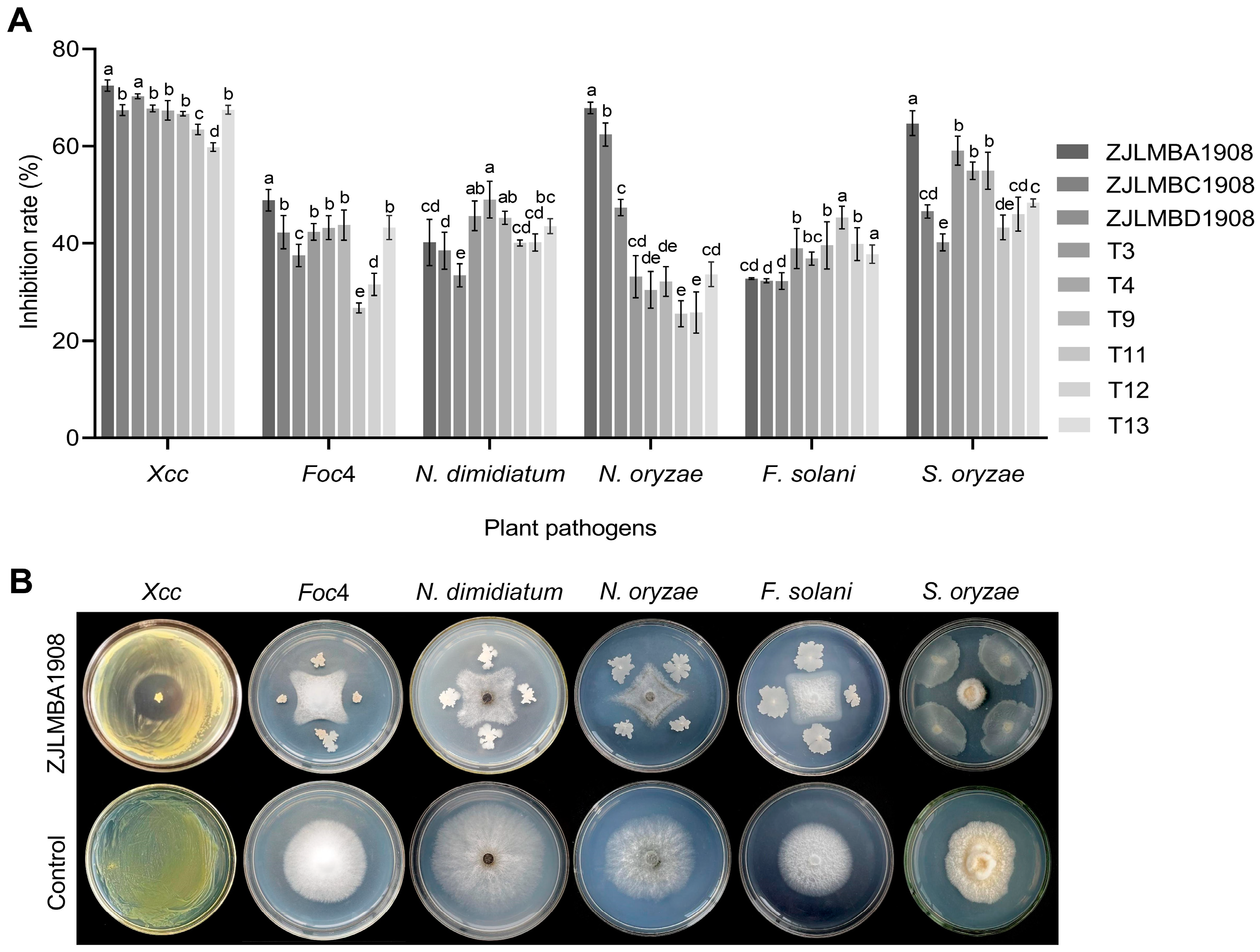
3.1. Antagonistic Effect of Different Endophytic Bacteria

A total of 52 bacterial strains were isolated from the lemon and rice leaves using the gradient dilution method, and 9 strains (ZJLMBA1908, ZJLMBC1908, ZJLMBD1908, T3, T4, T9, T11, T12 and T13) showed good antagonistic activity using the plate confrontation method; the results are shown in Figure 1A. The antagonistic activity of the nine strains against the target pathogen displayed significant variation. Via comparison with the other eight antagonistic bacteria, strain ZJLMBA1908 demonstrated a strong antimicrobial effect and its inhibition rates against X. citri subsp. citri, Foc4, N. oryzae and S. oryzae were, respectively, 36.33%, 48.90%, 67.91% and 64.52% (Figure 1B). For the plant pathogens N. dimidiatum and F. solani, although the inhibition rate was not the highest among the nine antagonists, strain ZJLMBA1908 also showed antibacterial potential with inhibition rates of 40.20% and 32.75%. Therefore, strain ZJLMBA1908 exhibited a broad spectrum against plant pathogens and was chosen for further investigation.

3.2. Identification of Strain ZJLMBA1908

The colonies of strain ZJLMBA1908 were white, round or oval with irregular-shaped edges and a rough, opaque surface on the LB medium (Figure 2A). Microscopically, the bacteria appeared rod-shaped with motility, with a size of (0.7–0.8) μm × (1.5–2.5) μm, produced oval endospores and exhibited the characteristics of a Gram-positive bacterium (Figure 2B). Thereby, they were shown to be a Bacillus sp. The physiological and biochemical characteristics of strain ZJLMBA1908 are documented in Table 1. Positive reactions were observed for the catalase activity test, starch hydrolysis, Voges–Proskauer test, methyl red test and gelatin liquefaction, while negative reactions were found in phenylalanine deaminase, anaerobic culture and 3 °C growth, and there was no salt tolerance at 20% (Table 1). Additionally, sugar and alcohol fermentation experiments demonstrated that strain ZJLMBA1908 could ferment mannose, xylose, glucose, sucrose, galactose and sorbitol. The strain was found to have characteristics similar to those of B. amyloliquefaciens.
The 16S rRNA gene, gyrA gene and rpoB gene sequences were submitted to the GenBank database with accession numbers OR060690, OR085992 and OR085993. Phylogenetic tree analysis of the 16S rRNA gene sequence showed that strain ZJLMBA1908 shared the closest genetic relationship with B. amyloliquefaciens NBRC15535 (Figure 2C). This result was further confirmed using a phylogenetic tree constructed from the sequences of two genes (gyrA and rpoB), which showed that B. amyloliquefaciens S499 and strain ZJLMBA1908 clustered to a single branch (Figure 2D). In conclusion, the strain ZJLMBA1908 isolated from lemon leaves was identified as B. amyloliquefaciens.

3.3. Antagonistic Effect Analysis of ZJLMBA1908 CFS

Various amounts of the CFS produced by strain ZJLMBA1908 were added to the medium to assess its antagonistic action against plant pathogens. The outcomes indicated that the CFS substantially suppressed the growth of all tested pathogens by increasing the dosage of the treatment (Figure 3 and Figure 4) and showed superior antagonistic effects compared to the strain itself. For X. citri subsp. citri, the inhibition rate increased with an increase in the CFS volume from 25.19% to 27.71% (Figure 3 and Table 2). For five different plant pathogen fungi, when the volume of the CFS increased from 150 µL to 2400 µL, the CFS showed antifungal activity with inhibition rates ranging from 17.41% to 33.72%, 2.90% to 40.32%, 4.25% to 63.75%, 1.97% to 53.86% and 6.35% to 39.79% against Foc4, N. dimidiatum, N. oryzae, F. solani and S. oryzae, respectively (Figure 4 and Table 3). Therefore, the CFS of strain ZJLMBA1908 contained potent antimicrobial compounds.

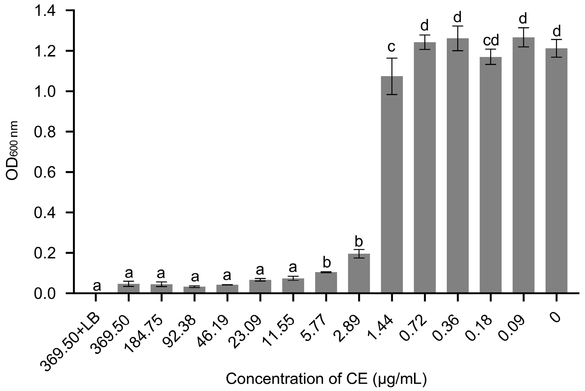
3.4. The MIC and MBC of Crude Extract (CE) against X. citri subsp. citri

An effective concentration of CE against X. citri subsp. citri was evaluated by determining its MIC and MBC values. The results indicate that when cultured in an LB medium containing 11.55 μg/mL of the CE, there was no significant difference in optical density compared to the negative control (Figure 5). Additionally, the absence of visible pathogen growth was observed with increasing concentrations, suggesting nearly complete inhibition of X. citri subsp. citri growth. Cell suspensions treated with the CE at concentrations ranging from 11.55 to 369.50 μg/mL were then transferred to agar plates and the growth was observed after 72 h. No colony formation was detected on the plates for suspensions treated with the CE at concentrations ≥184.75 μg/mL. Hence, the MIC and MBC values of the CE against X. citri subsp. citri were 11.55 and 184.75 μg/mL, respectively.

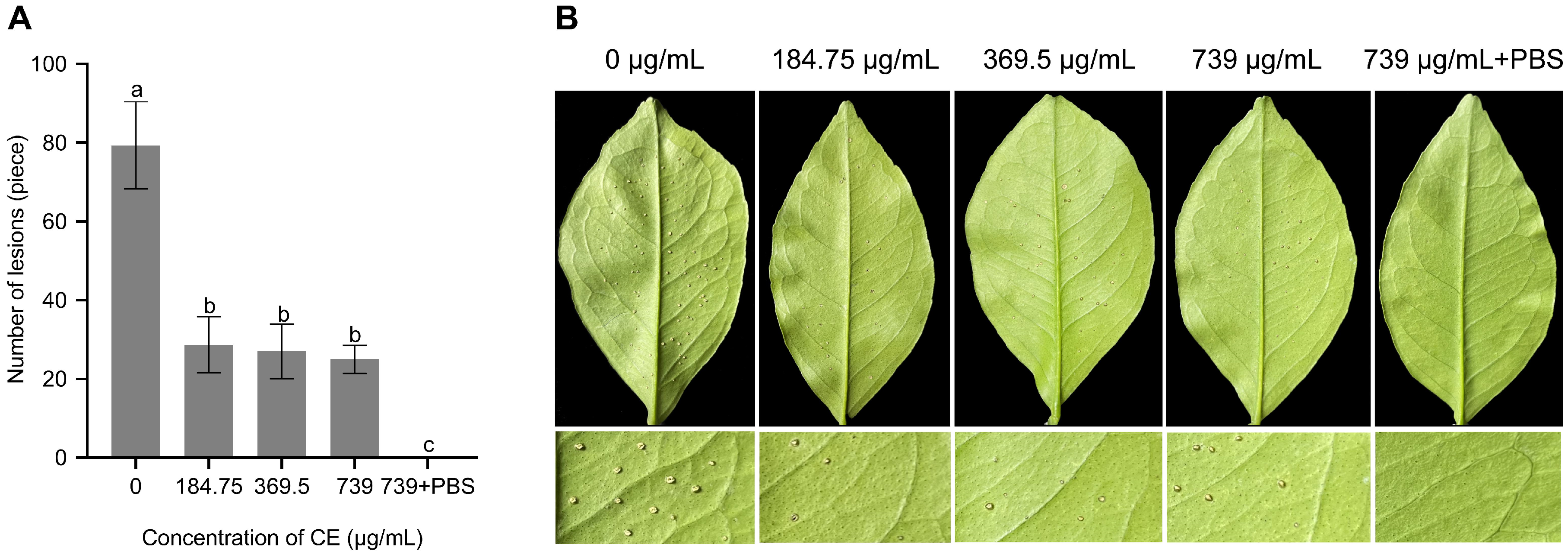
3.5. Evaluation of Biocontrol Efficiency of CE

After 30 days of inoculation, the foliage inoculated with pathogens exhibited severe raised and suberized lesions, whereas that treated with the CE had a noteworthy decrease in canker lesions. The symptoms of typical canker disease gradually decreased with increasing CE spray concentration, with 62.28%, 64.47% and 67.11% inhibition when applied at 184.75, 369.5 and 740 μg/mL, respectively (Figure 6A). Furthermore, leaves treated with the CE displayed minimal visual damage with a slight increase in diameter and only slightly raised pustules at the inoculation site. In contrast, positive control leaves tended to show massive lesions with large, round spots lifted to form a corky appearance (Figure 6B). The results showed effective leaf protection against X. citri subsp. citri infection and the prevention of further citrus canker development using foliar spraying of the CE.

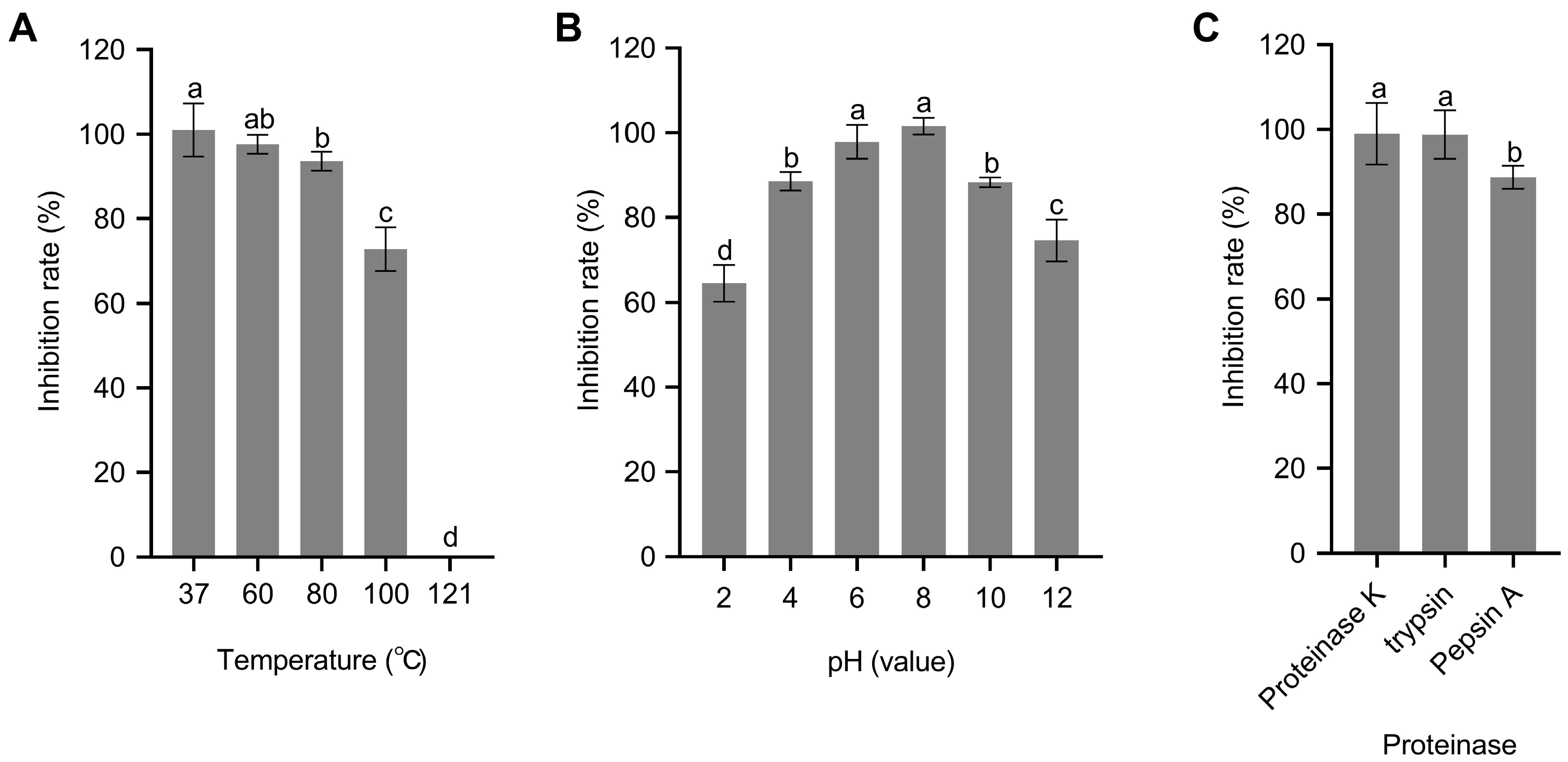
3.6. CE Stability Analysis

The CE had remarkable thermal stability, with over 70% inhibitory activity after exposure to temperatures of 37 °C, 60 °C, 80 °C and 100 °C for 30 min, while a significant loss of activity was observed at 121 °C (Figure 7A). Regarding the pH stability, the CE was stable over a wide range of pH values. In particular, the antimicrobial activity of the CE remained above 85% of the original activity between pHs of 4 and 10, with a gradual decrease observed when the pH was below 2 or above 10, but still maintaining inhibition rates of 64.52% and 74.54%, respectively (Figure 7B). Additionally, the CE also displayed excellent resistance to protease degradation, being insensitive to proteinase K and trypsin and only slightly sensitive to pepsin A (Figure 7C). These results implied that the CE possesses a wide range of activity, enabling it to be adaptable to various environmental conditions to effectively inhibit pathogenic microorganisms, and it might be utilized as a biocontrol agent in agriculture.

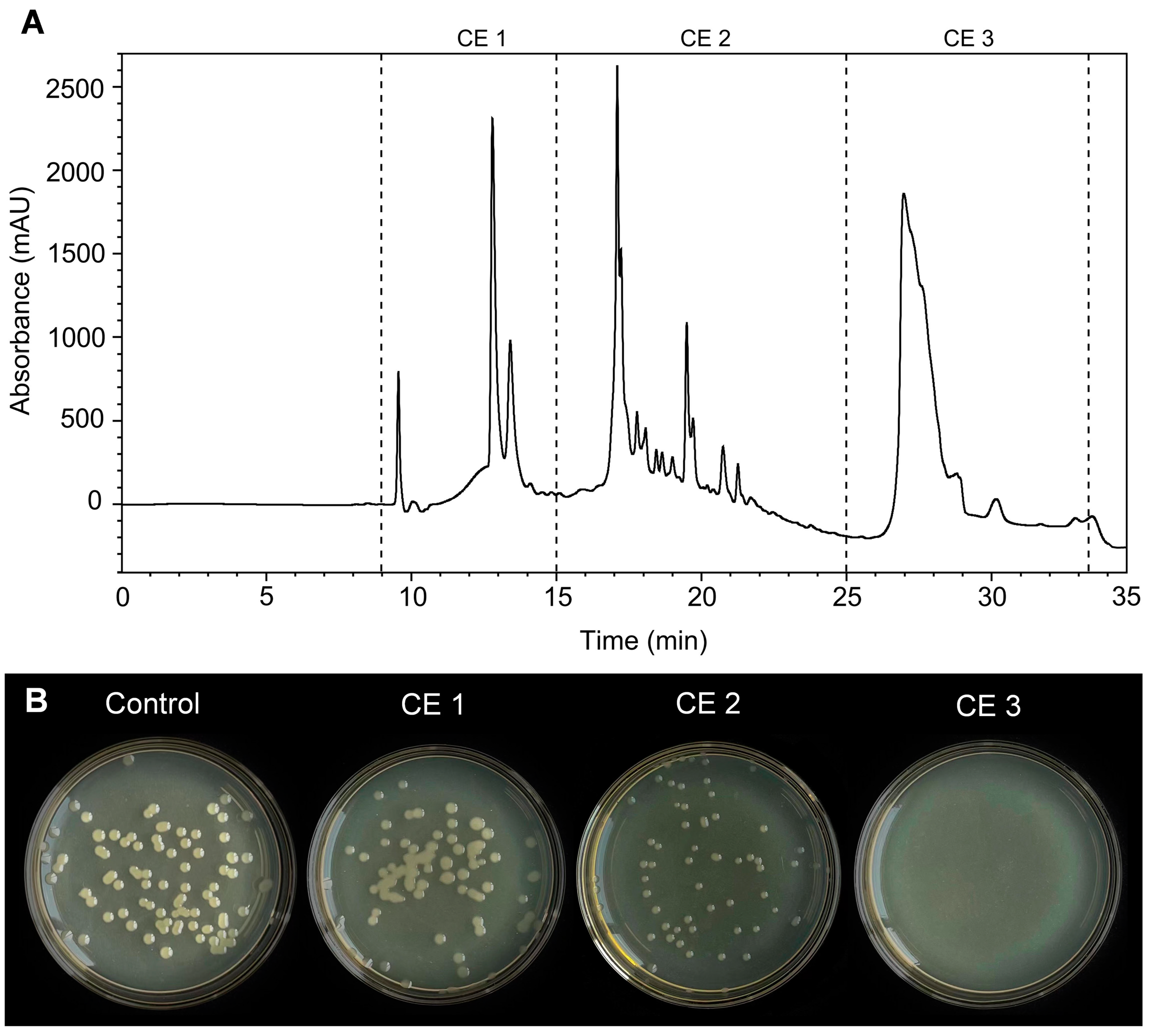
3.7. Analysis of the Antibacterial Activity of the Purified CE

The CE was separated and purified using HPLC, and obtained fractions with elution times of 9–34 min (sample numbers CE1–3) were further determined for antibacterial activity. Among these fractions, CE3 exhibited the strongest antagonistic activity against X. citri subsp. citri with 100% inhibition, as evidenced by the absence of growing colonies (Figure 8B). In contrast, CE1 and CE2 displayed low inhibition rates, amounting to 32.48% and 35.88%, respectively. Therefore, the CE3 fraction was collected and subjected to further mass spectrometric analysis to identify the active compounds.

3.8. Identification of Antibacterial Components from CE3

The LC–ESI–MS analysis revealed that CE3 of strain ZJLMBA1908 contained a variety of different organic compounds, including fatty acids, lipopeptides, sphingosine, phenols, flavonoids, alkaloids, cinnamic acid, steroids, terpenoids and esters (Table 4). Palmitic acid (36.46%), surfactin C15 (5.25%), dihydrosphingosine (4.28%) and phytosphingosine (2.58%) were the predominant components of CE3 in strain ZJLMBA1908. These findings suggest that fatty acids, lipopeptides and sphingosine may be the main metabolites of ZJLMBA1908 responsible for its anti-X. citri subsp. citri effects.

4. Discussion

Citrus canker caused by X. citri subsp. citri infects virtually all major citrus varieties, posing a severe threat to citrus production [32]. Biological control, employing microorganisms and their products, has proven effective in managing plant diseases caused by various pathogens. In contrast to conventional pesticides, biological agents are regarded as sustainable alternatives that pose no harm to mammals and exhibit minimal environmental impact [33]. In this research, the B. amyloliquefaciens strain ZJLMBA1908 was isolated and identified from lemon leaves. The strain and the cell-free supernatant (CFS) of this strain both exhibited antagonistic activity against X. citri subsp. citri and five pathogenic fungi, including Foc4, N. dimidiatum, N. oryzae, F. solani and S. oryzae. The crude extract (CE) obtained from the CFS of strain ZJLMBA1908 displayed strong biocontrol efficacy against X. citri subsp. citri in in vitro and in vivo experiments. The main anti-X. citri subsp. citri compounds in ZJLMBA1908 were identified as fatty acids, lipopeptides and sphingosine. These findings suggest that B. amyloliquefaciens ZJLMBA1908 and its cell extract could be employed as a biocontrol agent against X. citri subsp. citri and several fungal pathogens.
B. amyloliquefaciens is an antagonistic strain with demonstrated potential antimicrobial activities [34]. The B. amyloliquefaciens strain ZJLMBA1908 and its corresponding CFS exhibited remarkable and variable levels of antagonistic effects against the targeted plant pathogens in this study, probably because this strain can fine-tune its antagonistic strategy by producing different antibiotics depending on the nature of the different pathogens. Supporting evidence can be found in previous studies where B. amyloliquefaciens SQR9 modified antifungal substance production against six different soil-borne fungal pathogens [35]. B. amyloliquefaciens PGPBacCA1 was identified to have surfactin and iturin in the liquid culture, whereas for the solid medium, an additional class of lipopeptide called fengycin was identified within the bacterial–fungal growth inhibition zone of strain PGPBacCA1 [36].
Moreover, the biocontrol effects of B. amyloliquefaciens against citrus canker caused by X. citri subsp. citri were studied. The CE effectively inhibited the growth of X. citri subsp. citri in vitro, exhibiting low MIC values (11.55 μg/mL) compared to related studies. Rabbee et al. [37] reported MIC values of 46.9 μg/mL for the ethyl acetate CFS extract of endophytic B. velezensis against X. citri subsp. citri, while Nugroho et al. [38] reported that the MIC of Staphylococcus pasteuri B1 and Staphylococcus warneri C8 CFSs against X. citri subsp. citri was 25 μg/mL. The application of the CE also resulted in a substantial reduction in the occurrence of citrus canker in the host plants, achieving an approximate 60% reduction compared with those in the control. A single applied dose of 184.75 μg/mL of the CE was able to control X. citri subsp. citri infection in the citrus leaves, and no significant differences were observed between different applied doses. However, a trend emerged between the dosage of the CE and its effectiveness, with higher dosages corresponding to superior efficacy, suggesting that it could be meaningful to employ elevated dosages of the CE to control highly developed infections of X. citri subsp. citri. Conversely, for emerging infections, a lower dose of the BCA should suffice to exert adequate control measures. Additionally, the CE maintained stable activities in broad temperature and pH ranges, as well as in the presence of protease. These findings indicate that the anti-X. citri subsp. citri metabolites in the filtrate exhibited stable antagonistic properties, with similarity to the investigations of Nugroho et al. [38]. It also implies that the practical utilization of the CE in natural settings would not be affected by fluctuations in environmental factors.
The available evidence substantiates a correlation between the biocontrol efficacy of B. amyloliquefaciens and its capacity to synthesize a diverse array of antimicrobial compounds [39]. Wang et al. [40] reported B. amyloliquefaciens CE’s association with X. citri subsp. citri inhibition was likely due to the action of surfactin, fengycin and iturin, detected using electrospray ionization–mass spectrometry analysis of the culture extracts. However, the LC–ESI–MS analysis identified fatty acids, lipopeptides, sphingosine, alkaloids, cinnamic acid, flavonoids, phenols, esters and steroids as the main anti-X. citri subsp. citri substances produced by B. amyloliquefaciens. Among the anti-X. citri subsp. citri compounds, fatty acids, particularly palmitic acid, were identified as the most abundant main compounds produced by ZJLMBA1908. This suggests that long-chain fatty acids have a major influence on the anti-X. citri subsp. citri activity. Lin et al. [41] demonstrated linoleic acid, as a long-chain fatty acid, was found to be a potent compound against X. citri subsp. citri and it highly suppressed the growth of X. citri subsp. citri by disrupting the integrity of the cell membrane and inducing excessive production of reactive oxygen species (ROS). Lipopeptides, including surfactin A, surfactin B, surfactin C and maribasin B, dominated over the other groups. While the potent antibacterial activity of surfactin is widely acknowledged, maribasin B was identified for the first time as possessing antibacterial properties in B. amyloliquefacien [42]. Prior work by Zhang et al. [43] reported the broad-spectrum antifungal activity of maribasin B secreted by B. marinus against phytopathogens. Meanwhile, dihydrosphingosine and phytosphingosine were the other main components in strain ZJLMBA1908’s crude extract. These compounds mainly act as long-chain sphingoid bases, with reports of their potent anti-microbial activity inhibiting the growth of several yeast and fungal species in vitro [44]. Researchers have indicated the biological control potential of phenols, flavonoids, alkaloids, cinnamic acid, steroids, terpenoids and esters [45,46,47,48,49,50]. Thus, these compounds could altogether contribute to the antibacterial activity of B. amyloliquefaciens ZJLMBA1908. Although the exact mechanism of the antibacterial activity of these substances against X. citri subsp. citri has not been determined, the prime target of their action may be bacterial cell membranes, potentially disrupting processes essential for cellular defense and functionality.

5. Conclusions

In this study, a broad-spectrum antagonist, B. amyloliquefaciens ZJLMBA1908, was isolated and demonstrated a potent antibacterial ability against X. citri subsp. citri. The cell-free supernatant (CFS) obtained from strain ZJLMBA1908 also exhibited significant antimicrobial activity against a range of plant pathogens, including X. citri subsp. citri, Foc4, N. dimidiatum, N. oryzae, F. solani and S. oryzae. Application of the crude extract (CE) derived from the CFS remarkably restrained the growth of X. citri subsp. citri and reduced the severity of citrus canker symptoms. Using HPLC and LC–ESI–MS to analyze CE3 (separated from the CE), the main anti-X. citri subsp. citri active components of strain ZJLMBA1908 were identified as palmitic acid, phytosphingosine, dihydrosphingosine and surfactin C15. Furthermore, the CE maintained stable activities in a wide range of temperatures and pHs and in the presence of protease. These results indicate that the strain ZJLMBA1908 had great potential as a promising biocontrol agent for controlling the citrus canker caused by X. citri subsp. citri.

Author Contributions

Conceptualization, X.K. and Y.L. (Ya Li); methodology, X.K. and Z.W.; software, Y.L. (Yucheng Liu); validation, Y.L. (Yonglin Liang) and M.D.; formal analysis, X.K., Z.W. and Y.L. (Yucheng Liu); investigation, X.K., Y.L. (Yonglin Liang) and M.D.; data curation, X.K.; writing—original draft preparation, X.K.; writing—review and editing, Y.L. (Ya Li); visualization, X.K.; supervision, Y.L. (Ya Li); project administration, Y.L. (Ya Li); funding acquisition, Y.L. (Ya Li). All authors have read and agreed to the published version of the manuscript.

Funding

This work was supported by the Rural SciTech Special Commission Team Project of Zhanjiang from the Department of Science and Technology of Guangdong Province, grant/award number: A21413.

Data Availability Statement

The data that support the findings of this study are available from the corresponding author upon reasonable request.

Conflicts of Interest

The authors declare no conflict of interest.

References

  1. Pitino, M.; Armstrong, C.M.; Duan, Y. Rapid screening for citrus canker resistance employing pathogen-associated molecular pattern-triggered immunity responses. Hortic. Res. 2015, 2, 15042. [Google Scholar] [CrossRef] [PubMed]
  2. Naqvi, S.A.H.; Wang, J.; Malik, M.T.; Umar, U.-U.-D.; Ateeq-Ur-Rehman; Hasnain, A.; Sohail, M.A.; Shakeel, M.T.; Nauman, M.; Hafeez-ur-Rehman; et al. Citrus canker—distribution, taxonomy, epidemiology, disease cycle, pathogen biology, detection, and management: A critical review and future research agenda. Agronomy 2022, 12, 1075. [Google Scholar] [CrossRef]
  3. Behlau, F.; Hong, J.C.; Jones, J.B.; Graham, J.H. Evidence for acquisition of copper resistance genes from different sources in citrus-associated Xanthomonads. Phytopathology 2013, 103, 409–418. [Google Scholar] [CrossRef] [PubMed]
  4. O’Brien, P.A. Biological control of plant diseases. Australas. Plant Path. 2017, 46, 293–304. [Google Scholar] [CrossRef]
  5. Caicedo, J.C.; Villamizar, S.; Ferro, M.I.T.; Kupper, K.C.; Ferro, J.A. Bacteria from the citrus phylloplane can disrupt cell–cell signalling in Xanthomonas citri and reduce citrus canker disease severity. Plant Pathol. 2016, 65, 782–791. [Google Scholar] [CrossRef]
  6. Rabbee, M.F.; Islam, N.; Baek, K.-H. Biocontrol of citrus bacterial canker caused by Xanthomonas citri subsp. citri by Bacillus velezensis. Saudi J. Biol. Sci. 2022, 29, 2363–2371. [Google Scholar] [CrossRef] [PubMed]
  7. Wattana-Amorn, P.; Charoenwongsa, W.; Williams, C.; Crump, M.P.; Apichaisataienchote, B. Antibacterial activity of cyclo(L-Pro-L-Tyr) and cyclo(D-Pro-L-Tyr) from Streptomyces sp. strain 22-4 against phytopathogenic bacteria. Nat. Prod. Res. 2016, 30, 1980–1983. [Google Scholar] [CrossRef]
  8. Gholami, D.; Aminzadeh, S.; Alavi, S.M.; Kazemipour, N.; Ghoroghi, A.; Emruzi, Z. Comparison of antibiotics and bacteriocins antibacterial activity on Xanthomonas citri subsp. citri. Iran. J. Fish. Sci. 2018, 17, 162–178. [Google Scholar]
  9. Eid, A.M.; Fouda, A.; Abdel-Rahman, M.A.; Salem, S.S.; Elsaied, A.; Oelmüller, R.; Hijri, M.; Bhowmik, A.; Elkelish, A.; Hassan, S.E.-D. Harnessing bacterial endophytes for promotion of plant growth and biotechnological applications: An overview. Plants 2021, 10, 935. [Google Scholar] [CrossRef]
  10. Daungfu, O.; Youpensuk, S.; Lumyong, S. Endophytic bacteria isolated from citrus plants for biological control of citrus canker in lime plants. Trop. Life Sci. Res. 2019, 30, 73–88. [Google Scholar] [CrossRef]
  11. Chen, K.; Tian, Z.; Luo, Y.; Cheng, Y.; Long, C.A. Antagonistic activity and the mechanism of Bacillus amyloliquefaciens DH-4 against citrus green mold. Phytopathology 2018, 108, 1253–1262. [Google Scholar] [CrossRef]
  12. Bashan, Y.; de-Bashan, L.E.; Prabhu, S.; Hernández, J.-P. Advances in plant growth-promoting bacterial inoculant technology: Formulations and practical perspectives (1998–2013). Plant Soil 2014, 378, 1–33. [Google Scholar] [CrossRef]
  13. Pellegrini, M.; Pagnani, G.; Bernardi, M.; Mattedi, A.; Spera, D.M.; Gallo, M.D. Cell-Free supernatants of plant growth-promoting bacteria: A review of their use as biostimulant and microbial biocontrol agents in sustainable agriculture. Sustainability 2020, 12, 9917. [Google Scholar] [CrossRef]
  14. Fira, D.; Dimkić, I.; Berić, T.; Lozo, J.; Stanković, S. Biological control of plant pathogens by Bacillus species. J. Biotechnol. 2018, 285, 44–55. [Google Scholar] [CrossRef] [PubMed]
  15. Abdallah, D.B.; Tounsi, S.; Gharsallah, H.; Hammami, A.; Frikha-Gargouri, O. Bacillus amyloliquefaciens strain 32a as a source of lipopeptides for biocontrol of Agrobacterium tumefaciens strains. J. Appl. Microbiol. 2015, 119, 196–207. [Google Scholar] [CrossRef] [PubMed]
  16. Han, P.; Liu, T.; Zheng, Y.; Song, R.; Nan, T.; Yang, X.; Huang, L.; Yuan, Y. A mycorrhizal bacteria strain isolated from Polyporus umbellatus exhibits broad-spectrum antifungal activity. Front. Plant Sci. 2022, 13, 954160. [Google Scholar] [CrossRef]
  17. Li, Y.; Cai, Y.J.; Yu, Q.H.; Kong, S.L.; Zhou, X.Q.; Zhang, Y.H. Citrus bacterial canker disease: Pathogen identification and bactericides screening. Chin. Agri. Sci. Bull. 2021, 37, 146–153. [Google Scholar]
  18. Zhang, J.; Gu, S.B.; Zhang, T.R.; Wu, Y.; Ma, J.L.; Zhao, L.; Li, X.; Zhang, J. Characterization and antibacterial modes of action of bacteriocins from Bacillus coagulans CGMCC 9951 against Listeria monocytogenes. LWT-Food Sci. Technol. 2022, 160, 113272. [Google Scholar] [CrossRef]
  19. Cappuccino, J.C.; Sherman, N. Microbiology: A Laboratory Manual, 3rd ed.; Benjamin-Cummings Pub. Co.: New York, NY, USA, 1991. [Google Scholar]
  20. Holt, J.G.; Krieg, N.R.; Sneath, P.H.A.; Staley, J.T.; Williams, S.T. Bergey’s Manual of Determinative Bacteriology, 9th ed.; Williams and Wilkins Pub.: Philadelphia, PA, USA, 1994. [Google Scholar]
  21. Ludwig, W. Nucleic acid techniques in bacterial systematics and identification. Int. J. Food Microbiol. 2007, 120, 225–236. [Google Scholar] [CrossRef]
  22. Chun, J.; Bae, K.S. Phylogenetic analysis of Bacillus subtilis and related taxa based on partial gyrA gene sequences. Anton. Leeuw. Int. J. G. 2000, 78, 123–127. [Google Scholar] [CrossRef]
  23. Feng, D.; Zhang, A.; Yang, Y.; Yang, P. Coumarin-containing hybrids and their antibacterial activities. Arch. Pharm. 2020, 353, e1900380. [Google Scholar] [CrossRef] [PubMed]
  24. Li, S.B.; Fang, M.; Zhou, R.C.; Huang, J. Characterization and evaluation of the endophyte Bacillus B014 as a potential biocontrol agent for the control of Xanthomonas axonopodis pv. dieffenbachiae—Induced blight of Anthurium. Biol. Control 2012, 63, 9–16. [Google Scholar] [CrossRef]
  25. Wang, N.N.; Yan, X.; Gao, X.N.; Niu, H.J.; Kang, Z.S.; Huang, L.L. Purification and characterization of a potential antifungal protein from Bacillus subtilis E1R-J against Valsa mali. World J. Microb. Biotechnol. 2016, 32, 63. [Google Scholar] [CrossRef]
  26. Im, S.M.; Yu, N.H.; Joen, H.W.; Kim, S.O.; Park, H.W.; Park, A.R.; Kim, J.-C. Biological control of tomato bacterial wilt by oxydifficidin and difficidin-producing Bacillus methylotrophicus DR-08. Pestic. Biochem. Physiol. 2020, 163, 130–137. [Google Scholar] [CrossRef] [PubMed]
  27. Ying, T.T.; Wu, P.J.; Gao, L.L.; Wang, C.C.; Zhang, T.H.; Liu, S.S.; Huang, R.Q. Isolation and characterization of a new strain of Bacillus amyloliquefaciens and its effect on strawberry preservation. LWT-Food Sci. Technol. 2022, 165, 113712. [Google Scholar] [CrossRef]
  28. Yu, X.Y.; Armstrong, C.M.; Zhou, M.G.; Duan, Y.P. Bismerthiazol inhibits Xanthomonas citri subsp. citri growth and induces differential expression of citrus defense-related genes. Phytopathology 2016, 106, 693–701. [Google Scholar] [CrossRef] [PubMed]
  29. de Oliveira, A.G.; Spago, F.R.; Simionato, A.S.; Navarro, M.O.P.; da Silva, C.S.; Barazetti, A.R.; Cely, M.V.T.; Tischer, C.A.; San Martin, J.A.B.; de Jesus Andrade, C.G.T.; et al. Bioactive organocopper compound from Pseudomonas aeruginosa inhibits the growth of Xanthomonas citri subsp. citri. Front. Microbiol. 2016, 7, 113. [Google Scholar] [CrossRef]
  30. Ye, P.X.; Wang, J.W.; Liu, M.M.; Li, P.; Gu, Q. Purification and characterization of a novel bacteriocin from Lactobacillus paracasei ZFM54. LWT-Food Sci. Technol. 2021, 143, 111125. [Google Scholar] [CrossRef]
  31. Feng, C.L.; Lu, L.Z.; Liu, D.D.; Ning, Y.W.; Wang, Z.X. Purification, structure and characterization of the novel antimicrobial lipopeptides produced by Paenibacillus ehimensis HD. LWT-Food Sci. Technol. 2023, 177, 114603. [Google Scholar] [CrossRef]
  32. Tsugawa, H.; Satoh, A.; Uchino, H.; Cajka, T.; Arita, M.; Arita, M. Mass spectrometry data repository enhances novel metabolite discoveries with advances in computational metabolomics. Metabolites 2019, 9, 119. [Google Scholar] [CrossRef]
  33. Marin, V.R.; Ferrarezi, J.H.; Vieira, G.; Sass, D.C. Recent advances in the biocontrol of Xanthomonas spp. World J. Microb. Biotechnol. 2019, 35, 72. [Google Scholar] [CrossRef]
  34. Tariq, M.; Khan, A.; Asif, M.; Khan, F.; Ansari, T.; Shariq, M.; Siddiqui, M.A. Biological control: A sustainable and practical approach for plant disease management. Acta Agric. Scand. Sect. B Soil Plant Sci. 2020, 70, 507–524. [Google Scholar] [CrossRef]
  35. WoldemariamYohannes, K.; Wan, Z.; Yu, Q.L.; Li, H.Y.; Wei, X.T.; Liu, Y.L.; Wang, J.; Sun, B.G. Prebiotic, probiotic, antimicrobial, and functional food applications of Bacillus amyloliquefaciens. J. Agric. Food Chem. 2020, 68, 14709–14727. [Google Scholar] [CrossRef] [PubMed]
  36. Li, B.; Li, Q.; Xu, Z.H.; Zhang, N.; Shen, Q.R.; Zhang, R.F. Responses of beneficial Bacillus amyloliquefaciens SQR9 to different soilborne fungal pathogens through the alteration of antifungal compounds production. Front. Microbiol. 2014, 5, 636. [Google Scholar] [CrossRef]
  37. Torres, M.J.; Brandan, C.P.; Petroselli, G.; Erra-Balsells, R.; Audisio, M.C. Antagonistic effects of Bacillus subtilis subsp. subtilis and B. amyloliquefaciens against Macrophomina phaseolina: SEM study of fungal changes and UV-MALDI-TOF MS analysis of their bioactive compounds. Microbiol. Res. 2016, 182, 31–39. [Google Scholar] [CrossRef] [PubMed]
  38. Rabbee, M.F.; Ali, M.S.; Baek, K.-H. Endophyte Bacillus velezensis isolated from Citrus spp. controls streptomycin-resistant Xanthomonas citri subsp. citri that causes citrus bacterial canker. Agronomy 2019, 9, 470. [Google Scholar] [CrossRef]
  39. Nugroho, Y.A.; Suharjono, S.; Widyaningsih, S. Biological control of citrus canker pathogen Xanthomonas citri subsp. citri using Rangpur lime endophytic bacteria. Egypt. J. Biol. Pest Control 2022, 32, 63. [Google Scholar] [CrossRef]
  40. Wang, X.; Liang, L.Q.; Shao, H.; Ye, X.X.; Yang, X.B.; Chen, X.Y.; Shi, Y.; Zhang, L.H.; Xu, L.H.; Wang, J.X. Isolation of the novel strain Bacillus amyloliquefaciens F9 and identification of lipopeptide extract components responsible for activity against Xanthomonas citri subsp. citri. Plants 2022, 11, 457. [Google Scholar] [CrossRef]
  41. Lin, H.T.; Liang, Y.; Kaliaperumal, K.; Xiong, Q.; Duan, S.; Jiang, Y.M.; Zhang, J. Linoleic acid from the endophytic fungus Diaporthe sp. HT-79 inhibits the growth of Xanthomonas citri subsp. citri by destructing the cell membrane and producing reactive oxygen species (ROS). Pestic. Biochem. Physiol. 2023, 192, 105423. [Google Scholar] [CrossRef]
  42. Sreedharan, S.M.; Rishi, N.; Singh, R. Microbial lipopeptides: Properties, mechanics and engineering for novel lipopeptides. Microbiol. Res. 2023, 271, 127363. [Google Scholar] [CrossRef]
  43. Zhang, D.J.; Liu, R.F.; Li, Y.G.; Tao, L.M.; Tian, L. Two new antifungal cyclic lipopeptides from Bacillus marinus B-9987. Chem. Pharm. Bull. 2010, 58, 1630–1634. [Google Scholar] [CrossRef] [PubMed]
  44. Thevissen, K.; Hillaert, U.; Meert, E.M.; Chow, K.K.; Cammue, B.P.; Van Calenbergh, S.; François, I.E. Fungicidal activity of truncated analogues of dihydrosphingosine. Bioorg. Med. Chem. Lett. 2008, 18, 3728–3730. [Google Scholar] [CrossRef] [PubMed]
  45. Cushnie, T.P.; Cushnie, B.; Lamb, A.J. Alkaloids: An overview of their antibacterial, antibiotic-enhancing and antivirulence activities. Int. J. Antimicrob. Agents 2014, 44, 377–386. [Google Scholar] [CrossRef] [PubMed]
  46. do Nascimento, P.G.; Lemos, T.L.; Almeida, M.C.; de Souza, J.M.; Bizerra, A.M.; Santiago, G.M.; da Costa, J.G.; Coutinho, H.D. Lithocholic acid and derivatives: Antibacterial activity. Steroids 2015, 104, 8–15. [Google Scholar] [CrossRef]
  47. Santhakumari, S.; Jayakumar, R.; Logalakshmi, R.; Prabhu, N.M.; Abdul Nazar, A.K.; Karutha Pandian, S.; Veera Ravi, A. In vitro and in vivo effect of 2,6-Di-tert-butyl-4-methylphenol as an antibiofilm agent against quorum sensing mediated biofilm formation of Vibrio spp. Int. J. Food Microbiol. 2018, 281, 60–71. [Google Scholar] [CrossRef]
  48. Mrudulakumari Vasudevan, U.; Lee, E.Y. Flavonoids, terpenoids, and polyketide antibiotics: Role of glycosylation and biocatalytic tactics in engineering glycosylation. Biotechnol. Adv. 2020, 41, 107550. [Google Scholar] [CrossRef]
  49. Sahoo, C.R.; Sahoo, J.; Mahapatra, M.; Lenka, D.; Sahu, P.K.; Dehury, B.; Padhy, R.N.; Paidesetty, S.K. Coumarin derivatives as promising antibacterial agent(s). Arab. J. Chem. 2021, 14, 102922. [Google Scholar] [CrossRef]
  50. Xie, L.G.; Zhu, G.D.; Shang, J.J.; Chen, X.M.; Zhang, C.T.; Ji, X.L.; Zhang, Q.; Wei, Y. An overview on the biological activity and anti-cancer mechanism of lovastatin. Cell. Signal. 2021, 87, 110122. [Google Scholar] [CrossRef]
Figure 1. In vitro antagonistic effect of nine endophytic bacteria against six phytopathogens. (A) The inhibition rate of nine endophytic bacteria against six phytopathogens. Error bars indicate standard errors of the means from three repeated experiments. Different letters indicate a significant difference according to the Duncan’s multiple-range test (p < 0.05); (B) Antagonistic effect of strain ZJLMBA1908 against six phytopathogens.
Microorganisms 11 02928 g001
Figure 2. Identification of strain ZJLMBA1908. (A) Colony morphology of strain ZJLMBA1908 on LB solid medium; (B) cellular morphology of strain ZJLMBA1908 with Gram-positive staining; (C) phylogenetic tree of ZJLMBA1908 based on 16S rDNA gene sequence; (D) phylogenetic tree of ZJLMBA1908 based on joining the sequences of gyrA and rpoB genes. The scale bar represents the number of substitutions per base position and the letter T represents a type strain.
Microorganisms 11 02928 g002
Figure 3. Antibacterial effect of various volumes of CFS (cell-free supernatant) obtained from B. amyloliquefaciens ZJLMBA1908 against X. citri subsp. citri. The CK (control check) group was treated with sterile water.
Microorganisms 11 02928 g003
Figure 4. Antagonistic effect of various volumes of CFS (cell-free supernatant) obtained from B. amyloliquefaciens ZJLMBA1908 against five pathogenic fungi.
Microorganisms 11 02928 g004
Figure 5. The OD600 nm of X. citri subsp. citri after being treated with various concentrations of CE (crude extract) obtained from B. amyloliquefaciens ZJLMBA1908 in culture. Error bars indicate standard errors of the means from three repeated experiments. Different letters indicated a significant difference according to Duncan’s multiple-range test (p < 0.05).
Microorganisms 11 02928 g005
Figure 6. Biological control efficiency of CE (crude extract) obtained from B. amyloliquefaciens ZJLMBA1908 on X. citri subsp. citri under greenhouse conditions. (A) Quantification of canker lesions in citrus leaves; (B) the canker disease symptoms were treated with different concentrations of CE after 30 days post inoculation. Error bars indicate standard errors of the means from three repeated experiments. Different letters indicate a significant difference according to Duncan’s multiple-range test (p < 0.05).
Microorganisms 11 02928 g006
Figure 7. Stability of CE (crude extract) obtained from B. amyloliquefaciens ZJLMBA1908 under various conditions. (A) CE was treated at a wide range of temperatures; (B) CE was treated at a wide range of pHs; (C) CE was treated using different proteases. Standard deviations of three independent experiments are represented by error bars. Error bars indicate standard errors of the means from three repeated experiments. Different letters indicate a significant difference according to Duncan’s multiple-range test (p < 0.05).
Microorganisms 11 02928 g007
Figure 8. The antibacterial assay of purified CE (crude extract) obtained from B. amyloliquefaciens ZJLMBA1908 eluted fractions against X. citri subsp. citri. (A) The collected CE eluted fractions with different elution times in total ion current chromatogram; (B) the antagonistic activity of each fraction against X. citri subsp. citri.
Microorganisms 11 02928 g008
Table 1. Physiological and biochemical characteristics of strain ZJLMBA1908.
CharacteristicsResultsCharacteristicsResults
Catalase activity+D-mannose+
Starch hydrolysis+D-xylose+
Voges–Proskauer+D-Glucose+
Methyl red+Sucrose+
Gelatin liquefaction+D-galactose+
Phenylalanine deaminaseSalt tolerance test with 0.1% NaCl+
Anaerobic cultureSalt tolerance test with 5% NaCl+
3 °C growthSalt tolerance test with 10% NaCl+
Sorbitol+Salt tolerance test with 20% NaCl
Note: “+”, is Positive; “−”is Negative.
Table 2. Inhibition diameter and inhibition rate of various volumes of CFS (cell-free supernatant) obtained from B. amyloliquefaciens ZJLMBA1908 against X. citri subsp. citri.
CFS (µL)X. citri subsp. citri
Inhibition Diameter (mm) 1,2Inhibition Rate (%) 1,2
7522.67 ± 0.38 b25.19 ± 0.01 b
17524.38 ± 0.69 ab27.08 ± 0.01 ab
27524.94 ± 0.31 a27.71 ± 0.00 a
1 Numerical values are mean ± SD of triplicates. 2 Means were tested using Duncan’s multiple-range test. Means followed by the same letter are not significantly different (p < 0.05) within the same column.
Table 3. Inhibition rate of various volumes of CFS (cell-free supernatant) obtained from B. amyloliquefaciens ZJLMBA1908 against five pathogenic fungi.
CFS (µL)Inhibition Rate (%) 1,2
Foc4N. dimidiatumN. oryzaeF. solaniS. oryzae
15017.41 ± 0.48 d2.90 ± 1.09 d4.25 ± 0.15 e1.97 ± 0.55 e6.35 ± 0.19 e
30021.94 ± 0.36 c8.52 ± 0.42 c7.89 ± 0.09 d6.33 ± 0.51 d9.35 ± 0.03 d
60025.10 ± 0.64 b10.93 ± 1.76 c17.62 ± 0.58 c11.12 ± 0.43 c17.15 ± 1.05 c
120025.82 ± 0.72 b24.29 ± 0.32 b40.66 ± 0.20 b42.13 ± 0.65 b30.22 ± 0.45 b
240033.72 ± 0.67 a40.32 ± 0.13 a63.75 ± 0.76 a53.86 ± 1.62 a39.79 ± 1.08 a
1 Numerical values are mean ± SD of triplicates. 2 Means were tested using Duncan’s multiple-range test. Means followed by the same letter are not significantly different (p < 0.05) within the same column.
Table 4. Chemical composition of CE (crude extract) obtained from B. amyloliquefaciens ZJLMBA1908 eluted fraction CE3.
GroupMetaboliteIon TypeMeasured (m/z)Molecular FormulaPeak Area
Fatty acidsPalmitic acid[M−H]255.2428C16H32O27,943,267.400
Pentadecanoic acid[M−H]241.2269C15H30O2527,287.553
Linoleic acid[M−H]279.2437C18H32O2324,303.341
Petroselinic acid[M−H]281.2605C18H34O2284,596.411
Myristic acid[M−H]277.2086C14H28O2259,333.257
Docosahexanoic acid[M−H]327.2402C22H32O272,556.094
α-Linolenic acid[M−H]277.2271C18H30O232,681.762
LipopeptidesSurfactin C[M+H]+1036.687C53H93N7O13505,074.195
Surfactin B[M+Na]+1044.651C52H91N7O1370,432.186
Surfactin C[M+Na]+1058.671C53H93N7O1369,917.200
Surfactin A[M+H]+1008.656C51H89N7O1363,120.98
Maribasin B[M+H]+1057.564C49H76N12O1437,023.161
SphingosineDihydrosphingosine[M+H]+302.3047C18H39NO2411,920.009
Phytosphingosine[M+H]+318.2997C18H39NO3247,879.301
Phenols2,6-di-tert-butyl-4-methylphenol[M−H]219.1839C15H24O563,053.549
5-caffeoylquinic acid[M+Na]+377.0894C16H18O920,313.456
FlavonoidsProcyanidin A1[M+H]+577.1314C30H24O12219,703.147
Glabrol[M+H]+393.2101C25H28O492,746.922
Genistein[M+H]+271.0595C15H10O525,979.231
AlkaloidsAgelasine[M+NH4]+475.3233C26H40ClN5214,877.055
Vedelianin[M+H]+481.2607C29H36O672,421.615
Okaramine J[M+H]+525.2859C32H36N4O349,245.988
SteroidsLithocholic acid[M−H]375.2863C24H40O3304,811.408
Cinnamic acidCinnamaldehyde[M+H]+133.0651C9H8O35,660.410
4-methoxycinnamic acid[M−H]177.0638C10H10O3117,098.160
TerpenoidsCucurbitacin B[M+Na]+581.3050C32H46O828,239.383
Ginsenoside Rk2[M+H]+605.4328C36H60O726,324.767
EstersLovastatin[M+Na]+427.2459C24H36O519,164.271
Disclaimer/Publisher’s Note: The statements, opinions and data contained in all publications are solely those of the individual author(s) and contributor(s) and not of MDPI and/or the editor(s). MDPI and/or the editor(s) disclaim responsibility for any injury to people or property resulting from any ideas, methods, instructions or products referred to in the content.

