Article

Role of Resuscitation Promoting Factor-like Protein from Nocardiopsis halophila

1
College of Life Sciences, Hebei University, Baoding 071000, China
2
Institute of Life Science and Green Development, Hebei University, Baoding 071000, China
3
Key Laboratory of Microbial Diversity Research and Application of Hebei Province, Hebei University, Baoding 071000, China
4
Engineering Laboratory of Microbial Breeding and Preservation of Hebei Province, Hebei University, Baoding 071000, China
*
Author to whom correspondence should be addressed.
Microorganisms 2023, 11(2), 485; https://doi.org/10.3390/microorganisms11020485
Submission received: 20 December 2022 / Revised: 11 February 2023 / Accepted: 13 February 2023 / Published: 15 February 2023

Abstract

:
Resuscitation promoting factors (Rpf), a class of proteins secreted by gram-positive bacteria including actinobacteria, promote the resuscitation of dormant bacteria and spore germination. Here, we describe the reconstitution of the resuscitation promoting activity of the Rpf protein from Nocardiopsis halophila CGMCC 4.1195T in vitro and in vivo. The Rpf protein was expressed in the host Escherichia coli BL21 codon plus (DE3) and was confirmed to have a significant resuscitation effect on the viable but non-culturable (VBNC) N. halophila. Subsequently, the rpf gene of N. halophila was knocked out. We found that the growth rate of the mutant strain (Δrpf) was slower than that of the wild strain, and the former produced significantly shorter spores than the wild-type strain. Our results confirmed the activity of the Rpf protein in N. halophila to promote dormant bacteria resuscitation. This study will lay the foundation for the application of the Rpf protein from N. halophila to exploit actinomycetes resources.

1. Introduction

Rpf (resuscitation promoting factor) is a ≈16–17 kDa protein, first identified by Mukamolova et al. in 1998 in Micrococcus luteus [1], which could promote the resuscitation and growth of dormant, non-growing M. luteus [2]. The protein was also found to stimulate the growth of several other high G+C gram-positive bacteria [1]. Subsequently, the appearance of many sequenced genomes of various bacteria made clear that the Rpf of M. luteus was a member of a large family of the secreted bacterial Rpf-like proteins among the phylum Actinobacteria, including the representatives of Mycobacterium, Corynebacterium, Nocardia, Rhodococcus and Streptomyces genera. The number of the rpf gene orthologs for each species varies from 1 to 5 [3,4,5], and these collectively stimulate the growth of dormant bacteria [6,7,8,9,10]. To date, the Rpf-like proteins from M. luteus, Mycobacterium tuberculosis, Mycobacterium smegmatis and Mycobacterium bovis, have been used in vaccine development [6,11,12,13,14], diagnostic detection [15,16,17] and sewage treatment [18,19,20,21,22,23,24,25,26,27] due to its function of resuscitating dormant bacteria. In addition, our previous study confirmed that the Rpf protein of M. luteus could promote the isolation and culture of actinomycetes [28].
Actinomycetes are microorganisms that have considerable importance in the production of antibiotics [29]; they are responsible for producing more than 80% of antibiotics [30]. However, it has become increasingly difficult to isolate new actinomycetes and bioactive metabolites in normal soil [31]. During the past 20 years, a number of new actinomycetes strains which can produce bioactive compounds have been isolated from extreme or specific niches, including ocean, desert, and saline ecosystems [32,33,34,35], but these actinomycetes resources have not been fully exploited and utilized. The reason is that in these extreme environments, under certain as-yet-undefined stress conditions, actinomycetes tend to produce dormant exospores with thick cell walls to withstand adverse factors, resulting in difficulty germinating and growing under laboratory conditions. For dormant actinomycete bacteria, a return to active growth requires the so-called “resuscitation promoting factor” (Rpf) enzymes to break the thick protective cell wall [5,6,36,37,38,39,40,41].
Rpfs, acting as cell-wall-Lytic enzymes, have been implicated in the cleavage of dormant bacterial cell walls and the subsequent promotion of growth and metabolic reactivation in actinobacteria [42], which makes it possible to cultivate extreme environmental actinomycetes under laboratory conditions. Nocardiopsis halophila, a halophilic actinomycete isolated from a saline soil sample in Iraq [43], is an extreme environmental actinomycetes species. Our laboratory has previously analyzed the genome of N. halophila CGMCC 4.1195T, and found a single copy rpf gene with a size of 1251 bp encoding a protein of 416 amino acids. In this study, we aimed to clone and express the Rpf protein from N. halophila CGMCC 4.1195T and verify its resuscitation function, laying the foundation for mining extreme environmental actinomycetes resources.

2. Materials and Methods

2.1. Bioinformatics Analysis of Rpf Gene of N. halophila CGMCC 4.1195T

Multiple sequence alignment was performed on the UniProt portal (https://www.uniprot.org/) (accessed on 29 January 2023). Prediction of protein domain was carried out using HMMER (https://www.ebi.ac.uk/Tools/hmmer/) (accessed on 29 January 2023). Homology modeling was performed using Swiss-Model (https://swissmodel.expasy.org/interactive) (accessed on 29 January 2023) [44]. Alignment of the tertiary structure of two Rpf proteins was carried out using PyMol Version 2.5.4 [45]. MEGA X Version 10.2.0 [46] was used to construct phylogenetic tree.

2.2. Strains and Plasmids

The test strains, recipient strains and vectors used in this study are shown in Table 1.

2.3. Strain Culture

N. halophila CGMCC 4.1195T was cultured in Salt Tolerance Medium (STM) broth (10 g starch, 2 g (NH4)2SO4, 1 g K2HPO4, 1 g MgSO4·7H2O, 1g NaCl, 2 g CaCO3, add an additional 100 g NaCl and 1000 mL deionized H2O, pH 7.2) at 28 °C, 180 r/min for 3–5 d. The cultures were centrifuged at 10,000 rpm for 10 min at 4 °C, and the pellets were washed with sterile water and stored at −20 °C.

2.4. Gene Cloning and Construction of Expression Vector

The PCR primers in Table 2 were used to amplify the rpf gene from N. halophila CGMCC 4.1195T genomic DNA which was extracted using Bacterial Genome Small Quantity Extraction Kit (Sangon Biotech, Shanghai, China), referencing manufacturer’s instructions. The amplification procedure were as follows: predenaturation at 95 °C for 5 min, denatured 15 s at 95 °C, annealing 15 s at 65 °C, extension at 72 °C for 30 s, 30 cycles. The resulting PCR products were purified using a MiniBEST agarose gel DNA extraction kit (TaKaRa, Beijing, China) and sequentially digested with EcoRI and XholI and ligated with pMD18-T vector (TaKaRa, Beijing, China), which had been digested with the EcoRI and XholI, using T4 DNA ligase (TaKaRa, Beijing, China). The resulting ligation mixture (pMD18-T:rpf) was used to transform E. coli DH5α chemically competent cells (Sangon Biotech, Shanghai, China) referencing the manufacturer’s instructions and then plated on LB medium containing ampicillin (100 μg mL−1). A MiniBEST plasmid purification kit (TaKaRa, Beijing, China) was used to extract plasmid from transformants, and the predicted sequences confirmed by sequencing (Sangon Biotech, Shanghai, China).
The purified pMD18-T:rpf and pET 28a(+) plasmid was all digested using the EcoRI and XholI, and both digested rpf gene insert and vector were cut from agarose gels and purified using a MiniBEST agarose gel DNA extraction kit (TaKaRa) and sequentially ligated using T4 DNA ligase (TaKaRa). The resulting ligation mix (pET 28a(+):rpf) was used to transform chemically competent cells of E. coli BL21 codon plus (DE3)(Sangon Biotech) and then plated on LB medium containing kanamycin (50 μg mL−1). The pET 28a(+):rpf plasmid extraction and the rpf gene sequences confirmation were as above.

2.5. Expression and Purification of Rpf Protein

The positive clone of E. coli BL21 codon plus (DE3) colonies including pET 28a(+):rpf were inoculated in 10 mL cultures supplemented with kanamycin (as above) and chloramphenicol (25 μg mL−1) and grown at 37 °C overnight, and then the cultures were inoculated into 500 mL of LB medium containing kanamycin (as above) and chloramphenicol (as above) and grown at 37 °C until an optical density of 0.6 to 0.8 at 600 nm (OD600), followed by induction with 50 mM IPTG (isopropyl-b-D-thiogalactopyranoside). Cells containing pET 28a(+):rpf were grown at 37 °C for a further 3 h before harvesting by centrifugation (10,000 r/min, 4 °C, 10 min). The cell pellets were collected and resuspended in Native IMAC lysis buffer (300 mM KCl, 50 mM KH2PO4, 5 mM imidazole) containing lysozyme (1mg·mL−1). The mixture was then sonicated by Ultrasonic Process or inanicebath for 15 min with 5 s pulse and 5 s break, and centrifuged at 10,000 r/min for 20 min at 4 °C. Recombinant Rpf was purified with Ni2+ affinity chromatography Protein Purification Kit (Bio-Rad Laboratories Inc., Hercules, CA, USA) with reference to the manufacturer’s instructions, and analyzed using SDS-PAGE. The protein was filtered and sterilized using a sterile 0.22 µm sterile millipore polytetrafluoroethylene membrane and finally stored in a refrigerator at −20 °C. The concentration of the recombinant Rpf protein was determined by the Lowry method [50].

2.6. Induction of VBNC State

N. halophila CGMCC 4.1195T was inoculated in STM broth and cultured oscillatively at 200 r/min, 28 °C for 3-5 days. The cultures were collected and centrifuged at 4 °C at 6000 r/min for 10 min, and the cell pellets were washed using sterile saline solution (repeated three times). Finally, the cell pellets were re-suspended in sterile saline solution and stored in a refrigerator at 4 °C for at least three months (one year in this experiment) [18]. In the meantime, small amounts of the suspension were taken at two-week intervals and coated on the STM plate and cultured at 28 °C. When the growth could not be detected, it was considered to have entered the VBNC state.

2.7. Validation of Rpf Protein on the Resuscitation of VBNC N. halophila

To investigate the resuscitating ability of purified recombinant Rpf protein, a sample of VBNC N. halophila was diluted 10-fold with STM broth before adding purified recombinant Rpf protein to a final concentration of 1, 0.1 and 0.01 mg/mL, respectively, and then incubated on a shaker at 28 °C. The group without recombinant Rpf protein was used as the control group. Each group was repeated 3 times. The staining of resuscitated bacteria was observed by Confocal laser scanning microscope (FV3000, Olympus, Tokyo, Japan) on days 3, 5 and 7, on which propidium iodide (PI, red fluorescence) and fluorescein diacetate (FDA, green fluorescence) were used as fluorescent stains [51]. In addition, the growth of VBNC state bacteria under different concentrations of Rpf protein in STM broth was observed and monitored for 3 days using an automatic growth curve analyzer (Bioscreen C MBR, Helsinki, Finland). Each group had 3 replicates.

2.8. Construction of Rpf Gene Knockout Vector

DNA genome of N. halophila CGMCC 4.1195T was extracted as outlined above and used to amplify the homologous arms of rpf with the primers shown in Table 2. The left arm was amplified using the primers L-F/L-R, and inserted into the plasmid pJTU1278 vector between XbaI and BamHI sites to construct the recombinant plasmid pJTU1278: left. Similarly, the right arm was amplified using the primers R-F/R-R (Table 2) and inserted into the plasmid pJTU1278: left between BamHI and HindIII sites to generate the recombinant plasmid pJTU1278: left/right. All the plasmids constructed in this work were verified by sequencing (Sangon Biotech, Shanghai, China). The constructed knockout vector was transferred into E. coli ET12567 (pUZ8002) for later use.

2.9. Deletion of N. halophila CGMCC 4.1195T Rpf Gene

E. coli ET12567 (pUZ8002) with knockout vector was inoculated in LB liquid medium containing chloramphenicol (25 μg mL−1) and ampicillin (100 μg mL−1), and cultured overnight at 37 °C. On the second day, a small amount of culture was transferred to a fresh 50 mL LB medium containing chloramphenicol (as above) and ampicillin (as above), and cultured until an optical density of 0.5 to 0.6 at 600 nm (OD600). The cultures were centrifuged at 10,000 r/min for 10 min at 4 °C and the cell pellets were collected, washed with fresh LB medium 2–3 times, and then suspended with equal volume LB medium to be used as conjugation transfer donor.
Spores of N. halophila CGMCC 4.1195T from fresh cultures grown on the slant of STM were gently scraped off and added into 5 mL sterile water, and the spores suspension was placed in a sterilized 100 mL triangular flask with several sterile glass beads. The flask was shaken thoroughly to disperse the spores, filtered with sterilized cotton, and washed using 0.9% NaCl saline. Finally, the spores were resuspended with 0.9% NaCl saline and divided into 100 μL portions in 1.5 mL Eppendorf tubes.
The spore suspension was put into a 50 °C water bath pot for heat shock treatment for 10 min, and then transferred to 37 °C water bath pot for 0.5–1 h culture (pre-germination). The pre-germinated spores were washed in LB medium three times, and suspended in 100 μL LB medium as recipients. It was mixed with donors of 104~108 CFU (the recipients/donors were mixed in gradient ratios of 10:1, 1:1, 1:10, 1:30, respectively) for 30 min. The mixtures were spread on STM plates without antibiotics, blow-dried, and incubated at 28 °C for 16~20 h. The surfaces of the plates were covered with l mL sterile water containing nalidixic acid (75 μg mL−1) and thiostrepton (37.5 μg mL−1), and the white zygotes were observed after being blow-dried and cultured at 28 °C for 3–5 days. Finally, the zygotes were verified by colony PCR and sequencing, and the zygote lacking the rpf gene was used as deletion mutant Δrpf strain.

2.10. Comparison of the Growth of Wild Type Strain and Δrpf Strain

The wild-type strain and Δrpf strain were coated on STM agar plates, respectively. The growth of the strains were observed at days 3, 5, and 7. At the same time, the sporulation was observed with optical microscope and scanning electron microscopy. In addition, we observed and monitored the growth of wild-type and Δrpf strain in STM broth for 7 days using an automatic growth curve analyzer (Bioscreen C MBR, Helsinki, Finland). The above experiments were repeated 3 times.

2.11. Statistical Analysis

The GraphPad Prism (Version 9.4.1) was used for statistical analysis. Mean ± standard deviation (SD) for each parameter was calculated using the data of all three replicates. The unpaired T test was used to analyze the significance level (p < 0.05).

3. Results

3.1. Function Domains of Rpf Protein Encoded by Rpf Gene of N. halophila CGMCC 4.1195T

Domains in the N. halophila CGMCC 4.1195T rpf gene were predicted with HMMER (https://www.ebi.ac.uk/Tools/hmmer/) (accessed on 29 January 2023). The results showed that the Rpf coding chain has five domains as shown in Figure 1, comprising the following: three DUF348 domains (Pfam03990), which usually existed in the form of tandem repeats; one G5 domain, found in proteins involved in bacterial cell wall metabolism, which may have an adhesion function; and one Rpf catalytic domain, which acted to hydrolyze β-1,4 glycosidic bonds. Moreover, only the Rpf catalytic domains were ubiquitous in all Rpf-like proteins from M. tuberculosis, M. smegmatis, Streptomyces coelicolor, Rhodococcus erythropolis, Corynebacterium glutamicum, Mycobacterium ulcerans and M. luteus.

3.2. Homology of Rpf Proteins between N. halophila CGMCC 4.1195T and of Other Species

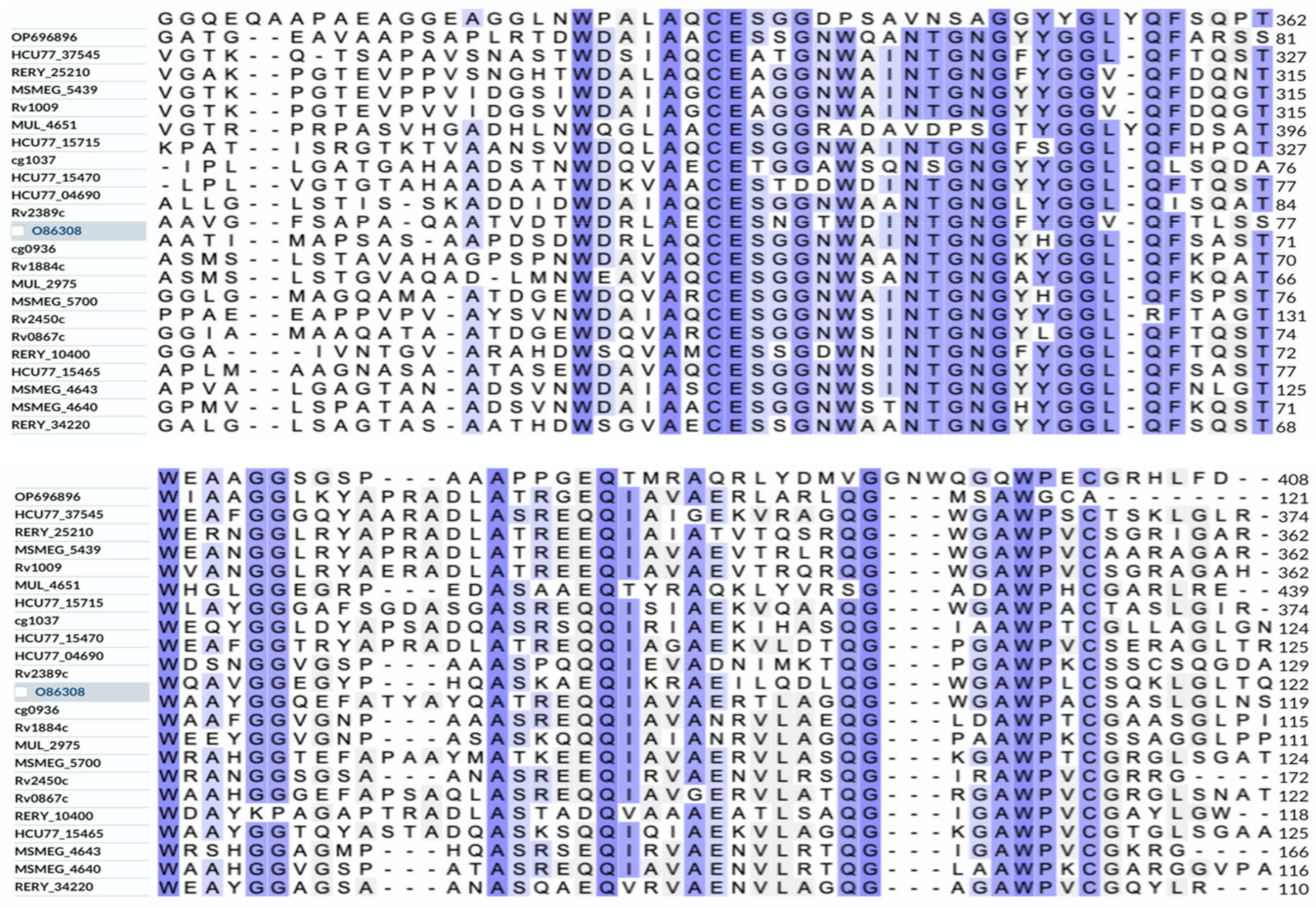
Multiple sequence alignment was performed on the UniProt portal (https://www.uniprot.org/align) (accessed on 29 January 2023) for the amino acid sequences of the Rpfs of N. halophila, M. luteus, S. coelicolor, M. tuberculosis, M. smegmatis, R. erythropolis, C. glutamicum and M. ulcerans. It was found that they had in common a conserved domain with a similar amino acid sequence (approximately 75 amino acid residues) (Figure 2). This was consistent with the reports in the literature [3,6]. This conserved domain is also the catalytic domain of Rpf and possesses lysozyme activity, which can promote bacteria growth. The complete Rpf proteins have the same biological function as the individual Rpf conserved domain.
A phylogenetic tree was constructed using the maximum likelihood method in MEGA X Version 10.2.0 for the amino acid sequences of the Rpfs of N. halophila, M. luteus, M. tuberculosis, M. smegmatis, R. erythropolis, S. coelicolor C. glutamicum and M. ulcerans. The results showed that the Rpf protein of N. halophila and the Rpf B protein of S. coelicolor belong to the same branch of evolution (Figure 3), which proved that they were closely related.

3.3. Tertiary Structure Prediction of Rpf Protein

Swiss-Model was used to predict the tertiary structure of the Rpf protein of N. halophila CGMCC 4.1195T (Figure 4A). The crystal structure of the Rpf protein included catalytic domain, G5 domain and DUF348, and the DUF348 domain adopted a compact fold, in which one α-helix was packed against a four-stranded β-sheet. The structure was consistent with the RpfB protein of M. tuberculosis [52].
Homology modeling of the Rpf protein of N. halophila CGMCC 4.1195T was performed using Swiss-Model. The modeled Rpf protein of N. halophila and the template (5e27.1.A) of the RpfB protein of M. tuberculosis were the most closely correlated, and the sequence identity between them was 28.22%. The alignment of the tertiary structures of the two Rpf proteins was assessed using PyMol Version 2.5.4 [46]. The tertiary structures of the two proteins were well matched (Figure 4B). The calculation of the RMSD between the two proteins was carried out using SuperPose Version 1.0 online protein structure superposition software (http://wishart.biology.ualberta.ca/SuperPose/) (accessed on 29 January 2023). If all atoms are considered, the RMSD between the overall structure is 3.79 Å. If only the carbon skeleton is considered, the RMSD between the overall structure is 3.36 Å. From the RMSD value, we think that the two proteins are similar in structure.

3.4. Purification and Characterization of Rpf Protein from N. halophila CGMCC 4.1195T

The genomic DNA of N. halophila CGMCC 4.1195T was used as a template to prepare rpf genes using primers in Table 2. The Rpf protein was expressed in E. coli BL21 codon plus (DE3) from plasmid pET 28a(+): rpf, and was purified from the cell extract with Ni2+ affinity chromatography. The purified recombinant Rpf protein was filtered and sterilized through a 0.22 μm sterile filter and adjusted to 2 mg/mL and stored in a refrigerator at −20 °C.

3.5. Validation of Rpf Protein on the Resuscitation of VBNC Bacteria

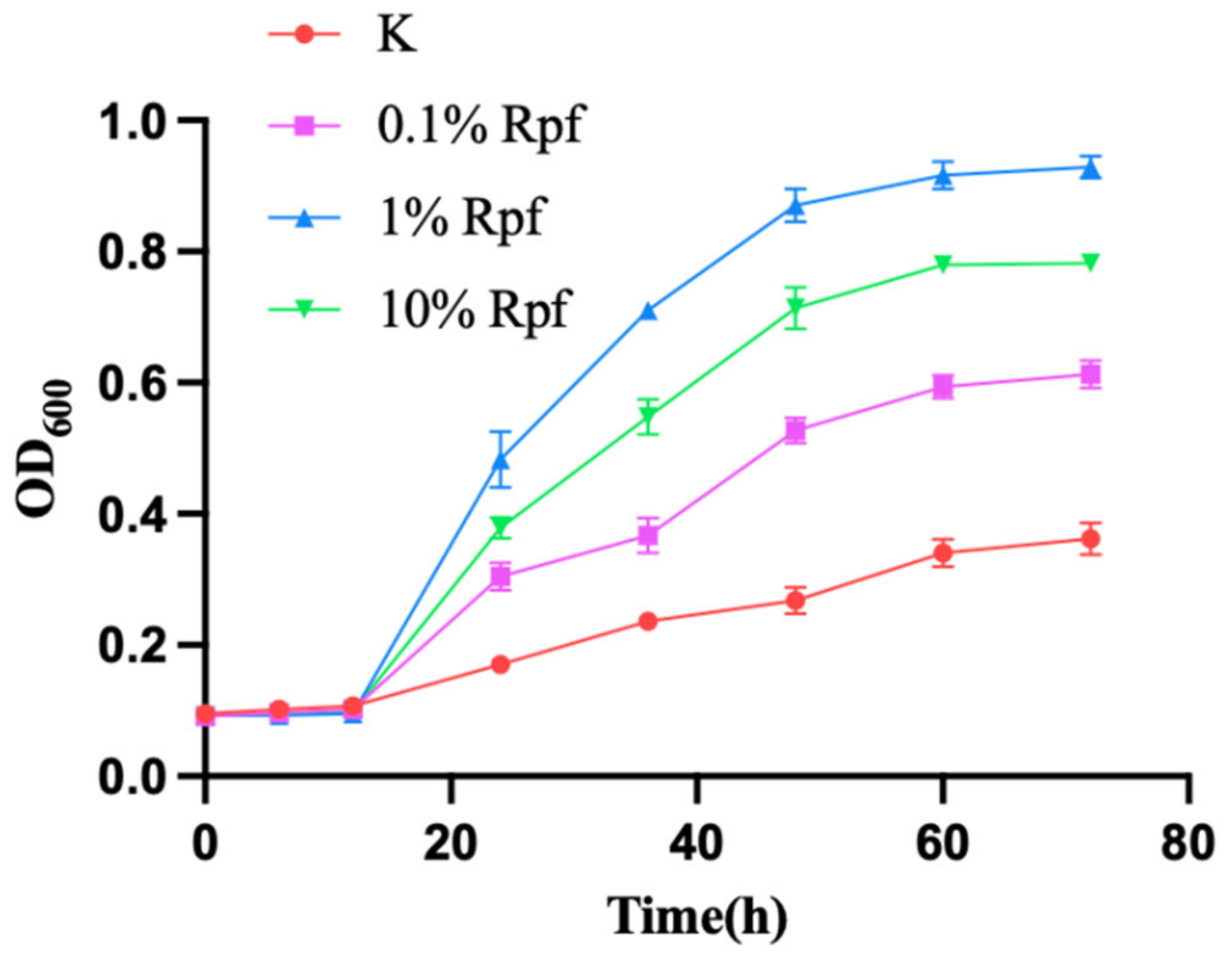
The recombinant Rpf protein, at concentrations of 0.01, 0.1 and 1 mg/mL, respectively, was added to the bacteria solution that entered the VBNC N. halophila CGMCC 4.1195T. The resuscitating effect of the Rpf on the bacteria was observed using Confocal laser scanning microscope (FV3000, Olympus, Tokyo, Japan) on days three, five and seven and is shown in Figure 5. It was found that at day three there were significantly more living bacteria (green fluorescence) in the experimental group supplemented with the Rpf protein than in the control group without the Rpf protein (Figure 5), and the resuscitation effect on the VBNC N. halophila CGMCC 4.1195T was most evident when 0.1 mg/mL of the recombinant Rpf protein was added to the medium. In addition, the same results were obtained from the growth curves of VBNC N. halophila CGMCC 4.1195T at different Rpf protein concentrations (Figure 6). This also confirmed that the Rpf protein from N. halophila CGMCC 4.1195T also had a resuscitation function similar to the M. luteus Rpf protein [2].
The Rpf protein obtained from N. halophila can be used for halophilic actinomycetes isolation from extreme halophilic environment samples in the future to further explore the effect of the Rpf protein on the culturability of halophilic actinomycetes. This will lay the foundation for further studies on the function of Rpf proteins derived from different actinomycetes.

3.6. Effects of Rpf Gene Deletion on Growth and Sporulation of Strain

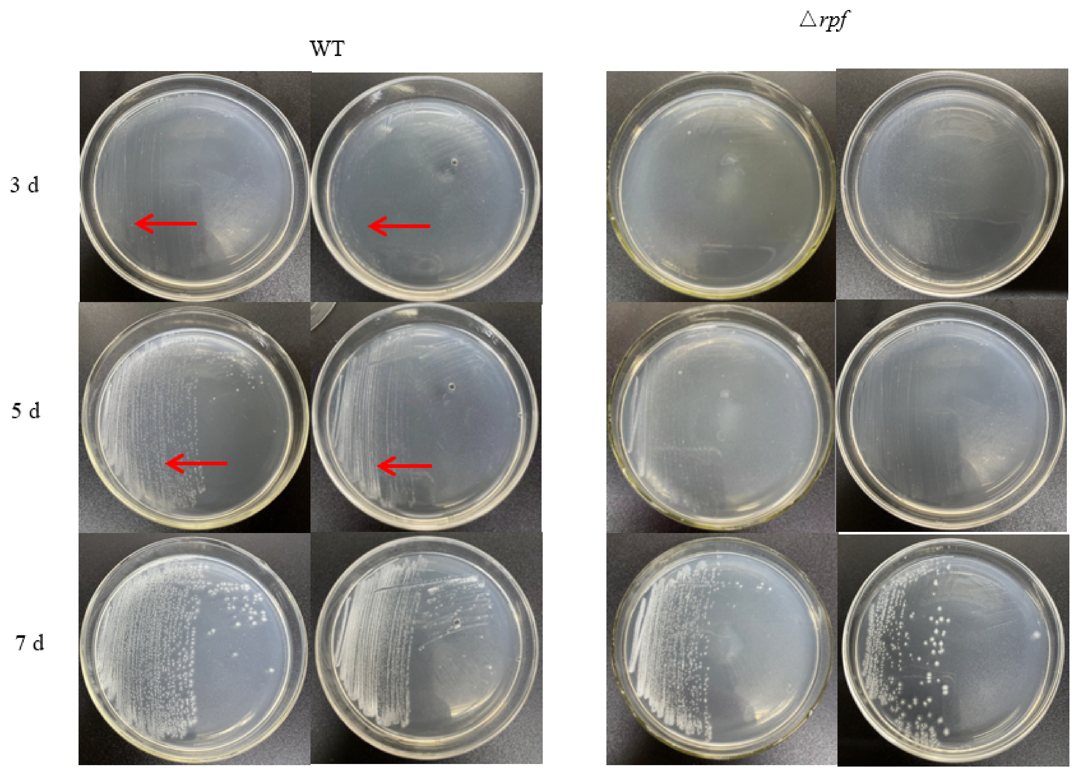
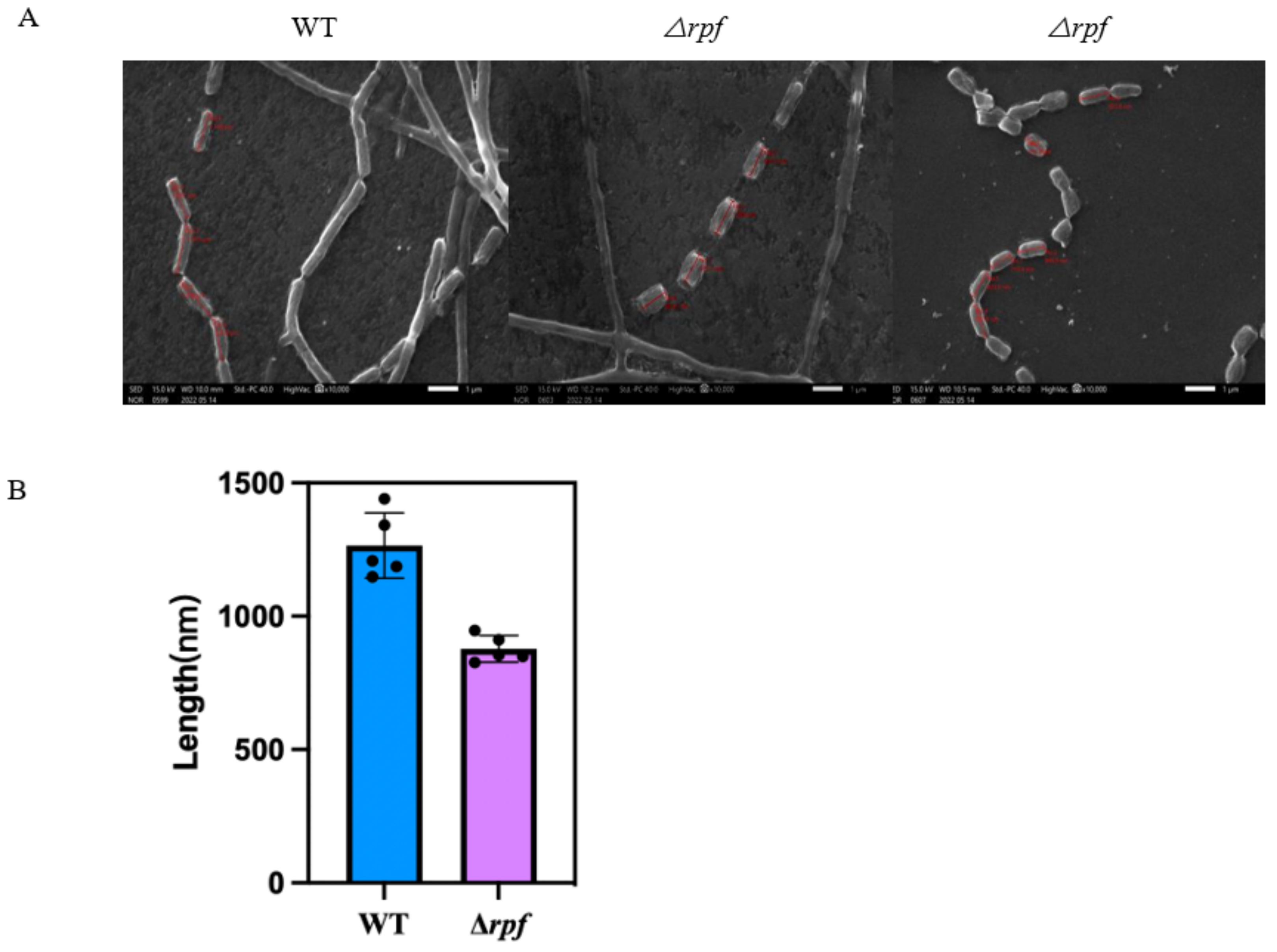
In order to investigate the role of the rpf gene in N. halophila CGMCC 4.1195T, the growth of mutant and wild-type strains on STM plates was observed. The results showed that the growth rate of the mutant strain (Δrpf) was slower than that of the wild strain in the early days. The wild-type strain had weak colonies at 3 days, however the Δrpf bacteria could not be observed. At 5 days, the wild-type had more colonies than the Δrpf group (Figure 7). Moreover, the growth curve also demonstrated that the mutant strain grew slightly slower than the wild-type strain (Figure 8), which is consistent with the results in Figure 7. Specifically, Combined with the growth curve, the growth of wild-type strain was significantly different from that of Δrpf strain at 1–5 days (p < 0.05); at three days p = 0.0027, at five days p = 0.0007 (Figure 7). The same was true of vegetative growth in liquid culture (Figure 8). Moreover, we found that the mutant strain (Δrpf) produced significantly shorter spores than the wild-type strain (p < 0.05) (Figure 9A,B). Whether this indicates that the deletion of rpf gene affects the spore morphology of the strain remains to be further verified.

4. Discussion

Rpf was first found in the culture supernatant of M. luteus, which could resuscitate VBNC bacteria [1]. Subsequently, the appearance of many sequenced genomes of various bacteria made clear that the Rpf of M. luteus was a member of a large family of the secreted bacterial Rpf-like proteins among many gram-positive (G+C)-rich bacteria belonging to the phylum Actinobacteria [3]. Moreover, the Rpf-like proteins derived from M. tuberculosis and S. coelicolor could also reactivate VBNC bacteria or dormant spores [9,36]. We previously found that there was a single copy of the rpf gene, with a size of 1251 bp, encoding an Rpf-like protein in the genome of N. halophila. Bioinformatics analyses showed that the Rpf-like protein had approximately 75 conserved amino acid residues similar to the M. luteus Rpf protein [42] (Figure 2), and the amino acid sequence of the Rpf-like protein was closely related to the RpfB protein of S. coelicolor [6] (Figure 3). The results of a tertiary structure prediction for the Rpf protein showed that the modeled Rpf protein of N. halophila and the template (5e27.1.A) of the RpfB protein of M. tuberculosis were the most closely correlated, and the RMSDs between the two proteins were 3.79 Å (where all atoms were considered) and 3.36 Å (where only the carbon skeleton was considered), respectively. So, we speculate that the Rpf-like protein has a resuscitation function similar to the Rpf proteins of M. tuberculosis and M. luteus.
In order to verify the resuscitation effect of the Rpf-like protein of N. halophila, we cloned the rpf gene of N. halophila and expressed it heterologous in E. coli BL21 Codon Plus (DE3). Different concentrations of Rpf protein were added to VBNC bacteria and observed with confocal fluorescence microscopy. The results showed that the recombinant Rpf-like protein from N. halophila had a significant resuscitation effect on the VBNC N. halophila, and the resuscitation effect was most obvious when 0.1 mg/mL of the recombinant Rpf protein was added (Figure 5 and Figure 6), indicating that high concentrations of the Rpf protein inhibited the growth and recovery of VBNC N. halophila. This phenomenon is consistent with the results reported in the literature [53]. However, FDA and PI staining are not the best method because PI is not effective in distinguishing VBNC bacteria from bacterial death. Therefore, in future experiments we can try to adopt some other methods, such as the RT-PCR technique [54,55], immunological method [56], flow cytometry [57,58,59], nucleic acid dye, western blotting and so on.
In addition, in order to investigate the role of the rpf gene in N. halophila CGMCC 4.1195T, we constructed the N. halophila CGMCC 4.1195T mutant strain with rpf gene deletion, and the growth of mutant and wild-type strains on STM plates was observed. The results showed that there were no significant differences in growth between the mutant strain and the wild-type strain (Figure 7 and Figure 8), but we found that the mutant strain sporulated slightly later than the wild-type strain, and had shorter spore sizes than the wild-type strain (Figure 9). Whether this indicates that the deletion of rpf gene can affect the spore morphology of the strain remains to be further verified.
It was reported that loss of the rpf gene was correlated with a delay in the onset of germination [6]. However, our experiment has not provided persuasive evidence, which we will need to complete in the future. In addition, bacterial spore germination is a complex physiological process. Usually, Bacillus subtilis forms heat- and desiccation-resistant spores under conditions of nutrient limitation [53]. These dormant spores germinate when stimulated by various nutrient sources. However, the molecular events leading to successful spore dormancy and later to germination remain to be fully elucidated [60]. The survival of an organism relies on its capacity to quickly respond and adapt to a constantly changing environment. Underlying this adaptive potential is the ability of cells to sense and transduce external and internal signals. Protein kinases, together with their cognate phosphatases, play a central role in signal transduction by catalyzing reversible protein phosphorylation. Eukaryote-like serine/threonine kinases and phosphatases are common in bacteria, such as such as PknB in Mycobacterium tuberculosis [54]; they might be able to sense and transduce external and internal signals and play an important role in spore germination. Moreover, Virmani et al. (2019) [61] reported that the spores of Bacillus anthracis inherit cellular components as phenotypic memory from the parent cell, and this memory plays a critical role in facilitating the spores’ revival. They found that increased expression of enolase (Eno) in the sporulating mother cell decreases germination efficiency. Eno is phosphorylated by the conserved Ser/Thr protein kinase PrkC which decreases the catalytic activity of Eno, and phosphorylation also regulates Eno expression and localization, thereby controlling the overall spore germination process [61]. There was evidence that spore germination was inhibited at the outgrowth step in the rpf null strain of Streptomyces [37]. However, whether Rpf protein is also regulated by Ser/Thr protein kinases to promote spore germination remains to be further investigated.
In short, based on the resuscitation function, the Rpf protein of N. halophila obtained in this study can be used to mine halophilic actinomycetes resources from extreme halophilic environment samples, and to improve the culturability of halophilic actinomycetes in the future. This will lay the foundation for further studying the function of Rpf proteins of different actinomycetes. Although this study did not focus on the relationship between the structure and function of Rpfs, nor the detailed mechanism of Rpf protein promoting spore germination, we plan to take them as the contents of further in-depth research in the future.

Author Contributions

Y.Z. (Yufan Zhang) conducted almost all experiments and wrote this manuscript. J.L. helped information processing and helped examine the manuscript. M.C. and Y.Z. (Yujia Zhang) participated in part of the experiment. X.Z. designed this study and revised the manuscript. All authors have read and agreed to the published version of the manuscript.

Funding

This work was supported by the grants from the National Natural Science Foundation of China (Grant No. 32270020), the S&T Program of Hebei (Grant No. 20326614D, 21326604D) and the Natural Science Foundation of Hebei Province (Grant No. C2020201005).

Data Availability Statement

All data and materials as well as software application or custom code support their published claims and comply with field standards.

Acknowledgments

The authors thank Natinoal Natural Science Foundation of China for the financial support given through 32270020 grant, and Hebei Provincial Department of Science and Technology given through 20326614D, 21326604D and C2020201005 grants.

Conflicts of Interest

The authors declare no conflict of interest.

References

  1. Mukamolova, G.V.; Kaprelyants, A.S.; Young, D.I.; Young, M.; Kell, D.B. A bacterial cytokine. Proc. Natl. Acad. Sci. USA 1998, 95, 8916–8921. [Google Scholar] [CrossRef] [PubMed] [Green Version]
  2. Mukamolova, G.V.; Kormer, S.; Kell, D.B.; Kaprelyants, A.S. Stimulation of the multiplication of Micrococcus luteus by an autocrine growth factor. Arch. Microbiol. 1999, 172, 9–14. [Google Scholar] [CrossRef] [PubMed]
  3. Nikitushkin, V.D.; Demina, G.R.; Kaprelyants, A.S. Rpf Proteins Are the Factors of Reactivation of the Dormant Forms of Actinobacteria. Biochemistry 2016, 81, 1719–1734. [Google Scholar] [CrossRef] [PubMed]
  4. Ravagnani, A.; Finan, C.; Young, M. A novel firmicute protein family related to the actinobacterial resuscitation-promoting factors by non-orthologous domain displacement. BMC Genom. 2005, 6, 39. [Google Scholar] [CrossRef] [PubMed] [Green Version]
  5. Biasini, M.; Bienert, S.; Waterhouse, A.; Arnold, K.; Studer, G.; Schmidt, T.; Kiefer, F.; Gallo Cassarino, T.; Bertoni, M.; Bordoli, L.; et al. SWISS-MODEL: Modelling protein tertiary and quaternary structure using evolutionary information. Nucleic Acids Res. 2014, 42, W252–W258. [Google Scholar] [CrossRef]
  6. Sexton, D.L.; St-Onge, R.J.; Haiser, H.J.; Yousef, M.R.; Brady, L.; Gao, C.; Leonard, J.; Elliot, M.A. Resuscitation-promoting factors are cell wall-lytic enzymes with important roles in the germination and growth of Streptomyces coelicolor. J. Bacteriol. 2015, 197, 848–860. [Google Scholar] [CrossRef] [Green Version]
  7. Hartmann, M.B.A.; Niehaus, K.; Pühler, A.; Tauch, A.; Kalinowski, J. The glycosylated cell surface protein Rpf2, containing a resuscitation-promoting factor motif, is involved in intercellular communication of Corynebacterium glutamicum. Arch. Microbiol. 2004, 182, 299–312. [Google Scholar] [CrossRef]
  8. Downing, K.J.; Mischenko, V.V.; Shleeva, M.O.; Young, D.I.; Young, M.; Kaprelyants, A.S.; Apt, A.S.; Mizrahi, V. Mutants of Mycobacterium tuberculosis lacking three of the five rpf-like genes are defective for growth in vivo and for resuscitation in vitro. Infect. Immun. 2005, 73, 3038–3043. [Google Scholar] [CrossRef] [Green Version]
  9. Kana, B.D.; Gordhan, B.; Downing, K.J.; Sung, N.; Vostroktunova, G.; Machowski, E.E.; Tsenova, L.; Young, M.; Kaprelyants, A.; Kaplan, G.; et al. The resuscitation-promoting factors of Mycobacterium tuberculosis are required for virulence and resuscitation from dormancy but are collectively dispensable for growth in vitro. Mol. Microbiol. 2008, 67, 672–684. [Google Scholar] [CrossRef] [Green Version]
  10. Russell-Goldman, E.; Xu, J.; Wang, X.; Chan, J.; Tufariello, J.M. A Mycobacterium tuberculosis Rpf. double-knockout strain exhibits profound defects in reactivation from chronic tuberculosis and innate immunity phenotypes. Infect. Immun. 2008, 76, 4269–4281. [Google Scholar] [CrossRef] [Green Version]
  11. Uhía, I.; Krishnan, N.; Robertson, B.D. Characterising resuscitation promoting factor fluorescent-fusions in mycobacteria. BMC Microbiol. 2018, 18, 30. [Google Scholar] [CrossRef] [Green Version]
  12. Rosser, A.; Stover, C.; Pareek, M.; Mukamolova, G.V. Resuscitation-promoting factors are important. determinants of the pathophysiology in Mycobacterium tuberculosis infection. Crit. Rev. Microbiol. 2017, 43, 621–630. [Google Scholar] [CrossRef] [Green Version]
  13. Pourazar, D.S.; Taala, A.; Masoumi, M.; Ebrahimzadeh, N.; Fateh, A.; Siadat, S.D.; Vaziri, F. Sub-minimum inhibitory concentration of rifampin: A potential risk factor for resuscitation of Mycobacterium tuberculosis. Antimicrob. Resist. Infect. Control 2017, 6, 116. [Google Scholar] [CrossRef] [Green Version]
  14. Gao, H.; Bai, Y.; Xue, Y.; Wang, L.; Fan, A.; Ding, X.; Xu, Z. Expression, purification, and. characterization of soluble RpfD with high bioactivity as a recombinant protein in Mycobacterium vaccae. Protein Expr. Purif. 2007, 55, 112–118. [Google Scholar] [CrossRef]
  15. Seidi, K.; Jahanban-Esfahlan, R. A novel approach to eradicate latent TB: Based on resuscitation. promoting factors. J. Med. Hypotheses Ideas 2013, 7, 69–74. [Google Scholar] [CrossRef] [Green Version]
  16. Huang, W.; Qi, Y.J.; Diao, Y.J.; Yang, F.; Zha, X.D.; Ren, C.P.; Huang, D.K.; Franken, K.L.; Ottenhoff, T.H.; Wu, Q.S.; et al. Use of resuscitation-promoting factor proteins improves the sensitivity of culture-based tuberculosis testing in special samples. Am. J. Respir. Crit. Care Med. 2014, 189, 612–614. [Google Scholar] [CrossRef]
  17. Arroyo, L.; Marín, D.; Franken, K.L.M.C.; Ottenhoff, T.H.M.; Barrera, L.F. Potential of DosR and Rpf. antigens from Mycobacterium tuberculosis to discriminate between latent and active tuberculosis in a tuberculosis endemic population of Medellin Colombia. BMC Infect. Dis. 2018, 18, 26. [Google Scholar] [CrossRef] [Green Version]
  18. Ye, Z.; Li, H.; Jia, Y.Y.; Fan, J.H.; Wan, J.X.; Guo, L.; Su, X.; Zhang, Y.; Wu, W.M.; Shen, C. Supplementing resuscitation-promoting factor (Rpf) enhanced biodegradation of polychlorinated biphenyls (PCBs) by Rhodococcus biphenylivorans strain TG9T. Environ. Pollut. 2020, 263, 114488. [Google Scholar] [CrossRef]
  19. Liu, Y.D.; Su, X.M.; Lu, L.; Ding, L.X.; Shen, C.F. A novel approach to enhance biological nutrient. removal using a culture supernatant from Micrococcus luteus containing resuscitation-promoting factor (Rpf) in SBR process. Environ. Sci. Pollut. Res. Int. 2016, 23, 4498–4508. [Google Scholar] [CrossRef]
  20. Murugan, K.; Vasudevan, N. Intracellular toxicity exerted by PCBs and role of VBNC bacterial strains in biodegradation. Ecotoxicol. Environ. Saf. 2018, 157, 40–60. [Google Scholar] [CrossRef]
  21. Su, X.M.; Li, S.; Xie, M.Q.; Tao, L.Q.; Zhou, Y.; Xiao, Y.Y.; Lin, H.J.; Chen, J.R.; Sun, F.Q. Enhancement of polychlorinatedbiphenyl biodegradation by resuscitation promoting factor (Rpf) and Rpf-responsive bacterial community. Chemosphere 2021, 26, 128283. [Google Scholar] [CrossRef] [PubMed]
  22. Su, X.M.; Wang, Y.; Xue, B.B.; Zhang, Y.G.; Mei, R.W.; Zhang, Y.; Hashmi, M.Z.; Lin, H.; Chen, J.; Sun, F. Resuscitation of functional bacterial community for enhancing biodegradation of phenol under high salinity conditions based on Rpf. Bioresour. Technol. 2018, 261, 394–402. [Google Scholar] [CrossRef] [PubMed]
  23. Yu, C.N.; Liu, Y.; Jia, Y.Y.; Su, X.M.; Lu, L.; Ding, L.X.; Shen, C. Extracellular organic matter from Micrococcus luteus containing resuscitation-promoting factor in sequencing batch reactor for effective nutrient and phenol removal. Sci. Total Environ. 2020, 727, 138627. [Google Scholar] [CrossRef] [PubMed]
  24. Jin, Y.; Gan, G.; Yu, X.Y.; Wu, D.D.; Zhang, L.; Yang, N.; Hu, J.D.; Liu, Z.H.; Zhang, L.X.; Hong, H.C. Isolation of viable but non-culturable bacteria from printing and dyeing wastewater bioreactor based on resuscitation promoting factor. Curr. Microbiol. 2017, 74, 787–797. [Google Scholar] [CrossRef]
  25. Su, X.M.; Shen, H.; Yao, X.; Ding, L.X.; Yu, C.; Shen, C.F. A novel approach to stimulate the. biphenyl-degrading potential of bacterial community from PCBs-contaminated soil of e-waste recycling sites. Bioresour. Technol. 2013, 146, 27–34. [Google Scholar] [CrossRef]
  26. Han, Z.; Lin, Q.H.; Zhang, S.S.; Zhou, X.R.; Li, S.; Sun, F.Q.; Shen, C.F.; Su, X.M. High PCBs mineralization capability of a resuscitated strain Bacillus sp. LS1 and its survival in PCB-contaminated soil. Sci. Total Environ. 2023, 856, 159224. [Google Scholar] [CrossRef]
  27. Su, X.M.; Xie, M.Q.; Han, Z.; Xiao, Y.Y.; Wang, R.; Shen, C.F.; Hashmi, M.Z.; Sun, F.Q. Resuscitation-promoting factor accelerates enrichment of highly active tetrachloroethene/polychlorinated biphenyl-dechlorinating cultures. Appl. Environ. Microbiol. 2023, 89, e0195122. [Google Scholar] [CrossRef]
  28. Wang, Y.; Shi, J.; Tang, L.; Zhang, Y.; Zhang, Y.; Zhang, X. Evaluation of Rpf protein of Micrococcus. luteus for cultivation of soil actinobacteria. Syst. Appl. Microbiol. 2021, 44, 126234. [Google Scholar] [CrossRef]
  29. Takahashi, Y.; Omura, S. Isolation of new actinomycete strains for the screening of new bioactive compounds. J. Gen. Appl. Microbiol. 2003, 49, 141–154. [Google Scholar] [CrossRef] [Green Version]
  30. Watve, M.G.; Tickoo, R.; Jog, M.M.; Bhole, B.D. How many antibiotics are produced by the genus. Streptomyces? Arch. Microbiol. 2001, 176, 386–390. [Google Scholar] [CrossRef]
  31. Messis, A.; Azzeddine, B.; Mouloud, K.; Said, B. Taxonomy and Study of Antimicrobial Activity of Two Halophilic Actinomycetes AH1 and AH2 Isolated from a Saline Soil of Bejaia (Algeria). Res. Rev. BioSci. 2012, 6, 328–334. [Google Scholar]
  32. Tian, S.Z.; Pu, X.; Luo, G.; Zhao, L.X.; Xu, L.H.; Li, W.J.; Luo, Y. Isolation and characterization of. new p-Terphenyls with antifungal, antibacterial, and antioxidant activities from halophilic actinomycete Nocardiopsis gilva YIM 90087. J. Agric. Food. Chem. 2013, 61, 3006–3012. [Google Scholar] [CrossRef]
  33. Kim, S.H.; Shin, Y.; Lee, S.H.; OH, K.B.; Lee, S.K.; Shin, J.; Oh, D.C. Salternamides A-D from a. Halophilic Streptomyces sp. Actinobacterium. J. Nat. Prod. 2015, 78, 836–843. [Google Scholar] [CrossRef]
  34. Zhang, L.; Zhang, J.; Ren, B.; Lu, W.; Hou, C.; Wang, J.; Ma, X.; Ma, R.; Liu, M.; Liu, Z.; et al. Characterization of anti-BCG benz[α]anthraquinones and new siderophores from a Xinjiang desert-isolated rare actinomycete Nocardia sp. XJ31. Appl. Microbiol. Biotechnol. 2020, 104, 8267–8278. [Google Scholar] [CrossRef]
  35. Choi, B.K.; Park, S.Y.; Choi, D.K.; Shin, B.; Shin, Y.H.; OH, D.C.; Lee, H.S.; Lee, H.S.; Lee, Y.J.; Lee, J.S.; et al. Streptoglycerides A-D with a Rare 6/5/5 Tricyclic Ring Skeleton from a Marine Actinomycete Streptomyces species. Org. Lett. 2018, 20, 6037–6040. [Google Scholar] [CrossRef]
  36. Sexton, D.L.; Herlihey, F.A.; Brott, A.S.; Crisante, D.A.; Shepherdson, E.; Clarke, A.J.; Elliot, M.A. Roles of LysM and LytM domains in resuscitation-promoting factor (Rpf) activity and Rpf-mediated peptidoglycan cleavage and dormant spore reactivation. J. Biol. Chem. 2020, 295, 9171–9182. [Google Scholar] [CrossRef]
  37. Cohen-gonsaud, M.B.P.; Bagnéris, C.; Henderson, B.; Ward, J.; Roumestand, C.; Keep, N.H. The. structure of a resuscitation-promoting factor domain from Mycobacterium tuberculosis shows homology to lysozymes. Nat. Struct. Mol. Biol. 2005, 12, 270–273. [Google Scholar] [CrossRef] [Green Version]
  38. Mukamolova, G.V.; Murzin, A.; Salina, E.G.; Demina, G.R.; Kell, D.B.; Kaprelyants, A.S.; Young, M. Muralytic activity of Micrococcus luteus Rpf and its relationship to physiological activity in promoting bacterial growth and resuscitation. Mol. Microbiol. 2006, 59, 84–98. [Google Scholar] [CrossRef] [Green Version]
  39. Hett, E.C.; Deng, L.L.; Rubin, E.J. A mycobacterial enzyme essential for cell division synergizes with. resuscitation-promoting factor. PLoS. Pathog. 2008, 4, e1000001. [Google Scholar] [CrossRef] [Green Version]
  40. Ruggiero, A.; Tizzano, B.; Pedone, E.; Pedone, C.; Wilmanns, M.; Berisio, R. Crystal structure of the resuscitation-promoting factor (△DUF) RpfB from M. tuberculosis. J. Mol. Biol. 2009, 385, 153–162. [Google Scholar] [CrossRef]
  41. Cohen-Gonsaud, M.K.N.; Davies, A.P.; Ward, J.; Henderson, B.; Labesse, G. Resuscitation-promoting. factors possess a lysozyme-like domain. Trends Biochem. Sci. 2004, 29, 7–10. [Google Scholar] [CrossRef] [PubMed] [Green Version]
  42. Telkov, M.V.; Demina, G.; Voloshin, S.A.; Salina, E.G.; Dudik, T.V.; Stekhanova, T.N.; Mukamolova, G.V.; Kazaryan, K.A.; Goncharenko, A.V.; Young, M.; et al. Proteins of the Rpf (resuscitation promoting factor) family are peptidoglycan hydrolases. Biochemistry 2006, 71, 414–422. [Google Scholar] [CrossRef] [PubMed]
  43. Keep, N.H.W.J.; Robertson, G.; Cohen-Gonsaud, M.; Henderson, B. Bacterial resuscitation factors: Revival of viable but non-culturable bacteria. Cell. Mol. Life Sci. 2006, 63, 2555–2559. [Google Scholar] [CrossRef] [PubMed]
  44. Al-Tai, A.M.; Ruan, J.S. Nocardiopsis halophila sp. nov., a New Halophilic Actinomycete. Isolated from Soil. Int. J. Syst. Evol. Microbiol. 1994, 44, 474–478. [Google Scholar] [CrossRef]
  45. Yuan, S.; Chan, H.; Filipek, S.; Vogel, H. Pymol and inkscape bridge the data and the data visualization. Structure 2016, 24, 2041–2042. [Google Scholar] [CrossRef] [Green Version]
  46. Kumar, S.; Stecher, G.; Li, M.; Knyaz, C.; Tamura, K. MEGA X: Molecular Evolutionary Genetics. Analysis across Computing Platforms. Mol. Biol. Evol. 2018, 35, 1547–1549. [Google Scholar] [CrossRef]
  47. Macneil, D.J.; Gewain, K.; Ruby, C.L.; Dezeny, G.; Gibbons, P.H.; Macneil, T. Analysis of. Streptomyces avermitilis genes required for avermectin biosynthesis utilizing a novel integration vector. Gene 1992, 111, 61–68. [Google Scholar] [CrossRef]
  48. Paget, M.S.; Chamberlin, L.; Atrih, A.; Foster, S.J.; Buttner, M.J. Evidence that the extracytoplasmic function sigma factor SigE is required for normal cell wall structure in Streptomyces coelicolor A3(2). J. Bacteriol. 1999, 181, 204–211. [Google Scholar] [CrossRef] [Green Version]
  49. Cheng, X.Y.; Wang, X.R.; Kang, Q.J.; Li, W.J.; Bai, L.Q. Establishment and Application of. Conjugation System for the Ansamitocin Producer Nocardiopsis ansamitocini EGI80425. Jiyinzuxue Yu Yingyong Shengwuxue (Genom. Appl. Biol.) 2020, 39, 1163–1171. [Google Scholar] [CrossRef]
  50. Ledoux, M.; Lamy, F. Determination of proteins and sulfobetaine with the Folin-phenol reagent. Anal. Biochem. 1986, 157, 28–31. [Google Scholar] [CrossRef]
  51. Yewalkar, S.; Wu, T.; Kuan, D.; Wang, H.; Li, D.; Johnson, A. Applicability of differential fluorescein. diacetate and propidium iodide fluorescence staining for monitoring algal growth and viability. Waste Dispos. Sustain. Energy 2019, 1, 199–206. [Google Scholar] [CrossRef] [Green Version]
  52. Ruggiero, A.; Squeglia, F.; Romano, M.; Vitagliano, L.; Simone, A.D.; Berisio, R. The structure of. Resuscitation p romoting factor B from M. tuberculosis reveals unexpected ubiquitin-like domains. Biochim. Biophys. Acta 2016, 860, 445–451. [Google Scholar] [CrossRef]
  53. Gong, X.; Lu, H.; Wu, J.; Zhou, Y.; Yang, L.; Wang, Y.; Shen, N.; Jiang, M. Enzymatic properties and biological activity of resuscitation-promoting factor B of Rhodococcus sp. (GX12401). Front. Microbiol. 2022, 13, 965843. [Google Scholar] [CrossRef]
  54. Lahtinen, S.J.; Ahokoski, H.; Reinikainen, J.P. Degradation of 16S rRNA and attributes of viability of viable but nonculturable probiotic bacteria. Lett. Appl. Microbiol. 2008, 46, 693–698. [Google Scholar] [CrossRef] [Green Version]
  55. Machado, A.; Bordalo, A.A. Diversity and dynamics of the Vibrio community in well water used for drinking in Guinea-Bissau (West Africa). Environ. Monit. Assess. 2014, 186, 5697–5709. [Google Scholar] [CrossRef]
  56. Liu, G.; Han, Y.; Xi, L.; Song, S. Applicability of a rapid method based on immunomagnetic capture-fluorescent PCR assay for Campylobacter jejuni. Food Control 2006, 17, 527–532. [Google Scholar] [CrossRef]
  57. Khan, M.M.T.; Pyle, B.H.; Camper, A.K. Specific and Rapid Enumeration of Viable but Nonculturable and Viable-Culturable Gram-Negative Bacteria by Using Flow Cytometry. Appl. Environ. Microbiol. 2010, 76, 5088–5096. [Google Scholar] [CrossRef] [Green Version]
  58. Berney, M.; Hammes, F.; Bosshard, F.; Weilenmann, H.U.; Egli, T. Assessment and interpretation of bacterial viability by using the LIVE/DEAD BacLight Kit in combination with flow cytometry. Appl. Environ. Microbiol. 2007, 73, 3283–3290. [Google Scholar] [CrossRef] [Green Version]
  59. Wang, Y.; Claeys, L.; Ha, D.; Verstraete, W.; Boon, N. Effects of chemically and electrochemically dosed chlorine on Escherichia coli and Legionella beliardensis assessed by flow cytometry. Appl. Microbiol. Biotechnol. 2010, 87, 331–341. [Google Scholar] [CrossRef]
  60. Pereira, S.F.; Goss, L.; Dworkin, J. Eukaryote-like serine/threonine kinases and phosphatases in bacteria. Microbiol. Mol. Biol. Rev. 2011, 75, 192–212. [Google Scholar] [CrossRef] [Green Version]
  61. Virmani, R.; Sajid, A.; Singhal, A.; Gaur, M.; Joshi, J.; Bothra, A.; Garg, R.; Misra, R.; Singh, V.P.; Molle, V.; et al. The Ser/Thr protein kinase PrkC imprints phenotypic memory in Bacillus anthracis spores by phosphorylating the glycolytic enzyme enolase. J. Biol. Chem. 2019, 294, 8930–8941. [Google Scholar] [CrossRef] [PubMed]
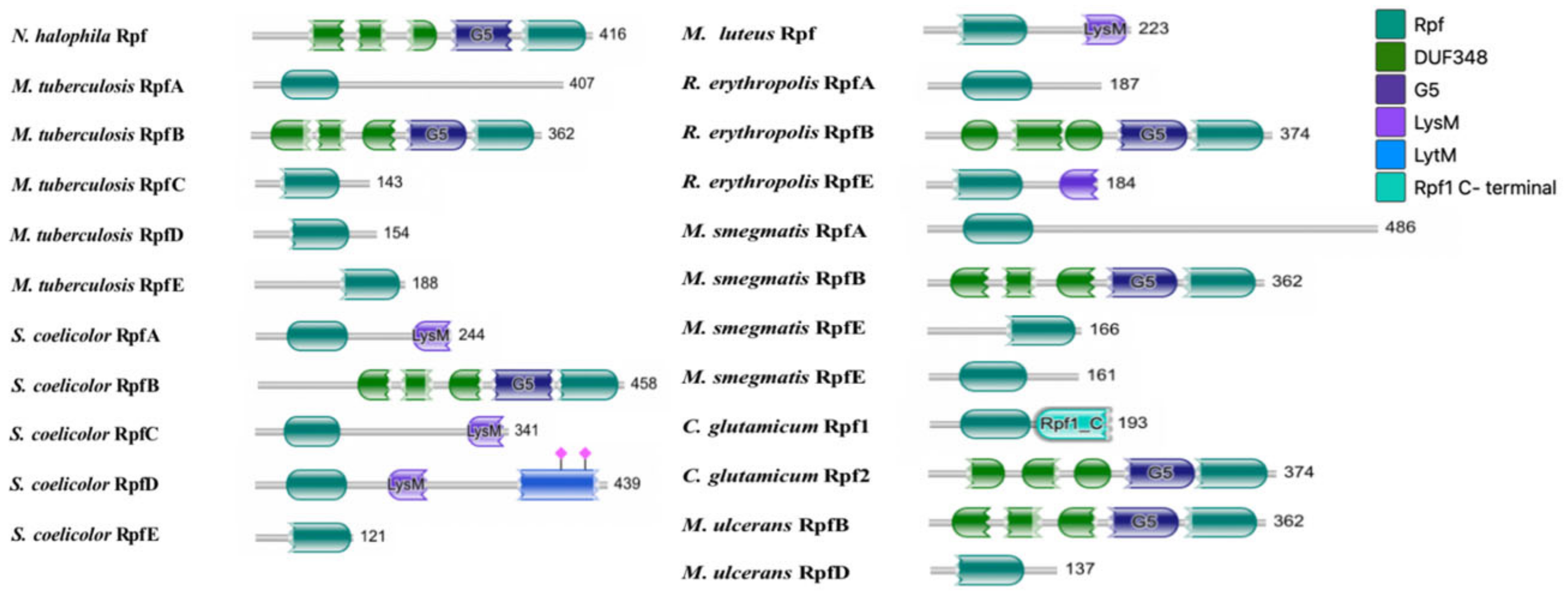
Figure 1. The function domain of Rpf proteins of N. halophila, M. tuberculosis, S. coelicolor, M. luteus, R. erythropolis, M. smegmatis, C. glutamicum and M. ulcerans.
Microorganisms 11 00485 g001
Figure 2. Multiple sequence alignment of Rpf (for resuscitation-promoting factors). Blue regions represent conserved domains. The alignment was performed on the UniProt portal (https://www.uniprot.org/align) (accessed on 29 January 2023). Sequence codes and sequences of M. tuberculosis are from UniProtKB database (https://www.uniprot.org/) (accessed on 29 January 2023) for the Rpfs of Rv2389c, Rv2450c, Rv1884c, Rv1009 and Rv0867c, M. smegmatis named MSMEG_5700, MSMEG_5439, MSMEG_4643 and MSMEG_4640, M. luteus named O86308, S. coelicolor named HCU77_15465, HCU77_15715, HCU77_15470, HCU77_04690 and HCU77_37545, R.erythropolis for RERY_10400, RERY_25210 and RERY_34220, C. glutamicum named cg0936 and cg1037, M. ulcerans named MUL_2975 and MUL_4651. Sequences of N. halophila are from NCBI Entrez database (http://www.ncbi.nlm.nih.gov/entrez) (accessed on 21 October 2022) the Rpf of OP696896.
Microorganisms 11 00485 g002
Figure 3. Phylogenetic tree based on amino acid sequences of Rpf proteins among N. halophila, M. tuberculosis, M. smegmatis, M. luteus, S. coelicolor, R.erythropolis, C. glutamicum and M. ulcerans. Phylogenetic tree was constructed using the maximum likelihood method in MEGA X Version 10.2.0. Numbers at nodes are based on 1000 resamplings; values above 50% are presented. Bar, 0.10 substitutions per amino acids position.
Microorganisms 11 00485 g003
Figure 4. Tertiary structure of Rpf protein of N. halophila CGMCC 4.1195T. ((A)(a)): The structure shows a ribbon diagram; ((A)(b)): surface-filled model; (B): Alignment of the tertiary structure of two Rpf proteins. Grey represents Rpf protein of N. halophila; Blue represents the one of M. tuberculosis.
Microorganisms 11 00485 g004
Figure 5. Growth of VBNC bacteria after the addition of Rpf from N. halophila CGMCC 4.1195T. (A). Wild-type strain (a) and VBNC strain (b). (B). Growth of VBNC bacteria after the addition of Rpf (0 mg/mL, 0.01 mg/mL, 0.1 mg/mL and 1 mg/mL) in 3 days (a), 5 days (b) and 7 days (c). In confocal fluorescence microscopy, dead bacteria were stained red by propidium iodide (PI) and live bacteria were stained green by fluorescein diacetate (FDA).
Microorganisms 11 00485 g005
Figure 6. The growth curves of VBNC N. halophila CGMCC 4.1195T at different Rpf protein concentrations.
Microorganisms 11 00485 g006
Figure 7. Growth of wild-type (WT) and rpf mutant strains (Δrpf). On the third day, there were almost no colonies on the the mutant strain (Δrpf) plate, while weak colonies appeared on the wild-type strain plate (Area indicated by the arrow); and on the fifth day, wild-type strain obviously formed a large number of white colonies (area indicated by the arrow), while the rpf mutant strain had less obvious and less white colonies.
Microorganisms 11 00485 g007
Figure 8. Growth curve of wild-type (WT) and rpf mutant strains (Δrpf). The unpaired T test was used to analyze the significance level (p < 0.05), (p > 0.05, NS; p < 0.05, *; p < 0.01, **; p < 0.001, ***; p < 0.0001, ****) The values are plotted to represent the means ± standard deviations of three independent experiments.
Microorganisms 11 00485 g008
Figure 9. Effects of rpf gene deletion on sporulation. (A) Scanning electron micrographs of wild-type (WT) and rpf mutant strains (Δrpf). Scale bar, 1 μm. (B) Analysis of spore length (p < 0.05). The unpaired T test was used to analyze the significance level (p < 0.05). The values are plotted to represent the mean ± standard deviations of at least five independent experiments for each strain.
Microorganisms 11 00485 g009
Table 1. Strains and plasmids used in this study.
Strain or PlasmidDescriptionSource or Reference
Strains
E. coli DH5αStrain used for cloning and plasmid maintenanceSangon Biotech
E. coli BL21 codon plus (DE3)Strain used for protein expressionSangon Biotech
E.coli ET12567(pUZ8002)Dam dcm, with transmobilizing plasmid pUZ8002[47,48]
N.halophila CGMCC 4.1195TStrain used for amplification of rpf gene.[43]
Plasmids
pMD18-TAmpr cloning vectorTaKaRa
pMD18-T:rpfpMD18-T containing rpf from strain CGMCC 4.1195TThis study
pET 28a(+)Ampr expression vectorTaKaRa
pET 28a(+):rpfpET 28a(+) containing rpf from strain CGMCC 4.1195TThis study
pJTU1278Conjugation vectors used for rpf gene knockout[49]
pJTU1278: left armpJTU1278 carrying the homologous arm of rpfThis study
pJTU1278: left/RightpJTU1278 carrying the left and right homologous arms of rpfThis study
Table 2. Primers used in the present study.
FragmentPrimersSequence (5′-3′)Restriction Enzyme
rpfrpf-FGGAATTCGGAAGAACCGTGCGCAATEcoRI
rpf-RCCGCTCGAGCATATGCTCAGTCGAACAGGTGGCXholI
Left homologous arm of rpfL-FGCTCTAGAGGGCCTCAGGGGAGCTCCACGCXbaI
L-RCGGGATCCGCCGCCGCGGCTCCGGTGBamHI
Right homologous arm of rpfR-FCGGGATCCGCCAGTGGCCCGAATGCGBamHI
R-RCCCAAGCTTGCCAGGAGACGGTGGCCCHindIII
Disclaimer/Publisher’s Note: The statements, opinions and data contained in all publications are solely those of the individual author(s) and contributor(s) and not of MDPI and/or the editor(s). MDPI and/or the editor(s) disclaim responsibility for any injury to people or property resulting from any ideas, methods, instructions or products referred to in the content.