Article

The Tubulin Superfamily in Apicomplexan Parasites

1
Molecular Biology and Biochemistry, University of California Irvine, Irvine, CA 92697, USA
2
Institute for Parasitology, University of Zurich, Winterthurerstrasse 266a, 8057 Zürich, Switzerland
*
Author to whom correspondence should be addressed.
Microorganisms 2023, 11(3), 706; https://doi.org/10.3390/microorganisms11030706
Submission received: 10 February 2023 / Revised: 3 March 2023 / Accepted: 6 March 2023 / Published: 9 March 2023
(This article belongs to the Special Issue Cellular Biology of Protozoan Parasites of Mammals)

Abstract

:
Microtubules and specialized microtubule-containing structures are assembled from tubulins, an ancient superfamily of essential eukaryotic proteins. Here, we use bioinformatic approaches to analyze features of tubulins in organisms from the phylum Apicomplexa. Apicomplexans are protozoan parasites that cause a variety of human and animal infectious diseases. Individual species harbor one to four genes each for α- and β-tubulin isotypes. These may specify highly similar proteins, suggesting functional redundancy, or exhibit key differences, consistent with specialized roles. Some, but not all apicomplexans harbor genes for δ- and ε-tubulins, which are found in organisms that construct appendage-containing basal bodies. Critical roles for apicomplexan δ- and ε-tubulin are likely to be limited to microgametes, consistent with a restricted requirement for flagella in a single developmental stage. Sequence divergence or the loss of δ- and ε-tubulin genes in other apicomplexans appears to be associated with diminished requirements for centrioles, basal bodies, and axonemes. Finally, because spindle microtubules and flagellar structures have been proposed as targets for anti-parasitic therapies and transmission-blocking strategies, we discuss these ideas in the context of tubulin-based structures and tubulin superfamily properties.

Graphical Abstract

1. Introduction

Microtubules are universal and essential components of eukaryotic cells that are indispensable for a variety of cellular processes, particularly mitosis, but also motility, cell shape, and intracellular trafficking. These filamentous polymers assemble from α-β-tubulin heterodimer subunits in a process that is regulated by GTP binding and hydrolysis [1,2]. The biosynthesis of functional heterodimers requires CCT and tubulin cofactors to coordinate the stable association of α- and β-tubulins, each of which binds to one molecule of GTP [3,4]. The polymerization of α-β heterodimers stimulates GTP hydrolysis in β-tubulin, causing a conformational change that weakens subunit interactions to permit microtubule disassembly. Disassembled heterodimers must exchange GDP for GTP to restore assembly competency. A dynamic equilibrium between free dimers and polymers is required for essential processes, including cell division.
α- and β-tubulins are members of a larger superfamily that consists of six subgroups (Figure 1). Members contain a tubulin signature motif ([S/A/G]-G-G-T-G-[S/A]-G) which coordinates binding to the phosphate portion of GTP [2]. Beyond this motif, sequence similarity and overall structural organization are conserved both within and between families from diverse eukaryotic lineages because functional constraints restrict variation [5]. All eukaryotes harbor genes for α- and β-tubulins to form heterodimer subunits which assemble into microtubules [5,6,7,8]. A third essential tubulin, γ-tubulin, complexes with additional proteins to form the γ-tubulin ring complex (γ-TuRC), which nucleates most microtubules [9,10]. The tubulin superfamily is thought to have arisen from gene duplication, with subsequent diversification such that tubulins in individual families carry out non-redundant functions. Tubulins arose near to the appearance of the last common eukaryotic ancestor, and α-, β-, and γ- families are found in all eukaryotes [5,11]. Gene expansion within these families led to multiple genes for α-, β- and/or γ-tubulins, known as isoforms or isotypes [12,13,14]. Isotypes are typically distinguished by minor amino acid differences that modify biochemical properties to fine-tune temporal, structural, or cell-specific requirements [12,13]. Eukaryotes have one to two genes to specify γ-tubulins and may have from one to seven or more genes that encode α- and β- tubulin isotypes [15,16,17]. Unicellular eukaryotes employ fewer α- and β- isotypes, typically specified by one to three genes.
Three other tubulins (δ-, ε-, and ζ-, known as the ZED module) contribute to centriole and basal body structures. Sequence conservation is weaker for ZED-tubulins, possibly due to lower functional constraints in these families, which are only found in some eukaryotes. Bioinformatic analysis links the function of ε-tubulin to δ- or ζ-tubulin; organisms harboring a gene for ε-tubulin also carry δ- and/or ζ-tubulin gene(s), and a lack of ε-tubulin signifies that δ- and ζ-tubulin genes will also be absent [18]. This likely reflects overlapping functions of δ- and ζ-tubulin, which are thought to be evolutionarily exchangeable [5,18]. ζ-tubulin genes are not found in apicomplexan genomes, although they are present in Leishmania and Trypanosoma species, which are in a distinct clade of protozoan parasites [6,18]. Genes for δ- and ε-tubulins have been eliminated from diverse eukaryotic lineages several times [5,19,20]. Loss is associated with an inability to form appendage-containing basal bodies, or to build basal bodies and motile flagella [20,21,22,23].
Apicomplexan parasites are a diverse group of protozoan pathogens that cause illness and death in humans and in animals, including agriculturally significant livestock. While some apicomplexans are transmitted by the bite of infected mosquito (Plasmodium) or tick (Babesia and Theileria) vectors, others are spread by the consumption of food or water harboring tissue cyst or oocyst stages (Toxoplasma, Cryptosporidium, Cyclospora, Cystoisospora). Perhaps the best-known apicomplexans are the Plasmodium species, which are categorized within the class Aconoidasida, order Haemosporida. Plasmodium species are the agents of human malaria, a deadly infection that is estimated to kill more than half a million individuals each year [24]. The Aconoidasida also includes the order Piroplasmida, containing Babesia and Theileria species. Babesia are important pathogens of cattle and emerging zoonotic infections of humans in many countries [25,26]. Bovine, ovine, and equine infections with Theileria cause significant livestock losses in endemic (tropical) areas [27,28]. The second class within the Apicomplexa, the Conoidasida, contains organisms that harbor a distinct tubulin-containing organelle, the conoid, which is proximal to apical secretory organelles and associated structures that give the phylum Apicomplexa its name. The Conoidasida includes orally infectious parasites in the order Eucoccidiorida, commonly known as coccidian parasites. Among these, parasites in the family Cryptosporidiidae (genus Cryptosporidium) are believed to represent an early branching lineage and have unusually streamlined genomes and reduced organelles relative to other apicomplexans [29]. Cryptosporidium resides in the intestinal epithelium and causes watery diarrhea and weight loss, resulting in serious and chronic complications in children and immunocompromised individuals [30]. Cystoisospora (formerly Isospora) and Cyclospora are zoonotic parasites of the Sarcocystidae and Eimeriidae families that cause serious diarrheal infections in immunocompromised individuals [31,32,33]. Besnoitia species (Sarcocystidae) cause pathology in grazing livestock and wildlife, and Eimeria species (Eimeriidae) infect poultry and livestock. Both are responsible for economic losses to the agricultural industry [34,35,36]. Lastly, Toxoplasma gondii (Sarcocystidae) is a globally distributed infection of all warm-blooded mammals, ranked by the Centers for Disease Control in the top five neglected infectious diseases in the US [37]. While infection can be asymptomatic in healthy human hosts, T. gondii causes birth defects, encephalitis, and blindness [38]. Moreover, because immunity controls but does not eliminate Toxoplasma, individuals remain at risk for the reactivation of latent parasites years after initial infection, particularly if immunity is compromised.
Apicomplexan parasites have complex life cycles that typically consist of asexual (vegetative) proliferation of zoite stages and genome reassortment during a transient sexual cycle. All parasite forms require spindle microtubules to coordinate chromosome segregation. In addition, zoites use cortical (subpellicular) microtubules to establish shape, rigidity, and polarity of the parasite pellicle. Subpellicular microtubules are non-dynamic and are unusually stable to detergent extraction. In Toxoplasma, this stability is conferred by a complex of microtubule inner proteins and is, therefore, extrinsic to tubulin [39,40]. Conoidasida class zoites also assemble the conoid, a distinctive tubulin-containing structure that incorporates repurposed proteins from the flagellar apparatus [41,42]. Importantly, apicomplexan zoites lack flagella, an organelle that underlies motility in other alveolate lineages. In Plasmodium and Toxoplasma zoites, motility has been shown to require posterior-directed capping of surface adhesins, which is driven by actin and myosin activity [43]. Although members of the Aconoidasida lack an obvious conoid, recent studies have identified conoid components in Plasmodium zoites, indicating the persistence of a less distinct structure [44]. In Toxoplasma, the conoid has been shown to bind to a formin that stimulates actin polymerization to activate gliding motility [45]. Microgametes (male) of some apicomplexans employ two flagella for motility required to fertilize macrogametes (female). Additional microtubules have been detected in electron microscopy studies of gametes, but little is known of the components or properties of these populations.
In this paper, we analyze the features of tubulins encoded by key apicomplexan organisms. We specifically consider the potential redundancy or specialization of α- and β-tubulin isotypes, the presence, absence, or divergence of δ- or ε-tubulins, and the stage-specific expression of tubulin family members. Ultimately, the properties of the tubulin superfamily inform proposed strategies to control apicomplexan parasites via tubulin-targeting antimitotic agents, or through transmission-blocking strategies that disrupt microgamete flagella [46,47,48,49,50,51].

2. Materials and Methods

2.1. Identification of Apicomplexan Tubulin Genes

Representative sequences for tubulin family members in the unicellular green alga Chlamydomonas reinhardtii and the ciliate Tetrahymena thermophila (deposited at UniProt: https://www.uniprot.org/, entries confirmed January 2023) were used to identify predicted homologs from VEuPathDB.org [52]. These organisms have well-defined members for each tubulin family and are grouped as Diaphoretickes along with apicomplexans [53]. BlastP searches [54] were used to collect all predicted tubulins from genomes of the apicomplexans Babesia microti, Besnoitia besnoiti, Cryptosporidium parvum, Cyclospora cayetanensis, Cystoisospora suis, Eimeria tenella, Hammondia hammondi, Neospora caninum, Plasmodium falciparum, Plasmodium vivax, Sarcocystis neurona, Theileria annulata and Toxoplasma gondii. Searches and analysis were initially limited to reference strains for these key pathogens. In the case that specific tubulin family members appeared to be missing, BlastP searches were expanded to encompass all sequenced genera and strains to assess whether this absence was a consistent feature or could be due to incomplete genome representation. To assign apicomplexan tubulins to specific families or to confirm existing annotations, sequences were evaluated relative to α-, β-, γ-, δ-, and ε-tubulins from C. reinhardtii and T. thermophila. Briefly, representative tubulin family members from C. reinhardtii and T. thermophila (Supplementary Table S1) were aligned with candidate apicomplexan tubulins using Clustal Omega [55,56,57]. The associated guide and phylogenetic trees were used to designate or confirm apicomplexan tubulins as members of specific families by proximity to individual Chlamydomonas and Tetrahymena tubulins.

2.2. Tubulin Protein Analysis

Predicted amino acid sequences for tubulins were aligned in Clustal Omega [55,56], and EMBOSS was used to determine pairwise conservation between tubulins [58]. In cases where tubulin genes are apparently mis-annotated, such as when they appear to lack typical regions or have extra sequences inserted or appended to conserved sequences, we have documented these in Table 1 and Table 2 notes and our observations have been added to VEuPathDB.org annotations.

2.3. Tubulin Transcriptomics and Proteomics

Relevant unique, sense strand tubulin transcript counts were extracted from several stage-specific RNA-Seq studies of P. falciparum [59,60,61,62] and T. gondii (unpublished data, Chandra Ramakrishnan and Adrian Hehl and [63]). The full datasets show trends but are not entirely consistent for studies that encompass overlapping stages (Supplementary Table S2). Qualitative evidence for protein expression (yes/no) was obtained from deposited mass spectrometry (MS) peptide datasets at VEuPathDB.org [52]. The appearance of one or more unique peptides that map to a given tubulin for a life cycle form was taken as evidence for protein presence in this stage.

3. Results

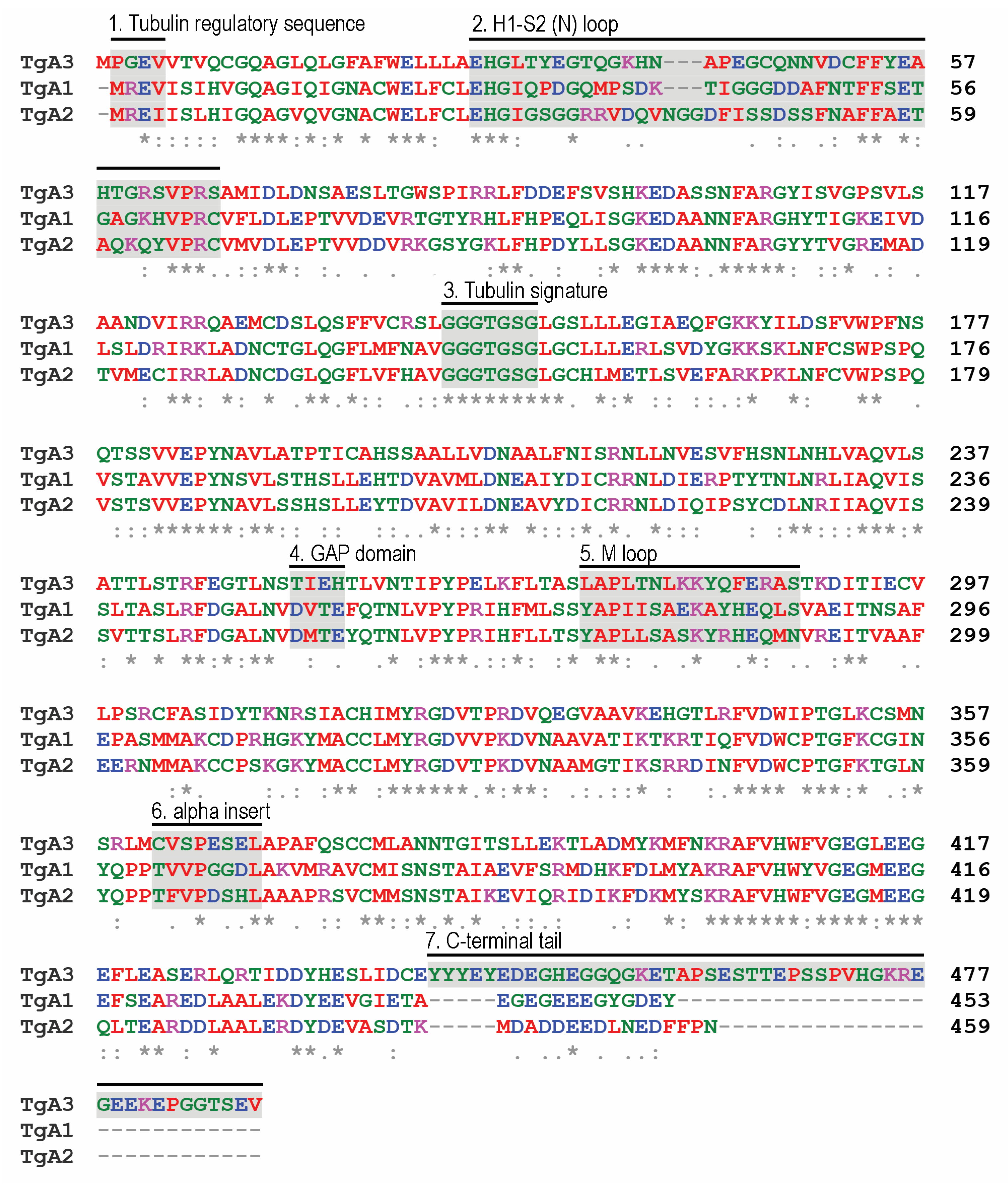
3.1. Apicomplexans Display Simple α-, β-, and γ-Isotype Repertoires

Tubulins are highly conserved proteins with several characteristic motifs which permit family members to be identified as belonging to one of six groups within the tubulin superfamily. By using Tetrahymena and Chlamydomonas tubulins to query the genomes of apicomplexan parasites, we ascertained that individual species harbor one to four genes for α- and β-tubulins, whereas γ-tubulin is specified by a single gene in all cases (Table 1). Among the Apicomplexa, P. falciparum and P. vivax are agents of the most consequential human malarias. Both have two α-tubulin genes and a single β-tubulin gene, as do the important agricultural pathogens T. annulata and B. microti. These organisms are assigned to orders (Haemosporida and Piroplasmida) within the Aconoidasida class of apicomplexans (Figure 2). Species classified within the Conoidasida harbor genes for one to four α- and one to three β-tubulin isotypes. The presence of genes for multiple tubulin isotypes is noteworthy. Because apicomplexans are haploid, conserved α- or β-tubulin isotypes with similar expression patterns would permit functional redundancy, essentially making the organism “merodiploid” for this tubulin family. Conversely, if isotypes display distinct amino acid sequences and/or differences in expression, they likely function in biochemically distinct and/or stage-specific roles.
In the Aconoidasida, a single β-tubulin gene functions for all tubulin-requiring contexts. Although most coccidians have three β-tubulin isotypes, the amino acid sequences of these are nearly identical to each other, as illustrated by the alignment of the T. gondii isotypes (Supplementary Figure S1). A genome-wide CRISPR screen performed on in vitro Toxoplasma tachyzoite cultures indicates that β3-tubulin is dispensable for growth in tachyzoites, whereas β1- and β2-tubulins have comparable negative fitness scores, indicating essential functions in tachyzoites [64]. The observation that Toxoplasma β3-tubulin expression is elevated during gametogenesis (see below) suggests that there may be a subtle feature of β3- that is critical for gametes. Notably, an essential sperm-specific isotype has been documented in a distinct biological context: testis-specific β-tubulin is required for the formation of the flagellar axoneme during Drosophila spermatogenesis [65].
In general, apicomplexan α-tubulins are much more diverse than β-tubulin isotypes. Aconoidasida species (Plasmodium, Babesia, Theileria) each harbor genes for two α-tubulin isotypes. Approximately 30 years ago, researchers recognized that P. falciparum α-tubulin II localized to the microgamete axoneme [66]. More recent studies reveal that α-tubulin II is co-expressed with α-tubulin I in other life cycle stages [67,68,69]. Isotypes I and II are 94% and 95% identical in P. falciparum and P. vivax, respectively. Alignments indicate that the H1-S2 (N) loop has the most differences between α- I and II. While this is more generally the region of greatest variation across all α-tubulins, it is also an area that is critical for lateral contacts between adjacent microtubule protofilaments. It is possible that, because flagellar axoneme microtubules are non-dynamic, the H1-S2 loop amino acids (QVVAGG) in α-tubulin II increase microtubule stability relative to α-tubulin I (KASRAN). In T. annulata and B. microti, one isotype harbors an insert of two or three amino acids in the H1-S2 loop relative to the other isotype. Inserted residues are not conserved between species, nor do they appear to correspond to the heterogeneity in the Plasmodium H1-S2 loops.
Figure 2. Tubulin superfamily members in apicomplexan species. Plasmodium, Babesia and Theileria species are classified in the Haemosporida and Piroplasmida, which are closely affiliated orders. The remaining apicomplexans analyzed in this study are classified as order Eucoccidiorida. Individual species have 1–4 α-tubulins and 1–3 β-tubulins (green). All have single genes for γ-tubulin (red), and most have single genes for δ- and ε-tubulin (blue). Cryptosporidium and Theileria lack genes for δ- and ε-tubulin, and their distinct positions in phylogenies suggest that these represent separate losses of δ- and ε-tubulin genes. Relationships between apicomplexan genera represent the basic organization of species from phylogenetic analyses, such as in the study by Kuo [68].
Microorganisms 11 00706 g002
Members of the Conoidasida harbor genes for one to four α-tubulin isotypes. Protein alignments reveal that the three Toxoplasma α-tubulin isotypes are extremely dissimilar (Figure 3). The α1- isotype (TGME49_316400) is most like conventional α-tubulins. However, α2- (TGME49_231770) and α3- (TGME49_231400) contain unique insertions and substitutions, suggesting that they do not serve housekeeping roles. For example, α2-tubulin has a three amino acid insert (GGD) in the H1-S2 loop, which may increase stability during microtubule deformation. There are three alterations to α3-tubulin motifs that may dramatically influence its expression and function. First, α3-harbors an insert to the amino (N)-terminal tubulin regulatory sequence (MREI to MPREI), which is required to regulate tubulin abundance in many systems [70]. It should be noted that this insert changes the absolute numbering of subsequent residues, which is important for describing the second difference in α3-tubulin. Genetic and biochemical studies indicate that the highly conserved α-tubulin residues D251 and E254 are essential for the GTPase activating (GAP) domain function [71]. The GAP domain stimulates polymerization-dependent GTP hydrolysis in an adjacent β-tubulin heterodimer to permit microtubule disassembly. These positions are occupied by T252 (251) and H255 (254) in α3-tubulin, although there is a glutamic acid residue at position 254 (253; Figure 3, box 4). Loss and/or repositioning of these canonical residues suggests that α3-tubulin has a reduced or nonexistent capacity to stimulate GTP hydrolysis. Finally, α3-tubulin has an extraordinarily long carboxy (C)-terminal tail (~40 residues) not observed in other tubulins. These differences may imply that α2- and α3- influence microtubule function in unconventional ways, such as in non-dynamic and/or long-lived microtubules in bradyzoites, sporozoites or microgametes. Alternatively, they may indicate that α2- and α3-tubulins are in the process of becoming non-functional and dispensable isotypes.
When the Neospora, Besnoitia, Hammondia, and Cystoisospora α-tubulin homologs are compared to the Toxoplasma isotypes, isotype-specific features are conserved. Hammondia and Besnoitia α2-tubulin homologs HHA_231770 and BESB_009760 retain the H1-S2 loop insert, and the Cystoisospora homolog (CSUI_001393) has a larger insert of seven amino acids. Unfortunately, the Neospora sequence (NCLIV_031800) is incomplete and missing this region. The Hammondia, Neospora, Besnoitia, and Cystoisospora α3- homologs (HHA_231400, NCLIV_031660, BESB_009930, CSUI_005294) possess unusually long C-terminal tails, albeit with little shared sequence identity. All are missing the canonical GTPase activating residues D251 and E254 (replaced by T and Q/H). Although the N-terminal region of Cystoisospora α3- is missing, the N-terminal tubulin regulatory sequence insertion (MREI to MPREI) is present in the other three homologs. Besnoitia has a fourth α-tubulin (BESB_039600) that is most like α1-tubulin, although it harbors a substitution in the N-terminal tubulin autoregulatory sequence (MKEI rather than MREI/V). Of the two E. tenella α-tubulins, ETH2_1417400 is most like conventional α1-tubulins and ETH2_0609600 has similarities to α3-, such as an insertion to the N-terminal tubulin regulatory sequence (MPGEV) and the loss of the GTPase activating residues (replaced by C and S). The single α-tubulins in S. neurona and C. parvum are conventional (α1-like) sequences, with Cryptosporidium α-tubulin being the most divergent of the coccidian α1-tubulins.
The head to tail association of α-β heterodimers endows microtubules with structural and biochemical polarity (Figure 1). Microtubules may be anchored to microtubule organizing centers (MTOCs). In this context, the plus (fast polymerizing) end is distal to the MTOC, with β-tubulins outermost at the microtubule tip. The minus end is often embedded in a γ-tubulin containing complex, such that terminal α-tubulin subunits interact with γ-tubulin. All apicomplexans have single genes for γ-tubulin. The alignment of the Toxoplasma, Cryptosporidium and Plasmodium γ-tubulin sequences with γ-tubulin from T. thermophila show reasonably close conservation. In general, γ-tubulin alignments agree for the first 350–400 amino acids, with heterogeneity in the C-terminal sequences that suggest that the last 50–70 amino acids are missing from some predicted proteins (Table 1).

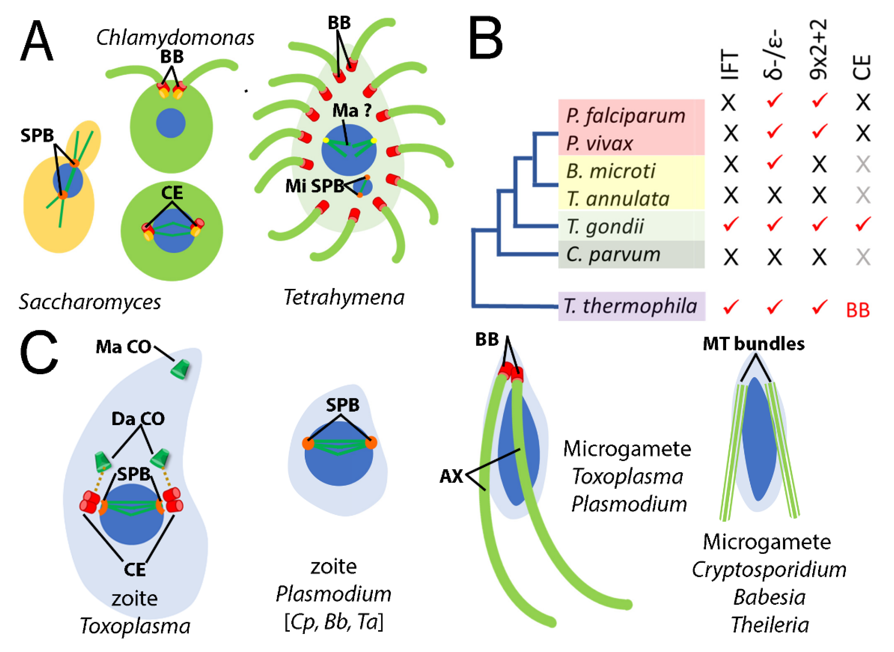
3.2. Not All Apicomplexans Have Genes for δ- and ε-Tubulins

Most surveyed apicomplexans harbor genes for apparent δ- and ε-tubulin homologs (Table 2). The exceptions are C. parvum and T. annulata, which lack genes for δ- and ε-tubulins. This absence was noted by Hodges and colleagues in a 2010 study of the evolutionary history of the centriole [22] and remains consistent in our reanalysis despite advancements in genome sequencing, assembly, and annotation for these genera. Species that lack δ- and ε-tubulin genes are members of two distinct apicomplexan lineages. The well-separated positions of Theileria (Piroplasmida) and Cryptosporidium (Eucoccidiorida) in phylogenetic analyses [72,73,74,75] are consistent with the conclusion that these genera independently lost δ- and ε-tubulin (Figure 2).
Meaningful comparisons of predicted δ- and ε-tubulin proteins from apicomplexans are limited by the lower level of homology among members of these families. However, it is notable that the tubulin signature motif, which is a highly conserved portion of the GTP-binding domain [2], deviates from the consensus or is missing altogether in some apicomplexan δ- and ε-tubulins (Figure 4). The tubulin signature appears to be entirely absent from the apparent S. neurona δ-tubulin homolog. Plasmodium δ-tubulin homologs have a non-canonical tubulin signature (AGGSGSG) rather than the expected [S/A/G]-G-G-T-G-[S/A]-G motif, and the C. suis signature has deteriorated to SEGGGSG. Surprisingly, the signature is preserved in B. microti δ-tubulin, despite studies that explicitly state that gametes are not flagellated. All apicomplexan ε-tubulins harbor a conserved tubulin motif, except for the apparent S. neurona homolog where, again, this sequence is undetectable. The low level of homology for S. neurona δ- and ε-tubulins and the apparent absence of a tubulin signature motif may indicate that these proteins are drifting into non-functional forms that no longer bind GTP.

3.3. Tubulin Expression Is Developmentally Regulated during the Parasite Life Cycle

To date, RNA-Seq and mass spectrometry (MS) studies have been carried out for several key P. falciparum and T. gondii stages. These data provide some insight into the expression of tubulin genes in zoite and gametocyte (pre-gamete) forms [59,60,61,62,63]. However, there are several caveats to these data. For example, although γ-tubulin is essential, peptides derived from it are not identified in all life cycle stages. This disparity likely represents a failure to detect peptides from less abundant proteins in stages that are technically more challenging to purify. Conversely, MS datasets for Toxoplasma tachyzoites and bradyzoites detect low levels of unique α2- and α3- peptides, while normalized RNA-Seq profiling does not detect significant transcripts for these tubulins. Lastly, while the Toxoplasma data are represented as normalized counts and were generated by the same research lab using comparable workflows, the Plasmodium data are from different labs and were not similarly processed by the original investigators. Despite these shortcomings, the existing highly normalized data extracted from VEuPathDB suggests that the transcription of δ- and ε-tubulin genes in Plasmodium is specific to microgametocytes, whereas the synthesis of δ- and ε-tubulin proteins in Toxoplasma may be regulated post-transcriptionally.
Late trophozoite and oocyst stage P. falciparum samples have comparable levels of β-, α-I, and α-II, and slightly lower levels for γ-tubulin transcripts (Figure 5). Peptides for α-I, α-II, and β- are present in red cell stage parasites (e.g., trophozoites) and in oocysts. As expected, given the absence of flagella in vegetative forms, δ- and ε-tubulin transcripts and peptides are absent or negligible in red cell stages and oocysts. While macrogametocytes have low levels of δ- and ε-tubulin transcripts, levels of α-II, δ-, and ε-tubulin are over a magnitude higher in microgametocytes. Moreover, δ- and ε-tubulin peptides are specifically detected in microgametocyte samples. These data are consistent with the conclusion that the Plasmodium expression of δ- and ε-tubulins is microgamete-specific.
Tachyzoites, bradyzoites and oocysts represent distinct T. gondii asexual forms obtained generally as highly enriched, defined stages. On the other hand, the EES5 sample is composed of a mixture of merozoites, microgametocytes, macrogametocytes, macrogametes, and microgametes at the late stage of enteroepithelial development in the cat intestine. Transcripts for the three β-tubulins and for γ-tubulin are found in all four samples (Figure 5). Transcripts for the unusual α2- and α3-tubulins are negligible or absent in tachyzoites, and α2- mRNA is also absent from bradyzoites. Surprisingly, δ- and ε-tubulin transcripts are found in all four samples, although there is no peptide evidence for protein expression in tachyzoite, bradyzoite and oocyte samples. Currently, it is not possible to obtain enriched gametocyte samples for MS studies to date.

4. Discussion

Tubulins are an ancient superfamily of GTP-binding proteins that emerged prior to the divergence of extant eukaryotes [5,20,76]. The highest sequence identity is found in the α-, β-, and γ-tubulin families, which are especially constrained by requisite interactions with GTP, other tubulins, and other proteins. In all eukaryotes, γ-tubulin nucleates microtubules that assemble from α-β-tubulin heterodimers. Their ubiquitous presence and sequence conservation makes α-, β-, and γ-tubulins valuable for molecular systematics of eukaryotes. The remaining three families, δ-, ε-, and ζ-tubulins, are components of centrioles and basal bodies, ancient organelles found in many eukaryotic groups, which suggests that they also emerged at around the time of the last eukaryotic common ancestor. ZED tubulins have been lost from diverse eukaryotic lineages [6,7,18]. These organisms illustrate that there are many routes to survive the loss or reduction of centriole and basal body functions.
Centrioles and basal bodies are found in lower plants, chytrid fungi, animals, and many protozoa. They typically consist of a cylinder of nine triplet microtubules (Figure 1) but may also be comprised of nine doublet or singlet microtubules in rarer circumstances. The construction of a transition zone converts a centriole into a basal body, which has the capacity to template and anchor an axoneme at the plasma membrane. In some eukaryotes, duplicated centrioles are located at spindle poles and serve to organize signaling molecules during chromosome segregation and cytokinesis. Separated centrioles are subsequently converted into basal bodies to construct flagella in daughter cells (Figure 6). However, early branching eukaryotes employ a closed mitosis, which occurs without nuclear envelope breakdown. For many of these (e.g., Tetrahymena), spindle microtubules emanate from acentriolar plaques/spindle pole bodies (SPB) embedded in the nuclear envelope and basal bodies are constructed exclusively for axoneme functions [77,78]. Other protozoa (e.g., Toxoplasma) have both SPBs (centrocones) and associated centrioles [79].
Apicomplexan parasites are located within the Alveolata, a large and diverse group of eukaryotic microorganisms that also includes ciliates and dinoflagellates [80,81,82]. Many alveolates construct canonical 9 × 2 + 2 axonemes to form motile cilia or flagella [83]. In fact, the ciliates Tetrahymena and Paramecium are well-established model systems for the study of ciliary function [78,84]. The recent expansion of sequenced apicomplexan genomes provides an opportunity to analyze members of the tubulin superfamily relative to those represented in ciliates. A previous study evaluated several apicomplexans, along with Tetrahymena and Paramecium, in the context of reconstructing the evolutionary history of the centriole [22]. That work demonstrated that Toxoplasma and Plasmodium have fewer homologs for core (ancestral) centriole components relative to Tetrahymena and Paramecium. While ciliates such as Tetrahymena generally occupy an extracellular, predatory niche that requires persistent axoneme-based motility, apicomplexans are obligate intracellular parasites. The asexual (zoite) stages of apicomplexans are not flagellated; these forms may use an unusual actin-based gliding motility to move and to invade host cells [43]. Therefore, the function of flagella in apicomplexans is, at most, reduced to a role in microgamete fertilization during the transient sexual cycle. Some, but not all apicomplexans harbor genes for δ- and ε-tubulins, and some but not all apicomplexan microgametes construct centrioles, basal bodies and flagellar axonemes (Figure 6).
Comparative genome analysis and ultrastructural studies indicate that δ- and ε-tubulin genes have been separately lost from many eukaryotic lineages, including the genera of model organisms Drosophila, Caenorhabditis, and Saccharomyces [5,19,20]. In each case, the consequences of this loss are different. Drosophila centrioles lack subdistal appendages, structural elements that typically distinguish the mature (mother) centriole [21,85,86,87]. These nine triplet-containing structures are converted into basal bodies that template axonemes, including for motile flagella in sperm cells. Caenorhabditis centrioles consist of nine singlet microtubules, and its non-flagellated sperm move by a distinct crawling mechanism [23,88,89]. C. elegans constructs non-motile (9 × 2 + 0) sensory cilia in some neurons that may be highly branched and originate in basal bodies consisting of doublet microtubules. Lastly, Saccharomyces and many other fungi do not form centrioles, basal bodies, or flagella. The presence of ε- and ζ-tubulin genes, basal bodies, and motile flagella in early branching chytrids indicates that both tubulins and structures were lost from other fungal lineages [5,18,90]. These examples of δ- and ε-tubulin loss and centriole, basal body and axoneme adaptations have apparent parallels in apicomplexan organisms.
As apicomplexans transitioned to an intracellular lifestyle, their need for flagellar motility was reduced to a stage-specific role in microgametes during the fertilization of macrogametes [91]. This shift reduced the pressure for the maintenance of centriole, basal body and axoneme structures, which led to the simplification or loss of these structures. For example, asexual stage Toxoplasma constructs atypical singlet centrioles, whereas asexual stage Plasmodium lacks these altogether [41,79,92,93]. Therefore, while Plasmodium microgametes must undergo de novo basal body assembly (a process that also occurs in Naegleria and Marsilea [94,95]), Toxoplasma may specifically synthesize δ- and ε-tubulin proteins during gametogenesis to convert singlet centrioles to triplet basal bodies [50,96]. δ- and ε-tubulins in individual apicomplexan species may be altered, vestigial, or absent.
The fullest picture of the relationship between apicomplexan δ- and ε-tubulins and tubulin-based structures is achieved by integrating ultrastructural observations of specific life cycle forms with analysis of parasite genomes. Unfortunately, only a few electron micrographs capture cross-sections through apicomplexan microgamete basal bodies, which are often described as electron-dense [92,97,98,99,100]. In Plasmodium, images show singlet microtubules which may represent the mature basal body form or reflect an intermediate stage of assembly. Similarly, a basal body from a microgamete of the coccidian S. suihominis shows a combination of doublet and triplet microtubules. The apparent Sarcocystis δ- and ε-tubulin homologs lack a discernible tubulin signature, and the Plasmodium δ-tubulin signature is non-canonical (Figure 4). Mutations that compromise the function of δ- or ε-tubulin in Chlamydomonas reduce basal body triplets to doublets or singlets [101,102,103]. Within the Piroplasmida, both Theileria and Babesia microgametes lack a 9 × 2 + 2 motile axoneme structure found in many eukaryotes, including Plasmodium [97,104]. T. annulata microgametes are described as spindle-shaped, with a pointed apex and flagellar-like protrusions containing two to six microtubules [105]. Remarkably, although the B. microti genome harbors δ- and ε-tubulin genes, its gametes lack axonemes and basal bodies [106,107,108]. Indeed, Weber and Friedhoff explicitly state that the “so-called strahlen (spiky rays) are cytoplasmic processes supported by a bundle of long, parallel microtubules that are distributed randomly and not configurated to a 9 + 2 or another pattern” [108]. Lastly, in the case of Cryptosporidium, the loss of δ- and ε-tubulin genes is in line with an overall reduction in genome complexity, including the loss of centriole components [22,29]. Super-resolution (fluorescence) microscopy of Cryptosporidium microgametes indicates that they lack flagella but possess sets of microtubules that run along either side of the length of the elongated nucleus [109]. As with Theileria and Babesia microgametes, these microtubule bundles are likely relics of the flagella observed in Plasmodium and Toxoplasma microgametes.
Previous analysis of committed male and female Plasmodium gametocytes reveals that the synthesis of flagellar components is restricted to this stage [110,111]. Microgametocytes specifically synthesize δ- and ε- tubulin transcripts and proteins (Figure 5). Several lines of indirect evidence suggest that Toxoplasma may specifically synthesize δ- and ε-tubulin proteins in microgametes. Toxoplasma tachyzoites construct singlet centrioles with structural similarity to δ- and ε-tubulin-deficient C. elegans centrioles [79]. MS surveys do not detect δ- or ε-tubulin in tachyzoites or other asexual stages. Lastly, there is ultrastructural evidence for triplet microtubule basal bodies in a coccidian (S. suihominis) microgamete. To date, it has not been possible to culture or isolate pure committed gametocytes, macrogametes, or microgametes from Toxoplasma for RNA-Seq or MS analysis. Felid enteroepithelial samples contain merozoites, gametocytes, macrogametes, and microgametes [112]. Although this mixed population may obscure differences between macrogametes and microgametes, the globally similar transcript levels in this and other samples (tachyzoites, bradyzoites and unsporulated oocysts) suggest that the synthesis of δ- and ε-tubulin proteins is regulated at a post-transcriptional level. Once it is possible to obtain pure samples of these forms, these hypotheses can be tested.
Because fertilization is essential for the transmission of some apicomplexans, the disruption of basal body and axoneme assembly has obvious relevance for reducing the incidence of disease. SAS-6 is a conserved and essential component of centrioles and basal bodies that establishes the nine-fold symmetry required for both duplication and function. Like δ- and ε-tubulin, its expression is microgamete-specific in Plasmodium, which also supports the interpretation that basal bodies are assembled de novo at this stage [49]. Targeted gene knockouts for SAS-6 and PF16 in Plasmodium have shown that basal body and axoneme components are important for the transmission of infection [49,113]. The disproportionately large impact of malaria on human morbidity and mortality means that a future method to compromise Plasmodium microgamete flagellar assembly or function could be transformative for human health. However, the specific set of basal body components, the existence of flagellated microgametes, and the importance of fertilization for transmission vary between individual apicomplexan lineages. Therefore, strategies to target microgamete flagella would not be universally applicable for all apicomplexan pathogens.
The individual evolutionary trajectories of apicomplexan parasites have led them to occupy distinct host cell and tissue niches, and consequently to have different therapeutic vulnerabilities. Most standard-of-care treatments for apicomplexan infections have limited utility for the inhibition of other apicomplexans. For example, erythrocytic stage Plasmodium parasites degrade hemoglobin as a source of nutrients, and many anti-malarial drugs interfere with this process [114]. These drug targets are non-essential for apicomplexans that infect other host cell types. Similarly, nitazoxanide, which interferes with electron transfer during anaerobic metabolism, is used to treat Cryptosporidium and Cyclospora infections, which remain in the relatively hypoxic environment of the intestine [115]. Because Toxoplasma disseminates from the intestine to tissue sites with higher oxygenation, it is less reliant on anaerobic metabolism and nitazoxanide is ineffective. Daraprim, the standard-of-care drug for toxoplasmosis, inhibits folate metabolism [116]. Cyclospora can also be treated by inhibiting folate metabolism, but Cryptosporidium has streamlined metabolic pathways, including an unexpected ability to salvage purines from the host [117]. In contrast, microtubules represent a conserved broad spectrum drug target shared by all apicomplexan pathogens.
Small molecules that bind to tubulin heterodimers alter the dynamic properties of microtubules and are used in clinical settings to treat cancer, inflammatory disorders, and helminth (worm) parasite infection. Typically, microtubule targeting agents disrupt dynamic spindle microtubules to impair mitosis. Phylogenetic analysis indicates that apicomplexan tubulins are most like tubulins from ciliates and green algae [118,119,120]. Not surprisingly, the phylogenetic relationships of α- and β-tubulins align with sensitivity to dinitroanilines, small molecules that inhibit microtubule assembly. Dinitroanilines prevent mitosis in protozoan parasites, green algae, and land plants, but are inactive in vertebrates or fungi [50,51]. Other tubulin-targeting compounds (MMV676477 and parabulin) also selectively inhibit apicomplexans and other protozoa [121,122]. Genes for distinct α- and β-tubulin isotypes may influence drug resistance. For example, aggressive and treatment refractory human cancers increase the expression of less sensitive isotypes to reduce the efficacy of tubulin targeting drugs [123]. This mechanism is less likely to arise in apicomplexans, which have simpler tubulin gene organization. However, drug resistance may arise by missense mutations that alter tubulin biochemistry, as observed for Toxoplasma oryzalin resistance which is conferred by α1-tubulin substitutions [124]. However, since apicomplexans are haploid, the co-expression of conserved isotypes may suppress the ability of missense mutations to confer resistance. Microtubules composed of a mixture of sensitive and resistant proteins remain somewhat susceptible to disruption. We hypothesize that drugs that bind to co-expressed tubulin subunits (e.g., Toxoplasma β-tubulins and possibly Plasmodium α-tubulins) will suppress the development of drug resistant strains.
Available genomes, transcript profiling, and morphological studies provide critical information on the organization, properties, and expression of tubulin superfamily members in apicomplexans. While the present data can be incorporated into a logical narrative, other details remain unanswered until researchers are able to collect robust and specific datasets for additional (currently inaccessible) life cycle stages. Nonetheless, existing evidence validates tubulin and microtubules as critical targets for anti-parasitic therapies and transmission-blocking strategies to control apicomplexan pathogens.

Supplementary Materials

The following supporting information can be downloaded at: https://www.mdpi.com/article/10.3390/microorganisms11030706/s1, Figure S1: Alignment of T. gondii β-tubulins; Table S1: Uniprot accession numbers for Tetrahymena and Chlamydomonas tubulins; Table S2: RNA-Seq datasets for P. falciparum; Table S3: RNA-Seq datasets for T. gondii.

Author Contributions

Conceptualization, N.M.; methodology, N.M. and C.R.; formal analysis, N.M., I.A. and C.R.; data curation, N.M., I.A. and C.R.; writing—original draft preparation, N.M.; writing—review and editing, N.M., I.A., C.R. and A.B.H.; visualization, N.M. and I.A. All authors have read and agreed to the published version of the manuscript.

Funding

I. A. was supported by graduate fellowships from the MBB GAANN (P200A210024) and T32 in Microbiology and Infectious Diseases (5T32AI141346) programs.

Data Availability Statement

All data supporting reported results can be found at VEupathDB.org or in supporting data for the papers cited in the methods.

Acknowledgments

Our ongoing research on the properties of Toxoplasma tubulin isotypes, supported by the ICRR Program (UCI Office of the Provost), inspired this computational analysis. This paper would not have been possible without the extraordinary bioinformatic resources available at VEupathDB.org, including feedback from David Roos and Omar Harb. We thank Ulrike Boehme for assistance with the reannotation of Toxoplasma δ-tubulin.

Conflicts of Interest

The authors declare no conflict of interest.

References

  1. Gudimchuk, N.B.; McIntosh, J.R. Regulation of microtubule dynamics, mechanics and function through the growing tip. Nat. Rev. Mol. Cell Biol. 2021, 22, 777–795. [Google Scholar] [CrossRef]
  2. Kristensson, M.A. The Game of Tubulins. Cells 2021, 10, 745. [Google Scholar] [CrossRef]
  3. Lopez-Fanarraga, M.; Avila, J.; Guasch, A.; Coll, M.; Zabala, J.C. Review: Postchaperonin tubulin folding cofactors and their role in microtubule dynamics. J. Struct. Biol. 2001, 135, 219–229. [Google Scholar] [CrossRef] [PubMed]
  4. Kelly, J.J.; Tranter, D.; Pardon, E.; Chi, G.; Kramer, H.; Happonen, L.; Knee, K.M.; Janz, J.M.; Steyaert, J.; Bulawa, C.; et al. Snapshots of actin and tubulin folding inside the TRiC chaperonin. Nat. Struct. Mol. Biol. 2022, 29, 420–429. [Google Scholar] [CrossRef]
  5. Findeisen, P.; Muhlhausen, S.; Dempewolf, S.; Hertzog, J.; Zietlow, A.; Carlomagno, T.; Kollmar, M. Six subgroups and extensive recent duplications characterize the evolution of the eukaryotic tubulin protein family. Genome Biol. Evol. 2014, 6, 2274–2288. [Google Scholar] [CrossRef] [Green Version]
  6. McKean, P.G.; Vaughan, S.; Gull, K. The extended tubulin superfamily. J. Cell Sci. 2001, 114, 2723–2733. [Google Scholar] [CrossRef]
  7. Dutcher, S.K. Long-lost relatives reappear: Identification of new members of the tubulin superfamily. Curr. Opin. Microbiol. 2003, 6, 634–640. [Google Scholar] [CrossRef]
  8. Marshall, W.F.; Rosenbaum, J.L. Tubulin superfamily: Giving birth to triplets. Curr. Biol. 2003, 13, R55–R56. [Google Scholar] [CrossRef] [Green Version]
  9. Zupa, E.; Liu, P.; Wurtz, M.; Schiebel, E.; Pfeffer, S. The structure of the γ-TuRC: A 25-years-old molecular puzzle. Curr. Opin. Struct. Biol. 2021, 66, 15–21. [Google Scholar] [CrossRef] [PubMed]
  10. Kollman, J.M.; Merdes, A.; Mourey, L.; Agard, D.A. Microtubule nucleation by γ-tubulin complexes. Nat. Rev. Mol. Cell Biol. 2011, 12, 709–721. [Google Scholar] [CrossRef] [PubMed]
  11. Fulton, C. The Amazing Evolutionary Complexity of Eukaryotic Tubulins: Lessons from Naegleria and the Multi-tubulin Hypothesis. Front. Cell Dev. Biol. 2022, 10, 867374. [Google Scholar] [CrossRef] [PubMed]
  12. Nsamba, E.T.; Gupta, M.L., Jr. Tubulin isotypes—Functional insights from model organisms. J. Cell Sci. 2022, 135, jcs259539. [Google Scholar] [CrossRef] [PubMed]
  13. Bera, A.; Gupta, M.L., Jr. Microtubules in Microorganisms: How Tubulin Isotypes Contribute to Diverse Cytoskeletal Functions. Front. Cell Dev. Biol. 2022, 10, 913809. [Google Scholar] [CrossRef] [PubMed]
  14. Wilson, P.G.; Borisy, G.G. Evolution of the multi-tubulin hypothesis. Bioessays 1997, 19, 451–454. [Google Scholar] [CrossRef] [PubMed]
  15. Ti, S.C.; Alushin, G.M.; Kapoor, T.M. Human β-Tubulin Isotypes Can Regulate Microtubule Protofilament Number and Stability. Dev. Cell 2018, 47, 175–190.e5. [Google Scholar] [CrossRef] [Green Version]
  16. Pamula, M.C.; Ti, S.-C.; Kapoor, T.M. The structured core of human β tubulin confers isotype-specific polymerization properties. J. Cell Biol. 2016, 213, 425–433. [Google Scholar] [CrossRef] [Green Version]
  17. Hausrat, T.J.; Radwitz, J.; Lombino, F.L.; Breiden, P.; Kneussel, M. α- and β-tubulin isotypes are differentially expressed during brain development. Dev. Neurobiol. 2021, 81, 333–350. [Google Scholar] [CrossRef] [Green Version]
  18. Turk, E.; Wills, A.A.; Kwon, T.; Sedzinski, J.; Wallingford, J.B.; Stearns, T. ζ-Tubulin Is a Member of a Conserved Tubulin Module and Is a Component of the Centriolar Basal Foot in Multiciliated Cells. Curr. Biol. 2015, 25, 2177–2183. [Google Scholar] [CrossRef] [Green Version]
  19. Stathatos, G.G.; Dunleavy, J.E.M.; Zenker, J.; O’Bryan, M.K. δ and ε tubulin in mammalian development. Trends Cell Biol. 2021, 31, 774–787. [Google Scholar] [CrossRef]
  20. Zhao, Z.; Liu, H.; Luo, Y.; Zhou, S.; An, L.; Wang, C.; Jin, Q.; Zhou, M.; Xu, J.R. Molecular evolution and functional divergence of tubulin superfamily in the fungal tree of life. Sci. Rep. 2014, 4, 6746. [Google Scholar] [CrossRef] [Green Version]
  21. Jana, S.C.; Bettencourt-Dias, M.; Durand, B.; Megraw, T.L. Drosophila melanogaster as a model for basal body research. Cilia 2016, 5, 22. [Google Scholar] [CrossRef] [PubMed] [Green Version]
  22. Hodges, M.E.; Scheumann, N.; Wickstead, B.; Langdale, J.A.; Gull, K. Reconstructing the evolutionary history of the centriole from protein components. J. Cell Sci. 2010, 123, 1407–1413. [Google Scholar] [CrossRef] [PubMed] [Green Version]
  23. Schwarz, A.; Sankaralingam, P.; O’Connell, K.F.; Muller-Reichert, T. Revisiting Centrioles in Nematodes-Historic Findings and Current Topics. Cells 2018, 7, 101. [Google Scholar] [CrossRef] [PubMed] [Green Version]
  24. The Lancet Editors. Malaria in 2022: A year of opportunity. Lancet 2022, 399, 1573. [Google Scholar] [CrossRef]
  25. Vannier, E.; Gewurz, B.E.; Krause, P.J. Human babesiosis. Infect. Dis. Clin. North Am. 2008, 22, 469–488. [Google Scholar] [CrossRef] [Green Version]
  26. Yang, Y.; Christie, J.; Koster, L.; Du, A.; Yao, C. Emerging Human Babesiosis with “Ground Zero” in North America. Microorganisms 2021, 9, 440. [Google Scholar] [CrossRef]
  27. Ullah, R.; Shams, S.; Khan, M.A.; Ayaz, S.; ul Akbar, N.; Din, Q.u.; Khan, A.; Leon, R.; Zeb, J. Epidemiology and molecular characterization of Theileria annulata in cattle from central Khyber Pakhtunkhwa, Pakistan. PLoS ONE 2021, 16, e0249417. [Google Scholar] [CrossRef]
  28. Roy, S.; Bhandari, V.; Barman, M.; Kumar, P.; Bhanot, V.; Arora, J.S.; Singh, S.; Sharma, P. Population Genetic Analysis of the Theileria annulata Parasites Identified Limited Diversity and Multiplicity of Infection in the Vaccine from India. Front. Microbiol. 2020, 11, 579929. [Google Scholar] [CrossRef]
  29. Keeling, P.J. Reduction and compaction in the genome of the apicomplexan parasite Cryptosporidium parvum. Dev. Cell 2004, 6, 614–616. [Google Scholar] [CrossRef] [Green Version]
  30. Gerace, E.; Lo Presti, V.D.M.; Biondo, C. Cryptosporidium Infection: Epidemiology, Pathogenesis and Differential Diagnosis. Eur. J. Microbiol. Immunol. 2019, 9, 119–123. [Google Scholar] [CrossRef]
  31. Barta, J.R.; Schrenzel, M.D.; Carreno, R.; Rideout, B.A. The genus Atoxoplasma (Garnham 1950) as a junior objective synonym of the genus Isospora (Schneider 1881) species infecting birds and resurrection of Cystoisospora (Frenkel 1977) as the correct genus for Isospora species infecting mammals. J. Parasitol. 2005, 91, 726–727. [Google Scholar] [CrossRef]
  32. Dubey, J.P.; Almeria, S. Cystoisospora belli infections in humans: The past 100 years. Parasitology 2019, 146, 1490–1527. [Google Scholar] [CrossRef]
  33. Li, J.; Cui, Z.; Qi, M.; Zhang, L. Advances in Cyclosporiasis Diagnosis and Therapeutic Intervention. Front. Cell. Infect. Microbiol. 2020, 10, 43. [Google Scholar] [CrossRef] [PubMed] [Green Version]
  34. Lopez-Osorio, S.; Chaparro-Gutierrez, J.J.; Gomez-Osorio, L.M. Overview of Poultry Eimeria Life Cycle and Host-Parasite Interactions. Front. Vet. Sci. 2020, 7, 384. [Google Scholar] [CrossRef]
  35. Blake, D.P.; Worthing, K.; Jenkins, M.C. Exploring Eimeria Genomes to Understand Population Biology: Recent Progress and Future Opportunities. Genes 2020, 11, 1103. [Google Scholar] [CrossRef] [PubMed]
  36. Gutierrez-Exposito, D.; Ferre, I.; Ortega-Mora, L.M.; Alvarez-Garcia, G. Advances in the diagnosis of bovine besnoitiosis: Current options and applications for control. Int. J. Parasitol. 2017, 47, 737–751. [Google Scholar] [CrossRef] [PubMed]
  37. Jones, J.L.; Kruszon-Moran, D.; Elder, S.; Rivera, H.N.; Press, C.; Montoya, J.G.; McQuillan, G.M. Toxoplasma gondii Infection in the United States, 2011–2014. Am. J. Trop. Med. Hyg. 2018, 98, 551–557. [Google Scholar] [CrossRef] [Green Version]
  38. Montoya, J.G.; Liesenfeld, O. Toxoplasmosis. Lancet 2004, 363, 1965–1976. [Google Scholar] [CrossRef]
  39. Tran, J.Q.; Li, C.; Chyan, A.; Chung, L.; Morrissette, N.S. SPM1 stabilizes subpellicular microtubules in Toxoplasma gondii. Eukaryot. Cell 2012, 11, 206–216. [Google Scholar] [CrossRef] [Green Version]
  40. Liu, J.; He, Y.; Benmerzouga, I.; Sullivan, W.J., Jr.; Morrissette, N.S.; Murray, J.M.; Hu, K. An ensemble of specifically targeted proteins stabilizes cortical microtubules in the human parasite Toxoplasma gondii. Mol. Biol. Cell 2016, 27, 549–571. [Google Scholar] [CrossRef] [Green Version]
  41. De Leon, J.C.; Scheumann, N.; Beatty, W.; Beck, J.R.; Tran, J.Q.; Yau, C.; Bradley, P.J.; Gull, K.; Wickstead, B.; Morrissette, N.S. A SAS-6-like protein suggests that the Toxoplasma conoid complex evolved from flagellar components. Eukaryot. Cell 2013, 12, 1009–1019. [Google Scholar] [CrossRef] [PubMed] [Green Version]
  42. Lévêque, M.F.; Berry, L.; Besteiro, S. An evolutionarily conserved SSNA1/DIP13 homologue is a component of both basal and apical complexes of Toxoplasma gondii. Sci. Rep. 2016, 6, 27809. [Google Scholar] [CrossRef] [PubMed] [Green Version]
  43. Frenal, K.; Dubremetz, J.F.; Lebrun, M.; Soldati-Favre, D. Gliding motility powers invasion and egress in Apicomplexa. Nat. Rev. Microbiol. 2017, 15, 645–660. [Google Scholar] [CrossRef] [PubMed] [Green Version]
  44. Wall, R.J.; Roques, M.; Katris, N.J.; Koreny, L.; Stanway, R.R.; Brady, D.; Waller, R.F.; Tewari, R. SAS6-like protein in Plasmodium indicates that conoid-associated apical complex proteins persist in invasive stages within the mosquito vector. Sci. Rep. 2016, 6, 28604. [Google Scholar] [CrossRef] [PubMed] [Green Version]
  45. Tosetti, N.; Dos Santos Pacheco, N.; Soldati-Favre, D.; Jacot, D. Three F-actin assembly centers regulate organelle inheritance, cell-cell communication and motility in Toxoplasma gondii. eLife 2019, 8, e42669. [Google Scholar] [CrossRef]
  46. Cruz-Bustos, T.; Feix, A.S.; Ruttkowski, B.; Joachim, A. Sexual Development in Non-Human Parasitic Apicomplexa: Just Biology or Targets for Control? Animals 2021, 11, 2891. [Google Scholar] [CrossRef]
  47. Bechtsi, D.P.; Waters, A.P. Genomics and epigenetics of sexual commitment in Plasmodium. Int. J. Parasitol. 2017, 47, 425–434. [Google Scholar] [CrossRef]
  48. Sinden, R.E. Targeting the Parasite to Suppress Malaria Transmission. Adv. Parasitol. 2017, 97, 147–185. [Google Scholar]
  49. Marques, S.R.; Ramakrishnan, C.; Carzaniga, R.; Blagborough, A.M.; Delves, M.J.; Talman, A.M.; Sinden, R.E. An essential role of the basal body protein SAS-6 in Plasmodium male gamete development and malaria transmission. Cell. Microbiol. 2015, 17, 191–206. [Google Scholar] [CrossRef] [Green Version]
  50. Morrissette, N. Targeting Toxoplasma tubules: Tubulin, microtubules and associated proteins in a human pathogen. Eukaryot. Cell 2015, 14, 2–12. [Google Scholar] [CrossRef] [Green Version]
  51. Bell, A. Microtubule inhibitors as potential antimalarial agents. Parasitol. Today 1998, 14, 234–240. [Google Scholar] [CrossRef] [PubMed]
  52. Amos, B.; Aurrecoechea, C.; Barba, M.; Barreto, A.; Basenko, E.Y.; Bażant, W.; Belnap, R.; Blevins, A.S.; Böhme, U.; Brestelli, J.; et al. VEuPathDB: The eukaryotic pathogen, vector and host bioinformatics resource center. Nucleic Acids Res. 2021, 50, D898–D911. [Google Scholar] [CrossRef] [PubMed]
  53. Adl, S.M.; Simpson, A.G.; Lane, C.E.; Lukes, J.; Bass, D.; Bowser, S.S.; Brown, M.W.; Burki, F.; Dunthorn, M.; Hampl, V.; et al. The revised classification of eukaryotes. J. Eukaryot. Microbiol. 2012, 59, 429–493. [Google Scholar] [CrossRef] [PubMed] [Green Version]
  54. Coordinators, N.R. Database resources of the National Center for Biotechnology Information. Nucleic Acids Res. 2018, 46, D8–D13. [Google Scholar] [CrossRef] [Green Version]
  55. Sievers, F.; Higgins, D.G. Clustal omega. Curr. Protoc. Bioinform. 2014, 48, 3–13. [Google Scholar] [CrossRef]
  56. Sievers, F.; Higgins, D.G. Clustal Omega for making accurate alignments of many protein sequences. Protein Sci. 2018, 27, 135–145. [Google Scholar] [CrossRef] [Green Version]
  57. Sievers, F.; Wilm, A.; Dineen, D.; Gibson, T.J.; Karplus, K.; Li, W.; Lopez, R.; McWilliam, H.; Remmert, M.; Soding, J.; et al. Fast, scalable generation of high-quality protein multiple sequence alignments using Clustal Omega. Mol. Syst. Biol. 2011, 7, 539. [Google Scholar] [CrossRef]
  58. Rice, P.; Longden, I.; Bleasby, A. EMBOSS: The European Molecular Biology Open Software Suite. Trends Genet. 2000, 16, 276–277. [Google Scholar] [CrossRef]
  59. Lasonder, E.; Rijpma, S.R.; van Schaijk, B.C.; Hoeijmakers, W.A.; Kensche, P.R.; Gresnigt, M.S.; Italiaander, A.; Vos, M.W.; Woestenenk, R.; Bousema, T.; et al. Integrated transcriptomic and proteomic analyses of P. falciparum gametocytes: Molecular insight into sex-specific processes and translational repression. Nucleic Acids Res. 2016, 44, 6087–6101. [Google Scholar] [CrossRef] [Green Version]
  60. Gomez-Diaz, E.; Yerbanga, R.S.; Lefevre, T.; Cohuet, A.; Rowley, M.J.; Ouedraogo, J.B.; Corces, V.G. Epigenetic regulation of Plasmodium falciparum clonally variant gene expression during development in Anopheles gambiae. Sci. Rep. 2017, 7, 40655. [Google Scholar] [CrossRef] [Green Version]
  61. Lopez-Barragan, M.J.; Lemieux, J.; Quinones, M.; Williamson, K.C.; Molina-Cruz, A.; Cui, K.; Barillas-Mury, C.; Zhao, K.; Su, X.Z. Directional gene expression and antisense transcripts in sexual and asexual stages of Plasmodium falciparum. BMC Genom. 2011, 12, 587. [Google Scholar] [CrossRef] [PubMed] [Green Version]
  62. Zanghi, G.; Vembar, S.S.; Baumgarten, S.; Ding, S.; Guizetti, J.; Bryant, J.M.; Mattei, D.; Jensen, A.T.R.; Renia, L.; Goh, Y.S.; et al. A Specific PfEMP1 Is Expressed in P. falciparum Sporozoites and Plays a Role in Hepatocyte Infection. Cell Rep. 2018, 22, 2951–2963. [Google Scholar] [CrossRef] [PubMed] [Green Version]
  63. Ramakrishnan, C.; Maier, S.; Walker, R.A.; Rehrauer, H.; Joekel, D.E.; Winiger, R.R.; Basso, W.U.; Grigg, M.E.; Hehl, A.B.; Deplazes, P.; et al. An experimental genetically attenuated live vaccine to prevent transmission of Toxoplasma gondii by cats. Sci. Rep. 2019, 9, 1474. [Google Scholar] [CrossRef]
  64. Sidik, S.M.; Huet, D.; Ganesan, S.M.; Huynh, M.H.; Wang, T.; Nasamu, A.S.; Thiru, P.; Saeij, J.P.J.; Carruthers, V.B.; Niles, J.C.; et al. A Genome-wide CRISPR Screen in Toxoplasma Identifies Essential Apicomplexan Genes. Cell 2016, 166, 1423–1435.e12. [Google Scholar] [CrossRef] [Green Version]
  65. Kemphues, K.J.; Raff, R.A.; Kaufman, T.C.; Raff, E.C. Mutation in a structural gene for a β-tubulin specific to testis in Drosophila melanogaster. Proc. Natl. Acad. Sci. USA 1979, 76, 3991–3995. [Google Scholar] [CrossRef] [Green Version]
  66. Rawlings, D.J.; Fujioka, H.; Fried, M.; Keister, D.B.; Aikawa, M.; Kaslow, D.C. α-tubulin II is a male-specific protein in Plasmodium falciparum. Mol. Biochem. Parasitol. 1992, 56, 239–250. [Google Scholar] [CrossRef] [PubMed]
  67. Fennell, B.J.; Al-shatr, Z.A.; Bell, A. Isotype expression, post-translational modification and stage-dependent production of tubulins in erythrocytic Plasmodium falciparum. Int. J. Parasitol. 2008, 38, 527–539. [Google Scholar] [CrossRef]
  68. Schwank, S.; Sutherland, C.J.; Drakeley, C.J. Promiscuous expression of α-tubulin II in maturing male and female Plasmodium falciparum gametocytes. PLoS ONE 2010, 5, e14470. [Google Scholar] [CrossRef] [Green Version]
  69. Kooij, T.W.; Franke-Fayard, B.; Renz, J.; Kroeze, H.; van Dooren, M.W.; Ramesar, J.; Augustijn, K.D.; Janse, C.J.; Waters, A.P. Plasmodium berghei α-tubulin II: A role in both male gamete formation and asexual blood stages. Mol. Biochem. Parasitol. 2005, 144, 16–26. [Google Scholar] [CrossRef]
  70. Lin, Z.; Gasic, I.; Chandrasekaran, V.; Peters, N.; Shao, S.; Mitchison, T.J.; Hegde, R.S. TTC5 mediates autoregulation of tubulin via mRNA degradation. Science 2020, 367, 100–104. [Google Scholar] [CrossRef]
  71. Anders, K.R.; Botstein, D. Dominant-lethal α-tubulin mutants defective in microtubule depolymerization in yeast. Mol. Biol. Cell 2001, 12, 3973–3986. [Google Scholar] [CrossRef] [PubMed]
  72. Salomaki, E.D.; Terpis, K.X.; Rueckert, S.; Kotyk, M.; Varadinova, Z.K.; Cepicka, I.; Lane, C.E.; Kolisko, M. Gregarine single-cell transcriptomics reveals differential mitochondrial remodeling and adaptation in apicomplexans. BMC Biol. 2021, 19, 77. [Google Scholar] [CrossRef]
  73. Janouškovec, J.; Paskerova, G.G.; Miroliubova, T.S.; Mikhailov, K.V.; Birley, T.; Aleoshin, V.V.; Simdyanov, T.G. Apicomplexan-like parasites are polyphyletic and widely but selectively dependent on cryptic plastid organelles. eLife 2019, 8, e49662. [Google Scholar] [CrossRef] [PubMed]
  74. Templeton, T.J.; Pain, A. Diversity of extracellular proteins during the transition from the ‘proto-apicomplexan’ alveolates to the apicomplexan obligate parasites. Parasitology 2016, 143, 1–17. [Google Scholar] [CrossRef] [Green Version]
  75. Kuo, C.H.; Wares, J.P.; Kissinger, J.C. The Apicomplexan whole-genome phylogeny: An analysis of incongruence among gene trees. Mol. Biol. Evol. 2008, 25, 2689–2698. [Google Scholar] [CrossRef] [PubMed]
  76. Aylett, C.H.S.; Duggin, I.G. The Tubulin Superfamily in Archaea. Sub-Cell. Biochem. 2017, 84, 393–417. [Google Scholar]
  77. Hufnagel, L.A. The Cilioprotist Cytoskeleton, a Model for Understanding How Cell Architecture and Pattern Are Specified: Recent Discoveries from Ciliates and Comparable Model Systems. Methods Mol. Biol. 2022, 2364, 251–295. [Google Scholar]
  78. Bayless, B.A.; Galati, D.F.; Pearson, C.G. Tetrahymena basal bodies. Cilia 2016, 5, 1. [Google Scholar] [CrossRef] [Green Version]
  79. Morrissette, N.S.; Sibley, L.D. Disruption of microtubules uncouples budding and nuclear division in Toxoplasma gondii. J. Cell Sci. 2002, 115, 1017–1025. [Google Scholar] [CrossRef]
  80. Cavalier-Smith, T. Kingdom Chromista and its eight phyla: A new synthesis emphasising periplastid protein targeting, cytoskeletal and periplastid evolution, and ancient divergences. Protoplasma 2018, 255, 297–357. [Google Scholar] [CrossRef] [Green Version]
  81. Kumar, S.; Rzhetsky, A. Evolutionary relationships of eukaryotic kingdoms. J. Mol. Evol. 1996, 42, 183–193. [Google Scholar] [CrossRef]
  82. Gajadhar, A.A.; Marquardt, W.C.; Hall, R.; Gunderson, J.; Ariztia-Carmona, E.V.; Sogin, M.L. Ribosomal RNA sequences of Sarcocystis muris, Theileria annulata and Crypthecodinium cohnii reveal evolutionary relationships among apicomplexans, dinoflagellates and ciliates. Mol. Biochem. Parasitol. 1991, 45, 147–154. [Google Scholar] [CrossRef] [PubMed]
  83. Ishikawa, T. Axoneme Structure from Motile Cilia. Cold Spring Harb. Perspect. Biol. 2017, 9, a028076. [Google Scholar] [CrossRef] [PubMed] [Green Version]
  84. Valentine, M.; Van Houten, J. Using Paramecium as a Model for Ciliopathies. Genes 2021, 12, 1493. [Google Scholar] [CrossRef]
  85. Ryu, H.; Lee, H.; Lee, J.; Noh, H.; Shin, M.; Kumar, V.; Hong, S.; Kim, J.; Park, S. The molecular dynamics of subdistal appendages in multi-ciliated cells. Nat. Commun. 2021, 12, 612. [Google Scholar] [CrossRef] [PubMed]
  86. Chong, W.M.; Wang, W.-J.; Lo, C.-H.; Chiu, T.-Y.; Chang, T.-J.; Liu, Y.-P.; Tanos, B.; Mazo, G.; Tsou, M.-F.B.; Jane, W.-N.; et al. Super-resolution microscopy reveals coupling between mammalian centriole subdistal appendages and distal appendages. eLife 2020, 9, e53580. [Google Scholar] [CrossRef] [Green Version]
  87. Pedersen, L.B.; Schroder, J.M.; Satir, P.; Christensen, S.T. The ciliary cytoskeleton. Compr. Physiol. 2012, 2, 779–803. [Google Scholar]
  88. Nechipurenko, I.; Sengupta, P. The rise and fall of basal bodies in the nematode Caenorhabditis elegans. Cilia 2017, 6, 9. [Google Scholar] [CrossRef] [Green Version]
  89. Smith, H. Nematode Sperm Motility. In WormBook; Chalfie, M., Ed.; 2006; Available online: http://www.wormbook.org (accessed on 1 January 2023).
  90. Medina, E.M.; Buchler, N.E. Chytrid fungi. Curr. Biol. 2020, 30, R516–R520. [Google Scholar] [CrossRef]
  91. Leander, B.S.; Keeling, P.J. Morphostasis in alveolate evolution. Trends Ecol. Evol. 2003, 18, 395–402. [Google Scholar] [CrossRef] [Green Version]
  92. Francia, M.E.; Dubremetz, J.F.; Morrissette, N.S. Basal body structure and composition in the apicomplexans Toxoplasma and Plasmodium. Cilia 2015, 5, 3. [Google Scholar] [CrossRef] [Green Version]
  93. Zeeshan, M.; Brady, D.; Markus, R.; Vaughan, S.; Ferguson, D.; Holder, A.A.; Tewari, R. Plasmodium SAS4: Basal body component of male cell which is dispensable for parasite transmission. Life Sci. Alliance 2022, 5. [Google Scholar] [CrossRef]
  94. Fritz-Laylin, L.K.; Fulton, C. Naegleria: A classic model for de novo basal body assembly. Cilia 2016, 5, 10. [Google Scholar] [CrossRef] [PubMed] [Green Version]
  95. Mizukami, I.; Gall, J. Centriole replication. II. Sperm formation in the fern, Marsilea and the cycad, Zamia. J. Cell Biol. 1966, 29, 97–111. [Google Scholar] [CrossRef] [Green Version]
  96. Walker, R.A.; Ferguson, D.J.; Miller, C.M.; Smith, N.C. Sex and Eimeria: A molecular perspective. Parasitology 2013, 140, 1701–1717. [Google Scholar] [CrossRef] [PubMed]
  97. Sinden, R.E.; Talman, A.; Marques, S.R.; Wass, M.N.; Sternberg, M.J. The flagellum in malarial parasites. Curr. Opin. Microbiol. 2010, 13, 491–500. [Google Scholar] [CrossRef]
  98. Mehlhorn, H.; Heydorn, A.O. Electron microscopical study on gamogony of Sarcocystis suihominis in human tissue cultures. Z Parasitenkd 1979, 58, 97–113. [Google Scholar] [CrossRef]
  99. Ferguson, D.J. Toxoplasma gondii and sex: Essential or optional extra? Trends Parasitol. 2002, 18, 355–359. [Google Scholar] [CrossRef]
  100. Fernando, M.A. Fine structural changes associated with microgametogenesis of Eimeria acervulina in chickens. Z. Parasitenkd. 1973, 43, 33–42. [Google Scholar] [CrossRef]
  101. Dutcher, S.K.; Trabuco, E.C. The UNI3 gene is required for assembly of basal bodies of Chlamydomonas and encodes δ-tubulin, a new member of the tubulin superfamily. Mol. Biol. Cell 1998, 9, 1293–1308. [Google Scholar] [CrossRef] [PubMed]
  102. Dutcher, S.K.; Morrissette, N.S.; Preble, A.M.; Rackley, C.; Stanga, J. ε-tubulin is an essential component of the centriole. Mol. Biol. Cell 2002, 13, 3859–3869. [Google Scholar] [CrossRef] [PubMed]
  103. Wingfield, J.L.; Lechtreck, K.F. Chlamydomonas Basal Bodies as Flagella Organizing Centers. Cells 2018, 7, 79. [Google Scholar] [CrossRef] [Green Version]
  104. Garnham, P.C.; Bird, R.G.; Baker, J.R. Electron microscope studies of motile stages of malaria parasites. V. Exflagellation in Plasmodium, Hepatocystis and Leucocytozoon. Trans. R. Soc. Trop. Med. Hyg. 1967, 61, 58–68. [Google Scholar] [CrossRef]
  105. Mehlhorn, H.; Weber, G.; Schein, E.; Büscher, G. Elektronenmikroskopische Untersuchung an Entwicklungsstadien von Theileria annulata (Dschunkowsky, Luhs, 1904) im Darm und in der Hämolymphe von Hyalomma anatolicum excavatum (Koch, 1844). Z. Parasitenkd. 1975, 48, 137–150. [Google Scholar] [CrossRef] [PubMed]
  106. Rudzinska, M.A.; Spielman, A.; Lewengrub, S.; Trager, W.; Piesman, J. Sexuality in piroplasms as revealed by electron microscopy in Babesia microti. Proc. Natl. Acad. Sci. USA 1983, 80, 2966–2970. [Google Scholar] [CrossRef] [Green Version]
  107. Rudzinska, M.A.; Spielman, A.; Lewengrub, S.; Piesman, J.; Karakashian, S. Penetration of the peritrophic membrane of the tick by Babesia microti. Cell Tissue Res. 1982, 221, 471–481. [Google Scholar] [CrossRef] [PubMed]
  108. Weber, G.; Friedhoff, K.T. Preliminary observations on the ultrastructure of supposed sexual stages of Babesia bigemina (Piroplasmea). Z. Parasitenkd. 1977, 53, 83–92. [Google Scholar] [CrossRef]
  109. Tandel, J.; English, E.D.; Sateriale, A.; Gullicksrud, J.A.; Beiting, D.P.; Sullivan, M.C.; Pinkston, B.; Striepen, B. Life cycle progression and sexual development of the apicomplexan parasite Cryptosporidium parvum. Nat. Microbiol. 2019, 4, 2226–2236. [Google Scholar] [CrossRef] [Green Version]
  110. Khan, S.M.; Franke-Fayard, B.; Mair, G.R.; Lasonder, E.; Janse, C.J.; Mann, M.; Waters, A.P. Proteome analysis of separated male and female gametocytes reveals novel sex-specific Plasmodium biology. Cell 2005, 121, 675–687. [Google Scholar] [CrossRef] [Green Version]
  111. Miao, J.; Chen, Z.; Wang, Z.; Shrestha, S.; Li, X.; Li, R.; Cui, L. Sex-Specific Biology of the Human Malaria Parasite Revealed from the Proteomes of Mature Male and Female Gametocytes. Mol. Cell. Proteom. 2017, 16, 537–551. [Google Scholar] [CrossRef] [Green Version]
  112. Tomasina, R.; Francia, M.E. The Structural and Molecular Underpinnings of Gametogenesis in Toxoplasma gondii. Front. Cell. Infect. Microbiol. 2020, 10, 608291. [Google Scholar] [CrossRef]
  113. Straschil, U.; Talman, A.M.; Ferguson, D.J.P.; Bunting, K.A.; Xu, Z.; Bailes, E.; Sinden, R.E.; Holder, A.A.; Smith, E.F.; Coates, J.C.; et al. The Armadillo Repeat Protein PF16 Is Essential for Flagellar Structure and Function in Plasmodium Male Gametes. PLoS ONE 2010, 5, e12901. [Google Scholar] [CrossRef] [PubMed] [Green Version]
  114. Olliaro, P.L.; Taylor, W.R. Antimalarial compounds: From bench to bedside. J. Exp. Biol. 2003, 206, 3753–3759. [Google Scholar] [CrossRef] [PubMed] [Green Version]
  115. Anderson, V.R.; Curran, M.P. Nitazoxanide: A review of its use in the treatment of gastrointestinal infections. Drugs 2007, 67, 1947–1967. [Google Scholar] [CrossRef]
  116. Leport, C.; Raffi, F.; Matheron, S.; Katlama, C.; Regnier, B.; Saimot, A.G.; Marche, C.; Vedrenne, C.; Vilde, J.L. Treatment of central nervous system toxoplasmosis with pyrimethamine/sulfadiazine combination in 35 patients with the acquired immunodeficiency syndrome. Efficacy of long-term continuous therapy. Am. J. Med. 1988, 84, 94–100. [Google Scholar] [CrossRef]
  117. Pawlowic, M.C.; Somepalli, M.; Sateriale, A.; Herbert, G.T.; Gibson, A.R.; Cuny, G.D.; Hedstrom, L.; Striepen, B. Genetic ablation of purine salvage in Cryptosporidium parvum reveals nucleotide uptake from the host cell. Proc. Natl. Acad. Sci. USA 2019, 116, 21160–21165. [Google Scholar] [CrossRef] [PubMed] [Green Version]
  118. Saldarriaga, J.F.; McEwan, M.; Fast, N.M.; Taylor, F.; Keeling, P.J. Multiple protein phylogenies show that Oxyrrhis marina and Perkinsus marinus are early branches of the dinoflagellate lineage. Int. J. Syst. Evol. Microbiol. 2003, 53, 355–365. [Google Scholar] [CrossRef] [Green Version]
  119. Fast, N.M.; Xue, L.; Bingham, S.; Keeling, P.J. Re-examining alveolate evolution using multiple protein molecular phylogenies. J. Eukaryot. Microbiol. 2002, 49, 30–37. [Google Scholar] [CrossRef]
  120. Keeling, P.J.; Doolittle, W.F. α-tubulin from early-diverging eukaryotic lineages and the evolution of the tubulin family. Mol. Biol. Evol. 1996, 13, 1297–1305. [Google Scholar] [CrossRef] [Green Version]
  121. Gaillard, N.; Sharma, A.; Abbaali, I.; Liu, T.; Shilliday, F.; Cook, A.D.; Ehrhard, V.; Bangera, M.; Roberts, A.J.; Moores, C.A.; et al. Inhibiting parasite proliferation using a rationally designed anti-tubulin agent. EMBO Mol. Med. 2021, 13, e13818. [Google Scholar] [CrossRef]
  122. Ullah, I.; Gahalawat, S.; Booshehri, L.M.; Niederstrasser, H.; Majumdar, S.; Leija, C.; Bradford, J.M.; Hu, B.; Ready, J.M.; Wetzel, D.M. An Antiparasitic Compound from the Medicines for Malaria Venture Pathogen Box Promotes Leishmania Tubulin Polymerization. ACS Infect. Dis. 2020, 6, 2057–2072. [Google Scholar] [CrossRef] [PubMed]
  123. Parker, A.L.; Teo, W.S.; McCarroll, J.A.; Kavallaris, M. An Emerging Role for Tubulin Isotypes in Modulating Cancer Biology and Chemotherapy Resistance. Int. J. Mol. Sci. 2017, 18, 1434. [Google Scholar] [CrossRef] [PubMed] [Green Version]
  124. Morrissette, N.S.; Mitra, A.; Sept, D.; Sibley, L.D. Dinitroanilines bind α-tubulin to disrupt microtubules. Mol. Biol. Cell 2004, 15, 1960–1968. [Google Scholar] [CrossRef] [PubMed]
Figure 1. Contribution of tubulin superfamily members to microtubule structures. (A) Microtubules are assembled by the reversible assembly of α-β tubulin heterodimer subunits (green). Tubulin heterodimers stack head to tail and these protofilaments associate laterally to form microtubules, which close at a seam located at a lateral junction of α- and β- interfaces. During depolymerization, protofilaments separate from each other and curl away from the microtubule tip prior to the dissociation of individual heterodimers. Microtubules are polarized, with β-tubulin subunits exposed at the fast-polymerizing (+) end and α-tubulin located at the tip of the (-) end. (B) Microtubules are typically templated by nucleation from the γ-tubulin ring complex (γ-TuRC), which consists of γ-tubulin (red) and associated proteins (yellow, orange, purple). This lock washer-like structure nucleates 13-protofilament microtubules by interaction of γ-tubulin with α-tubulin. (C) Axonemes extend from basal bodies (blue), which consist of a centriole and associated transition zone. Centrioles are formed by semi-conservative replication: the older mother (M) centriole contains subdistal appendages (SDA) that are absent from the younger daughter (D) centriole. While centrioles remain connected by proximal end filaments in non-motile sensory (9 × 2 + 0) cilia, they are disconnected in motile (9 × 2 + 2) flagella. Moreover, although metazoan sperm build a single flagellum, unicellular organisms including Plasmodium microgametes and Chlamydomonas have two flagella, with axonemes appended to both mother and daughter centrioles. Most basal bodies are constructed from 9 triplet microtubule blades (9 × 3) and require the expression of ε- and δ- or ζ- tubulins to assemble correctly. The motile flagellar axoneme consists of a central pair of singlet microtubules with nine surrounding doublet microtubules. Immotile axonemes in sensory cilia lack the central pair. Doublet and triplet microtubules consist of a conventional 13 protofilament microtubule (the A tubule) which shares a partial wall with an 11 protofilament “B” tubule. Triplet microtubules have a second 11 protofilament “C” tubule appended to the B tubule. Figure generated with images from Biorender.
Microorganisms 11 00706 g001
Figure 3. Alignment and features of the three Toxoplasma α-tubulin isotypes. The numbered boxes indicate motifs found in α-tubulins. While the tubulin signature is conserved in all three isotypes (3), other features of α2- and α3- are unusual. The M and H1-S2 loops (boxes 2 and 5) coordinate interactions between protofilaments and are not conserved with α1-tubulin. All α-tubulins have an 8 amino acid insert, which is thought to stabilize M loop interactions between protofilaments. Its sequence (6) is not conserved between isotypes. The GTPase activating domain (GAP, box 4) requires D251 and E254 to activate polymerization-dependent hydrolysis, which is essential for microtubule disassembly. The absence of D251 and the shift of E254 suggest that the α3-isotype has slowed, or non-existent GAP activity which may stabilize α3-containing polymers. A3-tubulin has an insertion to the tubulin regulatory sequence (MREI, box 1), which controls cytoplasmic levels of tubulin heterodimers, suggesting it may not be responsive to the same regulation. In addition, α3-tubulin has an extraordinarily long C-terminal tail (48 amino acids, box 7) not observed in other characterized tubulins. The notation “*” indicates a completely conserved residue, “:” indicates conservation between groups of strongly similar properties, and “.” denotes conservation between groups of weakly similar properties.
Microorganisms 11 00706 g003
Figure 4. Alignment of the tubulin signature motif in δ- and ε-tubulins from the unicellular green alga C. reinhardtii (Cr), the ciliate T. thermophila (Cr), and the apicomplexans B. microti (Bm), C. suis (Cs), T. gondii (Tg), P. falciparum (Pf), and P. vivax (Pv). The ε-tubulin (ET) motif is intact (right), but the δ-tubulin (DT) signature (left) has diverged in Plasmodium and Cystoisospora which may reduce GTP binding affinity. Although we predict that δ- and ε-tubulins are relict proteins in Babesia, they retain a consensus tubulin signature. The notation “*” indicates a completely conserved residue, “:” indicates conservation between groups of strongly similar properties, and “.” denotes conservation between groups of weakly similar properties. The highlighted amino acids in δ-tubulins indicate deviations from the tubulin signature.
Microorganisms 11 00706 g004
Figure 5. Expression of tubulin isotypes and family members in P. falciparum and T. gondii. (Top) Transcript abundance in late trophozoite, macrogametocyte, microgametocyte, and oocyst stages of P. falciparum. The presence (•) or absence (-) of unique peptides (P) for individual tubulins is indicated below the transcript data. The apparent absence of peptides for γ-tubulin in oocysts is likely due to sensitivity rather than lack of expression. Note the increased mRNA abundance of αII-, δ-, and ε-tubulins in microgametocytes relative to other stages. (Bottom) Transcript abundance in tachyzoites, bradyzoites, late enteroepithelial (EE) stages and unsporulated (unsp) oocysts from T. gondii. The presence or absence of unique peptides (P) for individual tubulins is indicated below the transcript data. At present, no MS dataset is comparable to the late EE stage sample. The apparent absence of peptides for γ-tubulin in bradyzoites and oocysts is likely to be due to detection issues rather than lack of expression. Note that there are comparable mRNA levels for δ- and ε-tubulins in all samples, indicating that transcription is not likely to be developmentally regulated.
Microorganisms 11 00706 g005
Figure 6. Organization of spindle poles, centrioles, basal bodies, and flagella in unicellular eukaryotes. (A) Distinct microtubule organization in the unicellular model organisms S. cerevisiae, C. reinhardtii, and T. thermophila. Saccharomyces lacks δ- and ε-tubulin, basal bodies, and flagella. Its spindle microtubules are organized by SPBs. Chlamydomonas harbors δ- and ε-tubulin genes and constructs two 9 × 2 + 2 flagella which emanate from basal bodies (BB). During cell division, flagella are resorbed and basal bodies become centrioles (CE), located at the poles of the spindle. Tetrahymena possesses two nuclei and is covered with cilia which originate in arrays of basal bodies. The germline (micronucleus) spindle emerges from SPB-like structures (Mi SPB), whereas organization of the somatic (macronuclear, Ma) spindle is poorly understood. (B) Variations in tubulins and tubulin structures in apicomplexan parasites and the related alveolate Tetrahymena. Intraflagellar transport (IFT) components, δ- and ε-tubulins, a 9 × 2 + 2 flagellar axoneme, and spindle associated centrioles are cataloged: red check marks indicate presence, while crosses indicate demonstrated (black) and inferred (gray) absence of characteristics. (C) Spindles, centrioles, centrocone (SPB) and axonemes in apicomplexans. Toxoplasma asexual stage zoites replicate by forming two internal daughter parasite buds. Spindle microtubules enter the nucleus through centrocones and are associated with centrioles at their poles. Centrioles are linked to emergent sets of daughter apical organelles, including the conoid (Da CO). Plasmodium zoites lack centrioles; their spindle microtubules emerge from centrocones. Centrioles have not been reported in ultrastructural studies of Cryptosporidium, Babesia, and Theileria zoites; therefore, their spindle poles are likely to be organized by centrocone plaques. Both Toxoplasma and Plasmodium microgametes are flagellated, with 9 × 2 + 2 axonemes. Cryptosporidium, Babesia, and Theileria microgametes are not flagellated, but have bundles of microtubules flanking the nucleus which likely represent vestigial axonemes.
Microorganisms 11 00706 g006
Table 1. Universal tubulins in apicomplexan species.
Speciesα-Tubulinβ-Tubulinγ-Tubulin
Babesia microtiBmR1_04g09455 Ch IV
BMR1_02g00915 1 Ch II
BMR1_01G01045 7 Ch IBmR1_04g07605 10 Ch IV
Besnoitia besnoitiaBESB_078340 Ch VII
BESB_039600 2 Ch II
BESB_009760 Ch I
BESB_009930 Ch I
BESB_049820 Ch III
BESB_055160 Ch IV
BESB_080330 Ch VII
BESB_059730 11 Ch V
Cryptosporidium parvumcgd4_2860 Ch 4 cgd6_4760 Ch 6cgd7_1980 12 Ch 7
Cyclospora cayetanensiscyc_07446 NA
cyc_01940 3 NA
cyc_02710 NA
cyc_00127 NA
cyc_08632 13 NA
Cystoisospora suisCSUI_008696 NA
CSUI_001393 NA
CSUI_005294 4 NA
CSUI_006267 NA
CSUI_009771 NA
CSUI_006169 NA
CSUI_002962 NA
Eimeria tenellaETH2_1417400 Ch 14
ETH2_0609600 5 Ch 6
ETH2_0401900 Ch 4ETH2_1576800 14 Ch 15
Hammondia hammondiaHHA_316400 NA
HHA_231770 NA
HHA_231400 NA
HHA_266960 NA
HHA_221620 NA
HHA_212240 NA
HHA_226870 NA
Neospora caninumNCLIV_058890 Ch XI
NCLIV_031800 6 Ch VIII
NCLIV_031660 Ch VIII
NCLIV_039100 Ch IX
NCLIV_005150 Ch II
NCLIV_049140 Ch X
NCLIV_046130 Ch X
Plasmodium falciparumPF3D7_0903700 Ch 9
PF3D7_0422300 Ch 4
PF3D7_1008700 Ch 10PF3D7_0803700 Ch 8
Plasmodium vivaxPVP01_0702100 Ch 7
PVP01_0530800 Ch 5
PVP01_0808400 Ch 8PVP01_0116500 Ch 1
Sarcocystis neuronaSN3_01900485 NA SN3_00103320 NA
SN3_02800250 8 NA
SN3_01300465 15 NA
Theileria annulataTA08335 Ch 4
TA21240 Ch 1
TA13315 9 Ch 2TA03470 16 Ch 3
Toxoplasma gondiiTGME49_316400 Ch XI
TGME49_231770 Ch VIII
TGME49_231400 Ch VIII
TGME49_266960 Ch IX
TGME49_221620 Ch II
TGME49_212240 Ch X
TGME49_226870 Ch X
Italicized accession numbers appear incomplete or inaccurately annotated. Ch: chromosome location; NA: chromosome location not assigned. 1 BMR1_02g00915 may be missing ~15 amino acids from the C-terminal tail. 2 BESB_039600 is a second α1-like tubulin. 3 cyc_01940 is fusion of a partial α-tubulin gene with Sec1 gene, partial sequence. 4 CSUI_005294 lacks an N-terminal region and has excess amino acids at the C-terminal region. 5 ETH2_0609600 appears to have two internal inserts (72, 42) and is missing a C-terminal domain. 6 NCLIV_031800 is missing 69 N-terminal amino acids. 7 BMR1_01G01045 is likely missing 6-10 carboxy terminal amino acids. 8 SN3_02800250 has an atypical insert of 88 residues beginning at position 33 (may be inaccurate). 9 TA13315 is likely missing 6-10 carboxy terminal amino acids. 10 BmR1_04g07605 is likely missing 4 N-terminal and 10 C-terminal amino acids, also internal gaps. 11 BESB_059730 may have ~20 excess amino acids at the C-terminal region. 12 cgd7_1980 likely has ~20 excess amino acids at the C-terminal region. 13 cyc_08632 has an atypical 25 amino acid insert at position 329. 14 ETH2_1576800 is missing ~8 amino acids at position 382. 15 SN3_01300465 appears to have artifactual inserts: 101-186; 301-318; 417-449. 16 TA03470 has an atypical 35 amino acid insert at position 240.
Table 2. δ- and ε-tubulins in apicomplexan species.
Speciesδ-Tubulinε-Tubulin
Babesia microtiBMR1_02g03305 Ch IIBMR1_03g04130 Ch III
Besnoitia besnoitiaBESB_022320 Ch XIIBESB_029970 Ch XIII
Cryptosporidium parvumNot identifiedNot identified
Cyclospora cayetanensiscyc_04504 1 NAcyc_05034 NA
Cystoisospora suisCSUI_009859 2 NACSUI_001319 NA
Eimeria tenellaETH2_1040000 Ch 10ETH2_0530700 Ch 5
Hammondia hammondiaHHA_207600 NAHHA_275870 NA
Neospora caninumNCLIV_002570 3 Ch IbNCLIV_007070 Ch III
Plasmodium falciparumPF3D7_0933800 4 Ch 9PF3D7_1475700 Ch 14
Plasmodium vivaxPVP01_0732500 5 Ch 7PVP01_1270600 Ch 12
Sarcocystis neuronaSN3_00102395 6 NASN3_01100650 7 NA
Theileria annulataNot identifiedNot identified
Toxoplasma gondiiTGME49_207600 Ch IbTGME49_275870 8 Ch III
Ch: chromosome location; NA: chromosome location not assigned. 1 cyc_04504 annotated as “tubulin gamma related” but is δ-tubulin. 2 CSUI_009859 has degenerate tubulin signature (ASEGGGSG). 3 NCLIV_002570 the 19 residue N-terminal extension is likely an artefact. 4 PF3D7_0933800 has degenerate tubulin signature (AGGSGSG). 5 PVP01_0732500 has a degenerate tubulin signature (AGGSGSG). 6 SN3_00102395 lacks a detectable tubulin signature. 7 SN3_01100650 lacks a detectable tubulin signature. 8 TGME49_275970 was reannotated to include an N-terminal region.
Disclaimer/Publisher’s Note: The statements, opinions and data contained in all publications are solely those of the individual author(s) and contributor(s) and not of MDPI and/or the editor(s). MDPI and/or the editor(s) disclaim responsibility for any injury to people or property resulting from any ideas, methods, instructions or products referred to in the content.

