Article

Interkingdom Signaling of the Insect Pathogen Photorhabdus luminescens with Plants Via the LuxR solo SdiA

1
Institute of Molecular Physiology (imP), Johannes-Gutenberg-University Mainz, Biocenter II, Microbiology and Biotechnology, Hanns-Dieter-Hüsch-Weg 17, 55128 Mainz, Germany
2
Institute for Biotechnology and Drug Research gGmbH (IBWF), Hanns-Dieter-Hüsch-Weg 17, 55128 Mainz, Germany
*
Author to whom correspondence should be addressed.
These authors contributed equally to this work.
Microorganisms 2023, 11(4), 890; https://doi.org/10.3390/microorganisms11040890
Submission received: 8 March 2023 / Revised: 24 March 2023 / Accepted: 27 March 2023 / Published: 30 March 2023

Abstract

:
In bacteria, group-coordinated behavior such as biofilm formation or virulence are often mediated via cell–cell communication, a process referred to as quorum sensing (QS). The canonical QS system of Gram-negative bacteria uses N-acyl homoserine lactones (AHLs) as communication molecules, which are produced by LuxI-type synthases and sensed by cognate LuxR-type receptors. These receptors act as transcriptional regulators controlling the expression of specific genes. Some bacteria harbor LuxR-type receptors lacking a cognate LuxI-type synthases, designated as LuxR solos. Among many other LuxR solos, the entomopathogenic enteric bacterium Photorhabdus luminescens harbors a SdiA-like LuxR solo containing an AHL signal-binding domain, for which a respective signal molecule and target genes have not been identified yet. Here we performed SPR analysis to demonstrate that SdiA acts as a bidirectional regulator of transcription, tightly controlling its own expression and the adjacent PluDJC_01670 (aidA) gene in P. luminescens, a gene supposed to be involved in the colonization of eukaryotes. Via qPCR we could further determine that in sdiA deletion mutant strains, aidA is upregulated, indicating that SdiA negatively affects expression of aidA. Furthermore, the ΔsdiA deletion mutant exhibited differences in biofilm formation and motility compared with the wild-type. Finally, using nanoDSF analysis we could identify putative binding ability of SdiA towards diverse AHLs, but also to plant-derived signals, modulating the DNA-binding capacity of SdiA, suggesting that this LuxR solo acts as an important player in interkingdom signaling between P. luminescens and plants.

1. Introduction

Like humans or animals, bacteria can communicate with each other to coordinate group behavior. Bacterial communication employs small diffusible signaling molecules in a process designated as quorum sensing (QS) in which the group-coordinated behavior is dependent on population density or quorum [1]. The most common and well-studied QS-based communication in bacteria is the canonical LuxI/LuxR-type communication in Gram-negative bacteria, where acylated homoserine lactones (AHLs) are produced via an autoinducer synthase LuxI and sensed by a cognate LuxR-type receptor when the AHLs exceed a specific threshold concentration. LuxR-type receptors usually consist of a variable N-terminal signal-binding domain (SBD), which binds the signal and is used to classify different LuxR-type receptors upon their signal-binding property. This domain is connected to the C-terminal helix-turn-helix (HTH) DNA-binding domain (DBD), which specifically binds the target promoters and modulates the transcription of respective genes [2,3,4]. Once AHLs bind to the LuxR-type receptor, the protein exhibits higher affinity towards the respective target promoters; therefore, it is constantly affecting the expression of the respective genes. Furthermore, a positive feedback loop that occurs as transcription of the cognate luxI gene is also regulated by LuxR upon signal binding, leading to excessive production of AHLs, for which reason these molecules are also designated as autoinducers [1,5,6,7]. Many proteobacteria harbor LuxR-type receptors; however, some of them lack a cognate LuxI synthase. Such LuxR homologs are designated as LuxR orphans or solos and are widespread among proteobacteria [8,9,10]. Many enteric bacteria such as Escherichia coli and Salmonella enterica as well as plant-associated bacteria such as Pseudomonas fluorescens, Xanthomonas campestris, or Agrobacterium tumefaciens contain LuxR solos [8]. These LuxR solos can be subgrouped into five classes, class A-E, having different origins, ligands, and ecological roles [11]. Some of them belong to non-AHL-producing bacteria and can sense exogenous AHLs or hormone-like signal molecules produced by bacteria or eukaryotes [10]. SdiA is a LuxR solo found in E. coli and S. enterica harboring an AHL signal-binding domain but without a cognate LuxI synthase and was therefore suggested to bind exogenous AHLs [12]. Recent docking studies revealed the ability of SdiA to bind long-chain AHLs with higher affinity compared with short-chain AHLs [13].
LuxR solos with a putative AHL-SBD are also found in enteric Photorhabdus species, i.e., among the 40 LuxR solos found in insect pathogenic P. luminescens ssp. laumondii DJC (recently renamed as P. laumondii [14]) there are two examples: PluR and SdiA [15,16]. However, for PluR, former studies have revealed a modification in the SBD that leads to perception of endogenous α-pyrones instead of AHLs as signals representing a novel type of cell–cell communication circuit [17]. In contrast, for SdiA in P. luminescens, no signal molecule has yet been identified. So far it is known that the SBD of P. luminescens SdiA shares high homology with SdiA from other bacteria and contains the conserved amino acid motif (WYDPWG) necessary for AHL binding [16]. For similar LuxR-type receptors, which are widely distributed in plant-associated bacteria such as pseudomonads, a possible sensing of AHL-like signaling molecules produced by plant hosts was suggested [18,19]. P. luminescens is an entomopathogenic bacterium that undergoes phenotypic switching. While P. luminescens primary cells (1°) live in symbiosis with Heterorhabditis bacteriophora nematodes, the secondary cells (2°) cannot interact with the nematodes anymore. Both cell forms are genotypically equal, revealing that the phenotypic differences are due to true phenotypic heterogeneity [20]. They appear in the infective life cycle within the insect when all the nutrients from the host cadaver are depleted. Since they cannot reassociate with the nematodes, they remain in the soil when the nematodes have left the depleted insect cadaver and interact with plant roots in the rhizosphere protecting them from phytopathogenic fungi [21,22,23]. To shed light on the role of SdiA in P. luminescens and its possible role as interkingdom receptor, we first investigated the effect of SdiA on specific phenotypes such as motility or biofilm formation. Surface plasmon resonance spectroscopy (SPR) was used to reveal the DNA-binding capacity of SdiA towards the promoters of sdiA and adjacent aidA, an interaction that was affected in presence of plant root exudates. Nano differential scanning fluorimetry (nanoDSF) analyses suggested putative signaling molecules binding towards SdiA, such as AHLs as well as plant compounds. In a quantitative RT-PCR, the expression of aidA was revealed to be influenced by the presence and absence of sdiA, suggesting an important role for SdiA in Photorhabdus–plant interkingdom signaling.

2. Materials and Methods

2.1. Bioinformatics Analyses

Among the 40 LuxR solos found in the genome of P. luminescens, of which two contain an AHL-like signal-binding domain, we investigated the SdiA-like LuxR solo PluDJC_01675, which was highlighted as putative AHL sensor in a previous analysis [16]. A multiple sequence alignment of SdiA with several AHL-LuxR solos from other bacteria was performed using ClustalW [24] to identify sequence motifs in the signal-binding site and the DNA-binding site. Furthermore, a tertiary protein structure of SdiA was predicted using SWISS-MODEL [25,26,27,28,29]. Finally, the BLAST tool was used to search homologous of aidA (gene upstream of sdiA) in other organisms. Promoter regions of the respective genes aidA and sdiA were analyzed for lux-box-like motifs using Benchling (https://www.benchling.com, accessed on 20 January 2022).

2.2. Bacterial Strains, Generation of Deletion Mutants and Expression Vectors

In this study, Photorhabdus luminescens DJC 1° and 2° wild-type and respective mutants were used [30]. Deletion mutants of PluDJC_01675 (sdiA) were obtained through in-frame deletion via double homologues recombination. For that purpose, ~500 bp fragments up- and downstream of the desired gene were cloned into pNPTS138-R6KT [31] suicide vector using primer pairs FA_sdiA_fwd_EagI (catCGGCCGATGAATATTAATCGACCATATGCC) + FA_sdiA_rev_ovl_FB (CCTGAGCTTTCAGCACAGGCCGGAAATTTAGAAC) for flank A and FB_sdiA_fwd_ovl_FB (AAGCTCAGGCCAGGCAATAGCTAAAGCTG) + FB_sdiA_rev_SalI (cctGTCGACCCCAAGCTCTGGAAGAATTCCCAT) for flank B for deletion of sdiA. Both flanks were fused and inserted into pNPTS138-R6KT using the respective restriction sites. Finally, the obtained vector was transferred into P. luminescens 1° and 2° cells via conjugation using E. coli ST18 strain [31,32]. E. coli BL21 (pLysS) strain was used for heterologous expression of sdiA. For that purpose, recombinant pBAD24-N-6xHis-sdiA vector, in which sdiA expression is under control of the arabinose (ara) inducible promoter [33], was generated by PCR using primers sdiA-N-6xHis_fwd_XmaI (gcgCCCGGGATGCATCATCACCACCACCATAATATTAATCGACCATATGCCTTA) + sdiA_rev_XbaI (gctTCTAGATTATATATAGCCAAGTAATACAGCTT) and genomic P. luminescens DJC DNA as template, and the obtained PCR product was ligated into pBAD24 using respective restriction sites. Bacterial cultures were inoculated into LB medium (1% [w/v] tryptone, 0.5% [w/v] yeast extract, 1% [w/v] NaCl) supplemented with or without the respective antibiotics and aerobically cultivated at 30 °C (P. luminescens) or 37 °C (E. coli), respectively. If designated, kanamycin was added with a final concentration of 60 µg/mL and carbenicillin with 100 µg/mL.

2.3. Motility and Biofilm Assays

To test whether LuxR solo SdiA is involved in modulation of motility or in biofilm formation in P. luminescens, swimming, twitching, and biofilm assays with the respective deletion mutant (ΔsdiA) in P. luminescens and the wild-type were performed. For that purpose, P. luminescens was grown overnight and the OD600 was adjusted for the respective assay. For swimming motility, 10 µL of an overnight culture with an OD600 = 0.1 was spotted in the center of swimming agar plates (0.3% [w/v] agar, 1% [w/v] tryptone, and 0.3% [w/v] NaCl) and incubated for 24 h at 30 °C. The resulting swimming halo diameter was measured using ImageJ (https://imagej.net/ij/index.html, accessed on 15 February 2022). For twitching motility, 10 µL of an overnight culture with an OD600 = 0.1 was spotted between the twitching agar (2% [w/v] agar, 1% [w/v] tryptone, and 0.3% [w/v] NaCl) and the petri dish by stabbing the pipette tip through the agar. Plates were incubated for 24 h at 30 °C. Then, the agar was removed from the petri dish, the plates were quickly washed with water, and after drying stained with 1% (w/v) crystal violet and incubated for 30 min. Afterwards, the plate was washed twice with water and dried overnight. The following day, twitching motility on the surface became visible as bacteria attached on the surface of the petri dish were stained by crystal violet. For quantification of biofilm production, a biofilm assay was performed [30,34,35,36]. Overnight cultures of P. luminescens were adjusted to an OD600 of 0.5 in LB and 135 µL per well of bacterial suspension was pipetted into transparent polystyrene 96-well microtiter plates and incubated for 24 h and 72 h, respectively, at 30 °C under static conditions. The wells were supplemented with or without PRE to analyze the effect of PRE on biofilm formation of P. luminescens. After incubation, the quantification occurred according to O’Toole, 2011 monitoring the absorbance at 575 nm using Tecan Spark plate reader (Tecan Group Ltd., Männedorf, Switzerland).

2.4. Extraction of Plant Root Exudates (PREs)

Plant root exudates were collected as described before [22]. Pisum sativum variant arvica (Bayrische Futtersaatbau, Ismaning, Germany) were grown at 24 °C; 16 h light/8 h dark for 2 weeks in vermiculite. A total of 75 plants were collected, washed, and put into vessels with 250 mL sterile ddH2O (for hydrophilic compounds) or methanol (for lipophilic compounds) under continuous shaking for 16 h. The solutions were then filter sterilized and stored at 4 °C in the dark until use.

2.5. Heterologous Overexpression of SdiA and Purification of 6His-SdiA

Heterologous expression of sdiA was performed in recombinant E. coli BL21 pLysS strain carrying the vector pBAD24-N-6xHis-sdiA. The 6xHis codon was N-terminally added to the gene via PCR and the resulting PCR product was inserted downstream of the Para in expression vector pBAD24. For protein production, an overnight culture of E. coli BL21::pBAD24-N-6xHis-sdiA cells was prepared and 1 l of LB medium supplemented with the respective antibiotic was inoculated at an OD600 = 0.1 and incubated at 37 °C under gentle stirring at 150 rpm until the culture reached an OD600 of 0.4. Then, gene expression was induced by adding 0.1% (v/v) L-arabinose to the culture and bacteria were further aerobically cultivated at 30 °C for 3 h. Cells were then harvested by centrifugation for 30 min at 4500 rpm at 4 °C whereupon the pellet was resuspended in lysis buffer (50 mM Tris/HCl, 300 mM NaCl, 5 mM imidazole, 0.5 mM PMSF, 2 mM DTT, pH 7.5). Cells were disrupted by performing three cycles in a French press cell disrupter at 1.35 kbar and cell debris was removed by low-speed centrifugation at 4500 rpm at 4 °C for 15 min. Afterwards, the cytosolic fraction was obtained via ultracentrifugation at 45,000 rpm and 4 °C for 45 min. Then, the cytosol fraction (supernatant) was incubated under gentle shaking at 4 °C with Ni2+-NTA-Agarose beads (Qiagen, Hilden, Germany) for purification. After 1 h of incubation, the bead–protein solution was loaded onto a column and the beads were washed twice using 15 mL washing buffer (50 mM Tris/HCL pH 7.5, 10% glycerol, 300 mM NaCl, 40 mM Imidazole). For the second washing step, the imidazole concentration was increased to 80 mM. The 6His-SdiA was eluted using elution buffer (50 mM Tris/HCL pH 7.5, 10% glycerol, 300 mM NaCl, and 250 mM Imidazole), where 6 × 500 µL of the protein solution were collected. To check for successful SdiA production, SDS-PAGE [37] and Western blot analysis using rabbit anti-His antibody (rabbit monoclonal, clone RM146, Sigma Aldrich, Deisenhofen, Germany) and anti-rabbit antibody (anti-rabbit lgG [whole molecule]—alkaline phosphatase antibody produced in goat, Sigma Aldrich) were performed.

2.6. Nano Differential Scanning Fluorimetry (nanoDSF)

NanoDSF is a microscale label-free method for rapid and easy detection of protein stability using the intrinsic aromatic amino acids tryptophane and tyrosine to determine protein folding and stability [38,39]. We used the nanoDSF technique to analyze both the stability of purified SdiA and putative binding of several compounds such as AHLs (10 nM C4-AHL, 10 nM C8-AHL, and 10 nM C12-AHL), 3.3% (v/v) PRE and their respective HPLC-separated fractions to SdiA via protein stability, assuming that ligand binding affects melting temperature of the protein. For that purpose, the protein sample was adjusted to a concentration of 0.3 mg/mL and loaded into capillaries, which were placed into a Prometheus NT.48 (NanoTemper Technologies GmbH, München, Germany) device. The measurement was performed in a temperature range between 20 °C and 90 °C with a temperature slope of 1.5 °C/min. The resulting data were analyzed using the PR.ThermControl software (NanoTemper).

2.7. Surface Plasmon Resonance (SPR) Spectroscopy

SPR analysis allows real-time detection of different types of biomolecular interactions, where bindings, specificities, kinetics, and affinities can be determined. Here, we performed SPR analysis using Biacore T200 (Cytiva LifeSciences, Freiburg, Germany) and precoated SA sensor chips (Cytiva), where streptavidin is covalently attached to a carboxymethyl dextran matrix. To test whether SdiA binds to the selected promoter regions, respective DNAs fragments were 5′biotinylated and amplified via PCR from P. luminescens DJC gDNA. To achieve ~180 bp fragments of each, [Btn]-PsdiA, [Btn]-PaidA, and [Btn]-PfliE, respective primer pairs [Btn]-PsdiA fwd + PsdiA rev, [Btn]-PaidA fwd + PaidA rev, and [Btn]-PfliE fwd + PfliE rev were used (Table 1). Chip equilibration occurred by injection of 90 µL 1 M NaCl/50 mM NaOH at a flow rate of 10 µL/min for three times. Then, 40 nM of the respective biotinylated promoters were injected with a contact time of 420 s at a flow rate of 10 µL/min and immobilized onto the SA chip. The DNA fragments were diluted in HBS-EP+ buffer (0.01 M HEPES pH 7.4, 0.15 M NaCl, 3 mM EDTA, 0.005% v/v Tween-20, filtered and degassed) with 0.5 M NaCl. The first flow cell of the chip was kept free and used to obtain a blank sensorgram for subtraction of bulk refractive index background for data evaluation. Then, different concentrations (1.5625 nM, 3.125 nM, 6.25 nM, 12.5 nM, 2 × 25 nM, 50 nM, 100 nM, 200 nM) at a final volume of 150 μL for each dilution of SdiA were prepared in HPS-EP+ buffer for SPR analysis. Additionally, SdiA binding properties to the different promoters were tested under the influence of 3.3% (v/v) PRE and the respective controls with 3.3% (v/v) methanol. The run started with an injection time of 180 s, a flow rate of 30 µL/min with a dissociation time of 420 s. Between every cycle the chip surface was regenerated by two regeneration steps, first injecting 2.5 M NaCl, for 60 s at a flow of 30 µL/min followed by an injection of 0.5% (w/v) SDS for 60 s at a flow rate of 30 µL/min. The resulting sensorgrams were recorded using the Biacore T200 control software 3.2.1 and analyzed with the Biacore T200 evaluation software 3.2.1 (Cytiva) to determine the binding kinetics parameter as well as the binding affinity of SdiA to the tested promoters using a 1:1 binding algorithm.

2.8. Real-Time qPCR (RT-PCR)

To investigate the influence of SdiA on the expression of aidA real-time qPCR was performed by comparing the expression level of the respective genes of P. luminescens 2° ΔsdiA with the wild-type. For that purpose, from overnight cultures of both wild-type and deletion mutant, 50 mL cultures with an OD600 of 0.05 were prepared and aerobically cultivated for 24 h at 30 °C. In total, three independent biological replicates were sampled, and the total RNA was extracted via the Aqua/Phenol-chloroform-isoamyl alcohol [21] and quality checked using the NanoDrop One (Thermo Fisher Scientifics, Waltham MA, USA). Synthesis of cDNA occurred using SuperScript III reverse transcriptase (Invitrogen, Waltham MA, USA) and qPCR followed using specific primers (Table 1). The gene rpoD was used as a housekeeping gene [22] and expression of fliE was also checked as negative control to confirm SPR data. Pfaffl and Simone equations were used to examine the relative expression values of the target genes and the standard error [40,41], while primer efficiencies were calculated with the LinRegPCR program (http://LinRegPCR.nl, accessed on 8 February 2023).

2.9. Preparative High-Performance Liquid Chromatography (HPLC)

To fractionate and enrich the putative plant-derived signal molecules binding to SdiA, the PRE (20 mg/mL in acetonitrile) were fractionized into 48-well plates via preparative HPLC on an Agilent LC system (Santa Clara, CA, USA) using LiChrospher 100 RP18 (125 × 4 mm, 5 μm) column at 40 °C. A linear gradient starting from 1% (v/v) acetonitrile to 99% (v/v) acetonitrile in 25 min and then maintaining 100% (v/v) acetonitrile for 3 min was used at a flowrate of 1 mL/min. The injection volume of the sample was 20 µL/run. Plates were dried to remove residual acetonitrile and stored at −20 °C until further use.

3. Results and Discussion

3.1. Structural Properties of SdiA

SdiA is a LuxR family transcriptional regulator with a proposed N-terminal AHL signal-binding domain (SBD) occurring in Gram-negative bacteria such as E. coli [42] and is also found in entomopathogenic P. luminescens [16]. Although SdiA of P. luminescens harbors the six conserved amino acid (AA) WYDPWG-motif (Figure 1A) [16] essential for binding AHLs, a respective signaling molecule has not yet been identified. Therefore, the SdiA protein sequence was analyzed and compared with other AHL-LuxR solos occurring in different bacteria. Indeed, the SBD of SdiA harbors in total 10 AA important for binding AHLs. Throughout the clustal alignment analysis the conserved WYDPWG-motif can be observed occurring in all analyzed AHL-LuxR receptors shaping the basic structure of the ligand-binding pocket (Figure 1A,B [marked in cyan]). However, the other 4 AAs in the essential part of the SBD vary between the different LuxR receptors (Figure 1B (highlighted in orange)), very likely resulting in an altered shape of the ligand-binding pocket, making the specificity towards different signal molecules [19,43]. These varieties also occur between the SdiA of different organisms, i.e., at position (3) (Figure 1B) Tyr73 of SdiA in P. luminescens is substituted with Phe76 in E. coli. Interestingly, variations in these regions in LuxR receptors of plant-associated bacteria were reported, whereupon these differences suggested specificity towards different molecules including plant compounds [19,44,45]. P. luminescens harbors a gene upstream of SdiA coding for PluDJC_01670, a one-domain protein harboring a PixA domain, similar to AidA2, found in plant pathogenic Ralstonia solanacearum. Interestingly, R. solanacearum harbors two AidA encoding genes (here named aidA2 and aidA1), both located upstream of solR, which codes for an AHL-LuxR receptors strongly regulating the expression of both aidA genes [46,47]. Although the orientation of aidA and sdiA in P. luminescens differs from the aidAsolR cluster in R. solanacearum, we suggest regulation of aidA expression via SdiA in P. luminescens as the genes share an intergenic promoter region (Figure 1C) harboring putative lux-box-like motifs (Figure 4A), which are important for DNA-binding of LuxR-type receptors [48]. Furthermore, BLAST analysis revealed similarity of about 27% between AidA of both bacteria, which lends support to the idea that P. luminescens AidA might be involved in host, predominantly in plant colonization, similar to that suggested for R. solanacearum AidA [47]. For that purpose, we supposed that P. luminescens SdiA might be involved in regulation of aidA gene and might therefore be involved in bacteria–plant interkingdom signaling.

3.2. Influence of SdiA on Motility and Biofilm Formation

In bacterial mutualism and virulence, not only biofilms, but also motility through swimming and twitching are essential for a successful host colonization. Especially twitching motility, which is a movement driven by pilus extension, attachment, and retraction on viscous or solid surfaces, plays a major role in pathogenesis [50,51]. Indeed, for plant pathogenic R. solanacearum, both motility strategies are critical for plant colonization and development of maximal virulence [52,53]. For P. luminescens, it is known that 2° cells are highly motile compared with 1° cells and react to PRE, which was suggested to be an important trait for plant colonization [21,22]. However, in the previous study, only swimming and not swarming capacity was investigated. Therefore, we first performed twitching motility assays, showing that 2° cells display significantly higher twitching motility on solid petri dish surface compared with 1° cells (Figure 2A), which might be an important trait that 2° cells use to move in the rhizosphere.
Generally, biofilm formation and motility can be regulated via QS as it was demonstrated for Vibrio harveyi [54,55]. Interestingly, LuxR solo SdiA of non-AHL-producing enteric bacteria such as Escherichia, Salmonella, or Klebsiella, influence expression of genes related to virulence factors such as biofilm formation or motility [56,57,58,59,60,61], which is not necessarily mediated upon signal binding [59,62,63,64,65,66]. For that reason, we first analyzed the influence of sdiA deletion on swimming and twitching motility as well as biofilm formation of P. luminescens. Remarkably, P. luminescens 2° ΔsdiA deletion mutant showed a totally impaired swimming as well as twitching capacity in comparison with 2° WT (Figure 2A,B), a behavior already observed for other bacteria such as Vibrio harveyi and plant pathogenic Acidovora citrulli, when deleting genes encoding LuxR receptors [55,67]. Furthermore, Yang and Defoirdt [55], as well as Wang and colleagues [67], also reported increased biofilm formation upon luxR deletion, which was also described for pathogenic Klebsiella lacking sdiA [68], an effect that we can also observe in P. luminescens ΔsdiA. Indeed, the mutants displayed an increased biofilm formation of about 67% compared with the wild-type in 2° cells, which was significantly visible after 24 h of incubation (Figure 2C, left panel). Based on these results we hypothesize a signal-independent regulatory crosstalk of P. luminescens SdiA positively regulating motility, whereas biofilm formation is repressed. It is likely, that SdiA plays a role in regulating a switch between a sessile and a motile lifestyle in P. luminescens 2°. Moreover, supplementation of 1° and 2° cells with PRE had the same effect as the deletion of sdiA with respect to motility and biofilm formation, which is most predominant in 2° cells. While motility of P. luminescens decreased in the presence of PRE [22], biofilm formation increases of about 54% (Figure 2C, right panel); however, this effect was only visible after 72 h. Therefore, from our finding we further propose that the regulatory connection between SdiA and biofilm formation and motility is distinctive in the absence of signaling molecules, but only affected in the presence of PRE. This is in accordance with the finding of the LuxR receptor EsaR in Pantoea stewartii, which was only active in the absence of the respective signal molecule [69]. Furthermore, it has been suggested for E. coli SdiA that the primary alteration of the protein had more impact on the expression of biofilm-related genes than binding of a signaling molecule. However, a plant-derived indole derivative binds SdiA leading to reduced biofilm formation indicating a negative effect on the regulatory role of SdiA upon signal binding [70,71]. Therefore, we hypothesized that SdiA is involved in interkingdom signaling (IKS) between P. luminescens and plants. It is likely that SdiA in its native conformation represses biofilm formation in 2° cells, while activating expression of genes responsible for motility. This regulation is then changed once 2° cells are close to the plants, so that SdiA binds a plant-derived molecule resulting in reduced motility and increased biofilm formation to colonize the new plant host.

3.3. Plant Root Exsudates and AHLs Influence Protein Stability Suggesting Putative Signals for SdiA

To further investigate the correlation observed for motility of P. luminescens 2° cells in presence of PRE and the ΔsdiA phenotype, the effect of PRE on purified SdiA protein stability was examined. For that purpose, nano differential scanning fluorimetry (nanoDSF) was used (i) to exclude possible buffer-derived denaturing of SdiA during protein purification and (ii) to test the protein stability in the presence of PRE and AHLs. Furthermore, PRE were separated into 48 fractions (Figure 3A) and tested on SdiA. It was described earlier that signal binding to LuxR-like receptors promotes conformational changes or changes in oligomerization [72], so that signal binding should result in differences in thermostability of SdiA. The measurement showed high thermostability of SdiA with an onset point (TON) on average of about 44 °C, where the protein started to unfold. The inflection point, on average at 53.3 °C, indicates the moment where half of the protein appears in an unfolded state and is equal to TM (Figure 3C). Control measurement supplementing SdiA solely with the respective solvent that used to suspend the AHLs to exclude solvent-derived effects on the melting temperature showed no significant effects (SdiA TM was determined as 46.1 °C) (Figure 3C, right panel). Furthermore, we tested the influence of several compounds such as short-chain C4-AHL, long-chain C8- and C12-AHL and PRE, as well as respective HPLC fractions on the protein folding properties in order to identify possible signal molecules recognized by SdiA. A putative ligand binding to LuxR-type receptors leading to conformational changes [72] becomes visible as the temperature shifts on the protein melting temperature TM, which can be measured. We observed that both, C4- and C12-AHL influenced SdiA folding temperature (C4-DTM= −3.9 °C and C12-DTM= +1.6 °C). However, upon binding of C12-AHL, thermostability of SdiA increased appearing as a ‘right-shifted’ curve with higher TM (47.7 °C). Surprisingly, SdiA was less stable in the presence of C4-AHL, as indicated by ‘left-shifted’ curve with lower TM (42.2 °C) compared with the control (Figure 3C, right panel). These data indicate a lower selectivity of SdiA towards different AHLs which is likely dependent on the lengths of the acyl chain. The signal-binding pocket of the receptor probably appears in different conformational states, upon signal binding, putatively influencing DNA-binding properties of SdiA. Similarly, for E. coli SdiA, it was shown that at least derivatives of AHL were also capable to act as folding switch autoinducers for the receptor [73]. Indeed, our data strengthen the hypothesis that the signal independent regulatory role of SdiA is disturbed by signal binding, since we could observe unstable conformations of the protein upon putative signal binding.
Additionally, it is known that plants produce molecules mimicking AHLs, which are sensed by, e.g., Pseudomonas fluorescens [18,19]. To gain insights whether SdiA of P. luminescens also responds to plant-derived compounds, we tested PRE as full mixture and subsequently as fractions separated through HPLC (Figure 3A). Indeed, our data indicate that a yet unknown compound in PRE binds to SdiA as the protein thermostability was affected, i.e., fractions B1, C3, and D7 decreased the SdiA unfolding temperature (lower TM) which was visible as a ‘left-shifted’ curve compared with the SdiA-control (Figure 3C, left and middle panel). Accordingly, we propose that PRE contain putative signal molecules, which are recognized by SdiA of P. luminescens. However, the chemical nature remains unclear. At this point various possible inducer molecules produced by plants could come into play. Different studies showed a variety of molecules, different to AHLs, which are produced by plants and bind LuxR regulators. Since we observed that AHLs bind to SdiA of P. luminescens, and it was earlier shown that those molecules were found in pea root exudates [74], it might therefore be possible that they are present in the PRE used in this study and influenced the thermostability of SdiA. Further possible candidates of signal molecules binding SdiA are glycerol and respective derivatives, which bound to SdiA of enterohaemorrhagic E. coli (EHEC) in the absence of AHLs [66], and that are also present in PRE [22]. Furthermore, plant-derived indole compounds, influenced SdiA-mediated regulation of gene expression in E. coli [70]. Interestingly, in recent studies plant-derived ethanolamine derivatives were shown to bind a LuxR-type regulator from Pseudomonas spec. that subsequently led to induction of the expression of different genes [45]. Therefore, ethanolamine derivatives should also be considered as putative signals for P. luminescens SdiA.
Taken together, we suggest a new communication circuit where SdiA is involved in IKS communication with plants, whereupon SdiA modulation of gene expression might be affected by signal binding due to conformational changes. Thereupon, SdiA of P. luminescens can be classified into the class D of the LuxR family regulators, which represent LuxR solos responding to non-AHL exogeneous signals [11]. Our findings suggest that it is likely that the SdiA conformational changes, indicated by the temperature shifts upon binding short-chain AHLs and PRE, result in impaired dimerization of the protein, and putatively in reduced DNA-binding affinity. This is in accordance with the findings for the LuxR-type receptor EsaR of Pantoea stewartii, where AHLs were found to block DNA-binding capacity [75,76,77,78]. This would also explain the phenotypic changes regarding biofilm formation and motility for P. luminescens, which were similar in the ΔsdiA mutant or exposing the respective wild-type to PRE. However, it must be determined, which plant-derived compound(s) bind to SdiA of P. luminescens, and whether SdiA-DNA-binding capacity is really affected upon signal binding.

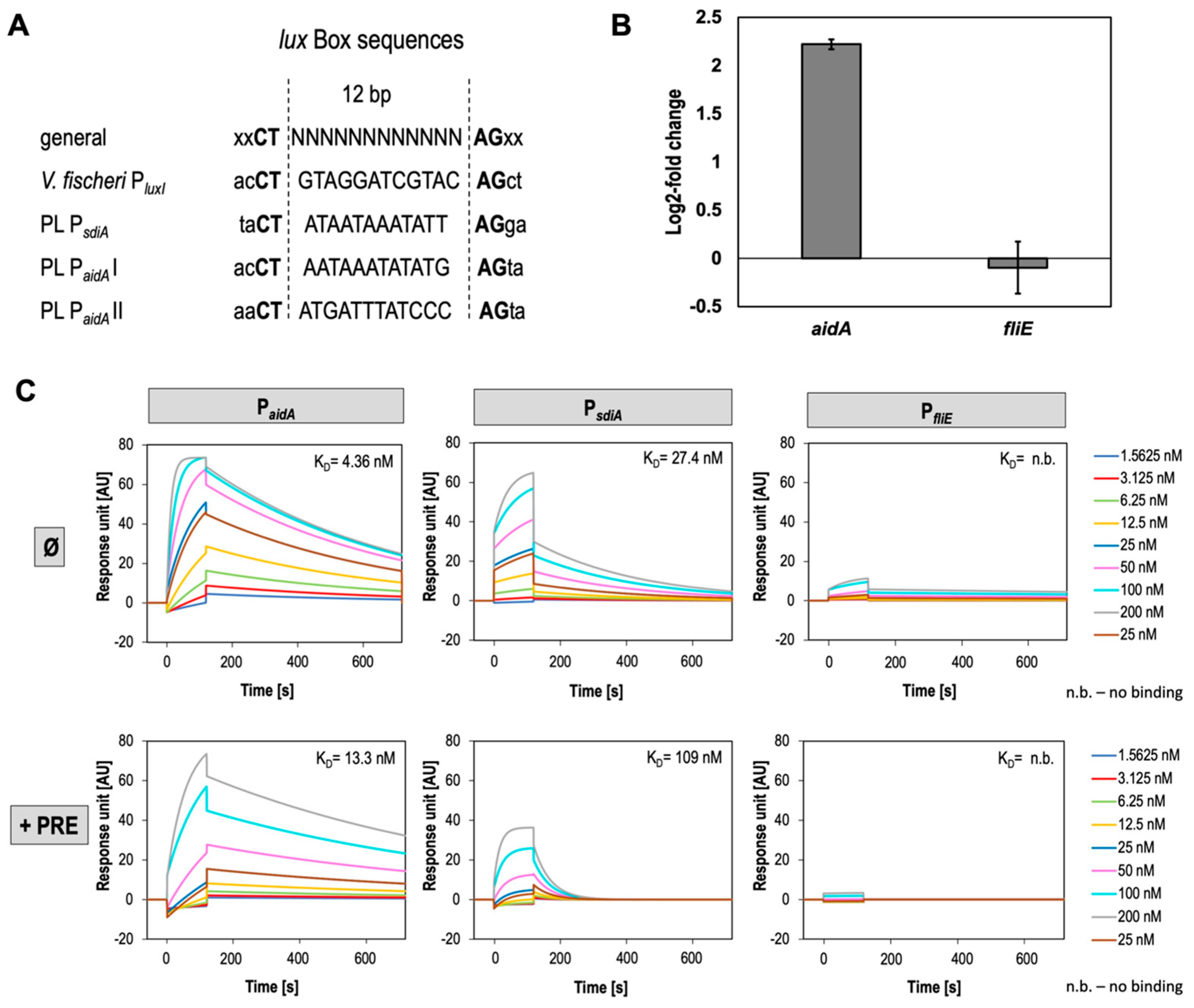
3.4. SdiA Binds PsdiA and PaidA with High Affinity and Negatively Affects Expression of AidA

It is well-known that transcriptional LuxR-regulators undergo conformational changes upon signal binding enabling the C-terminal HTH domain to bind target gene promoter at the lux-box conserved sites to control gene transcription [79,80]. In a prototypical QS circuit, LuxR-type regulators induce transcription of the cognate luxI, which is also designated as autoregulation [5]. However, since in P. luminescens SdiA is a LuxR solo as the bacteria do not harbor any luxI gene, we hypothesized that SdiA might regulate activity of its own promoter region. To validate this idea and the hypothesis that SdiA regulation is signal independent, we first studied the binding affinity of SdiA to its respective promoter (PsdiA) via surface plasmon resonance (SPR) spectroscopy without any putative ligand. Furthermore, we also analyzed the binding of SdiA towards the promoter of neighboring gene aidA (PaidA) since this gene is located upstream of sdiA and encodes in the opposite orientation. Both genes share a 310 bp long promoter containing intergenic region harboring putative lux-box-like motifs with a 12 bp long variable motif between with a 5′CT and a 3′AG (Figure 3A) [48,81], putatively necessary for SdiA-binding. As negative control we used PfliE not containing any lux-box-like motif in the respective promoter region (Figure 4C). Remarkably, SdiA showed a high binding affinity towards its own promoter PsdiA due to a high association rate (ka = 1.1 × 10⁵/M*s) and a dissociation rate of kd = 3.1 × 10⁻³/s resulting in an overall affinity of KD = 27.4 nM. This reveals a fast self-regulatory property of SdiA with rapid association and fast dissociation. However, SdiA bound with a ~3.5× higher association rate to the promoter of aidA (PaidA), caused by an extreme low dissociation rate (kd = 1.7 × 10³/s) with comparable association rate to PsdiA (ka = 3.9 × 10⁵/M*s), resulting in an overall affinity of 4.4 nM (Figure 4C). This demonstrated a very strong interaction of SdiA with PaidA, proving our hypothesis that SdiA regulates expression of aidA similar to as it was described for the LuxR-type receptor SolR of R. solanacearum. SolR was found to positively regulate expression of aidA genes and is required for virulence towards plants [47]. Additionally, for the SdiA-PaidA interaction we can observe a 1:2 binding stoichiometry indicated by a double Rmax value, which is in accordance with the occurrence of two putative LuxR-binding boxes in the respective promoter region, designated as PaidAI and PaidAII (Figure 4A). In contrast, for PsdiA, only a 1:1 binding stoichiometry could be observed by the SPR analysis indicating only putative one SdiA-binding site. Interestingly, these data suggest that the 310 bp long intergenic region between aidA and sdiA acts a bidirectional promoter. Thus, SdiA is capable of bidirectional stimulation of transcription of these two oppositely oriented genes. A similar binding mechanism is also described for LuxR of Vibrio fischeri, similarly stimulating lux operons located in the opposite direction [82].
To elucidate whether DNA-binding ability of SdiA is reduced in the presence of putative signal molecules, the binding properties of SdiA towards PsdiA and PaidAwere investigated using DNA-binding kinetics also in the presence of PRE. Indeed, the binding affinity of SdiA towards both promoters was reduced. While association towards PsdiA remained more or less similar, the dissociation from the DNA was observed to be much faster in the presence of PRE (ka = 1.8 × 10⁵/M*s, kd = 2.0 × 10⁻²/s) resulting in an overall affinity of 109 nM (Figure 4c bottom left panel). Similarly, a faster dissociation of SdiA from PaidA occurred in the presence of PRE (kd = 1.1 × 10⁻³/s), whereas here also the association rate was affected (ka = 8.2 × 10⁴/M*s), resulting in a lower overall affinity of 13.3 nM, (Figure 4c, bottom middle panel). Solvent-derived effects of PRE on binding kinetics can be excluded, as affinity of SdiA towards the respective promoters was not affected in the presence of the solvent methanol (N.D. and R.H. unpublished). SdiA did not significantly bound to PfliE, indicating that the interactions observed for PsdiA and PaidA were specific (Figure 4C, bottom right panel). Furthermore, we used real-time qPCR to confirm that SdiA-binding to PaidA also affects the expression of the corresponding gene. The housekeeping gene rpoD was used as control. Indeed, aidA expression showed to be negatively regulated by SdiA as we could detect an increased expression of the gene with a log2-fold change in 2.22 in the sdiA deletion mutant of P. luminescens (Figure 4B).
In summary, these data further support the hypothesis that a plant-derived signaling molecule binds to SdiA and subsequently influences DNA-binding of the receptor. Therefore, SdiA acts as LuxR-type receptor that is involved in interkingdom communication (IKS) of P. luminescens with plants. However, the molecular mechanism behind the chemical nature of the signaling molecule is unknown. Considering the role of AidA in R. solanacearum and the regulation of aidA expression mediated by SolR, our data further propose a putative role of AidA in P. luminescens in bacterial–plant interaction as aidA expression is under control of SdiA and the gene is clustered with sdiA, similar as found in R. solanacearum (Figure 1B), and as SdiA DNA-binding affinity is affected in the presence of PRE. In P. luminescens, it can be supposed that SdiA negatively affects expression of aidA in the absence of a signal molecule, and once SdiA senses a plant-derived signal, the affinity towards the promoter of aidA is reduced, leading to enhanced expression of the gene putatively important for plant host colonization. The exact molecular mechanism behind this regulation as well as the function of AidA in Photorhabdus–plant interaction remains unclear. Hence, further insights into the effect of the putative mentioned signal molecules on SdiA DNA-binding property should be gained, to understand the regulatory hierarchy of SdiA upon plant signals in P. luminescens.

4. Conclusions

Recent research showed potential biocontrol ability of P. luminescens 2° cells since the bacteria can colonize plant roots, protecting them from phytopathogenic fungi infection. As this cell variant does not re-associate with nematodes and interacts with plants, it was important to understand and to determine how the bacteria sense the rhizosphere environment. We can demonstrate that the LuxR solo SdiA harboring an AHL-like SBD is involved in IKS communication between P. luminescens and plants, potentially involving AidA in plant host colonization. First, in this work we demonstrated a regulatory crosstalk between SdiA motility as well as biofilm formation regulating a putative switch between a sessile and motile lifestyle of the bacteria (Figure 5). Nevertheless, which other genes beside aidA and sdiA are under control of SdiA is unknown and must be determined. From our findings we conclude that SdiA regulates expression of aidA and sdiA in a signal-independent manner (Figure 5). Here, we demonstrated that SdiA protein conformation is affected by short-chain AHLs and PRE and respective fractions indicating (i) plant-derived molecule(s) as signal for SdiA and (ii) an influence of signal binding on the regulatory role of SdiA. Although the nature of the plant-derived compound(s) must be determined, the data suggest a SdiA-mediated interkingdom communication of P. luminescens with plants as DNA binding was also influenced upon PRE. We also identified that SdiA acts as a bidirectional regulator binding in the intergenic region of sdiA and aidA since the receptor binds both promoters with high affinity, most probably negatively affecting expression of aidA. Lastly, for AidA of P. luminescens, of which homologs are also found in plant-associated bacteria and are regulated by a LuxR, we suggest an involvement in P. luminescens plant colonization whereby SdiA controls the expression of aidA depending on IKS with the plant host (Figure 5).

Author Contributions

Conceptualization, R.H. and A.R.; methodology, N.D, A.R. and L.W.; software, N.D.; validation, N.D., A.R.; formal analysis, A.R.; investigation, N.D.; resources, R.H.; data curation, N.D.; writing—original draft preparation, N.D.; writing—review and editing, R.H.; visualization, N.D.; supervision, R.H and A.R.; project administration, R.H.; funding acquisition, R.H. All authors have read and agreed to the published version of the manuscript.

Funding

This research was funded by DEUTSCHE FORSCHUNGSGEMEINSCHAFT (DFG), grant number “INST 247/992-1 FUGG”.

Institutional Review Board Statement

Not applicable.

Informed Consent Statement

Not applicable.

Data Availability Statement

Data is contained within the article or available on request from the corresponding author.

Acknowledgments

We thank Meike Saul-Reiss and Luis Antelo (IBWF Mainz) for the (semi-preparative) HPLC analyses and Kirsten Schaubruch (JGU Mainz) for SPR analyses and excellent technical assistance. SPR and nanoDSF analyses were performed in the Bioanalytics core facility of the JGU Biocenter.

Conflicts of Interest

The authors declare no conflict of interest.

References

  1. Waters, C.M.; Bassler, B.L. Quorum sensing: Cell-to-cell communication in bacteria. Annu. Rev. Cell Dev. Biol. 2005, 21, 319–346. [Google Scholar] [CrossRef] [PubMed] [Green Version]
  2. Choi, S.H.; Greenberg, E.P. The C-terminal region of the Vibrio fischeri LuxR protein contains an inducer-independent lux gene activating domain. PNAS 1991, 88, 11115–11119. [Google Scholar] [CrossRef] [PubMed] [Green Version]
  3. Hanzelka, B.L.; Greenberg, E.P. Evidence That the N-terminal region of the Vibrio fischeri LuxR protein constitutes an autoinducer-binding domain. J. Bacteriol. 1995, 177, 815–817. [Google Scholar] [CrossRef] [Green Version]
  4. Marchler-Bauer, A.; Zheng, C.; Chitsaz, F.; Derbyshire, M.K.; Geer, L.Y.; Geer, R.C.; Gonzales, N.R.; Gwadz, M.; Hurwitz, D.I.; Lanczycki, C.J.; et al. CDD: Conserved domains and protein three-dimensional structure. Nucleic. Acids Res. 2012, 41, D348–D352. [Google Scholar] [CrossRef] [PubMed] [Green Version]
  5. Fuqua, C.; Winans, S.C.; Greenberg, E.P. Census and consensus in bacterial ecosystems: The LuxR-LuxI family of quorum-sensing transcriptional regulators. Annu. Rev. Microbiol. 1996, 50, 727–751. [Google Scholar] [CrossRef]
  6. Fuqua, W.C.; Winans, S.C.; Greenberg, E.P. Quorum sensing in bacteria: The LuxR-LuxI family of cell density-responsive transcriptional regulators. J. Bacteriol. 1994, 176, 269–275. [Google Scholar] [CrossRef] [Green Version]
  7. Fuqua, C.; Parsek, M.R.; Greenberg, E.P. Regulation of gene expression by cell-to-cell communication: Acyl-homoserine lactone quorum sensing. Annu. Rev. Genet 2001, 35, 439–468. [Google Scholar] [CrossRef]
  8. Case, R.J.; Labbate, M.; Kjelleberg, S. AHL-driven quorum-sensing circuits: Their frequency and function among the proteobacteria. ISME J. 2008, 2, 345–349. [Google Scholar] [CrossRef]
  9. Patankar, A.V.; González, J.E. Orphan LuxR regulators of quorum sensing. FEMS Microbiol. Rev. 2009, 33, 739–756. [Google Scholar] [CrossRef] [Green Version]
  10. Subramoni, S.; Venturi, V. LuxR-family ‘solos’: Bachelor sensors/regulators of signalling molecules. Microbiol 2009, 155, 1377–1385. [Google Scholar] [CrossRef] [Green Version]
  11. Bez, C.; Geller, A.M.; Levy, A.; Venturi, V. Cell-cell signaling proteobacterial LuxR solos: A treasure trove of subgroups having different origins, ligands, and ecological roles. mSystems 2023, e01039-22. [Google Scholar] [CrossRef] [PubMed]
  12. Michael, B.; Smith, J.N.; Swift, S.; Heffron, F.; Ahmer, B.M.M. SdiA of Salmonella enterica is a LuxR homolog that detects mixed microbial communities. J. Bacteriol. 2001, 183, 5733–5742. [Google Scholar] [CrossRef] [PubMed] [Green Version]
  13. de Almeida, F.A.; Pinto, U.M.; Vanetti, M.C.D. Novel insights from molecular docking of SdiA from Salmonella enteritidis and Escherichia coli with quorum sensing and quorum quenching molecules. Microb. Pathog. 2016, 99, 178–190. [Google Scholar] [CrossRef]
  14. Machado, R.A.R.; Wüthrich, D.; Kuhnert, P.; Arce, C.C.M.; Thönen, L.; Ruiz, C.; Zhang, X.; Robert, C.A.M.; Karimi, J.; Kamali, S.; et al. Whole-genome-based revisit of Photorhabdus phylogeny: Proposal for the elevation of most Photorhabdus subspecies to the species level and description of one novel species Photorhabdus bodei sp. nov. and one novel subspecies Photorhabdus laumondii subsp. clarkei subsp. Nov. Int. J. Syst. Evol. Microbiol. 2018, 68, 2664–2681. [Google Scholar] [CrossRef]
  15. Heermann, R.; Fuchs, T.M. Comparative Analysis of the Photorhabdus luminescens and the Yersinia enterocolitica genomes: Uncovering candidate genes involved in insect pathogenicity. BMC Genom. 2008, 9, 40. [Google Scholar] [CrossRef] [Green Version]
  16. Brameyer, S.; Kresovic, D.; Bode, H.B.; Heermann, R. LuxR solos in Photorhabdus species. Front Cell Infect. Microbiol. 2014, 4, 166. [Google Scholar] [CrossRef] [Green Version]
  17. Brachmann, A.O.; Brameyer, S.; Kresovic, D.; Hitkova, I.; Kopp, Y.; Manske, C.; Schubert, K.; Bode, H.B.; Heermann, R. Pyrones as bacterial signaling molecules. Nat. Chem. Biol. 2013, 9, 573–578. [Google Scholar] [CrossRef] [PubMed]
  18. Bais, H.P.; Weir, T.L.; Perry, L.G.; Gilroy, S.; Vivanco, J.M. The role of root exudates in rhizosphere interactions with plants and other organisms. Annu. Rev. Plant Biol. 2006, 57, 233–266. [Google Scholar] [CrossRef] [Green Version]
  19. Bez, C.; Covaceuszach, S.; Bertani, I.; Choudhary, K.S.; Venturi, V. LuxR Solos from environmental fluorescent Pseudomonads. mSphere 2021, 6, e01322-20. [Google Scholar] [CrossRef]
  20. Dominelli, N.; Jäger, H.Y.; Langer, A.; Brachmann, A.; Heermann, R. High-throughput sequencing analysis reveals genomic similarity in phenotypic heterogeneous Photorhabdus luminescens cell populations. Ann. Microbiol. 2022, 72, 20. [Google Scholar] [CrossRef]
  21. Eckstein, S.; Dominelli, N.; Brachmann, A.; Heermann, R. Phenotypic heterogeneity of the insect pathogen Photorhabdus luminescens: Insights into the fate of secondary cells. Appl. Environ. Microbiol. 2019, 85, e01910-19. [Google Scholar] [CrossRef]
  22. Regaiolo, A.; Dominelli, N.; Andresen, K.; Heermann, R. The biocontrol agent and insect pathogen Photorhabdus luminescens interacts with plant roots. Appl. Environ. Microbiol 2020, 86, e00891-20. [Google Scholar] [CrossRef] [PubMed]
  23. Dominelli, N.; Platz, F.; Heermann, R. The insect pathogen Photorhabdus luminescens protects plants from phytopathogenic Fusarium graminearum via chitin degradation. Appl. Environ. Microbiol. 2022, 88, e00645-22. [Google Scholar] [CrossRef] [PubMed]
  24. Madeira, F.; Park, Y.M.; Lee, J.; Buso, N.; Gur, T.; Madhusoodanan, N.; Basutkar, P.; Tivey, A.R.N.; Potter, S.C.; Finn, R.D.; et al. The EMBL-EBI search and sequence analysis tools APIs in 2019. Nucleic. Acids. Res. 2019, 47, W636–W641. [Google Scholar] [CrossRef] [PubMed] [Green Version]
  25. Guex, N.; Peitsch, M.C.; Schwede, T. Automated comparative protein structure modeling with SWISS-MODEL and Swiss-PdbViewer: A historical perspective. Electrophoresis 2009, 30, S162–S173. [Google Scholar] [CrossRef]
  26. Bertoni, M.; Kiefer, F.; Biasini, M.; Bordoli, L.; Schwede, T. modeling protein quaternary structure of homo- and hetero-oligomers beyond binary interactions by homology. Sci. Rep. 2017, 7, 10480. [Google Scholar] [CrossRef] [Green Version]
  27. Bienert, S.; Waterhouse, A.; de Beer, T.A.P.; Tauriello, G.; Studer, G.; Bordoli, L.; Schwede, T. The SWISS-MODEL repository—New features and functionality. Nucleic. Acids. Res. 2017, 45, D313–D319. [Google Scholar] [CrossRef] [Green Version]
  28. Waterhouse, A.; Bertoni, M.; Bienert, S.; Studer, G.; Tauriello, G.; Gumienny, R.; Heer, F.T.; de Beer, T.A.P.; Rempfer, C.; Bordoli, L.; et al. SWISS-MODEL: Homology modelling of protein structures and complexes. Nucleic. Acids. Res. 2018, 46, W296–W303. [Google Scholar] [CrossRef] [Green Version]
  29. Studer, G.; Rempfer, C.; Waterhouse, A.M.; Gumienny, R.; Haas, J.; Schwede, T. QMEANDisCo—Distance constraints applied on model quality estimation. Bioinf. 2020, 36, 1765–1771. [Google Scholar] [CrossRef]
  30. Zamora-Lagos, M.-A.; Eckstein, S.; Langer, A.; Gazanis, A.; Pfeiffer, F.; Habermann, B.; Heermann, R. Phenotypic and genomic comparison of Photorhabdus luminescens subsp. laumondii TT01 and a widely used rifampicin-resistant Photorhabdus luminescens laboratory strain. BMC Genom. 2018, 19, 854. [Google Scholar] [CrossRef] [Green Version]
  31. Lassak, J.; Henche, A.-L.; Binnenkade, L.; Thormann, K.M. ArcS, the cognate sensor kinase in an atypical Arc system of Shewanella oneidensis MR-1. Appl. Environ. Microbiol. 2010, 76, 3263–3274. [Google Scholar] [CrossRef] [PubMed] [Green Version]
  32. Thoma, S.; Schobert, M. An improved Escherichia coli donor strain for diparental mating. FEMS Microbiol. Lett. 2009, 294, 127–132. [Google Scholar] [CrossRef] [PubMed] [Green Version]
  33. Guzman, L.M.; Belin, D.; Carson, M.J.; Beckwith, J. Tight regulation, modulation, and high-level expression by vectors containing the arabinose PBAD promoter. J. Bacteriol. 1995, 177, 4121–4130. [Google Scholar] [CrossRef] [PubMed] [Green Version]
  34. Christensen, G.D.; Simpson, W.A.; Younger, J.J.; Baddour, L.M.; Barrett, F.F.; Melton, D.M.; Beachey, E.H. Adherence of coagulase-negative Staphylococci to plastic tissue culture plates: A quantitative model for the adherence of Staphylococci to medical devices. J. Clin. Microbiol. 1985, 22, 996–1006. [Google Scholar] [CrossRef] [PubMed] [Green Version]
  35. O’Toole, G.A.; Kolter, R. Initiation of biofilm formation in Pseudomonas fluorescens WCS365 proceeds via multiple, convergent signalling pathways: A genetic analysis. Mol. Microbiol. 1998, 28, 449–461. [Google Scholar] [CrossRef]
  36. O’Toole, G.A. Microtiter dish biofilm formation assay. JoVE 2011, 47, 2437. [Google Scholar] [CrossRef]
  37. Laemmli, U.K. Cleavage of structural proteins during the assembly of the head of bacteriophage T4. Nature 1970, 227, 680–685. [Google Scholar] [CrossRef]
  38. Niesen, F.H.; Berglund, H.; Vedadi, M. The use of differential scanning fluorimetry to detect ligand interactions that promote protein stability. Nat. Protoc. 2007, 2, 2212–2221. [Google Scholar] [CrossRef]
  39. Alexander, C.G.; Wanner, R.; Johnson, C.M.; Breitsprecher, D.; Winter, G.; Duhr, S.; Baaske, P.; Ferguson, N. Novel microscale approaches for easy, rapid determination of protein stability in academic and commercial settings. Biochem. Biophys. Acta Proteins. Proteom. 2014, 1844, 2241–2250. [Google Scholar] [CrossRef] [Green Version]
  40. Pfaffl, M.W. A new mathematical model for relative quantification in real-time RT-PCR. Nucleic. Acids Res. 2001, 29, e45. [Google Scholar] [CrossRef]
  41. Simon, P. Q-Gene: Processing quantitative real-time RT–PCR data. Bioinformatics 2003, 19, 1439–1440. [Google Scholar] [CrossRef] [Green Version]
  42. Ahmer, B.M.M. Cell-to-cell signalling in Escherichia coli and Salmonella enterica: Quorum sensing in E. coli and Salmonella. Mol. Microbiol. 2004, 52, 933–945. [Google Scholar] [CrossRef] [PubMed]
  43. Covaceuszach, S.; Degrassi, G.; Venturi, V.; Lamba, D. Structural insights into a novel interkingdom signaling circuit by cartography of the ligand-binding sites of the homologous quorum sensing LuxR-family. IJMS 2013, 14, 20578–20596. [Google Scholar] [CrossRef] [PubMed] [Green Version]
  44. González, J.F.; Venturi, V. A novel widespread interkingdom signaling circuit. Trends Plant Sci. 2013, 18, 167–174. [Google Scholar] [CrossRef] [PubMed]
  45. Coutinho, B.G.; Mevers, E.; Schaefer, A.L.; Pelletier, D.A.; Harwood, C.S.; Clardy, J.; Greenberg, E.P. A Plant-responsive bacterial-signaling system senses an ethanolamine derivative. Proc. Natl. Acad. Sci. USA 2018, 115, 9785–9790. [Google Scholar] [CrossRef] [Green Version]
  46. Flavier, A.B.; Ganova-Raeva, L.M.; Schell, M.A.; Denny, T.P. Hierarchical autoinduction in Ralstonia solanacearum: Control of acyl-homoserine lactone production by a novel autoregulatory system responsive to 3-hydroxypalmitic acid methyl ester. J. Bacteriol. 1997, 179, 7089–7097. [Google Scholar] [CrossRef] [Green Version]
  47. Meng, F.; Babujee, L.; Jacobs, J.M.; Allen, C. Comparative transcriptome analysis reveals cool virulence factors of Ralstonia solanacearum race 3 biovar 2. PLoS ONE 2015, 10, e0139090. [Google Scholar] [CrossRef] [PubMed] [Green Version]
  48. Antunes, L.C.M.; Ferreira, R.B.R.; Lostroh, C.P.; Greenberg, E.P. A mutational analysis defines Vibrio fischeri LuxR binding sites. J. Bacteriol. 2008, 190, 4392–4397. [Google Scholar] [CrossRef] [Green Version]
  49. Crooks, G.E.; Hon, G.; Chandonia, J.-M.; Brenner, S.E. WebLogo: A sequence logo generator. Genome Res. 2004, 14, 1188–1190. [Google Scholar] [CrossRef] [Green Version]
  50. Mattick, J.S. Type IV pili and twitching motility. Annu. Rev. Microbiol. 2002, 56, 289–314. [Google Scholar] [CrossRef]
  51. Hall-Stoodley, L.; Costerton, J.W.; Stoodley, P. Bacterial biofilms: From the natural environment to infectious diseases. Nat. Rev. Microbiol. 2004, 2, 95–108. [Google Scholar] [CrossRef] [PubMed]
  52. Tans-Kersten, J.; Huang, H.; Allen, C. Ralstonia solanacearum needs motility for invasive virulence on tomato. J. Bacteriol. 2001, 183, 3597–3605. [Google Scholar] [CrossRef] [PubMed] [Green Version]
  53. Corral, J.; Sebastià, P.; Coll, N.S.; Barbé, J.; Aranda, J.; Valls, M. Twitching and swimming motility play a role in Ralstonia solanacearum pathogenicity. mSphere 2020, 5. [Google Scholar] [CrossRef] [Green Version]
  54. Anetzberger, C.; Pirch, T.; Jung, K. Heterogeneity in quorum sensing-regulated bioluminescence of Vibrio harveyi. Mol. Microbiol. 2009, 73, 267–277. [Google Scholar] [CrossRef]
  55. Yang, Q.; Defoirdt, T. Quorum sensing positively regulates flagellar motility in pathogenic Vibrio harveyi: QS regulation of motility in Vibrio harveyi. Environ. Microbiol. 2015, 17, 960–968. [Google Scholar] [CrossRef] [PubMed]
  56. Ahmer, B.M.M.; van Reeuwijk, J.; Timmers, C.D.; Valentine, P.J.; Heffron, F. Salmonella typhimurium encodes an SdiA homolog, a putative quorum sensor of the LuxR family, that regulates genes on the virulence plasmid. J. Bacteriol. 1998, 180, 1185–1193. [Google Scholar] [CrossRef] [Green Version]
  57. Kanamaru, K.; Kanamaru, K.; Tatsuno, I.; Tobe, T.; Sasakawa, C. SdiA, an Escherichia coli homologue of quorum-sensing regulators, controls the expression of virulence factors in enterohaemorrhagic Escherichia coli O157:H7. Mol. Microbiol. 2000, 38, 805–816. [Google Scholar] [CrossRef] [PubMed]
  58. Antunes, L.C.M.; Ferreira, R.B.R.; Buckner, M.M.C.; Finlay, B.B. Quorum sensing in bacterial virulence. Microbiol 2010, 156, 2271–2282. [Google Scholar] [CrossRef] [Green Version]
  59. Sharma, V.K.; Bearson, S.M.D.; Bearson, B.L. Evaluation of the effects of SdiA, a LuxR Homologue, on adherence and motility of Escherichia coli O157: H7. Microbiol 2010, 156, 1303–1312. [Google Scholar] [CrossRef] [Green Version]
  60. Tavio, M.M.; Aquili, V.D.; Poveda, J.B.; Antunes, N.T.; Sanchez-Cespedes, J.; Vila, J. Quorum-sensing regulator sdiA and marA overexpression is involved in in vitro-selected multidrug resistance of Escherichia coli. J. Antimicrob. Chemother. 2010, 65, 1178–1186. [Google Scholar] [CrossRef]
  61. Culler, H.; Couto, S.; Higa, J.; Ruiz, R.; Yang, M.; Bueris, V.; Franzolin, M.; Sircili, M. Role of SdiA on biofilm formation by atypical enteropathogenic Escherichia coli. Genes 2018, 9, 253. [Google Scholar] [CrossRef] [PubMed] [Green Version]
  62. Lindsay, A.; Ahmer, B.M.M. Effect of SdiA on biosensors of N-acylhomoserine lactones. J. Bacteriol. 2005, 187, 5054–5058. [Google Scholar] [CrossRef] [Green Version]
  63. Dyszel, J.L.; Soares, J.A.; Swearingen, M.C.; Lindsay, A.; Smith, J.N.; Ahmer, B.M.M. E. coli K-12 and EHEC genes regulated by SdiA. PLoS ONE 2010, 5, e8946. [Google Scholar] [CrossRef] [PubMed] [Green Version]
  64. Hughes, D.T.; Terekhova, D.A.; Liou, L.; Hovde, C.J.; Sahl, J.W.; Patankar, A.V.; Gonzalez, J.E.; Edrington, T.S.; Rasko, D.A.; Sperandio, V. Chemical sensing in mammalian host-bacterial commensal associations. PNAS 2010, 107, 9831–9836. [Google Scholar] [CrossRef] [Green Version]
  65. Shimada, T.; Shimada, K.; Matsui, M.; Kitai, Y.; Igarashi, J.; Suga, H.; Ishihama, A. Roles of cell division control factor SdiA: Recognition of quorum sensing signals and modulation of transcription regulation targets. Genes Cells 2014, 19, 405–418. [Google Scholar] [CrossRef]
  66. Nguyen, Y.; Nguyen, N.X.; Rogers, J.L.; Liao, J.; MacMillan, J.B.; Jiang, Y.; Sperandio, V. Structural and mechanistic roles of novel chemical ligands on the SdiA quorum-sensing transcription regulator. mBio 2015, 6, e02429-14. [Google Scholar] [CrossRef] [Green Version]
  67. Wang, T.; Guan, W.; Huang, Q.; Yang, Y.; Yan, W.; Sun, B.; Zhao, T. Quorum-sensing contributes to virulence, twitching motility, seed attachment and biofilm formation in the wild type strain Aac-5 of Acidovorax citrulli. Microb. Pathogen. 2016, 100, 133–140. [Google Scholar] [CrossRef] [Green Version]
  68. Pacheco, T.; Gomes, A.É.I.; Siqueira, N.M.G.; Assoni, L.; Darrieux, M.; Venter, H.; Ferraz, L.F.C. SdiA, a quorum-sensing regulator, suppresses fimbriae expression, biofilm formation, and quorum-sensing signaling molecules production in Klebsiella pneumoniae. Front Microbiol. 2021, 12, 597735. [Google Scholar] [CrossRef] [PubMed]
  69. Tsai, C.-S.; Winans, S.C. LuxR-type quorum-sensing regulators that are detached from common scents: LuxR-type apoproteins. Mol. Microbiol. 2010, 77, 1072–1082. [Google Scholar] [CrossRef] [PubMed] [Green Version]
  70. Lee, J.; Jayaraman, A.; Wood, T.K. Indole is an inter-species biofilm signal mediated by SdiA. BMC Microbiol. 2007, 7, 42. [Google Scholar] [CrossRef] [Green Version]
  71. Lee, J.; Maeda, T.; Hong, S.H.; Wood, T.K. Reconfiguring the quorum-sensing regulator SdiA of Escherichia coli to control biofilm formation via indole and N-acylhomoserine lactones. Appl. Environ. Microbiol. 2009, 75, 1703–1716. [Google Scholar] [CrossRef] [PubMed] [Green Version]
  72. Whitehead, N.A.; Barnard, A.M.L.; Slater, H.; Simpson, N.J.L.; Salmond, G.P.C. Quorum-sensing in Gram-negative bacteria. FEMS Microbiol. Rev. 2001, 25, 365–404. [Google Scholar] [CrossRef] [PubMed]
  73. Yao, Y.; Martinez-Yamout, M.A.; Dickerson, T.J.; Brogan, A.P.; Wright, P.E.; Dyson, H.J. Structure of the Escherichia coli quorum sensing protein SdiA: Activation of the folding switch by acyl homoserine lactones. J. Mol. Biol. 2006, 355, 262–273. [Google Scholar] [CrossRef] [PubMed]
  74. Teplitski, M.; Robinson, J.B.; Bauer, W.D. Plants secrete substances that mimic bacterial N-acyl homoserine lactone signal activities and affect population density-dependent behaviors in associated bacteria. Mol. Plant. Microbe Interact. 2000, 13, 637–648. [Google Scholar] [CrossRef] [Green Version]
  75. Castang, S.; Reverchon, S.; Gouet, P.; Nasser, W. Direct evidence for the modulation of the activity of the Erwinia chrysanthemi quorum-sensing regulator ExpR by acylhomoserine lactone pheromone. J. Biol. Chem. 2006, 281, 29972–29987. [Google Scholar] [CrossRef] [Green Version]
  76. Cui, Y.; Chatterjee, A.; Hasegawa, H.; Dixit, V.; Leigh, N.; Chatterjee, A.K. ExpR, a LuxR homolog of Erwinia carotovora subsp. carotovora, activates transcription of rsmA, which specifies a global regulatory RNA-binding protein. J. Bacteriol. 2005, 187, 4792–4803. [Google Scholar] [CrossRef] [Green Version]
  77. Minogue, T.D.; Trebra, M.W.; Bernhard, F.; von Bodman, S.B. The autoregulatory role of EsaR, a quorum-sensing regulator in Pantoea stewartii ssp. stewartii: Evidence for a repressor function: Autoregulatory role of EsaR. Mol. Microbiol. 2002, 44, 1625–1635. [Google Scholar] [CrossRef] [Green Version]
  78. Minogue, T.D.; Carlier, A.L.; Koutsoudis, M.D.; von Bodman, S.B. The cell density-dependent expression of stewartan exopolysaccharide in Pantoea stewartii ssp. stewartii is a function of EsaR-mediated repression of the rcsA gene: Quorum sensing control of stewartan EPS. Mol. Microbiol. 2005, 56, 189–203. [Google Scholar] [CrossRef]
  79. Devine, J.H.; Shadel, G.S.; Baldwin, T.O. Identification of the operator of the lux regulon from the Vibrio fischeri strain ATCC7744. PNAS 1989, 86, 5688–5692. [Google Scholar] [CrossRef] [Green Version]
  80. Stevens, A.M.; Greenberg, E.P. Quorum sensing in Vibrio fischeri: Essential elements for activation of the luminescence genes. J. Bacteriol. 1997, 179, 557–562. [Google Scholar] [CrossRef] [Green Version]
  81. Egland, K.A.; Greenberg, E.P. Quorum sensing in Vibrio fischeri: Elements of the luxI promoter. Mol. Microbiol. 1999, 31, 1197–1204. [Google Scholar] [CrossRef] [PubMed]
  82. Shadel, G.S.; Baldwin, T.O. The Vibrio fischeri LuxR protein is capable of bidirectional stimulation of transcription and both positive and negative regulation of the luxR gene. J. Bacteriol. 1991, 173, 568–574. [Google Scholar] [CrossRef] [PubMed] [Green Version]
Figure 1. Protein sequence analysis of AHL-LuxR solo SdiA (PluDJC01675) of P. luminescens. (A) Left panel: sequence logo motif of the six conserved amino acids (AAs) found in AHL-LuxR receptors made with WebLogo3 [49]. Right panel: SdiA tertiary structure predicted with SWISS-MODEL [25,26,27,28,29], pointing out the signal-binding pocket of SdiA. The numbers 1, 2, 4, 5, 8, and 9 below the logo indicate the position of the six conserved amino acids (WYDPWG) in the SdiA model essential for AHL binding. (B) Structure-based multiple sequence alignment of the protein sequences of the signal-binding domains (SBDs) of AHL-LuxRs TraR (B9K461) from Agrobacterium tumefaciens (ATs), CviR (Q7NQP7) from Chromobacterium violacaeum (CV), SdiA (Q7N9K5) from P. luminescens (PL), LasR (P25084) from Pseudomonas aeruginosa (PA), SdiA (P07026) from E. coli (EC), SdiA (A0A0H3GS53) from Klebsiella pneumoniae (KP), QscR (G3XD77) from P. aeruginosa (PA), and SolR (P58590) from Ralstonia solanacearum (RS). The six conserved AAs are highlighted in cyan. In orange are the variable AAs essential for AHL binding of the proteins. The numbers 1 to 10 indicate all AAs essential for AHL binding of P. luminescens SdiA in the SWISS-MODEL displayed above. The asterisk indicates fully conserved amino acid residues at the respective position within the alignment. The colon indicates conserved amino acid substitutions with strongly similar prop-erties and a score > 0.5. The period indicates semiconserved amino acid substitutions with weakly similar properties and a score ≤ 0.5. (C) Genetic loci of luxR-aidA cluster found in insect pathogenic and plant-associated P. luminescens and plant pathogenic R. solanacearum.
Microorganisms 11 00890 g001
Figure 2. Influence of SdiA on motility, twitching, and biofilm formation of P. luminescens 1° and 2° cells. (A) Twitching motility: at the top crystal violet staining of twitched cells adhering on the surface. The pictures represent one characteristic of at least three independently performed experiments with similar outcomes. (B) Swimming motility of ΔsdiA compared with the wild-type (WT). (C) Biofilm formation: crystal violet-stained biofilm quantified at 575 nm after 24 h of 1° and 2° cells wild-type (WT) and the respective ΔsdiA mutant. Below crystal violet staining. (D) Biofilm formation in presence of 3.3% (v/v) PRE after 72 h of incubation. Below crystal violet staining. The error bars represent the standard deviation of at least three biological replicates. *, **, *** all indicate p ≤ 0.05 of comparison between the different groups.
Microorganisms 11 00890 g002
Figure 3. HPLC chromatogram of plant root exudates, SdiA purification, and thermostability of SdiA in the presence of AHLs and PRE. (A) Chromatogram of semipreparative HPLC of plant root exudates separated in 48 fractions. The green dashed lines represent the fractions showing where putative signal molecules for SdiA are located. Light green: fraction B1; green: fraction C3; and dark green: fraction D7. (B) On the left panel SDS-PAGE representing purification of 6xHis-SdiA, which was confirmed via Western blot analysis (right panel). (C) NanoDSF analyses of SdiA with supplementation of lipophilic plant root exudates (L-PRE, left panel), the selected respective HPLC fractions (B1, C3, D7, middle panel) and 10 nM of C4- or lll C12-AHLs (right panel). The graphs represent the 1st derivative of the measured ratio of intrinsic tryptophane fluorescence at 350 and 330 nm of the protein. The maximum peak represents the melting temperature TM, where half of the protein molecules are denatured. TM values are indicated by the dotted lines colored respectively to the curve. In all measurements the black line shows the control protein measured with solvent, when necessary. Left shift of the graphs indicate different protein conformation upon molecule binding. In the left panel: PRE influence SdiA stability upon putative signal molecule binding with DTM −2.4°C. Middle panel: putative signaling molecule found in PRE-fractions indicated by DTM −3.8 °C (B1, light green), DTM −5.8 °C (C3, green), and DTM −3.4 °C (D7 dark green). Right panel: AHLs putatively bind to SdiA with different modes of action indicated by DTM −3.9 °C (C4-AHL, light blue) and DTM +1.6 °C (C12-AHL, blue), showing increased stability of the protein.
Microorganisms 11 00890 g003
Figure 4. DNA-binding of SdiA to PsdiA, PaidA, and PfliE. (A) Putative lux-like recognition site in the intergenic region between sdiA and aidA of P. luminescens (PL) compared with the lux-box motif within the luxI gene of Vibrio fischeri. PL PaidAI and II represent two lux-boxes found in the promoter region aidA. (B) Real-time qPCR demonstrating expression of aidA and fliE (control). The plot shows the log2-fold change in P. luminescens 2° ΔsdiA versus the wild-type. The analysis was performed after incubating the cells for 24 h at 30 °C in LB medium. Error bars represent standard deviation of at least three independently performed biological experiments (p ≤ 0.05). (C) Binding kinetics of SdiA to the promoters PaidA, PsdiA, and PfliE(negative control) using surface plasmon resonance (SPR) spectroscopy. The respective DNA fragments containing the putative promoter regions were immobilized onto a SA sensor chip and different concentrations of SdiA: 1.5625 nM (iris blue), 3.125 nM (red), 6.25 nM (green), 12.5 nM (yellow), 25 nM (blue and brown), 50 nM (pink), 100 nM (turquoise), and 200 nM (grey) were injected without (top panel indicated by Ø) and with addition of PRE; n.b.: no binding. All graphs and sensorgrams represent one characteristic measurement of at least three independently performed experiments with similar outcome.
Microorganisms 11 00890 g004
Figure 5. Model of the putative role of AHL-LuxR solo SdiA of P. luminescens. The LuxR-type receptor SdiA (PluDJC_016750) regulates the expression of several genes in absence of a signaling molecule, thereby modulating a switch of P. luminescens cells between a sessile and motile lifestyle. SdiA is involved in interkingdom signaling (IKS) communication with plants. After sensing a putative plant-derived signal, the protein undergoes conformational changes resulting in reduced DNA-binding capacity. SdiA acts as bidirectional regulator of transcription, binding within the intergenic region of sdiA and aidA genes. The regulation of aidA is negatively affected upon SdiA binding, which might be offset upon binding plant signals. AidA is therefore supposed to be involved in SdiA-mediated host colonization. However, SdiA reacts to different AHLs, thereby it might also be involved in interbacterial signaling (IBS); therefore, upon communication with neighboring AHL-producing bacteria, the expression of SdiA-controlled genes might be affected in a bacterial cell community.
Microorganisms 11 00890 g005
Table 1. Oligonucleotides used for amplification of biotinylated DNA for SPR analysis and real-time qPCR.
Primer NameSequence 5′-> 3′
[Btn]-PsdiA fwd[Btn]-GATTATTAGGATTTCAATCCTATTGATAT
PsdiA revTCAATGTCCTCTTGAAAATTAAG
[Btn]-PaidA fwd[Btn]-GACACCTCTTTACATATTTAAACTATT
PaidA revCTATATGAAGCAATACCTAATAAATATATG
[Btn]-PfliE fwd[Btn]-GTCATTATTCGCTGTTCACTC
PfliE revAAAAACCTCGTGTTAAACCAC
aidA-qPCR-fwdTCCAACAGTTATCCGTCAGC
aidA-qPCR-revGCCCTCCATCTAATATTCGCA
fliE-qPCR-fwdGTGCTGCAACTGATGCAAG
fliE-qPCR-revGAGCTCGTTTTGTGGCATTC
rpoD-qPCR-fwdCGGAAGATATCGTCGATTCCGA
rpoD-qPCR-revTGTCGTTAGCGGTTTCTGCT
Disclaimer/Publisher’s Note: The statements, opinions and data contained in all publications are solely those of the individual author(s) and contributor(s) and not of MDPI and/or the editor(s). MDPI and/or the editor(s) disclaim responsibility for any injury to people or property resulting from any ideas, methods, instructions or products referred to in the content.

