Article

The Effect of Rickettsia bellii on Anaplasma marginale Infection in Dermacentor andersoni Cell Culture

1
Department of Veterinary Microbiology & Pathology, Washington State University, Pullman, WA 99164, USA
2
Animal Disease Research Unit, Agricultural Research Service, United States Department of Agriculture, Pullman, WA 99164, USA
*
Author to whom correspondence should be addressed.
Microorganisms 2023, 11(5), 1096; https://doi.org/10.3390/microorganisms11051096
Submission received: 19 February 2023 / Revised: 16 April 2023 / Accepted: 18 April 2023 / Published: 22 April 2023
(This article belongs to the Special Issue Advanced Research on Ticks and Tick-Borne Diseases)

Abstract

:
Anaplasma marginale is a tick-borne pathogen that causes bovine anaplasmosis, which affects cattle around the world. Despite its broad prevalence and severe economic impacts, limited treatments exist for this disease. Our lab previously reported that a high proportion of Rickettsia bellii, a tick endosymbiont, in the microbiome of a population of Dermacentor andersoni ticks negatively impacts the ticks’ ability to acquire A. marginale. To better understand this correlation, we used mixed infection of A. marginale and R. bellii in D. andersoni cell culture. We assessed the impacts of different amounts of R. bellii in coinfections, as well as established R. bellii infection, on the ability of A. marginale to establish an infection and grow in D. andersoni cells. From these experiments, we conclude that A. marginale is less able to establish an infection in the presence of R. bellii and that an established R. bellii infection inhibits A. marginale replication. This interaction highlights the importance of the microbiome in preventing tick vector competence and may lead to the development of a biological or mechanistic control for A. marginale transmission by the tick.

1. Introduction

Anaplasma marginale is an obligate intracellular bacterium that parasitizes red blood cells and is the causative agent of bovine anaplasmosis, which is endemic in temperate, tropical, and subtropical areas around the globe [1]. Infection leads to anemia, jaundice, fever, weight loss, and, in some instances, death [2]. After the initial acute infection, A. marginale typically establishes lifelong persistence, making the bovine host a reservoir for ongoing transmission [3]. Feeding ticks acquire A. marginale in the bloodmeal and then the bacteria infect the tick midgut before spreading to the salivary glands, from where it can be transmitted to a new bovine host during a subsequent bloodmeal. This bacterium has been estimated in previous work to cost more than USD300 million annually to the US cattle industry, and over USD800 million across Latin America [1,4]. Despite the prevalence of bovine anaplasmosis, few effective treatment options exist for A. marginale. The current strategies for bovine anaplasmosis treatment and prevention include antibiotics, vaccines, and acaricides, which are not only ineffective, but in the case of the latter also have severe impacts on the environment [5,6,7]. Because control strategies for bovine anaplasmosis suffer from inefficiencies, we began to explore the relationship of A. marginale with the tick microbiome, with the long-term goal of being able to interfere with pathogen transmission.
In our previous study, we demonstrated that after antibiotic treatment of a cohort of F0 generation D. andersoni ticks, the F1 generation had a higher proportion of Rickettsia bellii in their microbiome than the control group of F1 ticks reared from an F0 cohort from the same population that were not exposed to antibiotics [8]. When F1-generation ticks were fed on an A. marginale-infected animal the cohort resulting from the treated F0 ticks acquired fewer A. marginale than the control F1 ticks. Both the treated group and the control acquired fewer A. marginale than a D. andersoni colony collected from a different location, which was negative for R. bellii. Rickettsia bellii is an obligate intracellular bacteria known to colonize more than 25 species of ticks [9]. Although many species of Rickettsia can cause vertebrate disease, R. bellii has never been associated with disease. Interestingly, tick endosymbionts within the genus have been shown to impact the maintenance of pathogens within tick hosts [10]. Wolbachia pipientis, also a Rickettsiales, has been shown to impact the maintenance of a wide range of pathogens within their insect hosts [11,12]. Additionally, Rickettsia buchneri, an endosymbiont of Ixodes scapularis, has recently been shown to reduce the infection of tick cells by multiple rickettsial pathogens, including Anaplasma phagocytophilum [13]. Our previous work indicates a correlation between the presence of R. bellii in the tick microbiome and reduced acquisition of A. marginale; however, the specifics of the interaction are unclear. It is unknown whether R. bellii directly causes the decrease in A. marginale, if its manipulation of the host causes a less hospitable environment for A. marginale, or if the presence of R. bellii decreases the prevalence of other organisms in the microbiome that facilitate A. marginale infection. This study aims to isolate the interaction among A. marginale, R. bellii, and the D. andersoni host cell from components of the microbiome that might be affecting the interaction and in doing so, reduce the number and type of complex interactions that might lead to the observed correlation in the previous work. We do so by conducting a series of mixed infections in tick cell cultures (DAE100 cells) with both R. bellii and A. marginale and examining different time points in infection, as well as different infection conditions, in order to narrow down a potential mechanism of action for further examination.

2. Materials and Methods

2.1. DAE100 Cell Maintenance

An embryonic cell line, DAE100, from the Rocky Mountain wood tick, D. andersoni, was used in these experiments. DAE100 cells were maintained in the presence of 15 mL of L15B complete media under similar conditions to those described previously in the absence of gentamicin [14,15]. Less than 24 h before infection, DAE100 cells were resuspended in fresh media. Cells were then quantified using hemocytometry and diluted to the appropriate concentration for the following experiments.

2.2. Bacteria Preparation

Anaplasma marginale strain St. Maries was grown in DAE100 cells cultured in L15B media buffered as described by Solyman et al. at 34 °C [15,16]. The media was changed two times per week. The infected cultures were maintained in Greiner Bio-One CELLSTAR™ TC Treated T-75 cell culture flasks (Sigma Aldrich, St. Louis, MO, USA) for approximately two weeks or until 90% of DAE100 cells were infected with A. marginale. The cells were then resuspended in media, transferred to 50 mL conical tubes, and centrifuged at 2500 RCF to pellet the cells. The supernatant was removed from the tubes and the pellet resuspended in sucrose–phosphate–glutamate buffer (SPG) [17]. The cell culture solution was then sonicated at 30% amplitude in 15 s increments until 80% of the host cells were lysed, as visualized on wet mount slides. The bacteria in SPG were then passed through a 5 µm filter to remove host cell debris and frozen at a rate of 1 °C per minute to −80 °C. An aliquot of A. marginale in SPG was then thawed and the DNA was extracted and used to calculate the concentration of A. marginale with qPCR (Table 1).
Rickettsia bellii RML369C containing the pRAM18 dSGA plasmid expressing green fluorescent protein (GFP) under streptomycin and spectinomycin selection was used for all experiments [17]. Rickettsia bellii was grown in DAE100 cells cultured in L15B complete media under the DAE100 growth conditions described above. The flasks were harvested when approximately 90% of the host cells were no longer adherent. The cells were resuspended in L15B and transferred to microcentrifuge tubes. Rickettsia bellii was then processed as above except cells were pelleted by centrifugation at 25,000 RCF, frozen in SPG, and quantified in the same manner as described for A. marginale.

2.3. Coinfections

Twenty-four-well plates were seeded with 5 × 10⁵ DAE100 cells in 500 µL of L15B complete media and were incubated at 34 °C [16]. Less than 24 h later, the L15B complete media was replaced with buffered L15B, along with A. marginale at an MOI of 50 and R. bellii at an MOI of one or ten, depending on the experiment. To accomplish this, stocks of A. marginale and R. bellii suspended in SPG were thawed at 37 °C for 3 min, centrifuged at 25,000 RCF for 10 min, resuspended in buffered L15B, and diluted to two times the final concentration. Two hundred and fifty microliters of the appropriate bacterial suspension was added to each well, and additional buffered L15B was added to bring each well to a final volume of 500 µL. The plates were then spun at 200 RCF for 5 min to bring the extracellular bacteria into proximity with the DAE100 cells. The plates were incubated at 34 °C for 1 h in a sealed EZ Campy container system (Fisher Scientific, Waltham, MA, USA) to maintain a microaerophilic environment appropriate for A. marginale growth. At this point, the media was removed and replaced with media containing 50 µg/mL of gentamicin to synchronize host cell infection by killing the extracellular bacteria, and the plates were again incubated in the Campy container system along with sachets for 1 h. The media containing gentamicin was replaced with buffered L15B complete media and fresh Campy sachets were added to the container. One plate was removed from the Campy containment system every 24 h and used for further analysis, and new Campy sachets were added to the system.

2.4. Superinfection

The quantified DAE100 cells were used to seed three Greiner Bio-One CELLSTAR™ TC Treated T-25 cell culture flasks (Sigma Aldrich) with 5 mL of L15B complete media, each with more than 0.4 times the total number of DAE100 cells needed for the infection. Less than 24 h later, the media was replaced in the flasks. Two flasks received 5 mL of media containing R. bellii at an MOI of 1, while the third received just 5 mL of L15B complete media. All flasks were centrifuged at 200 RCF for 5 min and incubated for one hour at 34 °C. The media was then replaced with fresh L15B containing 50 µg/mL of gentamicin and again incubated for 1 h under the same conditions to synchronize host cell infection by killing the extracellular bacteria. The media was again replaced with L15B complete media containing 100 µg/mL of both spectinomycin and streptomycin to maintain the R. bellii plasmid, and the flasks were held for 48 h at 34 °C. At this point, the media was again replaced, the cells were resuspended from the flask with mechanical agitation, and the R. bellii-infected cells were combined. The cells were quantified via hemocytometer and used to seed 24-well plates with 5 × 10⁵ DAE100 cells in 500 mL of media. From this point on, strep and spec were removed from media to prevent effects on A. marginale growth. Less than 24 h later, the appropriate wells were infected with A. marginale, and all plates were centrifuged and treated with gentamicin using the strategy described above for coinfection. Samples were collected, the Campy sachets were replaced at 24 h increments, and the samples were then used for further analysis.

2.5. RNA Extraction

The wells were rinsed once with buffered L15B complete media before the RNA was extracted using the Quick-RNA Microprep Kit (Zymo Research, Irvine, CA, USA). This was followed by a second DNA-digestion step in solution using the TURBO DNase kit (Invitrogen, Waltham, MA, USA) and concentrated using the RNA Clean & Concentrator kit (Zymo Research). The RNA samples were used to make cDNA in 10 µL reactions using the VERSO cDNA Synthesis kit (Thermo Fisher, Waltham, MA, USA).

2.6. RT-qPCR

The cDNA was used in quantitative PCR reactions using primers corresponding to D. andersoni GAPDH, A. marginale msp5, and R. bellii rpoB, as listed in Table 1. All reactions used Sso Advanced Universal SYBR qPCR super mix (Bio-Rad, Hercules, CA, USA). The bacterial cycle threshold (Ct) for all samples was normalized to D. andersoni from the same sample. For comparison of R. bellii growth, the conditions were normalized to the host RNA and then to the average of the 24 h time points for the MOI of one growth condition. Comparisons of DAE100 RNA run on separate plates were normalized using the D. andersoni positive control. The same positive controls were used for all plates within an experiment. All samples were run in technical duplicate, and the mean Ct value was used for further analysis.

2.7. Validation and Statistics

All coinfections and superinfections were paired with independent R. bellii and A. marginale infections under otherwise-identical conditions. All time points were grown in triplicate, and all qPCR quantifications were executed in duplicate. The bacterial growth curves were normalized to the host cell DNA from the same sample and to the 24 h time point from the same growth condition. Differences in bacterial growth were assessed using two-way ANOVA analysis, and significant differences between growth conditions were analyzed post hoc using Tukey’s test or Šídák’s multiple-comparison test depending on the number of conditions being compared (see Supplementary File S1). The twenty-four-hour time point comparisons were normalized to the host cell RNA and then to the average of the 24 h time points for the single-infection controls. These were analyzed using a two-tailed t-test. For the DAE100 growth curves, differences were assessed in the same manner as the bacterial growth curves. For comparison of R. belii growth, significance was assessed in the same manner as DAE100 growth curves.

3. Results

To understand the dynamics of the interaction among R. bellii, D. andersoni cells, and A. marginale, we conducted a series of three infections of DAE100 cells with both A. marginale and R. bellii. In the first two infections, 5 × 10⁵ DAE100 cells were concurrently infected with A. marginale and R. bellii. The only variation between these infections was the amount R. bellii, with an MOI of one for the first experiment (low-R. bellii coinfection) and ten for the second (high-R. bellii coinfection). In the third experiment, 5 × 10⁵ DAE100 cells were first infected with R. bellii at an MOI of 1, then three days later infected with A. marginale (superinfection) (Figure 1). All experiments used an MOI of 50 for A. marginale. These experiments address the effect of different amounts of R. bellii on A. marginale growth, as well as the impact of established R. bellii infection in the host cell as compared to concurrent infection by the two bacteria.

3.1. Coinfection with an R. bellii MOI of 1

In the coinfection experiment with a low MOI of R. bellii, A. marginale did not have a significant growth defect during coinfection compared to the A. marginale single-infection control (Figure 2A). There was, additionally, no difference in R. bellii replication in the coinfection compared to the R. bellii single-infection control (Figure 2B). Interestingly, however, when the 24 h time points for R. bellii and A. marginale in the coinfection were compared to their respective single-infection controls, A. marginale had a significant decrease in the coinfection (Figure 2C), with a mean difference of 21% across the three replicates. For R. bellii, there was not a significant difference in the amount of R. bellii present at the 24 h time point between the coinfection and the control (Figure 2C, Supplementary File S1). This indicates that fewer A. marginale organisms were able to enter the host cells in the presence of R. bellii, but once internalized, their growth was the same.

3.2. Coinfection with an R. bellii MOI of 10

For the high-R. bellii coinfection, mixed infection did not affect the replication of either A. marginale or R. bellii (Figure 3A,B). Interestingly, at the higher dose, R. bellii decreased in abundance over the four days. This occurred in both the presence and absence of A. marginale. A similar trend to the MOI of one experiment was observed with a lower amount of A. marginale present at the 24 h time point of the coinfection compared to the control, with a mean difference of 14%; however, the difference was not statistically significant (Figure 3C).

3.3. Superinfection

For the superinfection of A. marginale into R. bellii-infected cells, A. marginale replication was significantly impacted at the 96 h time point, and a marginally significant effect (p = 0.07) was observed at the 72 h time point (Figure 4A, Supplementary File S1). Similar to the high-MOI coinfection, R. bellii did not grow in either the control or the combined infection, and there was no significant difference between the two conditions at any of the four time points (Figure 4B). When the amount of R. bellii and A. marginale in the combined infection was compared to controls, both R. bellii and A. marginale showed a significant change, with A. marginale having an average of 27% less in the coinfection than the control and R. bellii having an average of 28% more (Figure 4C, Supplementary File S1).

3.4. DAE100 Growth Curves

To control for host cell death as a contributing factor to bacterial replication, or the lack thereof, the growth curve for DAE100 cells was plotted based on the quantity of DAE100 cell RNA from the three experiments above and their controls. In the coinfection with the lower MOI of R. bellii, DAE100 cells had slow growth across the 4-day period (Figure 5A). In the high-R. bellii-MOI coinfection, both the A. marginale and combined infection had little to no growth; however, the R. bellii control showed a significant decrease at the 72 and 96 h time points when compared to the other two growth conditions at a p value below 0.05 (Figure 5B, Supplementary File S1). Finally, the superinfected DAE100 cells had a similar lack of growth with the high-R. bellii coinfection and no significant difference between the coinfection and either control (Figure 5C).

3.5. R. bellii Growth Comparison

The amount of R. bellii used in the experiments is a major variable. To obtain a more complete view of its growth dynamics, R. bellii growth was compared among the three different experiments. As discussed in the previous sections, the amount of R. bellii decreases over the four days of the experiment in both the high-MOI coinfection and the superinfections. Even with the decrease in R. bellii, these two conditions still contain significantly more R. bellii than the MOI of one coinfection at all time points for the superinfection and at the first two time points for the MOI of ten coinfection (Figure 6, Supplementary File S1).

4. Discussion

We previously reported that D. andersoni ticks that contained R. bellii in their microbiome acquired fewer A. marginale and that the amount of R. bellii negatively correlated with the amount of A. marginale acquired [8]. To explore this observation further in the absence of the potentially confounding variable(s) of other microbiome components, we examined A. marginale infection of DAE100 cells with varying R. bellii infection levels and timing. Stated another way, we conducted coinfections of DAE100 cells with two different amounts of R. bellii while holding the amount of A. marginale constant. In the third experiment, we introduced A. marginale into cultures with established R. bellii infections. Our results showed that in the presence of R. bellii, A. marginale had a reduced ability to establish infection in DAE100 cells (Figure 2C, Figure 3C, and Figure 4C). Interestingly, if A. marginale and R. bellii were introduced as coinfections, the A. marginale growth was unaffected (Figure 2A and Figure 3A), i.e., A. marginale exhibited the same rate of replication when co-cultured with R. bellii as when grown on its own as a single infection in DAE100 cells. However, if A. marginale was introduced to the culture after R. bellii was established, A. marginale had a reduced capacity to establish infection and replicate, leading to a decline in bacterial numbers over time (Figure 4A,C). These results corroborate our earlier tick microbiome observations and identify the effects of R. bellii on both the early infection and longer-term growth of A. marginale.
The reduced levels of A. marginale at the 24 h time point across all experiments suggest that when A. marginale is grown in coinfections, as compared to when it is grown on its own, R. bellii inhibits the early phases of host cell infection. This could be due to the ability of R. bellii to manipulate the host cytoskeleton, preventing A. marginale from entering the host cell, or to disruption of morula formation. It is also possible that the presence of R. bellii triggers a response in the host cell that negatively impacts A. marginale within the first 24 h of infection. In the absence of an established R. bellii infection, however, this effect was insufficient to prevent A. marginale replication once it had gained entry to the cell.
When R. bellii infection was already established in DAE100 cells when A. marginale was introduced, not only were fewer A. marginale able to establish in the culture, but the A. marginale that did establish did not replicate. The impaired growth could be a result of the same mechanisms described above; however, it could be attributed to additional causes, such as host nutrient sequestration or mechanical disruption of the morula by motile Rickettsia within host cells infected by both bacteria [17]. Whatever the cause, the established infection by R. bellii more accurately represents the natural interaction of these two bacteria. R. bellii is a tick endosymbiont capable of vertical transmission in the tick, while A. marginale, though capable of transstadial transmission, is primarily transmitted between ruminants by male ticks that both acquire and transmit A. marginale while searching for females [8,18,19,20]. Thus, when these bacteria interact, R. bellii is likely established in the tick microbiome well before the introduction of A. marginale.
Given the nature of infecting cells with two different bacteria, one primary concern is the potential of either bacterium to negatively impact the host cell viability and, consequently, impact the viability of the other bacteria in culture. To examine the host cell viability, growth curves for the DAE100 cells were plotted. All bacterial growth curves were also normalized to the host cell RNA from the same sample. These two strategies allowed for the measurement of bacteria relative to the host cell population, which should account for host cell death that does not correlate to the presence of the measured bacteria. The separate DAE100 cell growth curves, in turn, aided in identifying instances in which the host cells were significantly affected by bacteria. Only the high-MOI coinfection showed a significant change in the DAE100 cell growth between the experimental conditions, and only at the 72 and 96 h time points of the R. bellii single-infection control. The other two conditions, however, tracked closely together. This may indicate that A. marginale has a positive effect on host cell survival in the presence of R. bellii. Interestingly, this same phenotype was not seen in the R. bellii control in the superinfection experiment, suggesting that the DAE100 cell death seen in the high-MOI coinfection experiment may be a result of the high R. bellii MOI introduced in that experiment.
With the exception of the low-MOI coinfection, R. bellii did not replicate effectively. In both the high-R. bellii-MOI coinfection and the superinfection, R. bellii had a twofold reduction during the four-day experiment. This phenotype was consistent in the single-infection controls, as well as the mixed infections, and was not reflected in a decrease in the number of DAE100 cells. It is also worth noting that although R. bellii did not replicate, this did not correlate to a lack of growth for A. marginale in the high-R. bellii-MOI coinfection. Rickettsia bellii moderating its infection level in tick cells would explain such a phenotype. This would be advantageous to an endosymbiont that relies on the reproduction of the tick host for transmission. It is also possible that all the host cells were infected by the 24 h time point and that the R. bellii was no longer able to replicate effectively under those conditions.
The comparison of the relative amount of R. bellii between growth conditions is also a potentially illuminating factor in the bacterial interaction. In both the high-MOI coinfection and superinfection, R. bellii did not grow over the course of the experiment; however, in both conditions, the amount of R. bellii present was higher than in the MOI of one coinfection (Figure 6). Moreover, the superinfection had a higher amount of R. bellii than even the high-MOI coinfection. Though the presence of R. bellii during A. marginale introduction is identified as a factor associated with the establishment of infection, it is possible that the higher level of R. bellii is a major contributing factor to the lack of A. marginale growth seen under this condition. In addition, the amount R. bellii at the 24 h time point in the superinfection was higher than would be expected for the 96 h time point for R. bellii (the amount of time that had passed since R. bellii infection at an MOI of one). This may be an artifact of the difference in strategies in the separate experiments. Under superinfection conditions, R. bellii was allowed to establish infection three days prior to a change in media and gentamicin treatment in association with A. marginale infection, while in both coinfection conditions the bacterial stocks were thawed and applied directly to the host cells. More bacterial survival would be expected from an established infection. This may also explain the different R. bellii growth phenotypes in the MOI of ten coinfection and the superinfection. Though both experiments showed a decrease in R. bellii between the 24 h and 48 h time points, R. bellii in the superinfection recovered to more than twice the 48 h amount by 96 hrs. In the high-MOI coinfection, however, the same recovery did not occur over that period. Even so, the MOI of ten coinfection maintained a living population of R. bellii equivalent to, or larger than, the 96 h time point of the MOI of one coinfection.

5. Conclusions

Bovine anaplasmosis is a major disease of cattle around the world. Despite its high prevalence and economic impact, there are few effective strategies for the treatment and prevention of the disease. Our previous work indicates a negative correlation between the presence and amount of R. bellii and the ability of the D. andersoni tick to acquire A. marginale. This work supports that correlation and indicates a causative relationship. When R. bellii was allowed to establish infection in DAE100 cells before A. marginale was introduced, A. marginale was unable to replicate in tick cells. Moreover, the presence of R. bellii reduced the amount of A. marginale in the host cells at 24 h. These findings suggest that the presence of R. bellii results in decreased entry of A. marginale and that an established infection, or very high infection levels of R. bellii, affect A. marginale growth. Whether these effects are due to a direct interaction between the two bacteria or to the compound effects of host cell manipulation by the two bacterial species is yet to be elucidated. The interaction between these two bacteria highlights the potential effects of arthropod endosymbionts on pathogen transmission by ticks. This may in turn lead to the development of a biocontrol agent for A. marginale transmission or lead to the discovery of a novel mechanism for prevention of host cell entry by A. marginale that can be exploited for disease prevention.

Supplementary Materials

The following supporting information can be downloaded at: https://www.mdpi.com/article/10.3390/microorganisms11051096/s1, Supplementary File S1: The results of statistical analysis from all experiments.

Author Contributions

Conceptualization, J.A.A. and K.A.B.; methodology, J.A.A., S.M.N. and K.A.B.; validation, J.A.A. and S.M.J.; formal analysis, J.A.A.; investigation, J.A.A.; resources, K.A.B. and S.M.N.; data curation, J.A.A.; writing—original draft preparation, J.A.A.; writing—review and editing, K.A.B., S.M.N. and S.M.J.; visualization, J.A.A.; supervision, K.A.B.; project administration, K.A.B.; funding acquisition, K.A.B. All authors have read and agreed to the published version of the manuscript.

Funding

This research was funded by an Agriculture & Food Research Initiative Competitive Grant, number 2018-67015-28304, from the USDA National Institute of Food and Agriculture.

Data Availability Statement

The data presented in this study are available in the body of this article, as well as Supplementary File S1.

Acknowledgments

We acknowledge Jessie Ujczo for her knowledge and advice in A. marginale growth in DAE100 cells, Evan Ott for his expertise in statistics, and Alyssa Aspinwall for help with editing and formatting.

Conflicts of Interest

The authors declare no conflict of interest. The funders had no role in the design of the study; in the collection, analyses, or interpretation of data; in the writing of the manuscript; or in the decision to publish the results.

References

  1. McCallon, B.R. Prevalence and Economic Aspects of Anaplasmosis. In Proceedings of the 6th National Anaplasmosis Conference, Las Vegas, NV, USA, 19–20 March 1973; pp. 1–3. [Google Scholar]
  2. Kocan, K.M.; de la Fuente, J. Co-Feeding Studies of Ticks Infected with Anaplasma marginale. Vet. Parasitol. 2003, 112, 295–305. [Google Scholar] [CrossRef] [PubMed]
  3. Eriks, I.S.; Stiller, D.; Palmer, G.H. Impact of persistent Anaplasma marginale rickettsemia on tick infection and transmission. J. Clin. Microbiol. 1993, 31, 2091–2096. [Google Scholar] [CrossRef] [PubMed]
  4. Kocan, K.M.; de la Fuente, J.; Guglielmone, A.A.; Meléndez, R.D. Antigens and Alternatives for Control of Anaplasma marginale Infection in Cattle. Clin. Microbiol. Rev. 2003, 16, 698–712. [Google Scholar] [CrossRef] [PubMed]
  5. Toillion, A.R.; Reppert, E.J.; Amachawadi, R.G.; Olson, K.C.; Coetzee, J.F.; Kang, Q.; Reif, K.E. Effect of Protracted Free-Choice Chlortetracycline-Medicated Mineral for Anaplasmosis Control on Escherichia coli Chlortetracycline Resistance Profile from Pastured Beef Cattle. Microorganisms 2021, 9, 2495. [Google Scholar] [CrossRef] [PubMed]
  6. George, J.E.; Pound, J.M.; Davey, R.B. Chemical Control of Ticks on Cattle and the Resistance of These Parasites to Acaricides. Parasitology 2004, 129, S353–S366. [Google Scholar] [CrossRef] [PubMed]
  7. Edvantoro, B.B.; Naidu, R.; Megharaj, M.; Singleton, I. Changes in Microbial Properties Associated with Long-Term Arsenic and DDT Contaminated Soils at Disused Cattle Dip Sites. Ecotoxicol. Environ. Saf. 2003, 55, 344–351. [Google Scholar] [CrossRef] [PubMed]
  8. Gall, C.A.; Reif, K.E.; Scoles, G.A.; Mason, K.L.; Mousel, M.; Noh, S.M.; Brayton, K.A. The Bacterial Microbiome of Dermacentor andersoni Ticks Influences Pathogen Susceptibility. ISME J. 2016, 10, 1846–1855. [Google Scholar] [CrossRef] [PubMed]
  9. Krawczak, F.S.; Labruna, M.B.; Hecht, J.A.; Paddock, C.D.; Karpathy, S.E. Genotypic Characterization of Rickettsia bellii Reveals Distinct Lineages in the United States and South America. BioMed. Res. Int. 2018, 2018, 8505483. [Google Scholar] [CrossRef] [PubMed]
  10. Burgdorfer, W.; Hayes, S.F.; Mavros, A.J. Nonpathogenic rickettsiae in Dermacentor andersoni: A limiting factor for the distribution of Rickettsia rickettsii. In Proceedings of the Rickettsiae and Rickettsial Diseases Proceedings, Hamilton, MT, USA, 3–5 September 1980; Burgdorfer, W., Anacker, R.L., Eds.; Academic Press: Cambridge, MA, USA, 1981; pp. 585–594. [Google Scholar]
  11. Aliota, M.T.; Peinado, S.A.; Velez, I.D.; Osorio, J.E. The WMel Strain of Wolbachia Reduces Transmission of Zika Virus by Aedes aegypti. Sci. Rep. 2016, 6, 28792. [Google Scholar] [CrossRef] [PubMed]
  12. Scoles, G.A.; Ueti, M.W.; Noh, S.M.; Knowles, D.P.; Palmer, G.H. Conservation of Transmission Phenotype of Anaplasma marginale (Rickettsiales: Anaplasmataceae) Strains among Dermacentor and Rhipicephalus Ticks (Acari: Ixodidae). J. Med. Entomol. 2007, 44, 484–491. [Google Scholar] [CrossRef] [PubMed]
  13. Cull, B.; Burkhardt, N.Y.; Wang, X.-R.; Thorpe, C.J.; Oliver, J.D.; Kurtti, T.J.; Munderloh, U.G. The Ixodes scapularis Symbiont Rickettsia buchneri Inhibits Growth of Pathogenic Rickettsiaceae in Tick Cells: Implications for Vector Competence. Front. Vet. Sci. 2022, 8, 748427. [Google Scholar] [CrossRef] [PubMed]
  14. Simser, J.A.; Palmer, A.T.; Munderloh, U.G.; Kurtti, T.J. Isolation of a Spotted Fever Group Rickettsia, Rickettsia peacockii, in a Rocky Mountain Wood Tick, Dermacentor andersoni, Cell Line. Appl. Environ. Microbiol. 2001, 67, 546–552. [Google Scholar] [CrossRef] [PubMed]
  15. Munderloh, U.G.; Kurtti, T.J. Formulation of Medium for Tick Cell Culture. Exp. Appl. Acarol. 1989, 7, 219–229. [Google Scholar] [CrossRef] [PubMed]
  16. Solyman, M.S.M.; Ujczo, J.; Brayton, K.A.; Shaw, D.K.; Schneider, D.A.; Noh, S.M. Iron Reduction in Dermacentor andersoni Tick Cells Inhibits Anaplasma marginale Replication. Int. J. Mol. Sci. 2022, 23, 3941. [Google Scholar] [CrossRef] [PubMed]
  17. Oliver, J.D.; Burkhardt, N.Y.; Felsheim, R.F.; Kurtti, T.J.; Munderloh, U.G. Motility Characteristics are Altered for Rickettsia bellii Transformed to Overexpress a Heterologous rickA Gene. Appl. Environ. Microbiol. 2014, 80, 1170–1176. [Google Scholar] [CrossRef] [PubMed]
  18. Kambris, Z.; Cook, P.E.; Phuc, H.K.; Sinkins, S.P. Immune Activation by Life-Shortening Wolbachia and Reduced Filarial Competence in Mosquitoes. Science 2009, 326, 134–136. [Google Scholar] [CrossRef] [PubMed]
  19. Ge, N.-L.; Kocan, K.M.; Blouin, E.F.; Murphy, G.L. Developmental Studies of Anaplasma marginale (Rickettsiales: Anaplasmataceae) in Male Dermacentor andersoni (Acari: Ixodidae) Infected as Adults by Using Nonradioactive in Situ Hybridization and Microscopy. J. Med. Entomol. 1996, 33, 911–920. [Google Scholar] [CrossRef] [PubMed]
  20. Munderloh, U.G.; Blouin, E.F.; Kocan, K.M.; Ge, N.L.; Edwards, W.L.; Kurtti, T.J. Establishment of the Tick (Acari: Ixodidae)-Borne Cattle Pathogen Anaplasma marginale (Rickettsiales: Anaplasmataceae) in Tick Cell Culture. J. Med. Entomol. 1996, 33, 656–664. [Google Scholar] [CrossRef] [PubMed]
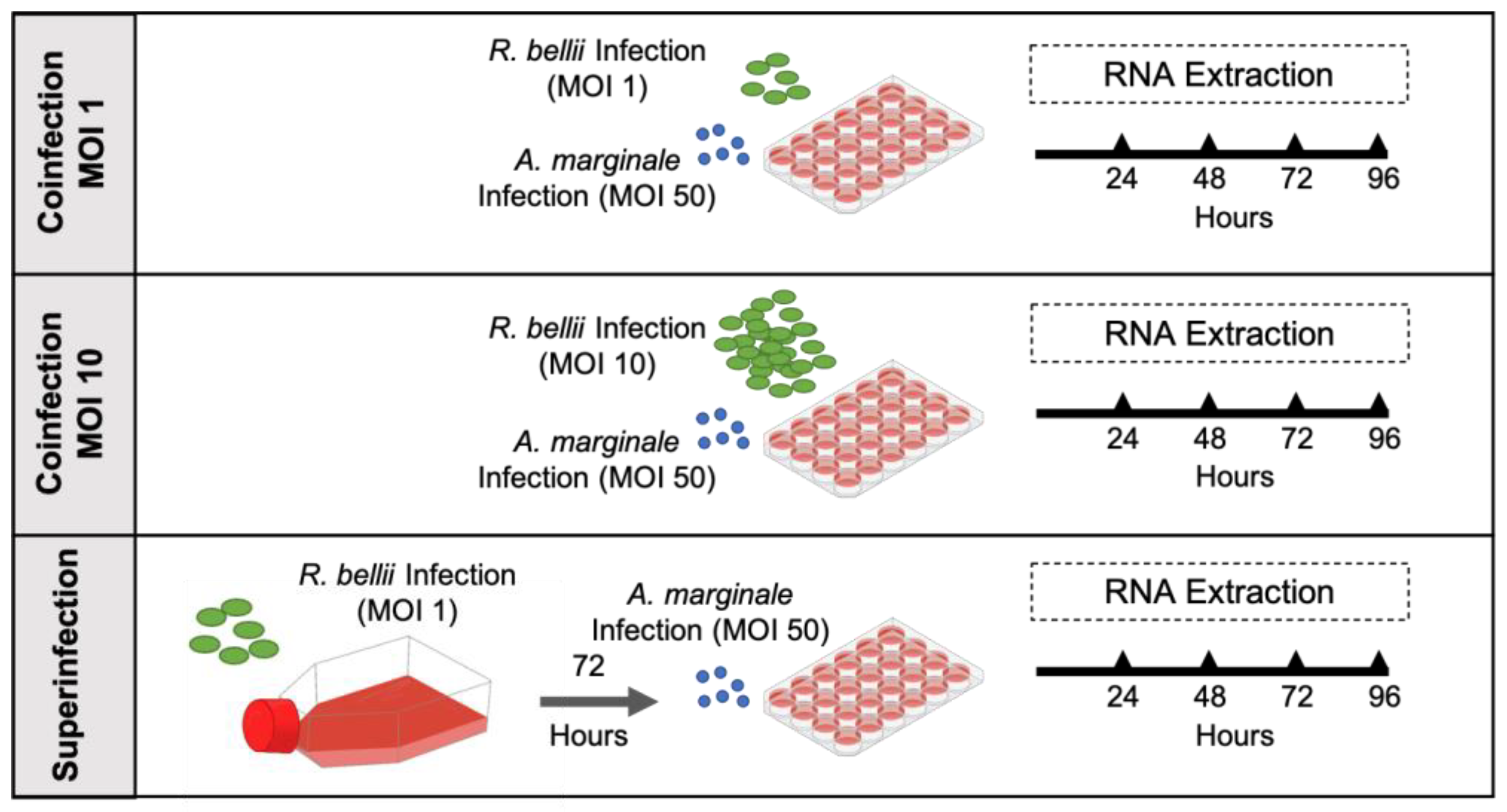
Figure 1. Overview of the three mixed-infection experiments: This figure illustrates the three growth-curve experiments, including different amounts of R. bellii and time points of infection for RNA collection. Anaplasma marginale was used at an MOI of 50 in all experiments.
Microorganisms 11 01096 g001
Figure 2. Growth and acquisition data for the low-R. bellii coinfection: (A) corresponds to the growth curve for A. marginale in both the A. marginale single-infection control (red) and the coinfection (black). (B) is the growth curve of R. bellii in the R. bellii single-infection control (blue) and the coinfection (black). The ΔΔCt values for both (A,B) are normalized to the host cell RNA and to the 24 h time point from the same growth condition and the significance of differences between conditions was analyzed with two-way ANOVAs. (C) is the amount of A. marginale and R. bellii present at the 24 h time point of the coinfection (gray), normalized to the host cell RNA, and to the 24 h time point of the corresponding single-infection control (black). Twenty-four-hour time points were compared using two-tailed t-tests. Error bars indicate standard error of the mean. * Represents a p value below 0.05.
Microorganisms 11 01096 g002
Figure 3. Growth and acquisition data for the high-R. bellii coinfection: (A) corresponds to the growth curve for A. marginale in both the A. marginale single-infection control (red) and the coinfection (black). (B) is the growth curve of R. bellii in the R. bellii single-infection control (blue) and the coinfection (black). The ΔΔCt values for both (A,B) are normalized to the host cell RNA and to the 24 h time point from the same growth condition and the significance of the differences between conditions was analyzed with two-way ANOVAs. (C) is the amount of A. marginale and R. bellii present at the 24 h time point of the coinfection (gray), normalized to host cell RNA, and to the 24 h time point of the corresponding single-infection control (black). Twenty-four-hour time points were compared with two-tailed t-tests. Error bars indicate standard error of the mean.
Microorganisms 11 01096 g003
Figure 4. Growth and acquisition data for the superinfection: (A) corresponds to the growth curve for A. marginale in both the A. marginale single-infection control (red) and the coinfection (black). (B) is the growth curve of R. bellii in the R. bellii single-infection control (blue) and the coinfection (black). The ΔΔCt values for both (A,B) are normalized to the host cell RNA and to the 24 h time point from the same growth condition and the significance of the differences between conditions was analyzed with two-way ANOVAs. Šídák’s multiple-comparison test was used in instances where ANOVA showed significance. (C) is the amount of A. marginale and R. bellii present at the 24 h time point of the coinfection (gray), normalized to the host cell RNA, and to the 24 h time point of the corresponding single-infection control (black). Twenty-four-hour time points were compared with two-tailed t-tests. Error bars indicate standard error of the mean. * Represents a p value below 0.05 ** Represents a p value below 0.01.
Microorganisms 11 01096 g004
Figure 5. DAE100 cell growth curves across three experimental conditions: (A) is the DAE100 growth curve for the low-R. bellii coinfection across all experimental conditions, (B) is the growth curve of DAE100 cells in the high-R. bellii coinfection, and (C) is the DAE100 cell growth curve for the superinfection. The ΔΔCt values for all growth curves are normalized to the genomic DNA positive control to facilitate comparison across samples and to the 24 h time point from the same growth condition. All growth curves include the R. bellii single-infection control (blue), the A. marginale single-infection control (red), and the combined infection (black). The significance of the differences between conditions was analyzed with two-way ANOVAs. Tukey’s test was used in instances where ANOVAs showed significance. Error bars indicate standard error of the mean. * represents a p value below 0.05.
Microorganisms 11 01096 g005
Figure 6. R. bellii growth in three experiments normalized to the MOI of 1 infection at 24 h time point: (A) is the growth curves for the R. bellii single-infection controls from the three experiments. (B) is the mixed infection from each of the three experiments. Both panels plot the growth of R. bellii normalized to host cell RNAand then to the average of the 24 h time points for the coinfection at an MOI of one. The significance of the differences between conditions was analyzed with two-way ANOVAs. Tukey’s test was used in instances where ANOVA showed significance. Error bars indicate standard error of the mean. * represents a p value below 0.05 ** represents a p value below 0.01.
Microorganisms 11 01096 g006
Table 1. Primer sets for RT-qPCR.
SpeciesGeneForwardReverseAmplicon Size (bp)
D. andersoniGAPDHggtcatctctgctccatctgtgctcacaatcttcatgcttg89
A. marginalemsp5cttccgaagttgtaagtgagggcacttatcggcatggtcgcctagttt203
R. belliirpoBgcttaaagatcgcaaagggattatagacgcctgccgacattctttcaactactg144
Disclaimer/Publisher’s Note: The statements, opinions and data contained in all publications are solely those of the individual author(s) and contributor(s) and not of MDPI and/or the editor(s). MDPI and/or the editor(s) disclaim responsibility for any injury to people or property resulting from any ideas, methods, instructions or products referred to in the content.