Article

The Impact of Anti-Inflammatory Drugs on the Prokaryotic Community Composition and Selected Bacterial Strains Based on Microcosm Experiments

1
Department of Microbiology, Doctoral School of Biology, Institute of Biology, Eötvös Loránd University, Pázmány Péter stny., 1/C, 1117 Budapest, Hungary
2
Department of Microbiology, Eötvös Loránd University, Pázmány Péter stny., 1/C, 1117 Budapest, Hungary
3
Department of Microbiology, Doctoral School of Environmental Sciences, Institute of Biology, Eötvös Loránd University, Pázmány Péter stny., 1/C, 1117 Budapest, Hungary
*
Authors to whom correspondence should be addressed.
Microorganisms 2023, 11(6), 1447; https://doi.org/10.3390/microorganisms11061447
Submission received: 26 April 2023 / Revised: 24 May 2023 / Accepted: 26 May 2023 / Published: 30 May 2023

Abstract

:
Non-steroidal anti-inflammatory drugs (NSAIDs) are increasingly recognized as potential environmental contaminants that may induce toxicity in aquatic ecosystems. This 3-week microcosm experiment explores the acute impacts of NSAIDs, including diclofenac (DCF), ibuprofen (IBU), and acetylsalicylic acid (ASA), on bacterial communities using a wide range of these substances (200–6000 ppm). The results showed that the NSAID-treated microcosms had higher cell count values than control samples, though the diversity of microbial communities decreased. The isolated heterotrophic bacteria mostly belonged to Proteobacteria, particularly Klebsiella. Next-generation sequencing (NGS) revealed that NSAIDs altered the structure of the bacterial community composition, with the proportion of Proteobacteria aligning with the selective cultivation results. Bacteria had higher resistance to IBU/ASA than to DCF. In DCF-treated microcosms, there has been a high reduction of the number of Bacteroidetes, whereas in the microcosms treated with IBU/ASA, they have remained abundant. The numbers of Patescibacteria and Actinobacteria have decreased across all NSAID-treated microcosms. Verrucomicrobia and Planctomycetes have tolerated all NSAIDs, even DCF. Cyanobacteria have also demonstrated tolerance to IBU/ASA treatment in the microcosms. The archaeal community structure was also impacted by the NSAID treatments, with Thaumarchaeota abundant in all microcosms, especially DCF-treated microcosms, while Nanoarchaeota is more typical of IBU/ASA-treated microcosms with lower NSAID concentrations. These results indicate that the presence of NSAIDs in aquatic environments could lead to changes in the composition of microbial communities.

1. Introduction

As a result of increasing drug consumption, a high number of pharmacologically active compounds (PhACs) are entering our natural water sources, posing an increasing ecological and health risk worldwide [1,2]. Today, the five most frequently used nonsteroidal anti-inflammatory drugs (NSAIDs) are acetylsalicylic acid, paracetamol, diclofenac, ibuprofen and naproxen [3]. These compounds have a negative effect on aquatic living organisms causing oxidative stress, DNA damage and behavioral changes in aquatic microscopic animals. Their photodegradation can produce compounds that are even more toxic than the original molecule [3,4,5,6]. To date, wastewater treatment plants (WWTPs) are not designed to entirely filter organic micropollutants, often characterized by high water solubility and poor biodegradability favoring their environmental persistence [7].
Many PhACs (e.g., antiepileptics, NSAIDs) have already been detected in aqueous environments, mainly in surface waters, groundwater, wastewater and, in some cases, in drinking water as well [8,9,10,11,12]. The Danube River, as one of the most important drinking water bases in Europe, is also endangered in this respect: the JDS3 (Joint Danube Survey 3) in 2013 [13] surveyed the entire length of the river and identified more than 100 different drug residues and other anthropogenic contaminants in it. Kondor et al. (2020) [14] also examined the Danube River and drinking water wells for drug residues; their experience showed that the gravel filtration bed of the Danube cannot remove chemical compounds completely.
One of the main concerns connected to the presence of pharmaceutical residues in the environment is their usual occurrence as complex mixtures rather than as a single compound. The different NSAIDs that are continuously and simultaneously used and released into the environment can interact synergistically [15,16]. Additionally, the environmental concentration of pharmaceutical compounds and their synergistic and antagonist effects are strongly related to the geographical area, climatologic conditions and the presence of wastewater discharges [17]. In the environment, the persistence of pharmaceutical products can be influenced by environmental factors, physicochemical properties of the molecule, molecular structure, polarity, photostability, chemical stability, volatility, presence of other pharmaceuticals in the same matrix and the presence and activity of microorganisms with the ability to degrade pharmaceuticals [18,19,20,21,22,23].
The ability of microorganisms to degrade drugs and their residues is becoming more important as it would be an economically and environmentally beneficial solution to increase organic micro-contamination. Microorganisms may be able to break down a number of difficult-to-degrade substrates (even polyaromatic compounds) facilitating their removal from the environment [24].
Numerous scientific papers have examined the impact of pharmaceuticals on bacteria across various taxonomic groups: Leão et al. [25] observed that the presence of salicylate increases S. aureus resistance to fluoroquinolones and fusidic acid, and it was also found [26] that this drug exhibits an increased resistance of E. coli to several antimicrobials (fluoroquinolones, ampicillin, cephalosporins, tetracycline and chloramphenicol).
Nevertheless, the environmental effects of drugs can extend far beyond the outcomes determined in toxicity assessments conducted on individual organisms—these substances have the potential to disrupt entire microbial communities. There are existing findings on the impact of drug residues on entire microbial communities (e.g., the finding of Gerbersdorf et al. [27]) but this research was found to be limited.
Based on a comprehensive review of the existing literature, no earlier studies have investigated the acute effects of NSAIDs in such a wide range and high concentrations as this research which aims to investigate the effect of three commonly used nonsteroidal anti-inflammatory drugs (NSAIDs): diclofenac (DCF), ibuprofen (IBU), and acetylsalicylic acid (ASA) on microbial communities in microcosm experiments originating water from the Danube River. This study combines multiple techniques such as (1) estimating cell count values by epifluorescent microscopy to reflect dynamic changes in the bacterial number; (2) selectively isolating bacteria utilizing NSAIDs as a sole carbon source; and (3) next-generation sequencing to further assess the impact of NSAIDs on microbial communities.
This study provides insights into the potential effect of NSAIDs on changes in microbial communities and highlights the importance of investigating their impact on natural water sources. The findings of this study may facilitate the development of novel bioremediation techniques to reduce the ecological and health risks posed by NSAIDs in natural water sources.

2. Materials and Methods

2.1. Experimental Design

Water samples have been taken two times from the right shore of the Danube in Budapest (Hungary, [47.477357° N; 19.061891° E]) into a 1.5 L clean, sterile, screw-capped glass bottle on 13 July 2020 (first experiment) and on 24 August 2020 (second experiment). The samples were transported to the laboratory at 4 °C temperature, and used for diclofenac (DCF), ibuprofen (IBU) and acetylsalicylic acid (ASA) exposure in laboratory microcosm experiments. An amount of 200 mL of water samples was dispensed into three types of microcosm systems (250 mL, clean, sterile glass bottles) separately loaded with the powdered form of the anti-inflammatory drugs to acquire 200, 500, 1000 and 2000 ppm concentration, respectively, regarding the first experiments (MK0_1, MK1, MD_200, MD_500, MD_1000, MD1_2000, MI_200, MI_500, MI_1000, MI1_2000, MA_200, MA_500, MA_1000, MA_2000), while 2000 and 6000 ppm in the case of the second experiments (MK0_2, MK2, MD2_2000, MD_6000, MI2_2000, MI_6000, MD_2000, MD_6000) (Table 1). Regarding both experiments, bottles were constantly shaking (80 rpm) for three weeks at 23 °C.

2.2. Determination of Physicochemical Parameters and Cell Count Values

Temperature, pH, conductivity and dissolved oxygen values were recorded during sampling on the field by a Hach HQ40D portable multimeter (Hach, Loveland, CO, USA). Determination of microscopic cell counts was carried out by using Nikon80i epifluorescent microscopy and NisElements program package [28]. For the investigations, 10 mL of water samples were filtered through isopore polycarbonate filters with a pore size of 0.22 µm (Millipore, Billerica, MA, USA) and fixed with 2% paraformaldehyde solution.

2.3. Selective Cultivation of Bacteria

Bacterial strains were isolated from MD_6000, MI_6000 and MA_6000 microcosms containing 6000 ppm of anti-inflammatory drugs. Isolation of strains was performed by standard dilution plate technique on DSM medium 1 supplemented with 2000 ppm of DCF, IBU and ASA, respectively. Incubation was carried out at 28 °C for 7 days. To better explore cultivable representatives utilizing the chosen drug residues, a 2nd selective isolation was carried out from separately created microcosms with 6000 ppm of drug residues based on the same experimental design, cultivation technique and laboratory circumstances as used previously.

2.4. DNA Extraction from the Bacterial Strains, 16S rRNA Gene Amplification and Taxonomic Identification

DNA was extracted from the isolated bacterial strains [29], then PCR amplification was done on the 16S rRNA gene using the primers 27F (5′–AGA GTT TGA TCM TGG CTC AG–3′) and 1492R (5′–GGT TAC CTT GTT ACG ACT T–3′) [30]. The 16S rRNA gene sequencing was carried out at LGC (Berlin, Germany). The sequenced bacterial strains were identified using EzBioCloud’s online identification system [31].
To analyze the genomic sequences and metabolic potential of selectively cultivated and identified bacterial strains, the genomic sequences of the type strains of these bacterial strains from the EzBioCloud database were obtained. By utilizing the Rapid Annotation using Subsystem Technology (RAST) pipeline [32], a web interface for functional annotation of genomic sequences to analyze the features and functions of these sequences, functions to the genes present in the genome based on their similarities to known genes in other organisms were assigned. Furthermore, the functional genes encoding the metabolism of aromatic intermediates in the bacterial strains were analyzed using the Kyoto Encyclopedia of Genes and Genomes (KEGG) database [33]. The genes responsible for the metabolism of these compounds in the bacterial strains were identified to gain insights into the potential metabolic capabilities of the strains.

2.5. DNA Extraction from Microcosms and Next-Generation DNA Sequencing

After incubation, the microcosms were filtered through a 0.22 μm pore size mixed cellulose filter (type GSWP; Millipore, Billerica, MA, USA). DNA was isolated from the filters using the Ultraclean® PowerSoil DNA Isolation Kit (MoBio, Carlsbad, CA, USA) according to the manufacturer’s instructions.
For the identification of bacterial and archaeal community structure, the 16S rRNA genes of the DNA samples were amplified with PCR in separate, triplicate reactions using primers with the following target-specific sequences: Bact_341F (5′–CCT ACG GGN GGC WGC AG–3′) and modified Bact_805R (5′–GAC TAC NVG GGT ATC TAA TCC–3′) for Bacteria as well as A519F (5′–CAG CMG CCG CGG TAA–3′) and Arch855R (5′–TCC CCC GCC AAT TCC TTT AA–3′) for Archaea [34]. Before sequencing, the DNA concentration of the PCR products was determined using a Qubit meter (Invitrogen Life Technologies, Carlsbad, CA, USA), and a minimum concentration of 4 ng/μL and 50 μL of PCR product was respected. DNA sequencing was performed on the Illumina MiSeq platform using MiSeq standard v2 chemistry by the Genomics Core Facility RTSF (Michigan State University, East Lansing, MI, USA). The forward and reverse fastq files obtained from the Illumina sequencer were processed and analyzed using the Mothur v1.40.5 software [35]. The contigs were acquired using the make.contigs with a deltaq value of 10 to get sequences with high-quality scores. The screen.seqs command was applied to keep only the sequences having the expected length, number of polymers and ambiguous bases. The sequences were aligned to the Silva database (silva.nr_v132.align) [36], and the non-aligned sequences and columns containing only “.” were removed by using the screen.seqs and filter.seqs commands based on the position of the archaeal and bacterial primers within the 16S rRNA gene. The pre.cluster command was used to remove sequences with Illumina sequencing errors. The chimeric sequences were removed by the UCHIME algorithm [37]. Only the abundant sequences were kept using the command split.abund to split the sequences into two groups with a cutoff value equal to 1. The taxonomic classification of the sequences was done using the Silva database silva.nr_v132.tax, and the non-archaeal and non-bacterial sequences were removed from the analyses based on the taxonomic classification output. The OTUs (Operational Taxonomic Units) were calculated using a distance matrix with distances larger than 0.15 obtained by using the dist.seqs and later the cluster commands assign sequences to OTUs, and eventually, the consensus taxa were determined using the classify.otu. In the end, data were normalized using the sub.sample and then rarefaction.single and summary.single were applied to calculate the rarefaction curve data and the values of the diversity indices. Shannon–Weaver, inverse Simpson (1/D) diversity indices and Chao-1 metrics were calculated using Mothur [35].
Non-metric multidimensional scaling ordination (NMDS) of the microcosms with the Bray–Curtis dissimilarity index based on their bacterial and archaeal OTU composition was carried out with the PAST 4.03 program.

3. Results

3.1. Physicochemical Parameters of the Danube Water

The physicochemical characteristics of the sampling sites are summarized in Table 2.

3.2. Microscopic Cell Counts and Diversity Indices of the Samples

The microscopic cell counts are presented together with Shannon diversity indices in Figure 1 for both Archaea and Bacteria based on amplicon sequencing. The Shannon diversity indices were determined by analyzing the population diversity in the samples based on operational taxonomic units (OTUs). The index was calculated using the following formula: H = i = 1 s ( P i ln P i ) where Pi represents the proportion (n/N) of individuals of a specific species found (n) divided by the total number of individuals found (N), ln denotes the natural logarithm, Σ signifies the sum of the calculations, and s represents the number of species.
When analyzing both experiments, a difference could be observed between the absolute controls (before incubation) and the microcosms treated with NSAIDs (after incubation): after comparing MK0_1 and MK0_2 absolute controls to the first and second treated experiments, it was revealed that the cell count values of the treated experiments were higher by at least one order of magnitude. When comparing MK1 and MK2 controls (after incubation) to the first and second treated experiments, a significant difference was not found regarding the cell count values. The number of cells was the most outstanding in DCF-treated microcosms. After comparing the cell counts of both treated experiments, values were similar; even a high number of cells was observed in microcosms with 6000 ppm of drug residues. At the same time, when considering Shannon diversity indices of Bacteria, it was found that microcosms treated with lower concentrations (200 and 500 ppm) of NSAIDs showed higher diversity than the microcosms containing drug residues in high concentrations (1000, 2000 and 6000 ppm) the decreased cell count values did not go together with lower diversity indices. Among microcosms treated with NSAIDs, the ASA-treated microcosms showed the highest overall bacterial diversity while it was the lowest in DCF-treated ones. Diversity indices of Archaea were only 0 and 0.69, meaning that compared to Bacteria, archaeal diversity was not observed by next-generation sequencing in the microcosms.

3.3. Results of Selective Cultivation and Genomic Analysis of Bacterial Strains

Selective cultivation was performed from MD_6000 (containing 6000 ppm of DCF), MI_6000 (containing 6000 ppm of IBU) and MD_6000 (containing 6000 ppm of ASA) microcosms. The results of the taxonomic identification of our selectively cultivated strains are presented in Table 3. Results of the second round of selective cultivation from microcosms with 6000 ppm of DCF, IBU and ASA are presented in Table 4.
From MD_6000, altogether 3, from MI_6000, 8 and also from MA_6000, altogether 8 strains were cultivated on DSM 1 medium containing 2000 ppm of DCF, IBU and ASA. From the separately created microcosms with 6000 ppm of DCF, 25 and 6000 ppm of IBU, 16 strains were cultivated, but cultivation from the 2nd ASA microcosm was not successful. Based on the 16S rRNA gene sequence similarities of all the cultivated strains, they showed 98.54–99.91% similarity values to the reference sequences of the type strains.
All isolated heterotrophic bacteria belonged to Proteobacteria with the exception of Paenibacillus lautus belonging to Firmicutes. At the class level of Proteobacteria, the isolates belonged to Alphaproteobacteria (7 strains [12%]), Betaproteobacteria (7 strains [12%]) and Gammaproteobacteria (45 strains [76%]). Among Alphaproteobacteria, genera Novosphingobium, Sphingobium and Ensifer were found, while the betaproteobacterial representatives were mostly assigned to the genera Burkholderia, Pseudoduganella and Cupriavidus. Regarding Gammaproteobacteria, Klebsiella, Pseudomonas, Aeromonas, Rahnella, Cytrobacter, Phytobacter, Raoultella and Kosakonia were cultivated. Among them, Klebsiella was the most abundant bacterial genus.
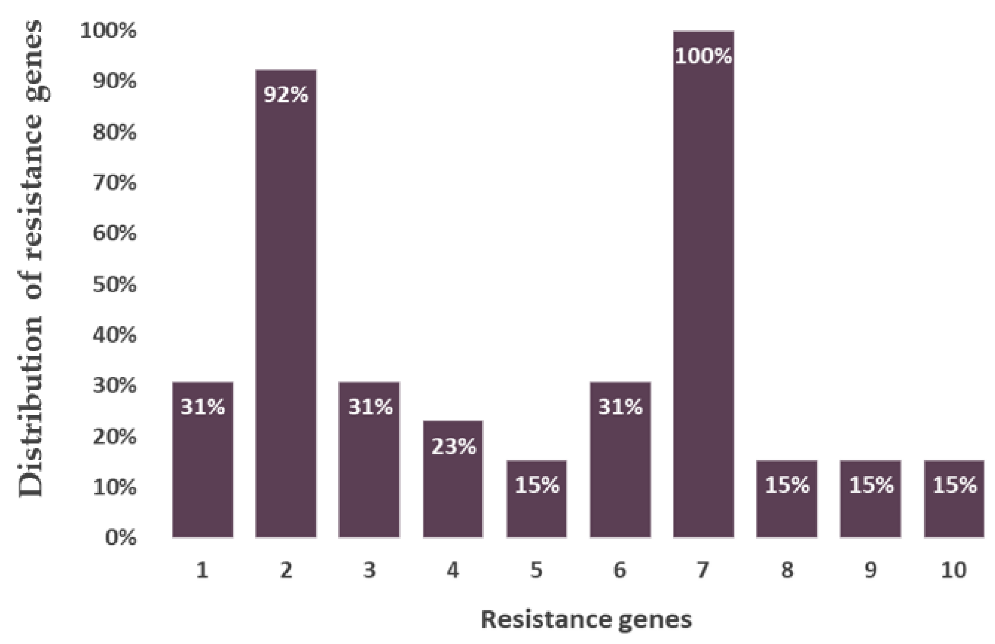
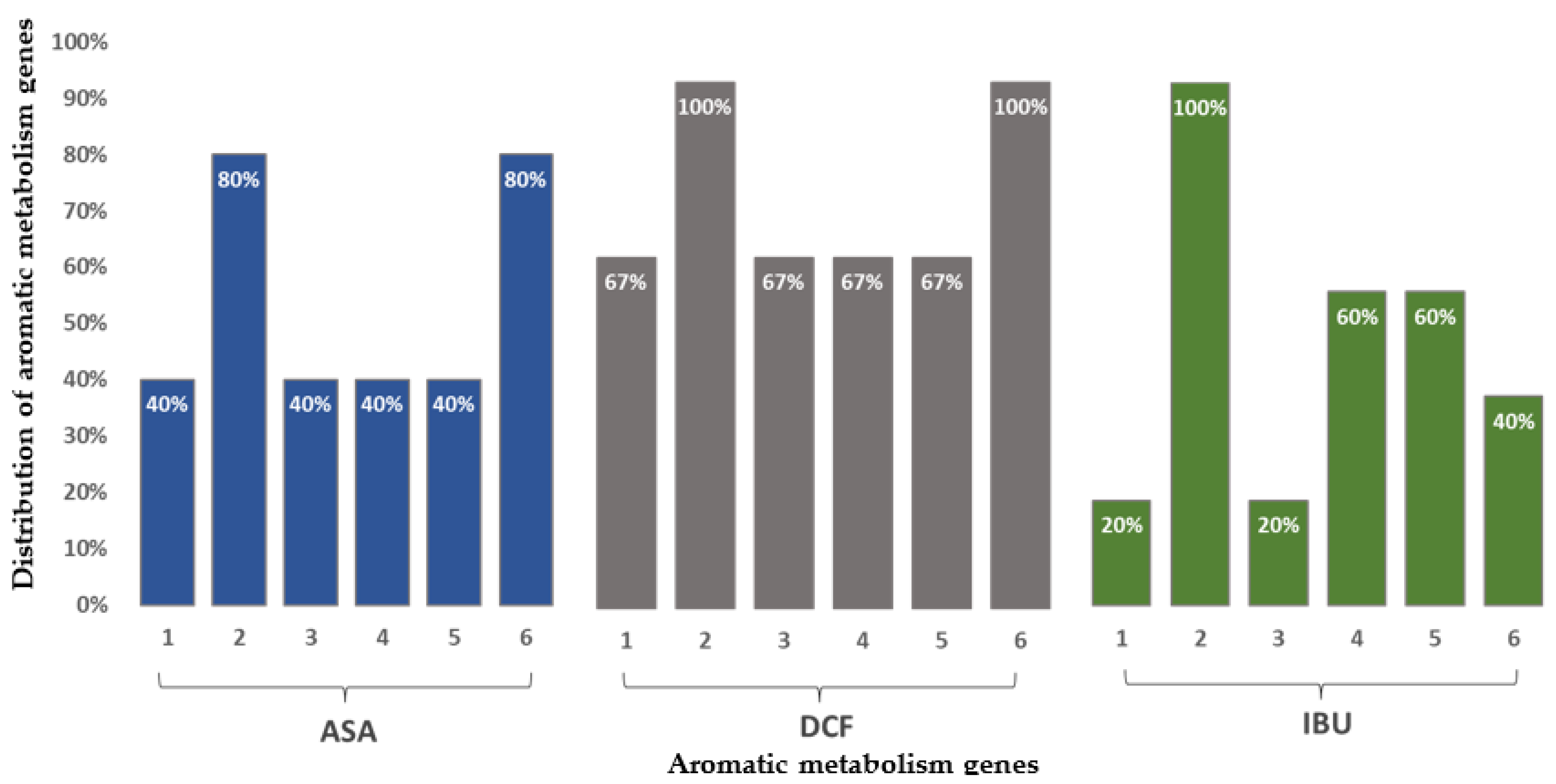
The analyzed bacterial strains contained two outstanding genes responsible for beta-lactamase and fluoroquinolone resistance. Their occurrence was characteristic in almost all bacterial strains breaking down NSAIDs. Four genes were present in most of the bacterial species encoding the catechol and protocatechuate branches of beta-ketoadipate metabolism, the salicylate and gentisate metabolism, as well as the homogenization pathway for the breakdown of aromatic compounds. The beta-lactamase resistance gene was present in more than 90% of the bacteria while the fluoroquinolone resistance gene was found in all the studied species (Figure 2). In the case of bacteria degrading ASA and DCF, the catechol branch of the beta-ketoadipate pathway and the metabolism of salicylate and gentisate were encoded. Bacterial species degrading IBU, the gene encoding the homogenous pathway for the degradation of aromatic compounds and the protocatechol branch of the beta-ketoadipate pathway were the most common (Figure 3).

3.4. Results of Amplicon Sequencing

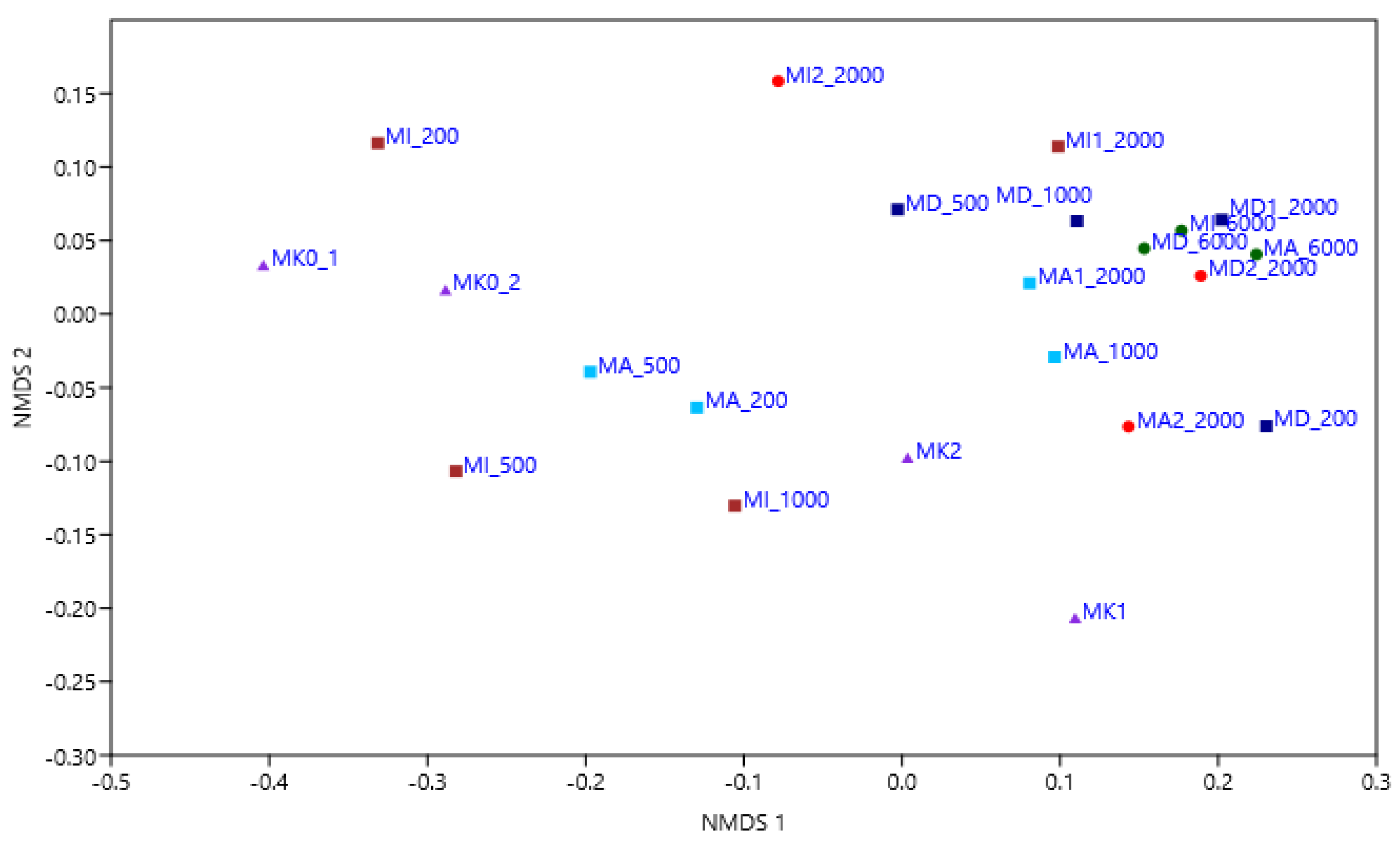
Bacterial sequences were more abundant than archaeal sequences in our amplicon libraries. NMDS ordination (Figure 4) of the microcosms with the Bray–Curtis dissimilarity index based on their bacterial and archaeal OTU composition confirmed that NSAIDs affect the bacterial and archaeal community structure of the microcosms; microcosms containing 2000 and 6000 ppm of NSAIDs are separated from the controls and treated microcosms with a lower concentration of drug residues.

3.4.1. Bacterial Community Composition of the Microcosms Based on Amplicon Sequencing

Altogether, 36 bacterial phyla were identified by amplicon sequencing; the distribution of the most abundant members is presented in Figure 5a,b. Regarding MK0_1 and MK0_2 as the original samples (serving as absolute controls), the analysis revealed that Proteobacteria emerged as the dominant taxon, displaying the highest relative abundance. It was closely followed by Bacteroidetes and Patescibacteria, which exhibited significant representation within the samples. Actinobacteria were also found to be abundant in the analyzed samples. In contrast, Verrucomicrobia, Cyanobacteria and Planctomycetes displayed comparatively lower levels of representation. The taxonomic groups Chlamydiae, Firmicutes and Dependentiae exhibited even lower relative abundances within these samples.
The absolute dominance of Proteobacteria could be observed in all microcosms but their proportional ratio increases in the treated microcosms, especially in those treated with DCF: among Alphaproteobacteria, Caulobacteraceae (genus Caulobacter), Sphingomonadaceae and Rhizobiales (genus Aquabacter and genus Sandarakinorhabdus), regarding Gammaproteobacteria, Burkholderiaceae (genus Polynucleobacter), Rhodocyclaceae (genus Aquicella), Enterobacteriaceae (genus Legionella and Acinetobacter), as well as Pseudomonadaceae (genus Pseudomonas) were the most common representatives. The proportion of Alpha- and Gammaproteobacteria were similar in microcosms with 200–2000 ppm of NSAIDs, but the 6000 ppm of concentration was more favorable to Gammaproteobacteria. Among them, the two most abundant bacterial genera were Pseudomonas and Acinetobacter. In the absolute control samples, as already mentioned, the ratio of Proteobacteria was in balance with Patescibacteria and Bacteroidetes, especially in MK0_1, and then as the concentration of drugs was increased, Proteobacteria proliferated the treated microcosms. Their number was the most significant in the second experiment containing a high concentration of DCF (MD_6000), IBU (MI_6000) and ASA (MA_6000). When comparing the first and second microcosm experiments, it can be said that at 6000 ppm, Proteobacteria almost completely suppressed the other bacterial members (such as Actinobacteria, Verrucomicrobia or Planctomycetes) of the community except for Bacteroidetes, their abundance was still relatively high at this concentration. When comparing the DCF, IBU and ASA-treated microcosms with each other, it can be stated that Proteobacteria dominated the DCF-treated microcosms as they were the most abundant representatives in microcosms containing this anti-inflammatory drug. Actinobacteria (Ilumatobacteraceae, Sporichthyaceae, Microbacteriaceae) characterized the controls and microcosms treated with 200–500 ppm of IBU and ASA, were also abundant in MA2_2000 but low in DCF-treated microcosms. The ratio of Unclassified Bacteria was lower in DCF-treated microcosms than in the IBU- and ASA-treated ones. Bacteroidetes (Chitinophagaceae, Saprospiraceae, Microscillaceae, Spirosomaceae, Ignavibacteria) were revealed in all microcosms except MI1_2000, and they mostly dominated the IBU- and ASA-treated microcosms. Among Chlamydiae, Chlamydiales, Parachlamydiaceae and Simkaniaceae were present in the highest number, especially in MK0_1, MI_1000 and MA1_2000. Cyanobacteria were found in the controls and microcosms treated with lower, 200 ppm of IBU and ASA. Among Dependentiae, which ratio was high at 2000 ppm of ASA, Babeliales were mainly present. Firmicutes could only be found in the controls (Bacillales, Clostridiales, Selenomonadales and Veillonellaceae). As for Patescibacteria, consistently being observed in DCF-treated microcosms, Parcubacteria were the most abundant bacterial members. The number of Planctomycetes (Phycisphaeraceae, Pirellulaceae, Rubinisphaeraceae), similar to Bacteroidetes, was high in IBU- and ASA-treated microcosms. Verrucomicrobia (Chthoniobacteraceae, Opitutaceae, Pedosphaeraceae, Verrucomicrobiaceae) utilized DCF and IBU most successfully but were able to use lower concentrations (200–500 ppm) of ASA as well. Phyla contributing to less than 2% of the microcosms were Acidobacteria, Armatimonadetes, Chloroflexi, Elusimicrobia, Entotheonellaeota, Epsilonbacteraeota, Fibrobacteres, Fusobacteria, Gemmatimonadetes, Hydrogenedentes, Kiritimatiellaeota, Latescibacteria, Lentisphaerae, Margulisbacteria, Nitrospirae, Omnitrophicaeota, Rokubacteria, Spirochaetes, Tenericutes and Zixibacteria.

3.4.2. Archaeal Community Composition of the Microcosms Based on Amplicon Sequencing

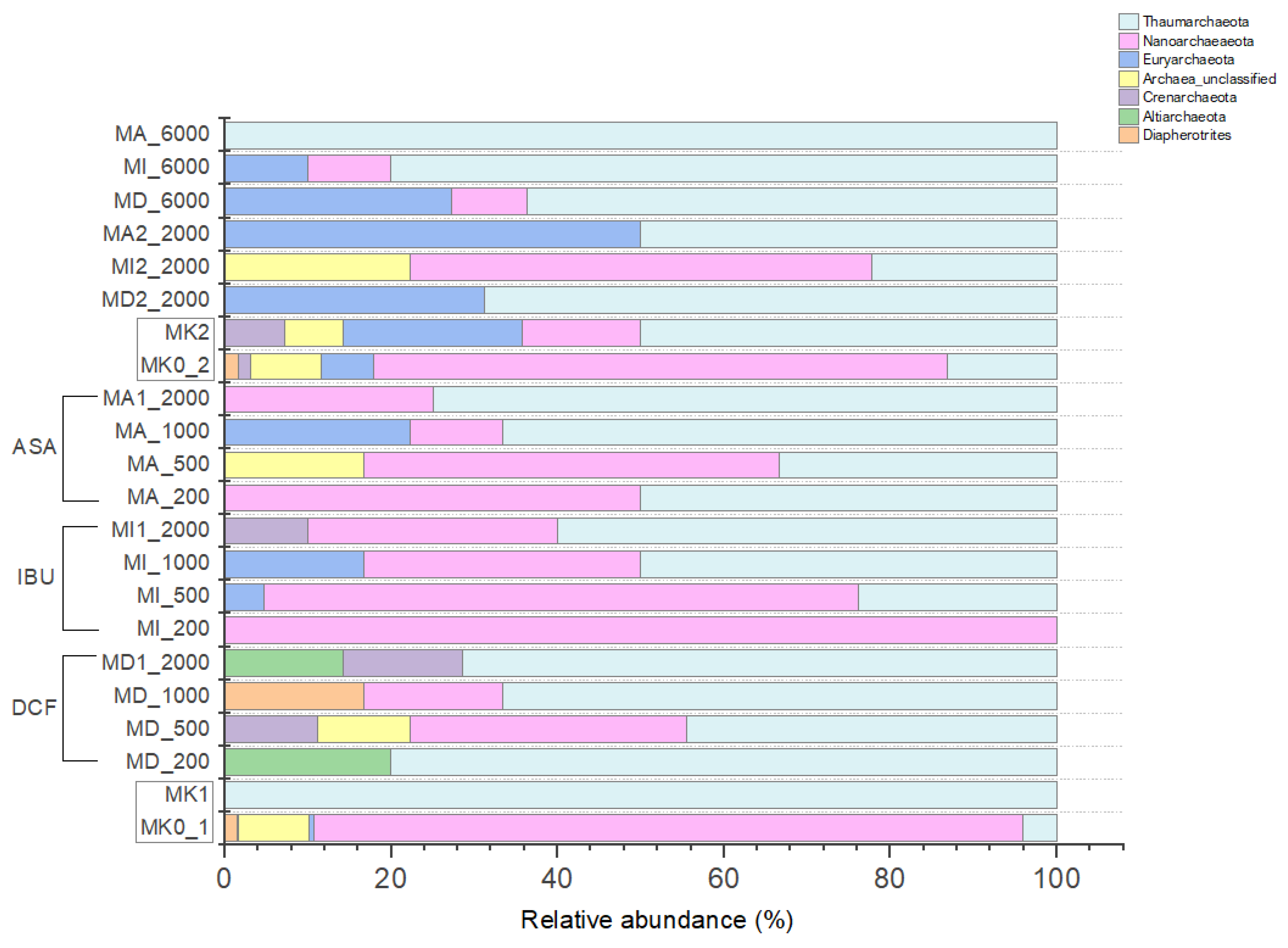
Altogether 8 archaeal phyla were identified by amplicon sequencing; the distribution of the most abundant members is presented in Figure 6. Regarding MK0_1 and MK0_2 as the original samples (serving as absolute controls), our analysis revealed that Crenarchaeota exhibited a minor representation, indicating a relatively lower abundance within the samples. Conversely, Diapherotrites and Euryarchaeota demonstrated a more pronounced presence, suggesting a higher abundance compared to Crenarchaeota. Nanoarchaeaeota displayed a substantial presence, indicating a significant abundance within the microbial community. Thaumarchaeota were also abundant in the samples, though they represented a relatively lower level compared to Nanoarchaeota.
Compared to the NSAID-treated microcosms, the archaeal communities of the control samples were more diverse. When analyzing the first and second experiments at the same time, it can be stated that the archaeal community has significantly changed in the microcosms, especially the treated ones: Thaumarchaeota (ammonia-oxidizing archaea [AOA]: Nitrosopumilaceae, Nitrososphaeraceae and Nitrosotaleaceae) were abundant in all microcosms but they dominated the microcosms treated with 200–2000 ppm of DCF and microcosms containing 6000 ppm of NSAIDs. Nanoarchaeota (Nanohaloarchaeia and Woesearchaeia) were also present in each microcosm, but they were more typical to IBU- and ASA-treated-microcosms with lower, 500–1000 ppm of concentrations. Further, Thamarchaeota, Euryarchaeota (Thermoplasmata and methanogenic archaeal representatives: Methanobacterium, Methanobrevibacter, Methanocella, Methanocorpusculum, Methanoregula, Methanosaeta and Methanosarcina) used high concentrations (6000 ppm) of drug-residues. When analyzing only the treated microcosms, it can be concluded that the archaeal composition of IBU- and ASA-treated microcosms were similar while the DCF-treated ones are differentiated from them mainly because of their lower Nanoarchaeota ratio. Altiarchaeota (Altiarchaeia) and Crenarchaeota (Bathyarchaeia) were typical to DCF-treated microcosms but the latter also could be found in MI1_2000. Diapherotrites (Iainarchaeales and Micrarchaeia) were observed in the controls and MD_1000. Unclassified Archaea were found in the controls, MD_500, MA_500 and MI2_2000.

4. Discussion

As mentioned before, a 3-week microcosm experiment was conducted in 2020 to assess microbial community changes influenced by exposure to anti-inflammatory drugs at various concentrations (200, 500, 1000, 2000 and 6000 ppm). NSAIDs trigger increasing toxic phenomena in aquatic environments. Based on JDS3 (2013), 20–40 ng L−1 of DCF, 550 ng L−1 of IBU and 470 ng L−1 ASA was measured in the Danube River. Several bacteria are able to utilize these compounds thus contributing to their biotransformation as well as degradation in the environment [24,25,26]. Our experiments had been focusing on microorganisms able to use NSAIDs as a sole carbon source rather than measuring their biodegradation capabilities. After cultivation, though, the genes of our isolated strains were analyzed upon NSAID metabolism and amplicon sequencing results were also compared to the literature data on the biodegradation possibility of NSAIDS.
In the microcosms, cell count values of the treated experiments were higher than the absolute controls, and then 6000 ppm of NSAID concentrations facilitated the proliferation of bacteria even more. When a community structure is interfered with in any way (e.g., it receives a selective inhibitor or some of the representatives are able to grow on a selective substrate), it always shifts the composition of the community and it is common that selective enrichment leads to a decreased diversity. The reduced diversity indices and the NGS analysis showed other interesting bacterial and archeal community changes that will be discussed below.
Among our cultivated bacterial strains, 1 belonged to Firmicutes while the rest of the strains were members of Proteobacateria. From the first isolation, representatives belonging to class Alpha-, Beta- and Gammaproteobacteria were found, while the second isolation resulted in strains found to be members only of Gammaproteobacteria. Regarding the first selective isolation, more taxa were cultivated than in the second; cultivation of representatives in the presence of ASA was also successful. Regarding the second selective cultivation, mostly genera Aeromonas and Rahnella were able to utilize DCF, while Klebsiella dominated the microcosms containing IBU. It is not surprising that mostly Proteobacteria were found since it was also revealed by the NGS analysis that the treatment of NSAIDs shifted the composition of the microbial communities in this direction.
The analyzed bacterial strains contained two outstanding genes responsible for beta-lactamase and fluoroquinolone resistance. Their occurrence was observable in almost all bacterial strains breaking down NSAIDs, but their presence hardly proves bacterial resistance to NSAIDs. Both fluoroquinolones and beta-lactam derivatives are widely used and distributed antibiotics. NSAIDs make microorganisms more sensitive to certain antibiotics, but regarding beta-lactam derivatives and fluoroquinolones, the opposite has been found, i.e., NSAIDs make bacteria more resistant to these compounds. This may happen since part of the bacterial resistance to these antibiotics is due to multidrug efflux pumps, which are substrates of NSAIDs and may be able to stimulate them. In addition, NSAIDs can alter the permeability of bacterial membranes [38,39].
Four genes were present in the bacterial species encoding the catechol and protocatechuate branches of beta-ketoadipate metabolism, the salicylate and gentisate metabolism, as well as the homogenization pathway for the breakdown of aromatic compounds. In this case, the drug molecule was the substrate of the efflux pump involved in the resistance against the given antibiotic or a gene encoding the breakdown of non-specific aromatic intermediates, which contributes to the resistance of the microorganisms to drug molecules as the aromatic ring is present in them [38,39,40,41,42].
By next-generation sequencing, it was found that the diversity of the treated microcosms was reduced compared to the controls, and except for Proteobacteria, a lower ratio of representatives could be observed in them. The type of pharmaceutical compound used for preparing the microcosms affected the selection of the microbial communities: when considering microcosms containing 200–2000 ppm of DCF, the abundance of Proteobacteria was high compared to microcosms with IBU and ASA in which a high number of Bacteroidetes and Planctomycetes could be observed in addition to Proteobacteria. At 6000 ppm, Proteobacteria almost completely overgrew other bacterial members except for Bacteroidetes.
Proteobacteria was described as the most diverse and representative phylum in marine and soil environments [43] dominating biological wastewater treatments due to the role of its high number of representatives in organic and nutrient removal [44]. It is also one of the most common phyla involved in the biodegradation of different pollutants, e.g., hydrocarbons [45] and pharmaceuticals [46]. A bacterial strain, Labrys portucalensis (Alphaproteobacteria), first was isolated from an industrially contaminated sediment in northern Portugal [47] and was able to biodegrade several fluoroanilines [48]. 4-Fluoroaniline is a common fluorine-containing aromatic amine, widely used in the synthesis of medicines and pesticides, such as the drug ezetimibe, norfloxacin and a 4-amidogen quinoline pesticide. In a study conducted by [49], Labrys portucalensis F11 was shown to be able to biodegrade DCF. In another study, a considerable increase of Alphaproteobacteria was detected in activated sludge containing 500 and 1000 ppm of IBU [50], strongly correlating with our findings. Sphingomonas species can degrade salicylate and ibuprofen as well [51,52].
The two most abundant bacterial genera regarding Gammaproteobacteria were Pseudomonas and Acinetobacter. This finding is in correlation with a study examining the biodegradation of paroxetine (an antidepressant of the selective serotonin reuptake inhibitor [SSRI] class) and bezafibrate (a fibrate drug used as a lipid-lowering agent to treat hyperlipidemia) where these genera were also present in the highest number among Gammaproteobacteria [53]. Another finding also proved that the genus Pseudomonas is capable of aromatic compound degradation [24]. Research performed by Rutere et al. (2020) [54] found that Pseudomonas thivervalensis was able to grow by utilizing IBU as a sole carbon and energy source validating its ibuprofen degradation capabilities. Murdoch and Hay (2005) [52] summarized the metabolism of some compounds the most similar to ibuprofen and identified patterns in which Pseudomonas putida F1 and Pseudomonas cepacia had a role. Pseudomonas putida R1 was able to biodegrade salicylate, too [55]. It has been reported that bacteria belonging to Methylococcaceae could also be relevant to the metabolic degradation of IBU [56]. Interestingly, common aspects of the response of microcosms containing high, 2000–6000 ppm of IBU as well as gut microbiome response to IBU could be considered. As published, NSAIDs lead to changes in the composition and diversity of gut microbes in humans [57]. Even though the change in the gut microbial community is not directly connected to our study, the dominance of Enterobacteriaceae in the presence of a high concentration of IBU in both of these environments may suggest basic mechanisms that could be necessary to study further. Regarding the DCF biodegradation ability of the members of the family Enterobacteriaceae, it was also observed that an isolated bacterium Enterobacter hormaechei could degrade DCF at an elimination rate of 52.8%, while in the presence of glucose as a supplementary carbon source, the degradation rate increased to approximately 82% [58].
Bacteroidetes are heterotrophic bacteria, and some of them are able to degrade high molecular weight organic compounds like petroleum hydrocarbons [59]. The relatively higher percentage of Bacteroidetes in MD_6000, MI_6000 and MA_6000 might be explained by the increased concentration of NSAIDs. This could be supported by Drury et al. (2013) [60] who found that Bacteroidetes increased at an urban site with a higher concentration of complex organic compounds. Among Bacteroidetes, Chitinophagaceae was one of the most abundant families. Although a previous report was not found on their role in NSAID degradation, Kang et al. (2016) [61] published that Chitinophagaceae bacterium was capable of degrading 2-MIB, a T&O-causing compound.
The number of Planctomycetes, similar to Bacteroidetes, was high in IBU and ASA-treated microcosms except for MD_200 and was insignificant in the DCF-containing ones. De Bento Flores (2013) [62] described that DCF was the only studied chemical compound besides acetaminophen and caffeine that negatively affected the growth of Planctomycetes, causing the complete loss of cell viability. This effect of DCF could be in agreement with results reported by Yu et al. (2013) [63] about experiments with Vibrio fischeri and with our findings as well.
Actinobacteria characterized IBU- and ASA-treated microcosms (similarly to both Bacteroidetes and Planctomycetes) with 200–500 ppm of concentration, but could not use DCF. Compared to the controls, the number of Actinobacteria decreased but remained relatively high assuming adaption to the selected NSAIDs. Actinobacteria may degrade various aromatic compounds as well as biopolymers such as polyisoprenes (rubbers) [64]. According to Zhang et al. (2016) [65], the abundance of Actinobacteria increased in IBU-enriched planed beds. Pala-Ozkok et al. (2014) [66] also published that continuous exposure of activated sludge to erythromycin caused the predominance of members belonging to Actinobacteria. Another research study showed Actinobacteria could hydroxylate DCF to different products [67] with which our findings are not in harmony.
Members of Verrucomicrobia were present mainly in the DCF- and the IBU-treated microcosms but were also found in microcosms with lower concentrations (200–500 ppm) of ASA. In spite of that, this is one of the phyla abundant in microcosms treated with DCF, in addition to Proteobacteria and Bacteroidetes. Thelusmond et al. (2018) [68] observed that DCF negatively impacted Verrucomicrobia in DCF-amended soil samples. The first representatives of the phylum were first isolated as aerobic chemoorganotrophs from aquatic habitats [69], which is known to be related to several other phyla forming a cluster called a superphylum, including Planctomycetes and Chlamydiae, referring to them as the PVC superphylum (Planctomycetes-Verrucomicrobia-Chlamydiae) [70]. The group is little studied and has unknown scientific potential but often can be found in extreme environments [71]. Navrozidu et al. (2019) [4] reported that the proportion of Verrucomibrobia was high in the examined bacterial community when analyzed in immobilized biomass of a bioreactor fed with wastewater containing commercial ibuprofen tablets, but they were degraded after adding only pure ibuprofen to the samples.
The ratio of Cyanobacteria was the highest in 200 ppm of NSAID-containing microcosms, especially with IBU and ASA. Bácsi et al. (2016) [72] conducted an experiment similar to ours with Cyanobacteria: in addition to other NSAIDs, they also used DCF and IBU for testing and found that ibuprofen and piroxicam were less toxic than DCF, with which our study is in agreement.
Under extreme conditions (including the presence of toxic compounds), the most extremophilic phenotypes described so far belong to the Archaea domain [73], but upon their role, our knowledge is limited: most of the novelty discovered phyla do not have any cultured representatives [74], and are affiliated primarily with Thaumarchaeota, Nanoarchaeota and Euryarchaeota. They tended to increase in response to NSAID treatments, except for Nanoarchaeota, which seemed to decrease after raised concentrations of NSAIDs. Rutere et al. (2020) [54] reported similar mechanisms regarding the response of these archaeal phyla to IBU. According to Osorio et al. (2021) [75], biotransformation of DCF and its related NSAIDs can be microbially mediated by ammonia-oxidizing bacteria and archaea with which the high number of AOA Thaumarchaeota present in each of our NSAID-containing microcosms are in complete correlation. Euryarchaeota includes all methanogenic microorganisms [76]. Their presence in our microcosms can be connected to important metabolic pathways involved in the degradation of NSAIDs. Granatto et al. (2020) [77] also published that the abundant genus Methanosaeta was found in their examined anaerobic sludge containing DCF and IBU.
Overall, the bacterial strains isolated from microcosms enriched with anti-inflammatory residues indicate that these bacteria may have an active role in the degradation of these compounds. Cell count values did not decrease even at extremely high concentrations of NSAIDs, though the diversity of the communities decreased. The next-generation sequencing results showed that a few bacterial representatives (mainly Proteobacteria and Bacteroidetes) successfully proliferated during the test period in spite of high drug concentrations. The cultivation of NSAID-degrading bacteria allowed us to confirm that bacteria using drug residues as a sole carbon source can be resistant to different pharmaceutical pollutants. Further examinations should be performed to investigate the individual potential of our isolated strains degrading the selected pharmaceuticals as well as the findings by NGS to have the possibility to optimize the production of bacterial biomass for bioremediation applications and to contribute to various bioremediation procedures.

5. Conclusions

Despite the widespread use of NSAIDs, their unintended presence in the environment, the mechanisms of biological degradation and their genetic bases are becoming more and more understood. The need for isolation and characterization of bacterial strains being able to degrade anti-inflammatory residues is still important. The results of this work demonstrated that microorganisms are capable of adapting and responding to the presence of emergent pollutants, like pharmaceutical drugs. The isolated bacterial strains exhibited two genes associated with beta-lactamase and fluoroquinolone resistance. These genes were present in most bacterial strains capable of degrading NSAIDs, but their presence alone did not indicate resistance to NSAIDs. However, NSAIDs were found to make bacteria more resistant to beta-lactam derivatives and fluoroquinolones, potentially due to their effect on multidrug efflux pumps and bacterial membrane permeability. The taxonomic analysis revealed that the natural samples (serving as absolute controls) were dominated by Proteobacteria, with a high representation of Bacteroidetes and Patescibacteria. Actinobacteria were also found to be abundant, while Verrucomicrobia, Cyanobacteria, and Planctomycetes exhibited lower levels of representation. Regarding archaea, Nanoarchaeaeota were significantly present, indicating a substantial abundance within the microbial community, whereas Thaumarchaeota, although abundant, were relatively less represented compared to Nanoarchaeota. After three weeks of incubation, NSAIDs altered the structure of the microbial community of the samples, with an excessive proportion of Proteobacteria. Bacteria had higher resistance to IBU/ASA than DCF: in DCF-treated microcosms, there has been a high reduction in the number of Bacteroidetes, whereas in the microcosms treated with IBU/ASA, they have remained abundant. The numbers of Patescibacteria and Actinobacteria have decreased across all NSAID-treated microcosms. Verrucomicrobia and Planctomycetes have tolerated all NSAIDs, even DCF. Cyanobacteria have also demonstrated tolerance to IBU/ASA treatment in the microcosms. The changes in microbial communities in the presence of high concentrations of DCF, IBU and ASA suggest that environmental sites contaminated with these NSAIDs are likely to be recovered through bioremediation procedures. Verrucomicrobia may be the most important representative in the elimination of DCF, while Bacteroidetes, Planctomycetes, Actinobacteria, and even Verrucomicribia could all utilize IBU/ASA.

Author Contributions

Methodology, E.T.; Formal analysis, R.F., T.M., M.T., G.A. and N.S.; Resources, E.T.; Writing—original draft, R.F.; Supervision, E.T. All authors have read and agreed to the published version of the manuscript.

Funding

This study was funded by the ELTE Thematic Excellence Programme 2020, supported by the National Research Development and Innovation Office, TKP2020-IKA-05.

Data Availability Statement

The data that support the findings of this study are available on request from the corresponding author, [R.F.].

Conflicts of Interest

The authors declare no conflict of interest.

References

  1. Aus der Beek, T.; Weber, F.-A.; Bergmann, A.; Hickmann, S.; Ebert, I.; Hein, A.; Küster, A. Pharmaceuticals in the environment—Global occurrences and perspectives. Environ. Toxicol. Chem. 2016, 35, 823–835. [Google Scholar] [CrossRef] [PubMed]
  2. Loos, L.; Gawlik, B.M.; Locoro, G.; Rimaviciute, E.; Contini, S.; Bidoglio, G. EU-wide survey of polar organic persistent pollutants in European river waters. Environ. Pollut. 2009, 157, 561–568. [Google Scholar] [CrossRef] [PubMed]
  3. Gómez-Oliván, L.M.; Neri-Cruz, N.; Galar-Martínez, M.; Islas-Flores, H.; García-Medina, S. Binary mixtures of diclofenac with paracetamol, ibuprofen, naproxen, and acetylsalicylic acid and these pharmaceuticals in isolated form induce oxidative stress on Hyalella azteca. Environ. Monit. Assess. 2014, 186, 7259–7271. [Google Scholar] [CrossRef] [PubMed]
  4. Navrozidou, E.; Melidis, P.; Ntougias, S. Biodegradation aspects of ibuprofen and identification of ibuprofen-degrading microbiota in an immobilized cell bioreactor. Environ. Sci. Pollut. Res. 2019, 26, 14238–14249. [Google Scholar] [CrossRef]
  5. Fortunato, M.S.; Fuentes Abril, N.P.; Martinefski, M.; Trípodi, V.; Papalia, M.; Rádice, M.; Gutkind, G.; Gallego, A.; Korol, S.E. Aerobic degradation of ibuprofen in batch and continuous reactors by an indigenous bacterial community. Environ. Technol. 2016, 37, 2617–2626. [Google Scholar] [CrossRef]
  6. Żur, J.; Piński, A.; Wojcieszyńska, D.; Smułek, W.; Guzik, U. Diclofenac Degradation-Enzymes, Genetic Background and Cellular Alterations Triggered in Diclofenac-Metabolizing Strain Pseudomonas moorei KB4. Int. J. Mol. Sci. 2020, 21, 6786. [Google Scholar] [CrossRef]
  7. Candido, J.P.; Andrade, S.J.; Fonseca, A.L.; Silva, F.S.; Silva, M.R.A.; Kondo, M.M. Ibuprofen removal of heterogeneous photocatalysis and ecotoxicological evaluation of the treated solutions. Environ. Sci. Pollut. Res. 2017, 23, 19911–19920. [Google Scholar] [CrossRef]
  8. Bueno, M.J.M.; Gomez, M.J.; Herrera, S.; Hernando, M.D.; Agüera, A.; Fernández-Alba, A.R. Occurrence and persistence of organic emerging contaminants and priority pollutants in five sewage treatment plants of Spain: Two years pilot survey monitoring. Environ. Pollut. 2012, 164, 267–273. [Google Scholar] [CrossRef]
  9. Jonkers, N.; Sousa, A.; Galante-Oliveira, S.; Barroso, C.M.; Kohler, H.P.; Giger, W. Occurrence and sources of selected phenolic endocrine disruptors in Ria de Aveiro Portugal. Environ. Sci. Pollut. Res. Int. 2010, 17, 834–843. [Google Scholar] [CrossRef]
  10. Oketola, A.A.; Fagbemigun, T.K. Determination of nonylphenol, octylphenol, and bisphenol-A in water and sediments of two major rivers in Lagos, Nigeria. J. Environ. 2013, 4, 34127. [Google Scholar] [CrossRef]
  11. Bradley, P.M.; Barber, L.B.; Duris, J.W.; Foreman, W.T.; Furlong, E.T.; Hubbard, L.E.; Hutchinson, K.J.; Keefe, S.H.; Kolpin, D.W. Riverbank filtration potential of pharmaceuticals in a wastewater-impacted stream. Environ. Pollut. 2014, 193, 173–180. [Google Scholar] [CrossRef]
  12. Davoli, E.; Zuccato, E.; Castiglioni, S. Illicit drugs in drinking water. Curr. Opin. Environ. Sci. Health 2019, 7, 92–97. [Google Scholar] [CrossRef]
  13. JDS3. Available online: http://www.danubesurvey.org/jds3/jds3-files/nodes/documents/jds3_final_scientific_report_1.pdf (accessed on 25 April 2023).
  14. Kondor, A.C.; Jakab, G.; Vancsik, A.; Filep, T.; Szeberényi, J.; Szabó, L.; Maász, G.; Ferincz, Á.; Dobosy, P.; Szalai, Z. Occurrence of pharmaceuticals in the Danube and drinking water wells: Efficiency of riverbank filtration. Environ. Pollut. 2020, 265, 114893. [Google Scholar] [CrossRef]
  15. Daughton, C.G.; Ternes, T.A. Pharmaceuticals and personal care products in the environment: Agents of subtle change? Environ. Health Perspect. 1999, 107, 907–938. [Google Scholar] [CrossRef]
  16. Calisto, V.; Esteves, V.I. Psychiatric pharmaceuticals in the environment. Chemosphere 2009, 77, 1257–1274. [Google Scholar] [CrossRef]
  17. Santos, J.L.; Aparicio, I.; Callejón, M.; Alonso, E. Occurrence of pharmaceutically active compounds during a 1-year period in wastewaters from four wastewater treatment plants in Seville (Spain). J. Hazard. Mater. 2009, 164, 1509–1516. [Google Scholar] [CrossRef]
  18. Onesios, K.M.; Yu, J.T.; Bouwer, E.J. Biodegradation and removal of pharmaceuticals and personal care products in treatment systems: A review. Biodegradation 2009, 20, 441–466. [Google Scholar] [CrossRef]
  19. Caracciolo, A.; Topp, E.; Grenni, P. Pharmaceuticals in the environment: Biodegradation and effects on natural microbial communities. A review. J. Pharm. Biomed. Anal. 2015, 106, 25–36. [Google Scholar] [CrossRef]
  20. Gros, M.; Petrović, M.; Barceló, D. Wastewater treatment plants as a pathway for aquatic contamination by pharmaceuticals in the Ebro river basin (Northeast Spain). Environ. Toxicol. Chem. 2007, 26, 1553. [Google Scholar] [CrossRef]
  21. Dordio, A.V.; Carvalho, A.J.P. Organic xenobiotics removal in constructed wetlands, with emphasis on the importance of the support matrix. J. Hazard. Mater. 2013, 252–253, 272–292. [Google Scholar] [CrossRef]
  22. Vasquez, M.I.; Lambrianides, A.; Schneider, M.; Kümmerer, K.; Fatta-Kassinos, D. Environmental side effects of pharmaceutical cocktails: What we know and what we should know. J. Hazard. Mater. 2014, 279, 169–189. [Google Scholar] [CrossRef] [PubMed]
  23. Tiwari, B.; Sellamuthu, B.; Ouarda, Y.; Drogui, P.; Tyagi, R.D.; Buelna, G. Review on fate and mechanism of removal of pharmaceutical pollutants from wastewater using biological approach. Bioresour. Technol. 2017, 224, 1–12. [Google Scholar] [CrossRef] [PubMed]
  24. Seo, J.-S.; Keum, Y.-S.; Li, Q.X. Bacterial Degradation of Aromatic Compounds. Int. J. Environ. Res. Public Health 2009, 6, 278–309. [Google Scholar] [CrossRef] [PubMed]
  25. Leão, C.; Borges, A.; Simões, M. NSAIDs as a drug repurposing strategy for biofilm control. Antibiotics 2020, 9, 591. [Google Scholar] [CrossRef] [PubMed]
  26. Dai, L.; Wu, T.; Xiong, Y.; Ni, H.; Ding, Y.; Zhang, W. Ibuprofen-mediated potential inhibition of biofilm development and quorum sensing in Pseudomonas aeruginosa. Life Sci. 2019, 237, 116947. [Google Scholar] [CrossRef]
  27. Gerbersdorf, S.U.; Hollert, H.; Brinkmann, M.; Wieprecht, S.; Schuttrumpf, H. Anthropogenic pollutants affect ecosystem services of freshwater sediments: The need for a “triad plus x” approach. J. Soils Sediments 2011, 11, 1099–1114. [Google Scholar] [CrossRef]
  28. Kéki, Z.; Makk, J.; Barkács, K.; Vajna, B.; Palatinszky, M.; Márialigeti, K.; Tóth, E. Critical point analysis and biocide treatment in a microbiologically contaminated water purification system of a power plant. SN Appl. Sci. 2019, 1, 820–831. [Google Scholar] [CrossRef]
  29. Szuróczki, S.; Kéki, Z.; Káli, S.; Lippai, A.; Márialigeti, K.; Tóth, E. Microbiological investigations on the water of a thermal bath at Budapest. Acta Microbiol. Immunol. Hung. 2016, 63, 229–241. [Google Scholar] [CrossRef]
  30. Kalwasinska, A.; Felföldi, T.; Walczak, M.; Kosobucki, P. Physiology and Molecular Phylogeny of Bacteria Isolated from Alkaline Distillery Lime. Pol. J. Microbiol. 2015, 64, 369–377. [Google Scholar] [CrossRef]
  31. Yoon, S.H.; Sung, M.H.; Soonjae, K.; Jeongmin, L.; Yeseul, K.; Hyungseok, S.; Jongsik, C. Introducing EzBioCloud: A Taxonomically United Database of 16S RRNA Gene Sequences and Whole-Genome Assemblies. Int. J. Sys. Evol. Microbiol. 2017, 67, 1613–1617. [Google Scholar] [CrossRef]
  32. Aziz, R.K.; Bartels, D.; Best, A.A.; DeJongh, M.; Disz, T.; Edwards, R.A.; Formsma, K.; Gerdes, S.; Glass, E.M.; Kubal, M.; et al. The RAST server: Rapid annotations using subsystems technology. BMC Genom. 2008, 9, 75. [Google Scholar] [CrossRef]
  33. Kanehisa, M.; Furumichi, M.; Tanabe, M.; Sato, Y.; Morishima, K. KEGG: New perspectives on genomes, pathways, diseases and drugs. Nucleic Acids Res. 2017, 45, 353–361. [Google Scholar] [CrossRef]
  34. Klindworth, A.; Pruesse, E.; Schweer, T.; Peplies, J.; Quast, C.; Horn, M.; Glöckner, F.O. Evaluation of general 16S ribosomal RNA gene PCR primers for classical and next-generation sequencingbased diversity studies. Nucleic Acid Res. 2013, 41, e1. [Google Scholar] [CrossRef]
  35. Schloss, P.D.; Westcott, S.L.; Ryabin, T.; Hall, J.R.; Hartmann, M.; Hollister, E.B.; Lesniewski, R.A.; Oakley, B.B.; Parks, D.H.; Robinson, C.J.; et al. Introducing Mothur: Open-source, platformindependent, community supported software for describing and comparing microbial communities. Appl. Environ. Microbiol. 2009, 75, 7537–7541. [Google Scholar] [CrossRef]
  36. Quast, C.; Pruesse, E.; Yilmaz, P.; Gerken, J.; Schweer, T.; Yarza, P.; Peplies, J.; Glöckner, F.O. The SILVA Ribosomal RNA Gene Database Project: Improved Data Processing and Web-Based Tools. Nucleic Acids Res. 2013, 41, 590–596. [Google Scholar] [CrossRef]
  37. Edgar, R.C.; Haas, B.J.; Clemente, J.C.; Quince, C.; Knight, R. UCHIME Improves Sensitivity and Speed of Chimera Detection. Bioinformatics 2011, 27, 2194–2200. [Google Scholar] [CrossRef]
  38. Zimmermann, P.; Curtis, N. Antimicrobial Effects of Antipyretics. Antimicrob. Agents Chemother. 2017, 61, e02268-16. [Google Scholar] [CrossRef]
  39. Laudy, A.E.; Mrowka, A.; Krajewska, J.; Tyski, S. The Influence of Efflux Pump Inhibitors on the Activity of Non-Antibiotic NSAIDS against Gram-Negative Rods. PLoS ONE 2016, 11, 0147131. [Google Scholar] [CrossRef]
  40. Murdoch, R.W.; Hay, A.G. Genetic and chemical characterization of ibuprofen degradation by Sphingomonas Ibu-2. Microbiology 2013, 159, 621–632. [Google Scholar] [CrossRef]
  41. Rocheleau, H.; Al-Harthi, R.; Ouellet, T. Degradation of salicylic acid by Fusarium graminearum. Fungal Biol. 2019, 123, 77–86. [Google Scholar] [CrossRef]
  42. Palyzová, A.; Marešová, H.; Novák, J.; Zahradník, J.; Řezanka, T. Effect of the anti-inflammatory drug diclofenac on lipid composition of bacterial strain Raoultella sp. KDF8. Folia Microbiol. 2020, 65, 763–773. [Google Scholar] [CrossRef] [PubMed]
  43. von Mering, C.; Hugenholtz, P.; Raes, J.; Tringe, S.G.; Doerks, T.; Jensen, L.J.; Ward, N.; Bork, P. Quantitative phylogenetic assessment of microbial communities in diverse environments. Science 2007, 315, 1126–1130. [Google Scholar] [CrossRef] [PubMed]
  44. Vasiliadou, I.A.; Molina, R.; Martinez, F.; Melero, J.A.; Stathopoulou, P.M.; Tsiamis, G. Toxicity assessment of pharmaceutical compounds on mixed culture from activated sludge using respirometric technique: The role of microbial community structure. Sci. Total Environ. 2018, 630, 809–819. [Google Scholar] [CrossRef] [PubMed]
  45. Shahi, A.; Aydin, S.; Ince, B.; Ince, O. Reconstruction of bacterial community structure and variation for enhanced petroleum hydrocarbons degradation through biostimulation of oil contaminated soil. Chem. Eng. J. 2016, 306, 60–66. [Google Scholar] [CrossRef]
  46. Alexandrino, D.A.M.; Mucha, A.P.; Almeida, C.M.; Gao, W.; Jia, Z.; Carvalho, M.F. Biodegradation of the veterinary antibiotics enrofloxacin and ceftiofur and associated microbial community dynamics. Sci. Total Environ. 2017, 581–582, 359–368. [Google Scholar] [CrossRef]
  47. Carvalho, M.F.; De Marco, P.; Duque, A.F.; Pacheco, C.C.; Janssen, D.B.; Castro, P.M. Labrys portucalensis sp. nov., a fluorobenzene-degrading bacterium isolated from an industrially contaminated sediment in northern Portugal. Int. J. Syst. Evol. Microbiol. 2008, 58, 692–698. [Google Scholar] [CrossRef]
  48. Zeyer, J.; Wasserfallen, A.; Timmis, K.N. Microbial Mineralization of Ring-Substituted Anilines through an Ortho-Cleavage Pathway. Appl. Environ. Microbiol. 1985, 50, 447–453. [Google Scholar] [CrossRef]
  49. Moreira, I.S.; Bessa, V.S.; Murgolo, S.; Piccirillo, C.; Mascolo, G.; Castro, P.M.L. Biodegradation of Diclofenac by the bacterial strain Labrys portucalensis F11. Ecotoxicol. Environ. Saf. 2018, 15, 104–113. [Google Scholar] [CrossRef]
  50. Davids, M.; Gudra, D.; Radovica-Spalvina, I.; Fridmanis, D.; Bartkevics, V.; Muter, O. The effects of ibuprofen on activated sludge: Shift in bacterial community structure and resistance to ciprofloxacin. J. Hazard. Mater. 2017, 340, 291–299. [Google Scholar] [CrossRef]
  51. Jouanneau, Y.; Micoud, J.; Meyer, C. Purification and characterization of a from Sphingomonas sp. strain CHY-1 three-component salicylate 1-hydroxylase. Appl. Environ. Microbiol. 2007, 73, 7515–7521. [Google Scholar] [CrossRef]
  52. Murdoch, R.W.; Hay, A.G. The biotransformation of ibuprofen to trihydroxyibuprofen in activated sludge and by Variovorax Ibu-1. Biodegradation 2015, 26, 105–113. [Google Scholar] [CrossRef]
  53. Fernandes, G.L.; Shenoy, B.D.; Damare, S.R. Diversity of bacterial community in the oxygen minimum zones of Arabian Sea and Bay of Bengal as deduced by illumina sequencing. Front. Microbiol. 2020, 10, 3153. [Google Scholar] [CrossRef]
  54. Rutere, C.; Knoop, K.; Posselt, M.; Ho, A.; Horn, M.A. Ibuprofen Degradation and Associated Bacterial Communities in Hyporheic Zone Sediments. Microorganisms 2020, 8, 1245. [Google Scholar] [CrossRef]
  55. Chakrabarty, A.M. Genetic basis of the biodegradation of salicylate in Pseudomonas. J. Bacteriol. 1972, 112, 815–823. [Google Scholar] [CrossRef]
  56. Li, Y.; Wu, B.; Zhu, G.; Liu, Y.; Ng, W.J.; Appan, A.; Tan, S. High-throughput pyrosequencing analysis of bacteria relevant to cometabolic and metabolic degradation of ibuprofen in horizontal subsurface flow constructed wetlands. Sci. Total Environ. 2016, 562, 604–613. [Google Scholar] [CrossRef]
  57. Lanas, A.; Scarpignato, C. Microbial flora in NSAID-induced intestinal damage: A role for antibiotics? Digestion 2016, 73, 136–150. [Google Scholar] [CrossRef]
  58. Aissaoui, S.; Ouled-Haddar, H.; Sifour, M.; Harrouche, K.; Sghaier, H. Metabolic and co-metabolic transformation of diclofenac by Enterobacter hormaechei D15 isolated from activated sludge. Curr. Microbiol. 2017, 74, 381–388. [Google Scholar] [CrossRef]
  59. Gargouri, B.; Karray, F.; Mhiri, N.; Aloui, F.; Sayadi, S. Bioremediation of petroleum hydrocarbons-contaminated soil by bacterial consortium isolated from an industrial wastewater treatment plant. J. Chem. Technol. Biotech. 2014, 89, 978–987. [Google Scholar] [CrossRef]
  60. Drury, B.; Rosi-Marshall, E.; Kelly, J.J. Wastewater treatment effluent reduces the abundance and diversity of benthic bacterial communities in urban and suburban rivers. Appl. Environ. Microbiol. 2013, 79, 1897–1905. [Google Scholar] [CrossRef]
  61. Kang, D.; Jian, L.; Beihai, Z.; Rongfang, Y. Isolation of bacteria capable of removing 2-methylisoborneol and effect of cometabolism carbon on biodegradation. Environ. Eng. Res. 2016, 21, 256–264. [Google Scholar]
  62. de Bento Flores, C.E. Planctomycetes Cell Viability Studies: Perspectives of Toxicity Assessment Using Zeta Potential. Master’s Thesis, Dissertação de Mestrado apresentada à Faculdade de Ciências da Universidade do Porto, Centro Interdisciplinar de Investigação Marinha Ambiental, Biologia Celular e Molecular, Porto, Portugal, 2013. [Google Scholar]
  63. Yu, H.; Nie, E.; Xu, J.; Yan, S.; Cooper, W.J.; Song, W. Degradation of Diclofenac by Advanced Oxidation and Reduction Processes: Kinetic Studies, Degradation Pathways and Toxicity Assessments. Water Res. 2013, 47, 1909–1918. [Google Scholar] [CrossRef] [PubMed]
  64. Basik, A.A.; Sanglier, J.J.; Yeo, C.T.; Sudesh, K. Microbial Degradation of Rubber: Actinobacteria. Polymers 2021, 13, 1989. [Google Scholar] [CrossRef] [PubMed]
  65. Zhang, D.; Luo, J.; Lee, Z.M.P.; Maspolim, Y.; Gersberg, R.M.; Liu, Y.; Tan, S.K.; Ng, W.J. Characterization of bacterial communities in wetland mesocosms receiving pharmaceutical-enriched wastewater. Ecol. Eng. 2016, 90, 215–224. [Google Scholar] [CrossRef]
  66. Pala-Ozkok, I.; Rehman, A.; Ubay-Cokgor, E.; Jonas, D.; Orhon, D. Pyrosequencing reveals the inhibitory impact of chronic exposure to erythromycin on activated sludge bacterial community structure. Biochem. Eng. J. 2014, 90, 195–205. [Google Scholar] [CrossRef]
  67. Prior, J.E.; Shokati, T.; Christians, U.; Gill, R.T. Identification and characterization of a bacterial cytochrome P450 for the metabolism of diclofenac. Appl. Microbiol. Biotechnol. 2010, 85, 625–633. [Google Scholar] [CrossRef]
  68. Thelusmond, J.R.; Kawka, E.; Strathmann, T.J.; Cupples, A.M. Diclofenac, carbamazepine and triclocarban biodegradation in agricultural soils and the microorganisms and metabolic pathways affected. Sci. Total Environ. 2018, 640–641, 1393–1410. [Google Scholar] [CrossRef]
  69. Schlesner, H. Verrucomicrobium spinosum gen. nov., sp. nov.: A Fimbriated Prosthecate Bacterium. Syst. Appl. Microbiol. 1987, 10, 54–56. [Google Scholar] [CrossRef]
  70. Wagner, M.; Horn, M. The Planctomycetes, Verrucomicrobia, Chlamydiae and sister phyla comprise a superphylum with biotechnological and medical relevance. Curr. Opin. Biotechnol. 2006, 17, 241–249. [Google Scholar] [CrossRef]
  71. Lage, O.M.; Bondoso, J. Planctomycetes and macroalgae, a striking association. Front. Microbiol. 2014, 5, 267. [Google Scholar] [CrossRef]
  72. Bácsi, I.; B-Béres, V.; Kókai, Z.; Gonda, S.; Novák, Z.; Nagy, S.A.; Vasas, G. Effects of non-steroidal anti-inflammatory drugs on cyanobacteria and algae in laboratory strains and in natural algal assemblages. Environ. Pollut. 2016, 212, 508–518. [Google Scholar] [CrossRef]
  73. Lemmens, L.; Maklad, H.R.; Bervoets, I.; Peeters, E. Transcription regulators in Archaea: Homologies and differences with bacterial regulators. J. Mol. Biol. 2019, 431, 4132–4146. [Google Scholar] [CrossRef]
  74. Castelle, C.J.; Banfield, J.F. Major new microbial groups expand diversity and alter our understanding of the tree of life. Cell 2018, 172, 1181–1197. [Google Scholar] [CrossRef]
  75. Osorio, V.; Cruz-Alcalde, A.; Pérez, S. Nitrosation and nitration of diclofenac and structurally related nonsteroidal anti-inflammatory drugs (NSAIDs) in nitrifying activated sludge. Sci. Total Environ. 2022, 807, 150533. [Google Scholar] [CrossRef]
  76. Li, A.; Chu, Y.; Wang, X.; Ren, L.; Yu, J.; Liu, X.; Yan, J.; Zhang, L.; Wu, S.; Li, S. A pyrosequencing-based metagenomic study of methane-producing microbial community in solid-state biogas reactor. Biotechnol. Biofuels 2013, 6, 3. [Google Scholar] [CrossRef]
  77. Granatto, C.F.; Grosseli, G.M.; Sakamoto, I.K.; Fadini, P.S.; Varesche, M.B.A. Methanogenic potential of diclofenac and ibuprofen in sanitary sewage using metabolic cosubstrates. Sci. Total Environ. 2020, 742, 140530. [Google Scholar] [CrossRef]
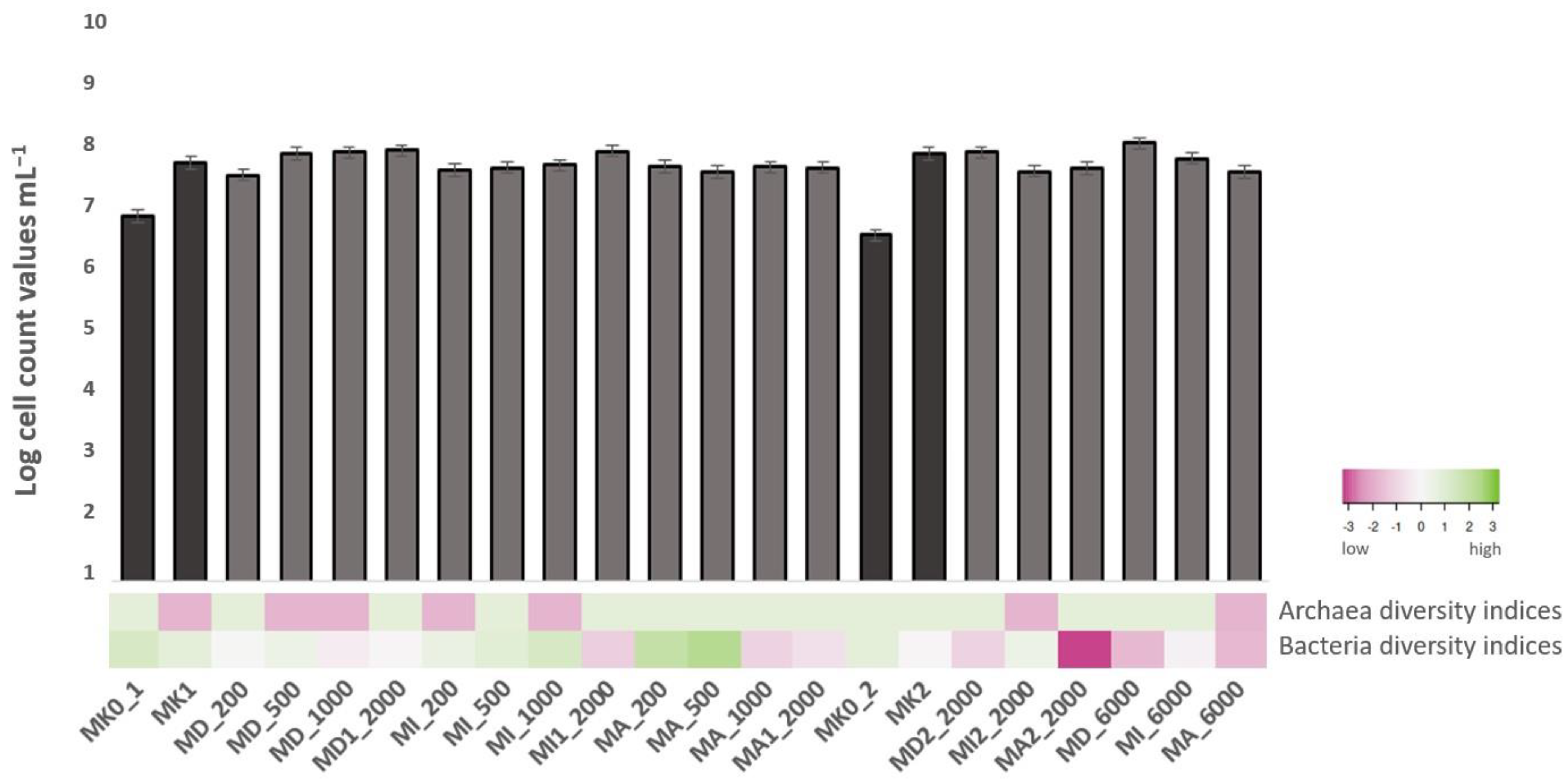
Figure 1. Cell count values with 0.1 standard error and Shannon–Wiener diversity indices of the microcosms before incubation (MK0_1; MK0_2) and after 3 weeks incubation (MK1; MD_200; MD_500; MD_1000; MD1_2000; MI_200; MI_500; MI_1000; MI1_2000; MA_200; MA_500; MA_1000; MA1_2000; MK2; MD2_2000; MI2_2000; MA2_2000; MD_6000; MA_6000).
Microorganisms 11 01447 g001
Figure 2. Distribution of antibiotic resistance genes in the studied, isolated bacterial strains. (1) Adaptation to D-cysteine; (2) beta-lactamase resistance; (3) bile hydrolysis; (4) phosphomycin resistance; (5) MexC-MexD-OprJ multidrug efflux system; (6) multidrug resistance efflux pumps; (7) fluoroquinolone resistance; (8) streptothricin resistance; (9) tetracycline resistance, ribosome protection type; (10) tetracycline resistance, ribosome protection type.
Microorganisms 11 01447 g002
Figure 3. Distribution of aromatic metabolism genes in the studied, isolated bacterial strains degrading acetylsalicylic acid (ASA), diclofenac (DCF) and ibuprofen (IBU), respectively. (1) catabolic pathway of 4-hydroxyphenylacetic acid; (2) catechol branch of the beta-ketoadipate pathway; (3) central, meta-cleavage of aromatic compounds; (4) homogentisate pathway of catabolism of aromatic compounds; (5) protocatechol branch of the beta-ketoadipate pathway, (6): salicylate and gentisate.
Microorganisms 11 01447 g003
Figure 4. NMDS ordination of the microcosms with the Bray–Curtis dissimilarity index based on their bacterial and archaeal OTU composition (triangles: control samples, squares: first experiments (dark blue—with DCF, brown—with IBU, light blue—with ASA); dots: second experiments (red—2000 ppm of DCF, IBU and ASA; green: 6000 ppm of DCF, IBU and ASA)).
Microorganisms 11 01447 g004
Figure 5. (a) Distribution of the abundant bacterial phyla based on 16S rRNA gene amplicon sequencing in the microcosms. (b) Distribution of the detected classes of the phylum Proteobacteria based on 16S rRNA gene amplicon sequencing in the microcosms.
Microorganisms 11 01447 g005
Figure 6. Distribution of the abundant archaeal phyla based on 16S rRNA gene amplicon sequencing in the microcosms.
Microorganisms 11 01447 g006
Table 1. Name of the samples, control and first/second experiments of microcosms with anti-inflammatory drug content (ppm) and type (A: ASA [acetylsalicylic acid], D: DCF: [diclofenac], I: IBU [ibuprofen]).
Names of the SamplesControls and ExperimentsAnti-Inflammatory Drug Content (ppm)Anti-Inflammatory Drug Applied
MK0_1absolute control * (first)0none
MK1control ** (first)0none
MD_200first (July 2020)200DCF
MD_500first (July 2020)500DCF
MD_1000first (July 2020)1000DCF
MD1_2000first (July 2020)2000DCF
MI_200first (July 2020)200IBU
MI_500first (July 2020)500IBU
MI_1000first (July 2020)1000IBU
MI1_2000first (July 2020)2000IBU
MA_200first (July 2020)200ASA
MA_500first (July 2020)500ASA
MA_1000first (July 2020)1000ASA
MA1_2000first (July 2020)2000ASA
MK0_2absolute control * (second)0none
MK2control ** (second)0none
MD2_2000second (August 2020)2000DCF
MI2_2000second (August 2020)2000IBU
MA2_2000second (August 2020)2000ASA
MD_6000second (August 2020)6000DCF
MI_6000second (August 2020)6000IBU
MA_6000second (August 2020)6000ASA
* absolute control: Danube water at the starting date of the experiments. ** control: the absolute control sample incubated for three weeks under the same conditions just like the treated microcosms, without NSAID.
Table 2. Physicochemical parameters of the sampling sites.
ExperimentsLocationT °CConductivity (μS m−1)pHO2 (%)O2 (mg L−1)
first (July 2020)47.477357° N; 19.061891° E172237.8395.912.54
second (August 2020)18.52037.5793.613.22
Table 3. Results of the selective cultivation in the 1st cultivation experiment.
Strain DesignationTaxonomic IdentificationStrainSimilarity (%)
MA1Burkholderia ambifariaAMMD99.79
MA2Burkholderia ambifariaAMMD99.81
MA4Novosphingobium arvoryzaeJYI-02100
MA6Paenibacillus lautusNBRC 1538098.92
MA7Burkholderia vietnamiensisLMG 1092999.89
MA8Burkholderia vietnamiensisLMG 1092999.89
MA9Pseudoduganella danionisE3/298.54
MA10Burkholderia vietnamiensisLMG 1092999.89
MD7Sphingobium yanoikuyaeATCC 51230100
MD8Ensifer adhaerensCasida A100
MD9Klebsiella grimontii06D021100
MI1Pseudomonas nitritireducensWZBFD3-5A299.70
MI3Novosphingobium barchaimiiLL0299.43
MI4Klebsiella pneumoniae subsp. ozaenaeATCC 1129699.90
MI10Sphingobium yanoikuyaeATCC 51230100
MI11Novosphingobium barchaimiiLL0299.62
MI12Pseudomonas nitroreducensDSM 1439999.77
MI13Sphingobium yanoikuyaeATCC 51230100
MI14Cupriavidus agavaeASC-984298.70
Table 4. Results of the selective cultivation in the 2nd cultivation experiment.
Strain DesignationTaxonomic IdentificationStrainSimilarity (%)
MD1Aeromonas sanarelliiLMG 2468299.90
MD2Aeromonas sanarelliiLMG 2468299.90
MD6Rahnella acerisSAP-1998.49
MD7_2Rahnella acerisSAP-19100.00
MD8_2Aeromonas caviaeCECT 838100.00
MD10Klebsiella michiganensisW1498.92
MD12Aeromonas sanarelliiLMG 2468299.91
MD13Rahnella acerisSAP-19100.00
MD15Aeromonas caviaeCECT 83898.95
MD17Rahnella acerisSAP-19100.00
MD18Rahnella acerisSAP-19100.00
MD19Klebsiella michiganensisW1498.92
MD21Citrobacter bitternisSKKUI-TP799.81
MD22Aeromonas caviaeCECT 83898.95
MD23Rahnella acerisSAP-19100.00
MD24Phytobacter diazotrophicusLS 899.39
MD25Rahnella acerisSAP-19100.00
MD26Klebsiella grimontii06D021100.00
MD27Klebsiella grimontii06D021100.00
MD28Rahnella acerisSAP-19100.00
MD29Aeromonas caviaeCECT 83898.95
MD30Aeromonas taiwanensisLMG 2468399.50
MD31Aeromonas sanarelliiLMG 2468299.81
MD32Aeromonas sanarelliiLMG 2468299.90
MD33Klebsiella michiganensisW1499.61
MI1_2Klebsiella quasivariicolaKPN170599.80
MI2Klebsiella pneumoniae subsp. pneumoniaeDSM 3010499.61
MI3_2Pseudomonas aeruginosaJCM 596299.74
MI6Klebsiella huaxiensisWCHKl090001 ATCC 1129699.84
MI9Klebsiella pneumoniae subsp. ozaenaeATCC 1129699.90
MI10_2Klebsiella quasivariicolaKPN170599.80
MI11_2Klebsiella quasivariicolaKPN170599.80
MI12_2Klebsiella huaxiensisWCHKl09000198.73
MI14_2Klebsiella huaxiensisWCHKl090001 W1499.24
MI15Klebsiella michiganensisW1499.65
MI17Raoultella ornithinolyticaJCM 6096100.00
MI18Klebsiella huaxiensisWCHKl090001100.00
MI21Klebsiella michiganensisW1499.44
MI22Kosakonia radicincitansDSM 1665699.66
MI23Klebsiella huaxiensisWCHKl090001100.00
MI24Klebsiella huaxiensisWCHKl09000199.20
Disclaimer/Publisher’s Note: The statements, opinions and data contained in all publications are solely those of the individual author(s) and contributor(s) and not of MDPI and/or the editor(s). MDPI and/or the editor(s) disclaim responsibility for any injury to people or property resulting from any ideas, methods, instructions or products referred to in the content.