Article

Biofilm Formation by Mutant Strains of Bacilli under Different Stress Conditions

1
Institute of Fundamental Medicine, Kazan Federal University, Kremlevskaya St. 18, 420008 Kazan, Russia
2
Interdisciplinary Center of Analytical Microscopy, Kazan Federal University, Paris Commune St. 9, 420008 Kazan, Russia
3
Department of Molecular Pathology, NYU College of Dentistry, 345 E. 24th Street, New York, NY 10010, USA
*
Authors to whom correspondence should be addressed.
Microorganisms 2023, 11(6), 1486; https://doi.org/10.3390/microorganisms11061486
Submission received: 19 April 2023 / Revised: 27 May 2023 / Accepted: 31 May 2023 / Published: 2 June 2023
(This article belongs to the Special Issue Advances in Microbial and Plant Biotechnology)

Abstract

:
Bacillus subtilis is traditionally classified as a PGPR that colonizes plant roots through biofilm formation. The current study focused on investigating the influence of various factors on bacilli biofilm formation. In the course of the study, the levels of biofilm formation by the model strain B. subtilis WT 168 and on its basis created regulatory mutants, as well as strains of bacilli with deleted extracellular proteases under conditions of changes in temperature, pH, salt and oxidative stress and presence of divalent metals ions. B. subtilis 168 forms halotolerant and oxidative stress-resistant biofilms at a temperature range of 22 °C–45 °C and a pH range of 6–8.5. The presence of Ca2+, Mn2+ and Mg2+ upsurges the biofilm development while an inhibition with Zn2+. Biofilm formation level was higher in protease-deficient strains. Relative to the wild-type strain, degU mutants showed a decrease in biofilm formation, abrB mutants formed biofilms more efficiently. spo0A mutants showed a plummeted film formation for the first 36 h, followed by a surge after. The effect of metal ions and NaCl on the mutant biofilms formation is described. Confocal microscopy indicated that B. subtilis mutants and protease-deficient strains differ in matrix structure. The highest content of amyloid-like proteins in mutant biofilms was registered for degU-mutants and protease-deficient strains.

1. Introduction

The rhizosphere is a complex ecosystem that includes plant roots and a layer of soil adjacent to the roots with a high content of microorganisms, which is the site of interaction between plants and bacteria. The interaction occurs as a result of root colonization by plant growth-promoting rhizobacteria (PGPR), among which representatives of the Bacillaceae family are deemed highly significant. The stages of colonization include chemotaxis, adhesion, aggregation, and biofilm formation which are largely determined by biological and environmental factors [1]. Colonization and formation of interactions of bacteria with plant roots activate chemotaxis [2,3]. Interaction in the rhizosphere occurs through a complex set of signaling molecules that control the behavior of microorganisms in communities. Specific gene products, among which transcription factors play a key role mediate the formation and development of the rhizomicrobiome [4].
Bacillus sp. are Gram-positive PGPRs useful for agriculturally important plants. Bacillus subtilis is a model organism for the study of biofilms, and the molecular networks and regulatory systems for biofilm formation in these bacteria have been well characterized. The transcription factors Spo0A, DegU, and AbrB are involved in the control of biofilm formation; they regulate the expression of genes associated with the production of the cell matrix [5,6,7]. Several studies have confirmed root exudates and their individual components (sugars, organic acids, amino acids) as important promoters of the growth and biofilm formation of PGPR [8,9,10]. Nevertheless, the influence of environmental and stress factors on these processes remains poorly understood. Understanding the process of colonization by Bacillus strains and the search for factors influencing this process is necessary to control the use of rhizobacteria in agriculture [11]. An important role in the process of colonization is played by the secreted enzymes of bacilli, which are involved in the transformation of proteins to peptides, many of which are important signaling molecules in the rhizosphere [12]. In addition to physical and chemical factors, bacteria united by a biofilm develop and differentiate under the action of enzymes. Information on the control of enzymes in the biofilm development cycle is insufficient. Such knowledge is useful for the development of a general strategy for the management of biofilms of spore-forming B. subtilis bacteria, which is considered an effective biocontrol tool.
This study aimed to investigate the biological and genetic factors influencing the biofilm formation of the potential plant root colonizer B. subtilis (strain B. subtilis 168). The results obtained from this study are essential for determining the optimal conditions for biocontrol activity to maximize crop productivity.

2. Materials and Methods
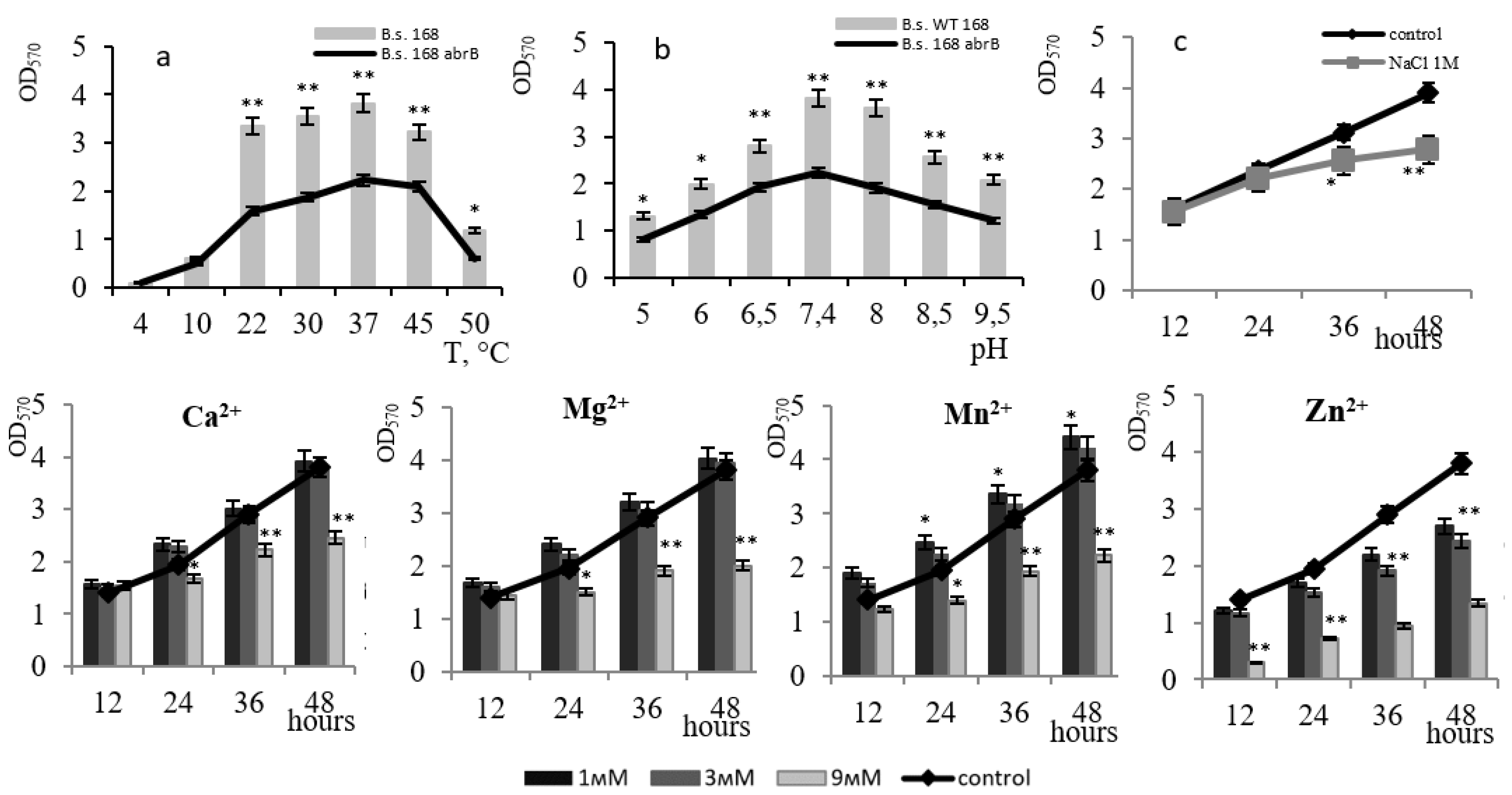
The strains used in the study are shown in the Table 1.
Cultivation of the strains was performed on a synthetic-E medium, of the following composition (g/L): L-glutamic acid—20; citric acid—12; glycerol—80; NH4Cl—7; MgSO4*7H2O—0.5; K2HPO4—0.5; CaCl2*2H2O—0.15; FeCl3*6H2O—0.04, MnSO4*H2O—0.148, pH 7.4, T = 37 °C [13]. Seed served 16-h inoculum (1% v/v). Bacterial growth was monitored by the change in the optical density of the culture at 600 nm (xMark spectrophotometer BioRad, Hercules, CA, USA). Biomass was expressed in absorbance units. Spore formation was determined by counting cells and spores by Peshkov microscopy method mode microscope Carl Zeiss Jena (Germany) in 1600 times magnification in four visual fields. The number of free spores was expressed as a percentage of the total number of vegetative and sporulating cells. Solutions of antibiotics (kanamycin and tetracycline) were prepared separately and added to the medium immediately before inoculation of the culture at a final concentration of 10 μg/mL. NaCl was added to the medium in the dry form before autoclaving to a final concentration in the medium of 1 M. Hydrogen peroxide (H2O2) was added sterile for 24 h of biofilm at final concentrations of 1, 5, and 10 mM. Metal ions in the form of salts of CaCl2, MgCl2, MnCl2, and ZnCl2 were sterilized separately and introduced into the medium in final concentrations of 1, 3, and 9 mM before inoculation. The assessment of the influence of all stress factors (NaCl, H2O2, metal ions) was carried out on an E-medium, pH 7.4 at 37 °C. Biofilm formation is defined by the method set incubation with crystal violet (CV) [14] with modification [15]. The cell culture with optical density OD600 = 0.1 was introduced into 96-well plates with a U-shaped bottom, 100 µL per well, and incubated at 37 °C under a lid. After incubation, the culture liquid with the plankton culture was removed from the plates, the plate was washed with water and dried at room temperature. 125 μL of 0.1% crystal violet dye (Sigma-Aldrich, St. Louis, MO, USA) was added to the wells and kept for 10 min at room temperature. The plates were then washed again with water and dried at room temperature. 200 μL of dimethyl sulfoxide (DMSO) (Sigma-Aldrich, USA) was added to each well and kept for 15 min at room temperature, during which the crystal violet bound to the biofilm structure dissociates into a DMSO solution. The contents of the wells were taken (125 μL), and transferred to the wells of a flat-bottomed 96-well plate for measurement on an xMark spectrophotometer (BioRad, USA) at a wavelength of 570 nm. Sterile E-medium without cell culture was used as a control for the biofilms formation during the experiment. The presence of the ability to synthesize amyloids in the studied strain was established by testing using a medium containing 25 μg/mL of Congo Red dye [16]. The culture plates were incubated at a temperature of 37 °C for 12–48 h and the binding of the Congo Red dye by the cells was noted by changing the color of the colonies.
The relative content of amyloid fibrils in the bacterium B. subtilis was estimated according to the method described in the article by Gophna et al., 2001 [17]. To study the relative content of amyloid fibrils, B. subtilis bacteria were grown on plates with agar medium E for 2 days at 37 °C. Colonies were scraped off and resuspended in a sterile 0.9% NaCl solution. The concentration of bacteria was determined by measuring the optical density at 570 nm. The cell suspension was diluted to a turbidity index OD570 = 0.9, and 1 mL was taken and centrifuged for 10 min at 14,000 rpm. After removing the supernatant, bacteria were resuspended in 1 mL of Congo Red solution (0.002% in 0.9% NaCl) and incubated for 10 min at room temperature. Next, centrifugation was repeated under the same conditions, the supernatant was taken, and the optical density was measured at 500 nm. The intensity of binding of the Congo Red dye by the cells was assessed by the decrease in optical density relative to the initial solution of the Congo Red dye. To prepare biofilms for SEM, a 16-h cell culture grown at 37 °C was used. Bacteria were grown on PVC catheters placed in the wells of a plastic 12-well cell culture plate (Corning Costar, Corning, NY, USA). Catheters were cut out 1 × 1 cm2 in size and sterilized at 1 atm., 121 °C within 30 min. The required volume of cell culture was added to each well so that the final optical density (OD600) was 0.2. The plates were incubated for 12 h to 2 days at 37 °C. The catheters were washed with phosphate buffer PBS and the biofilm samples were chemically fixed in 1.5% glutaraldehyde for 12–16 h. Next, the samples were dehydrated in a series of alcohols with increasing concentration—30%, 40%, 50%, 70%, 80%, and 90% for 15 min each.
All analyses were performed at least on four biological replicates. The obtained data were processed using Statgraphics Plus 5.0. and GraphPad Prism 7.05 statistical software, and are presented as the mean ± standard deviation (SD). Student’s t-test analysis was used to calculate the data variance with p < 0.05 (*) and p < 0.01 (**) representing a significant difference.

3. Results

Analysis of the growth dynamics, biofilm formation, and sporulation of the B. subtilis strain showed that it develops biofilm beginning at 12 h of bacterial growth and reaches a maximum at 48 h in the stationary phase of culture growth. Biofilm dispersion, which began after 70 h correlated with the cell culture dying phase. Spores appeared in the medium at 46 h during the phase of biofilm development, and active sporulation occurred after 60 h at the stage of biofilm dispersion. Thus, it was observed that the processes of biofilm formation and sporulation, are separate and do not occur simultaneously, but develop sequentially (Figure 1a).
Based on the ultrastructural data, the B. subtilis 168 biofilm is formed in 2 days (Figure 1b). At 24 h of cultivation, cells begin to adhere to the surface. By the 36th h, the cells produce extracellular polymeric compounds and form clusters, a matrix appears that binds culture cells together, and at 48 h a mature biofilm with bacterial cells immersed in a dense protective matrix is observed.
The organization of the B. subtilis biofilm can begin at 10 °C, but the structure is well favored and maintained at a temperature range from 22 °C to 45 °C. At 50 °C the process occurs at a very lagged rate (Figure 2a). The bacilli are capable of forming biofilms in a slightly acidic environment (pH 5–pH 6.0). However, the optimal pH for biofilm formation ranges from 7.4–pH 8, a subsequent increase in pH to 8.5–9.0 plummets the level of biofilm formation (Figure 2b). The optimal pH for biofilm formation is closer to neutral. Under natural conditions, such a pH range is essential for the active colonization of plant roots with bacilli biofilms, which in turn facilitates the formation of mutually beneficial symbiotic relationships [18]. The upsurge in studies of this kind is relevant and promising for the advancement of agrobiotechnology since soil strains of B. subtilis are considered effective biocontrol agents in plant protection against soil pathogens [19,20,21,22].
The effectiveness of colonization control to a large extent depends on the content of metals in the rhizosphere, therefore, deciphering the role of metal ions in the formation of bacilli biofilms is imperative [23]. We studied the effect of divalent metal ions on the development of B. subtilis 168 biofilms (Figure 2).
The introduction of Ca2+ ions at a concentration of 3 mM or lower led to an increase in the level of B. subtilis 168 biofilm formation by up to 45% at 12 h of growth and up to 11% at 36 h of growth. With an increase in the concentration of exogenous calcium to 9 mM, the film formation was partially inhibited by 10%. Interestingly, it was noted that calcium effectively acts precisely at the early stage of B. subtilis biofilm maturation (Figure 2). There is evidence that Ca2+ ions play an important role in the adhesion of bacilli cells during biofilm formation [24]. The presence of calcium in the medium affects the regulatory pathways of B. subtilis 168 during flagella development and the synthesis of biofilm matrix components [25]. While analyzing the transcriptome of B. subtilis 168, the authors identified 305 genes, the expression of which is affected by calcium, including genes for the biofilm matrix biosynthesis and genes encoding the biosynthesis of spore coat polysaccharides. [25]. The interactions between the microorganisms and Ca2+ enhance both the extent and the rate of biofilm development with increasing Ca2+ concentration [26]. The authors note that the Ca2+ concentration also correlates with the B. subtilis biofilm morphology. Recent studies have shown that microbial biofilms contain an organized internal mineral structure composed of crystalline calcium carbonate (calcite), which largely determines their three-dimensional morphology [27].
Similar data were obtained when Mn2+ ions were introduced into the medium. The results showed that Mn2+ is essential at the starting point of B. subtilis 168 biofilm maturation; an increase in the level of film formation was observed up to the 36th h at Mn2+ concentrations of 1–3 mM (Figure 2). The absence of inhibition following the treatment of cells with Ca2+ and Mn2+ indicates their non-toxicity to biofilm formation. There is evidence that the exogenous presence of Mn2+ ions promotes the formation of stable biofilms in bacilli by participating in the activation of phosphorel, which is necessary for phosphorylation of the global transcription factor Spo0A, presumably acting as a signaling cofactor with histidine kinase KinD and protein components of phosphorele, Spo0F and Spo0B phosphotransferases [28]. The effect of manganese ion stimulation on biofilm formation has been found for many bacilli and suggests that this signaling pathway for biofilm formation is highly conserved for this group of microorganisms [29,30].
The effect of Mg2+ ions at final concentrations of 1, 3, and 9 mM on the development of B. subtilis 168 biofilm was studied (Figure 2). It was established that Mg2+ ions at a concentration of 1–3 mM contribute to biofilm structure at the early stages of its organization; after 24 h, we observed a decrease in film formation of up to 10% in the presence of 3 mM Mg2+. An increase in the magnesium concentration in the medium to 9 mM led to a decrease in the level of biofilms of B. subtilis 168 by 60%. Per the literature data, Mg2+ ions play an important role in regulating the adhesion of cells that have lost their mobility [31]. The authors emphasize that the additional introduction of divalent magnesium cations into the cultivation solution leads to a significant increase in adhesion during biofilm colonization of bacilli.
Zn2+ ions at concentrations of 1 and 3 mM did not affect the formation of B. subtilis 168 biofilms within 12 h of growth. However, at a Zn2+ concentration of 9 mM, a decrease in the level of biofilm formation (from 60% to 70%) was observed up to 48 h (Figure 2). Moreover, even at high concentrations of Zn2+ ions, only partial suppression of biofilm formation was observed under structured community conditions. The toxic effect of Zn2+ ions is associated with its ability to complex formation, which in turn can disrupt protein function in the cell membrane including enzyme systems of the respiratory chain [32].
The addition of 1 M NaCl to the culture medium led to a 1.5-fold decrease in the formation of B. subtilis 168 biofilms by the 48th hour of growth (Figure 2c). In the presence of 1 M NaCl, a structured community of halotolerant bacteria is preserved in the biofilm, which supports population survival under salt stress. There is evidence that B. subtilis histidine kinase can function as an osmosensor due to the extracellular domain of CACHE, which seems to be important for the continued colonization of B. subtilis under stress conditions [28].
Reactive oxygen species and oxidizing agents, such as H2O2, are involved in lipid peroxidation, protein degradation, and enzyme inactivation, causing significant damage to bacteria [33]. Bacteria have developed protective mechanisms against oxidative stress. Among such mechanisms are the synthesis of catalase and superoxide dismutase, essential for the removal of free radicals. Three families of catalase genes have been identified in the genomes of bacteria, namely: monofunctional catalases, heme-containing bifunctional catalase-peroxidases, and Mn-containing catalases [33,34]. Together they provide a well-balanced defense system for aerobic bacteria to ensure the survival of living cells by removing reactive oxygen species.
The effect of oxidative stress on the formation of a B. subtilis 168 biofilm was studied when hydrogen peroxide was added to the cultivation medium at concentrations from 1 to 10 mM. Inhibition of the growth of planktonic culture by 1.9 times for 48 h was observed. At a peroxide concentration of 10 mM (Figure 3A), 1.4-fold suppression of biofilm development was registered under the same conditions (Figure 3B). In addition, the degree of inhibition increased with increasing peroxide concentration; however, the degree of resistance to peroxide in a biofilm culture is higher than that of planktonic culture. Thus, H2O2 inhibits film formation to a lesser extent than growth in free bacteria.
To comparatively study the formation of B. subtilis biofilm, we used regulatory mutants, i.e., strains whose genome contains inactivated genes for global transcription factors that affect the formation of bacilli biofilms: Spo0A, DegU, and AbrB [5,6,7]. The following strains were used in the experiment: B. subtilis 168 (wild type), B. subtilis abrB with an inactivated abrB gene (pleiotropic regulator of the vegetative phase), B. subtilis degU with an inactivated degU gene (a component of the DegS/DegU regulation system), and B. subtilis spo0A—with an inactivated spo0A gene (stationary phase regulator). The formation of biofilms by these strains was studied (Figure 4).
The strain with the inactivated pleiotropic repressor AbrB showed an increase in the level of biofilm formation of up to 20% within the first 48 h; further, the level of film formation remained similar to that of the control (Figure 4). The degU-mutant showed a decrease in biofilm formation over 72 h, with a maximum decrease of up to 40% at 48 h. spo0A-deficiency contributed to a decrease in the level of film formation for the first 48 h of growth, while at the late stage (after 60 h) in spo0A mutants, an increase in the level of biofilm formation was observed in comparison to the wild-type strain (Figure 4). At later stages, the transcription factor Spo0A initiates the sporulation process. Our data are consistent with genetic data that, at the stage of vegetative growth, the AbrB repressor is involved in the suppression of the expression of operon genes encoding the biofilm matrix; later, the DegU regulator is involved in the activation of biofilm matrix genes; at the biofilm dispersion stage, the Spo0A factor blocks the expression of biofilm matrix genes and activates spore formation. The process of biofilm formation is complex and its regulation is under the control of a multiple-branched regulatory network. In our experiments, none of the regulatory proteins blocked the formation of biofilm completely. This process is subject to the integrated control of regulatory systems that evaluate the input of many different signals. Therefore, the inactivation of one of the regulators was not critical; in the absence of one of the regulatory proteins, the biofilm is formed under the control of another regulatory network.
The biofilms of the spo0A-mutant and the wild-type strain, showed maximum growth at 37 °C and pH 7.4 (Figure 5a). However, the response of the spo0A-mutant to the presence of metals differed from that of the wild-type strain: in the presence of 3 mM Ca2+ and 3 mM Mn2+ ions in the medium, biofilm inhibition was observed at a level of up to 20%; this effect was absent for Mg2+ at concentrations of 3 mM. The data reflect the participation of Ca2+ and Mn2+ in activating the phosphorylation of the transcription factor Spo0A (Figure 5a). The toxic effect of Zn2+ on film formation was only noticed in the wild-type strain. Data on the resistance of the spo0A-mutant to 1 M NaCl are comparable with that of the wild-type strain.
Activation of the Spo0A factor depends on the phosphorylating action of at least five different kinases (KinA–E), which are responsible for the transfer of the phosphate group to the Spo0A regulator [35,36]. Kinase activation is regulated by specific signals, some of which have been identified. KinC induces low levels of Spo0A~P activation in response to changes in the content of potassium cations [37]. KinD is sensitive to the content of manganese ions [28,38]. KinB is activated due to interruptions in electron transport resulting from environmental influences (low oxygen content or large concentrations of iron ions). KinA responds to NAD/NADH levels in the cytoplasm [39]. The level of Spo0A activation is determined by the cumulative signal input, in which metal ions are involved as cofactors. The obtained data on an increase in the level of film formation of spo0A mutants in the presence of Ca2+ and Mn2+ ions indicate an important role of these metals in the activation of the Spo0A transcription factor. The activated form of Spo0A~P in turn inhibits the expression of AbrB, which is an alternative repressor for the expression of biofilm matrix genes [37].
Interesting results were obtained for the degU mutant biofilm (Figure 6). The optimal conditions for biofilm formation were 37 °C and pH 7.4, and were the same as the optimal conditions for the wild-type strain. However, the dynamics of biofilm formation changed with the presence of divalent metal ions: the biofilm reached its maximum level at 36 h of growth, followed by the stage of biofilm dispersion (Figure 6). In contrast to the spo0A-mutant, biofilm formation was activated in the presence of 1–3 mM Ca2+, Mg2+, and Mn2+ averaged by ~20%, with a maximum effect at a concentration of 1 mM. With an increase in concentration to 9 mM, the film formation activity decreased with a profound effect by Mg2+ and Mn2+ ions. For Zn2+ ions, the resistance of the degU-mutant biofilm to their toxic effects increased by 10% compared to the wild-type strain. When 1 M sodium chloride was introduced into the medium, the ability to form a biofilm in the deg-mutant decreased by 70% within 48 h, as compared to 30% for the cells of the wild-type strain under identical conditions. Thus, the inactivation of the transcription factor DegU leads to an early dispersion of the biofilm and a decrease in the strain’s salt tolerance.
The abrB-mutant formed biofilms at the same optimal temperature and pH as the wild strain (Figure 7a,b). The mutation did not affect the level of biofilm formation when 1 M sodium chloride was added to the medium (Figure 7c). In the presence of Ca2+, Mg2+, and Mn2+ at concentrations ≤ 3 mM, biofilm formation was activated with the maximum effect (by 17%) of 1 mM Mn2+. Increasing the concentration to 9 mM declined the film-forming activity, with a greater effect by Mg2+ and Mn2+ ions. The resistance of the abrB-mutant biofilm to the toxic effect of Zn2+ ions increased by 10% compared to the wild-type strain. A similar effect was also observed in the deg-mutant. Thus, biofilm formation of the abrB-mutant depends on the presence of metal ions in the medium with a strong effect by 3 mM Mn2+ ions.
Biofilms of B. subtilis are the main type of cooperative existence of bacteria in the rhizosphere. In addition to physical and chemical factors, bacteria united by a biofilm are regulated and differentiated into subpopulations inside the biofilm under enzymatic influences. There is a dearth of information on the effect of enzymes on biofilm development. Such knowledge is essential for the development of a general strategy for the management of biofilms of spore-forming B. subtilis bacteria, which are considered to be an effective biocontrol tool.
We studied the contribution of the proteolytic activity of bacilli to the formation of B. subtilis biofilm; these bacteria effectively secrete various proteases into the medium. B. subtilis strains defective in the synthesis of extracellular proteases were used. 62 protease genes have been identified in the B. subtilis genome, of which 14 are secreted [40]. We used the B. subtilis 20–36 strain, with two inactivated extracellular proteinases, the B. subtilis BRB8 strain with nine inactivated extracellular proteinases, and the B. subtilis BRB14 strain, with eleven inactivated extracellular proteinases. The dynamics of biofilm formation by the mutant strains were studied at pH 7.4 and a cultivation temperature of 37 °C (Figure 8).
The mutant strains demonstrated an increase in the level of biofilm formation compared to the wild-type strain, and the greater the number of inactivated proteinase genes, the higher the level of biofilm formation (Figure 8). The maximum difference in biofilm optical density from the wild-type strain was 15%, which was registered in the mutant strain with 11 inactivated genes of extracellular proteinases (Figure 8).
A comparative study of the characteristics of the biofilm of the B. subtilis BRB14 strain showed that the mutant strain formed a biofilm analogous to that of the wild-type while maintaining a film-formation maximum at 48 h (Figure 9a,b). The biofilm of B. subtilis BRB14 was more resistant to 1 M NaCl relative to that of the wild-type strain (Figure 9c). In the presence of Ca2+, Mg2+, and Mn2+ ions in the medium at concentrations ≤ 3 mM, biofilm formation was sustainably activated with a maximum effect by Mn2+ ions at concentrations ≤ 3 mM (up to 10%). Ca2+ and Mg2+ concentrations ≤ 3 mM showed a positive effect just until the 36th h. With an upsurge in concentration to 9 mM, the film formation activity declined, but to a lesser extent as compared to the wild-type strain. The resistance of the B. subtilis BRB14 biofilm to the toxic effect of Zn2+ ions increased by 6% relative to the wild-type strain. Comparative analysis depicted the B. subtilis BRB14 biofilm as more resistant to 1 M NaCl than that of the other mutant strains.
Extracellular proteases can potentially participate in the processes of control over the formation of the biofilm matrix during the processing of regulatory peptides, as well as in the dispersion of the biofilm. All genetic cascades of B. subtilis are negatively controlled by the Rap family of phosphatases that specifically dephosphorylate global regulators. Each Rap phosphatase is inactivated by a specific cognate extracellular regulatory peptide (Phr peptides) [41]. Rap-Phr pairs coordinate various biological processes with population density, including biofilms [42]. RapGH phosphatase is known to dephosphorylate DegU~P while RapABEJ phosphatases dephosphorylate Spo0A~P. Regulatory peptides are an alternative signaling program between cells as a mode of interaction [43]. Extracellular proteinases may be involved in peptide turnover [40]. Our data showed that the inactivation of 11 genes of B. subtilis extracellular proteinases leads to an increase in film formation (Figure 8).
A common feature of bacilli biofilms is the abundance of architectural features, the complexity of which depends on ecological and stress factors of the environment and is controlled by the cooperative interaction of regulatory networks. The ultrastructure of B. subtilis 168 biofilms and mutant strains defective in transcription factor genes and extracellular proteinase genes (BRB14) was studied by scanning electron microscopy (Figure 10 168). The B. subtilis BRB14 biofilm is distinguished by a more intensive formation of an extracellular matrix with a branched structure (Figure 10 BRB14). The absence of extracellular proteinases leads to the formation of a biofilm with a denser structure. The biofilm of the spo0A-mutant is characterized by the formation of a matrix with embedded bacilli cells, while the three-dimensional structure of the degU-mutant biofilm differs in morphology and more pronounced architecture compared to the biofilm of the spo0A-mutant and the wild type strain (Figure 10 spo0A, degU). The biofilm of the abrB-mutant differs from the others and contains more matrix; the cells are immersed in it, which is associated with the inactivation of the repressor of the matrix gene operons (Figure 10 abrB). Inactivation of each of these genes has a different effect on the structure and morphology of the biofilm matrix as a result of changes in regulatory networks; however, even if one of the transcription factors is switched off, bacilli proceed to form biofilms, which confirms the complexity of the ways to control its formation.
The three-dimensional architecture of the B. subtilis biofilm has been understudied. The extracellular biofilm matrix of B. subtilis includes an exopolysaccharide that is critical for community architecture and function. Understanding of the mechanism of biosynthesis and molecular composition of B. subtilis exopolysaccharide remains incomplete [44]. The structural integrity of the matrix is provided by two secreted proteins, TasA and TapA, which are encoded by the operon of three tapA-sipW-tasA genes (tapA operon) [45]. TasA is a functional amyloid protein that is secreted into the extracellular space, where it self-assembles into fibers that are attached to the cell wall by the TapA protein [46]. This architecture ensures the resistance of the biofilm to external influences.
The ability to synthesize amyloid-like proteins in the biofilm of B. subtilis strains was established by testing on medium E with the Congo Red dye (Figure 11).
On the 2nd day of cultivation, the colonies on the agar medium turned red, indicating the presence of amyloid-like proteins in the B. subtilis biofilm. We studied the binding of the Congo Red dye by extracellular amyloids of biofilms of different strains of B. subtilis: a protease-deficient strain and regulatory mutants spo0A and degU and compared with the data for the wild-type strain B. subtilis 168 (Figure 11). It was found that relative to the wild-type strain, BRB14 and the degU mutant contain more amyloid-like proteins in the biofilm, while the spo0A mutant, on the contrary, contains less. Based on the results of studies on the ultrastructure and relative content of amyloid-like proteins in biofilms of different strains, it can be concluded that extracellular proteinases can participate in the dissociation of the protein components of the B. subtilis biofilm and contribute to the functioning of this complex structure at the dispersion stage.

4. Conclusions

Biofilms and sporulation are predominantly the natural modes of bacilli life, in particular in the rhizosphere. Biofilm formation is a flexible system of adaptation to environmental factors and a symbiotic way of interacting with macroorganisms such as plants, for example. The processes of biofilm formation and sporulation are regulatorily separated and do not occur simultaneously but develop in accordance with incoming signals. Biofilms of B. subtilis develop at a temperature range of 20 °C to 45 °C and a pH range of 6.5–8.5; sporulation begins outside these parameters. The B. subtilis genome contains genes regulating mechanisms of biofilm formation and development. These mechanisms are based on complex genetic programs that are implemented in compliance with a combination of external factors and signals. A study of the ultrastructure of biofilms of B. subtilis mutant strains defective in the genes of extracellular proteinases and regulatory proteins showed that they differ in architecture, structural heterogeneity of the matrix, and in the content of amyloid-like proteins, the maximum level of which was established for deg-mutants and strains defective in extracellular proteinases. The level of biofilm formation is partially inhibited in deg-mutants by about 30–40%, and upsurged in abrB-mutants by 25% relative to the wild-type strain. This means that the loss of one of the regulators Spo0A, DegU, and AbrB did not cause a critical change in biofilm formation; together they exert multiple control of film formation. Since B. subtilis naturally inhabits more complex environments, such as plant roots, the presence of other bacteria, and rhizosphere organisms, control mechanisms, and interactions can even be more bewildering.

Author Contributions

Conceptualization, M.S. and N.R.; methodology, M.S. and A.M.; software, I.D.; validation, M.S., N.R. and A.S.; formal analysis, M.S., N.R. and A.M.; investigation, N.R. and I.D.; resources, M.S., V.E.; data curation, M.S., N.R.; writing—original draft preparation, M.S.; writing—review and editing, M.S., N.R. and Y.A.; visualization, N.R.; supervision, M.S.; project administration, M.S.; funding acquisition, M.S. All authors have read and agreed to the published version of the manuscript.

Funding

This research was supported by the RSF grant (Russian Science Foundation) №22-16-00138.

Data Availability Statement

The data obtained in this study are available upon request.

Acknowledgments

The study was carried out thanks to the technical resources of the Strategic Academic Leadership Program of the Kazan Federal University (PRIORITET-2030).

Conflicts of Interest

The authors declare no conflict of interest. The funders had no role in the design of the study; in the collection, analyses, or interpretation of data; in the writing of the manuscript; or in the decision to publish the results.

References

  1. Huang, R.; Feng, H.; Xu, Z.; Zhang, N.; Liu, Y.; Shao, J.; Shen, Q.; Zhang, R. Identification of adhesins in plant beneficial rhizobacteria Bacillus velezensis SQR9 and their effect on root colonization. Mol. Plant-Microbe Interact. 2022, 35, 64–72. [Google Scholar] [CrossRef]
  2. Feng, H.; Fu, R.; Hou, H.; Lv, Y.; Zhang, N.; Liu, Y.; Xu, Z.; Miao, Y.; Krell, T.; Shen, Q.; et al. Chemotaxis of beneficial rhizobacteria to root exudates: The first step towards root–microbe rhizosphere interactions. Int. J. Mol. Sci. 2021, 22, 6655. [Google Scholar] [CrossRef]
  3. Allard-Massicotte, R.; Tessier, L.; Lécuyer, F.; Lakshmanan, V.; Lucier, J.-F.; Garneau, D.; Caudwell, L.; Vlamakis, H.; Bais, H.V.; Beauregard, P.B. Bacillus subtilis early colonization of Arabidopsis thaliana roots involve multiple chemotaxis receptors. MBio 2016, 7, e01664-16. [Google Scholar] [CrossRef] [Green Version]
  4. Hashem, A.; Tabassum, B.; Abd Allah, E.F. Bacillus subtilis: A plant-growth promoting rhizobacterium that also impacts biotic stress. Saudi J. Biol. Sci. 2019, 26, 1291–1297. [Google Scholar] [CrossRef]
  5. Arnaouteli, S.; Bamford, N.C.; Stanley-Wall, N.R.; Kovács, A.T. Bacillus subtilis biofilm formation and social interactions. Nat. Rev. Microbiol. 2021, 19, 600–614. [Google Scholar] [CrossRef]
  6. Cairns, L.S.; Hobley, L.; Stanley-Wall, N.R. Biofilm formation by Bacillus subtilis: New insights into regulatory strategies and assembly mechanisms. Mol. Microbiol. 2014, 93, 587–598. [Google Scholar] [CrossRef] [Green Version]
  7. Vlamakis, H.; Chai, Y.; Beauregard, P.; Losick, R.; Kolter, R. Sticking together: Building a biofilm the Bacillus subtilis way. Nat. Rev. Microbiol. 2013, 11, 157–168. [Google Scholar] [CrossRef] [Green Version]
  8. Liu, Y.; Feng, H.; Fu, R.; Zhang, N.; Du, W.; Shen, Q.; Zhang, R. Induced root-secreted D-galactose functions as a chemoattractant and enhances the biofilm formation of Bacillus velezensis SQR9 in an McpA-dependent manner. Appl. Microbiol. Biotechnol. 2020, 104, 785–797. [Google Scholar] [CrossRef]
  9. Jiao, H.; Xu, W.; Hu, Y.; Tian, R.; Wang, Z. Citric acid in rice root exudates enhanced the colonization and plant growth-promoting ability of Bacillus altitudinis LZP02. Microbiol. Spectr. 2022, 10, e0100222. [Google Scholar] [CrossRef]
  10. Vora, S.M.; Joshi, P.; Belwalkar, M.; Archana, G. Root exudates influence chemotaxis and colonization of diverse plant growth promoting rhizobacteria in the pigeon pea-maize intercropping system. Rhizosphere 2021, 18, 100331. [Google Scholar] [CrossRef]
  11. Pandin, C.; Le Coq, D.; Canette, A.; Aymerich, S.; Briandet, R. Should the biofilm mode of life be taken into consideration for microbial biocontrol agents? Microb. Biotechnol. 2017, 10, 719–734. [Google Scholar] [CrossRef] [PubMed] [Green Version]
  12. Mahapatra, S.; Yadav, R.; Ramakrishna, W. Bacillus subtilis impact on plant growth, soil health and environment: Dr. Jekyll and Mr. Hyde. J. Appl. Microbiol. 2022, 132, 3543–3562. [Google Scholar] [CrossRef] [PubMed]
  13. Morikawa, M.; Kagihiro, S.; Haruki, M.; Takano, K.; Branda, S.; Kolter, R.; Kanaya, S. Biofilm formation by a Bacillus subtilis strain that produces γ-polyglutamate. Microbiology 2006, 152, 2801–2807. [Google Scholar] [CrossRef] [PubMed] [Green Version]
  14. O’Toole, G.A.; Pratt, L.A.; Watnick, P.I.; Newman, D.K.; Weaver, V.B.; Kolter, R. Genetic approaches to study of biofilms. Methods Enzymol. 1999, 310, 91–109. [Google Scholar] [CrossRef]
  15. Merritt, J.H.; Kadouri, D.E.; O’Toole, G.A. Growing and analyzing static biofilms. Curr. Protoc. Microbiol. 2005, 1, Unit 1B.1. [Google Scholar] [CrossRef] [Green Version]
  16. Reichhardt, C.; Jacobson, A.N. Congo Red interactions with curli-producing E. coli and native curli amyloid fibers. PLoS ONE 2015, 10, e0140388. [Google Scholar] [CrossRef] [Green Version]
  17. Gophna, U.; Barlev, M.; Seijffers, R.; Oelschlager, T.A.; Hacker, J.; Ron, E.Z. Curli fibers mediate internalization of Escherichia coli by eukaryotic cells. Infect. Immun. 2001, 69, 2659–2665. [Google Scholar] [CrossRef] [Green Version]
  18. Khan, A.R.; Mustafa, A.; Hyder, S.; Valipour, M.; Rizvi, Z.F.; Gondal, A.S.; Yousuf, Z.; Iqbal, R.; Daraz, U. Bacillus spp. as bioagents: Uses and application for sustainable agriculture. Biology 2022, 11, 1763. [Google Scholar] [CrossRef]
  19. Ahmad, I.; Akhtar, M.J.; Mehmood, S.; Akhter, K.; Tahir, M.; Saeed, M.F.; Hussain, M.B.; Hussain, S. Combined application of compost and Bacillus sp. CIK-512 ameliorated the lead toxicity in radish by regulating the homeostasis of antioxidants and lead. Ecotoxicol. Environ. Saf. 2018, 148, 805–812. [Google Scholar] [CrossRef]
  20. Batool, T.; Ali, S.; Seleiman, M.F.; Naveed, N.H.; Ali, A.; Ahmed, K.; Abid, M.; Rizwan, M.; Shahid, M.R.; Alotaibi, M.; et al. Plant growth promoting rhizobacteria alleviates drought stress in potato in response to suppressive oxidative stress and antioxidant enzymes activities. Sci. Rep. 2020, 10, 16975. [Google Scholar] [CrossRef]
  21. Khedher, S.B.; Mejdoub-Trabelsi, B.; Tounsi, S. Biological potential of Bacillus subtilis V26 for the control of Fusarium wilt and tuber dry rot on potato caused by Fusarium species and the promotion of plant growth. Biol. Control 2021, 152, 104444. [Google Scholar] [CrossRef]
  22. Chu, D.; Wang, X.; Fan, J.; Peng, L.; Liang, J.; Zheng, Y.; Wang, R.; You, X.; Jing, C.; Clark, J.; et al. Ulva prolifera polysaccharide enhances the root colonisation by Bacillus amyloliquefaciens strain Cas02. Carbohydr. Polym. 2023, 299, 120171. [Google Scholar] [CrossRef] [PubMed]
  23. Yang, W.; Yan, H.; Zhang, J.; Gao, Y.; Xu, W.; Shang, J.; Luo, Y. Inhibition of biofilm formation by Cd2+ on Bacillus subtilis 1JN2 depressed its biocontrol efficiency against Ralstonia wilt on tomato. Microbiol. Res. 2018, 215, 1–6. [Google Scholar] [CrossRef] [PubMed]
  24. Wang, T.; Flint, S.; Palmer, J. Magnesium and calcium ions: Roles in bacterial cell attachment and biofilm structure maturation. Biofouling 2019, 35, 959–974. [Google Scholar] [CrossRef]
  25. Oomes, S.J.; Jonker, M.J.; Wittink, F.R.; Hehenkamp, J.O.; Breit, T.M.; Brul, T.M. The effect of calcium on the transcriptome of sporulating B. subtilis cells. Int. J. Food Microbiol. 2009, 133, 234–242. [Google Scholar] [CrossRef]
  26. Tian, L.; Chen, X.D.; Yang, Q.P.; Chen, J.C.; Shi, L.; Li, Q. Effect of calcium ions on the evolution of biofouling by Bacillus subtilis in plate heat exchangers simulating the heat pump system used with treated sewage in the 2008 Olympic Village. Colloids Surf. B: Biointerfaces 2012, 94, 309–316. [Google Scholar] [CrossRef]
  27. Keren-Paz, A.; Maan, H.; Karunker, I.; Olender, T.; Kapishnikov, S.; Dersch, S.; Kartvelishvily, E.; Wolf, S.G.; Gal, A.; Graumann, P.L.; et al. The roles of intracellular and extracellular calcium in Bacillus subtilis biofilms. Iscience 2022, 25, 104308. [Google Scholar] [CrossRef]
  28. Shemesh, M.; Chai, Y. A combination of glycerol and manganese promotes biofilm formation in Bacillus subtilis via histidine kinase KinD signaling. J. Bacteriol. 2013, 195, 2747–2754. [Google Scholar] [CrossRef] [Green Version]
  29. Mhatre, E.; Troszok, A.; Gallegos-Monterrosa, R.; Lindstädt, S.; Hölscher, T.; Kuipers, O.P.; Kovács, A.T. The impact of manganese on biofilm development of Bacillus subtilis. Microbiology 2016, 162, 1468–1478. [Google Scholar] [CrossRef]
  30. Ran, X.; Zhu, Z.; Long, H.; Tian, Q.; You, L.; Wu, X.; Liu, Q.; Huang, S.; Li, S.; Niu, X.; et al. Manganese stress adaptation mechanisms of Bacillus safensis strain ST7 from mine soil. Front. Microbiol. 2021, 12, 758889. [Google Scholar] [CrossRef]
  31. He, X.; Wang, J.; Abdoli, L.; Li, H. Mg2+/Ca2+ promotes the adhesion of marine bacteria and algae and enhances following biofilm formation in artificial seawater. Colloids Surf. B: Biointerfaces 2016, 146, 289–295. [Google Scholar] [CrossRef] [PubMed]
  32. Yang, L.; Kuang, H.; Liu, Y.; Xu, H.; Aguilar, Z.P.; Xiong, Y.; Wei, H. Mechanism of enhanced antibacterial activity of ultra-fine ZnO in phosphate buffer solution with various organic acids. Environ. Pollut. 2016, 218, 863–869. [Google Scholar] [CrossRef] [PubMed]
  33. Sooch, B.S.; Kauldhar, B.S.; Puri, M. Recent insights into microbial catalases: Isolation, production and purification. Biotechnol. Adv. 2014, 32, 1429–1447. [Google Scholar] [CrossRef]
  34. Shaeer, A.; Aslam, M.; Rashid, N. Structural and functional analyses of a novel manganese-catalase from Bacillus subtilis R5. Int. J. Biol. Macromol. 2021, 180, 222–233. [Google Scholar] [CrossRef] [PubMed]
  35. Jiang, M.; Shao, W.; Perego, M.; Hoch, J.A. Multiple histidine kinases regulate entry into stationary phase and sporulation in Bacillus subtilis. Mol. Microbiol. 2000, 38, 535–542. [Google Scholar] [CrossRef]
  36. Chen, Z.; Srivastava, P.; Zarazúa-Osorio, B.; Marathe, A.; Fujita, M.; Igoshin, O.A. Bacillus subtilis histidine kinase KinC activates biofilm formation by controlling heterogeneity of single-cell responses. MBio 2022, 13, e0169421. [Google Scholar] [CrossRef]
  37. Grau, R.R.; de Oña, P.; Kunert, M.; Leñini, C.; Gallegos-Monterrosa, R.; Mhatre, E.; Vileta, D.; Donato, V.; Hölscher, T.; Boland, W.; et al. A duo of potassium-responsive histidine kinases governs the multicellular destiny of Bacillus subtilis. MBio 2015, 6, e00581. [Google Scholar] [CrossRef] [Green Version]
  38. Beauregard, P.B.; Chai, Y.; Vlamakis, H.; Losick, R.; Kolter, R. Bacillus subtilis biofilm induction by plant polysaccharides. Proc. Natl. Acad. Sci. USA 2013, 110, 1621–1630. [Google Scholar] [CrossRef] [Green Version]
  39. Kolodkin-Gal, I.; Elsholz, A.K.; Muth, C.; Girguis, P.R.; Kolter, R.; Losick, R. Respiration control of multicellularity in Bacillus subtilis by a complex of the cytochrome chain with a membrane-embedded histidine kinase. Genes Dev. 2013, 27, 887–899. [Google Scholar] [CrossRef] [Green Version]
  40. Rey, M.W.; Ramaiya, P.; Nelson, B.A.; Brody-Karpin, B.A.; Zaretsky, E.J.; Tang, M.; de Leon, A.L.; Xiang, H.; Gusti, V.; Clausen, I.G.; et al. Complete genome sequence of the industrial bacterium Bacillus licheniformis and comparisons with closely related Bacillus species. Genome Biol. 2004, 5, r77. [Google Scholar] [CrossRef] [Green Version]
  41. Cardoso, P.F.; Perchat, S.; Vilas-Boas, L.A.; Lereclus, D.; Vilas-Bôas, G.T. Diversity of the Rap-Phr quorum-sensing systems in the Bacillus cereus group. Curr. Genet. 2019, 65, 1367–1381. [Google Scholar] [CrossRef] [PubMed]
  42. Boguslawski, K.M.; Hill, P.A.; Griffith, K.L. Novel mechanisms of controlling the activities of the transcription factors Spo0A and ComA by the plasmid-encoded quorum sensing regulators Rap60-Phr60 in Bacillus subtilis. Mol. Microbiol. 2015, 96, 325–348. [Google Scholar] [CrossRef] [PubMed] [Green Version]
  43. Gallegos-Monterrosa, R.; Christensen, M.N.; Barchewitz, T.; Koppenhöfer, S.; Priyadarshini, B.; Bálint, B.; Maróti, G.; Kempen, P.J.; Dragoš, A.; Kovács, Á.T. Impact of Rap-Phr system abundance on adaptation of Bacillus subtilis. Commun. Biol. 2021, 4, 468. [Google Scholar] [CrossRef] [PubMed]
  44. Arbour, C.A.; Nagar, R.; Bernstein, H.M.; Ghosh, S.; Al-Sammarraie, Y.; Dorfmueller, H.C.; Ferguson, M.A.J.; Stanley-Wall, N.R.; Imperiali, B. Preprint defining early steps in B. subtilis biofilm biosynthesis. bioRxiv 2023. [Google Scholar] [CrossRef]
  45. Musik, J.E.; Zalucki, Y.M.; Day, C.J.; Jennings, M.P. Expression of the Bacillus subtilis TasA signal peptide leads to cell death in Escherichia coli due to inefficient cleavage by LepB. Biochim. et Biophys. Acta (BBA)-Biomembr. 2021, 1863, 183768. [Google Scholar] [CrossRef]
  46. Romero, D.; Vlamakis, H.; Losick, R.; Kolter, R. Functional analysis of the accessory protein TapA in Bacillus subtilis amyloid fiber assembly. J. Bacteriol. 2014, 196, 1505–1513. [Google Scholar] [CrossRef] [Green Version]
Figure 1. Dynamics of growth, sporulation, and biofilm formation of B. subtilis 168 (a); Scanning electron microscopy (SEM) of the biofilm surface of B. subtilis 168; ×1000 k (b).
Microorganisms 11 01486 g001
Figure 2. The effect of temperature (a) and pH (b) on biofilm formation by the B. subtilis 168 strain. The effect of 1 M NaCl on the growth of B. subtilis 168 biofilm at pH 7.4 and temperature 37 °C (c). The effect of divalent metal ions (Ca2+, Mg2+, Mn2+, Zn2+) on B. subtilis 168 biofilm formation. * p ≤ 0.05; ** p ≤ 0.01.
Microorganisms 11 01486 g002
Figure 3. The effect of hydrogen peroxide on the growth of plankton culture (A) and the formation of biofilms (B) of strain B. subtilis 168. The arrow indicates the time of hydrogen peroxide introduction into the culture. * p ≤ 0.05; ** p ≤ 0.01.
Microorganisms 11 01486 g003
Figure 4. Formation of biofilms by B. subtilis strains with inactivated genes of regulatory proteins (B. subtilis abrB, B. subtilis degU, B. subtilis spo0A). * p ≤ 0.05; ** p ≤ 0.01.
Microorganisms 11 01486 g004
Figure 5. The effect of temperature (a) and pH (b) on biofilm formation by B. subtilis spo0A. The effect of 1 M NaCl on B. subtilis spo0A biofilm growth at pH 7.4 and 37 °C (c). The effect of divalent metal ions (Ca2+, Mg2+, Mn2+, Zn2+) on biofilm formation by B. subtilis spo0A. * p ≤ 0.05; ** p ≤ 0.01.
Microorganisms 11 01486 g005
Figure 6. The effect of temperature (a) and pH (b) on biofilm formation by B. subtilis degU. The effect of 1 M NaCl on biofilm development of the B. subtilis degU strain at pH 7.4 and 37 °C (c). The effect of divalent metal ions (Ca2+, Mg2+, Mn2+, Zn2+) on biofilm formation by B. subtilis degU strain. * p ≤ 0.05; **p ≤ 0.01.
Microorganisms 11 01486 g006
Figure 7. The effect of temperature (a) and pH (b) on biofilm formation by B. subtilis abrB strain. The effect of 1 M NaCl on the biofilm formation of the B. subtilis abrB strain at pH 7.4 and 37 °C (c). The effect of divalent metal ions (Ca2+, Mg2+, Mn2+, Zn2+) on biofilm formation by B. subtilis abrB strain. * p ≤ 0.05; ** p ≤ 0.01.
Microorganisms 11 01486 g007
Figure 8. Biofilm formation by protease-deficient strains B. subtilis BG2036, B. subtilis BRB8 and B. subtilis BRB14. The B. subtilis 168 strain was taken as a control for comparison. * p ≤ 0.05.
Microorganisms 11 01486 g008
Figure 9. The effect of temperature (a) and pH (b) on biofilm formation by B. subtilis BRB14. The effect of 1 M NaCl on the growth of B. subtilis BRB14 biofilm at pH 7.4 and 37 °C (c). The effect of divalent metal ions (Ca2+, Mg2+, Mn2+, Zn2+) on the formation of B. subtilis BRB14 biofilm. * p ≤ 0.05; ** p ≤ 0.01.
Microorganisms 11 01486 g009
Figure 10. Scanning electron microscopy of the surface of a 48-h biofilm of B. subtilis 168, B. subtilis BRB14, B. subtilis spo0A, B. subtilis degU, B. subtilis abrB; ×1000 k, mark 1 µm.
Microorganisms 11 01486 g010
Figure 11. Binding of Congo Red dye to extracellular amyloids of mutant strains and the wild-type strain of B. subtilis at 48 h of biofilm formation. * p ≤ 0.05; ** p ≤ 0.01.
Microorganisms 11 01486 g011
Table 1. Strains used in the study.
StrainsMutation DescriptionSource
Bacillus subtilis WT 168Natural isolate (wild-type)Proffessor J. Stuelke, University of Göttingen, Germany
Protease-deficient strains
Bacillus subtilis BG20-36ΔnprE–522; Δapr–684Eugenio Ferrarri, Genencor Int. Inc., Rochester, NY, USA
Bacillus subtilis BRB08ΔtrpC2, ΔnprB, ΔaprE, Δepr, Δbpr, ΔnprE, Δmpr, Δvpr, ΔwprACobra Biologics, Keele, UK
Bacillus subtilis BRB14ΔtrpC2, ΔnprB, ΔaprE, Δepr, Δbpr, ΔnprE, Δmpr, Δvpr, ΔwprA, ΔhtrA, ΔhtrBCobra Biologics, Keele, UK
Strains with regulatory mutations
Bacillus subtilis 168 abrB::kan (amyE::pAT606)ΔabrB (Kan)Dr. Prof. T. Masher
Ludwig-Maximillian’s University, Munich, Germany
Bacillus subtilis 168 degU::kan (amyE::pAT606)ΔdegU (Kan)Dr. Prof. T. Masher
Ludwig-Maximillian’s University, Munich, Germany
Bacillus subtilis 168 spo0A::tet (amyE::pAT612)Δspo0A (Tet)Dr. Prof. T. Masher
Ludwig-Maximillian’s University, Munich, Germany
Disclaimer/Publisher’s Note: The statements, opinions and data contained in all publications are solely those of the individual author(s) and contributor(s) and not of MDPI and/or the editor(s). MDPI and/or the editor(s) disclaim responsibility for any injury to people or property resulting from any ideas, methods, instructions or products referred to in the content.

