Abstract
Environmental contamination with Bacillus anthracis spores poses clear threats to livestock that play key roles in the economies of pastoral communities. Regular monitoring of contaminated sites is particularly important in anthrax-endemic parts of the world, such as Kars province in eastern Türkiye, where the Veterinary Microbiology Department of Kafkas University has conducted an anthrax surveillance programme for over 30 years. We reviewed the microbiological results of 232 soil samples collected during 2009–2023, from sites known to be contaminated with B. anthracis spores following burial or butchering of infected animal carcasses. Twenty-five contaminated sites in 16 villages were studied. Samples were taken from a total of 61 different positions within these sites and viable spores were detected in 136 (58.6%) of the samples examined. Of the 96 samples from which spores were not recovered, subsequent samples from the same positions proved positive on 21 occasions. Using a standardised sampling plan, it was discovered that samples taken 1–2 m on a downward slope from the centre-point of contamination had higher (p < 0.001) spore concentrations than those taken from other positions. Although spore concentrations at some sampling positions varied over time, the overall values remained stable. This finding contrasts with observations in other parts of the world where spore concentrations tend to decline with time and may reflect regional differences in soil composition that permit more prolonged spore persistence. Concentrations of >100 spores/g soil were found in 10 (66.7%) of the 15 samples taken 10–13 years following a contamination event. These results demonstrate the longevity of viable anthrax spores in the soil of agricultural environments following decomposition of infected animal carcasses, and therefore the need for prolonged bacteriological monitoring of contaminated sites. Furthermore, they underline the importance of appropriate decontamination, as burial on its own does not eliminate all spores.
1. Introduction
The natural reservoir of the pathogen Bacillus anthracis is soil. The bacterium is able to survive for long periods in soil due to its ability to form resistant spores that are able to infect grazing domestic and wild herbivores. Transmission to humans occurs primarily through close contact with the meat and products of infected animals [1].
Environmental contamination with B. anthracis spores poses clear threats to the health of livestock, particularly cattle and sheep, which play a key role in the economies of agricultural communities. Furthermore, in some nomadic pastoral cultures, cattle, goats and sheep constitute the main repository of family wealth, so their loss from disease can have a significant impact on future generations. Monitoring of contaminated sites is therefore particularly important in anthrax-endemic parts of the world, such as the Kars region in eastern Türkiye. The Veterinary Microbiology Department of Kafkas University in Kars has conducted an anthrax surveillance programme in this region for over 30 years and has undertaken planned surveys of known contaminated sites as well as analysis of samples routinely delivered to the department by farmers. Since 2008 veterinarians have carried out a sustained animal vaccination programme, accompanied by systematic education of livestock owners regarding the presenting features of anthrax and the risks of infection. Burial procedures for infected animal carcasses have also improved and these measures, together with the government compensation scheme that was introduced in 2012 [2], are believed to be responsible for the decrease in animal and human cases observed in the Kars region over the last decade.
It is well known that humans can contract anthrax following inhalation of airborne spores of B. anthracis. Historically, this has occurred in the context of occupational exposure to spore-contaminated animal fleeces in textile mills, where inhalation anthrax was known as Wool-sorter’s Disease. However, not all exposed individuals in these industrial environments develop clinical disease [3,4]. Indeed, studies of workers in a Belgian wool-processing factory [5] with previously documented environmental anthrax spore contamination [6], and in a New Hampshire goat-hair processing factory [3], found that individuals with no history of infection or vaccination can have detectable circulating antibodies against anthrax protective antigen (PA).
These studies led to the hypothesis proposed by Kissling et al. [5], amongst others, that healthy individuals living and working in naturally contaminated, non-industrial environments could also develop antibodies against anthrax toxin antigens as a consequence of subclinical exposure. The validity of this hypothesis was strengthened by a recent paper by Buyuk et al. [7], reporting the presence of anti-anthrax toxin IgG antibodies in the serum of healthy individuals living in the Kars region of eastern Türkiye, who had no history of previous clinical anthrax infection.
In an effort to estimate the level of B. anthracis spore exposure of individuals participating in the Buyuk study, we reviewed the microbiological analyses of soil samples collected from sites across the Kars region which were known to be contaminated following the disposal of infected animal carcases. In addition to determining the level of spore contamination at each site, when practical, we monitored the impact of location and time since contamination on spore viability.
2. Methods
2.1. Soil Sampling
Results are reported for all 232 soil samples taken during 2009–2023 from sites known to have been contaminated with B. anthracis; dates of contamination ranged from 2006 to 2023. Soil samples were derived from sites where an animal that had died of anthrax had been buried, or a carcass of an anthrax-infected animal had been butchered on open ground. Nearly all (>98%) the animals involved were cattle, the remainder being sheep. Samples were either collected during surveys planned by the Microbiology Department (n = 216) or taken from a known contaminated site by farmers and brought to the department for routine analysis (n = 16). These latter samples consisted of 0.5–1 kg of superficial soil from the centre of the contaminated site; they were not taken or handled in any systematic way before arrival at the laboratory. In contrast, samples obtained during planned surveys were taken in a standardised manner [8,9] with 0.1–0.5 kg of soil being collected from the top 10 cm of ground by a trowel that was decontaminated between each sampling. Where possible, multiple samples were collected from different positions at a given site to increase the likelihood of isolating B. anthracis, as it had previously been shown that the distribution of spores across a contaminated site was uneven [8,10]. Sixty-one sampling positions from 25 contaminated sites in 16 villages were studied altogether.
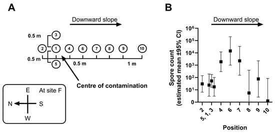
Sites where animal carcasses had been buried were relatively well defined, around 2 m by 2 m in extent, while areas with superficial contamination following butchering of an infected carcass on open ground were more irregular. An envelope sampling method [11] was employed at both types of sites for the Microbiology Department surveys, with 0.1–0.5 kg of soil being collected from the top 10 cm of ground at different positions within each contaminated site. One sample (number 1) was taken from the centre of a 1 m2 area within the site, without placing a metal peg in the carcass remains. As a general rule, a further four samples (numbers 2–5) were taken from positions round the periphery of the contaminated area [8], at a distance of 0.5 m along lines towards the four points of the compass (N, E, S, W). For sites with contamination areas less than 1 m2 in extent, either the distances between the four cardinal sampling points were reduced or fewer samples were taken. For large sites, up to five additional samples (numbered 6–10) were taken at intervals for a further 3 m down the slope (the maximum gradient at any site being <15°). Sampling positions were recorded on a map of the site (an example is shown in Figure 1A) to ensure that subsequent samples could be taken as closely as possible from the same places. All soil samples were stored in the laboratory at room temperature prior to culture.

Figure 1.
Spore counts in samples from different positions at contaminated sites in the Kars region. Panel (A): Standardised site sampling diagram: Position 1 was at the centre-point of the contaminated area. Panel (B): Estimated means (±95% CI) of spore counts at sampling positions 1–10 derived from a mixed model analysis of data from all the sites studied. The output of the mixed model analysis is shown in Table S3 and the estimated means (±95% CI) of spore counts are listed in Table S4.
The information collected for each soil sample included the following: location (district, village, site and position number), reason for sampling (planned survey or routine analysis), type of contamination (ground surface or animal burial), date of contamination and date of sampling. These details are listed together with the spore concentration (count/g soil) in Table S1, and a map of Kars province with the administrative districts in which the villages are situated is shown in Figure S1.
The precision of recording contamination dates at individual sites varied. Due to lack of written records in the villages concerned, the dates of contamination for the earliest sites studied could only be identified to within a range of 1–2 years. Sampling dates were more precise, with nearly all being recorded to the nearest month. In order to avoid the risk of overestimating the longevity of viable spores in soil, the interval between contamination and sampling (Column I in Table S1) was therefore calculated as the number of days between the final day of the period listed for the contamination date and the first day of the period listed for the sampling date.
2.2. Culture and Identification of B. anthracis
Following collection, all soil samples and isolated bacteria were processed in the biocontainment facilities at Kafkas University within BSL 2-Plus (A2 type) bio-safety cabinets (ESCO, SG). A portion of soil (40 g) from each sample was mixed with distilled water (200 mL), shaken vigorously by hand, and the resulting suspension left at room temperature for 30 min. A 1 mL aliquot was then taken from the suspension and subjected to 10-fold serial dilutions, which were heat treated at 62.5–63 °C for 20 min to inactivate vegetative bacteria. Following this, a 150 µL aliquot from each dilution was spread across the surface of a 7% (v/v) sheep blood agar (Thermo Fisher Scientific, Waltham, MA, USA) plate in duplicate and incubated overnight in air at 37 °C. The plates were then examined for the presence of colonies with characteristic B. anthracis morphology (rough appearance, non-hemolytic when cultured on sheep blood agar) [1]. The limit of detection was 13.2 cfu per gram of soil.
Suspected B. anthracis colonies were sub-cultured onto fresh sheep blood agar plates to determine sensitivity to diagnostic gamma phage and penicillin (10 units) (Oxoid, UK). The plates were then incubated overnight at 37 °C and examined for signs of inhibition (lysis and antibiotic inhibition, respectively). If these tests produced contradictory results, colonies were subjected to further phenotypic analysis which included Gram staining, determination of motility and capsule production when incubated in the presence of bicarbonate and carbon dioxide. Finally, pXO1 and pXO2 plasmid-based PCRs were carried out, using PA 5/8 for protective antigen and Cap 6/103 for capsule, respectively [12].
Degrees of genetic relatedness in selected isolates were determined by single nucleotide polymorphism (SNP) sub-typing, followed by high-resolution genotyping using 25-loci variable-number tandem repeat analysis (MLVA-25). This was carried out by the Lugar Center for Public Health Research at the National Center for Disease Control, Tbilisi, Georgia [13].
2.3. Statistical Analyses
Data (see Table S1) were analysed using the software SPSS V27.1 (IBM, Armonk, NY, USA). Graphs were prepared using Graphpad PRISM V9.0. For analysis, bacterial count data were transformed to better fit a normal distribution. Linear mixed modelling was used to analyse these data. Only a main effect model was fitted, and no attempt was made to include interactions. The residuals from the final model were Gaussian (assessed by QQ plot), and there was no evidence for heteroscedasticity (assessed by residual plot). Data were analysed by a mixed model in which the experimental unit was held as the site. Sampling Position and Time since Contamination were used as explanatory variables. Village and Administrative District were nested and tested for effect in separate models. Descriptive statistics were calculated using Graphpad PRISM V9.0.
3. Results
Throughout a period of 14 years, 25 sites known to have been previously contaminated with B. anthracis, were sampled and spore counts determined at multiple times and sampling positions. A total of 136 (58.6%) soil samples yielded B. anthracis. Genotype data generated from 10 of these isolates, as expected, confirmed that they were closely related with minimal diversity (Table S2, [13]).
Sampling position was a good predictor for spore count (). Although no contaminated site had a slope with a gradient > 15°, plotting the estimated, modelled means of spore count at different sampling positions demonstrated that counts were greater at a short distance down a slope from the centre (Position 1) of the contaminated area (Figure 1B). While we did not find any records of significant flooding at any of the sites studied, the Kars region is, however, subject to repeated sessions of freezing and thawing due to the melting of winter snow [14], and as a consequence the water table level is likely to fluctuate.
Although there were notable variations in spore counts between sites (Figure 2), statistical modelling was unable to find any association between the degree of contamination at a site and its location at either Village or District level ( and respectively).

Figure 2.
Spore counts at multiple sites across the Kars region of Türkiye. Counts are shown as a data point for each sample with the median line added. The points are partially transparent to enable data density to be visualised; thus darker shades of grey show spore count values where data points overlap. Samples were taken at one or more times and/or positions relative to the centre-point of each contaminated site (coded A to Y). The -axis shows the closest Village (coded to ) to the site and the administrative District within Kars province. Data points presented in red represent spore levels in excess of 105 spores/gram of soil, which is a level estimated to be a lethal dose for grazing cattle [15].
While there were no reported cases of human infection associated with living in close proximity to spore contaminated sites [7], levels of >105 spores per gram of soil have been estimated to be a lethal dose for cattle grazing in Etosha [15]. Whether this is the same for cattle in the Kars region of eastern Türkiye has yet to be determined.
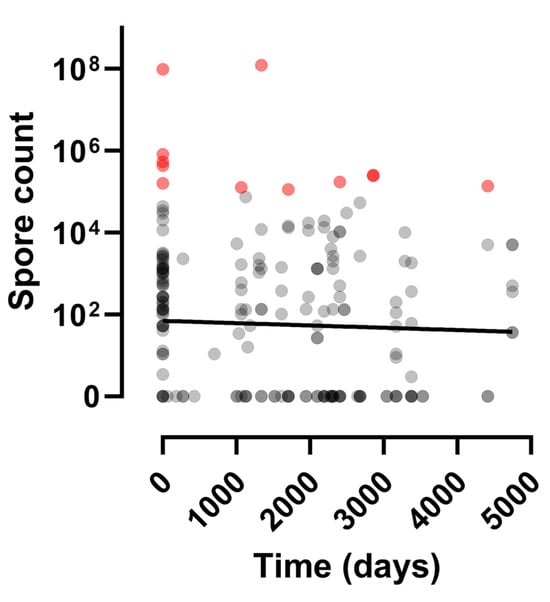
In contrast to studies in other parts of the world, there was no evidence that spore counts were linked to the time interval between contamination and the time of sampling (Figure 3). The effect of time was considered while holding the sampling position constant throughout the statistical modelling and this further confirmed a lack of effect (), suggesting that the spores retained their viability throughout the time period studied.

Figure 3.
A plot of B. anthracis spore yield relative to time since contamination at multiple sites across the Kars region. This scatter plot ignores sampling position. A linear regression line has been added to illustrate the lack of time effect. Spore concentrations of >102/g soil were found in 10 (66.7%) of the 15 samples taken 10–13 years following a contamination event. The points are partially transparent to enable data density to be visualised; thus darker shades of grey show spore count values where data points overlap. Data points presented in red represent spore levels in excess of 105 spores/gram of soil, which is a level estimated to be a lethal dose for grazing cattle [15].
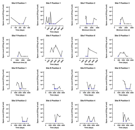
While a main time effect was not obvious, it is clear that there is considerable variation in spore count. Two sites (F in Village , Kars District; R in Village , Selim District) in particular, were sampled repeatedly over time across a range of positions, enabling assessment of the variability of viable spore detection over time at a specific position. Figure 4 is a graphical representation of spore counts found at different time intervals in soil samples from 16 of the 25 sites studied.

Figure 4.
Plots of B. anthracis spore counts relative to time since contamination, at 16 of the 25 sites studied. The panels show the individual sites that were monitored across repeated time points. The line on these individual plots shows the geometric mean. Samples fluctuated between positive and negative.
On 21 occasions, spores were not isolated at one time point but were subsequently detected at the same position at a later date; these samples are highlighted yellow in Table S1 and the changing spore counts over time for 16 of the sampling positions involved are shown in Figure S2. At seven of these positions, samples fluctuated from positive through negative and then to positive for B. anthracis spores once more, and for a further four positions, they fluctuated repeatedly between negative and positive at different time points (Figure S2).
4. Discussion
The study reported in this paper provides background information to a recently published clinical study of the anthrax toxin specific immune response of individuals living in B. anthracis spore contaminated areas of the Kars region of eastern Türkiye [7]. The clinical study identified individuals with antibodies to toxin components suggestive of subclinical exposure to the pathogen. As the nature of this exposure was unknown, we sought to characterise the background level of environmental contamination by monitoring spore levels at known contaminated sites.
Locations known to be contaminated with B. anthracis spores were repeatedly sampled for the presence of the pathogen in some cases over a 14-year time frame. While there were notable variations in spore levels recovered at different times and sites, statistical modelling did not identify any association between the degree of contamination and geographic location. This suggests that the sites studied did not differ significantly in factors that affect spore survival.
While there was no statistical association between spore numbers and specific locations in our study, the position at which samples were taken from a contaminated site was a good predictor for recovery of the pathogen. In a study of spore contaminated sites in Canada, only 28.4% of samples taken from locations where the soil had been saturated with body fluids escaping the carcass were positive for B. anthracis spores [8]. We also found that recovery of B. anthracis spores from a specific sampling point varied with time in that 21 previously negative sampling positions yielded positive results upon retesting at a later date. A similar switch from negative to positive was also seen in a study of spore contaminated sites in Etosha National Park in Namibia [9,10]. The reasons for these fluctuations in spore recovery are unclear, but highlight the importance of collecting multiple samples in different positions over extended periods of time from a contaminated site, so as not to miss the presence of the pathogen.
In contrast to studies in other parts of the world, there was no evidence of a reduction in spore viability over the time course of the study. Indeed, spore concentrations of >102/g soil were found in 10 (66.7%) of the 15 samples taken 10 or more years following initial contamination. We also observed no difference between sites where animal carcasses had been butchered on open ground and where they had been buried, highlighting the importance of appropriate decontamination, as burial on its own does not eliminate spores from the environment.
These results differ from those reported from a series of studies in Etosha which observed that B. anthracis spore numbers in soil declined exponentially over time with numbers as high as 1.19 × 10⁹ spores/g of soil reducing to no detectable spores over a 10-year period [15,16,17]. However, in some natural conditions, spores are known to persist for decades, with spores of the Vollum strain of B. anthracis being detected more than 40 years after soils were experimentally inoculated on Gruinard Island off the coast of Scotland [18]. Similarly, re-emergence of anthrax in reindeer in the Yamal region of Siberia in 2016 more than 70 years after the last known case, together with sporadic cases originating from unknown environmental sources in Sweden, strongly suggests that persistence times can exceed several decades under certain conditions [19,20].
Whilst the factors affecting persistence of spore reservoirs in soil are not fully understood, it is possible that changes in local weather patterns due to global warming could contribute to an increase in spore numbers and a corresponding increase in outbreaks [21]. Melting of the permafrost combined with changes in rainfall patterns [22] may result in an increase in local flooding that brings trapped spores to the surface from previously undisturbed reservoirs, replenishing any spores that may have died [23].
The rate at which a spore pool decays is also likely to depend on local environmental conditions; for example, larger concentrations of spores are found in soils having slightly alkaline pH (>6.0), higher organic matter and higher calcium content [24]. Features of the exosporium have also been shown to affect the ability of B. anthracis spores to bind to different soil types [25]. Spores persist best in dry soils where microbial activity is minimal. In moist soils viability is usually in the range of 3 months to 3–4 years, but rarely longer [24].
While an association has been made between the level of environmental contamination and infectivity in grazing cattle in Etosha, with >105 spores/gram of soil being identified as a lethal dose [15], the same cannot be said for humans. Cases of clinically confirmed anthrax infection arising from direct contact with contaminated soil are extremely rare [26]. A Daily Telegraph report [27] in 2023, of Russian troops contracting anthrax as a consequence of digging trenches in the Zaporizhzhia region of Ukraine, appears at odds with a review by Finke et al. [28] which concluded that “there is no scientific evidence proving for soil-borne anthrax in military animals and soldiers even in case of intensive exposure during heavy disturbance of soil structure in known endemic areas”. Interestingly, these authors go on to say that in highly endemic areas there may be a risk of infection due to contact with soil from recent burial sites of infected animals. Ukraine has 10,000 known anthrax burial sites and it is estimated that there are a further 6000 unregistered sites [29]. In our study, we determined the level of B. anthracis spores in soil collected from known contaminated locations cross the Kars region of Türkiye, some in close proximity to sites of human occupation. While spore numbers at some sites exceeded those predicted in experimental models to infect humans, the lack of recorded clinical cases of infection associated with environmental exposure suggests that infections, if they do occur, are extremely rare [30].
Animal studies have shown that grazing animals in Etosha develop antibody responses to the B. anthracis toxin, suggesting exposure to the pathogen at levels insufficient to cause an active infection [31,32]. A recent study of humans in the Kars region also detected toxin specific antibody responses in individuals with no history of clinical infection suggesting that they had also been exposed to low levels of the pathogen [7]. Whether this exposure was a consequence of contact with contaminated animals and their products or spore contaminated environments is unknown.
The ability to live and work in spore contaminated environments is of particular interest to those tasked with rendering an area safe for human occupation following the release of large numbers of B. anthracis spores [33]. The limited studies which are available, including our own, suggest that healthy humans can tolerate low level spore exposure without ill effects [3,4,5,6,7,34]. Further work is needed to identify what constitutes a “low level of exposure” given that a community is likely to contain individuals who vary in their susceptibility to the pathogen [35].
5. Study Limitations
This was an observational study, reporting common features of all samples received by the Veterinary Microbiology Department of Kafkas University in Kars during the period 2009–2023, that proved positive for viable Bacillus anthracis spores. Therefore, the results do not give an indication of the prevalence or distribution of anthrax-contaminated sites in the Kars region. Rather, this study describes common features of samples from known contaminated sites, emphasises the longevity of infectious anthrax spores in soil, and underlines the importance of thorough decontamination and appropriate disposal of infected animal carcasses and tissues.
Supplementary Materials
The following supporting information can be downloaded at: https://www.mdpi.com/article/10.3390/microorganisms12101944/s1, Figure S1: Map of Kars Province in eastern Türkiye showing its eight administrative districts; Figure S2: Plots of B. anthracis spore counts relative to time since contamination, at sampling positions where spores were not isolated at one time but were detected subsequently at the same position; Table S1: Excel table with data on the Kars soil samples; Table S2: SNP genotype data for ten B. anthracis strains isolated from soil samples examined in this study reported by Khmaladze et al. [13]; Table S3: Mixed model analysis of spore counts at different sampling positions at contaminated sites; Table S4: Estimated means of spore counts at different sampling positions at known contaminated sites.
Author Contributions
Conceptualization, M.S., H.D., M.D., F.B. and L.B.; Methodology, M.S., T.R.L., H.D., O.C., M.D., F.B. and L.B.; Software, T.R.L.; Validation, F.B. and L.B.; Formal analysis, T.R.L. and H.D.; Investigation, M.S., O.C., F.B. and L.B.; Resources, M.S., M.D., F.B. and L.B.; Data curation, M.S., H.D., F.B. and L.B.; Writing—original draft, T.R.L., H.D. and L.B.; Writing—review & editing, M.S., T.R.L., H.D., M.D., F.B. and L.B.; Project administration, M.S. and F.B.; Funding acquisition, M.S. and L.B. All authors have read and agreed to the published version of the manuscript.
Funding
This research was funded by the UK Government Decontamination Service, which is now named Defra (Department for Environment, Food & Rural Affairs) CBRN Emergencies.
Data Availability Statement
The original contributions presented in the study are included in the article/Supplementary Materials, further inquiries can be directed to the corresponding author.
Acknowledgments
We are grateful to the veterinary microbiology staff at Kafkas University who assisted with sample preparation, culture and analysis. We are particularly thankful for the encouragement given by David Elliott and the UK International Biosecurity Programme (IBSP) in ensuring that these results were prepared for publication in the academic literature.
Conflicts of Interest
The authors declare no conflict of interest.
References
- Turnbull, P.C.B. WHO Guidance: Anthrax in Humans and Animals, 4th ed.; WHO: Geneva, Switzerland, 2008; Available online: https://www.who.int/publications/i/item/9789241547536 (accessed on 23 July 2024).
- 14/1/2012-28173; Animal Disease Compensation Regulations, Türkiye. Regulation on Compensated Animal Diseases and Compensation Rates. Ministry of Food, Agriculture and Livestock: Ankara, Türkiye, 2012.
- Norman, P.S.; Ray, J.G.; Brachman, P.S.; Plotkin, S.A.; Pagano, J.S. Serologic testing for anthrax antibodies in workers in a goat hair processing mill. Am. J. Epidemiol. 1960, 72, 32–37. [Google Scholar] [CrossRef] [PubMed]
- Plotkin, S.A.; Brachman, P.S.; Utell, M.; Bumford, F.H.; Atchison, M.M. An epidemic of inhalation anthrax, the first in the twentieth century: I. Clinical features. Am. J. Med. 1960, 29, 992–1001. [Google Scholar] [CrossRef] [PubMed]
- Kissling, E.; Wattiau, P.; China, B.; Poncin, M.; Fretin, D.; Pirenne, Y.; Hanquet, G.B. anthracis in a wool-processing factory: Seroprevalence and occupational risk. Epidemiol. Infect. 2012, 140, 879–886. [Google Scholar] [CrossRef] [PubMed]
- Wattiau, P.; Klee, S.R.; Fretin, D.; Van Hessche, M.; Ménart, M.; Franz, T.; Chasseur, C.; Butaye, P.; Imberechts, H. Occurrence and genetic diversity of Bacillus anthracis strains isolated in an active wool-cleaning factory. Appl. Environ. Microbiol. 2008, 74, 4005–4011. [Google Scholar] [CrossRef][Green Version]
- Buyuk, F.; Dyson, H.; Laws, T.R.; Celebi, O.; Doganay, M.; Sahin, M.; Baillie, L. Human exposure to naturally occurring Bacillus anthracis in the Kars Region of Eastern Türkiye. Microorganisms 2024, 12, 167. [Google Scholar] [CrossRef]
- Dragon, D.C.; Bader, D.E.; Mitchell, J.; Woollen, N. Natural dissemination of Bacillus anthracis spores in Northern Canada. Appl. Environ. Microbiol. 2005, 71, 1610–1615. [Google Scholar] [CrossRef]
- Bellan, S.E.; Turnbull, P.C.; Beyer, W.; Getz, W.M. Effects of experimental exclusion of scavengers from carcasses of anthrax-infected herbivores on Bacillus anthracis sporulation, survival, and distribution. Appl. Environ. Microbiol. 2013, 79, 3756–3761. [Google Scholar] [CrossRef]
- Lindeque, P.M.; Turnbull, P.C. Ecology and epidemiology of anthrax in the Etosha National Park, Namibia. Onderstepoort J. Vet. Res. 1994, 61, 71–83. [Google Scholar]
- Kashparov, V.A.; Ahamdach, N.; Zvarich, S.I.; Yoschenko, V.; Maloshtan, I.M.; Dewiere, L. Kinetics of dissolution of Chernobyl fuel particles in soil in natural conditions. J. Environ. Radioact. 2004, 72, 335–353. [Google Scholar] [CrossRef]
- Buyuk, F.; Sahin, M.; Cooper, C.; Celebi, O.; Saglam, A.G.; Baillie, L.; Celik, E.; Akca, D.; Otlu, S. The effect of prolonged storage on the virulence of isolates of Bacillus anthracis obtained from environmental and animal sources in the Kars Region of Türkiye. FEMS Microbiol. Let. 2015, 362, fnv102. [Google Scholar] [CrossRef]
- Khmaladze, E.; Su, W.; Zghenti, E.; Buyuk, F.; Sahin, M.; Nicolich, M.P.; Baillie, L.; Obiso, R.; Kotorashvili, A. Molecular genotyping of Bacillus anthracis strains from Georgia and Northeastern Part of Turkey. J. Bacteriol. Mycol. 2017, 4, 1053. [Google Scholar]
- Wikipedia Kars Description. 2023. Available online: https://en.wikipedia.org/wiki/Kars (accessed on 23 July 2024).
- Turner, W.C.; Kausrud, K.L.; Beyer, W.; Easterday, W.R.; Barandongo, Z.R.; Blaschke, E.; Cloete, C.; Lazak, J.; Van Ert, M.N.; Ganz, H.H.; et al. Lethal exposure: An integrated approach to pathogen transmission via environmental reservoirs. Sci. Rep. 2016, 6, 27311. [Google Scholar] [CrossRef]
- Turner, W.C.; Kausrud, K.L.; Krishnappa, Y.S.; Cromsigt, J.P.G.M.; Ganz, H.H.; Mapaure, I.; Cloete, C.C.; Havarua, M.; Kusters, M.; Getz, W.M.; et al. Fatal attraction: Vegetation responses to nutrient inputs attract herbivores to infectious anthrax carcass sites. Proc. R. Soc. B 2014, 281, 20141785. [Google Scholar] [CrossRef] [PubMed]
- Barandongo, Z.R.; Dolfi, A.C.; Bruce, S.A.; Rysava, K.; Huang, Y.H.; Joel, H.; Hassim, A.; Kamath, P.L.; van Heerden, H.; Turner, W.C. The persistence of time: The lifespan of Bacillus anthracis spores in environmental reservoirs. Res. Microbiol. 2023, 174, 104029. [Google Scholar] [CrossRef] [PubMed]
- Manchee, R.J.; Broster, M.G.; Anderson, I.S.; Henstridge, R.M.; Melling, J. Decontamination of Bacillus anthracis on Gruinard Island. Nature 1983, 303, 239–240. [Google Scholar] [CrossRef]
- Elvander, M.; Persson, B.; Sternberg, L.S. Historical cases of anthrax in Sweden 1916–1961. Transbound. Emerg. Dis. 2017, 64, 892–898. [Google Scholar] [CrossRef][Green Version]
- Timofeev, V.; Bahtejeva, I.; Mironova, R.; Titareva, G.; Lev, I.; Christiany, D.; Borzilov, A.; Bogun, A.; Vergnaud, G. Insights from Bacillus anthracis strains isolated from permafrost in the tundra zone of Russia. PLoS ONE 2019, 14, e0209140. [Google Scholar] [CrossRef]
- Railean, V.; Sobolewski, J.; Jaśkowski, J.M. Anthrax in one health in Southern and Southeastern Europe—The effect of climate change? Vet. Res. Commun. 2024, 48, 623–632. [Google Scholar] [CrossRef]
- Pittiglio, C.; Shadomy, S.; El Idrissi, A.; Soumare, B.; Lubroth, J.; Makonnen, Y. Seasonality and Ecological Suitability Modelling for Anthrax (Bacillus anthracis) in Western Africa. Animals 2022, 12, 1146. [Google Scholar] [CrossRef]
- Maksimović, Z.; Cornwell, M.S.; Semren, O.; Rifatbegović, M. The apparent role of climate change in a recent anthrax outbreak in cattle. Rev. Sci. Tech. Off. Int. Epiz. 2017, 36, 959–963. [Google Scholar] [CrossRef]
- Hugh-Jones, M.; Blackburn, J. The ecology of Bacillus anthracis. Mol. Asp. Med. 2009, 30, 356–367. [Google Scholar] [CrossRef] [PubMed]
- Williams, G.; Linley, E.; Nicholas, R.; Baillie, L. The role of the exosporium in the environmental distribution of anthrax. J. Appl. Microbiol. 2013, 114, 396–403. [Google Scholar] [CrossRef] [PubMed]
- Doganay, M. Anthrax. In Infectious Diseases, 4th ed.; Cohen, J., Powderly, W.G., Opal, S.M., Eds.; Elsevier: Beijing, China, 2017; pp. 1123–1128. [Google Scholar]
- Barnes, J. Russian Troops Digging Trenches in Ukraine Reportedly Infected with Anthrax. Brussels Correspondent, Daily Telegraph. 26 April 2023. Available online: https://www.telegraph.co.uk/world-news/2023/04/26/anthrax-reportedly-infects-russian-troops-in-ukraine/ (accessed on 23 July 2024).
- Finke, E.J.; Beyer, W.; Loderstädt, U.; Frickmann, H. Review: The risk of contracting anthrax from spore-contaminated soil—A military medical perspective. Eur. J. Microbiol. Immunol. (Bp) 2020, 10, 29–63. [Google Scholar] [CrossRef]
- Sinitsyn, V.A.; Yanenko, U.M.; Zaviryukha, G.A.; Vasileva, T.B.; Tarasov, O.A.; Kosyanchuk, N.I.; Muzykina, L.M. The situation of anthrax which is on the territory of Ukraine. Ukr. J. Ecol. 2019, 9, 113–119. [Google Scholar] [CrossRef] [PubMed]
- Cohen, M.L.; Whalen, T. Implications of low level human exposure to respirable B. anthracis. Appl. Biosaf. 2007, 12, 109–115. [Google Scholar] [CrossRef]
- Cizauskas, C.A.; Bellan, S.E.; Turner, W.C.; Vance, R.E.; Getz, W.M. Frequent and seasonally variable sublethal anthrax infections are accompanied by short-lived immunity in an endemic system. J. Anim. Ecol. 2014, 83, 1078–1090. [Google Scholar] [CrossRef]
- Ochai, S.O.; Crafford, J.E.; Hassim, A.; Byaruhanga, C.; Huang, Y.H.; Hartmann, A.; Dekker, E.H.; van Schalkwyk, O.L.; Kamath, P.L.; Turner, W.C.; et al. Immunological evidence of variation in exposure and immune response to Bacillus anthracis in herbivores of Kruger and Etosha National Parks. Front. Immunol. 2022, 13, 814031. [Google Scholar] [CrossRef]
- National Research Council. Reopening Public Facilities after a Biological Attack: A Decision Making Framework; The National Academies Press: Washington, DC, USA, 2005. [Google Scholar] [CrossRef]
- Wattiau, P.; Govaerts, M.; Frangoulidis, D.; Fretin, D.; Kissling, E.; Van Hessche, M.; China, B.; Poncin, M.; Pirenne, Y.; Hanquet, G. Immunologic response of unvaccinated workers exposed to anthrax, Belgium. Emerg. Infect. Dis. 2009, 15, 1637–1640. [Google Scholar] [CrossRef]
- Barakat, L.A.; Quentzel, H.L.; Jernigan, J.A.; Kirschke, D.L.; Griffith, K.; Spear, S.M.; Kelley, K.; Barden, D.; Mayo, D.; Stephens, D.S.; et al. Fatal inhalational Anthrax in a 94-year-old Connecticut Woman. JAMA 2002, 287, 863868. [Google Scholar] [CrossRef]
Disclaimer/Publisher’s Note: The statements, opinions and data contained in all publications are solely those of the individual author(s) and contributor(s) and not of MDPI and/or the editor(s). MDPI and/or the editor(s) disclaim responsibility for any injury to people or property resulting from any ideas, methods, instructions or products referred to in the content. |
© 2024 by the authors. Licensee MDPI, Basel, Switzerland. This article is an open access article distributed under the terms and conditions of the Creative Commons Attribution (CC BY) license (https://creativecommons.org/licenses/by/4.0/). Content includes material subject to © 2024 Crown copyright (2024), Dstl. This material is licensed under the terms of the Open Government Licence except where otherwise stated. To view this licence, visit https://www.nationalarchives.gov.uk/doc/open-government-licence/version/3 or write to the Information Policy Team, The National Archives, Kew, London TW9 4DU, or email: psi@nationalarchives.gov.uk.