Article

The Cultivation of Halophilic Microalgae Shapes the Structure of Their Prokaryotic Assemblages

by
Elena A. Selivanova
1,*,
Michail M. Yakimov
2,
Vladimir Y. Kataev
1,
Yuri A. Khlopko
1,
Alexander S. Balkin
1 and
Andrey O. Plotnikov
1,*
1
Institute for Cellular and Intracellular Symbiosis of the Ural Branch of Russian Academy of Sciences, Orenburg Federal Research Center of the Ural Branch of Russian Academy of Sciences, 460000 Orenburg, Russia
2
Extreme Microbiology, Biotechnology and Astrobiology Group, Institute of Polar Research, The Institute of Polar Sciences of the National Research Council (ISP-CNR), 98122 Messina, Italy
*
Authors to whom correspondence should be addressed.
Microorganisms 2024, 12(10), 1947; https://doi.org/10.3390/microorganisms12101947
Submission received: 30 August 2024 / Revised: 16 September 2024 / Accepted: 19 September 2024 / Published: 26 September 2024
(This article belongs to the Special Issue Omics Research in Microbial Ecology)

Abstract

:
The influence of microalgae on the formation of associated prokaryotic assemblages in halophilic microbial communities is currently underestimated. The aim of this study was to characterize shifts in prokaryotic assemblages of halophilic microalgae upon their transition to laboratory cultivation. Monoalgal cultures belonging to the classes Chlorodendrophyceae, Bacillariophyceae, Trebouxiophyceae, and Chlorophyceae were isolated from habitats with intermediate salinity, about 100 g/L, nearby Elton Lake (Russia). Significant changes were revealed in the structure of algae-associated prokaryotic assemblages, indicating that microalgae supported sufficiently diverse and even communities of prokaryotes. Despite some similarities in their prokaryotic assemblages, taxon-specific complexes of dominant genera were identified for each microalga species. These complexes were most different among Alphaproteobacteria, likely due to their close association with microalgae. Other taxon-specific bacteria included members of phylum Verrucomicrobiota (Coraliomargarita in assemblages of Navicula sp.) and class Gammaproteobacteria (Salinispirillum in microbiomes of A. gracilis). After numerous washings of algal cells, only alphaproteobacteria Marivibrio remained in all assemblages of T. indica, likely due to a firm attachment to the microalgae cells. Our results may be useful for further efforts to develop technologies applied for industrial cultivation of halophilic microalgae and for developing approaches to obtain new prokaryotes with a microalgae-associated lifestyle.

1. Introduction

Large-scale microalgae cultivation is one of the current priorities in aquaculture, aiming not only to combat the increasing greenhouse gas production by capturing the immense amount of carbon dioxide, but also to produce a biomass with considerable nutritional value [1,2]. In addition to easily digestible proteins, it contains many valuable nutrients such as vitamins, polyunsaturated fatty acids, carotenoids, etc. [3,4]. Considering that axenic microalgae cultures are too unrealistic, unsuitable, and labor-intensive for use, always exposed to the risk of obvious contamination in large-scale and long-term applications, the isolation of algological pure cultures of microalgae with their subsequent application in biotechnology is the first step towards obtaining a valuable and stable biotechnological product [5]. In precise terms, algologically pure (monoalgal) culture means mechanical (under microscopy examination) selection of a single microalga cell floating in at least 2–5 µL of environ, obviously containing a great number of prokaryotic cells. It has been suggested that the microenvironment of prokaryotes associated with algae provides protection to the latter from many environmental disturbances [6].
In 1972, Bell and Mitchell proposed the term “phycosphere” to describe an area surrounding the microalgal cell and extending for some distance on the µm scale, within which bacterial growth is stimulated by algal extracellular products [7]. Recent studies have shown that more complex interactions occur between marine microalgae and their associated bacteria [8,9,10,11]. In addition to interactions based on the uptake by prokaryotes of dissolved organic matter released by microalgae, a diverse set of complex mutual chemical signaling and metabolite exchanges were detected [8,9,10,11]. Historically, associations of microalgae with prokaryotes have been studied intensively in freshwater and marine ecosystems, as well as in some extreme habitats [12,13,14]. Long-term cultures of microalgae that maintain symbiotic relationships with bacteria are also the focus of research and therefore used as a convenient model for studying trophic interactions in complex microbial communities, as well as the stimulation of algae growth and mutual suppression and competition for nutrients [15,16,17,18,19]. Interactions based on an uptake by prokaryotes of microalga-derived dissolved organic matter, exchange of vitamins, iron compounds, action of auxins, antimicrobial compounds, etc., have important ecological consequences for water communities. In general, the relations can be divided into competitive, synergistic, and parasitic [20]. Bacteria can either compete with microalgae for limited resources or even produce toxic substances against microalgae. Moreover, prokaryotes can either enhance or suppress the growth and development of microalgae, influencing the process known as “algal blooming” [18,19,21]. Certainly, the character of microbial interactions should be considered in large-scale microalgae cultivation due to possible impacts on the culture stability, biomass growth, and yield of useful products. Knowledge on the composition of the microalga microbiomes and understanding their formation modes will help to support the sustainable stable growth of microalgae for biotechnological application and industrial use [6]. As mentioned above, the biotechnological role of microalgae is growing, particularly as components of food supplements, animal feed, and medicines, as well as for energy production and phytoremediation [3,4,15].
Halophilic microalgae, despite the technological difficulties of their use, are promising objects for biotechnology due to their ability to accumulate a significant amount of carotenoids, compatible solutes, lipids, and polyunsaturated fatty acids compared to freshwater and marine algae [22,23,24,25]. However, associations of microalgae with prokaryotes in hypersaline conditions have been studied to a much lesser extent. This is especially true for the intermediate salinity level, at which the microbiomes of halophilic microalgae have been practically unstudied. In reservoirs with a salinity of about 100 g/L, the composition of microalgae differs from both seawater and reservoirs with a salinity more than 200 g/L. In contrast to conditions of extreme salinity where the diversity of microalgae is sharply reduced and limited to extreme halophiles, under the intermediate salinity level, more diverse communities of chlorophyte algae and diatoms develop, many of which are poorly studied or even unidentified [26,27].
Taking into account all mentioned above, in this study, we obtained a couple of samples from inland saline waters, isolated monoalgal cultures from them, and analyzed the algae microbiomes compared to natural prokaryotic communities using DNA metabarcoding. We attempted to address the following main questions in this study:
  • How do the diversity and composition of the prokaryotic communities change upon isolation of halophilic algae in laboratory cultures?
  • Does the taxonomic affiliation of halophilic microalgae determine the composition of their prokaryotic assemblages?
  • Are the prokaryotic assemblages of halophilic microalgae composed randomly, or are there specific prokaryotic taxa featuring microalgal species?

2. Materials and Methods

2.1. Sampling, Alga Cultures, and Growth Conditions

Samples were collected in 2019 from two hypersaline sites in the basin of Lake Elton, Volgograd region, Russia, the mouth of the Malaya Smorogda River, also named Malaya Samoroda River (MS), with salinity of 110 ppt (49.0960N 46.7333E), and an ephemeral pond near the Solyanka River (EPS) with salinity 100 ppt (49.1845N 46.5946E). Water samples of 0.5 L were collected at each site from a depth of 10 cm in sterile bottles. Sampled water of 50–100 mL volume was filtered sequentially through 5.0 and 0.22 µm membranes immediately after collection. The membranes were placed in sterile Eppendorf tubes with 200 μL of DNA/RNA Shield™ (Zymo Research, Irvine, CA, USA), transported to the laboratory, and stored at −80 °C until DNA extraction. Remaining water from the samples was transported to the laboratory, examined under an Axioskop light microscope (Carl Zeiss, Oberkochen, Germany), and used for microalgae isolation and the preparation of the cultivation medium. A total of 26 monoalgal cultures assigned to 5 species (4–5 cultures of each species from each site) were obtained by direct isolation of single cells under a Nikon TS2 inverted microscope (Nikon, Tokyo, Japan) with glass Pasteur pipettes (Table 1). Isolation of monoalgal cultures of T. indica from the MS sample was complicated by a huge amount of tiny Picochlorum sp. cells. Thus, we had to ‘wash’ the T. indica cells through several (4–5) sequential transfers from one drop of sterile medium to another under microscope examination. At the initial stages, clonal cultures were grown in bacteria-free natural water obtained by filtration through the 0.22 μm membrane with subsequent autoclaving. Then, the algae were cultivated at 25 °C under periodic 5 klx illumination with luminescent lamps (12 h—day, 12 h—night) in modified OPS medium that contained 82.3 g NaCl, 17.0 g MgSO4, 2.5 g KNO3, 0.2 g K2HPO4, and 1.0 g NaHCO3 per 1 L (salinity 100 g/L). The purity of the obtained cultures was controlled microscopically and using 18S metabarcoding. The cultivation lasted three months; during that time, the cultures underwent five transfers to fresh medium. As a control, the samples without microalgae cells were picked up with Pasteur pipettes under a Nikon TS2 inverted microscope (Nikon, Tokyo, Japan) to assess a prokaryotic community in the absence of algae. The mineral medium applied for microalgae cultivation was used for the control samples to evaluate the specific impact of algae on the structure of prokaryotic communities, and to identify possible contamination of the algal cultures in the laboratory.

2.2. DNA Extraction

Total DNA was extracted from the microalgae cultures at the late logarithmic growth phase, as well from the control samples and natural samples (eDNA). Cultures and control samples (1.5 mL) were centrifuged for 5 min at 14,000 rpm and 6 °C. Supernatants were removed up to 100 μL residual volume. The pellets were homogenized on a TissueLyser LT (Qiagen, Hilden, Germany) using the Lysing Matrix Y (MP Biomedicals, Solon, OH, USA) for 1 min at 50 Hz and supplemented with 50 μL of TE-buffer. After centrifugation for 5 min at 14,000 rpm, the supernatants were transferred to clean tubes and heated at 95 °C for 10 min. The samples were supplemented with TE-buffer up to a volume of 800 μL. Total DNA from the extracts was cleaned and concentrated using a NucleoSpin® gDNA Clean-up XS column kit (Macherey-Nagel GmbH & Co. KG, Düren, Germany). For eDNA extraction, each membrane was transferred into a Lysing Matrix E tube (MP Biomedicals, LLC, Solon, OH, USA) with the addition of 2× volume of tris-saline buffer (1 M Tris-HCl; 0.5 M EDTA; 5 M NaCl; MQ) and homogenized for 5 min at 50 Hz (TissueLyser LT, Qiagen, Hilden, Germany). Then, biomass on membranes was enzymatically digested (lysozyme, proteinase K, SDS in total conc. 1%) and DNA was extracted by phenol/chloroform (phenol/chloroform 1:1 v/v; chloroform/isoamyl alcohol 24:1 v/v) and precipitated from the aqueous phase by a threefold volume of absolute ethanol by adding 10% v/v 10 M ammonium acetate at −20 °C overnight. After centrifugation and double washing with 80% ethanol, DNA was air-dried and dissolved in 30 μL of autoclaved deionized water. To estimate possible contamination, negative controls were used, for which autoclaved deionized water (100 μL) was treated along with experimental samples as described above. The concentration, quality, and quantity of DNA were estimated by electrophoresis in 1% agarose gel and via photometry using a NanoDrop 8000 (Thermo Fisher Scientific Inc., Waltham, MA, USA).

2.3. Preparation of DNA Libraries and Sequencing

16S rDNA libraries for high-throughput sequencing were prepared according to the Illumina protocol (Part no. 15044223, Rev. B). DNA amplification was performed using S-D-Bact-0341-b-S-17 (forward) and S-D-Bact-0785-a-A-21 (reverse) primers targeting the V3 and V4 regions of the 16S rRNA gene [28]. 18S rDNA libraries for algae identification were prepared from all 26 monoalgal cultures using forward TAReuk454FWD1 and reverse TAReukRev3 primers targeting the hypervariable V4 region of the 18S rRNA gene [29], producing amplicons with lengths of about 500 bp. The libraries were sequenced on the MiSeq platform (Illumina, San Diego, CA, USA) using a 2 × 300 bp paired-end v3 MiSeq Reagent Kit in the “Persistence of microorganisms” Science Resource Centre, the Institute for Cellular and Intracellular Symbiosis, the Ural Branch of the Russian Academy of Sciences.

2.4. Bioinformatic Pipeline

Paired-end reads were merged with a minimal overlap of 30 bp, a p-value of 0.0001, and a Q-score of 30 using PEAR v. 0.9.10 [30]. At the next stage, if there were adapters, they were trimmed by the program Trimmomatic V. 0.36 [31]. Subsequent treatment of merged and trimmed reads was conducted using the UPARSE algorithm of the USEARCH v. 10.0.240 program [32] and included quality filtering and amplicon size selection (400 bp—minimal size for 16S rRNA data; 360 bp—minimal size for 18S rRNA data). The reads with Ns and a maximum number of expected errors above one were discarded during the filtering procedure. The filtering quality was evaluated with FastQC v. 0.11.7. As a result of dereplication and clustering with the UPARSE algorithm from USEARCH, operational taxonomic units (OTUs) were formed, while singletons and doubletons were removed. For clustering of OTUs, we used a 97% threshold. Chimeric sequences were detected and removed using USEARCH v. 10.0.240 at the stage of OTU clustering [33]. The resulting OTUs were globally aligned to merged reads that resulted in an OTU table. Every sample was checked for contaminant 16S rRNA gene fragments that originated from bacterial cells or DNA, which were possibly present in reagents, water, air, laboratory plastic, or other contaminants on hands. OTUs resulting from contaminant 16S rRNA fragments were identified at a similarity level of 98% and removed via the USEARCH command ublast by matching the sequences from the samples with the positive control and negative control ones. The OTUs corresponding to chloroplast DNA were also excluded from the library for subsequent analysis. Classification of the OTUs was conducted against the RDP database with an 80% threshold [34]. OTUs with low support were additionally checked using the NCBI GenBank database (https://www.ncbi.nlm.nih.gov/genbank/, accessed on 1 May 2024) and built-in tool BLAST (https://blast.ncbi.nlm.nih.gov/Blast.cgi, accessed on 1 May 2024). Taxonomic affiliation is indicated in accordance with the NCBI Taxonomy Database (https://www.ncbi.nlm.nih.gov/taxonomy/, accessed on 1 May 2024).

2.5. Statistical Analysis and Visualization

After bioinformatic processing, the data normalized by total sum scaling were analyzed using the MicrobiomeAnalyst tool (https://www.microbiomeanalyst.ca/, accessed on 1 May 2024) [35]. The observed number of OTUs and Chao1, Shannon, and Simpson indices were calculated using MicrobiomeAnalyst [35]. Principal coordinate analysis (PCoA) 2D plots demonstrating the ordination of the samples were constructed using permutational multivariate analysis of variance (PERMANOVA) based on the Bray–Curtis dissimilarities using MicrobiomeAnalyst [35]. Mantel’s test was carried out, applying the abundance tables of dominant prokaryote genera depending on the presence of different microalgae species, and visualized using the ‘LinkET’ R package v. 0.0.7.4 in the R 4.4.0 environment [36]. Bar charts were created using Microsoft Excel 2019 v. 1808. Bubble charts were built using the package ggplot2 [37].

3. Results

3.1. Characteristics of the Isolated Monoalgal Cultures

First, we examined under an Axioskop light microscope (Carl Zeiss, Oberkochen, Germany) several microalgal communities sampled from two hypersaline habitats, and obtained a number of monoalgal cultures. Green microalgae were abundant in the sample from the ephemeral pond near the Solyanka River (EPS) with a salinity of 100 ppt. Direct isolation of individual algal cells resulted in clones of three different morphotypes. According to the sequences of the V4 region of their 18S rRNA genes, the obtained cultures were identified as Dunaliella sp., Tetraselmis indica, and Asteromonas gracilis (Table 1). As for the sample from the Malaya Smorogda River (MS) with 110 ppt salinity, there were diatoms in mass together with green microalgae. Clone cultures of a diatom Navicula spp., green alga Picochlorum sp. and Tetraselmis indica were obtained and identified in the same way. Results of 18S rRNA gene sequencing clearly indicated that the cultures of each species could be assigned to a single common OTU, including both MS and EPS cultures of T. indica (Table 1). However, MS cultures of Navicula sp. were assigned to two barely distinguishable OTUs, which had similarity of 99.28 and 98.89 with the closest homologue from NCBI, Navicula salinicola MT012298.

3.2. General Characteristics of the 16S rDNA Metabarcoding Data

The composition of the natural prokaryotic communities, assemblages associated with monoalgal cultures after five passages through OPS laboratory medium, and control ones was analyzed using DNA metabarcoding. The number of paired-end reads with a maximum length of 2 × 300 base pairs (bp) varied from 10,432 to 80,277 per sample (Tables S3 and S4). The number of high-quality reads obtained after dereplicating, filtering, and removal of chimeric sequences varied from 4020 to 36,431 per sample. The total amount of prokaryotic OTUs ranged from 2 to 196 per sample after clustering at the 97% level and the removal of singletons and doubletons, contaminant sequences, and OTUs assigned to chloroplasts (Tables S3 and S4).

3.3. Alpha-Diversity and Taxonomic Composition of the Natural Prokaryotic Communities from the Hypersaline Sites

The structure of natural prokaryotic communities in the EPS and MS water samples was quite different despite their similar salinity. A total of 196 OTUs belonging to 97 genera were identified in the EPS community. The Shannon and Simpson biodiversity indices for the EPS prokaryotic community were 2.96 and 0.87, respectively. Classes Gammaproteobacteria and Alphaproteobacteria and a phylum of uncultivated bacteria, “Candidatus Parcubacteria”, were predominant (Figure 1). Interestingly, sequences belonging to unclassified Francisellaceae dominated among Gammaproteobacteria (Table S1). Genus Roseivirga dominated among Alphaproteobacteria together with unclassified OTUs. In addition, phyla Bacteroidota, Balneolota, Verrucomicrobiota, and Bdellovibrionota, represented mainly by genus Halobacteriovorax, had significant proportions. Verrucomicrobiota contained the dominant genera Puniceicoccus and Coraliomargarita. Phyla Balneolota and Bacteroidota were represented by the dominant genera Gracilimonas and Roseivirga, respectively.
In the MS samples, 117–134 OTUs were detected, which belonged to 70–75 genera. The Shannon and Simpson biodiversity indices were 2.70–2.77 and 0.84–0.86, respectively. More than a third of the prokaryotic reads were attributed to phylum Actinomycetota; classes Betaproteobacteria and Gammaproteobacteria also made up a large proportion (Figure 1). Phyla Balneolota, Bacillota, and Bacteroidota occupied slightly less proportions. The dominant actinomycetes were represented by genera Rhodoluna, Pontimonas, and Longivirga (Table S2). Betaproteobacteria were represented only by genus Bordetella. The most abundant genera of Gammaproteobacteria were Spiribacter and Wenzhouxiangella (Chromatiales). Among Bacteroidota, the dominant genera included Psychroflexus, Mesohalobacter, and Owenweeksia. Genera Roseovarius (Alphaproteobacteria), Rhodohalobacter (Balneolota), and unclassified Erysipelotrichaceae (Bacillota) also dominated in the MS community.

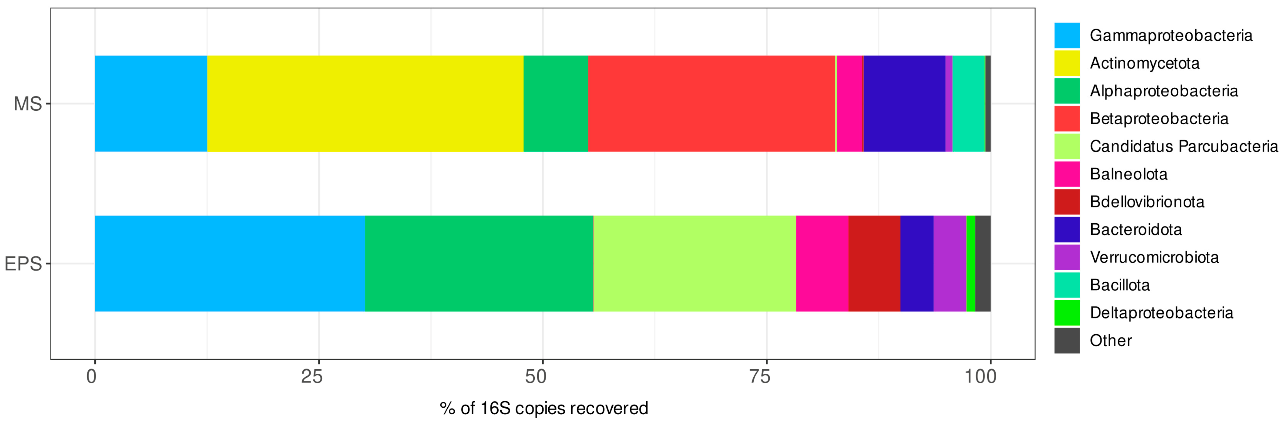
3.4. Alpha-Diversity and Taxonomic Composition of the Control Prokaryotic Communities

Most control samples of prokaryotic communities grown in mineral medium without algae did not produce amplicons with prokaryotic primers. This indicates a very insignificant growth of prokaryotes in mineral medium without microalgae. Ultimately, we managed to obtain amplicons from only two control EPS samples of ten, and for one control MS sample of six.
The taxonomic richness and alpha-diversity of both EPS and MS prokaryotic communities decreased sharply upon the start of laboratory cultivation without microalgae. Thus, in the EPS control samples, a significant decrease in taxonomic richness was observed; namely, 21–37 OTUs belonged to 12–19 genera and 5–6 classes of prokaryotes (Figure 2A, Table S3). The Shannon and Simpson indices were 1.00–1.74 and 0.42–0.70, respectively (Figure 2B,C). The same trend was noted for the MS control sample, where 23 OTUs belonging to 11 genera and 4 classes of prokaryotes were observed (Figure 2D, Table S4). The Shannon and Simpson indices were 1.28 and 0.54, respectively (Figure 2E,F).
While major phyla observed in the natural communities disappeared in the EPS and MS control samples, Pseudomonadota, occupying a lower proportion in the natural communities, increased its abundance. This accompanied opposite changes in percentages of the main Pseudomonadota classes, namely a sharp increase in Gammaproteobacteria and a decrease in Alphaproteobacteria. The taxonomic composition of all control samples was characterized by the considerable predominance of genus Marinobacter, more than 50%. For both control samples, Alphaproteobacteria were presented by the same dominant genera as in natural communities, for instance, Roseovarius. An increase in the proportion of class Saprospiria was also noted for most control samples. The other taxa were detected occasionally (Tables S1 and S2).

3.5. Alpha-Diversity of Prokaryotic Assemblages Associated with the Monoalgal Cultures

The taxonomic richness of prokaryotic assemblages in the monoalgal cultures decreased upon transition to laboratory cultivation. In the EPS monoalgal cultures, we found only 30–77 OTUs assigned to 14–45 genera and 3–10 classes (Figure 2A, Table S3). At the same time, the Shannon and Simpson indices decreased to a lesser extent and remained at relatively high levels compared to the control communities. The highest diversity indices of prokaryotic assemblages were recorded in the EPS cultures of A. gracilis (Shannon index 2.48–2.95, Simpson index 0.84–0.93) (Figure 2B,C). The lowest diversity indices were found in the assemblages of Dunaliella sp. (Shannon index 1.52–2.08, Simpson index 0.68–0.82).
Prokaryotic assemblages of the MS alga cultures showed similar trends in taxonomic richness and alpha-diversity as described above. In particular, 2–46 OTUs from 2–8 classes were found in the MS alga cultures (Figure 2D, Table S4). The Shannon index for bacterial assemblages of both Navicula sp. (2.03–2.56) and Picochlorum sp. (2.08–2.37) cultures was slightly different from the MS natural communities (2.70–2.77) in contrast to its very low value in the control community (1.28) (Figure 2E,F). The same trend was noted for the Simpson index, which was similar between prokaryotic assemblages of both Navicula sp. (0.78–0.90) and Picochlorum sp. (0.81–0.87) cultures. Prokaryotic assemblages associated with T. indica differed sharply from those in the other algal cultures by low diversity indices due to the presence of only 2–5 OTUs per culture. This phenomenon was probably determined by a thorough washing of single algal cells through repeated (4–5 times) transfers in a sterile medium to remove the T. indica cultures of small cells of predominant Picochlorum spp.

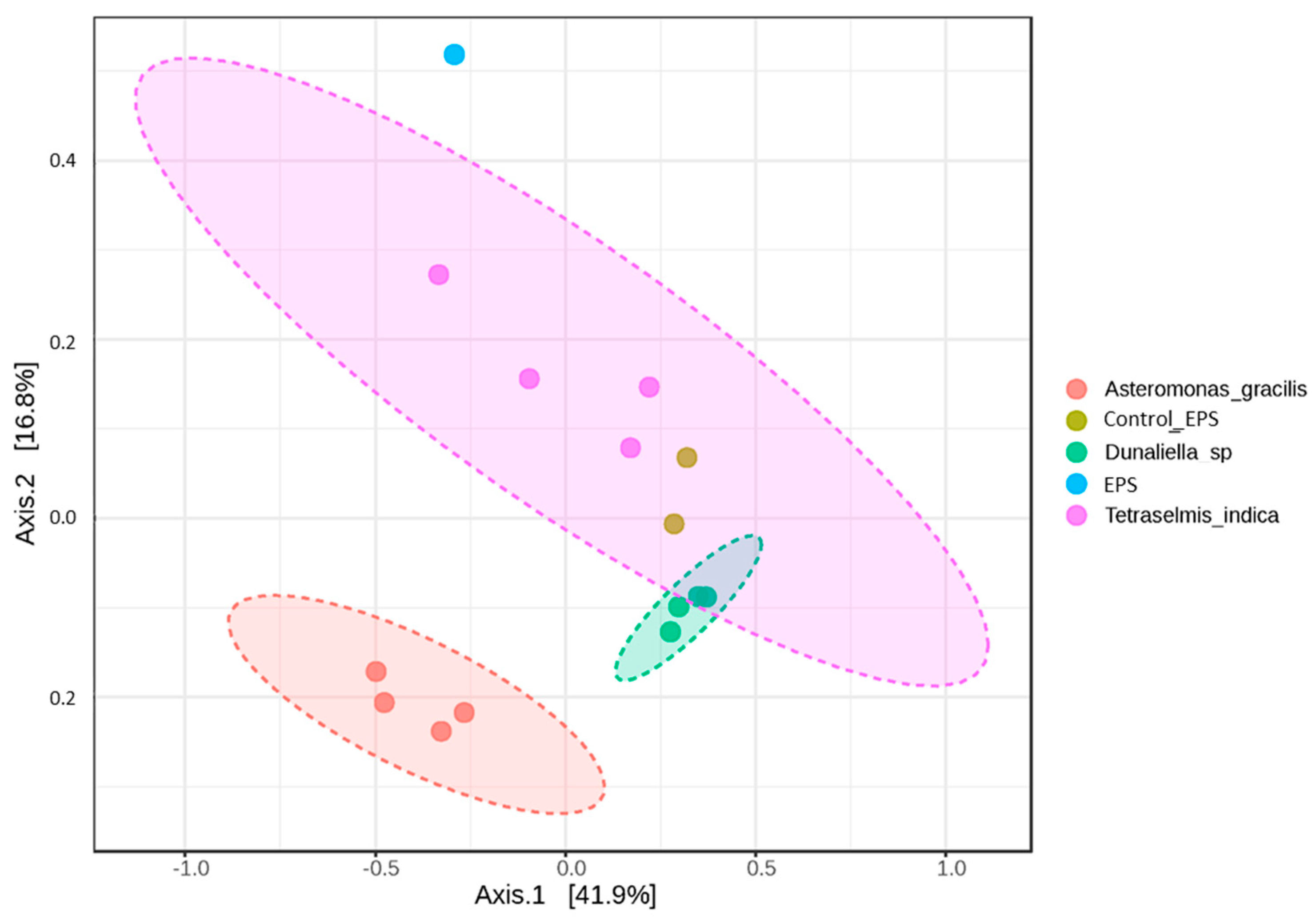
3.6. Beta-Diversity of Prokaryotic Assemblages Associated with the Monoalgal Cultures

PCoA analysis based on the Bray–Curtis metrics of the prokaryotic assemblages associated with the EPS monoalgal cultures revealed three separated clusters belonging to Asteromonas gracilis, Tetraselmis indica, and Dunaliella sp., respectively (Figure 3). The control assemblages were closer to those of Dunaliella sp. The dendrogram built on the Bray–Curtis metrics demonstrated the same clusters (Figure S1).
Prokaryotic assemblages associated with the MS cultures of Picochlorum sp. and Navicula sp. were close to each other, whereas the assemblages of T. indica formed a separate cluster located far from them (Figure 4 and Figure S2). After removing the T. indica assemblages from the analysis, the differences between the natural prokaryotic community and the alga assemblages became more evident.

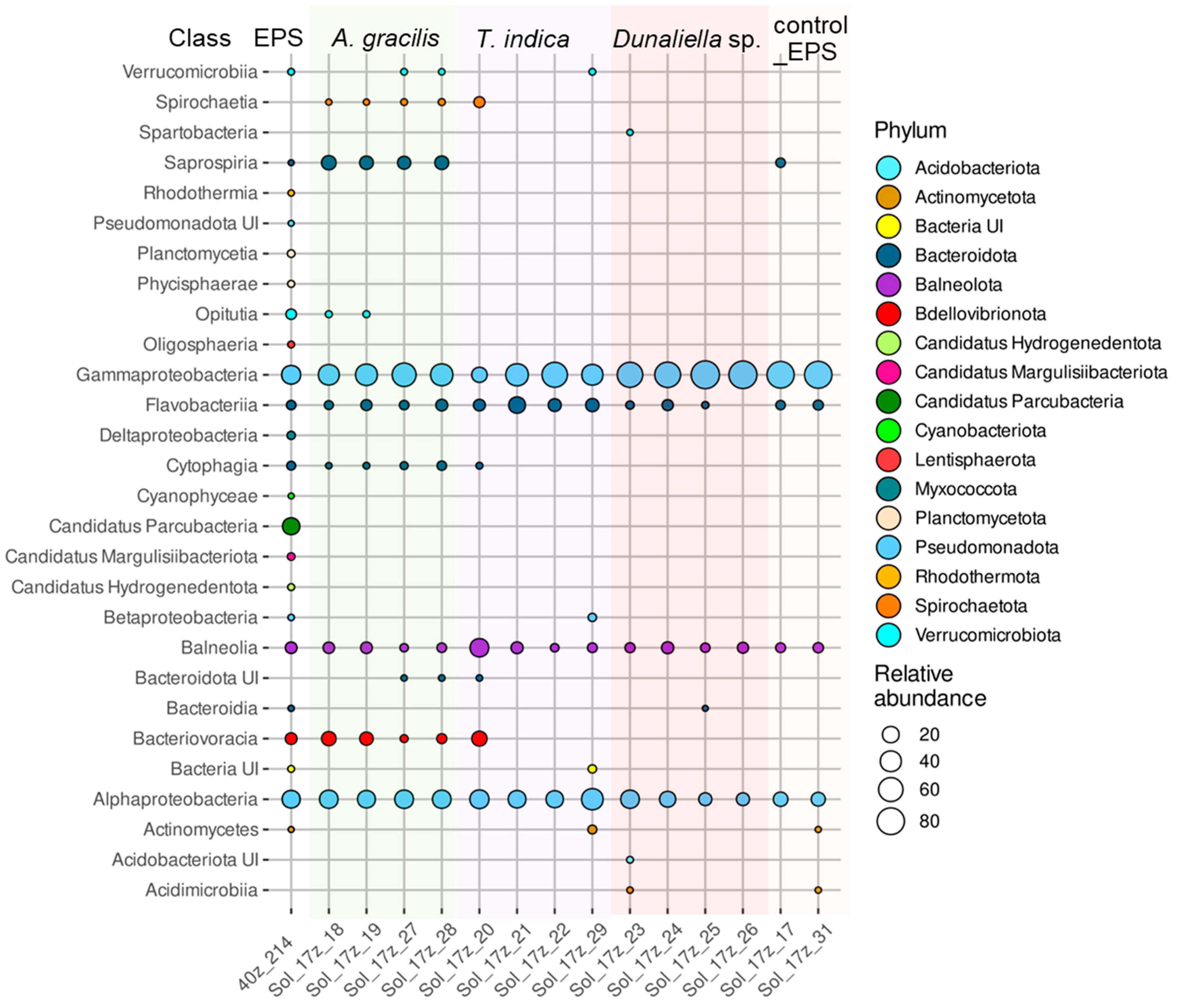
3.7. Taxonomic Composition of the Prokaryotic Assemblages Associated with the Monoalgal Cultures

3.7.1. Taxonomic Composition of the Prokaryotic Assemblages Associated with the Monoalgal Cultures from the Ephemeral Pond near Solyanka River (EPS)

Phyla Cyanobacteriota, “Candidatus Hydrogenedentota”, Lentisphaerota, “Candidatus Parcubacteria”, Planctomycetota, and Rhodothermota and class Deltaproteobacteria disappeared from the assemblages of the EPS monoalgal cultures upon laboratory cultivation compared to the natural EPS community (Figure 5). These taxa constituted a small part of the natural community, except for “Candidatus Parcubacteria” with a great relative abundance 22.6% (Figure 5, Table S1).
Phylum Bacteroidota dominated and its proportion increased significantly in assemblages with A. gracilis and T. indica (Figure 5, Table S1). However, class Bacteroidia assigned to this phylum disappeared, and class Cytophagia, represented by a single genus, Marivirga, was maintained only in the cultures of A. gracilis. In contrast, class Flavobacteriia retained its relative abundance in most monoalgal cultures, and increased its share in assemblages of T. indica. In most monoalgal cultures, one or two genera of flavobacteria were present as dominants including Brumimicrobium, Salibacter, Mesohalobacter, Muricauda, and Psychroflexus (Table S1). Their distribution was largely occasional; only Mesohalobacter was detected in all assemblages of A. gracilis. In addition, class Saprospiria, represented by a single OTU of unclassified Saprospirales, exclusively increased its percentage in association with A. gracilis.
Phylum Balneolota maintained its share in the EPS monoalgal cultures, and was represented by two genera, Gracilimonas and Rhodohalobacter (Table S1).
The proportion of Pseudomonadota increased in the EPS monoalgal cultures (Figure 5, Table S1). It was represented mainly by classes Alphaproteobacteria and Gammaproteobacteria. Alphaproteobacteria had a similar high proportion in the assemblages of A. gracilis and T. indica as in the natural community, while its share decreased in association with Dunaliella sp. Taxonomic composition of Alphaproteobacteria differed in the natural community and monoalgal cultures (Table S1). Along with Roseovarius and unclassified Rhodobacteraceae, which dominated in the natural community, Rhodovulum, Roseivivax, Marivibrio, and Thalassospira dominated in the assemblages of A. gracilis. In all assemblages of T. indica, genera Roseovarius and Thalassospira dominated. In assemblages of Dunaliella sp. genus Rhodovulum was characterized by the highest relative abundance.
The relative abundance of class Gammaproteobacteria increased, especially in the cultures of Dunaliella sp. (Table S1). Genus Marinobacter, presented by one OTU closely related to Marinobacter adhaerens (MN595037), dominated in all samples, although its proportion in the natural community was insignificant (Table S1). The dominant genus Spiribacter reached the highest relative abundance in assemblages of Dunaliella sp. Another representative of order Chromatiales, genus Wenzhouxiangella, was detected only in association with A. gracilis, although its relative abundance was significantly lower compared to Spiribacter. Only assemblages of A. gracilis were featured with dominant genera Salinispirillum and Saccharospirillum. The hydrocarbon-degrading genus Alloalcanivorax was also detected in association with A. gracilis and T. indica. Methylotrophic Gammaproteobacteria Methylophaga were present in assemblages of all monoalgal cultures, and their relative abundance was greater than in the natural community, reaching 20.3% in association with T. indica.
In some monoalgal cultures, the predatory bacterium Halobacteriovorax, a genus of phylum Bdellovibrionota, persisted and its proportion even increased. In those assemblages, the proportion of Gammaproteobacteria decreased, perhaps due to predation.

3.7.2. Taxonomic Composition of the Prokaryotic Assemblages Associated with the Monoalgal Cultures from Malaya Smorogda River (MS)

Compared to the natural MS community, many phyla completely disappeared in the MS monoalgal cultures including phyla Euryarchaeota, Actinomycetota (whose dominance in the natural community was 35%), Campilobacterota, Deinococcota, Bacillota, Fusobacteriota, “Candidatus Parcubacteria”, Planctomycetota, and Thermotogota, class Deltaproteobacteria and unclassified Pseudomonadota (Figure 6, Table S2). Although Bacteroidota retained its abundance in the algal assemblages, classes Cytophagia and Bacteroidia disappeared. At the same time, the proportion of phyla Balneolota and Pseudomonadota increased in associations with microalgae. An increase in the proportion of Verrucomicrobiota was noted only in assemblages of diatoms.
The dominance of phylum Bacteroidota in most monoalgal cultures was accompanied with the prevalence of classes Flavobacteriia and Saprospiria (Figure 6, Table S2). The relative abundance of class Flavobacteriia reached 13 and 18% in the Picochlorum sp. and Navicula sp. cultures, respectively. However, the genus composition of Flavobacteriia in assemblages of microalgae was different (Table S2). For example, Psychroflexus was the dominant genus in few assemblages, whereas Salibacter or Owenweeksia dominated in others. Class Saprospiria was found in all Navicula sp. and Picochlorum sp. cultures. Similar to the EPS monoalgal assemblages, the proportion of Balneolota, represented by Gracilimonas and Rhodohalobacter, increased in association with Navicula sp. and Picochlorum sp. Genus Roseovarius was a common dominant Alphaproteobacteria for assemblages of both Navicula sp. and Picochlorum sp. Genus Saliniramus and unclassified Rhodovibrionaceae were observed in all Navicula sp. cultures. Genera Marivibrio, Tepidicaulis, and unclassified Rhodovibrionaceae dominated in some Picochlorum sp. assemblages, whereas Saliniramus and Oceanicaulis prevailed in others. The relative abundance of Marinobacter, presented by the same OTU as in the EPS assemblages, was significantly larger in association with Navicula sp. and Picochlorum sp. Also, genus Spiribacter accounted for a significant proportion in association with Navicula sp. and Picochlorum sp. Genus Methylophaga dominated in all Picochlorum sp. cultures and reached 39.2%.
Genus Wenzhouxiangella was present only in association with Navicula sp. The hydrocarbon-degrading genus Alloalcanivorax was present in some assemblages of Navicula sp. and Picochlorum sp., although its relative abundance was rather low. Genus Coraliomargarita (phylum Verrucomicrobiota) accounted for up to 35% in association with Navicula sp., not being found in other assemblages.
The assemblages of T. indica differed from other MS cultures in poor diversity of classes Alphaproteobacteria and Gammaproteobacteria, represented by single OTUs (Table S2). As mentioned above, such a sharp decrease in taxonomic richness is probably due to the numerous washings of algal cells during isolation of the monoalgal cultures. As a result, in all MS assemblages of T. indica, one OTU assigned to genus Marivibrio prevailed, reaching proportions of 98.0–99.9% in three samples and 30.2% in the fourth one, where genus Alloalcanivorax prevailed (Table S2). Interestingly, single Marivibrio-related sequences were detected in the MS natural communities. In our opinion, this phenomenon may be determined by a tight attachment of Marivibrio to the surface of algal cells.

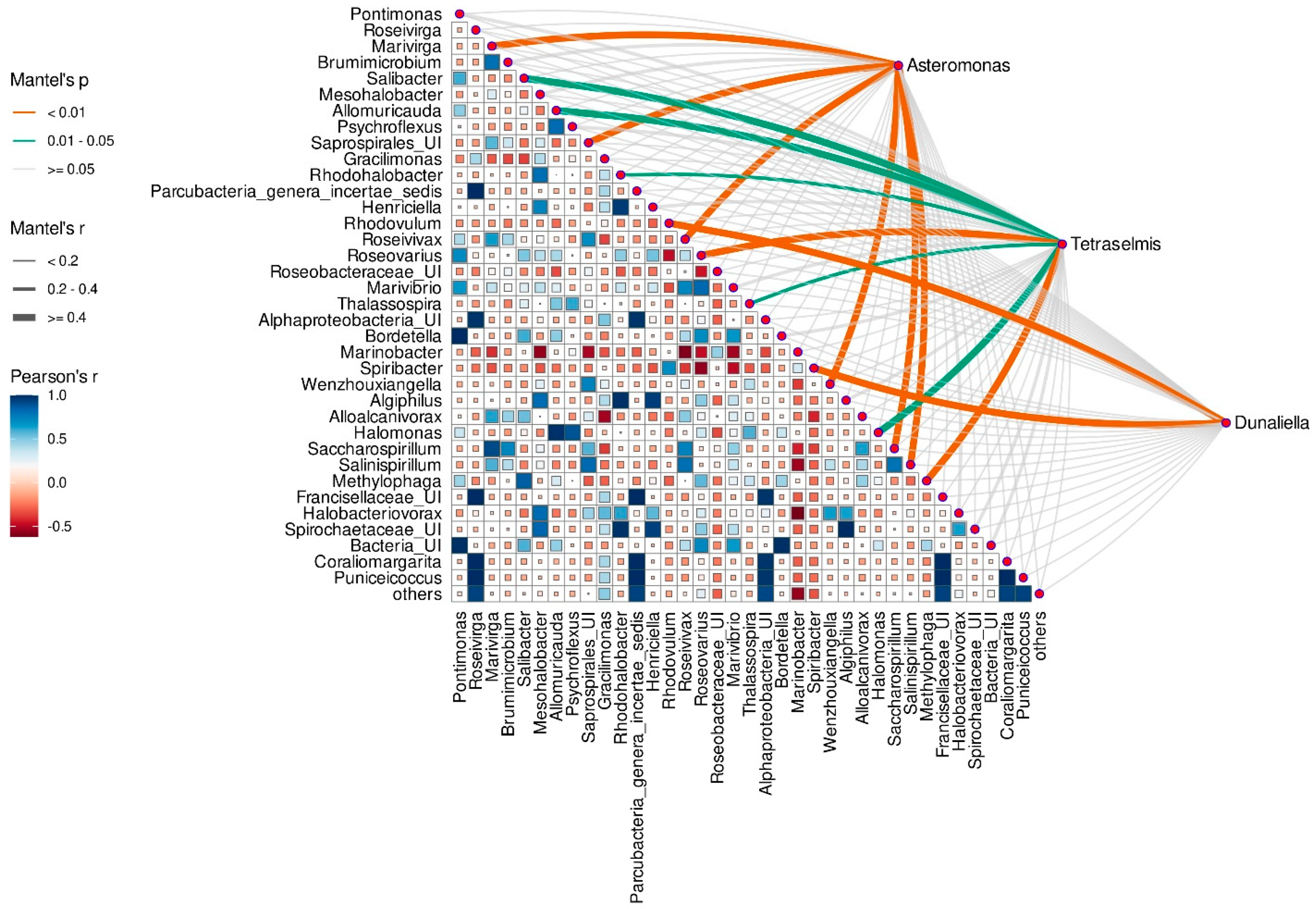
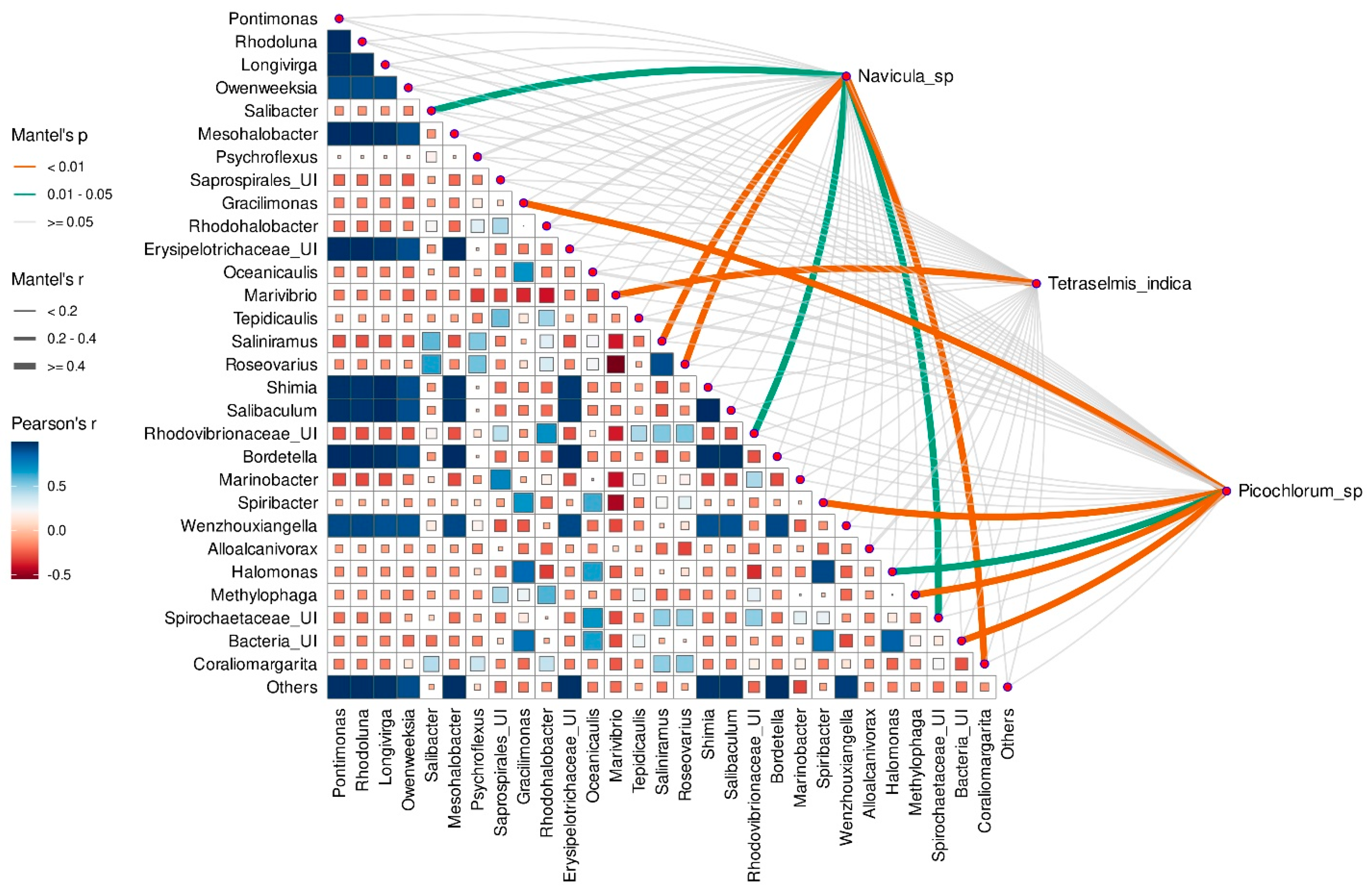
3.8. Correlation between Microalgae Species and Taxonomic Composition of Prokaryotic Assembleges

The Mantel test was additionally applied to assess the correlation between the microalgae species and the relative abundance of prokaryotic genera. Each microalgal species from both EPS and MS assemblages correlated significantly with some associated prokaryotic genera.
A. gracilis had significant correlations with genera Marivirga, Roseivivax, Wenzhouxiangella, Salinispirillum, Saccharospirillum, and unidentified Saprospirales (Figure 7). Dunaliella sp. correlated significantly with abundant Alpha- and Gammaproteobacteria, Rhodovulum and Spiribacter, respectively. Genera Roseovarius and Methylophaga (Mantel p value < 0.01), Salibacter, Allomuricauda, and Halomonas (Mantel p value < 0.05) were strongly associated with T. indica, whereas Rhodohalobacter and Thalassospira correlated with this alga moderately (Figure 7).
Navicula spp. correlated significantly with Roseovarius, Saliniramus, Coraliomargarita (Mantel p value < 0.01), Salibacter, unidentified Rhodovibrionaceae and Spirochaetaceae (Mantel p value < 0.05) (Figure 8). Genera Gracilimonas, Spiribacter, Methylophaga (Mantel p value < 0.01), and Halomonas (Mantel p value < 0.05) were strongly associated with Picochlorum sp., whereas Marivibrio greatly correlated with T. indica (Figure 8).

4. Discussion

In this study, we have analyzed the microbiomes of monoalgal cultures obtained from inland saline waters using DNA metabarcoding, and compared them to natural prokaryotic communities. The monoalgal cultures were isolated from two sites near Elton Lake with salinity levels of 100–110‰. It is worth noting that interactions between microalgae and bacterial communities under intermediate salinity are less studied than in marine [15,20,38,39] or at near-saturated salinities [40,41]. Moreover, to date, few studies have focused on changes in prokaryotic communities associated with halophilic microalgae upon their transfer from environment to laboratory cultures. For this study, dominant microalgae were taken including green algae Dunaliella sp., T. indica, and A. gracilis in the ephemeral pond near Solyanka River (EPS) and diatom Navicula sp., green algae Picochlorum sp., and T. indica in Malaya Smorogda River (MS). Results of sequencing the V4 region of the 18S rRNA gene convincingly confirmed the microscopic identification of the isolated cultures. All these taxa are halophilic and have been previously isolated from hypersaline water bodies [24,42,43,44,45]. Previously, Dunaliella sp., Picochlorum sp., and T. indica have been detected by NGS in saline rivers of the Elton Lake basin [27]. However, their cultures were isolated from these habitats for the first time in this study. A. gracilis and Navicula salinicola have not been previously recorded there. Given their potential as promising sources of carotenoids, biodiesel products, and polyunsaturated fatty acids [25,46,47,48,49], studying prokaryotic assemblages associated with these microalgae is of particular importance.
In both natural communities and microalgae cultures, the most abundant phylotypes belonged to the bacterial genera common in habitats with intermediate salinity [50], and were represented by moderately halophilic bacteria [51]. The taxonomic richness of bacteria in the laboratory monoalgal cultures was significantly lower than in natural communities, which is consistent with literature data on the change in microalgae-associated bacterial communities upon transition to laboratory cultivation [38]. However, a decrease in the prokaryotic richness and diversity was much more pronounced in the control communities without microalgae. Moreover, most control samples did not produce PCR amplicons at all, which can be explained by the attenuation and cessation of prokaryotic growth in the minimal mineral medium in the absence of microalgae acting as the primary producer of organic matter. It should be noted that diversity indices remained quite high in most microalgae cultures except for those washed many times during the isolation procedure. Algae likely contribute to the maintenance of sufficiently diverse, balanced, and even prokaryotic assemblages in laboratory cultures.
At the level of high-ranking taxa, classes Alphaproteobacteria and Gammaproteobacteria of phylum Pseudomonadota, as well as phylum Bacteroidota, turned out to be dominant groups in the microalgae cultures of all species, despite significant differences between the taxonomic composition of EPS and MS natural samples. The predominance of these taxa in microalgae associations has been described previously in both natural and pilot cultures [52]. Thus, it was shown that Pseudomonadota and Bacteroidota dominated in all samples of marine communities associated with Haematococcus lacustris, despite the pronounced difference in environmental conditions and bacterial diversity [38]. Furthermore, the predominance of Bacteroidota and Pseudomonadota has reached up to 90% in bacterial communities associated with laboratory cultures of the biofuel-producing green microalgae Nannochloropsis [52] and in all industrial algae production systems [53]. Both of the two prevalent bacterial phyla, Proteobacteria and Bacteroidota, are capable of rapidly decomposing complex organic matter, facilitating a direct transfer of organic carbon from algae to more demanding prokaryotes [54,55]. Our data demonstrated that phylum Balneolota also dominated in bacterial assemblages associated with halophilic microalgae, reaching 25–28%. Its prevalence is determined mainly by the development of Balneolaceae family members, which include bacteria growing aerobically at 5–10% NaCl [51]. Although there is no information on the dominance of this group in assemblages of halophilic microalgae, one member of the phylum Balneolota was shown to be adapted to a cyanobacteria-associated lifestyle in a hypersaline soda lake [56]. However, in the Tetraselmis indica culture, freed from most free-living bacteria as a result of numerous washings of individual algal cells, Bacteroidota and Balneolota were absent completely. This indicates their likely free-living state, not being attached to the surface of microalgal cells. This suggestion is consistent with data demonstrating that Bacteroidota are the most abundant in free-living bacterial communities associated with microalgae regardless of the kind of alga, whereas Pseudomonadota have been dominant in the cell-attached fractions of algal cultures [57]. In addition to the similarity in taxonomic composition at the level of high-ranking taxa, differences depending on the taxonomic position of microalgae were revealed. Namely, phylum Verrucomicrobiota was detected only in the assemblages of diatoms, and its relative abundance reached 35%. This fact can probably be related to the ability of free-living Verrucomicrobiota to consume fucose- and rhamnose-containing sulfated polysaccharides produced by diatoms, which are difficult for other bacteria to decompose [58].
Analysis of the taxonomic composition of prokaryotic assemblages at the family and genus levels revealed common, occasional, and species-specific taxa of bacteria associated with different microalgae species. The distribution of most Bacteroidota representatives in microalgae cultures was occasional, probably due to a similar function of different taxa, consuming high-molecular dissolved organic matter and providing low-molecular products to microalgae. Some species of the dominant genus Psychroflexus have been previously found and isolated from associations with algae, and their dependence on algal cells due to epiphytism has been described [59]. For another species, Mesohalobacter halotolerans, porphyrin and chlorophyll metabolism has been predicted [50]. Some dominated phylotypes were related to the family Cryomorphaceae, which has been associated with phytoplankton blooms [60]. Moreover, some representatives of the family were isolated from assemblages of microalgae [61]. The presence of unidentified Saprospiraceae only in association with Navicula sp. and Picochlorum sp. may indicate their specificity to respective algal taxa. In addition, members of the family Cryomorphaceae are known to be largely associated with marine sediments and eukaryotes, and are not free-living organisms. This likely reflects their propensity to attach to surfaces, as well as their ability to degrade associated complex nutrient sources [62,63] and cause lysis of the diatom Chaetoceros ceratosporum [53,64,65]. Representatives of another family, Saprospiraceae, have been described in 100% of samples from an industrial production system of Nannochloropsis salina [53].
Phylotypes of Balneolaceae that dominated all monoalgal cultures were assigned to two genera, Rhodohalobacter and Gracilimonas, regardless of the microalga species. Some representatives of Gracilimonas have been previously isolated from cyanobacterial culture Synechococcus and from seaweed [66,67]. Rhodohalobacter mucosus is able to hydrolyze starch, alginate, casein, and other complex organic substances produced by algae [68].
Alphaproteobacteria and Gammaproteobacteria were the most abundant and diverse prokaryotes, and also included specific complexes of genera for the microalgae species (species-specific complexes): Roseivivax for A. gracilis; Rhodovulum for Dunaliella sp.; Rhodovulum, Thalassospira, and Marivibrio for T. indica; and Roseovarius, Saliniramus, and unidentified Rhodovibrionaceae for Navicula spp. This fact indicates the significance of these bacterial taxa for the growth of the microalgae. Among Alphaproteobacteria, the phylotypes belonging to family Rhodobacteraceae were the most abundant and were present in all microalgae cultures, except for T. indica washed out from free-living bacterial associates. The presence of Rhodobacteraceae in the phycosphere of marine diatoms is well known and may be explained by the ability of these Alphaproteobacteria to degrade NH3-containing compatible solutes, such as methylamines and glycine betaine, releasing ammonium as a nitrogen source for some diatoms [69]. Moreover, some members of this family have been shown to possess the complete adenosylcobalamin (vitamin B12) pathway, and therefore they may be potential suppliers of B12 to microalgae, providing a beneficial effect on their long-term survival [39]. In addition, bacteria of the Roseobacter clade can supply microalgae with plant growth promoters [70], thereby exerting a positive effect on growth [71] and ensuring proper algal morphogenesis [72]. A significant proportion of unidentified sequences belonging to families Rhodobacteraceae and Rhodovibrionaceae opens up the prospect of searching for new bacterial taxa in these assemblages of the halophilic alga.
In addition, it is important to emphasize that in the monoalgal cultures of T. indica, washed many times from accompanied bacteria, only one alphaproteobacterial phylotype belonging to genus Marivibrio was an absolute dominant. The possibility of epiphytic growth of Marivibrio has not been described yet, since the representatives of this monospecific genus had been isolated from underground rock salt [73]. In contrast, adhesion of Alphaproteobacteria belonging to genus Ruegeria to the cell surface of T. indica has been previously shown [74].
Genus Marinobacter, a representative of class Gammaproteobacteria, was the most frequently occurring dominant in the monoalgal assemblages. Its relative abundance increased significantly upon transition of the algae to laboratory cultivation, but the highest proportion of Marinobacter was noted in the alga-free controls, which demonstrates an ability of this oligotrophic bacterium to survive under starvation. At the same time, Marinobacter adhaerens is known to stimulate the growth of various diatoms including Thalassiosira weissflogii [71,75,76]. Genus Marinobacter also dominated in the studied assemblages of green halophilic microalgae. A necessity of Marinobacter for microalgae may be related to the production and release of Fe-chelating siderophores, which enhance algal production [77]. Genus Spiribacter was another common dominant for all studied microalgal cultures. The relative abundance of its phylotypes was significant both in the natural communities and in assemblages of the microalga cultures. This is not surprising, because Spiribacter is a widespread planktonic moderately halophilic bacterium [78]. However, the highest relative abundances of Spiribacter were recorded in the assemblages of Dunaliella sp., a well-known halophilic microalga, due to its ability to produce significant amounts of glycerol as a compatible solute [40]. In turn, many Spiribacter species are able to catabolically degrade glycerol [79].
The presence of genus Methylophaga among the dominants common to all cultures most likely indicates its role as a symbiont of halophilic microalgae. The mutual trophic relationship is probably determined by the ability of these bacteria to use the methylation metabolites (such as trimethylamines, dimethylsulfopropionate and dimethylsulfide) produced by algae as carbon and energy sources. On the other hand, it is known that some strains of halophilic methylobacteria isolated from associations with algae produce auxins (indole-3-acetic acid) [80]. In addition, for the strain Methylophaga sp. isolated from the association with Microchloropsis salina, synthesis of vitamins B1 (thiamine), B7 (biotin), and B12 (cobalamin) has been predicted [81]. Findings of sequences assigned to the genus of hydrocarbon-degrading bacteria Alloalcanivorax in the microalgae assemblages are consistent with current observations. Previously, Alcanivorax borkumensis has been isolated from cultures of dinoflagellate Gymnodinium catenatum [82]. However, according to our data, Alloalcanivorax are not obligatory in the associations with the halophilic microalgae unlike Marinobacter. The specificity of particular bacterial associates for a certain algal species can be demonstrated using assemblages of A. gracilis cultures, where genera Salinispirillum and Saccharospirillum were found, in contrast to assemblages of other algae where these bacteria were absent. Representatives of these genera are able to form specific mutual trophic relationships with microalgae like the bacterial taxa described above [83,84]. Coraliomargarita belonging to phylum Verrucomicrobiota is another crucial bacterial taxon, which is specific for assemblages of Navicula sp., where its relative abundance reached 35%. At the same time, it was absent in all other algal assemblages. This genus has been previously found in association with the ichthyotoxic dinoflagellate Cochlodinium (Margalefidinium) polykrikoides, which, under bloom conditions, release complex high-molecular organic compounds serving as a preferential carbon and energy source for Coraliomargarita [85].

5. Conclusions

In conclusion, the following statements are formulated: (i) Prokaryotic assemblages of halophilic microalgae change considerably upon transition to laboratory cultivation. Microalgae support rather diverse and even assemblages of prokaryotes despite the disappearance of a significant number of taxa. (ii) Classes Alphaproteobacteria and Gammaproteobacteria, phyla Balneolota and Bacteroidota, are the most considerable dominants regardless of algal taxonomic affiliation and site of isolation. (iii) Despite a certain similarity of prokaryotic assemblages, microalgal species feature taxon-specific complexes of dominant prokaryotic genera as a result of selective advantages during cultivation. The Alphaprotheobacteria class contains the largest number of taxon-specific genera, likely due to their close association with the microalgae. (iv) The distribution of most Bacteroidota genera in microalgal assemblages is random except for unidentified Saprospirales, which reveal a strong association with A. gracilis. (v) Alphaproteobacteria of the Marivibrio genus became dominant in association with T. indica being cleared from most free-living prokaryotes, likely due to a firm attachment to the microalgae cells.
Our results provide insights into the structure and functioning of halophilic microbial communities formed by microalgae and prokaryotes. They may be useful for further developing cultivation and control systems applied for biotechnologies of halophilic algae. On the other hand, the data will contribute to the development of approaches for obtaining new prokaryotes with a microalgae-associated lifestyle.

Supplementary Materials

The following supporting information can be downloaded at https://www.mdpi.com/article/10.3390/microorganisms12101947/s1, Figure S1: Dendrogram based on the Bray–Curtis metrics of the prokaryotic assemblages associated with cultures of halophilic microalgae, derived from the ephemeral pond near Solyanka River; Figure S2: Dendrogram based on the Bray–Curtis metrics of the prokaryotic assemblages associated with cultures of halophilic microalgae, derived from Malaya Smorogda River; Table S1: Relative abundances of dominant genera (more than 1% of reads in a sample) in the prokaryotic assemblages from the monoalgal and control (without algae) cultures from the ephemeral pond near Solyanka River; Table S2: Relative abundances of dominant genera (more than 1% of reads in a sample) in the prokaryotic assemblages from the monoalgal and control (without algae) cultures from Malaya Smorogda River; Table S3: Numbers of raw reads, high-quality reads, and taxonomic richness in DNA libraries from the prokaryotic community of the ephemeral pond near Solyanka River and the prokaryotic assemblages associated with the derived monoalgal cultures; Table S4: Numbers of raw reads, high-quality reads, and taxonomic richness in DNA libraries from the prokaryotic community of Malaya Smorogda River and the prokaryotic assemblages associated with the derived monoalgal cultures.

Author Contributions

Conceptualization, E.A.S. and A.O.P.; methodology, E.A.S., A.O.P. and M.M.Y.; software, Y.A.K. and A.S.B.; validation, E.A.S. and Y.A.K.; formal analysis, E.A.S. and Y.A.K.; investigation, E.A.S. and V.Y.K.; resources, E.A.S., V.Y.K., A.S.B. and A.O.P.; data curation, E.A.S.; writing—original draft preparation, E.A.S.; writing—review and editing, E.A.S., M.M.Y. and A.O.P.; visualization, E.A.S. and A.S.B.; supervision, M.M.Y. and A.O.P.; project administration, A.O.P.; funding acquisition, E.A.S. and A.O.P. All authors have read and agreed to the published version of the manuscript.

Funding

This research was funded by the Russian Science Foundation, grant number 23-24-10062, https://rscf.ru/project/23-24-10062/ (accessed on 1 September 2023).

Data Availability Statement

The raw sequence reads were deposited into the Sequence Read Archive (SRA) of NCBI under the accession numbers SRX25767012-SRX25767043. The Bioproject accession number is PRJNA1149946. The partial 18S rRNA genes of microalgae obtained in this study were deposited to the GenBank (NCBI) under accession numbers OR037277-OR037282.

Acknowledgments

The study was conducted in the “Persistence of microorganisms” Science Resource Centre, the Institute for Cellular and Intracellular Symbiosis, the Ural Branch of the Russian Academy of Sciences. The authors express their sincere gratitude to Ivan Bekpergenov for his technical assistance with sampling, and to Ekaterina Filonchikova for her assistance with algae cultivation.

Conflicts of Interest

The authors declare no conflicts of interest.

Abbreviations

MS, Malaya Smorogda River; EPS, ephemeral pond near Solyanka River; OTU, operational taxonomic unit.

References

  1. Tiwari, T.; Kaur, G.A.; Singh, P.K.; Balayan, S.; Mishra, A.; Tiwari, A. Emerging Bio-Capture Strategies for Greenhouse Gas Reduction: Navigating Challenges towards Carbon Neutrality. Sci. Total Environ. 2024, 929, 172433. [Google Scholar] [CrossRef] [PubMed]
  2. Dolganyuk, V.; Belova, D.; Babich, O.; Prosekov, A.; Ivanova, S.; Katserov, D.; Patyukov, N.; Sukhikh, S. Microalgae: A Promising Source of Valuable Bioproducts. Biomolecules 2020, 10, 1153. [Google Scholar] [CrossRef] [PubMed]
  3. Spolaore, P.; Joannis-Cassan, C.; Duran, E.; Isambert, A. Commercial Applications of Microalgae. J. Biosci. Bioeng. 2006, 101, 87–96. [Google Scholar] [CrossRef]
  4. Molina Grima, E.; Belarbi, E.H.; Acién Fernández, F.G.; Robles Medina, A.M.; Chisti, Y. Recovery of Microalgal Biomass and Metabolites: Process Options and Economics. Biotechnol. Adv. 2003, 20, 491–515. [Google Scholar] [CrossRef] [PubMed]
  5. Subashchandrabose, S.R.; Ramakrishnan, B.; Megharaj, M.; Venkateswarlu, K.; Naidu, R. Consortia of Cyanobacteria/Microalgae and Bacteria: Biotechnological Potential. Biotechnol. Adv. 2011, 29, 896–907. [Google Scholar] [CrossRef]
  6. Ramanan, R.; Kim, B.-H.; Cho, D.-H.; Oh, H.-M.; Kim, H.-S. Algae-Bacteria Interactions: Evolution, Ecology and Emerging Applications. Biotechnol. Adv. 2016, 34, 14–29. [Google Scholar] [CrossRef]
  7. Bell, W.; Mitchell, R. Chemotactic and growth responses of marine bacteria to algal extracellular products. Biol. Bull. 1972, 143, 265–277. [Google Scholar] [CrossRef]
  8. Amin, S.A.; Hmelo, L.R.; van Tol, H.M.; Durham, B.P.; Carlson, L.T.; Heal, K.R.; Morales, R.L.; Berthiaume, C.T.; Parker, M.S.; Djunaedi, B.; et al. Interaction and Signalling between a Cosmopolitan Phytoplankton and Associated Bacteria. Nature 2015, 522, 98–101. [Google Scholar] [CrossRef]
  9. Durham, B.P.; Sharma, S.; Luo, H.; Smith, C.B.; Amin, S.A.; Bender, S.J.; Dearth, S.P.; Van Mooy, B.A.S.; Campagna, S.R.; Kujawinski, E.B.; et al. Cryptic Carbon and Sulfur Cycling between Surface Ocean Plankton. Proc. Natl. Acad. Sci. USA 2015, 112, 453–457. [Google Scholar] [CrossRef]
  10. Seymour, J.R.; Amin, S.A.; Raina, J.-B.; Stocker, R. Zooming in on the Phycosphere: The Ecological Interface for Phytoplankton–Bacteria Relationships. Nat. Microbiol. 2017, 2, 17065. [Google Scholar] [CrossRef]
  11. Shibl, A.A.; Isaac, A.; Ochsenkühn, M.A.; Cárdenas, A.; Fei, C.; Behringer, G.; Arnoux, M.; Drou, N.; Santos, M.P.; Gunsalus, K.C.; et al. Diatom Modulation of Select Bacteria through Use of Two Unique Secondary Metabolites. Proc. Natl. Acad. Sci. USA 2020, 117, 27445–27455. [Google Scholar] [CrossRef] [PubMed]
  12. Helliwell, K.E.; Shibl, A.A.; Amin, S.A. The Diatom Microbiome: New Perspectives for Diatom-Bacteria Symbioses. In The Molecular Life of Diatoms; Falciatore, A., Mock, T., Eds.; Springer International Publishing: Cham, Switzerland, 2022; pp. 679–712. ISBN 978-3-030-92499-7. [Google Scholar]
  13. Krug, L.; Erlacher, A.; Markut, K.; Berg, G.; Cernava, T. The Microbiome of Alpine Snow Algae Shows a Specific Inter-Kingdom Connectivity and Algae-Bacteria Interactions with Supportive Capacities. ISME J. 2020, 14, 2197–2210. [Google Scholar] [CrossRef] [PubMed]
  14. Bruto, M.; Oger, P.M.; Got, P.; Bernard, C.; Melayah, D.; Cloarec, L.A.; Duval, C.; Escalas, A.; Duperron, S.; Guigard, L.; et al. Phytoplanktonic Species in the Haloalkaline Lake Dziani Dzaha Select Their Archaeal Microbiome. Mol. Ecol. 2023, 32, 6824–6838. [Google Scholar] [CrossRef]
  15. Park, Y.; Je, K.-W.; Lee, K.; Jung, S.-E.; Choi, T.-J. Growth Promotion of Chlorella ellipsoidea by Co-Inoculation with Brevundimonas sp. Isolated from the Microalga. Hydrobiologia 2008, 598, 219–228. [Google Scholar] [CrossRef]
  16. Grossart, H.; Simon, M. Interactions of Planktonic Algae and Bacteria: Effects on Algal Growth and Organic Matter Dynamics. Aquat. Microb. Ecol. 2007, 47, 163–176. [Google Scholar] [CrossRef]
  17. Gonzalez, L.E.; Bashan, Y. Increased Growth of the Microalga Chlorella vulgaris When Coimmobilized and Cocultured in Alginate Beads with the Plant-Growth-Promoting Bacterium Azospirillum brasilense. Appl. Environ. Microbiol. 2000, 66, 1527–1531. [Google Scholar] [CrossRef]
  18. Mayali, X.; Doucette, G.J. Microbial Community Interactions and Population Dynamics of an Algicidal Bacterium Active against Karenia brevis (Dinophyceae). Harmful Algae 2002, 1, 277–293. [Google Scholar] [CrossRef]
  19. Joint, I.; Henriksen, P.; Fonnes, G.A.; Bourne, D.; Thingstad, T.F.; Riemann, B. Competition for Inorganic Nutrients between Phytoplankton and Bacterioplankton in Nutrient Manipulated Mesocosms. Aquat. Microb. Ecol. 2002, 29, 145–159. [Google Scholar] [CrossRef]
  20. Amin, S.A.; Parker, M.S.; Armbrust, E.V. Interactions between Diatoms and Bacteria. Microbiol. Mol. Biol. Rev. 2012, 76, 667–684. [Google Scholar] [CrossRef]
  21. Fukami, K.; Nishijima, T.; Ishida, Y. Stimulative and Inhibitory Effects of Bacteria on the Growth of Microalgae. In Live Food in Aquaculture; Hagiwara, A., Snell, T.W., Lubzens, E., Tamaru, C.S., Eds.; Springer: Dordrecht, The Netherlands, 1997; pp. 185–191. [Google Scholar]
  22. Hernández Acevedo, H.E.; Flores Ramos, L.; Villamón Cifuentes, F.; Ruiz Soto, A.; Aguilar Samanamud, C.P. Characterization and Production Potential of Carotenes in Peruvian Strains of Dunaliella salina Teodoresco. J. World Aquac. Soc. 2022, 53, 765–780. [Google Scholar] [CrossRef]
  23. Avron, M.; Benamotz, A. Dunaliella: Physiology, Biochemistry, and Biotechnology; CRC Press: Boca Raton, FL, USA, 1992. [Google Scholar]
  24. Hotos, G.N. A Short Review on the Halotolerant Green Microalga Asteromonas gracilis Artari with Emphasis on Its Uses. Asian J. Fish. Aquat. Res. 2019, 4, 1–8. [Google Scholar] [CrossRef]
  25. Fawzy, M.A. Fatty Acid Characterization and Biodiesel Production by the Marine Microalga Asteromonas gracilis: Statistical Optimization of Medium for Biomass and Lipid Enhancement. Mar. Biotechnol. 2017, 19, 219–231. [Google Scholar] [CrossRef]
  26. Kirkwood, A.E.; Henley, W.J. Algal community dynamics and halotolerance in a terrestrial, hypersaline environment. J. Phycol. 2006, 42, 537–547. [Google Scholar] [CrossRef]
  27. Ignatenko, M.E.; Selivanova, E.; Khlopko, Y.A.; Yatsenko-Stepanova, T.N. Algal and Cyanobacterial Diversity in Saline Rivers of the Elton Lake Basin (Russia) Studied via Light Microscopy and next-Generation Sequencing. Biosyst. Divers. 2021, 29, 59–66. [Google Scholar] [CrossRef]
  28. Klindworth, A.; Pruesse, E.; Schweer, T.; Peplies, J.; Quast, C.; Horn, M.; Glöckner, F.O. Evaluation of general 16S ribosomal RNA gene PCR primers for classical and next-generation sequencing-based diversity studies. Nucleic Acids Res. 2013, 41, e1. [Google Scholar] [CrossRef] [PubMed]
  29. Stoeck, T.; Bass, D.; Nebel, M.; Christen, R.; Jones, M.D.M.; Breiner, H.-W.; Richards, T.A. Multiple marker parallel tag environmental DNA sequencing reveals a highly complex eukaryotic community in marine anoxic water. Mol. Ecol. 2010, 19, 21–31. [Google Scholar] [CrossRef] [PubMed]
  30. Zhang, J.; Kobert, J.K.; Flouri, T.; Stamatakis, A. PEAR—A fast and accurate Illumina Paired-End reAd mergeR. Bioinformatics 2014, 30, 614–620. [Google Scholar] [CrossRef]
  31. Bolger, A.M.; Lohse, M.; Usadel, B. Trimmomatic: A flexible trimmer for Illumina sequence data. Bioinformatics 2014, 30, 2114–2120. [Google Scholar] [CrossRef]
  32. Edgar, R.C. UPARSE: Highly accurate OTU sequences from microbial amplicon reads. Nat. Methods 2013, 10, 996–998. [Google Scholar] [CrossRef]
  33. Edgar, R.C.; Haas, B.J.; Clemente, J.C.; Quince, C.; Knight, R. UCHIME improves sensitivity and speed of chimera detection. Bioinformatics 2011, 27, 2194–2200. [Google Scholar] [CrossRef]
  34. Wang, Q.; Garrity, G.M.; Tiedje, J.M.; Cole, J.R. Naive Bayesian Classifier for Rapid Assignment of RRNA Sequences into the New Bacterial Taxonomy. Appl. Environ. Microbiol. 2007, 73, 5261–5267. [Google Scholar] [CrossRef]
  35. Chong, J.; Liu, P.; Zhou, G.; Xia, J. Using Microbiome Analyst for Comprehensive Statistical, Functional, and Meta-Analysis of Microbiome Data. Nat. Protoc. 2020, 15, 799–821. [Google Scholar] [CrossRef] [PubMed]
  36. Huang, H. linkET: Everything Is Linkable, R Package Version 0.0.3. 2021. Available online: https://github.com/Hy4m/linkET (accessed on 1 July 2024).
  37. Wickham, H. Ggplot2: Elegant Graphics for Data Analysis, 2nd ed.; Use R; Springer: Cham, Switzerland, 2016; ISBN 978-3-319-24277-4. [Google Scholar]
  38. Kublanovskaya, A.; Solovchenko, A.; Fedorenko, T.; Chekanov, K.; Lobakova, E. Natural Communities of Carotenogenic Chlorophyte Haematococcus lacustris and Bacteria from the White Sea Coastal Rock Ponds. Microb. Ecol. 2020, 79, 785–800. [Google Scholar] [CrossRef] [PubMed]
  39. Vacant, S.; Benites, L.F.; Salmeron, C.; Intertaglia, L.; Norest, M.; Cadoudal, A.; Sanchez, F.; Caceres, C.; Piganeau, G. Long-Term Stability of Bacterial Associations in a Microcosm of Ostreococcus tauri (Chlorophyta, Mamiellophyceae). Front. Plant Sci. 2022, 13, 814386. [Google Scholar] [CrossRef] [PubMed]
  40. Bardavid, R.E.; Khristo, P.; Oren, A. Interrelationships between Dunaliella and halophilic prokaryotes in saltern crystallizer ponds. Extremophiles 2008, 12, 5–14. [Google Scholar] [CrossRef]
  41. Williams, T.J.; Allen, M.; Tschitschko, B.; Cavicchioli, R. Glycerol metabolism of haloarchaea. Environ. Microbiol. 2017, 19, 864–877. [Google Scholar] [CrossRef] [PubMed]
  42. Oren, A. A Hundred Years of Dunaliella Research: 1905–2005. Saline Syst. 2005, 1, 2. [Google Scholar] [CrossRef]
  43. Arora, M.; Anil, A.C.; Leliaert, F.; Delany, J.; Mesbahi, E. Tetraselmis indica (Chlorodendrophyceae, Chlorophyta), a New Species Isolated from Salt Pans in Goa, India. Eur. J. Phycol. 2013, 48, 61–78. [Google Scholar] [CrossRef]
  44. Fayó, R.; Pan, J.; Espinosa, M.A. Microbial Mat and Surface Sediment Communities from a Shallow Oxbow Lake in the Colorado River Floodplain, Argentina. Geomicrobiol. J. 2020, 37, 937–949. [Google Scholar] [CrossRef]
  45. Henley, W.J.; Hironaka, J.L.; Guillou, L.; Buchheim, M.A.; Buchheim, J.A.; Fawley, M.W.; Fawley, K.P. Phylogenetic analysis of the ‘Nannochloris-like’ algae and diagnoses of Picochlorum oklahomensis gen. et sp. nov. (Trebouxiophyceae, Chlorophyta). Phycologia 2004, 43, 641–652. [Google Scholar] [CrossRef]
  46. Solovchenko, A.E.; Selivanova, E.A.; Chekanov, K.A.; Sidorov, R.A.; Nemtseva, N.V.; Lobakova, E.S. Induction of secondary carotenogenesis in new halophile microalgae from the genus Dunaliella (Chlorophyceae). Biochemistry 2015, 80, 1508–1513. [Google Scholar] [CrossRef] [PubMed]
  47. Mohammadi, M.; Kazeroni, N.; Baboli, M. Fatty Acid Composition of the Marine Micro Alga Tetraselmis chuii Butcher in Response to Culture Conditions. J. Algal. Biomass Utln 2015, 6, 49–55. [Google Scholar]
  48. Pugkaew, W.; Meetam, M.; Yokthongwattana, K.; Leeratsuwan, N.; Pokethitiyook, P. Effects of Salinity Changes on Growth, Photosynthetic Activity, Biochemical Composition, and Lipid Productivity of Marine Microalga Tetraselmis suecica. J. Appl. Phycol. 2019, 31, 969–979. [Google Scholar] [CrossRef]
  49. Etesami, E.; Saba, F.; Noroozi, M.; Amoozegar, M.A.; Khaniki, G.B.; Fazeli, S.A.S. Caspian Sea’s Navicula Salinicola Hustedt 1939 and Effect of the Prolonged Culture on Its Fatty Acid Profile. Int. J. Aquat. Biol. 2017, 5, 268–274. [Google Scholar] [CrossRef]
  50. Feng, X.; Zhang, J.-Y.; Sang, J.; Mu, D.; Du, Z. Mesohalobacter halotolerans Gen. Nov., Sp. Nov., Isolated from a Marine Solar Saltern. Int. J. Syst. Evol. Microbiol. 2020, 70, 3588–3596. [Google Scholar] [CrossRef]
  51. Munoz, R.; Rosselló-Móra, R.; Amann, R. Revised Phylogeny of Bacteroidetes and Proposal of Sixteen New Taxa and Two New Combinations Including Rhodothermaeota Phyl. Nov. Syst. Appl. Microbiol. 2016, 39, 281–296. [Google Scholar] [CrossRef]
  52. Wang, H.; Hill, R.T.; Zheng, T.; Hu, X.; Wang, B. Effects of Bacterial Communities on Biofuel-Producing Microalgae: Stimulation, Inhibition and Harvesting. Crit. Rev. Biotechnol. 2016, 36, 341–352. [Google Scholar] [CrossRef]
  53. Fulbright, S.P.; Robbins-Pianka, A.; Berg-Lyons, D.; Knight, R.; Reardon, K.F.; Chisholm, S.T. Bacterial Community Changes in an Industrial Algae Production System. Algal Res. 2018, 31, 147–156. [Google Scholar] [CrossRef]
  54. Abell, G.C.J.; Bowman, J.P. Colonization and Community Dynamics of Class Flavobacteria on Diatom Detritus in Experimental Mesocosms Based on Southern Ocean Seawater. FEMS Microbiol. Ecol. 2005, 53, 379–391. [Google Scholar] [CrossRef]
  55. Krüger, K.; Chafee, M.; Ben Francis, T.; Glavina del Rio, T.; Becher, D.; Schweder, T.; Amann, R.I.; Teeling, H. In Marine Bacteroidetes the Bulk of Glycan Degradation during Algae Blooms Is Mediated by Few Clades Using a Restricted Set of Genes. ISME J. 2019, 13, 2800–2816. [Google Scholar] [CrossRef]
  56. Sorokin, D.Y.; Muntyan, M.S.; Toshchakov, S.V.; Korzhenkov, A.A.; Kublanov, I.V. Phenotypic and Genomic Properties of a Novel Deep-Lineage Haloalkaliphilic Member of the Phylum Balneolaeota from Soda Lakes Possessing Na+-Translocating Proteorhodopsin. Front. Microbiol. 2018, 9, 2672. [Google Scholar] [CrossRef]
  57. Kimbrel, J.A.; Samo, T.J.; Ward, C.; Nilson, D.; Thelen, M.P.; Siccardi, A.; Zimba, P.; Lane, T.W.; Mayali, X. Host Selection and Stochastic Effects Influence Bacterial Community Assembly on the Microalgal Phycosphere. Algal Res. 2019, 40, 101489. [Google Scholar] [CrossRef]
  58. Orellana, L.H.; Francis, T.B.; Ferraro, M.; Hehemann, J.-H.; Fuchs, B.M.; Amann, R.I. Verrucomicrobiota Are Specialist Consumers of Sulfated Methyl Pentoses during Diatom Blooms. ISME J. 2022, 16, 630–641. [Google Scholar] [CrossRef] [PubMed]
  59. Feng, S.; Powell, S.M.; Wilson, R.; Bowman, J.P. Extensive Gene Acquisition in the Extremely Psychrophilic Bacterial Species Psychroflexus torquis and the Link to Sea-Ice Ecosystem Specialism. Genome Biol. Evol. 2014, 6, 133–148. [Google Scholar] [CrossRef]
  60. Pinhassi, J.; Sala, M.M.; Havskum, H.; Peters, F.; Guadayol, Ò.; Malits, A.; Marrasé, C. Changes in Bacterioplankton Composition under Different Phytoplankton Regimens. Appl. Environ. Microbiol. 2004, 70, 6753–6766. [Google Scholar] [CrossRef] [PubMed]
  61. Zhou, Y.; Su, J.; Lai, Q.; Li, X.; Yang, X.; Dong, P.; Zheng, T. Phaeocystidibacter luteus Gen. Nov., Sp. Nov., a Member of the Family Cryomorphaceae Isolated from the Marine Alga Phaeocystis globosa, and Emended Description of Owenweeksia hongkongensis. Int. J. Syst. Evol. Microbiol. 2013, 63, 1143–1148. [Google Scholar] [CrossRef]
  62. DeLong, E.F.; Franks, D.G.; Alldredge, A.L. Phylogenetic Diversity of Aggregate-attached vs. Free-living Marine Bacterial Assemblages. Limnol. Oceanogr. 1993, 38, 924–934. [Google Scholar] [CrossRef]
  63. Burke, C.; Thomas, T.; Lewis, M.; Steinberg, P.; Kjelleberg, S. Composition, Uniqueness and Variability of the Epiphytic Bacterial Community of the Green Alga Ulva australis. ISME J. 2011, 5, 590–600. [Google Scholar] [CrossRef]
  64. Furusawa, G.; Yoshikawa, T.; Yasuda, A.; Sakata, T. Algicidal Activity and Gliding Motility of Saprospira sp. SS98-5. Can. J. Microbiol. 2003, 49, 92–100. [Google Scholar] [CrossRef]
  65. McIlroy, S.J.; Nielsen, P.H. The Family Saprospiraceae. In The Prokaryotes: Other Major Lineages of Bacteria and The Archaea; Rosenberg, E., DeLong, E.F., Lory, S., Stackebrandt, E., Thompson, F., Eds.; Springer: Berlin/Heidelberg, Germany, 2014; pp. 863–889. ISBN 978-3-642-38954-2. [Google Scholar]
  66. Choi, D.H.; Zhang, G.I.; Noh, J.H.; Kim, W.-S.; Cho, B.C. Gracilimonas tropica Gen. Nov., Sp. Nov., Isolated from a Synechococcus Culture. Int. J. Syst. Evol. Microbiol. 2009, 59, 1167–1172. [Google Scholar] [CrossRef]
  67. Lee, Y.; Weerawongwiwat, V.; Kim, J.-H.; Yoon, J.-H.; Lee, J.-S.; Sukhoom, A.; Kim, W. Gracilimonas sediminicola Sp. Nov., a Moderately Halotolerant Bacterium Isolated from Seaweed Sediment Collected in the East Sea, Republic of Korea. Int. J. Syst. Evol. Microbiol. 2023, 73, 005912. [Google Scholar] [CrossRef] [PubMed]
  68. Wang, T.-J.; Liu, Z.-W.; Feng, X.; Zou, Q.-H.; Du, Z.-J. Rhodohalobacter mucosus Sp. Nov., Isolated from a Marine Solar Saltern. Arch. Microbiol. 2021, 203, 2419–2424. [Google Scholar] [CrossRef]
  69. Zecher, K.; Hayes, K.R.; Philipp, B. Evidence of Interdomain Ammonium Cross-Feeding from Methylamine- and Glycine Betaine-Degrading Rhodobacteraceae to Diatoms as a Widespread Interaction in the Marine Phycosphere. Front. Microbiol. 2020, 11, 533894. [Google Scholar] [CrossRef] [PubMed]
  70. Wienhausen, G.; Noriega-Ortega, B.E.; Niggemann, J.; Dittmar, T.; Simon, M. The Exometabolome of Two Model Strains of the Roseobacter Group: A Marketplace of Microbial Metabolites. Front. Microbiol. 2017, 8, 1985. [Google Scholar] [CrossRef]
  71. Ling, T.; Zhang, Y.; Cao, J.; Xu, J.; Kong, Z.; Zhang, L.; Liao, K.; Zhou, C.; Yan, X. Analysis of Bacterial Community Diversity within Seven Bait-Microalgae. Algal Res. 2020, 51, 102033. [Google Scholar] [CrossRef]
  72. Kessler, R.W.; Weiss, A.; Kuegler, S.; Hermes, C.; Wichard, T. Macroalgal-Bacterial Interactions: Role of Dimethylsulfoniopropionate in Microbial Gardening by Ulva (Chlorophyta). Mol. Ecol. 2018, 27, 1808–1819. [Google Scholar] [CrossRef] [PubMed]
  73. Chen, S.; Xu, Y.; Zheng, C.; Ke, L.-X. Marivibrio halodurans Gen. Nov., Sp. Nov., a Marine Bacterium in the Family Rhodospirillaceae Isolated from Underground Rock Salt. Int. J. Syst. Evol. Microbiol. 2017, 67, 4266–4271. [Google Scholar] [CrossRef]
  74. Arora, M.; AC, A.; Delany, J.; Rajarajan, N.; Emami, K.; Mesbahi, E. Carbohydrate-Degrading Bacteria Closely Associated with Tetraselmis indica: Influence on Algal Growth. Aquat. Biol. 2012, 15, 61–71. [Google Scholar] [CrossRef]
  75. Kaeppel, E.C.; Gärdes, A.; Seebah, S.; Grossart, H.-P.; Ullrich, M.S. Marinobacter adhaerens Sp. Nov., Isolated from Marine Aggregates Formed with the Diatom Thalassiosira weissflogii. Int. J. Syst. Evol. Microbiol. 2012, 62, 124–128. [Google Scholar] [CrossRef]
  76. Sonnenschein, E.C.; Syit, D.A.; Grossart, H.-P.; Ullrich, M.S. Chemotaxis of Marinobacter adhaerens and Its Impact on Attachment to the Diatom Thalassiosira weissflogii. Appl. Environ. Microbiol. 2012, 78, 6900–6907. [Google Scholar] [CrossRef]
  77. Amin, S.A.; Green, D.H.; Hart, M.C.; Küpper, F.C.; Sunda, W.G.; Carrano, C.J. Photolysis of Iron-Siderophore Chelates Promotes Bacterial-Algal Mutualism. Proc. Natl. Acad. Sci. USA 2009, 106, 17071–17076. [Google Scholar] [CrossRef] [PubMed]
  78. Gong, Y.; Ma, L.; Du, Z.-Z.; Zheng, W.-S.; Lu, D.-C.; Du, Z.-J. Spiribacter halobius Sp. Nov., a Novel Halophilic Gammaproteobacterium with a Relatively Large Genome. Front. Mar. Sci. 2022, 9, 1028967. [Google Scholar] [CrossRef]
  79. López-Pérez, M.; Ghai, R.; Leon, M.J.; Rodríguez-Olmos, Á.; Copa-Patiño, J.L.; Soliveri, J.; Sanchez-Porro, C.; Ventosa, A.; Rodriguez-Valera, F. Genomes of “Spiribacter”, a streamlined, successful halophilic bacterium. BMC Genom. 2013, 14, 787. [Google Scholar] [CrossRef] [PubMed]
  80. Li, T.D.; Doronina, N.V.; Ivanova, E.G.; Trotsenko, I.A. Vitamin B12-independent Strains of Methylophaga marina Isolated from Red Sea Algae. Mikrobiologiia 2007, 76, 88–94. [Google Scholar] [CrossRef] [PubMed]
  81. Morris, M.M.; Kimbrel, J.A.; Geng, H.; Tran-Gyamfi, M.B.; Yu, E.T.; Sale, K.L.; Lane, T.W.; Mayali, X. Bacterial Community Assembly, Succession, and Metabolic Function during Outdoor Cultivation of Microchloropsis salina. mSphere 2022, 7, e0023122. [Google Scholar] [CrossRef]
  82. Chernikova, T.N.; Bargiela, R.; Toshchakov, S.V.; Shivaraman, V.; Lunev, E.A.; Yakimov, M.M.; Thomas, D.N.; Golyshin, P.N. Hydrocarbon-Degrading Bacteria Alcanivorax and Marinobacter Associated With Microalgae Pavlova lutheri and Nannochloropsis oculata. Front. Microbiol. 2020, 11, 572931. [Google Scholar] [CrossRef]
  83. Li, J.; Li, Z.; Gong, H.; Ma, M.; Li, S.; Yang, H.; Zhang, H.; Liu, J. Identification and Characterization of a Novel High-Activity Amylosucrase from Salinispirillum sp. LH10-3-1. Appl. Microbiol. Biotechnol. 2023, 107, 1725–1736. [Google Scholar] [CrossRef]
  84. Yang, Q.; Jiang, Z.; Zhou, X.; Xie, Z.; Wang, Y.; Wang, D.; Feng, L.; Yang, G.; Ge, Y.; Zhang, X. Saccharospirillum alexandrii Sp. Nov., Isolated from the Toxigenic Marine Dinoflagellate Alexandrium catenella LZT09. Int. J. Syst. Evol. Microbiol. 2020, 70, 820–826. [Google Scholar] [CrossRef]
  85. Hattenrath-Lehmann, T.K.; Jankowiak, J.; Koch, F.; Gobler, C.J. Prokaryotic and Eukaryotic Microbiomes Associated with Blooms of the Ichthyotoxic Dinoflagellate Cochlodinium (Margalefidinium) polykrikoides in New York, USA, Estuaries. PLoS ONE 2019, 14, e0223067. [Google Scholar] [CrossRef]
Figure 1. Taxonomic composition of water prokaryotic communities from the hypersaline sites the ephemeral pond near the Solyanka River (EPS) and the Malaya Smorogda River (MS) at the phylum level. Classes are indicated for phylum Pseudomonadota only. The top 10 most abundant taxa in every community are shown.
Microorganisms 12 01947 g001
Figure 2. Alpha-diversity metrics for prokaryotic assemblages associated with the monoalgal cultures derived from the ephemeral pond near the Solyanka River (AC), and from the Malaya Smorogda River (DF) in comparison with natural prokaryotic communities (Solyanka_River, Malaya_Samoroda_River) and control samples (control_Sol, control_MS). Black diamonds mean average values.
Microorganisms 12 01947 g002
Figure 3. PCoA 2D plot based on the Bray–Curtis metrics of the prokaryotic assemblages associated with the monoalgal cultures derived from the ephemeral pond near Solyanka River, natural prokaryotic community (EPS), and control samples (Control_EPS); PERMANOVA F-value: 5.6194; p-value: 0.001.
Microorganisms 12 01947 g003
Figure 4. PCoA 2D plots based on the Bray–Curtis metrics of the prokaryotic assemblages associated with the monoalgal cultures derived from Malaya Smorogda River, natural prokaryotic communities (MS), and control sample (control_MS): (a) Tetraselmis indica assemblages are included; PERMANOVA F-value: 8.8769; p-value: 0.001: (b) Tetraselmis indica assemblages are not included; PERMANOVA F-value: 6.4412; p-value: 0.001.
Microorganisms 12 01947 g004
Figure 5. Relative abundances of prokaryotic classes belonging to different phyla in the assemblages associated with the monoalgal cultures derived from the ephemeral pond near Solyanka River.
Microorganisms 12 01947 g005
Figure 6. Relative abundances of prokaryotic classes belonging to different phyla in the assemblages associated with the monoalgal cultures derived from Malaya Smorogda River.
Microorganisms 12 01947 g006
Figure 7. Mantel’s test showing Spearman’s correlations between the microalga species and the relative abundance of prokaryotic genera from microbiomes of the EPS monoalgal cultures. The heat map shows Pearson’s correlations between abundances of prokaryotes. Edge width, size and color of block denote the Mantel r statistic, whereas edge color denotes the Mantel p value based on 9999 permutations.
Microorganisms 12 01947 g007
Figure 8. Mantel’s test showing Spearman’s correlations between the microalga species and the relative abundance of prokaryotic genera from microbiomes of the MS monoalgal cultures. The heat map shows Pearson’s correlations between abundances of prokaryotes. Edge width, size and color of block denote the Mantel r statistic, whereas edge color denotes the Mantel p value based on 9999 permutations.
Microorganisms 12 01947 g008
Table 1. Taxonomy of monoalgal cultures isolated from hypersaline sites and their closest homologues in 18S rRNA gene sequences from database GenBank NCBI.
OTU (NCBI Accession No.)Closest Homologue (NCBI Accession No.)Query Cover (%)Similarity (%)Identified as (Number of Cultures)Taxonomy
(Phylum, Class)
Sampling Site
19Z-93_132-05-7 (OR037277)Dunaliella sp. (MN907401)10099.76Dunaliella sp. (4)Chlorophyta,
Chlorophyceae
EPS
19Z-93_132-05-9 (OR037278)Navicula salinicola (MT012298)10099.28Navicula sp. 1 (2)Bacillariophyta,
Bacillariophyceae
MS
19Z-93_132-05-24 (OR037279)Navicula salinicola (MT012298)10098.89Navicula sp. 2 (3)Bacillariophyta,
Bacillariophyceae
MS
19Z-93_132-05-1 (OR037280)Tetraselmis indica (HQ651184)10099.76Tetraselmis indica (4)Chlorophyta,
Chlorodendrophyceae
EPS
19Z-93_132-05-1 (OR037280)Tetraselmis indica (HQ651184)10099.76Tetraselmis indica (4)Chlorophyta,
Chlorodendrophyceae
MS
19Z-93_132-05-3 (OR037281)Picochlorum sp. (MK973100)10099.76Picochlorum sp. (5)Chlorophyta,
Trebouxiophyceae
MS
19Z-93_132-05-2 (OR037282)Asteromonas gracilis
(JN033244)
10099.76%Asteromonas gracilis (4)Chlorophyta,
Chlorophyceae
EPS
Disclaimer/Publisher’s Note: The statements, opinions and data contained in all publications are solely those of the individual author(s) and contributor(s) and not of MDPI and/or the editor(s). MDPI and/or the editor(s) disclaim responsibility for any injury to people or property resulting from any ideas, methods, instructions or products referred to in the content.

