Article

A New Isolated Fungus and Its Pathogenicity for Apis mellifera Brood in China

1
State Key Laboratory of Resource Insects, Institute of Apicultural Research, Chinese Academy of Agricultural Sciences, Beijing 100093, China
2
College of Agriculture and Environmental Science, Bahir Dar University, Bahir Dar P.O. Box 26, Ethiopia
*
Author to whom correspondence should be addressed.
These authors have contributed equally to this work.
Microorganisms 2024, 12(2), 313; https://doi.org/10.3390/microorganisms12020313
Submission received: 10 November 2023 / Revised: 13 December 2023 / Accepted: 14 December 2023 / Published: 1 February 2024
(This article belongs to the Section Veterinary Microbiology)

Abstract

:
In this article, we report the pathogenicity of a new strain of fungus, Rhizopus oryzae to honeybee larvae, isolated from the chalkbrood-diseased mummies of honeybee larvae and pupae collected from apiaries in China. Based on morphological observation and internal transcribed spacer (ITS) region analyses, the isolated pathogenic fungus was identified as R. oryzae. Koch’s postulates were performed to determine the cause-and-effect pathogenicity of this isolate fungus. The in vitro pathogenicity of this virulent fungus in honeybees was tested by artificially inoculating worker larvae in the lab. The pathogenicity of this new fungus for honeybee larvae was both conidial-concentration and exposure-time dependent; its highly infectious and virulent effect against the larvae was observed at 1 × 10⁵ conidia/larva in vitro after 96 h of challenge. Using probit regression analysis, the LT50 value against the larvae was 26.8 h at a conidial concentration of 1 × 10⁵ conidia/larva, and the LC50 was 6.2 × 10³ conidia/larva. These results indicate that the new isolate of R. oryzae has considerable pathogenicity in honeybee larvae. Additionally, this report suggests that pathogenic phytofungi may harm their associated pollinators. We recommend further research to quantify the levels, mechanisms, and pathways of the pathogenicity of this novel isolated pathogen for honeybee larvae at the colony level.

1. Introduction

Honeybees are globally important to food production, as they facilitate the pollination of about 80% of all insect-pollinated crops and the conservation of flowering plant biodiversity [1,2,3,4]. Honeybees explore their ecological zones to collect honey, pollen, propolis, water, and other essentials they need for survival [5,6,7]. During this collection process, honeybees inevitably interact with either beneficial or harmful substances in the ecosystem, such as microbes, parasites, and fungal spores, which they then carry to the hive [8,9]. Honeybees live in large social colonies, with thousands of individuals living together; they exhibit trophallaxis and nursing behaviors [10,11], which makes it easy for microbes to spread in honeybee colonies. Additionally, honeybee products can also be contaminated with foreign substances the honeybees bring in, such as dust and airborne microbes [12,13]. Therefore, it is particularly important to evaluate the safety of honeybee collection processes.
Honeybees interact with fungi in a wide range of ways, from parasitic to symbiotic. The majority of both beneficial and harmful microbial communities associated with honeybees are acquired from foraging activity [14,15]. Although most of the collected fungal spores do not establish themselves on the honeybee or within the beehive, some fungal species are pathogenic to honeybees [16]. The well-studied honeybee fungi are those belonging to the genus Ascosphaera, the causal agent of honeybee chalkbrood disease [17,18]. Another extensively documented bee-associated fungus is in the genus Aspergillus, which parasites both honeybee adults and larvae, producing mycotoxins that are toxic to honeybees [19]. Nosemosis is a common bee disease that is closely related to fungi in the genus Vairimorpha [20]. Others, such as Rhizopus spp. and Mucor hiemalis, are considered to be pathogenic to honeybees in certain stress conditions [21]. Some fungal species are saprophytic on honeybee products and combs, and their pathogenic spores are harmful to both brood and adult honeybees [22], predisposing them to further attacks from parasites and predators, and thus hampering productive beekeeping [23,24]. Physical, chemical, and biological stressors may influence the development and difficulty of treatment of entomo-pathogenic fungal infections in honeybees, including mycelial growth that may lead to the death of larvae due to mechanical and enzymatic tissue damage [25,26].
Recently, a number of studies have shown that fungi have extensive roles in bee behavior, development, survival, and fitness [27]. However, less research has been conducted on honeybees. Flower-associated yeasts may serve as an olfactory cue to attract bee foraging, but this does not seem to work on honeybees or may even repel the honeybee [28,29]. Honeybees have occasionally been observed to collect fungal spores in the genera Melampsora, Uromyces, Zaghouania, and Podosphaera when floral resources are scarce for unknown reasons, these spores may aid in pollen nutrition and preservation, which is beneficial for honeybee health [30,31]. Fungal cells and their metabolites may act directly as food sources or nutritious supplements, such as amino acids, sterols, and vitamins [32,33]. Fungi can reduce the pathogen load of honeybees by competing with pathogens for growth or may enhance bee immunity [34,35]. There is also a case report that yeast diets can reduce Nosema infection in honeybees [36].
Rhizopus (Zygomycetes) is a cosmopolitan, ubiquitous, mass-spore-forming genus that includes entomopathogenic fungi [37]. It includes species such as R. oryzae, R. oligosporus, R. circinans, R. arrhizus, R. delemar, R. microspores, and R. stolonifera [38]. Rhizopus is fast-growing and their infection of fruits and other types of food is characterized by white mycelia and black sporangiospores during the decay process; another principal characteristic is the formation of rhizoids [39,40]. Some Rhizopus species, such as R. oryzae and R. stolonifera, are weak parasites of ripening honeybee-pollinated fruit crops, including apple, peach, strawberry, citrus, persimmon, pear, and pumpkin [39,41,42,43,44]. Following the bees’ foraging activities on infected crops, fungal spores gain entry and become established in the beehive by dissemination through direct contact and food contamination [45,46]. Rhizopus spp. has been detected in honeybee bread, crop, midgut, and prepupae [47,48]. Some studies have defined it as a pollen provision contamination; it can spoil bee bread in the right conditions, resulting in a shortage of food supply and a population decline [49], while other studies considered it a natural defense against the pathogenic fungal disease of chalkbrood via microbe-microbe competition or by producing growth-inhibitory substances, but this is only speculation [50,51]. Rhizopus was found to be a primary cause of provision spoilage for some soil-nesting bees; Rhizopus stolonifer was isolated from the dead brood of the alkali bee and was considered to be the cause of death [48]. Thus, the aims of this study were to (1) observe the occurrence of and isolate R. oryzae in cadavers taken from managed honeybee colonies in China and (2) analyze in vitro infection and its pathogenicity for honeybee brood.

2. Materials and Methods

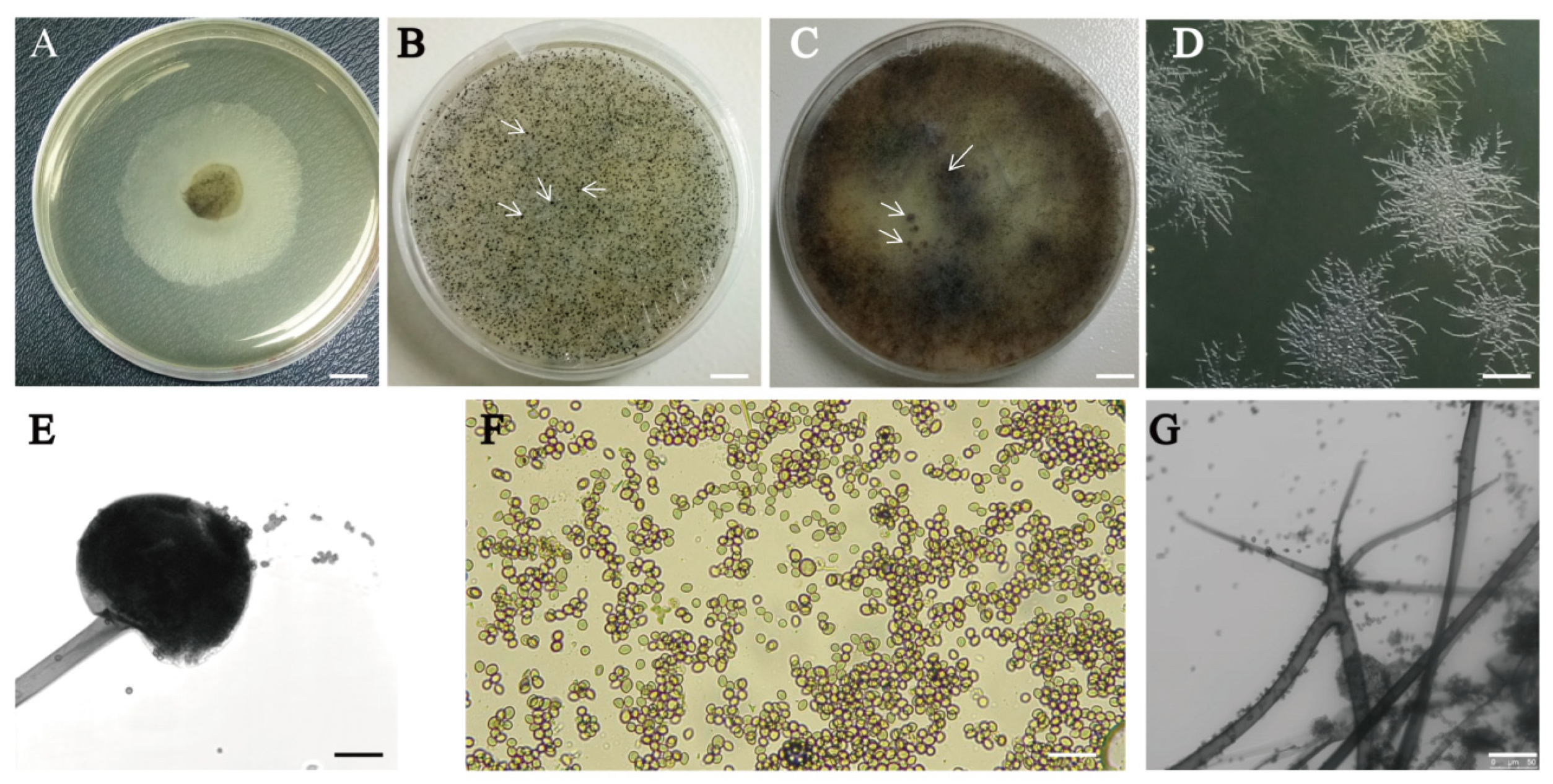
2.1. Isolation, Morphological Characterization, and Culture Conditions of R. oryzae

Between April and May 2018, brood mummy samples were collected from outside of colonies from four beekeeping sites in China (one in Henan province, one in Jiangsu province, and two in Beijing, where honeybee brood deaths had been recorded. A sample surface was sterilized using 70% ethanol for 1 min and then washed with de-ionized water three times. The brood mummies were then cut into smaller sizes and cultured on potato dextrose agar (PDA) Petri dishes, with a diameter of 9 cm, containing 30 mg/L streptomycin to suppress bacterial growth. The Petri dishes were placed in an incubator at 30 °C for 3 d. Fungal growth was observed using a light microscope, prior to strain selection and isolation. Slides containing hyphal parts were also prepared and observed using a light microscope. To purify the fungus, we collected samples from the isolates using the single-spore isolation method [52] with slight modifications. After purification, we observed the growing hyphal morphology, sporangiospores, sporangia, and sporangiophores of each single fungal mycelium from the inoculated plates, to characterize the fungal hyphal growth, conidia, texture, and spore structures. We collected pure spores from the PDA Petri dishes to grow the mycelia in potato dextrose broth (PDB) (Biomed Co., Beijing, China) for DNA extraction and molecular identification. The experiments were conducted in triplicate and samples were stored at −80 °C prior to analysis.

2.2. DNA Extraction and PCR Amplification

Mycelia from the fungal isolates and in vitro-infected larvae were prepared for DNA extraction by culturing spores in PDB, which were then placed on a reciprocal shaker at 140 rpm and incubated at 30 °C for 6 d. A Qiagen fungal DNA extraction kit was used for fungi DNA extraction. The concentration and other quality indicator values of the DNA were measured using the NanoDrop 2000 spectrophotometer (Thermo Fisher Scientific Inc., Waltham, MA, USA). Once high-quality DNA had been extracted from the fungal isolates, PCR was carried out to amplify the complete ITS sequences, using 40 ng/µL of DNA with a forward primer (5’-TCCGTAGGTGAACCTGCGG-3’) and reverse primer (5’-TCCTCCGCTTATTGATATGC-3’). Then, 25 μL of Taq polymerase-based reaction mix was employed in a PCR reaction under the following conditions: initial denaturation at 94 °C for 3 min, followed by 30 cycles of denaturation at 94 °C for 30 s, 59 °C for 30 s, and 72 °C for 30 s, with a final extension at 72 °C for 5 min and rapid cooling to 4 °C. Electrophoresis was used to check the quality of the PCR products, using 4 μL of PCR reaction product on 2% (w/v) agarose gel. The resulting PCR products (601 bp) were sent to the Biomedical Molecular Research Laboratory, Beijing, China for determination of the complete ITS sequences; these sequences were analyzed using the NCBI nucleotide BLAST tool. The experiments were replicated three times and samples were stored at −80 °C until analysis.

2.3. Phylogenetic Analysis

To determine the isolated fungus species’ identity and the infrageneric relationships in the genus, phylogenetic analysis was carried out for the highly identified sequences of these fungi. Multiple sequences collected from NCBI were aligned using MAFFT (version 7.453). Gblocks (version 0.91b) was used to filter poorly aligned positions and divergent regions. A phylogenetic tree of the isolated fungus, based on ITS region, was constructed using the IQ-tree program (version 1.6.12), using the ultrafast bootstrap parameters and suggested models. The reliability of the tree was measured with bootstrapping using 1000 replicates. An outgroup in the analysis was the fungus Mucor indicus ITS sequence.

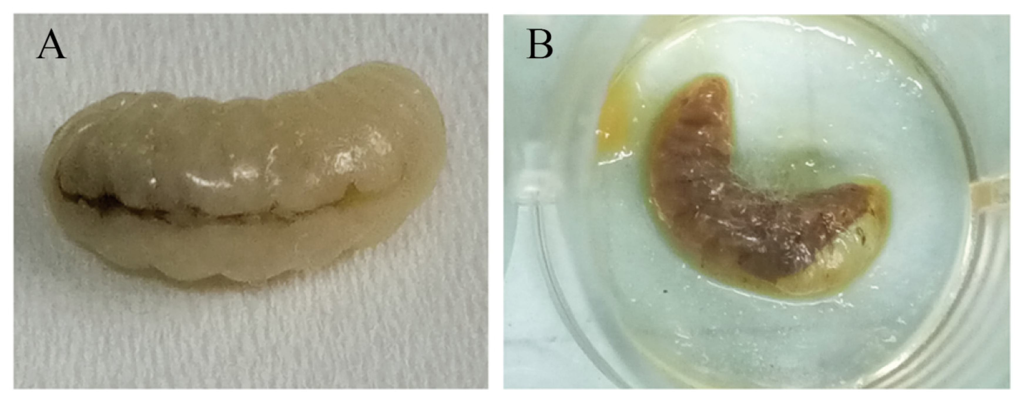
2.4. Bioassay of Pathogenicity

We tested the pathogenicity of the isolated fungal strain in vitro using spores collected in the laboratory, following the method described by Wubie [53] with slight modifications. In brief, oral inoculation of in vitro-reared second-instar honeybee worker larvae was used to investigate the differences in the pathogenicity of R. oryzae. Here, 1-day-old honeybee worker larvae were grafted to 24-well individual larva-rearing cells. Royal jelly-based larval food [54,55] was prepared, comprising 50% fresh royal jelly mixed with a solute of 6% D-fructose (w/v), 6% D-glucose (w/v), and 1% yeast extract (w/v) in 37% of sterile deionized water, which was pre-warmed to 34 °C. The larvae were fed on this diet once daily and were reared in an incubator at 34 ± 0.5 °C with 70% relative humidity.
To assess the isolate’s pathogenicity, we conducted a challenge inoculation experiment, in which about 1 × 10², 1 × 10³, 1 × 10⁴, 1 × 10⁵, and 1 × 10⁶ R. oryzae spores were mixed with 5 µL of larval diet and orally administered to the honeybee worker larvae. The infected larvae, showing abnormal dark grey skin coloration and back apical midrib melanization, were observed daily and the mortality rates were recorded. The number of hours post inoculation (hpi) (0 h, 24 h, 48 h, 72 h, 96 h, and 144 h) was set to determine whether the fungal infection depended on time. Koch’s postulates [56] were applied by using healthy second-instar honeybee worker larvae, which were milky white and shiny, to establish that R. oryzae caused diseases in the honeybee brood. After the infection experiment, identification of the fungus at the DNA level was carried out, following the protocol described above.

2.5. Statistical Analysis

All the bioassay data were analyzed using the SPSS (version 20.0 SPSS Inc., Chicago, IL, USA) statistical software package. To observe the effects of conidial concentration and exposure time and their interaction with the mortality of larvae, a repeated-measures ANOVA was performed. The mean difference of mortality was compared using Tukey’s multiple range test, where p < 0.05 was used as an indicator of statistical significance. The median lethal concentration (LC50) and time (LT50) values were calculated using Probit regression analysis.

3. Results

3.1. Isolation and Morphological Characterization of the New Fungal Isolate

The brood mummy samples were collected from the apiary where larval disease had occurred and where samples showed symptoms of honeybee chalkbrood disease. Among the fungi isolated from the brood mummy samples, one was different from the previously isolated Ascosphaera apis. This fungal colony grew aggressively on PDA within 24 h and was preliminarily identified as R. oryzae, based on the morphological profile and conidia analysis (Figure 1A,B) [57].
The colonies growing on PDA were initially white and cottony, then they became heavily speckled with sporangia, and finally became brown-grey to black-grey (Figure 2A–C). They spread rapidly, with stolons fixed to the substrate by rhizoids at various points (Figure 2D). Following incubation for 48 h at 30 °C, the mycelial height was 10–15 mm, touching the Petri dish cover plates (Figure 2B). The sporangiophores were straight, smooth-walled, simple, or branched, non-septate, and long and arose from the stolon opposite rhizoids, usually in groups (Figure 2E). The sporangia were globose, almost transparent at first, before turning black with many spores (Figure 2E). The sporangiospores were unequal, numerous, irregular, oval, and angular, with striations (Figure 2F). The rhizoids and stolons were dark brown (Figure 2G).

3.2. Internal Transcribed Spacer (ITS) Sequence Analysis

PCR product quality (Figure 3A,B) and amplified ITS DNA complete sequence alignment confirmed that the isolated fungus was R. oryzae, with 99.83% nucleotide BLAST similarity (Figure 4A). Further phylogenetic analysis showed that the isolated fungus in this analysis was classified into the R. oryzae clade with high bootstrap support (100%), indicating that the isolated strain was R. oryzae (Figure 4B). Taken together, based on multiple sequence alignments and phylogenetic analysis of the ITS region, the results show that the isolated fungus in this study can be considered a new pathogenic isolate of the R. oryzae involved in A. mellifera.

3.3. Pathogenicity Evaluation

Different concentrations of fungal spores caused significant differences in the death rate of honeybee larvae (Table S1 in the Supplementary Materials). The mortality of infected honeybee worker larvae significantly occurred within 24 h post-inoculation at spore concentrations of 1 × 10⁵ and 1 × 10⁶ (Tables S2 and S3 in the Supplementary Materials). This was characterized by a change in larval skin color from dark brown to black. Most orally spore-inoculated larvae died and exhibited dehydration, abnormal dark grey skin color, rigidity, and back apical midrib melanization (Figure 5). After 144 h, the surfaces of the dead larvae were extensively covered with conidia and mycelium (Figure 6). Conidia inoculated larvae showed slower growth rates, darker brown skin color, higher mortality rates, and greater mycelial growth, which was preceded by the production of dense yellow sporangiospores, than the control larvae (Figure 6). The disease symptoms on larvae following inoculation with the R. oryzae isolate were similar to those seen in the honeybee larvae mummies collected from the apiary sites.
There were significant differences in the in vitro pathogenicity of R. oryzae to honeybee worker larvae between the experimental and control groups across the developmental stages (p < 0.05). Repeated-measures ANOVA analysis revealed that the mean percentage mortality of R. oryzae was conidial-concentration- and time-dependent (Tables S4–S6 in the Supplementary Materials). The mortality rate increased with an increase in conidial concentrations and incubating time (Figure 7A,B). The mortality increased rapidly from 24 h to 72 h at conidial concentrations of 1 × 10⁵ and 1 × 10⁶, and the maximum mortality (98%) was recorded at 96 h (Figure 7A).
Probit regression analysis was performed to assess the virulence of R. oryzae for honeybee larvae. The median lethal concentration (LC50) value was 6.2 × 10³. The median lethal time (LT50) value was 25.50 h at 1 × 10⁶ conidia per larvae and 26.8 h at 1 × 10⁵ conidia per larvae (Table S1 in the Supplementary Materials).

4. Discussion

Recent studies have found that the cross-infection of host species by pathogens occurs more frequently than previously reported [58,59]. R. oryzae is common in the natural environment. Honeybees inevitably carry back this fungus to their hive when they collect nectar and pollen [60]. Although the infection of honeybee larvae by Rhizopus spp. is not common, this study successfully isolated and demonstrated the potential pathogenicity of this new R. oryzae strain from honeybee mummies to larvae, as a brood pathogen. The evaluation of the pathogenicity and virulence of this new fungus showed that it had high pathogenicity for honeybee larvae in laboratory cultures. Thus, the spread of this new strain to honeybee colonies could have deleterious effects on their individual health and pollinating functions. This study is the first report published on the pathogenicity of R. oryzae in honeybees, which suggests a potential risk to the health of honeybee colonies and crop pollination safety.

4.1. R. oryzae, a New Isolated Fungus That Infects Honeybee Larvae in Artificial Culture

With the development of biotechnology, morphological identification using light microscopy and electron microscopy, combined with molecular identification, has made fungal identification more accurate. Based on morphological and molecular analyses, the isolated fungus in this study was placed within a clade comprising R. oryzae reference isolates. It forms a long-grouped rhizoid, which arose from the stolon opposite rhizoids. This morphology is different from species in the Eurotiomycetes (Ascomycota) class, such as Aspergillus and Ascosphaera, which represent common subclasses of fungi that cause disease in honeybee larvae by germinating within the gut and ultimately mummifying the larvae when they ingest the fungal spores [47]. Furthermore, molecular analysis, based on the generated sequence of the Rhizopus 18S ITS region, confirmed that the fungus is R. oryzae. Also, BLAST analysis of the ITS region revealed a 99.83% sequence similarity with previously sequenced strains of R. oryzae [61]. Phylogenetic analysis also indicates that the isolated fungus in this study can be considered as R. oryzae.

4.2. The Virulence of R. oryzae for Honeybee Larvae Is Time- and Dosage-Dependent

Rhizopus is commonly detected in pollen, bee bread, and bee bodies. Early in 1974, Rhizopus arrhizus was isolated from the foraging honeybee midgut [49]. In 1988, Rhizopus sp. was identified in bee bread and the nurse honeybee midgut as disease-preventing fungi [51]. Recently, lower numbers of Rhizopus have been reported to be present in honeybee bread [50] and in stingless bee Trigona collina collection [62]. There have been many cases where bees collect fungal spores in place of pollen for unknown reasons [63,64]. Most collected fungi belong to either rust fungi, powdery mildews, or molds. Some of them may act as a form of nutrition or may be beneficial for bee health [63], while others have been found to spoil some soil-nesting bee bread and are a cause of larva death [48], including Rhizopus, whereas the mold-caused mortality of honeybee larvae is rare, and there are no data on the evaluation of the pathogenicity of these molds for honeybee larvae. The present study successfully isolated the strain of R. oryzae from brood mummy samples, and first evaluated its virulence for honeybee larvae. Our results revealed that the virulence of this pathogen for honeybee larvae was time- and dosage-dependent. This result is in line with the reports that R. oryzae showed pathogenicity for the soil-nesting bee, silkworm, wax moth [65,66,67], and fly [68]. It caused mortality at a lower concentration of 6.5 × 10² CFU per larva g−1 in silkworms [66]. These documents reveal that R. oryzae can infect insects; here, we found that R. oryzae being virulent for honeybee larvae is not accidental. Previous research has found that R. oryzae causes disease in bee-pollinated plants [41,69]. Therefore, we hypothesize that R. oryzae may infect both the host plant and its pollinators after their visitation.

4.3. The Potential Influence of R. oryzae on Honeybee Colonies Requires Further Investigation

Although Rhizopus has been commonly found in honeybee bread and assessed causally for some bee diseases, this study is the first to report the detection of R. oryzae in managed honeybee colonies and an evaluation of its pathogenicity for honeybee brood in the laboratory. It may probably be due to the fact that R. oryzae is an opportunistic, facultative pathogen, like A. apis, which needs a specific trigger to switch from a saprobe to a parasite. In this study, R. oryzae was isolated from the samples of A. apis-infected bee larvae collected from four apiaries in China during the summer. When chalkbrood disease is prevailing, the weather is moist, and the state of disease of the colony is weak, this provides an ideal environment for R. oryzae growth. Additionally, due to their similar symptoms, the presence of R. oryzae may have been overlooked by beekeepers and researchers in the past. Thirdly, when co-existing in the hive, the dominant growth of A. apis inhibits the growth of R. oryzae. It can be speculated from the single-spore isolated method used in this study, that when the suppression by A. apis was absent, R. oryzae grew aggressively on PDA within 24 h. This result is consistent with the hypothesis that A. apis prevents other fungal growth when a mixed infection is present in the honeybee colony [15].
A honeybee colony is a superorganism; it possesses various potential defenses against microbial infections, such as propolis [70,71], hygiene behaviors [72], grooming behaviors [73], and colony thermoregulation [74]. A hygienic colony can detect infected brood earlier and will remove them from the colony before they become mummies, thus reducing the risk of disease epidemics [75]. There are several studies showing that honeybees can sense the spores of fungi and remove them by grooming. Taken together, although we detected the strong pathogenicity of R. oryzae for A. mellifera larvae, it did not raise the possibility of colony prevalence.
The complex interactions between disease-causing agents, such as fungi, parasitic mites, bacteria, and viruses, and environmental factors, such as plant protection agents and pesticides, can cause honeybee colony collapse [76]. Therefore, future studies are needed to determine whether this strain of R. oryzae has pathogenicity for honeybees at the colony level, and if it can interact with other causative agents or if it has undergone genetic mutations that may increase its potential to infect honeybee larvae under natural conditions. Even though A. apis has been the predominantly isolated fungus of honeybee larvae, as indicated by chalky mummies following pupation [77,78], our finding regarding R. oryzae indicates that this new fungal species may cause economically important disease levels in managed honeybee colonies, which can result in significant brood loss. Additionally, a comparative study at the genomic and transcriptomic levels of this strain and the R. oryzae strains found on plants may show whether a host shift from plants to insects has occurred. Further research is also needed to determine whether adult worker bees spread this pathogen and, if so, to identify the mechanisms and pathways involved.

5. Conclusions

To our knowledge, this is the first mycological and molecular identification of R. oryzae, which could cause disease in honeybee broods in China and may add to the existing challenges facing the honeybee industry. In this study, we successfully isolated the fungus, R. oryzae, from dead honeybee broods, and described the morphological characteristics of its hyphae, sporangiospores, sporangia, and sporangiophores. The taxonomic classification was confirmed, based on morphology and molecular identification. Koch’s postulates were used to verify the pathogenicity of this newly isolated fungus. Its virulence to honeybee larvae is both time- and dosage-dependent. In the present study, we hypothesize that host plant pathogens can be transferred to their pollinators, but the spread pathway and the pathogenicity for honeybee colonies need further investigation.

Supplementary Materials

The following supporting information can be downloaded at: https://www.mdpi.com/article/10.3390/microorganisms12020313/s1, Table S1: Pathogenicity of different concentrations of R. oryzae spores to honeybee larvae. Table S2: Death rate of honeybee larvae inoculated with different concentrations of spores, during incubation time. Table S3: Death number of honeybee larvae inoculated with R. oryzae spores with different concentration during incubation time. Table S4: Multiple comparisons of virulence of different concentration R. oryzae spores to honeybee larvae. Table S5: Multiple comparisons of virulence of R. oryzae spores to honeybee larvae during incubation time. Table S6: Multivariate Tests.

Author Contributions

T.A., S.X. and L.M. conceptualized and investigated the study; A.G. and J.W. analyzed the data; H.Y., J.T. and N.L. carried out the experiments; T.A. and L.M. wrote the original draft manuscript; A.G. and S.X. revised the manuscript. All authors have read and agreed to the published version of the manuscript.

Funding

This work was supported by the Special Fund of the Modern Agro-industry Technology Research System (CARS-44-KXJ6) and the Agricultural Science and Technology Innovation Program (CAAS-ASTIP-IAR).

Data Availability Statement

The isolated fungus complete sequence has been uploaded to the NCBI; the GenBank accession number is MT1805651.1.

Acknowledgments

We would like to acknowledge all the supporting staff of the honeybee pathology research group at the Institute of Apicultural Research, Chinese Academy of Agricultural Sciences, Beijing, China for their generous provision of research materials. We also gratefully acknowledge the support received from a Special Fund of the Modern Agro-industry Technology Research System (CARS-44-KXJ6) and the Agricultural Science and Technology Innovation Program (CAAS-ASTIP-IAR) for their financial support.

Conflicts of Interest

The authors declare that they have no competing financial interests.

References

  1. Armstrong, A.; Brown, L.; Davies, G.; Whyatt, J.D.; Potts, S.G. Honeybee pollination benefits could inform solar park business cases, planning decisions and environmental sustainability targets. Biol. Conserv. 2021, 263, 109332. [Google Scholar] [CrossRef]
  2. Hung, K.L.J.; Kingston, J.M.; Albrecht, M.; Holway, D.A.; Kohn, J.R. The worldwide importance of honey bees as pollinators in natural habitats. Proc. R. Soc. B-Biol. Sci. 2018, 285, 20172140. [Google Scholar] [CrossRef] [PubMed]
  3. Croft, S.; Brown, M.; Wilkins, S.; Hart, A.; Smith, G.C. Evaluating european food safety authority protection goals for honeybees (Apis mellifera): What do they mean for pollination? Integr. Environ. Assess. Manag. 2018, 14, 750–758. [Google Scholar] [CrossRef] [PubMed]
  4. Stabentheiner, A.; Kovac, H. Honeybee economics: Optimisation of foraging in a variable world. Sci. Rep. 2016, 6, 28339. [Google Scholar] [CrossRef] [PubMed]
  5. Olszewski, K.; Dziechciarz, P.; Trytek, M.; Borsuk, G. A scientific note on the strategy of wax collection as rare behavior of Apis mellifera. Apidologie 2022, 53, 40. [Google Scholar] [CrossRef]
  6. Bobis, O. Plants: Sources of diversity in propolis properties. Plants 2022, 11, 2298. [Google Scholar] [CrossRef] [PubMed]
  7. Mair, B.; Wolf, M. Studies on the botanical origin and the residues of pesticides in corbicular pollen loads and bee bread of bee colonies in the proximity of apple orchards in South Tyrol. J. Kult. 2023, 75, 225–234. [Google Scholar] [CrossRef]
  8. Parish, J.B.; Scott, E.S.; Hogendoorn, K. Collection of conidia of by honey bee workers. Australas. Plant Pathol. 2020, 49, 245–247. [Google Scholar] [CrossRef]
  9. Evans, J.D.; Schwarz, R.S. Bees brought to their knees: Microbes affecting honey bee health. Trends Microbiol. 2011, 19, 614–620. [Google Scholar] [CrossRef]
  10. Zhang, F.; Cao, W.J.; Zhang, Y.H.; Luo, J.; Hou, J.A.; Chen, L.C.; Yi, G.Q.; Li, H.H.; Huang, M.F.; Dong, L.X.; et al. S-dinotefuran affects the social behavior of honeybees (Apis mellifera) and increases their risk in the colony. Pestic. Biochem. Physiol. 2023, 196, 105594. [Google Scholar] [CrossRef]
  11. Laomettachit, T.; Liangruksa, M.; Termsaithong, T.; Tangthanawatsakul, A.; Duangphakdee, O. A model of infection in honeybee colonies with social immunity. PLoS ONE 2021, 16, e0247294. [Google Scholar] [CrossRef] [PubMed]
  12. Santorelli, L.A.; Wilkinson, T.; Abdulmalik, R.; Rai, Y.; Creevey, C.J.; Huws, S.; Gutierrez-Merino, J. Beehives possess their own distinct microbiomes. Environ. Microbiome 2023, 18, 1. [Google Scholar] [CrossRef] [PubMed]
  13. Sinkeviciene, J.; Taraseviciene, Ä.; Tamutis, V. Fungi and mycotoxins in bee pollen collected in Lithuania. Appl. Sci. 2023, 13, 1571. [Google Scholar] [CrossRef]
  14. Cui, P.; Kong, K.; Yao, Y.; Huang, Z.D.; Shi, S.P.; Liu, P.; Huang, Y.C.; Abbas, N.; Yu, L.S.; Zhang, Y.L. Community composition, bacterial symbionts, antibacterial and antioxidant activities of honeybee-associated fungi. BMC Microbiol. 2022, 22, 168. [Google Scholar] [CrossRef]
  15. Cheng, X.F.; Zhang, L.; Luo, J.; Yang, S.; Deng, Y.C.; Li, J.H.; Hou, C.S. Two pathogenic fungi isolated from chalkbrood samples and honey bee viruses they carried. Front. Microbiol. 2022, 13, 843842. [Google Scholar] [CrossRef] [PubMed]
  16. Rutkowski, D.; Weston, M.; Vannette, R.L. Bees just wanna have fungi: A review of bee associations with nonpathogenic fungi. Fems Microbiol. Ecol. 2023, 99, fiad077. [Google Scholar] [CrossRef] [PubMed]
  17. Sevim, A.; Akpinar, R.; Karaoglu, S.A.; Bozdeveci, A.; Sevim, E. Prevalence and phylogenetic analysis of (Maassen ex Claussen) LS Olive & Spiltoir (1955) isolates from honeybee colonies in Turkey. Biologia 2022, 77, 2689–2699. [Google Scholar] [CrossRef]
  18. Getachew, A.; Wubie, A.J.; Wu, J.L.; Xu, J.; Wu, P.J.; Abejew, T.A.; Tu, Y.Y.; Zhou, T.; Xu, S.F. Molecular identification of pathogenicity associated genes in honeybee fungal pathogen, by restricted enzyme-mediated integration (REMI) constructed mutants. Int. J. Agric. Biol. 2018, 20, 2879–2890. [Google Scholar]
  19. Niu, G.D.; Johnson, R.M.; Berenbaum, M.R. Toxicity of mycotoxins to honeybees and its amelioration by propolis. Apidologie 2011, 42, 79–87. [Google Scholar] [CrossRef]
  20. Grupe, A.C.; Quandt, C.A. A growing pandemic: A review of Nosema parasites in globally distributed domesticated and native bees. PLoS Pathog. 2020, 16, e1008580. [Google Scholar] [CrossRef]
  21. Ayo Fasasi, K. Microbiota of honeybees, Apis mellifera Adansonii (Hymenoptera: Apidae) from selected ecozones, South West Nigeria. Pak. J. Biol. Sci. 2018, 21, 232–238. [Google Scholar] [CrossRef] [PubMed]
  22. Vojvodic, S.; Jensen, A.B.; James, R.R.; Boomsma, J.J.; Eilenberg, J. Temperature dependent virulence of obligate and facultative fungal pathogens of honeybee brood. Vet. Microbiol. 2011, 149, 200–205. [Google Scholar] [CrossRef] [PubMed]
  23. Klinger, E.G.; Vojvodic, S.; DeGrandi-Hoffman, G.; Welker, D.L.; James, R.R. Mixed infections reveal virulence differences between host-specific bee pathogens. J. Invertebr. Pathol. 2015, 129, 28–35. [Google Scholar] [CrossRef] [PubMed]
  24. Vojvodic, S.; Boomsma, J.J.; Eilenberg, J.; Jensen, A.B. Virulence of mixed fungal infections in honey bee brood. Front. Zool. 2012, 9, 5. [Google Scholar] [CrossRef] [PubMed]
  25. Keller, K.M.; Deveza, M.V.; Koshiyama, A.S.; Tassinari, S.; Barth, O.M.; Castro, R.N.; Lorenzon, M.C. Fungi infection in honeybee hives in regions affected by Brazilian sac brood. Arq. Bras. Med. Vet. E Zootec. 2014, 66, 1471–1478. [Google Scholar] [CrossRef]
  26. Dolezal, A.G.; Toth, A.L. Feedbacks between nutrition and disease in honey bee health. Curr. Opin. Insect Sci. 2018, 26, 114–119. [Google Scholar] [CrossRef] [PubMed]
  27. Dosselli, R.; Grassl, J.; Carson, A.; Simmons, L.W.; Baer, B. Flight behaviour of honey bee (Apis mellifera) workers is altered by initial infections of the fungal parasite. Sci. Rep. 2016, 6, 36649. [Google Scholar] [CrossRef] [PubMed]
  28. Schaeffer, R.N.; Mei, Y.Z.; Andicoechea, J.; Manson, J.S.; Irwin, R.E. Consequences of a nectar yeast for pollinator preference and performance. Funct. Ecol. 2017, 31, 613–621. [Google Scholar] [CrossRef]
  29. Rering, C.C.; Rudolph, A.B.; Beck, J.J. Pollen and yeast change nectar aroma and nutritional content alone and together, but honey bee foraging reflects only the avoidance of yeast. Environ. Microbiol. 2021, 23, 4141–4150. [Google Scholar] [CrossRef]
  30. Didaras, N.A.; Karatasou, K.; Dimitriou, T.G.; Amoutzias, G.D.; Mossialos, D. Antimicrobial activity of bee-collected pollen and beebread: State of the art and future perspectives. Antibiotics 2020, 9, 811. [Google Scholar] [CrossRef]
  31. Dharampal, P.S.; Carlson, C.; Currie, C.R.; Steffan, S.A. Pollen-borne microbes shape bee fitness. Proc. R. Soc. B Biol. Sci. 2019, 286, 20182894. [Google Scholar] [CrossRef]
  32. Menezes, C.; Vollet-Neto, A.; Marsaioli, A.J.; Zampieri, D.; Fontoura, I.C.; Luchessi, A.D.; Imperatriz-Fonseca, V.L. A Brazilian social bee must cultivate fungus to survive. Curr. Biol. 2015, 25, 2851–2855. [Google Scholar] [CrossRef] [PubMed]
  33. Parish, J.B.; Scott, E.S.; Hogendoorn, K. Nutritional benefit of fungal spores for honey bee workers. Sci. Rep. 2020, 10, 15671. [Google Scholar] [CrossRef]
  34. Disayathanoowat, T.; Li, H.; Supapimon, N.; Suwannarach, N.; Lumyong, S.; Chantawannakul, P.; Guo, J. Different dynamics of bacterial and fungal communities in hive-stored bee bread and their possible roles: A case study from two commercial honey bees in China. Microorganisms 2020, 8, 264. [Google Scholar] [CrossRef]
  35. Vocadlova, K.; Lüddecke, T.; Patras, M.A.; Marner, M.; Hartwig, C.; Benes, K.; Matha, V.; Mraz, P.; Schäberle, T.F.; Vilcinskas, A. Extracts of Talaromyces purpureogenus strains from Apis mellifera bee bread inhibit the growth of Paenibacillus spp. in vitro. Microorganisms 2023, 11, 2067. [Google Scholar] [CrossRef] [PubMed]
  36. Skerl, M.I.S.; Gajger, I.T. Performance and Nosema spp. spore level in young honeybee (Apis mellifera carnica, Pollmann 1879) colonies supplemented with candies. Slov. Vet. Res. 2022, 59, 159–167. [Google Scholar] [CrossRef]
  37. Gryganskyi, A.P.; Lee, S.C.; Litvintseva, A.P.; Smith, M.E.; Bonito, G.; Porter, T.M.; Anishchenko, I.M.; Heitman, J.; Vilgalys, R. Structure, function, and phylogeny of the mating locus in the Rhizopus oryzae complex. PLoS ONE 2010, 5, e15273. [Google Scholar] [CrossRef] [PubMed]
  38. Gryganskyi, A.P.; Golan, J.; Dolatabadi, S.; Mondo, S.; Robb, S.; Idnurm, A.; Muszewska, A.; Steczkiewicz, K.; Masonjones, S.; Liao, H.L.; et al. Phylogenetic and phylogenomic definition of Rhizopus species. G3-Genes Genomes Genet. 2018, 8, 2007–2018. [Google Scholar] [CrossRef] [PubMed]
  39. Li, W.Q.; Jiang, Y.Y.; Hu, C.J.; Liu, G.A.; Li, Y.G.; Wang, S. Identification, pathogenic mechanism and control of causing postharvest fruit rot in pumpkin. Postharvest Biol. Technol. 2023, 204, 112460. [Google Scholar] [CrossRef]
  40. Febriani, R.; Sjamsuridzal, W.; Oetari, A.; Santoso, I.; Roosheroe, I.G. ITS regions of rDNA sequence and morphological analyses clarify five strains from tempeh as Rhizopus oryzae. In Proceedings of the 3rd International Symposium on Current Progress in Mathematics and Sciences 2017 (Iscpms2017), Bali, Indonesia, 26–27 July 2017; Volume 2023. [Google Scholar] [CrossRef]
  41. Khokhar, I.; Mukhtar, I.; Wang, J.H.; Jia, Y.; Yan, Y.C. A report of Rhizopus oryzae causing postharvest soft rot of apple fruit in China. Australas. Plant Dis. Notes 2019, 14, 7. [Google Scholar] [CrossRef]
  42. Cloutier, A.; Tran, S.; Avis, T.J. Suppressive effect of compost bacteria against grey mould and Rhizopus rot on strawberry fruit. Biocontrol Sci. Technol. 2020, 30, 143–159. [Google Scholar] [CrossRef]
  43. Wu, F.; Tong, X.Q.; Zhang, L.Q.; Mei, L.; Guo, Y.B.; Wang, Y.J. Suppression of Rhizopus fruit rot by volatile organic compounds produced by CF05. Biocontrol Sci. Technol. 2020, 30, 1351–1364. [Google Scholar] [CrossRef]
  44. Khokhar, I.; Jia, Y.; Mukhtar, I.; Wang, J.H.; Ruth, N.; Eltoukhy, A.; Fan, S.H.; Li, X.J.; Wang, J.Y.; Yan, Y.C. First report of causing postharvest fruit rot on pear in China. Plant Dis. 2019, 103, 1423. [Google Scholar] [CrossRef]
  45. Yoder, J.A.; Hedges, B.Z.; Heydinger, D.J.; Sammataro, D.; DeGrandi-Hoffman, G. Differences among fungicides targeting beneficial fungi associated with honey bee colonies. In Honey Bee Colony Health: Challenges and Sustainable Solutions; CRC Press: Boca Raton, FL, USA, 2012; pp. 181–192. [Google Scholar]
  46. Zhao, Y.Z.; Zhang, Z.F.; Cai, L.; Peng, W.J.; Liu, F. Four new filamentous fungal species from newly-collected and hive-stored bee pollen. Mycosphere 2018, 9, 1089–1116. [Google Scholar] [CrossRef]
  47. Gilliam, M.; Prest, D.B.; Lorenz, B.J. Microbiology of pollen and bee bread:taxonomy and enzymology of molds. Apidologie 1989, 20, 53–68. [Google Scholar] [CrossRef]
  48. Batra, L.R.; Batra, S.W.; Bohart, G.E. The mycoflora of domesticated and wild bees (Apoidea). Mycopathol. Mycol. Appl. 1973, 49, 13–44. [Google Scholar] [CrossRef]
  49. Gilliam, M. Fungi isolated from honey bees, Apis mellifera. J. Invertebr. Pathol. 1974, 24, 213–217. [Google Scholar] [CrossRef] [PubMed]
  50. Yoder, J.A.; Heydinger, D.J.; Hedges, B.Z.; Sammataro, D.; Finley, J.; DeGrandi-Hoffman, G.; Croxall, T.J.; Christensen, B.S. Fungicides Reduce Symbiotic Fungi in Bee Bread and the Beneficial Fungi in Colonies. In Honey Bee Colony Health: Challenges and Sustainable Solutions; CRC Press: Boca Raton, FL, USA, 2012; pp. 193–214. [Google Scholar]
  51. Gilliam, M. Factors affecting development of chalkbrood disease in colonies of honey bees, Apis mellifera, fed pollen contaminated with Ascosphaera apis. J. Invertebr. Pathol. 1988, 52, 314–325. [Google Scholar] [CrossRef]
  52. Georg, L.K. A simple and rapid method for obtaining monospore cultures of fungi. Mycologia 1974, 39, 368–371. [Google Scholar] [CrossRef]
  53. Wubie, A.J.; Hu, Y.; Li, W.; Huang, J.; Guo, Z.; Xu, S.; Zhou, T. Factors analysis in protoplast isolation and regeneration from a chalkbrood fungus, Ascosphaera apis. Int. J. Agric. Biol. 2014, 16, 89–96. [Google Scholar]
  54. Jensen, A.B.; Aronstein, K.; Flores, J.M.; Vojvodic, S.; Palacio, M.A.; Spivak, M. Standard methods for fungal brood disease research. J. Apic. Res. 2013, 52, 1–20. [Google Scholar] [CrossRef]
  55. Crailsheim, K.; Brodschneider, R.; Aupinel, P.; Behrens, D.; Genersch, E.; Vollmann, J.; Riessberger-Gallé, U. Standard methods for artificial rearing of larvae. J. Apic. Res. 2013, 52, 1–16. [Google Scholar] [CrossRef]
  56. Carlberg, D. Koch’s postulates revisited. Scientist 2000, 14, 6. [Google Scholar]
  57. Samson, R.A.; Visagie, C.M.; Houbraken, J.; Hong, S.B.; Hubka, V.; Klaassen, C.H.W.; Perrone, G.; Seifert, K.A.; Susca, A.; Tanney, J.B.; et al. Phylogeny, identification and nomenclature of the genus. Aspergrillus. Stud. Mycol. 2014, 78, 141–173. [Google Scholar] [CrossRef] [PubMed]
  58. Fürst, M.A.; McMahon, D.P.; Osborne, J.L.; Paxton, R.J.; Brown, M.J.F. Disease associations between honeybees and bumblebees as a threat to wild pollinators. Nature 2014, 506, 364–366. [Google Scholar] [CrossRef] [PubMed]
  59. Evison, S.E.F.; Jensen, A.B. The biology and prevalence of fungal diseases in managed and wild bees. Curr. Opin. Insect Sci. 2018, 26, 105–113. [Google Scholar] [CrossRef] [PubMed]
  60. Sinkeviciene, J.; Amsiejus, A. Mycobiota in bee pollen collected by different types of traps. Zemdirb. Agric. 2019, 106, 377–382. [Google Scholar] [CrossRef]
  61. Abe, A.; Oda, Y.; Asano, K.; Sone, T. The molecular phylogeny of the genus Rhizopus based on rDNA sequences. Biosci. Biotechnol. Biochem. 2006, 70, 2387–2393. [Google Scholar] [CrossRef] [PubMed]
  62. Eltz, T.; Brühl, C.A.; Görke, C. Collection of mold (Rhizopus sp.) spores in lieu of pollen by the stingless bee Trigona collina. Insectes Sociaux 2002, 49, 28–30. [Google Scholar] [CrossRef]
  63. Shaw, D.E. Honeybees collecting Neurospora spores from steamed Pinus logs in Queensland. Mycologist 1993, 7, 182–185. [Google Scholar] [CrossRef]
  64. Oliveira, M.L.; Morato, E.F. Stingless bees (Hymenoptera, Meliponini) feeding on stinkhorn spores (Fungi, Phallales): Robbery or dispersal? Revta Bras. Zool. 2000, 17, 881–884. [Google Scholar] [CrossRef]
  65. Maurer, E.; Hörtnagl, C.; Lackner, M.; Grässle, D.; Naschberger, V.; Moser, P.; Segal, E.; Semis, M.; Lass-Flörl, C.; Binder, U. Galleria mellonella as a model system to study virulence potential of mucormycetes and evaluation of antifungal treatment. Med. Mycol. 2019, 57, 351–362. [Google Scholar] [CrossRef] [PubMed]
  66. Tominaga, T.; Uchida, R.; Koyama, N.; Tomoda, H. Anti-Rhizopus activity of tanzawaic acids produced by the hot spring-derived fungus Penicillium sp. BF-0005. J. Antibiot. 2018, 71, 626–632. [Google Scholar] [CrossRef] [PubMed]
  67. Kurakado, S.; Matsumoto, Y.; Sugita, T. Efficacy of posaconazole against infection in silkworm. Med. Mycol. J. 2021, 62, 53–57. [Google Scholar] [CrossRef] [PubMed]
  68. Shirazi, F.; Farmakiotis, D.; Yan, Y.; Albert, N.; Do, K.A.; Kontoyiannis, D.P. Diet modification and metformin have a beneficial effect in a fly model of obesity and mucormycosis. PLoS ONE 2014, 9, e108635. [Google Scholar] [CrossRef] [PubMed]
  69. Fan, Z.Q.; Xia, Q.; Zhao, Z.Q.; Duan, Y.A.; Zhao, L.; Wang, H.Y.; Jiang, W.T.; Chen, X.S.; Yin, C.M.; Mao, Z.Q. Screening and identification of XERF-1 and its effect on apple replant disease. Sci. Hortic. 2022, 305, 111400. [Google Scholar] [CrossRef]
  70. Popova, M.; Reyes, M.; Le Conte, Y.; Bankova, V. Propolis chemical composition and honeybee resistance against Varroa destructor. Nat. Prod. Res. 2014, 28, 788–794. [Google Scholar] [CrossRef] [PubMed]
  71. Popova, M.; Antonova, D.; Bankova, V. Chemical composition of propolis and American foulbrood: Is there any relationship? Bulg. Chem. Commun. 2017, 49, 171–175. [Google Scholar]
  72. Swanson, J.A.I.; Torto, B.; Kells, S.A.; Mesce, K.A.; Tumlinson, J.H.; Spivak, M. Odorants that induce hygienic behavior in honeybees: Identification of volatile compounds in chalkbrood-infected honeybee larvae. J. Chem. Ecol. 2009, 35, 1108–1116. [Google Scholar] [CrossRef]
  73. Foose, A.M.; Westwick, R.R.; Vengarai, M.; Rittschof, C.C. The survival consequences of grooming in the honey bee. Insectes Sociaux 2022, 69, 279–287. [Google Scholar] [CrossRef]
  74. Becher, M.A.; Hildenbrandt, H.; Hemelrijk, C.K.; Moritz, R.F.A. Brood temperature, task division and colony survival in honeybees: A model. Ecol. Model. 2010, 221, 769–776. [Google Scholar] [CrossRef]
  75. Medina-Flores, C.A.; Medina, L.A.M.; Guzmán-Novoa, E. Effect of hygienic behavior on resistance to chalkbrood disease (Ascosphaera apis) in Africanized bee colonies (Apis mellifera). Rev. Mex. Cienc. Pecu. 2022, 13, 225–239. [Google Scholar] [CrossRef]
  76. Pent, K.; Naudi, S.; Raimets, R.; Jürison, M.; Liiskmann, E.; Karise, R. Overlapping exposure effects of pathogen and dimethoate on honeybee (Apis mellifera Linnaeus) metabolic rate and longevity. Front. Physiol. 2023, 14, 1198070. [Google Scholar] [CrossRef]
  77. Jensen, A.B.; Welker, D.L.; Kryger, P.; James, R.R. Polymorphic DNA sequences of the fungal honey bee pathogen Ascosphaera apis. Fems Microbiol. Lett. 2012, 330, 17–22. [Google Scholar] [CrossRef]
  78. Aronstein, K.A.; Holloway, B.A. Honey bee fungal pathogen, Ascosphaera apis; current understanding of host-pathogen interactions and host mechanisms of resistance. Microb. Pathog. 2013, 13, 402–410. [Google Scholar]
Figure 1. Collection of honeybee brood mummies and the selective inoculation of R. oryzae on PDA culture. (A) White mummies. (B) The growth of white R. oryzae mycelia from mummies on PDA culture. Bars = 1 cm.
Microorganisms 12 00313 g001
Figure 2. Mycelial growth pattern, venation, sporulation, and conidial structure of R. oryzae. (A) Mycelial structure. (B) Young conidia. (C) Matured conidia. (D) Mycelial venation. (E) Matured conidia head, observed with a 10× objective lens. (F) Conidial structure, observed with a 20× objective lens. (G) Mycelial rhizoid. The bars on A to C represent 1 cm, and those on D to G represent 50 μm. The white arrow points to the conidia.
Microorganisms 12 00313 g002
Figure 3. Internal transcribed spacer (ITS) bands of R. oryzae before and after honeybee worker larvae inoculation. (A) ITS of the isolated R. oryzae before the infection bioassay, where images 1–4 represent samples isolated from four beekeeping sites in China. (B) ITS of R. oryzae after infection bioassay, where images 5–8 represent samples isolated from four locations, respectively.
Microorganisms 12 00313 g003
Figure 4. Complete sequence alignment and phylogenetic analysis of the ITS region of the isolated fungus. (A) Complete sequence alignment of the ITS region from the isolated R. oryzae to the NCBI-uploaded reference genome. Isltd R. oryzae represents isolated R. oryzae, R. oryzae represents the NCBI-referenced R. oryzae. The boxes show the base pairs of ITS with differences. (B) Phylogenetic analysis of the isolated fungus, based on the ITS sequence region. GenBank accession numbers for the sequences are adjacent to the corresponding species names. ‘MT103595.1_Rhizopus_oryzae’ in the red box represents the current sequence data from this study.
Microorganisms 12 00313 g004
Figure 5. Observed physical expressions of R. oryzae inoculation in honeybee larvae. (A) Back apical midrib melanization in post-inoculation larvae. (B) Body melanization in post-inoculation larvae.
Microorganisms 12 00313 g005
Figure 6. Observed physical expressions of R. oryzae on inoculated honeybee larvae compared with control group. (A) Non-infected control larvae. (B) R. oryzae: initially infected larvae. (C) R. oryzae: post-infection larvae. White arrows show health larvae and red arrows show infected larvae respectively.
Microorganisms 12 00313 g006
Figure 7. In vitro pathogenicity of R. oryzae and the mortality rate of honeybee worker larvae. (A) Cumulative mortality rate of larvae across inoculation times. (B) Percentage mortality rate of honeybee worker larvae at different conidial concentrations of R. oryzae and time. The error bars represent the SD. Lowercase letters show significant differences in the average larvae death rate among the concentrations. T1, T2, T3, T4, and T5 stand for R. oryzae spore-inoculated larvae at 1 × 10², 1 × 10³, 1 × 10⁴, 1 × 10⁵, and 1 × 10⁶ conidia/larva, respectively. T0 is the control group, in which larvae were fed on a normal diet.
Microorganisms 12 00313 g007
Disclaimer/Publisher’s Note: The statements, opinions and data contained in all publications are solely those of the individual author(s) and contributor(s) and not of MDPI and/or the editor(s). MDPI and/or the editor(s) disclaim responsibility for any injury to people or property resulting from any ideas, methods, instructions or products referred to in the content.

