Article

EnvC Homolog Encoded by Xanthomonas citri subsp. citri Is Necessary for Cell Division and Virulence

1
Agricultural and Livestock Microbiology Graduation Program, School of Agricultural and Veterinarian Sciences, São Paulo State University (UNESP), Jaboticabal 14884-900, SP, Brazil
2
Department of Plant Pathology and Weed Research, Institute of Plant Protection Agricultural Research Organization (ARO), Volcani Institute, Rishon LeZion 7505101, Israel
3
Biochemistry Building, Institute of Biosciences, São Paulo State University (UNESP), Rio Claro 13506-900, SP, Brazil
4
Department of Agricultural, Livestock and Environmental Biotechnology, School of Agricultural and Veterinary Sciences, São Paulo State University (UNESP), Jaboticabal 14884-900, SP, Brazil
5
Citrus Research and Education Center, Department of Microbiology and Cell Science, Institute of Food and Agricultural Sciences, University of Florida, Lake Alfred, FL 33850, USA
*
Author to whom correspondence should be addressed.
These authors contributed equally to this work.
Current address: Department of Plant Pathology, University of Georgia, Tifton, GA 31793, USA.
Microorganisms 2024, 12(4), 691; https://doi.org/10.3390/microorganisms12040691
Submission received: 9 October 2023 / Revised: 16 March 2024 / Accepted: 21 March 2024 / Published: 29 March 2024
(This article belongs to the Section Plant Microbe Interactions)

Abstract

:
Peptidoglycan hydrolases are enzymes responsible for breaking the peptidoglycan present in the bacterial cell wall, facilitating cell growth, cell division and peptidoglycan turnover. Xanthomonas citri subsp. citri (X. citri), the causal agent of citrus canker, encodes an Escherichia coli M23 peptidase EnvC homolog. EnvC is a LytM factor essential for cleaving the septal peptidoglycan, thereby facilitating the separation of daughter cells. In this study, the investigation focused on EnvC contribution to the virulence and cell separation of X. citri. It was observed that disruption of the X. citri envC gene (ΔenvC) led to a reduction in virulence. Upon inoculation into leaves of Rangpur lime (Citrus limonia Osbeck), the X. citri ΔenvC exhibited a delayed onset of citrus canker symptoms compared with the wild-type X. citri. Mutant complementation restored the wild-type phenotype. Sub-cellular localization confirmed that X. citri EnvC is a periplasmic protein. Moreover, the X. citri ΔenvC mutant exhibited elongated cells, indicating a defect in cell division. These findings support the role of EnvC in the regulation of cell wall organization, cell division, and they clarify the role of this peptidase in X. citri virulence.

1. Introduction

Xanthomonas citri subsp. citri (X. citri) is a phytopathogenic Gram-negative bacterium and the causal agent of citrus canker, a severe disease, which affects all economically important citrus varieties worldwide, causing significant economic losses [1]. Gram-negative bacteria feature a dense peptidoglycan (PG) layer situated in the periplasmic space, positioned between their outer and inner membranes [2]. This polymer is composed of glycan strands of β-1,4-glycosidic bond-linked N-acetylglucosamine (GlcNAc) and N-acetylmuramic acid (MurNAc) disaccharides, cross-linked by short peptides, playing a crucial role in upholding and sustaining the shape and integrity of the cell [3]. Two main classes of peptidoglycan-lytic enzymes are responsible for the PG’s assembly: the glycosidases cleaving the glycan backbone and the amidases (or peptidases) cleaving the peptide side chain [4].
The X. citri strain 306 (GenBank accession number: AE008923.1) possesses nine proteins sharing the M23 peptidase domain, with four of them being hypothetical proteins (Supplementary Table S1). One of these proteins—XAC0024 (GenBank accession number: AAM34916.1)—is a homolog of Escherichia coli (E. coli) EnvC (GenBank accession number: EGO4467787.1), a widely distributed protein in bacteria [5]. EnvC is highly conserved among Gram-negative bacteria and functions as part of the septal ring apparatus [6]. In E. coli, EnvC is a periplasmic peptidase, which plays a role in septal peptidoglycan splitting and daughter-cell separation [7]. Deletion of the E. coli envC gene, like other genes encoding LytM domain hydrolases, such as nlpD, ygeR and uebA, leads to the formation of long cell chains, suggesting a defect in cell separation [7]. It was demonstrated that EnvC controls cell separation by activating PG-degrading amidases AmiA and AmiB [8]. Homologs of envC have been shown to perform similar functions in various bacterial species and are essential for the pathogenicity of several animal bacterial pathogens, including Vibrio cholera, enterohemorrhagic E. coli and Fusobacterium nucleatum [9,10,11]. The envC homolog of Pseudomonas aeruginosa was identified to be functionally redundant to nlpD, as deletion of both led to the formation of long cell chains and enhanced sensitivity to high temperature and antimicrobial compounds [12].
The role of LytM factors and PG amidases in pathogenicity and cell division was recently demonstrated in X. campestris pv. campestris [13]. Deficiency in cell separation was observed in either nlpD or envC deletion strains; however, deletion of the single gene nlpD had a significant effect on virulence and induction of hypersensitive response in non-host plants, while deletion of envC did not significantly affect host interactions [13].
In the present study, the envC homolog (GenBank XAC0024) of X. citri strain 306 was characterized. It was observed that envC was essential for virulence but did not completely compromise the ability of X. citri to trigger weak symptoms in a susceptible host genotype. Additionally, X. citri ΔenvC displayed an altered cell shape compared with the wild-type (wt) strain. Similar to observations in other Gram-negative bacteria, the X. citri envC gene seems to play a role in daughter-cell separation. Moreover, the sub-cellular localization of X. citri EnvC protein fused with the mCherry fluorophore (EnvC-mCherry) is consistent with the protein occupying the periplasmic region.

2. Materials and Methods

2.1. Bacterial Strains, Plasmids and Culture Condition

The bacterial strains and plasmids utilized in this study are detailed in Supplementary Table S2 [14,15,16]. E. coli strains DH10B, SM10ʎpir and HST08–Stellar used for cloning were cultivated at 37 °C in a LB/LB-agar medium [17]. Growth in the liquid medium was conducted at 250 rpm (shaker) for 14–16 h, while growth in the solid medium took place in a bacteriological incubator for 14–16 h. X. citri 306 strains were cultivated at 30 °C in a NYG-rich medium (3 g/L yeast extract, 5 g/L peptone, 20 g/L glycerol, pH 7.0), nutrient broth (NB) medium (3 g/L meat extract, 5 g/L peptone) or on NB-agar plates (NB medium containing 15 g/L agar) supplemented with L-arabinose (0.05% w/v), starch (0.2% w/v) and sucrose (5% w/v) when required. X. citri was grown for 48 h either in liquid NYG-rich medium at 250 rpm (shaker) or in solid NB medium in a bacteriological incubator. The antibiotics carbenicillin and kanamycin or ampicillin and spectinomycin were used when required at a concentration of 50 μg/mL and 100 μg/mL, respectively.

2.2. Mutant Construction

2.2.1. Partial Deletion of XAC0024 Nucleotide Sequence

The X. citri strain containing the disrupted XAC0024 gene (mutant ΔenvC) was obtained through site-directed mutagenesis using the overlap extension approach in the polymerase chain reaction [18]. To generate the XAC0024 mutant, X. citri genomic DNA served as a template in a two-step PCR process. In the initial step, primers A(F) and B(R), as well as C(F) and D(R) (Supplementary Table S3), were employed in separate reactions to generate fragments AB and CD, respectively. Subsequently, these fragments were fused through a double-joint PCR. The PCR reaction, each with a final volume of 20 µL, contained 26.5 ng of DNA, 0.2 mM of each dNTP, 1U of Phusion High Fidelity DNA Polymerase (New England Biolabs, Ipswich, MA, USA), 0.5 μM of each primer and 3% of DMSO for primer pairs A–B and 5% of DMSO for primer pairs C–D. The PCR conditions comprised an initial denaturation at 98 °C for 30 s, followed by 35 cycles of denaturation at 98 °C for 10 s, annealing at 69 °C for 30 s and extension at 72 °C for 30 s. The reaction concluded with a final extension at 72 °C for 10 min, executed in a Veriti® 96-Well Thermal Cycler (Applied Biosystems, Waltham, MA, USA). The resulting PCR products (fragments AB and CD) underwent purification using the Wizard® SV Gel and PCR Clean-Up System (Promega, Madison, WI, USA), and they were quantified using the NanoDrop® ND-1000 spectrophotometer (Thermo Fisher Scientific, Waltham, MA, USA). The size of the amplified fragments AB and CD was confirmed via agarose gel electrophoresis (Supplementary Figure S1). The double-joint PCR step was performed using fragments AB and CD as templates and the primers A(F) and D(R) to generate the AD fragment, using 3% DMSO and the same PCR conditions described above, except for a final volume adjustment to 50 µL. The resulting product was subjected to 0.7% agarose gel electrophoresis (Supplementary Figure S1), and the band with the expected size was recovered from the gel using the Wizard® SV Gel and PCR Clean-Up System (Promega). The purified DNA fragment was quantitated in a NanoDrop® ND-1000 spectrophotometer (Thermo Fisher Scientific) and submitted to a PCR reaction to add a 3′-A overhang to the ends of the AD fragment. This reaction utilized a final concentration of 0.2 mM dATP and 1 U of Platinum® Taq DNA Polymerase Recombinant (Invitrogen, Waltham, MA, USA) in a 50 µL reaction for 10 min at 72 °C in a GeneAmp® PCR System 9700 (Applied Biosystems).

2.2.2. Deletion Vector Construction

The PCR-amplified AD fragment, featuring 3′-A overhangs, was ligated into the pGEM®-T Easy plasmid (Promega) using T4 DNA ligase, according to the manufacturer’s instructions. The ligation reaction comprised 50 ng of plasmid and 118 ng of insert in a final volume of 10 µL. Subsequently, 2 µL of the ligation reaction was used to transform 50 µL of chemically competent E. coli DH10B cells (Invitrogen), following the protocol described in Ref [17]. Recombinant bacteria carrying the plasmid DNA harboring the AD fragment were selected by plating them onto agar plates containing solid LB medium, 100 µg/mL of ampicillin, 0.1 mM of Isopropyl-β-D-1-thiolgalactopyranoside (IPTG) and 0.0032% of 5-bromo-4-chloro-3-indolyl-β-D-galactoside (X-gal). After a 16 h incubation period at 37 °C, two white colonies were picked, and their recombinant plasmid was isolated using the Wizard® Plus SV Minipreps DNA Purification System (Promega), following the manufacturer’s instructions. The presence of the AD fragment was checked via PCR using the vector primers M13/pUC F-20 and M13/pUC R-48 (Table S2), with subsequent confirmation of the sequence via sequencing on an ABI 3730xl DNA analyzer (Applied Biosystems) utilizing the same vector primers. Next, the recombinant plasmid was digested with ApaI and SalI restriction enzymes (New England Biolabs), and the AD fragment was recovered from agarose gel, as previously described. This fragment was then ligated into the suicide pOK1 plasmid previously digested with the same enzymes. The ligation reaction was used to transform chemically competent E. coli SM10 λpir cells, following the protocol described in Ref [17]. The recombinant bacteria carrying the recombinant pOK1 plasmid were selected by plating them onto agar plates containing solid LB medium and 100 µg/mL of spectinomycin. The purification of pOK1 plasmid DNA was accomplished using the Wizard® Plus SV Minipreps DNA Purification System (Promega), and the presence of the AD fragment was confirmed by PCR using A(F) and D(R) primers.

2.2.3. Mutant Generation

The pOK1 suicide vector, containing the A–D sequence (Supplementary Figure S1), was employed to excise bases 370–962 of the X. citri XAC0024 gene (1236 bp). This deletion was achieved by integrating the suicide vector into chromosomal DNA through double-cross-over homologous recombination. Electrocompetent X. citri 306 wild-type cells were transformed with the recombinant pOK1 vector, as described in Ref. [19]. The screening of the deletion mutant followed the methodology described in Ref. [20], using NB-agar medium with the addition of spectinomycin antibiotic. Since the pOK1 vector has the SacB gene, a positive selection for loss of the vector was achieved via growth on sucrose. Colonies, which grew in sucrose but not in the presence of spectinomycin, were selected, and their genomic DNA extraction was carried out using the Wizard® Genomic DNA Purification Kit (Promega), following the manufacturer’s instructions. The deletion was confirmed by PCR reaction using 50 ng of mutant and X. citri 306 wt genomic DNAs, GoTaq® DNA Polymerase, as well as primers A(F) and D(R). The amplicons were visualized in 1% agarose gel, and amplicons with the expected length were then sequenced, and the confirmed mutant was named X. citri ΔenvCenvC).

2.3. Mutant Complementation

For complementation of the ΔenvC mutant, a 2236 bp fragment encompassing the genome region in bases 25382–27617 was amplified. This fragment contained 1236 bp of the ORF XAC0024, along with a region spanning 500 base pairs upstream of the 5′ end and 500 base pairs downstream of the 3′ end. The amplification was carried out using the primers 0024_500_IF_F/0024_500_IF_R (Supplementary Table S3) and Q5 High-Fidelity DNA Polymerase (New England Biolabs). Subsequently, the PCR-amplified fragment was ligated into the XhoI site of the pMAJIIc plasmid [17], employing the In-Fusion HD Cloning Kit (Takara Bio USA, Inc., San Jose, CA, USA) according to the manufacturer’s recommendations. The complementation plasmid was confirmed by DNA sequencing (primer XAC0024_mcherry_F; Supplementary Table S3) and was used to transform the X. citri ΔenvC strain, resulting in production of the X. citri ΔenvC pMAJIIc-envC (X. citri ΔenvC amy:pMAJIIc-envC) strain.

2.4. Sub-Cellular Localization

2.4.1. Vector Construction

For construction of the pMAJIIc-envC plasmid, enabling the sub-cellular localization of proteins fused with mCherry fluorescent protein, the X. citri envC gene (XAC0024) sequence was amplified via PCR with primers 0024_IF_F and 0024_IF_R (Supplementary Table S3) using X. citri 306 genomic DNA as a template, and the resulting product was ligated into the XhoI site of the pMAJIIc plasmid [17] using the In-Fusion HD Cloning Kit (Takara Bio USA, Inc.), following the manufacturer’s guidelines. The plasmid construction was confirmed by DNA sequencing (primers pGCD21-F and XAC0024_mcherry-F; Supplementary Table S3) and was used to transform electrocompetent X. citri 306 strain cells, generating the X. citri pMAJIIc-envC (X.citri amy:pMAJIIc-envC) strain.

2.4.2. Fluorescence Microscopy

The initial cultures of X. citri wt and X. citri pMAJIIc-envC were prepared by cultivating bacteria in 5.0 mL of NB medium for approximately 16 h at 30 °C and 200 rpm. The cultures were then diluted to an OD 600 nm of 0.1 using fresh NB medium for a final volume of 5.0 mL and subsequently cultivated under the same conditions until an OD 600 nm of 0.3 was reached. At this point, arabinose was added to the medium to a final concentration of 0.05%, and the cultures were maintained at 30 °C and 200 rpm. After a minimum of 2 h of induction, 5 µL drops of cell cultures were placed on agarose-covered microscope slides for direct microscope observation [21]. For chromosome visualization, X. citri wt, ΔenvC and ΔenvC pMAJIIc-envC cells were cultivated under the same conditions described above and stained with DAPI using the protocol described in Ref [22]. Bacterial visualization was conducted using an Olympus BX61 microscope equipped with a monochromatic OrcaFlash2.8 camera (Hamamatsu, Japan) and TxRed and DAPI filters. Data collection and analysis were carried out using the CellSens Version 11 software (Olympus). Statistical analyses were conducted using GraphPad Prism version 6.

2.5. Pathogenicity and Bacterial Viability Analyses

Pathogenicity tests were conducted in triplicate using Rangpur lime (Citrus limonia) as the plant host. Bacterial cultures (X. citri wt, ΔenvC and ΔenvC pMAJIIc-envC) were adjusted to 108 CFU/mL (OD 600 nm of 0.3) using sterile 0.9% NaCl solution and then inoculated on the abaxial surface of leaves using a needleless syringe. The negative control consisted of inoculation with sterile 0.9% NaCl solution. The inoculated plants were kept in a controlled environmental plant laboratory equipped with a HEPA filter to maintain air particle purity. The conditions were set at 28–30 °C, 55% humidity and a 12 h light cycle, and the plants were observed for up to 30 days to monitor the appearance of citrus canker symptoms. The inoculated plants were assessed at 3, 5, 7, 10, 12 and 15 days after inoculation (DAI).

2.6. Growth Curves

X. citri wt, ΔenvC and ΔenvC pMAJIIc-envC were initially cultivated in NB medium for 16 h at 30 °C and 200 rpm. For the in vitro growth curves, the cultures were subsequently diluted in fresh NB medium to an OD 600 nm of 0.1 in a final volume of 1.5 mL (OD 600 nm of 0.3 corresponds to 108 CFU/mL). Cell cultures were then distributed in the wells of a 24-well microtiter plate and incubated in a microtiter plate reader (Synergy H1N1; BioTek, Winooski, VT, USA) at 30 °C with constant agitation (200 rpm), and the OD at 600 nm was measured every 30 min for 72 h [23].

2.7. Phylogenetic Analyses and Protein Modeling

A single gene alignment—focusing on the XAC0024 gene sequence and sequences obtained from various Xanthomonas species (Supplementary Table S4)—was carried out using ClustalW [24]. For probabilistic analyses, the best evolutionary model was determined using jModelTest performed on the CIPRES resource [25]. Maximum likelihood (ML) analyses were conducted on RAxML version 8.0.24 [26], and branch support was assessed using bootstrap analysis [27] with 1000 replicates. The cladogram was drawn using MEGA X software version 10.1.5 [28].

3. Results

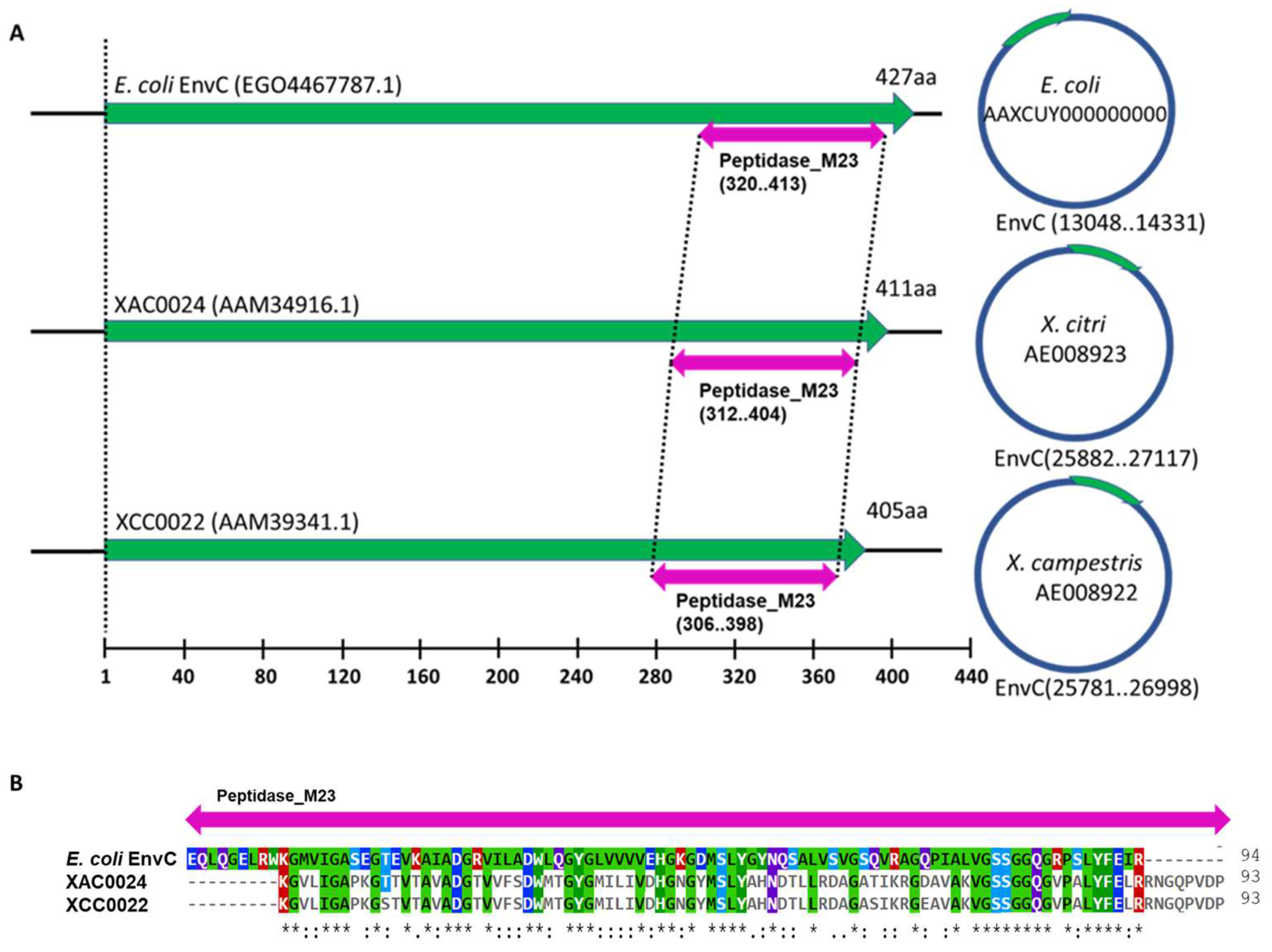
3.1. X. citri Encodes an EnvC Homolog, Which Is Conserved in MANY Bacteria

The XAC0024 protein exhibits a high degree of conservation among other sequenced Xanthomonas species, as shown in the maximum likelihood tree (Figure 1). Despite the fact that the single gene employed in the reconstruction may not precisely reflect the established phylogeny of the Xanthomonas genus, which is typically based on core genome alignment [29], our analysis successfully recovered some anticipated clusters. The species X. citri and X. fuscans demonstrate a close relationship and—in conjunction with X. axonopodis and X. euvesicatoria (X. campestris 85-10)—comprise the clade “X. axonopodis” [30]. This clade, incorporating the species X. arboricola, X. fragariae, X. hortorum and X. gardneri, aligns consistently with the topology of clade A, as reported in Ref [29], wherein X. hortorum and X. gardneri form a cohesive cluster [31].
A comparison of the nucleotide sequence between XAC0024 from X. citri 306 and its homolog XCC0022 from X. campestris pv. campestris str. ATCC 33913 showed an identity of 85% (Supplementary Figure S2). Analysis of the protein sequences revealed that XAC0024 and XCC0022 shared an identity of 89%, a coverage of 98% and a similarity of 93% (Supplementary Figure S3). These findings indicate a considerable level of sequence conservation between the two proteins. However, the comparison between the EnvC protein from E. coli and XAC0024 from X. citri revealed a 33% identity and a 52% similarity in terms of amino acid sequence (Supplementary Figure S4). The EnvC protein from E. coli contains 16 additional amino acids at the N-terminus compared to its homolog XAC0024 from X. citri. To explore the shared conserved domains among these proteins, we utilized the NCBI Batch Web CD-Search tool. Figure 2 displays a visualization of the identified conserved domains between the three protein sequences from X. citri 306 (XAC0024), X. campestris (XCC0022) and E. coli (EnvC) (see Supplementary Figure S5 for a detailed visualization of the nine ORFs of X. citri sharing the M23 domain).
To reinforce the similarity between the sequences of Xanthomonas strains used for phylogeny reconstruction, we utilized the NCBI Batch Web CD-Search tool, which revealed that the conserved M23 peptidase domain is shared by 16 representative Xanthomonas species (Supplementary Figure S6). This observation suggests a potential conserved function of this domain among homologs in other bacteria [32]. An amino acid sequence analysis performed by the SignalP 5.0 server [33] revealed that EnvC from X. citri, E. coli and X. campestris possesses a signal peptide with specific cleavage sites (Supplementary Figure S7). The cleavage site for EnvC in X. citri is between amino acids 20 and 21 (Supplementary Figure S7A), while for E. coli, it is between amino acids 42 and 43, and for X. campestris, it is between amino acids 14 and 15 (Supplementary Figure S7B,C).

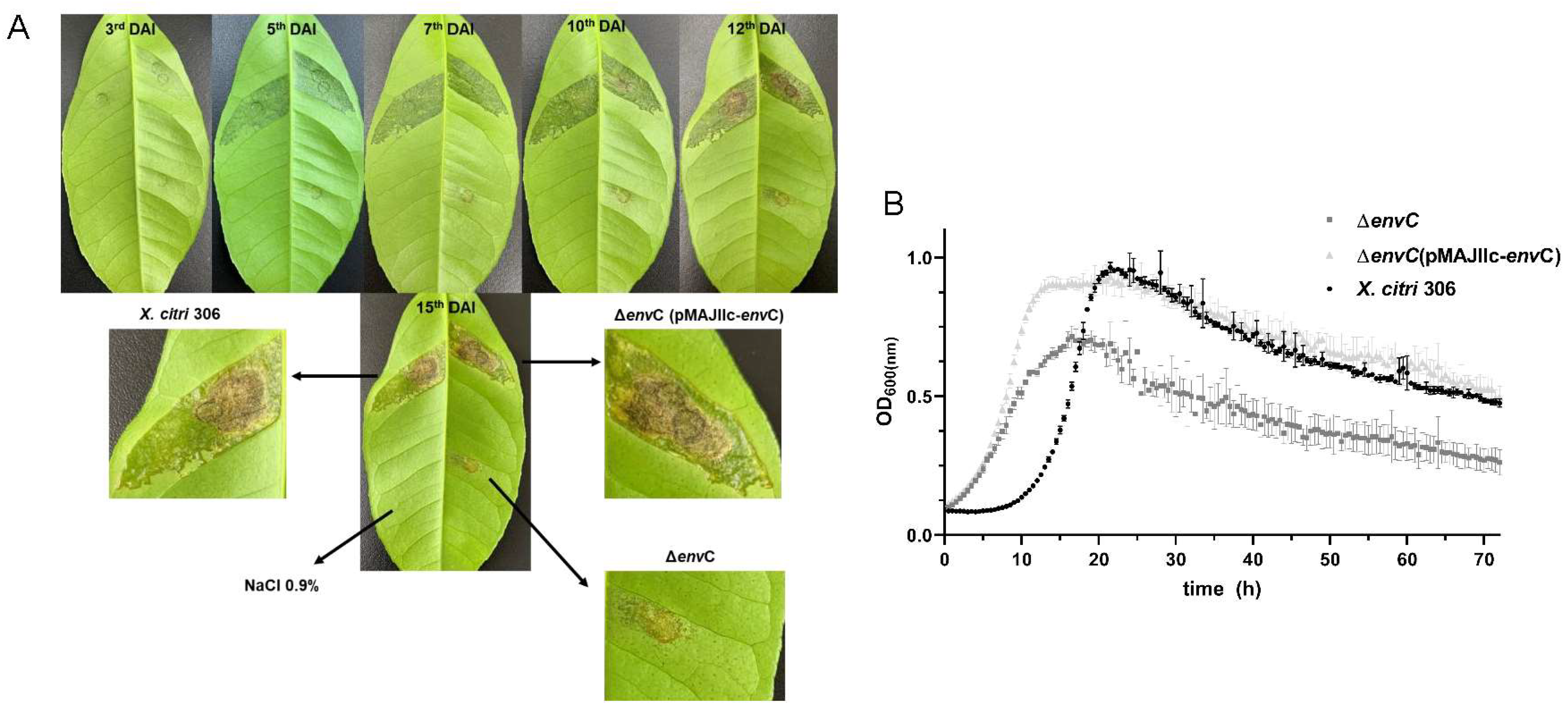
3.2. Disruption of envC Affects X. citri Virulence

To investigate the role of envC, we generated an envC deletion mutant (ΔenvC) and assessed its virulence and viability in comparison with the wild-type isolate X. citri 306. These assessments included examining symptom development in planta and conducting in vitro growth curves. To determine the contribution of EnvC to virulence, Rangpur lime leaves were inoculated with X. citri wt, ΔenvC and ΔenvC complemented strain (ΔenvC pMAJIIc-envC) and monitored for 15 days to observe the onset and progression of citrus canker symptoms. The temporal progression of symptoms showed hypertrophy/hyperplasia followed by water soaking and formation of brownish necrotic lesions at the late stage of infection, characteristic of citrus canker disease, in both X. citri wt and ΔenvC pMAJIIc-envC strains (Figure 3A). However, the ΔenvC mutant displayed a delay in the induction of citrus canker symptoms and produced lesions of reduced severity. These appeared to be concentrated close to the point of inoculation (Figure 3A).
Furthermore, we examined whether envC is essential for cell viability and proliferation by monitoring its growth in NB medium. Our results demonstrated that ΔenvC exhibited a distinct growth pattern compared to X. citri wt (Figure 3B). At the outset, the mutant exhibited an accelerated growth rate; however, ΔenvC eventually reached a plateau with a significantly lower population compared to both X. citri wt and ΔenvC pMAJIIc-envC strains. When inoculated into leaves of Rangpur lime (Citrus limonia), ΔenvC achieved a population 500 times less than that of the X. citri wt at 10 days after inoculation (results not shown). These findings underscore the necessity of envC for the virulence of X. citri and its ability to colonize the host. Additionally, disruption of envC adversely affects cell viability and proliferation.

3.3. EnvC Is Required for X. citri Daughter-Cell Separation

To determine the sub-cellular localization of EnvC encoded by X. citri, we expressed a version of the protein as an mCherry fusion (EnvC-mCherry) (Figure 4). X. citri EnvC-mCherry expressing cells (pMAJIIc-envC) exhibited a strong fluorescence signal, primarily concentrated around the edges of the cells, while the cytoplasm remained non-fluorescent (Figure 4B,C). This phenotype suggests that X. citri EnvC is predominantly localized in the periplasmic region of the cells. Importantly, the wild-type X. citri strain used as a control exhibited no detectable fluorescence emission (Figure 4E,F).
Next, the possible roles of EnvC in cell division and chromosome segregation were investigated by examining DAPI-stained cells of X. citri wild-type, ΔenvC and ΔenvC pMAJIIc-envC strains (Figure 5). The X. citri ΔenvC cells exhibited abnormally shaped rods, somewhat curved (Figure 5D–F). Many cells were arranged in long-chained structures, displaying clear division constrictions, which occasionally gave rise to minicells (Figure 5G–I; indicated by arrows). Although ΔenvC mutants appeared competent in the initial stages of the division process, they exhibited noticeable and detectable defects in late-stage division/separation processes.
For cells, which were not part of a chain, thus displaying an overall normal shape, we observed a significant difference in their average cell length compared to X. citri wild-type and ΔenvC pMAJIIc-envC strains (Table 1). To quantitatively evaluate this difference, we measured 400 individual cells from each culture: X. citri wild type, ΔenvC and ΔenvC pMAJIIc-envC. X. citri wild type had an average cell length of 1.18 ± 0.22 µm, while ΔenvC pMAJIIc-envC had an average cell length of 1.22 ± 0.29 µm. In contrast, X. citri ΔenvC mutants exhibited an average cell length of 1.89 ± 0.38 µm. Additionally, we scored the percentages of observed abnormalities for X. citri ΔenvC mutants (Table 1), with filamented chains comprising approximately 55.5% of the cells, while minicells accounted for 5.25% (n = 400).
Chromosome organization was visualized using 4′,6-diamidino-2-phenylindole (DAPI) staining (Figure 5). Cultures of the ΔenvC mutant displayed a continuous distribution of chromosomal mass spanning across the elongated cells (Figure 5E,F,H,I). This continuous distribution in some cells possibly hindered septal closure. In contrast, both X. citri and ΔenvC pMAJIIc-envC strains displayed a bilobed chromosome organization pattern with the expected normal distribution, indicating successful complementation of the mutant (Figure 5B,C,K,L). In all strains, a strong nucleoid accumulation signal was also noticeable in the middle or pole of the cells (Figure 5B,H,K), consistent with previous observations in the E. coli wt strain [34] and X. citri 306 wt strain [17].

4. Discussion

The XAC0024 protein from X. citri isolate 306 [15] exhibits significant homology with both EnvC proteins from E. coli and EnvC (XCC0022) from X. campestris. This homology was confirmed through amino acid sequence comparison analyses. Notably, the proteins from X. citri and X. campestris share a remarkable 93% similarity in amino acid sequence, while the X. citri protein demonstrates a 52% similarity with the EnvC protein from E. coli. Furthermore, a previous study reported homology between the XCC0022 protein from X. campestris and the E. coli EnvC protein [13].
The EnvC protein encoded by X. citri has a predicted M23 peptidase domain, which is part of a superfamily of metallopeptidase, characterized by the presence of zinc in its active enzyme site [35]. This enzyme family includes the Nlpd from E. coli, a LytM factor, as well as EnvC, responsible for activating N-acetylmuramoyl-L-alanine amidases (AmiA, AmiB and AmiC). These amidases play a crucial role in daughter-cell separation, and the inactivation of genes encoding them results in long cell chain formation [7,8,36].
In this study, we demonstrated that the EnvC protein of X. citri was predominantly localized in the periplasmic region of the cells and that the envC mutant of X. citrienvC) displayed a defect in cell separation, accompanied by distinct changes in cell morphology. Cultures of the ΔenvC mutant exhibited elongated rods, long-chained cells and minicells, which indicated impaired daughter-cell separation. These findings are consistent with the results previously reported for X. campestris strains lacking nlpD, envC or amiC1 [13], which also exhibited severe defects in daughter-cell separation. The physiological roles of peptidoglycan hydrolases, including EnvC, are still not fully understood, as the loss of an individual enzyme has little effect on growth and division, suggesting a functional overlap between numerous hydrolases [2]. To shed light on the specific role of EnvC, a collection of E. coli mutants lacking individual LytM factors (EnvC, NlpD, YgeR and YebA), as well as all possible combinations of them, were previously analyzed [7]. Among these mutants, only those with envC deletion failed to normally separate, further confirming the fundamental role of EnvC in proper cell separation.
Our observations of chromosome segregation errors in the X. citri ΔenvC mutant suggest that they are linked to the delayed division induced by the absence of EnvC. Interestingly, complementation of the mutant with X. citri ΔenvC pMAJIIc-envC restored a normal phenotype, with no detectable morphological discrepancies compared to the wild-type strain. Although further investigation is warranted, these results indicate that the ΔenvC mutant indeed experiences a late-division/separation defect, possibly leading to longer cell compartments and allowing the chromosomal mass to span across.
The X. citri mutant lacking envC exhibited a delay in citrus canker symptomatology compared with the wt and complemented strains. This delay in symptom development demonstrated that the deletion of EnvC had a significant impact on the bacteria’s virulence, as evidenced by its reduced ability to induce symptoms when inoculated into citrus leaves, which are susceptible hosts. The complemented strain ΔenvC pMAJIIc-envC fully restored virulence, confirming that the phenotype observed in the ΔenvC mutant was specifically caused by the absence of EnvC and was not a result of polar mutation.
Interestingly, Xanthomonas campestris strains lacking either nlpD or amiC1 almost completely lost their virulence, but the mutant lacking envC showed levels of virulence comparable to those of the wt strain [13]. This indicates that the contribution of EnvC to virulence is species-specific and that EnvC may be directly or indirectly involved in specific virulence pathways, which differ between Xanthomonas strains. The species-specific role of EnvC in virulence highlights the complex and intricate nature of bacterial pathogenesis, where different strains of the same genus may employ distinct mechanisms to establish disease. Further investigations into the specific pathways influenced by EnvC in different Xanthomonas strains could provide valuable insights into the molecular basis of pathogenicity and potential targets for disease control strategies.
The presence of a signal peptide in EnvC from X. citri, E. coli and X. campestris, which can serve as a membrane anchor, suggests that EnvC proteins are likely to be transported to the periplasmic region, a common localization for proteins in many pathogenic bacteria, often facilitated by tat/sec systems in Gram-negative bacteria [37,38,39,40,41,42,43,44,45,46]. The localization patterns observed here for EnvC-mCherry fusion protein in Xanthomonas citri and E. coli [7] are consistent with the fusion protein occupying the periplasmic region of the bacterium [17]. This sub-cellular localization supports the notion that EnvC functions in the periplasm, where it may play a crucial role in late-division and daughter-cell separation processes, as supported by similar findings in other bacterial models [6,7,12,13].
Taken together, our results—as well as similar results from many other bacterial models—support the notion that EnvC has an important function in late-division and daughter-cell separation processes [6,7,12,47]. However, the mechanism by which X. citri EnvC operates in daughter-cell separation, considering its periplasmic location, and how and whether this protein interacts with other hydrolases are still intriguing questions, which require further investigation.

5. Conclusions

The present study demonstrates the critical role of the EnvC protein in X. citri virulence, as disruption of the envC gene significantly reduces the bacterium’s ability to cause citrus canker. This underscores the importance of EnvC in disease progression. Additionally, EnvC is shown to be indispensable for proper cell division, as its absence results in morphological alterations and defects in daughter-cell separation, indicating its involvement in division processes. The comparison with X. campestris strains lacking envC suggests that the role of EnvC in virulence may vary among different species within the Xanthomonas genus. This highlights the intricate nature of bacterial pathogenesis and underscores the necessity for species-specific investigations.

Supplementary Materials

The following supporting information can be downloaded at: https://www.mdpi.com/article/10.3390/microorganisms12040691/s1, Figure S1: Deletion of the central portion of gene XAC0024 from X. citri via double-joint PCR; Figure S2: Nucleotide sequence alignment between XAC0024 from X. citri and its homolog XCC0022 from X. campestris; Figure S3: Protein sequence alignment between XAC0024 from X. citri and its homolog XCC0022 from X. campestris; Figure S4: Protein sequence alignment between XAC0024 from X. citri and EnvC from E. coli.; Figure S5: Domain multiple sequence alignment of the nine ORFs of X. citri sharing the M23 peptidase domain; Figure S6: Domain multiple sequence alignment of 16 representative species within the Xanthomonas species used in maximum likelihood phylogeny; Figure S7: The predicted signal peptide for XAC0024 from X. citri, EnvC from E. coli and XCC0022 from X. campestris; Table S1: Proteins of Xanthomonas citri subsp. citri 306 strain sharing the M23 peptidase domain according to an in silico search using the IMG “find function” tool; Table S2: List of strains and plasmids used in this work; Table S3: Primers used in this study; Table S4: Xanthomonas species used for phylogenetic reconstruction based on the XAC0024 nucleotide sequence of X. citri.

Author Contributions

Conceptualization, M.M.P., H.F. and J.A.F.; Formal analysis, M.M.P., T.Z.M., C.Z. and D.T.; Funding acquisition, N.W., M.I.T.F. and J.A.F.; Investigation, M.M.P., T.Z.M., D.T., C.Z., H.A.A. and H.F.; Methodology, M.M.P., T.Z.M., H.F. and J.A.F.; Project administration, M.M.P. and J.A.F.; Resources, N.W. and M.I.T.F.; Supervision, H.F., N.W. and J.A.F.; Validation, M.M.P. and J.A.F.; Visualization, M.M.P. and J.A.F.; Writing—original draft, M.M.P.; Writing—review and editing, M.M.P., D.T., H.F., N.W. and J.A.F. All authors have read and agreed to the published version of the manuscript.

Funding

This work is part of M. M. Pena’s Ph.D. thesis and T. Z. Martins’ master’s dissertation, and it was financed in part by the Coordenação de Aperfeiçoamento de Pessoal de Nível Superior, Brasil (CAPES), Finance Code 001, and by a fellowship grant from Conselho Nacional de Desenvolvimento Científico e Tecnológico, Brasil (CNPq), to JAF (Grant No. 312089/2019-8). M.I.T.F. is a recipient of a CNPq productivity fellowship (Grant No. 310102/2019-7).

Data Availability Statement

Data are contained within the article and Supplementary Materials.

Acknowledgments

We thank the Citrus Research and Education Center at the University of Florida for allowing us to use their microscope facilities. We also thank Naiara Zancanari for support with the analyses and the sequencing facility at the Center for Biological Resources and Genomic Biology (CREBIO) at the University of São Paulo State (UNESP) at Jaboticabal Campus, Brazil. We thank Roberto Hirochi Herai for critically reading the manuscript and providing valuable suggestions.

Conflicts of Interest

The authors declare no conflicts of interest.

References

  1. Das, A.K. Citrus Canker—A review. J. Appl. Hortic. 2003, 5, 52–60. [Google Scholar] [CrossRef]
  2. Vollmer, W.; Blanot, D.; de Pedro, M.A. Peptidoglycan structure and architecture. FEMS Microbiol. Rev. 2008, 32, 149–167. [Google Scholar] [CrossRef]
  3. Silhavy, T.J.; Kahne, D.; Walker, S. The bacterial cell envelope. Cold Spring Harb. Perspect. Biol. 2010, 2, a000414. [Google Scholar] [CrossRef]
  4. Frirdich, E.; Gaynor, E.C. Peptidoglycan hydrolases, bacterial shape, and pathogenesis. Curr. Opin. Microbiol. 2013, 16, 767–778. [Google Scholar] [CrossRef]
  5. Firczuk, M.; Bochtler, M. Folds and activities of peptidoglycan amidases. FEMS Microbiol. Rev. 2007, 31, 676–691. [Google Scholar] [CrossRef]
  6. Bernhardt, T.G.; de Boer, P.A. The Escherichia coli amidase AmiC is a periplasmic septal ring component exported via the twin-arginine transport pathway. Mol. Microbiol. 2003, 5, 1171–1182. [Google Scholar] [CrossRef] [PubMed]
  7. Uehara, T.; Dinh, T.; Bernhardt, T.G. LytM-domain factors are required for daughter cell separation and rapid ampicillin-induced lysis in Escherichia coli. J. Bacteriol. 2009, 191, 5094–5107. [Google Scholar] [CrossRef] [PubMed]
  8. Uehara, T.; Parzych, K.R.; Dinh, T.; Bernhardt, T.G. Daughter cell separation is controlled by cytokinetic ring-activated cell wall hydrolysis. EMBO J. 2010, 29, 1412–1422. [Google Scholar] [CrossRef]
  9. Wu, C.; Al Mamun, A.A.M.; Luong, T.T.; Hu, B.; Gu, J.; Lee, J.H.; D’Amore, M.; Das, A.; Ton-That, H. Forward Genetic Dissection of Biofilm Development by Fusobacterium nucleatum: Novel Functions of Cell Division Proteins FtsX and EnvC. mBio 2018, 9, e00360-18. [Google Scholar] [CrossRef]
  10. Möll, A.; Dörr, T.; Alvarez, L.; Chao, M.C.; Davis, B.M.; Cava, F.; Waldor, M.K. Cell separation in Vibrio cholerae is mediated by a single amidase whose action is modulated by two nonredundant activators. J. Bacteriol. 2014, 196, 3937–3948. [Google Scholar] [CrossRef]
  11. Warr, A.R.; Hubbard, T.P.; Munera, D.; Blondel, C.; Wiesch, P.A.Z.; Abel, S.; Wang, X.; Davis, B.M.; Waldor, M.K. Transposon-insertion sequencing screens unveil requirements for EHEC growth and intestinal colonization. PLoS Pathog. 2019, 15, e1007652. [Google Scholar] [CrossRef] [PubMed]
  12. Yakhnina, A.A.; Mcmanus, H.R.; Bernhardt, T.G. The cell wall amidase AmiB is essential for Pseudomonas aeruginosa cell division, drug resistance and viability. Mol. Microbiol. 2015, 97, 957–973. [Google Scholar] [CrossRef]
  13. Yang, L.C.; Gan, Y.L.; Yang, L.Y.; Jiang, B.L.; Tang, J.L. Peptidoglycan hydrolysis mediated by the amidase AmiC and its LytM activator NlpD is critical for cell separation and virulence in the phytopathogen Xanthomonas campestris. Mol. Plant Pathol. 2018, 19, 1705–1718. [Google Scholar] [CrossRef] [PubMed]
  14. da Silva, A.C.; Ferro, J.A.; Reinach, F.C.; Farah, C.S.; Furlan, L.R.; Quaggio, R.B.; Monteiro-Vitorello, C.B.; Van Sluys, M.A.; Almeida, N.F.; Alves, L.M.; et al. Comparison of the genomes of two Xanthomonas pathogens with differing host specificities. Nature 2002, 417, 459–463. [Google Scholar] [CrossRef]
  15. Huguet, E.; Hahn, K.; Wengelnik, K.; Bonas, U. hpaA mutants of Xanthomonas campestris pv. vesicatoria are affected in pathogenicity but retain the ability to induce host-specific hypersensitive reaction. Mol. Microbiol. 1998, 29, 1379–1390. [Google Scholar] [CrossRef]
  16. Pena, M.M.; Teper, D.; Ferreira, H.; Wang, N.; Sato, K.U.; Ferro, M.I.T.; Ferro, J.A. mCherry fusions enable the subcellular localization of periplasmic and cytoplasmic proteins in Xanthomonas sp. PLoS ONE 2020, 15, e0236185. [Google Scholar] [CrossRef] [PubMed]
  17. Sambrook, J.; Fritsch, E.R.; Maniatis, T. Molecular Cloning: A Laboratory Manual, 2nd ed.; Cold Spring Harbor Laboratory Press: New York, NY, USA, 1989; Volume 1. [Google Scholar]
  18. Lee, J.; Lee, H.-J.; Shin, M.-K.; Ryu, W.-S. Versatile PCR-mediated insertion or deletion mutagenesis. BioTechniques 2004, 36, 398–400. [Google Scholar] [CrossRef]
  19. do Amaral, A.M.; Toledo, C.P.; Baptista, J.C.; Machado, M.A. Transformation of Xanthomonas axonopodis pv. citri by electroporation. Fitopatol. Bras. 2005, 30, 292–294. [Google Scholar] [CrossRef]
  20. Kaniga, K.; Delor, I.; Cornelis, G.R. A wide-host-range suicide vector for improving reverse genetics in Gram-negative bacteria: Inactivation of the blaA gene of Yersinia enterocolitica. Gene 1991, 109, 137–141. [Google Scholar] [CrossRef]
  21. Martins, P.M.M.; Lau, I.F.; Bacci, M., Jr.; Belasque, J.; Do Amaral, A.M.; Taboga, S.R.; Ferreira, H. Subcellular localization of proteins labeled with GFP in Xanthomonas citri ssp. citri: Targeting the division septum. FEMS Microbiol. Lett. 2010, 310, 76–83. [Google Scholar] [CrossRef]
  22. Morão, L.G.; Polaquini, C.R.; Kopacz, M.; Torrezan, G.S.; Ayusso, G.M.; Dilarri, G.; Cavalca, L.B.; Zielinska, A.; Scheffers, D.-J.; Regasini, L.O.; et al. A simplified curcumin targets the membrane of Bacillus subtilis. MicrobiologyOpen 2019, 8, e683. [Google Scholar] [CrossRef] [PubMed]
  23. Lacerda, L.A.; Cavalca, L.B.; Martins, P.M.M.; Govone, J.S.; Bacci, M., Jr.; Ferreira, H. Protein depletion using the arabinose promoter in Xanthomonas citri subsp. citri. Plasmid 2017, 90, 44–52. [Google Scholar] [CrossRef] [PubMed]
  24. Thompson, J.D.; Higgins, D.G.; Gibson, T.J. CLUSTAL W: Improving the sensitivity of progressive multiple sequence alignment through sequence weighting, positions-specific gap penalties and weight matrix choice. Nucleic Acids Res. 1994, 22, 4673–4680. [Google Scholar] [CrossRef]
  25. Miller, M.A.; Pfeiffer, W.; Schwartz, T. Creating the CIPRES Science Gateway for Inference of Large Phylogenetic Trees. In Proceedings of the Gateway Computing Environments Workshop, New Orleans, LA, USA, 14 November 2010; pp. 1–8. [Google Scholar] [CrossRef]
  26. Stamatakis, A. RAxML Version 8: A tool for phylogenetic analysis and post-analysis of large phylogenies. Bioinformatics 2014, 30, 1312–1313. [Google Scholar] [CrossRef] [PubMed]
  27. Felsenstein, J. Confidence limits on phylogenies: An approach using the bootstrap. Evolution 1985, 39, 783–791. [Google Scholar] [CrossRef] [PubMed]
  28. Kumar, S.; Stecher, G.; Li, M.; Knyaz, C.; Tamura, K. MEGA X: Molecular evolutionary genetics analysis across computing platforms. Mol. Biol. Evol. 2018, 35, 1547–1549. [Google Scholar] [CrossRef]
  29. Mirdita, M.; Schütze, K.; Moriwaki, Y.; Heo, L.; Ovchinnikov, S.; Steinegger, M. ColabFold: Making protein folding accessible to all. Nat. Methods 2022, 19, 679–682. [Google Scholar] [CrossRef]
  30. Meng, E.C.; Goddard, T.D.; Pettersen, E.F.; Couch, G.S.; Pearson, Z.J.; Morris, J.H.; Ferrin, T.E. UCSF ChimeraX: Tools for structure building and analysis. Protein Sci. 2023, 32, e4792. [Google Scholar] [CrossRef]
  31. The Xanthomonas sp. Portal—INRA. Available online: https://iant.toulouse.inra.fr/bacteria/annotation/cgi/xansp.cgi (accessed on 24 July 2023).
  32. Timilsina, S.; Potnis, N.; Newberry, E.A.; Liyanapathiranage, P.; Iruegas-Bocardo, F.; White, F.F.; Goss, E.M.; Jones, J.B. Xanthomonas diversity, virulence and plant–pathogen interactions. Nat. Rev. Microbiol. 2020, 18, 415–427. [Google Scholar] [CrossRef]
  33. Rodriguez-R, L.M.; Grajales, A.; Arrieta-Ortiz, M.L.; Salazar, C.; Restrepo, S.; Bernal, A. Genomes-based phylogeny of the genus Xanthomonas. BMC Microbiol. 2012, 12, 43. [Google Scholar] [CrossRef]
  34. Timilsina, S.; Kara, S.; Jacques, M.A.; Potnis, N.; Minsavage, G.V.; Vallad, G.E.; Jones, J.B.; Fischer-Le Saux, M. Reclassification of Xanthomonas gardneri (ex Šutič 1957) Jones et al. 2006 as a later heterotypic synonym of Xanthomonas cynarae Trébaol et al. 2000 and description of X. cynarae pv. cynarae and X. cynarae pv. gardneri based on whole genome analyses. Int. J. Syst. Evol. Microbiol. 2019, 69, 343–349. [Google Scholar] [CrossRef]
  35. Altenhoff, A.M.; Studer, R.A.; Robinson-Rechavi, M.; Dessimoz, C. Resolving the ortholog conjecture: Orthologs tend to be weakly, but significantly, more similar in function than paralogs. PLoS Comput. Biol. 2012, 8, e1002514. [Google Scholar] [CrossRef] [PubMed]
  36. Freiesleben, U.V.; Krekling, M.A.; Hansen, F.G.; Lobner-Olesen, A. The eclipse period of Escherichia coli. EMBO J. 2000, 19, 6240–6248. [Google Scholar] [CrossRef] [PubMed]
  37. Wu, Q.; Lahti, J.M.; Air, G.M.; Burrows, P.D.; Cooper, M.D. Molecular cloning of the murine BP-1/6C3 antigen: A member of the zinc-dependent metallopeptidase family. Proc. Natl. Acad. Sci. USA 1990, 87, 993–997. [Google Scholar] [CrossRef] [PubMed]
  38. Heidrich, C.; Templin, M.F.; Ursinus, A.; Merdanovic, M.; Berger, J.; SCHWARZ, H.; de Pedro, M.A.; Holtje, J.V. Involvement of N-acetylmuramyl-L-alanine amidases in cell separation and antibiotic-induced autolysis of Escherichia coli. Mol. Microbiol. 2001, 41, 167–178. [Google Scholar] [CrossRef] [PubMed]
  39. Armenteros, J.J.A.; Tsirigos, K.D.; Sonderby, C.K.; Peterson, T.N.; Winther, O.; Brunak, S.; Heijne, G.V.; Nielsen, H. SignalP 5.0 improves signal peptide predictions using deep neural networks. Nat. Biotechnol. 2019, 37, 420–423. [Google Scholar] [CrossRef] [PubMed]
  40. Brunings, A.M.; Gabriel, D.W. Xanthomonas citri: Breaking the surface. Mol. Plant Pathol. 2003, 4, 141–157. [Google Scholar] [CrossRef] [PubMed]
  41. Jha, G.; Rajeshwari, R.; Sonti, R.V. Bacterial Type Two Secretion System Secreted Proteins: Double-Edged Swords for Plant Pathogens. Mol. Plant Microbe Interact. 2005, 18, 891–898. [Google Scholar] [CrossRef] [PubMed]
  42. Büttner, D.; Bonas, U. Regulation and secretion of Xanthomonas virulence factors. FEMS Microbiol. Rev. 2010, 34, 107–133. [Google Scholar] [CrossRef] [PubMed]
  43. Soares, M.R.; Facincani, A.P.; Ferreira, R.M.; Moreira, L.M.; De Oliveira, J.C.F.; Ferro, J.A.; Ferro, M.I.T.; Meneghini, R.; Gozzo, F.C. Proteome of the phytopathogen Xanthomonas citri subsp. citri: A global expression profile. Proteome Sci. 2010, 8, 55. [Google Scholar] [CrossRef]
  44. Ryan, R.P.; Vorhölter, F.J.; Potnis, N.; Jones, J.B.; Sluys, M.-A.V.; Bogdanove, A.J.; Dow, J.M. Pathogenomics of Xanthomonas: Understanding bacterium–plant interactions. Nat. Rev. Microbiol. 2011, 9, 344–355. [Google Scholar] [CrossRef] [PubMed]
  45. Yan, Q.; Wang, N. The ColR/ColS two-component system plays multiple roles in the pathogenicity of the citrus canker pathogen Xanthomonas citri subsp. citri. J. Bacteriol. 2011, 193, 1590–1599. [Google Scholar] [CrossRef] [PubMed]
  46. Zhou, X.; Hu, X.; Li, J.; Wang, N. A Novel Periplasmic Protein, VrpA, Contributes to Efficient Protein Secretion by the Type III Secretion System in Xanthomonas spp. Mol. Plant Microbe Interact. 2015, 28, 143–153. [Google Scholar] [CrossRef] [PubMed]
  47. Zimaro, T.; Thomas, L.; Marondedze, C.; Sgro, G.G.; Garofalo, C.G.; Ficarra, F.A.; Gehring, C.; Ottado, J.; Gottig, N. The type III protein secretion system contributes to Xanthomonas citri subsp. citri biofilm formation. BMC Microbiol. 2014, 14, 96. [Google Scholar] [CrossRef] [PubMed]
Figure 1. Maximum likelihood phylogeny based on the nucleotide sequences of 31 strains representing the Xanthomonas genus. The phylogenetic tree was inferred using RaxMLversion 8.0.24 and drawn using MEGA X software version 10.1.5. Values on the branches indicate bootstrap values for each branch, expressed as percentages. E. coli K12 was used as the outgroup.
Microorganisms 12 00691 g001
Figure 2. Multiple amino acid sequence alignment of M23 peptidase domain for XAC0024 from X. citri 306 (AAM34916.1), XCC0022 from X. campestris (AAM39341.1) and EnvC from E. coli (EGO4467787.1). (A) M23 peptidase domain within the protein sequences of E. coli EnvC, XAC0024 and XCC0022 (purple arrows) and their corresponding locus within genomes (circles on the right). (B) Multiple amino acid sequence alignment of M23 peptidase domain showing matching amino acids for the three organisms. The protein sequences were uploaded from the NCBI Batch Web CD-Search tool. Each protein is depicted with a Peptidase_M13 domain (pfam01551) and a conserved protein domain family EnvC (COG4942). * Conserved sequence (identical); : Conservative mutation; . Semi-conservative mutation.
Microorganisms 12 00691 g002
Figure 3. Pathogenicity assay and growth curve for X. citri 306, ΔenvC and ΔenvC-pMAJIIc-envC. (A) Rangpur lime leaves were infiltrated with cell suspensions of the specified X. citri strains. Pictures were taken at 3, 5, 7, 10, 12 and 15 days after inoculation (DAI). (B) In vitro growth curve. X. citri 306, ΔenvC and ΔenvC-pMAJIIc-envC were cultivated in NB medium, and OD 600 nm readings were taken every 30 min for 72 h. The points on the curves represent the average of triplicate cultures, and the vertical lines indicate the standard deviation values of the means. All experiments were performed in triplicate.
Microorganisms 12 00691 g003
Figure 4. Sub-cellular localization of EnvC-mCherry in X. citri. X. citri strains expressing EnvC-mCherry fusions were cultivated up to OD 600 nm of 0.3, followed by induction with 0.05% arabinose for 2 h before microscope observation. The panels depict the phase contrast images (left), TxRed channels (middle) and the overlay, respectively, for (AC): X. citri pMAJIIc-envC, (DF): X. citri 306. Magnification 100X; scale bar 5 µm.
Microorganisms 12 00691 g004
Figure 5. Cell morphology and nucleoid distribution analyses of X. citri 306, ΔenvC and ΔenvC pMAJIIc-envC strains. The panels show the phase contrast (left), DAPI channels (middle) and the overlay, respectively, for (AC): X. citri 306, (DI): ΔenvC, (HL): ΔenvC-pMAJIIc-envC. Arrows indicate the minicell position. Magnification 100×; scale bar 5 µm.
Microorganisms 12 00691 g005
Table 1. Morphological analysis of X. citri strains according to cell length.
Cell Length µmFilaments %Minicells %
X. citri 3061.18 ± 0.22 a00
ΔenvC1.89 ± 0.38 b55.55.25
ΔenvC pMAJIIc-envC1.22 ± 0.29 a00
Total n = 400 cells measured. Data correspond to the average cell length ± standard deviation. Same letters mean no significant difference according to the Tukey test—0.05.
Disclaimer/Publisher’s Note: The statements, opinions and data contained in all publications are solely those of the individual author(s) and contributor(s) and not of MDPI and/or the editor(s). MDPI and/or the editor(s) disclaim responsibility for any injury to people or property resulting from any ideas, methods, instructions or products referred to in the content.

