Article

Repeated Occurrence of Mobile Colistin Resistance Gene-Carrying Plasmids in Pathogenic Escherichia coli from German Pig Farms

1
Institute of Hygiene and Infectious Diseases of Animals, Justus Liebig University Giessen, 35392 Giessen, Germany
2
Department of Infectious Diseases and Microbiology, University of Luebeck, 23538 Luebeck, Germany
3
Microbial Genomics, Robert Koch Institute, 13353 Berlin, Germany
*
Author to whom correspondence should be addressed.
Microorganisms 2024, 12(4), 729; https://doi.org/10.3390/microorganisms12040729
Submission received: 6 March 2024 / Revised: 26 March 2024 / Accepted: 28 March 2024 / Published: 3 April 2024
(This article belongs to the Special Issue Bacterial Infections and Antibiotic Resistance in Veterinary Medicine)

Abstract

:
The global spread of plasmid-mediated mobile colistin resistance (mcr) genes threatens the vital role of colistin as a drug of last resort. We investigated whether the recurrent occurrence of specific E. coli pathotypes and plasmids in individual pig farms resulted from the continued presence or repeated reintroduction of distinct E. coli strains. E. coli isolates (n = 154) obtained from three pig farms with at least four consecutive years of mcr detection positive for virulence-associated genes (VAGs) predicting an intestinal pathogenic pathotype via polymerase chain reaction were analyzed. Detailed investigation of VAGs, antimicrobial resistance genes and plasmid Inc types was conducted using whole genome sequencing for 87 selected isolates. Sixty-one E. coli isolates harbored mcr-1, and one isolate carried mcr-4. On Farm 1, mcr-positive isolates were either edema disease E. coli (EDEC; 77.3%) or enterotoxigenic E. coli (ETEC; 22.7%). On Farm 2, all mcr-positive strains were ETEC, while mcr-positive isolates from Farm 3 showed a wider range of pathotypes. The mcr-1.1 gene was located on IncHI2 (Farm 1), IncX4 (Farm 2) or IncX4 and IncI2 plasmids (Farm 3). These findings suggest that various pathogenic E. coli strains play an important role in maintaining plasmid-encoded colistin resistance genes in the pig environment over time.

1. Introduction

The emergence and spread of multidrug-resistant bacteria is a rising problem that threatens the effective treatment of infectious diseases in humans and animals [1]. Escherichia (E.) coli is a ubiquitous Gram-negative bacterium that is regularly found in the intestinal tract of humans and animals. Intestinal pathogenic E. coli (InPEC) strains can cause a number of different diseases, including diarrhea.
In pigs, neonatal and post-weaning diarrhea is a widespread and often severe disease, resulting in significant economic losses in the swine industry worldwide [2]. Certain E. coli pathotypes are associated with causing enteric diseases in piglets, e.g., enterotoxigenic E. coli (ETEC) and atypical enteropathogenic E. coli (aEPEC) [3]. E. coli isolates producing the Shiga toxin subtype Stx2e, encoded by the stx2e gene, are the causative agent of edema disease in weaned piglets [4]. Colistin has been widely used to treat intestinal infections in swine caused by InPEC [5]. In 2015, the use of colistin came under scrutiny due to the emergence of colistin-resistant bacteria due to a transferable colistin resistance gene [6]. A recent study from 2022 reported that 51.9% of 662 veterinarians surveyed stopped the use of colistin and 33.4% reduced their usage. The main indication for the use of colistin was gastrointestinal disease in pigs [6]. The increasing occurrence of antimicrobial resistance (AMR) against this last resort antibiotic incites new debates for additional regulations of its usage, especially in veterinary medicine [7].
Until a few years ago, acquired colistin resistance in bacteria was mainly attributed to chromosomal mutations such as modifications of two-component systems like pmrA/pmrB and phoP/phoQ [8,9]. In 2015, Liu et al. described for the first time a mobile colistin resistance gene that was found on a transmissible plasmid in one E. coli isolate from a Chinese pig, thereafter named mcr-1 [10]. Since then, ten different mcr genes (mcr-1mcr-10) have been reported with numerous variants, mostly found in Gram-negative bacterial species [11,12,13,14,15,16,17,18,19]. Over the years, mcr-mediated colistin resistance has been reported in Enterobacterales isolated from several sources, including humans, livestock, companion animals, wildlife and the environment [20,21,22,23].
The conjugative transfer of mcr-harboring plasmids between bacteria plays a vital role in the dissemination of AMR against colistin [24]. The first discovered mcr-1 gene was located on a plasmid (pHNSHP45) of 64,015 bp in size, possessing a typical IncI2-type backbone [10]. Since then, mcr-1 has been found on numerous plasmid types, such as IncHI1, IncHI2, IncF, IncN, IncP, IncX4 and IncN1-IncHI2 [25,26,27]. Resistance genes mcr-2, mcr-3, mcr-6, mcr-7, mcr-8, mcr-9 and mcr-10 were detected frequently on plasmids of types IncX4, IncHI2, IncHI2, IncI2, IncF type, IncHI2/HI2A and IncF, respectively [24].
The temporal and spatial distribution of mcr-positive isolates among pathogenic E. coli at individual farm levels has rarely been studied. Miguela-Villoldo et al. investigated mcr-1 prevalence in healthy Spanish food-producing pigs from 1998 to 2021, selecting 50 caecal pig samples across 14 years [28]. While the frequency of mcr-1-positive samples increased from 2004 (16%) to 2015 (66%), a downward trend was observed from 2017 (54%) to 2021 (17%). In Germany, the mcr-1 gene was detected in 15 different pig-fattening farms where pooled feces and boot swabs had been collected from 2011 to 2012 [29]. Further analyses of one representative mcr-1-containing E. coli isolate per farm showed that mcr-1 was mainly located on IncX4 plasmids (n = 9) but also on IncHI2 (n = 3), IncX4/N (n = 1), IncHI2/FIB/FII/X3 (n = 1) and integrated into the chromosome (n = 1).
This study investigated the occurrence and genomic location of mcr genes in pathogenic E. coli isolates obtained from three German pig farms over at least four years, which were chosen from a comprehensive in-house database. The genomes of a representative set of isolates were sequenced to determine the presence of virulence-associated genes (VAGs) characteristic for intestinal pathogenic E. coli (InPEC) pathotypes and AMR genes as well as their plasmid location. Based on core genome and plasmid comparisons, the repeated occurrence of distinct E. coli clones, as well as distinct resistance and virulence plasmids on different farms, was examined.

2. Materials and Methods

2.1. Study Inclusion Criteria for Farms

For this retrospective survey on the repeated occurrence of mcr-positive porcine pathogenic E. coli, suitable pig farms were selected based on specific criteria. From our database of more than 3000 registered pig farms in Germany that had sent samples for molecular typing of E. coli isolates in the past, we selected only farms from which at least 25 pathogenic E. coli isolates had been obtained and preserved over the years (n = 23 farms). This was based on our recently published collection of 10,573 E. coli isolates, each harboring at least one of ten VAGs, which were tested using PCR for the presence of mcr-1 to mcr-10 genes [30]. Briefly, E. coli isolates were obtained from feces or mucosal swabs (rectum or small intestine). Upon arrival at the laboratory, the samples were streaked for single bacterial colonies on blood agar plates (blood agar base, Merck Chemicals, Darmstadt, Germany) containing 5% sheep blood and on Gassner agar (sifin diagnostics GmbH, Berlin, Germany). After approximately 18 h of incubation at 37 °C, up to six morphologically different, putative E. coli colonies were picked per sample and tested for the presence of VAGs. More details of sample collection and processing were published recently [30].
Subsequently, pig farms with less than 25 pigs sampled (n = 11) or no detection of mcr genes (n = 5) were excluded from the study, as well as farms with no isolates for six consecutive years (n = 3). Finally, only farms were selected where mcr-positive E. coli had been isolated from pig fecal samples for at least four consecutive years.

2.2. Whole Genome Sequencing

The genomic DNA of E. coli bacteria was extracted using the Master Pure™ DNA Purification Kit (Biozym Scientific GmbH, Hessisch Oldendorf, Germany). Bacterial genomes were sequenced using an Illumina MiSeq sequencer (MiSeq Reagent Kit V.3; Illumina Inc., San Diego, CA, USA) via multiplexing of 30 samples per flow cell using 2 × 150 bp paired-end reads to achieve an average coverage of 90-fold. Quality control, including contamination removal and adapter trimming, was carried out using an in-house pipeline. De novo assemblies were generated using the SPAdes Genome Assembler (v3.15.5) with the “-isolate” flag [31]. The Bakta pipeline (v1.8.2) was employed using species-specific databases for genomic annotation of the bacterial genomes [32].

2.3. Phenotypic Resistance Testing, Antimicrobial Resistance Genes, Virulence-Associated Genes

All 87 whole genome-sequenced E. coli isolates were tested for antimicrobial susceptibilities using the broth microdilution method. An individual panel layout was used from the MICRONAUT system (Merlin Diagnostics, Bornheim-Hersel, Germany). Fourteen antimicrobial substances and/or combinations (in µg/mL: amikacin (0.25–32), amoxicillin/clavulanic acid (1/0.5–16/8), ampicillin (1–16), cefotaxime (0.016–2), ceftazidime (0.031–8), colistin (0.125–8), enrofloxacin (0.004–2), florfenicol (1–16), gentamicin (0.125–8), piperacillin/tazobactam (0.5/4–64/4), spectinomycin (4–64), sulfamethoxazole (4–256), tetracycline (0.25–8) and trimethoprim (0.25–8)) were included in this layout. E. coli strains ATCC 25922 and NCTC 13846 were used for quality control. Minimum inhibitory concentration (MIC) values were interpreted by means of defined clinical breakpoints set by the Clinical and Laboratory Standards Institute (CLSI) for veterinary and human Enterobacterales isolates [33]. For antibiotics without a defined breakpoint for E. coli (in the case of cefotaxime, sulfamethoxazole and trimethoprim), human breakpoints of Enterobacterales according to CLSI [34] were used, except for colistin, which was interpreted according to the European Committee on Antimicrobial Susceptibility Testing (EUCAST) guidelines [35]. No veterinary or human breakpoints are available for enrofloxacin and spectinomycin from either CLSI or EUCAST. For both substances, the epidemiological cut-off (ECOFF) was used for E. coli provided by EUCAST (https://mic.eucast.org/search/, accessed on 5 March 2024), separating the E. coli population into a population without acquired or mutational resistance (wild-type) and a population with phenotypically detectable acquired resistance mechanisms (non-wild-type) against enrofloxacin and spectinomycin.
Antimicrobial resistance (AMR) genes and chromosomal point mutations related to antimicrobial resistance were identified in all whole genome-sequenced isolates by using the online tool ResFinder 4.1, available on the website of the Center for Genomic Epidemiology (CGE) (https://cge.food.dtu.dk/services/ResFinder/, accessed on 17 December 2023). All isolates were investigated for VAGs using VirulenceFinder 2.0 (https://cge.food.dtu.dk/services/VirulenceFinder/, accessed on 17 December 2023). The results were additionally verified with multiple genome analysis accessible online from BacWGSTdb (http://bacdb.cn/BacWGSTdb/Tools.php, accessed on 17 December 2023).

2.4. Determination of Genoserotypes, Clonotypes, Multilocus Sequence Types, Core Genome MLS Types and Phylogroups

The genoserotypes of the whole genome-sequenced E. coli isolates were determined by applying the web-based SerotypeFinder 2.0, provided by the Center for Genomic Epidemiology (https://cge.food.dtu.dk/services/SerotypeFinder/, accessed on 22 November 2023). The internal 469- and 489-nucleotide sequences of the fumC and fimH genes, respectively, were used for clonotyping [36]. Clonotypes (CH types), which are the combinations of allele assignments for fumC and fimH, were determined using CHTyper 1.0 (https://cge.food.dtu.dk/services/CHTyper/, accessed on 1 January 2024).
Multilocus sequence types (STs) were determined by applying MLST 2.0 (https://cge.food.dtu.dk/services/MLST/, accessed on 22 November 2023), which is based on the Achtman seven-gene MLST scheme that includes seven housekeeping genes (adk, fumC, gyrB, icd, mdh, purA and recA). E. coli isolates were defined as clonal when they displayed the same sequence type in addition to identical virulence and resistance gene profiles.
The core genome was calculated using Roary [37]. Phylogenetic analysis was performed by using the web-based tool ClermontTyper (IAME, http://clermontyping.iame-research.center/index.php, accessed 18 December 2023), allowing us to assign tested strains to E. albertii, E. fergusonii, Escherichia cryptic clades I-V, E. coli sensu strico as well as to the main phylogroups A, B1, B2, C, D, E, F and G [38,39]. The remaining non-sequenced E. coli isolates were tested in a quadruplex PCR, allowing for the assignment to the eight main phylogroups A, B1, B2, C, D, E, F, G and Escherichia cryptic clades I-V [39,40].

2.5. Genomic Location of Virulence-Associated Genes and mcr Genes, Plasmid Analysis

To determine the location of mcr genes, two methods were used. Whole genome sequence data were analyzed for the location of AMR and virulence genes using the tool “Chromosome & Plasmid Overview” implemented in the software package Ridom SeqSphere+ (http://www3.ridom.de/seqsphere, accessed 22 January 2024). To identify plasmid replicon types, PlasmidFinder 2.1 (https://cge.food.dtu.dk/services/PlasmidFinder/, accessed 22 January 2024) and Ridom SeqSphere+ were employed. The BacWGST database (http://bacdb.cn/BacWGSTdb/Tools.php, accessed on 17 December 2023) and RidomSeqSphere+ were applied to detect closely related plasmids via sequence comparison. To illustrate circular comparisons between the plasmids, we used the blast ring image generator software BRIG Version 0.95 [41].
To confirm the results of sequence analysis regarding the genomic location of mcr-1 genes, an additional method was employed. The location of mcr-1 genes in 38 E. coli isolates was determined via the S1 nuclease digestion of genomic DNA and pulsed-field gel electrophoresis (PFGE), followed by Southern blot hybridization (SBH). For this method, digoxigenin-labeled DNA probes (“DIG luminescent detection Kit” Boehringer Mannheim GmbH, Mannheim, Germany) were generated, targeting the PCR fragments specific for the mcr-1 gene [10].

3. Results

3.1. Farm Selection

Farms in Hesse (n = 2) and North Rhine Westphalia (n = 1), Germany, met the chosen study inclusion criteria. Farm 1 sent samples (41 × feces, 1 × intestine + feces, and 1 × E. coli isolate) from May 2002 to October 2019. Samples were obtained from 43 pigs, most of them suffering from diarrheal disease. From this sample material, 50 E. coli isolates were cultivated that were positive for at least one VAG associated with certain pathotypes of InPEC as determined by PCR [30]. Twenty-two (44%) of these E. coli isolates, obtained from July 2009 to September 2012, tested positive for mcr-1 genes (Table 1).
Farm 2 submitted samples from 69 pigs between June 2004 and February 2021. Samples were mainly feces (n = 43), followed by directly submitted E. coli isolates (n = 22) and by isolates obtained from the intestine and feces (n = 4). From these samples, 78 VAG-positive E. coli isolates were stored. Twenty-four (30.8%) isolates, obtained from July 2013 to February 2018, proved mcr-1-positive (Table 1).
From October 2014 to April 2019, 26 VAG-positive E. coli were isolated from 25 sampled pigs of Farm 3. Most isolates (n = 23) were received through submissions from other veterinary diagnostic laboratories for further molecular typing in our institute, supplemented by two fecal samples. In total, 16 (61.5%) mcr-positive pathogenic E. coli were retrieved over more than four years (Table 1).
In total, 154 intestinal pathogenic E. coli isolates were obtained from 137 pigs housed on the selected three farms from May 2002 to February 2021. The total number of mcr-positive isolates was 62 (40.3%).

3.2. E. coli Pathotypes

A prediction of pathotypes was conducted for all 154 E. coli isolates included in this study based on the presence of certain VAGs obtained from PCR analysis, as described previously [30]: adhesive fimbriae E. coli (in the following termed AdhF-Ec), positive only for at least one adhesive fimbriae gene (faeG, fanA, fasA, fedA and fimF41a); AEEC (often also referred to as atypical EPEC), positive for eae; EDEC, positive for fedA and stx2; ETEC, positive for at least one adhesive fimbriae gene (faeG, fanA, fasA, fedA and fimF41a) and at least one enterotoxin gene (eltB-Ip, estap and estb); ETEC-like, positive only for at least one enterotoxin gene (eltB-Ip, estap and estb); ETEC/STEC hybrid (in the following simply termed ETEC/STEC), positive for at least one adhesive fimbriae gene (faeG, fanA, fasA, fedA and fimF41a) and at least one enterotoxin gene (eltB-Ip, estap and estb) and stx2; STEC, positive only for stx2.
The most common pathotypes detected in all farms were ETEC (n = 78; 50.7%) and EDEC (n = 32; 20.8%). ETEC/STEC (n = 16; 10.4%) and ETEC-like (n = 12; 7.8%) were found in farms 2 and 3 and all three farms, respectively. AEEC (n = 6; 3.9%), STEC (n = 6; 3.9%) and AdhF-Ec (n = 4; 2.6%) were rarely identified. Details about the distribution of E. coli pathotypes according to farms are given in Table 1.
Additionally, with regard to mcr-1-positive pathogenic E. coli, ETEC (n = 29; 48.3%) and EDEC (n = 18; 30%) were the most common pathotypes, followed by ETEC/STEC (n = 11; 18.3%) and STEC (n = 2; 3.3%). On Farm 1, 81% of the mcr-1-positive isolates were EDEC (n = 17) and 19% ETEC (n = 4) (Table 1). On Farm 2, all mcr-1-positive isolates were ETEC. The distribution of mcr-1-positive isolates on Farm 3 was as follows: ETEC/STEC (n = 11; 73.3%), followed by EDEC (n = 2; 13.3%), STEC (n = 1; 6.7%), and ETEC (n = 1; 6.7%).

3.3. Virulence Associated Genes and Virulence Plasmids

The genomes of 87 E. coli isolates were sequenced to conduct further molecular analysis, specifically regarding VAGs, as well as mcr detection and genomic localization. The isolates included 38 mcr-1.1-positive, 1 mcr-4.8-positive and 48 mcr-negative E. coli isolates (Table 2). At least one isolate per year and farm was selected for genome sequencing. If there were several mcr-positive isolates of different pathotypes per year and farm, at least one representative isolate per pathotype was selected. The mcr-negative isolates (one per pathotype) were additionally sequenced from the years with detected mcr occurrence.
The majority of the VAGs identified via PCR (Section 3.1) were also identified in the genomic data (Table 2). The adhesive fimbriae F18 (encoded by the fedA gene) were further classified into F18 fimbrial subtypes F18ab (fedAab) and F18ac (fedAac). In total, 19 isolates encoded for the F18ab subtype, while 21 isolates encoded for F18ac. One isolate carrying the fedA gene was not further typeable (Farm 1, IHIT52950). EDEC, defined as isolates positive for fedA and stx2e, was predicted in 94.7% of all F18ab-positive bacteria. Isolates encoding for F18ac mostly harbored enterotoxin genes (ETEC; 61.9%) or enterotoxin and Shiga toxin genes (ETEC/STEC; 33.3%). All of the isolates that tested positive for F4 fimbriae (encoded by faeG) in the PCR were identified as the F4ac subtype (faeGac) via genome analysis. This subtype is known to be the most prevalent in piglets with post-weaning diarrhea [2]. Genomic data from 30 isolates positive for the stx2 gene via PCR revealed that they encoded for Shiga toxin subtype Stx2e (stx2e), which plays a pivotal role in the pathogenesis of edema disease in swine [4].
Over the years, E. coli isolates from all three farms repeatedly harbored similar virulence plasmids that either carried fimbrial genes, enterotoxin genes or both. Two reference plasmids were identified in the NCBI database, which showed high similarities to these virulence plasmids. To illustrate similarities between the study and reference plasmids, we selected one representative isolate per year and farm for BRIG analysis (Figure 1). Table 2 provides details on the E. coli strains used as representative isolates, including isolation year, pathotype, and ST.
Reference plasmid p15ODTXV (NCBI Reference Sequence: MG904998.1) was identified in an ETEC/STEC strain isolated from a diarrheic pig in Switzerland in 2014/2015. It carried estap, estb, fedA and hlyDBAC virulence genes. Representative plasmids identified in four isolates from Farm 3 were highly similar to the IncFII/IncX1 multivirulence reference plasmid used in Figure 1A. Virulence plasmids detected in Farm 1 (n = 19) and Farm 2 (n = 10) resembling p15ODTXV showed varying similarities over the years and mostly carried either fedAab (n = 15) or fedAac (n = 11; Table S1) genes.
Plasmids with high similarity to the IncFII reference plasmid pUMNK88_K88 (NCBI Reference Sequence: CP002730.1) were found in all three farms. In 2007, one ETEC strain from a pig with neonatal diarrhea in the USA was found to contain the pUMNK88_K88 plasmid, which carried faeG as a virulence gene. BLAST analysis revealed two very similar IncFII plasmids harbored by two strains positive for faeGac from Farm 1 and Farm 3, respectively (Table S1). In Farm 2, IncFII plasmids encoding for faeGac were found in ten ETEC strains (all ST100) isolated in the years 2005 to 2018 (Figure 1B). A comparison of the plasmids from this study with reference plasmids demonstrated structural resemblance of virulence plasmids over several years within and, in part, also across farms (e.g., plasmid pUMNK88_K88).
Table S2 provides a detailed distribution of all additionally detected VAGs among whole genome-sequenced isolates, sorted by farms and the categories: adhesion, tissue damage (hemolysin/toxin genes), invasion and protection, iron acquisition and secretion system.

3.4. Resistance Phenotypes and Genotypes

Most of the 87 isolates were resistant to ampicillin (86.2%; >16 µg/mL), sulfamethoxazole (88.5%; >256 µg/mL) and tetracycline (86.2%; >8 µg/mL). Lower resistance rates were observed with trimethoprim (42.5%; ≥16 µg/mL), gentamicin (9.2%; >8 µg/mL), florfenicol (4.6%; >16 µg/mL) and cefotaxime (1.1%; >2 µg/mL). Low MIC values were determined for enrofloxacin (83.9%; <0.125 µg/mL), assigning most of the isolates to a wild-type population, as defined by ECOFFs provided by EUCAST. More than half of the tested strains showed MIC values over 64 µg/mL for spectinomycin (62.1%), categorizing these isolates into a non-wild-type population. No isolate was resistant to amoxicillin/clavulanic acid (≥32/16 µg/mL), amikacin (≥64 µg/mL), ceftazidime (≥16 µg/mL) and piperacillin/tazobactam (≥128/4 µg/mL).
Colistin MICs ranged from 0.25 to 8 µg/mL. Fourty-one isolates proved susceptible to colistin, while fourty-six strains were resistant (52.9%; ≥4 µg/mL). Almost all mcr-positive isolates showed resistance to colistin, with a MIC ≥ 4 µg/mL, except for IHIT25408 (Farm 2) and IHIT47044 (Farm 3), which showed MICs of 0.5 µg/mL. The chromosomal point mutation in the pmrB gene (V161G), which is presumably associated with colistin resistance, was detected in four mcr-negative isolates (Farm 1: 2 ST42-ETEC; Farm 2: 1 ST42-AdhF-Ec; 1 ST42-ETEC). Two of these isolates showed phenotypic resistance to colistin (Table 2).
The same AMR genes were prevalent in all three farms, with sul2 (85.2%), aph(3``)-Ib (77.8%), aph(6)-Id (77.8%), blaTEM-1B (77.8%) and tet(A) (74.1%) being the most frequently detected AMR genes in Farm 1. In Farm 2, the most prevalent AMR genes detected were tet(A) (79.6%), blaTEM-1B (72.7%), sul2 (68.2%), aadA1 (45.5%), aph(3``)-Ib (43.2%) and aph(6)-Id (43.2%). In Farm 3, the AMR genes most frequently found were dfrA1 (75%), followed by aph(3``)-Ib (68.8%), aph(6)-Id (68.8%), blaTEM-1B (68.8%), sul1 (68.8%), sul2 (68.8%), aadA1 (62.5) and tet(A) (62.5%). Chromosomal mutations in the gyrA and parE genes were identified in all three farms. The highest prevalence was observed in Farm 3, with 50% of the isolates showing the gyrA S83L mutation and 43.8% showing the parE I355T mutation. Details on the distribution of AMR genes and chromosomal point mutations are provided in Table S3.
AMR gene patterns (excluding mcr genes) differed among the E. coli isolates over time. In Farm 1, ST1-EDEC isolates from December 2005 to October 2019 revealed five different combinations of AMR genes (Table S3). In Farm 2, ST100-ETEC strains exhibited mainly two different AMR gene patterns over the years. Between November 2011 and May 2014, ST100-ETEC strains were found to carry multiple AMR genes, including aadA5, aph(3``)-Ib, aph(6)-Id, blaTEM-1B, catB3, dfrA1, dfrA14, sul1, sul2 and tet(A). However, ST100-ETEC strains isolated from October 2014 to February 2018 only tested positive for the genes blaTEM-1B, sul2 and tet(A). On Farm 3, all isolates obtained over the time revealed different AMR gene patterns.

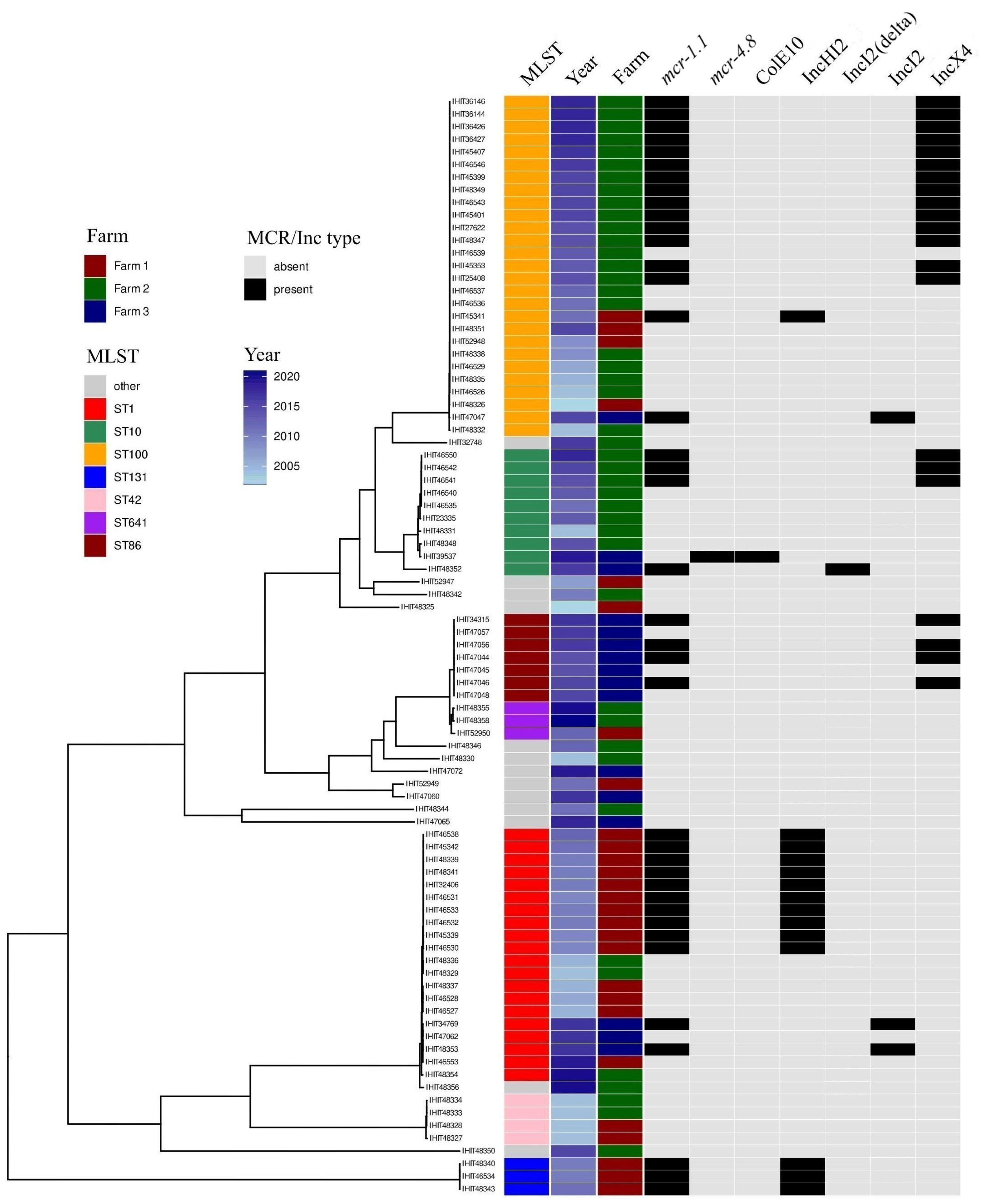
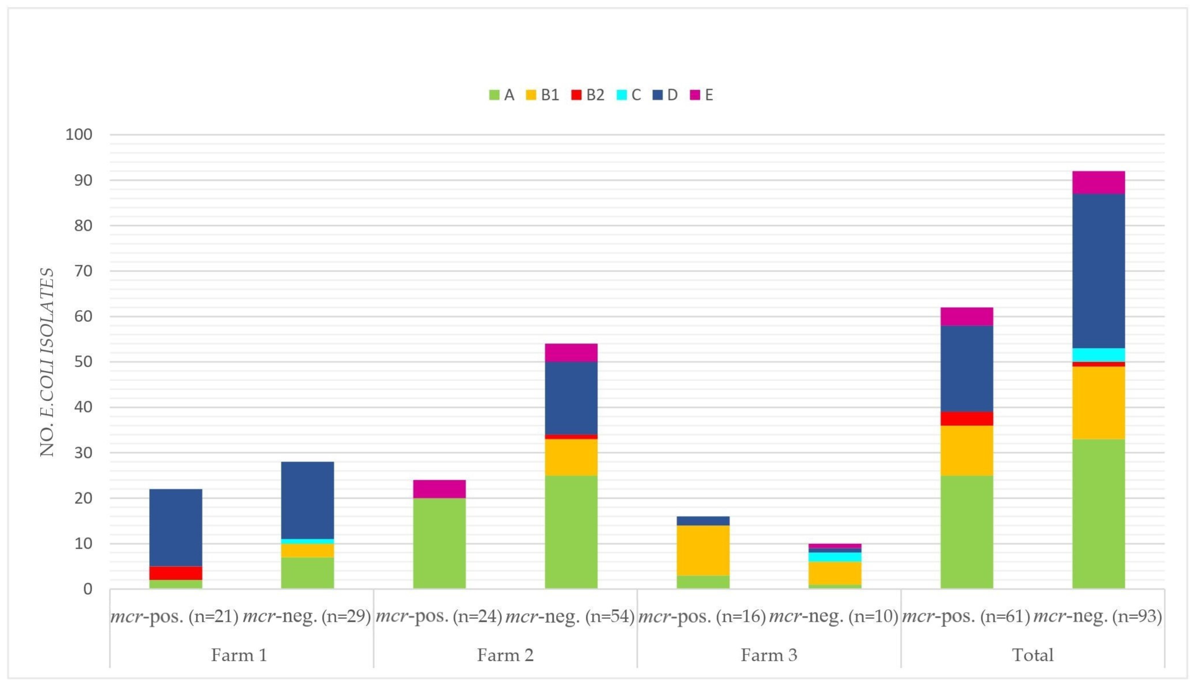
3.5. Multilocus Sequence Types, Clonotypes, Phylogenetic Groups

In the phylogenetic tree of all whole genome-sequenced E. coli isolates in this study, the majority of isolates from each farm clustered together, irrespective of the year of isolation (Figure 2). Clustering was also observed for mcr-positive isolates on each farm.
Overall, 20 known multilocus sequence types (STs) were determined (Table 2). ST1 was the predominant ST (60.9% of the isolates) on Farm 1, while the most prevalent STs on farms 2 and 3 were ST100 (n = 21; 48.8%) and ST86 (n = 7; 43.8%), respectively (Figure 2). Three mcr-1-positive ETEC isolates from Farm 2 (isolated between June 2010 and June 2011) belonged to ST131. This sequence type has previously been detected in ETEC strains isolated from pigs with diarrhea in Spain [3]. It is also known as the predominant E. coli lineage among extraintestinal pathogenic E. coli (ExPEC) worldwide, frequently associated with an ESBL phenotype and multidrug resistance [42].
Nine, fifteen and eight different clonotypes (CH types) were found in E. coli isolates from farms 1, 2 and 3, respectively (Table 2). The most frequent CH types on Farm 1 and Farm 2 were 2-54 (51.9%; all ST1-EDEC) and 27-0 (50%; all ST100-ETEC), respectively. CH type 6–32 was most common in Farm 3 (43.8%, 7 ST86-ETEC/STEC and one ST86-ETEC).
Phylogroups A (n = 58; 37.7%) and D (n = 53; 34.4%) were found to be the most common phylogroups among all 154 E. coli isolates (Figure 3). While the majority of E. coli isolates of Farm 1 were assigned to phylogroup D (n = 34; 68%), phylogroups A (n = 45; 57.7%) and B1 (n = 16; 61.5%), which were most frequent in isolates from Farms 2 and 3, respectively. Overall, only four, three and nine isolates were assigned to phylogenetic groups B2, C and E. No isolate belonged to phylogroup F or G.
Considering only isolates that were positive for mcr genes, a similar distribution of phylogroups per farm and in total was observed. The predominant phylogroups for farms 1 to 3 were D (n = 17; 81%), A (n = 20; 83.3%) and B1 (n = 11; 68.8%), respectively. Only phylogroups A and E (n = 4; 16.7%) were determined for mcr-positive isolates of Farm 2. No mcr-positive E. coli belonged to phylogroup C.

3.6. Genoserotypes

A total of 27 different genoserotypes were identified among 87 whole genome-sequenced E. coli isolates (Table 2). Fourteen isolates were not typeable (Ont). The most common genoserotype in isolates obtained from Farm 1 was O139:H1 (n = 13; 56.5%). This genoserotype has been associated with edema disease-causing E. coli in pigs in Europe [43]. Genoserotype O149:H10 was found repeatedly in isolates from Farm 2 (n = 18; 41.9%). Commonly reported O antigens of ETEC and EDEC isolates, such as O45, O139, O141, O147 and O149, were predominant (n = 52; 71.2%) among all typeable isolates of this study.

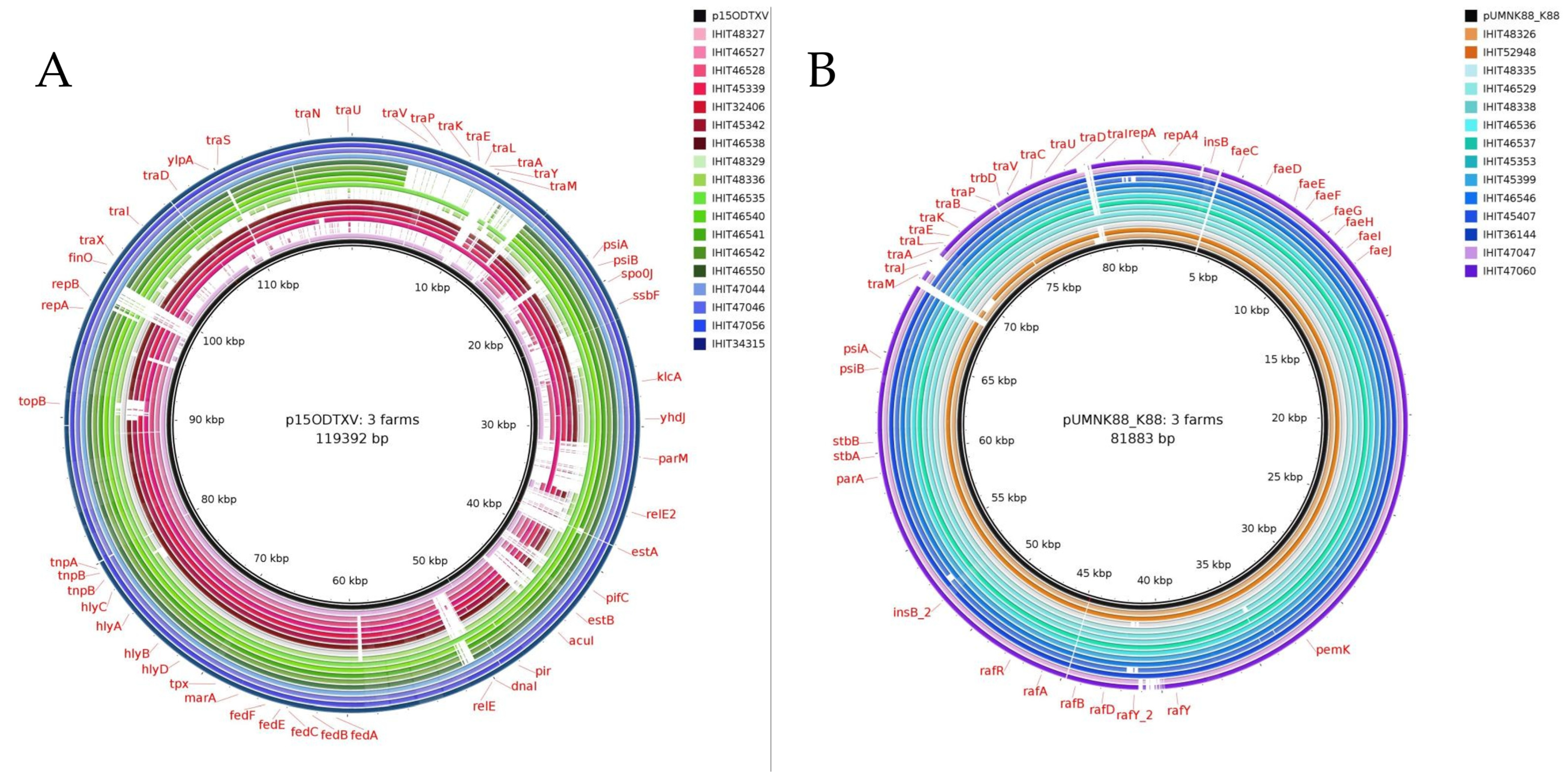
3.7. Genomic Location of mcr Genes and Plasmid Analysis

The genomic location of mcr-1.1 in 38 whole genome-sequenced E. coli isolates was determined via Southern blot hybridization (SBH) of S1 nuclease-digested whole-cell DNA and analysis of genomic sequence data. We identified mcr-1.1 on four different plasmids in this study. S1 nuclease PFGE and SBH initially revealed mcr genes on plasmids with approximate sizes of 250 kbp (n = 13), 35 kbp (n = 19) and 65 kbp (n = 3). Three isolates (IHIT34315, IHIT34769, and IHIT36427) did not reveal the genomic location of mcr genes through SBH despite repeated attempts.
Additionally, whole genome sequence analysis confirmed the occurrence of identical mcr-1.1-harboring plasmids per farm. All of the tested mcr-positive E. coli of Farm 1 carried mcr-1.1 on IncHI2 plasmids (n = 14), while all 17 mcr-positive isolates of Farm 2 harbored mcr-1.1 on IncX4 plasmids. E. coli isolates obtained from Farm 3 carried the mcr-1.1 gene on IncX4 (n = 4), IncI2 (n = 3) and IncI2 (delta) (n = 1) plasmids. The mcr-4.8 gene was located on a ColE10 plasmid, as previously reported [30].
The BacWGST database was used to identify closely related MCR-1 plasmids, which were then used as reference plasmids to illustrate circular comparisons with MCR-1 plasmids of representative isolates of this study (Figure 4).
For comparison, we selected one mcr-positive representative E. coli isolate per year and farm. When two different pathotypes tested positive for mcr in the same year, both positive pathotypes were included. Six representative plasmids were detected in Farm 1 and identified as similar to two IncHI2 reference plasmids. The plasmids were found in IHIT45339 (EDEC; 2009), IHIT32406 (EDEC; 2010), IHIT46534 (ETEC; 2010), IHIT45341 (ETEC; 2011), IHIT45342 (EDEC; 2011) and IHIT46538 (EDEC; 2012), with nucleotide sequence identities ranging from 98.8% to 99.99% and coverage ranging from 90% to 97% compared to the references. The reference plasmids harboring mcr-1.1 had been identified in E. coli strains isolated from swine in China (pDJB-3; NCBI Reference Sequence: MK574666.1; used as a reference plasmid in Figure 4A) and broiler chicken in China (pGD27-70; NCBI Reference Sequence: MN232195.1).
The reference plasmid used for mcr-harboring plasmids from ETEC isolates in Farm 2 was the mcr-1.1-carrying IncX4 plasmid identified in an E. coli isolate obtained from pig feces in Italy in 2016 (pMCR-1-IHIT35346; identity ≥ 99.4%, coverage ≥ 95%; NCBI Reference Sequence: KX894453.1). Another very similar mcr-1.1-carrying IncX4 plasmid (pGMI17-004_2; NCBI Reference Sequence: NZ_CP028167.1) obtained from an E. coli isolate from poultry in Denmark in 2010 is also represented in Figure 4B.
The IncX4 plasmid pMCR-1-IHIT35346 was also used as a reference for all IncX4 plasmids detected in Farm 3 (identity ranging from 97.4% to 100%, coverage ≥ 98%) (Figure 4C). Additionally, one E. coli strain, isolated from raw milk cheese in Egypt in 2016, harbored one mcr-1.1-carrying IncX4, which showed high identity (≥ 99.96%, coverage ≥ 98%) to all four IncX4 plasmids from Farm 3 (pCFSAN061769_01; NCBI Reference Sequence: CP042970.1).
An E. coli isolate obtained from influent municipal wastewater in Japan harbored mcr-1.1-carrying IncI2 plasmid pC2 (NCBI Reference Sequence: LC473131.1), which was used as a reference in Figure 4D. This plasmid was highly similar to plasmids from Farm 3 (isolates IHIT47047, IHIT34769, IHIT48353), with identities ranging from 92.66% to 99.99% and coverage greater than 99%. However, IHIT48352 harbored one IncI2 (delta), which was similar to one mcr-1.1-carrying plasmid from a Salmonella enterica isolate obtained from a child in Ecuador (p778; 98.4% identity, coverage 100%; NCBI Reference Sequence: MN746292.1). None of the selected reference plasmids contained additional resistance genes besides mcr-1.1.

4. Discussion

The global dissemination of colistin resistance genes in bacteria from humans, animals and the environment has been reported over the last years [24,44]. The occurrence of mcr genes has been investigated in various studies, especially those conducted concerning pig and poultry production [45,46,47]. However, data on recurrent mcr-mediated resistance in livestock farms are scarce [48,49]. We were not able to identify clones of pathogenic E. coli over the years on the specific farms. However, our study described three German pig farms with the repeated occurrence of highly similar mcr-carrying plasmids in pathogenic E. coli isolates over two to more than four years, respectively.
In our study, ETEC was the prevalent pathotype for all collected isolates (78/154; 50.7%) as well as for mcr-1-positive isolates (30/61; 49.2%). The most common pathotypes for each farm also coincided with the prevalent pathotypes of mcr-1-harboring E.coli. ETEC has been reported to be the most common cause of post-weaning diarrhea (PWD) in swine, but is also known as a pathogen in humans [5,50]. An epidemiological study from Spain reported a significant association of ETEC (67%) with the occurrence of PWD in swine [3]. That study investigated 481 E. coli isolates from diarrheic pigs, of which 123 (25.6%) strains carried the mcr-1 gene, with 57.7% belonging to the ETEC pathotype. This study also investigated the prevalence of the Shiga toxin gene stx2, which was found to be lower than in our study (10% vs. 35%) [3].
We identified IncFII/IncX1 and IncFII plasmids as the most frequent virulence plasmid types among the isolates from all three farms over the years. VAGs like eltB-Ip, estap, estb, and fedABCEF have previously been reported to be encoded on IncF or IncFII/IncX1 plasmids of E. coli strains isolated from diarrheic pigs [51,52]. Virulence plasmids have occasionally been described in ETEC and ETEC/STEC strains [52,53], but to the best of our knowledge, not in individual pig farms over the years. The striking similarity of certain virulence plasmids in single farms, such as in Farm 3 for four consecutive years (Figure 1A), suggests a local clonal distribution of virulence plasmids. However, it should be taken into account that specific virulence plasmids can be distributed across farms (Figure 1B) and countries. In summary, our data do not support clones (according to the strict definition used in our study design) of E. coli strains carrying specific virulence plasmids over the years.
Effelsberg et al. investigated the occurrence of mcr-1 to mcr-5 genes in 318 porcine fecal samples from 81 pig farms in northwest Germany collected from March 2018 to September 2020 [54]. Overall, ten farms (12.3%) provided fecal samples that contained E. coli harboring mcr-1. Two farms showed repeated presence of mcr-1-positive strains from two different dates of sampling. A retrospective study examined 436 boot swab and pooled fecal samples from 58 German pig-fattening farms for the presence of mcr-1 and mcr-2 [29]. The mcr-1 gene was detected in 43 (9.9%) E. coli isolates obtained from 15 (25.9%) farms, which were almost evenly distributed in northern/western (25%), southern (25%), middle (21%) and eastern (36%) Germany in 2011 to 2012. Considering the presence of mcr-positive pathogenic E. coli strains on the farms examined in our study, the prevalence was high, with 42%, 30.8% and 61.5% for farms 1, 2 and 3, respectively. A longitudinal study from Thailand investigated the occurrence of mcr genes in one representative pig farm from 2017 to 2020 after the cessation of prophylactic colistin usage [48]. Samples were taken from pigs (n = 70), farm workers (n = 50) and wastewater (n = 50). While mcr-1-positive isolates (n = 4; 8%) were detected in humans only in 2017, the prevalence of colistin-resistant strains was 28.6% in pigs, with a declining trend over the years, and 18% in wastewater. Another study obtained mixed bacterial cultures (n = 35) from the environment of three German pig farms from 2011 to 2012 [55]. Seven E. coli isolates tested positive for mcr-1-harboring IncX4 plasmids. The positive samples were obtained from boot swabs, barn dog feces, stable flies and manure. In our study, we did not expect the recurrent identification of highly similar mcr-positive pathogenic E. coli isolates in three German pig farms over more than four years. This is a concerning observation because it suggests that the occurrence of resistant strains over the years may be largely undetected due to missing systematic observational data.
The predominant mcr-1.1-harboring plasmid types in our study were IncHI2 for farm 1, IncX4 for Farm 2 and both IncX4 and IncI2 plasmids for Farm 3. A systematic review by Matamoros et al. reported a total of 13 plasmid incompatibility types for mcr-1-carrying plasmids for 217 Enterobacteriaceae isolated from human, animal and environmental samples [56]. The majority of plasmid types were IncX4 (35.2%), IncI2 (34.7%) and IncHI2 (20.5%), with a significant geographical clustering of IncHI2 plasmids in Europe and a regional spread of IncI2 plasmids in Asia. A recent study from France investigated the occurrence of mcr-1mcr-5 and mcr-9 genes in colistin-resistant E. coli isolates obtained from over 1500 goats of 80 breeding and five fattening goat farms [57]. In total, 149 mcr-1-positive E. coli were identified, with 146 mcr-1 genes located on either IncX4 (38.9%) or IncHI2 (26.8%) plasmids and on the chromosome (32.2%). The mcr-1-carrying plasmids of types IncX4 and IncHI2 were never detected on the same farm in that study.
The most frequently detected sequence types in our study were ST100 (n = 27; all ETEC) and ST1 (n = 20; 19 EDEC, 1 AdhF-Ec). Kusumoto et al. (2016) investigated 967 swine-pathogenic E. coli strains that were isolated from diseased pigs in Japan between 1991 and 2014 [58]. Isolates that were classified as EDEC in that study predominantly belonged to ST1, while ETEC strains were mostly typed as ST100. Sequence type ST10, which was the third most common sequence type in our study (n = 10, 50% mcr-positive isolates), was the most prevalent ST in mcr-positive swine-pathogenic E. coli in a study from Spain [3]. Three mcr-1.1-positive ST131 ETEC strains (harboring ETEC-typical genes fedAac, estap and estb) were isolated from Farm 2 in June 2010 (n =2) and June 2011 (n = 1). All strains harbored additional virulence genes fimH, fyuA, hlyA, ibeA, iss, kpsMII, ompA, papB, papC, sitA and traT, which are typical for ExPEC [59].
Bok et al. analyzed 274 E. coli strains isolated from healthy post-weaning piglets and sows in Poland with the phylogenetic assignment of isolates mainly to phylogroups A (37.6%) and B1 (33.2%) [60]. This is in line with our findings of phylogroups A and B1 being the most prevalent phylogroups for Farm 2 and Farm 3, respectively. Another study from Thailand reported phylogroups A (44.3%), B1 (34.4%) and D (14.8%) being the predominant phylogroups for mcr-harboring E. coli from slaughtered pigs in 2014–2015 [61]. Here, we observed similar prevalences for collected mcr-positive E. coli isolates.
Our study has some limitations. This retrospective survey was based on the receipt of samples for diagnostic purposes and allows for bias due to the random nature of sample submissions. Only porcine E. coli isolates that proved positive for certain VAGs were documented and stored, leaving out non-pathogenic E. coli. In addition, viable but non-culturable (VBNC) E. coli were not studied. Bacteria in the VBNC state are viable but unable to grow on nutrient culture media. Pathogenic E. coli entering the VBNC state as a survival strategy has been reported [62], and would have been missed in our study design. Furthermore, comprehensive data were not available for all three farms, including information on farm characteristics such as potential external contamination, management practices related to animal health, and previous veterinary treatments, including the use of colistin or other antimicrobials.
The collection of a large pool of pathogenic E. coli isolates over a period of more than twenty years is a major advantage for the selection and evaluation of sampled pig farms in Germany. To the best of our knowledge, this study is the first to present phenotypic and molecular data on mcr-mediated resistance and intestinal pathogenic E. coli isolates from individual pig farms over an extended period of time.

5. Conclusions

We describe three German pig farms in which pathotypes of E. coli, including ETEC and EDEC, and various MCR plasmids have been found repeatedly over the years. Isolates obtained on each farm over 17.4, 26.6, and 4.5 years, respectively, differed from each other in their multilocus sequence types and/or VAG and AMR gene profiles and were therefore not considered clonally related. However, a comparison of the plasmid sequence data with reference plasmids demonstrated the structural resemblance of virulence plasmids over several years within and, in part, also across farms.
The data suggest that the repeated occurrence of mcr-carrying plasmids in pathogenic E. coli isolates may be due to the local long-term occurrence of mcr-carrying plasmids rather than reinfection with different plasmids. In affected farms, specific eradication programs, such as rigorous hygiene management and prevention of pig trafficking, may be at least as equally important and effective in reducing the burden of colistin resistance as general recommendations to reduce colistin usage.

Supplementary Materials

The following supporting information can be downloaded at: https://www.mdpi.com/article/10.3390/microorganisms12040729/s1, Table S1: 87 E. coli were analyzed to determine the location of virulence genes associated with intestinal pathogenic E. coli. Isolates are sorted by farms, sequence type (ST), and date of isolation; Table S2: Distribution of virulence-associated genes among 87 E. coli isolates from swine, sorted by farms, sequence types (ST), and date of isolation. Only genes that were at least present in one of the isolates with an identity of >90% (VirulenceFinder 2.0 and BacWGSTdb), are listed; Table S3: Pathotypes, sequence types (STs), resistance genes and chromosomal point mutations related to antimicrobial resistance of 87 representative whole genome-sequenced E. coli isolates, sorted by farms, STs, and date of isolation.

Author Contributions

Conceptualization, C.E.; methodology, C.E., R.B., L.G., T.S. and S.A.W.; validation, C.E. and L.G.; investigation, C.E., R.B., E.P.-B. and L.G.; resources, C.E., R.B. and T.S.; data curation, C.E., L.G., E.P.-B., T.S. and S.A.W.; writing—original draft preparation, L.G.; writing—review and editing, C.E., R.B. and L.G.; visualization, L.G.; supervision, C.E.; project administration, C.E. and R.B. All authors have read and agreed to the published version of the manuscript.

Funding

This research received no external funding.

Institutional Review Board Statement

Ethical review and approval was waived for this study due to the fact that sample collection was not for research but for diagnostic purposes, and only the results obtained were used for scientific purposes. No additional pain, suffering, or harm was inflicted on the animals as a result of our study.

Data Availability Statement

All relevant data are provided in the paper and its Supplements. Raw sequence reads of 87 E. coli genomes are provided under NCBI Bioproject ID PRJNA916215. Further raw data can be made available on reasonable request.

Acknowledgments

We thank all our colleagues from the in-house microbiology diagnostic for collecting E. coli isolates. We thank Anja Schwanitz and Ursula Leidner for their excellent technical assistance. We thank the Sequencing Core Facility of the Genome Competence Centre, Robert Koch Institute, for providing excellent sequencing services. We thank Sébastien Boutin for supporting data visualization.

Conflicts of Interest

The authors declare no conflicts of interest.

References

  1. van Duin, D.; Paterson, D.L. Multidrug-Resistant Bacteria in the Community: An Update. Infect. Dis. Clin. N. Am. 2020, 34, 709–722. [Google Scholar] [CrossRef] [PubMed]
  2. Luppi, A. Swine enteric colibacillosis: Diagnosis, therapy and antimicrobial resistance. Porc. Health Manag. 2017, 3, 16. [Google Scholar] [CrossRef] [PubMed]
  3. García-Meniño, I.; García, V.; Mora, A.; Díaz-Jiménez, D.; Flament-Simon, S.C.; Alonso, M.P.; Blanco, J.E.; Blanco, M.; Blanco, J. Swine Enteric Colibacillosis in Spain: Pathogenic Potential of mcr-1 ST10 and ST131 E. coli Isolates. Front. Microbiol. 2018, 9, 2659. [Google Scholar] [CrossRef] [PubMed]
  4. Fairbrother, J.M.; Gyles, C.L. Colibacillosis. In Disease of Swine, 10th ed.; Zimmerman, J.J., Karriker, L.A., Ramirez, A., Schwartz, K.J., Stevenson, G.W., Eds.; Wiley: Hoboken, NJ, USA, 2012; pp. 723–747. [Google Scholar]
  5. Rhouma, M.; Fairbrother, J.M.; Beaudry, F.; Letellier, A. Post weaning diarrhea in pigs: Risk factors and non-colistin-based control strategies. Acta Vet. Scand. 2017, 59, 31. [Google Scholar] [CrossRef] [PubMed]
  6. Jansen, W.; van Hout, J.; Wiegel, J.; Iatridou, D.; Chantziaras, I.; de Briyne, N. Colistin Use in European Livestock: Veterinary Field Data on Trends and Perspectives for Further Reduction. Vet. Sci. 2022, 9, 650. [Google Scholar] [CrossRef] [PubMed]
  7. European Medicines Agency (EMA). Updated Advice on the Use of Colistin Products in Animals within the European Union: Development of Resistance and Possible Impact on Human and Animal Health; EMA: London, UK, 2016. [Google Scholar]
  8. Gunn, J.S.; Lim, K.B.; Krueger, J.; Kim, K.; Guo, L.; Hackett, M.; Miller, S.I. PmrA-PmrB-regulated genes necessary for 4-aminoarabinose lipid A modification and polymyxin resistance. Mol. Microbiol. 1998, 27, 1171–1182. [Google Scholar] [CrossRef] [PubMed]
  9. Olaitan, A.O.; Diene, S.M.; Kempf, M.; Berrazeg, M.; Bakour, S.; Gupta, S.K.; Thongmalayvong, B.; Akkhavong, K.; Somphavong, S.; Paboriboune, P.; et al. Worldwide emergence of colistin resistance in Klebsiella pneumoniae from healthy humans and patients in Lao PDR, Thailand, Israel, Nigeria and France owing to inactivation of the PhoP/PhoQ regulator mgrB: An epidemiological and molecular study. Int. J. Antimicrob. Agents 2014, 44, 500–507. [Google Scholar] [CrossRef] [PubMed]
  10. Liu, Y.-Y.; Wang, Y.; Walsh, T.R.; Yi, L.-X.; Zhang, R.; Spencer, J.; Doi, Y.; Tian, G.; Dong, B.; Huang, X.; et al. Emergence of plasmid-mediated colistin resistance mechanism MCR-1 in animals and human beings in China: A microbiological and molecular biological study. Lancet Infect. Dis. 2016, 16, 161–168. [Google Scholar] [CrossRef] [PubMed]
  11. Xavier, B.B.; Lammens, C.; Ruhal, R.; Kumar-Singh, S.; Butaye, P.; Goossens, H.; Malhotra-Kumar, S. Identification of a novel plasmid-mediated colistin-resistance gene, mcr-2, in Escherichia coli, Belgium, June 2016. Euro Surveill. 2016, 21, 30280. [Google Scholar] [CrossRef]
  12. Yin, W.; Li, H.; Shen, Y.; Liu, Z.; Wang, S.; Shen, Z.; Zhang, R.; Walsh, T.R.; Shen, J.; Wang, Y. Novel Plasmid-Mediated Colistin Resistance Gene mcr-3 in Escherichia coli. mBio 2017, 8, e0054317. [Google Scholar] [CrossRef]
  13. Carattoli, A.; Villa, L.; Feudi, C.; Curcio, L.; Orsini, S.; Luppi, A.; Pezzotti, G.; Magistrali, C.F. Novel plasmid-mediated colistin resistance mcr-4 gene in Salmonella and Escherichia coli, Italy 2013, Spain and Belgium, 2015 to 2016. Euro Surveill. 2017, 22, 30589. [Google Scholar] [CrossRef]
  14. Borowiak, M.; Fischer, J.; Hammerl, J.A.; Hendriksen, R.S.; Szabo, I.; Malorny, B. Identification of a novel transposon-associated phosphoethanolamine transferase gene, mcr-5, conferring colistin resistance in d-tartrate fermenting Salmonella enterica subsp. enterica serovar Paratyphi B. J. Antimicrob. Chemother. 2017, 72, 3317–3324. [Google Scholar] [CrossRef]
  15. AbuOun, M.; Stubberfield, E.J.; Duggett, N.A.; Kirchner, M.; Dormer, L.; Nunez-Garcia, J.; Randall, L.P.; Lemma, F.; Crook, D.W.; Teale, C.; et al. mcr-1 and mcr-2 (mcr-6.1) variant genes identified in Moraxella species isolated from pigs in Great Britain from 2014 to 2015. J. Antimicrob. Chemother. 2018, 73, 2904. [Google Scholar] [CrossRef] [PubMed]
  16. Yang, Y.-Q.; Li, Y.-X.; Lei, C.-W.; Zhang, A.-Y.; Wang, H.-N. Novel plasmid-mediated colistin resistance gene mcr-7.1 in Klebsiella pneumoniae. J. Antimicrob. Chemother. 2018, 73, 1791–1795. [Google Scholar] [CrossRef]
  17. Wang, X.; Wang, Y.; Zhou, Y.; Li, J.; Yin, W.; Wang, S.; Zhang, S.; Shen, J.; Shen, Z.; Wang, Y. Emergence of a novel mobile colistin resistance gene, mcr-8, in NDM-producing Klebsiella pneumoniae. Emerg. Microbes Infect. 2018, 7, 122. [Google Scholar] [CrossRef]
  18. Carroll, L.M.; Gaballa, A.; Guldimann, C.; Sullivan, G.; Henderson, L.O.; Wiedmann, M. Identification of Novel Mobilized Colistin Resistance Gene mcr-9 in a Multidrug-Resistant, Colistin-Susceptible Salmonella enterica Serotype Typhimurium Isolate. mBio 2019, 10, e0085319. [Google Scholar] [CrossRef] [PubMed]
  19. Wang, C.; Feng, Y.; Liu, L.; Wei, L.; Kang, M.; Zong, Z. Identification of novel mobile colistin resistance gene mcr-10. Emerg. Microbes Infect. 2020, 9, 508–516. [Google Scholar] [CrossRef] [PubMed]
  20. Bastidas-Caldes, C.; de Waard, J.H.; Salgado, M.S.; Villacís, M.J.; Coral-Almeida, M.; Yamamoto, Y.; Calvopiña, M. Worldwide Prevalence of mcr-mediated Colistin-Resistance Escherichia coli in Isolates of Clinical Samples, Healthy Humans, and Livestock-A Systematic Review and Meta-Analysis. Pathogens 2022, 11, 659. [Google Scholar] [CrossRef] [PubMed]
  21. Hamame, A.; Davoust, B.; Cherak, Z.; Rolain, J.-M.; Diene, S.M. Mobile Colistin Resistance (mcr) Genes in Cats and Dogs and Their Zoonotic Transmission Risks. Pathogens 2022, 11, 698. [Google Scholar] [CrossRef]
  22. Wang, J.; Ma, Z.-B.; Zeng, Z.-L.; Yang, X.-W.; Huang, Y.; Liu, J.-H. The role of wildlife (wild birds) in the global transmission of antimicrobial resistance genes. Zool. Res. 2017, 38, 55–80. [Google Scholar] [CrossRef]
  23. Dantas Palmeira, J.; V Cunha, M.; Ferreira, H.; Fonseca, C.; Tinoco Torres, R. Worldwide Disseminated IncX4 Plasmid Carrying mcr-1 Arrives to Wild Mammal in Portugal. Microbiol. Spectr. 2022, 10, e0124522. [Google Scholar] [CrossRef] [PubMed]
  24. Mmatli, M.; Mbelle, N.M.; Osei Sekyere, J. Global epidemiology, genetic environment, risk factors and therapeutic prospects of mcr genes: A current and emerging update. Front. Cell. Infect. Microbiol. 2022, 12, 941358. [Google Scholar] [CrossRef] [PubMed]
  25. Lima, T.; Loureiro, D.; Henriques, A.; Ramos, F.; Pomba, C.; Domingues, S.; Da Silva, G.J. Occurrence and Biological Cost of mcr-1-Carrying Plasmids Co-harbouring Beta-Lactamase Resistance Genes in Zoonotic Pathogens from Intensive Animal Production. Antibiotics 2022, 11, 1356. [Google Scholar] [CrossRef] [PubMed]
  26. McGann, P.; Snesrud, E.; Maybank, R.; Corey, B.; Ong, A.C.; Clifford, R.; Hinkle, M.; Whitman, T.; Lesho, E.; Schaecher, K.E. Escherichia coli Harboring mcr-1 and blaCTX-M on a Novel IncF Plasmid: First Report of mcr-1 in the United States. Antimicrob. Agents Chemother. 2016, 60, 4420–4421. [Google Scholar] [CrossRef] [PubMed]
  27. Mei, C.-Y.; Jiang, Y.; Ma, Q.-C.; Lu, M.-J.; Wu, H.; Wang, Z.-Y.; Jiao, X.; Wang, J. Chromosomally and Plasmid-Located mcr in Salmonella from Animals and Food Products in China. Microbiol. Spectr. 2022, 10, e0277322. [Google Scholar] [CrossRef]
  28. Miguela-Villoldo, P.; Moreno, M.A.; Rodríguez-Lázaro, D.; Gallardo, A.; Hernández, M.; Serrano, T.; Sáez, J.L.; de Frutos, C.; Agüero, M.; Quesada, A.; et al. Longitudinal study of the mcr-1 gene prevalence in Spanish food-producing pigs from 1998 to 2021 and its relationship with the use of polymyxins. Porc. Health Manag. 2022, 8, 12. [Google Scholar] [CrossRef] [PubMed]
  29. Roschanski, N.; Falgenhauer, L.; Grobbel, M.; Guenther, S.; Kreienbrock, L.; Imirzalioglu, C.; Roesler, U. Retrospective survey of mcr-1 and mcr-2 in German pig-fattening farms, 2011–2012. Int. J. Antimicrob. Agents 2017, 50, 266–271. [Google Scholar] [CrossRef] [PubMed]
  30. Göpel, L.; Prenger-Berninghoff, E.; Wolf, S.A.; Semmler, T.; Bauerfeind, R.; Ewers, C. Occurrence of Mobile Colistin Resistance Genes mcr-1mcr-10 including Novel mcr Gene Variants in Different Pathotypes of Porcine Escherichia coli Isolates Collected in Germany from 2000 to 2021. Appl. Microbiol. 2024, 4, 70–84. [Google Scholar] [CrossRef]
  31. Bankevich, A.; Nurk, S.; Antipov, D.; Gurevich, A.A.; Dvorkin, M.; Kulikov, A.S.; Lesin, V.M.; Nikolenko, S.I.; Pham, S.; Prjibelski, A.D.; et al. SPAdes: A new genome assembly algorithm and its applications to single-cell sequencing. J. Comput. Biol. 2012, 19, 455–477. [Google Scholar] [CrossRef]
  32. Schwengers, O.; Jelonek, L.; Dieckmann, M.A.; Beyvers, S.; Blom, J.; Goesmann, A. Bakta: Rapid and standardized annotation of bacterial genomes via alignment-free sequence identification. Microb. Genom. 2021, 7, 000685. [Google Scholar] [CrossRef]
  33. CLSI. Performance Standards for Antimicrobial Disk and Dilution Susceptibility Tests for Bacteria Isolated From Animals, 6th ed.; CLSI supplement VET01S; Clinical and Laboratory Standards Institute: Berwyn, PA, USA, 2023. [Google Scholar]
  34. CLSI. Performance Standards for Antimicrobial Susceptibility Testing, 32nd ed.; CLSI supplement M100; Clinical and Laboratory Standards Institute: Berwyn, PA, USA, 2022. [Google Scholar]
  35. The European Committee on Antimicrobial Susceptibility Testing. Breakpoint Tables for Interpretation of MICs and Zone Diameters. Version 12.0. 2022. Available online: http://www.eucast.org (accessed on 16 December 2023).
  36. Weissman, S.J.; Johnson, J.R.; Tchesnokova, V.; Billig, M.; Dykhuizen, D.; Riddell, K.; Rogers, P.; Qin, X.; Butler-Wu, S.; Cookson, B.T.; et al. High-resolution two-locus clonal typing of extraintestinal pathogenic Escherichia coli. Appl. Environ. Microbiol. 2012, 78, 1353–1360. [Google Scholar] [CrossRef] [PubMed]
  37. Page, A.J.; Cummins, C.A.; Hunt, M.; Wong, V.K.; Reuter, S.; Holden, M.T.G.; Fookes, M.; Falush, D.; Keane, J.A.; Parkhill, J. Roary: Rapid large-scale prokaryote pan genome analysis. Bioinformatics 2015, 31, 3691–3693. [Google Scholar] [CrossRef]
  38. Beghain, J.; Bridier-Nahmias, A.; Le Nagard, H.; Denamur, E.; Clermont, O. ClermonTyping: An easy-to-use and accurate in silico method for Escherichia genus strain phylotyping. Microb. Genom. 2018, 4, e000192. [Google Scholar] [CrossRef] [PubMed]
  39. Clermont, O.; Dixit, O.V.A.; Vangchhia, B.; Condamine, B.; Dion, S.; Bridier-Nahmias, A.; Denamur, E.; Gordon, D. Characterization and rapid identification of phylogroup G in Escherichia coli, a lineage with high virulence and antibiotic resistance potential. Environ Microbiol 2019, 21, 3107–3117. [Google Scholar] [CrossRef] [PubMed]
  40. Clermont, O.; Christenson, J.K.; Denamur, E.; Gordon, D.M. The Clermont Escherichia coli phylo-typing method revisited: Improvement of specificity and detection of new phylo-groups. Environ. Microbiol. Rep. 2013, 5, 58–65. [Google Scholar] [CrossRef] [PubMed]
  41. Alikhan, N.-F.; Petty, N.K.; Ben Zakour, N.L.; Beatson, S.A. BLAST Ring Image Generator (BRIG): Simple prokaryote genome comparisons. BMC Genom. 2011, 12, 402. [Google Scholar] [CrossRef] [PubMed]
  42. Nicolas-Chanoine, M.-H.; Bertrand, X.; Madec, J.-Y. Escherichia coli ST131, an intriguing clonal group. Clin. Microbiol. Rev. 2014, 27, 543–574. [Google Scholar] [CrossRef] [PubMed]
  43. Fairbrother, J.M.; Nadeau, E.; Gyles, C.L. Escherichia coli in postweaning diarrhea in pigs: An update on bacterial types, pathogenesis, and prevention strategies. Anim. Health Res. Rev. 2005, 6, 17–39. [Google Scholar] [CrossRef]
  44. Martiny, H.-M.; Munk, P.; Brinch, C.; Szarvas, J.; Aarestrup, F.M.; Petersen, T.N. Global Distribution of mcr Gene Variants in 214 K Metagenomic Samples. mSystems 2022, 7, e0010522. [Google Scholar] [CrossRef]
  45. Hamame, A.; Davoust, B.; Hasnaoui, B.; Mwenebitu, D.L.; Rolain, J.-M.; Diene, S.M. Screening of colistin-resistant bacteria in livestock animals from France. Vet. Res. 2022, 53, 96. [Google Scholar] [CrossRef]
  46. Tu, Z.; Shui, J.; Liu, J.; Tuo, H.; Zhang, H.; Lin, C.; Feng, J.; Feng, Y.; Su, W.; Zhang, A. Exploring the abundance and influencing factors of antimicrobial resistance genes in manure plasmidome from swine farms. J. Environ. Sci. 2023, 124, 462–471. [Google Scholar] [CrossRef] [PubMed]
  47. Tang, B.; Wang, J.; Zheng, X.; Chang, J.; Ma, J.; Wang, J.; Ji, X.; Yang, H.; Ding, B. Antimicrobial resistance surveillance of Escherichia coli from chickens in the Qinghai Plateau of China. Front. Microbiol. 2022, 13, 885132. [Google Scholar] [CrossRef] [PubMed]
  48. Khine, N.O.; Lugsomya, K.; Niyomtham, W.; Pongpan, T.; Hampson, D.J.; Prapasarakul, N. Longitudinal Monitoring Reveals Persistence of Colistin-Resistant Escherichia coli on a Pig Farm Following Cessation of Colistin Use. Front. Vet. Sci. 2022, 9, 845746. [Google Scholar] [CrossRef] [PubMed]
  49. Randall, L.P.; Horton, R.A.; Lemma, F.; Martelli, F.; Duggett, N.A.D.; Smith, R.P.; Kirchner, M.J.; Ellis, R.J.; Rogers, J.P.; Williamson, S.M.; et al. Longitudinal study on the occurrence in pigs of colistin-resistant Escherichia coli carrying mcr-1 following the cessation of use of colistin. J. Appl. Microbiol. 2018, 125, 596–608. [Google Scholar] [CrossRef] [PubMed]
  50. Qadri, F.; Svennerholm, A.-M.; Faruque, A.S.G.; Sack, R.B. Enterotoxigenic Escherichia coli in developing countries: Epidemiology, microbiology, clinical features, treatment, and prevention. Clin. Microbiol. Rev. 2005, 18, 465–483. [Google Scholar] [CrossRef]
  51. Villa, L.; García-Fernández, A.; Fortini, D.; Carattoli, A. Replicon sequence typing of IncF plasmids carrying virulence and resistance determinants. J. Antimicrob. Chemother. 2010, 65, 2518–2529. [Google Scholar] [CrossRef] [PubMed]
  52. Brilhante, M.; Perreten, V.; Donà, V. Multidrug resistance and multivirulence plasmids in enterotoxigenic and hybrid Shiga toxin-producing/enterotoxigenic Escherichia coli isolated from diarrheic pigs in Switzerland. Vet. J. 2019, 244, 60–68. [Google Scholar] [CrossRef]
  53. Shepard, S.M.; Danzeisen, J.L.; Isaacson, R.E.; Seemann, T.; Achtman, M.; Johnson, T.J. Genome sequences and phylogenetic analysis of K88- and F18-positive porcine enterotoxigenic Escherichia coli. J. Bacteriol. 2012, 194, 395–405. [Google Scholar] [CrossRef] [PubMed]
  54. Effelsberg, N.; Kobusch, I.; Linnemann, S.; Hofmann, F.; Schollenbruch, H.; Mellmann, A.; Boelhauve, M.; Köck, R.; Cuny, C. Prevalence and zoonotic transmission of colistin-resistant and carbapenemase-producing Enterobacterales on German pig farms. One Health 2021, 13, 100354. [Google Scholar] [CrossRef] [PubMed]
  55. Guenther, S.; Falgenhauer, L.; Semmler, T.; Imirzalioglu, C.; Chakraborty, T.; Roesler, U.; Roschanski, N. Environmental emission of multiresistant Escherichia coli carrying the colistin resistance gene mcr-1 from German swine farms. J. Antimicrob. Chemother. 2017, 72, 1289–1292. [Google Scholar] [CrossRef]
  56. Matamoros, S.; van Hattem, J.M.; Arcilla, M.S.; Willemse, N.; Melles, D.C.; Penders, J.; Vinh, T.N.; Thi Hoa, N.; Bootsma, M.C.J.; van Genderen, P.J.; et al. Global phylogenetic analysis of Escherichia coli and plasmids carrying the mcr-1 gene indicates bacterial diversity but plasmid restriction. Sci. Rep. 2017, 7, 15364. [Google Scholar] [CrossRef] [PubMed]
  57. Treilles, M.; Châtre, P.; Drapeau, A.; Madec, J.-Y.; Haenni, M. Spread of the mcr-1 colistin-resistance gene in Escherichia coli through plasmid transmission and chromosomal transposition in French goats. Front. Microbiol. 2022, 13, 1023403. [Google Scholar] [CrossRef] [PubMed]
  58. Kusumoto, M.; Hikoda, Y.; Fujii, Y.; Murata, M.; Miyoshi, H.; Ogura, Y.; Gotoh, Y.; Iwata, T.; Hayashi, T.; Akiba, M. Emergence of a Multidrug-Resistant Shiga Toxin-Producing Enterotoxigenic Escherichia coli Lineage in Diseased Swine in Japan. J. Clin. Microbiol. 2016, 54, 1074–1081. [Google Scholar] [CrossRef] [PubMed]
  59. Sora, V.M.; Meroni, G.; Martino, P.A.; Soggiu, A.; Bonizzi, L.; Zecconi, A. Extraintestinal Pathogenic Escherichia coli: Virulence Factors and Antibiotic Resistance. Pathogens 2021, 10, 1355. [Google Scholar] [CrossRef] [PubMed]
  60. Bok, E.; Kożańska, A.; Mazurek-Popczyk, J.; Wojciech, M.; Baldy-Chudzik, K. Extended Phylogeny and Extraintestinal Virulence Potential of Commensal Escherichia coli from Piglets and Sows. Int. J. Environ. Res. Public Health 2020, 17, 366. [Google Scholar] [CrossRef] [PubMed]
  61. Khanawapee, A.; Kerdsin, A.; Chopjitt, P.; Boueroy, P.; Hatrongjit, R.; Akeda, Y.; Tomono, K.; Nuanualsuwan, S.; Hamada, S. Distribution and Molecular Characterization of Escherichia coli Harboring mcr Genes Isolated from Slaughtered Pigs in Thailand. Microb. Drug Resist. 2021, 27, 971–979. [Google Scholar] [CrossRef]
  62. Pazos-Rojas, L.A.; Cuellar-Sánchez, A.; Romero-Cerón, A.L.; Rivera-Urbalejo, A.; van Dillewijn, P.; Luna-Vital, D.A.; Muñoz-Rojas, J.; Morales-García, Y.E.; Del Bustillos-Cristales, M.R. The Viable but Non-Culturable (VBNC) State, a Poorly Explored Aspect of Beneficial Bacteria. Microorganisms 2023, 12, 39. [Google Scholar] [CrossRef]
Figure 1. Schematic circular representation of E. coli virulence plasmids detected in all three farms over the years (details given in Table 2). For comparison, one representative isolate per year and farm was selected. The reference plasmids used were p15ODTXV (A); NCBI Reference Sequence: MG904998.1) and pUMNK88_K88 (B); NCBI Reference Sequence: CP002730.1), shown in the inner rings. The plasmids detected in isolates from the three farms are arranged according to farm and year of isolation. Isolates from the same farm are color-coded (A): Farm 1 = red, Farm 2 = green, Farm 3 = blue; (B): Farm 1 = orange, Farm 2 = turquoise/blue, Farm 3 = violet). The color of the earliest isolation is displayed as the lightest, while the color of the latest isolation is displayed as the darkest.
Microorganisms 12 00729 g001
Figure 2. Neighbor-joining tree based on the comparison of 2670 core genome genes of 87 E. coli isolates from Farm 1, Farm 2 and Farm 3, with the respective multilocus sequence types (MLST) and isolation years color-coded. The occurrence of mcr genes and their location on different plasmids are shown. Farm specific clustering of isolates from different years can be observed for Farm 1, Farm 2 and Farm 3.
Microorganisms 12 00729 g002
Figure 3. Overview of the phylogenetic grouping of mcr-positive and mcr-negative E. coli isolates sorted by Farms 1 to 3 and in total.
Microorganisms 12 00729 g003
Figure 4. Schematic circular representation of detected MCR-1 plasmids from three farms in our study compared to most similar reference plasmids predicted using the BacWGST database. One representative mcr-positive E. coli isolate per year and farm was selected for comparison (details given in Table 2). If two different mcr-positive pathotypes were detected per year and farm, we selected both pathotypes. The plasmids from our study are arranged in order of isolation year, with the earliest (light color) on the third innermost ring and the latest (dark color) on the outermost ring in (AD). The reference plasmids are shown in the two innermost rings. Reference plasmids (NCBI Reference Sequence in brackets) included are as follows: pDJB-3 (MK574666.1), pGD27-70 (MN232195.1) for Farm 1 (A); pMCR-1-IHIT35346 (KX894453.1), pGMI17-004_2 (NZ_CP028167.1) for Farm 2 (B); pMCR-1-IHIT35346 (KX894453.1), pCFSAN061769_01 (CP042970.1) for Farm 3 (C); pC2 (LC473131.1) and p778 (MN746292.1) for Farm 3 (D).
Microorganisms 12 00729 g004
Table 1. Sample collection from three farms and predicted pathotypes from all E. coli isolates and mcr-positive E. coli isolates per farm.
E. coli Isolates Possessing InPEC-Related VAGs *mcr-Positive E. coli Isolates Possessing InPEC-Related VAGs
Farm No.No. of Sampled PigsSample Type
(No.)
No.Predicted
Pathotypes **
Sample Collection PeriodNo.Predicted
Pathotypes
Isolate
Collection
Period
Farm 143feces (41), intestine + feces (1), isolate (1)50EDEC (23), ETEC (21), STEC (3), ETEC-like (2), AdhF-Ec (1)05/2002–10/201922EDEC (17), ETEC (5)07/2009–09/2012
Farm 269feces (43), isolate (22), intestine + feces (4)78ETEC (53), ETEC-like (7), EDEC (6), AEEC (6), AdhF-Ec (3), ETEC/STEC (2), STEC (1)06/2004–02/202124ETEC (24)07/2013–02/2018
Farm 325isolate (23), feces (2)26ETEC/STEC (14), ETEC (4), EDEC (3), ETEC-like (3), STEC (2)10/2014–04/201916ETEC/STEC (11), EDEC (2), ETEC (1), ETEC-like (1), STEC (1)10/2014–04/2019
* VAGs = virulence-associated genes; InPEC = positive for at least one of the following virulence factors (and encoding genes): F4 (faeG), F5 (fanA), F6 (fasA), F18 (fedA) and F41 (fimF41a) (adhesive fimbriae); LT-Ia, LT-Ib (eltB-Ip), ST-Ia (estap) and ST-II (estb) (heat-labile/stable enterotoxins); stx2, including stx2e (stx2) (Shiga toxin) or intimin (eae) in a modified multiplex PCR [30]. ** Pathotypes: AdhF-Ec, positive for at least one adhesive fimbriae gene (faeG, fanA, fasA, fedA and fimF41a); AEEC, positive for eae; EDEC, positive for fedA and stx2; ETEC, positive for at least one adhesive fimbriae gene (faeG, fanA, fasA, fedA and fimF41a) and at least one enterotoxin gene (eltB-Ip, estap and estb); ETEC-like, positive for at least one enterotoxin gene (eltB-Ip, estap and estb); ETEC/STEC, positive for at least one adhesive fimbriae gene (faeG, fanA, fasA, fedA and fimF41a) and at least one enterotoxin gene (eltB-Ip, estap and estb) and stx2; STEC, positive for stx2.
Table 2. Characteristics of 87 (27/44/16) Escherichia coli isolates, sorted by Farms 1–3, MLST and date of isolation.
Strain IDSource *Date of IsolationVAGs **PathotypeMLSTClonotypeGenoserotype ***Phylogroupmcr Gene/pmrB MutationColistin MIC (µg/mL)
Farm 1 (n = 27)
IHIT46527Feces06/2005fedAab, stx2eEDEC12-54O139:H1D-1
IHIT48337Feces12/2005fedAab, stx2eEDEC12-54Ont:H1D-8
IHIT46528Feces01/2006fedAab, stx2eEDEC12-54O139:H1D-4
IHIT46530Feces07/2009fedAab, stx2eEDEC12-54O139:H1Dmcr-1.18
IHIT45339Feces08/2009fedAab, stx2eEDEC12-54O139:H1Dmcr-1.18
IHIT46531Feces12/2009fedAab, stx2eEDEC12-54O139:H1Dmcr-1.14
IHIT46532Feces01/2010fedAab, stx2eEDEC12-54O139:H1Dmcr-1.18
IHIT46533Feces03/2010fedAab, stx2eEDEC12-54O139:H1Dmcr-1.18
IHIT48339Feces04/2010fedAab, stx2eEDEC12-54O139:H1Dmcr-1.18
IHIT32406Feces06/2010fedAab, stx2eEDEC12-54O139:H1Dmcr-1.18
IHIT48341Feces06/2010fedAab, stx2eEDEC12-54O139:H1Dmcr-1.18
IHIT45342Feces07/2011fedAab, stx2eEDEC12-54O139:H1Dmcr-1.18
IHIT46538Feces09/2012fedAab, stx2eEDEC12-54O139:H1Dmcr-1.18
IHIT46553Isolate10/2019fedAab, stx2eEDEC12-54O139:H1D-0.5
IHIT52949Feces06/2011estb, estap, fedAacETEC234-54O8:H17C- 0.25
IHIT48327Feces04/2004estb, eltB-Ip, fedAacETEC4228-65O147:H14DpmrB V161G2
IHIT48328Feces07/2004estb, estap #, fedAacETEC4228-65Ont:H14DpmrB V161G0.5
IHIT48326Feces05/2002estb, eltB-Ip, faeGacETEC10027-0O149:H10A-1
IHIT52948Feces02/2008estb, eltB-Ip, faeGacETEC10027-0O149:H10A-0.25
IHIT45341Feces07/2011estb, eltB-Ip, faeGacETEC10027-65O149:H10Amcr-1.18
IHIT48351Feces12/2015estb #, eltB-Ip, faeGacETEC10027-0Ont:H10A-0.5
IHIT46534Feces06/2010estb, estap, fedAacETEC13140-683O25:H4B2mcr-1.14
IHIT48340Feces06/2010estb, estap, fedAacETEC13140-683O25:H4B2mcr-1.14
IHIT48343Feces06/2011estb, estap #, fedAacETEC13140-683O25:H4B2mcr-1.18
IHIT52950Feces07/2012fedAAdhF-Ec6416-289O121:H10B1- 0.5
IHIT48325Feces05/2002stx2eSTEC710153-1582Ont:H30A-0.5
IHIT52947Feces12/2007stx2eSTEC1200911-23O142:H27A-0.5
Farm 2 (n = 44)
IHIT48329Feces07/2004fedAab, stx2eEDEC12-54O139:H1D-0.5
IHIT48336Feces08/2005fedAab, stx2eEDEC12-54O139:H1D-8
IHIT48354Int + Fec11/2020fedAabAdhF-Ec12-54O139:H1D-0.5
IHIT48331Feces10/2004estb #ETEC-like1011-54O163:H10A-0.5
IHIT46535Feces02/2011estb, estap, fedAacETEC1011-24O141:H4A-0.5
IHIT46540Isolate07/2013estb, estap, fedAacETEC1011-24O141:H4A-0.25
IHIT23335Feces07/2013estb, estap, fedAac, stx2eETEC/STEC1011-24O141:H4A-0.25
IHIT48348Feces11/2014estbETEC-like1011-45Ont:H6A-8
IHIT46541Feces11/2014estb, estap #, fedAacETEC1011-24O141:H4Amcr-1.14
IHIT46542Isolate01/2015estb, estap, fedAacETEC1011-24O141:H4Amcr-1.14
IHIT46550Isolate02/2018estb, estap, fedAacETEC1011-24O141:H4Amcr-1.14
IHIT48346Feces12/2012eaeAEEC204-25Ont:H49B1-0.5
IHIT48330Feces07/2004eaeAEEC294-24O123:H11B1-0.5
IHIT48333Feces12/2004estb, eltB-Ip, fedAacETEC4228-65O147:H14DpmrB V161G4
IHIT48334Feces12/2004fedAacAdhF-Ec4228-65O147:H14DpmrB V161G4
IHIT48342Feces06/2010eae #AEEC9311-0O5:H4A-0.25
IHIT46526Feces06/2004estb, estap, eltB-Ip, faeGacETEC10027-0O149:H10A-1
IHIT48332Feces10/2004estb, eltB-Ip, faeGacETEC10027-0O149:H10A-0.5
IHIT48335Feces03/2005estb, estap, eltB-Ip, faeGacETEC10027-0O149:H10A-0.25
IHIT46529Feces02/2006estb, estap, eltB-Ip, faeGacETEC10027-0O149:H10A-8
IHIT48338Feces08/2008estb, eltB-Ip, faeGacETEC10027-0Ont:H10A-4
IHIT46536Feces11/2011estb, estap, eltB-Ip, faeGacETEC10027-0O149:H10A-0.5
IHIT46537Feces01/2012estb, estap #, eltB-Ip, faeGacETEC10027-0O149:H10A-0.5
IHIT45353Isolate07/2013estb, estap, eltB-Ip, faeGacETEC10027-0O149:H10Amcr-1.18
IHIT46539Isolate07/2013estb, estap, eltB-Ip, faeGacETEC10027-0O149:H10A-0.25
IHIT25408Isolate03/2014estb, estap, eltB-Ip, faeGacETEC10027-0Ont:H10Amcr-1.10.5
IHIT48347Isolate05/2014estb, estap, eltB-Ip, faeGacETEC10027-0O149:H10Amcr-1.18
IHIT27622Isolate10/2014estb, estap, eltB-Ip, faeGacETEC10027-0O149:H10Amcr-1.18
IHIT45399Isolate01/2015estb, estap, eltB-Ip, faeGacETEC10027-0O149:H10Amcr-1.18
IHIT48349Isolate07/2015estb, estap, eltB-Ip, faeGacETEC10027-0Ont:H10Amcr-1.18
IHIT45401Isolate09/2015estb, estap, eltB-Ip, faeGacETEC10027-0O149:H10Amcr-1.18
IHIT46543Isolate09/2015estb, estap, eltB-Ip, faeGacETEC10027-0O149:H10Amcr-1.14
IHIT46546Isolate09/2016estb, estap, eltB-Ip, faeGacETEC10027-0O149:H10Amcr-1.18
IHIT45407Isolate08/2017estb, estap, eltB-Ip, faeGacETEC10027-0O149:H10Amcr-1.18
IHIT36144Isolate01/2018estb, estap, eltB-Ip, faeGacETEC10027-0O149:H10Amcr-1.18
IHIT36146Isolate01/2018estb, estap, eltB-Ip, faeGacETEC10027-0O149:H10Amcr-1.18
IHIT36426Isolate02/2018estb, estap, eltB-Ip, faeGacETEC10027-0O149:H10Amcr-1.18
IHIT36427Isolate02/2018estb, estap, eltB-Ip, faeGacETEC10027-0O149:H10Amcr-1.18
IHIT48355Int + Fec11/2020estb #ETEC-like6416-832O45:H10B1-2
IHIT48358Feces02/2021estbETEC-like6416-289O115:H10B1-0.25
IHIT32748Isolate09/2016eaeAEEC793168-555O49:H10A-0.5
IHIT48344Feces12/2011eaeAEEC79984-305O108:H9E-0.25
IHIT48356Int + Fec 11/2020stx2eSTEC9552-65O139:H1D-0.5
IHIT48350Feces09/2015estbETEC-like2944224-1082O17/O77:H28D-1
Farm 3 (n = 16)
IHIT34769Isolate06/2017fedAab, stx2eEDEC12-54O139:H1Dmcr-1.14
IHIT48353Isolate06/2017faeGac, stx2eEDEC12-54Ont:H1Dmcr-1.18
IHIT47062Isolate10/2017fedAab, stx2eEDEC12-54O139:H1D-1
IHIT48352Feces12/2016stx2eSTEC1011-23Ont.:H32Amcr-1.18
IHIT39537Isolate04/2019estbETEC-like1011-2594O35:H6Amcr-4.84
IHIT47044Isolate10/2014estb, estap, fedAac, stx2eETEC/STEC866-32Ont:H10B1mcr-1.10.5
IHIT47045Isolate10/2014estb, estap, fedAac, stx2 #ETEC/STEC866-32O86:H10B1-1
IHIT47046Isolate06/2015estb, estap, fedAac, stx2eETEC/STEC866-32O86:H10B1mcr-1.14
IHIT47048Isolate06/2015estb, estap, fedAac, stx2eETEC/STEC866-32O86:H10B1-0.25
IHIT47056Isolate06/2016estb, estap, fedAac, stx2eETEC/STEC866-32Ont:H10B1mcr-1.14
IHIT47057Isolate11/2016estb, estap, fedAacETEC866-32O86:H10B1-0.5
IHIT34315Isolate04/2017estb, estap, fedAac, stx2eETEC/STEC866-32Ont:H10B1mcr-1.14
IHIT47060Isolate02/2017estb, eltB-Ip, faeGacETEC904-54O8:H19C-0.5
IHIT47047Isolate06/2015estb, eltB-Ip, faeGacETEC10027-0O149:H10Amcr-1.18
IHIT47065Isolate07/2018estbETEC-like1184-331O15:H45E-0.5
IHIT47072Isolate03/2019estbETEC-like16265-32O8:H19B1-0.25
* Int + Fec = intestine and feces. ** VAGs = virulence-associated genes for InPEC: faeGac, fedAab and fedAac (adhesive fimbriae); eltB-Ip, estap and estb (heat-labile/stable enterotoxins); stx2 (Shiga toxin) or eae (intimin). VAGs marked with # were positive in the PCR but negative for the respective virulence gene according to whole genome data. *** Ont = O not typeable.
Disclaimer/Publisher’s Note: The statements, opinions and data contained in all publications are solely those of the individual author(s) and contributor(s) and not of MDPI and/or the editor(s). MDPI and/or the editor(s) disclaim responsibility for any injury to people or property resulting from any ideas, methods, instructions or products referred to in the content.

